


Abstract
Lipomas are benign adipose masses that are rarely associated with synovial membranes. In addition, there are only a few reports describing synovial lipomas in the foot. No reported occurrence of this lesion in the subtalar joint currently exists. This case report documents the presentation, clinical evaluation, advanced imaging, and surgical management of a 45-year-old man with a large synovial lipoma of the subtalar joint.
Lipomas are benign and well-encapsulated soft-tissue tumors composed of mature adipose tissue. They are fairly common throughout the body, including the foot. In the classic paper by Kirby et al, [1] lipomas accounted for approximately 8% of all benign soft-tissue tumors found in the foot.
Typically, these lesions occur in extra-articular spaces. However, intra-articular or synovial origins have been reported. The occurrence of these ‘‘synovial lipomas’’ is very uncommon. [2] Reports involving foot and ankle joints are even more rare. [3,4] Furthermore, during the current review of the literature, no reports involving the subtalar joint were found.
Because any intra-articular, space-occupying mass of the foot can result in significant pain, disruption of joint motion, and gait abnormalities, reports of synovial lipomas in the foot are important contributions to podiatric medical, orthopedic, and rheumatologic practices. [5] The present case report describes the presentation, diagnosis, magnetic resonance imaging, and surgical treatment of a synovial lipoma originating from the posterior facet of the subtalar joint.
Case Report
A 45-year-old man presented to our clinic with a 6-month history of pain and stiffness in his left ankle and hindfoot. There was no preceding trauma. The patient reported that the pain was aggravated by prolonged walking and standing. He did not report any history of rheumatologic disorder.
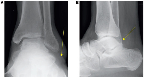
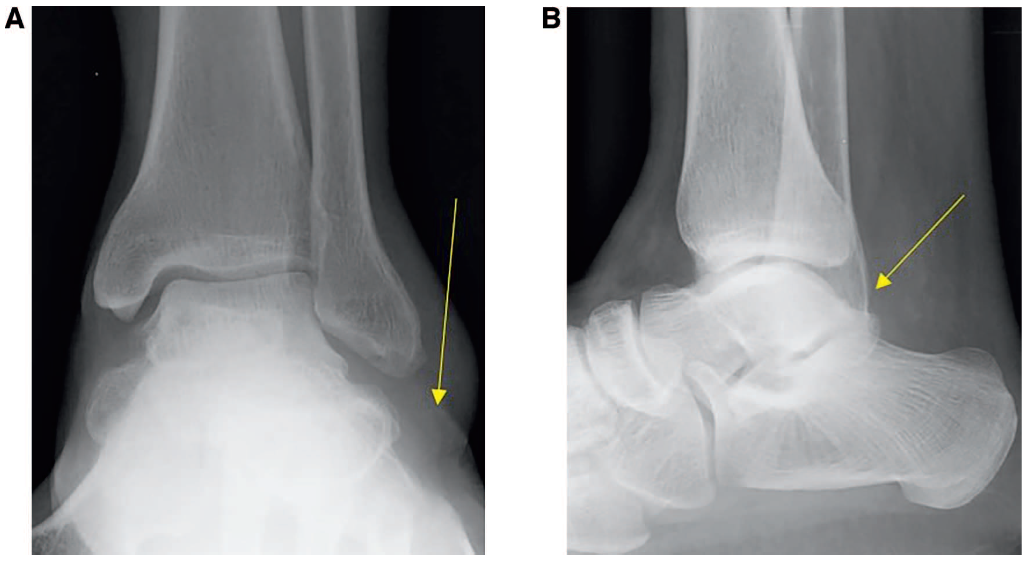
On examination, the patient demonstrated limited active and passive subtalar joint motion. Ankle joint motion, however, seemed unaffected. In addition, a focal area of tissue fullness was noted posterior and inferior to the lateral malleolus. Plain film radio-graphic evaluation revealed a focal area of soft-tissue expansion distal to the lateral malleolus (Figure 1A) and mild degenerative changes in the posterior subtalar joint (Figure 1B).
Figure 1.
A, Preoperative anteroposterior radiograph; the yellow arrow indicates soft-tissue expansion at the site of the mass. B, Preoperative lateral radiograph; the yellow arrow indicates degenerative change in the posterior subtalar joint.
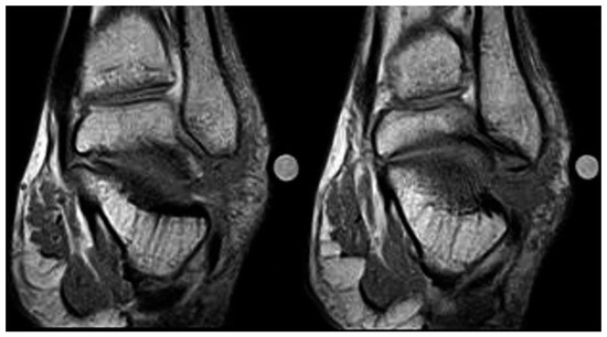
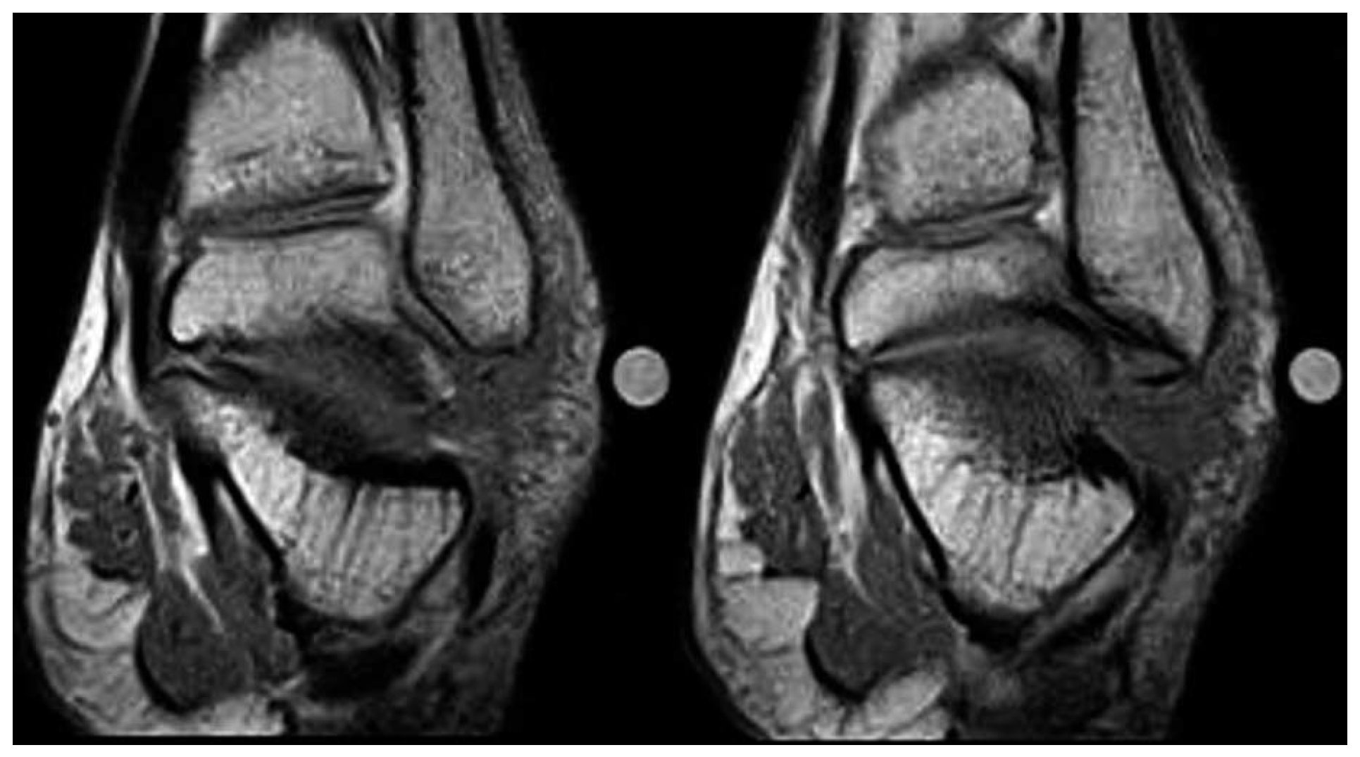
Magnetic resonance imaging was performed to evaluate for a possible intra-articular or tendon sheath mass. The sequential coronal T1-weighted images in Figure 2 show an irregular extension of a mixed fat/water signal emanating laterally from the subtalar joint.
Figure 2.
Coronal T1-weighted magnetic resonance images.
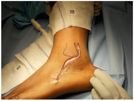
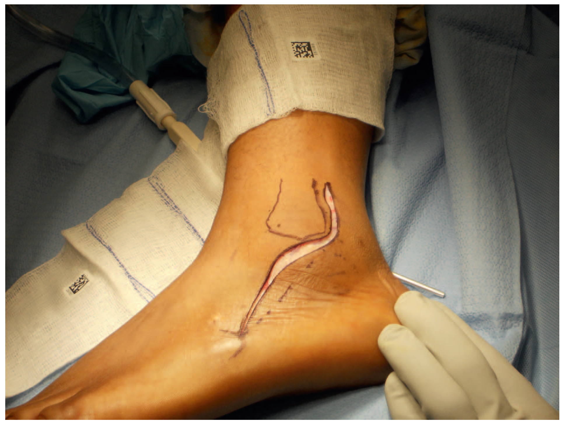
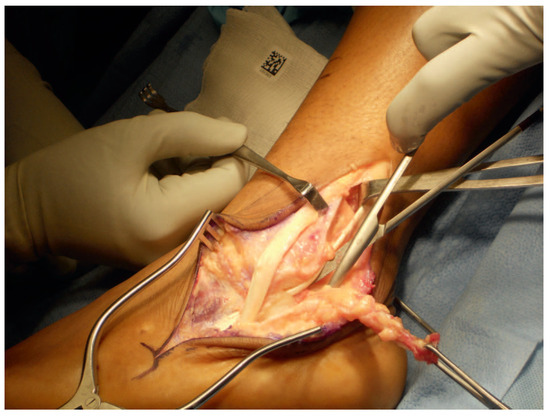
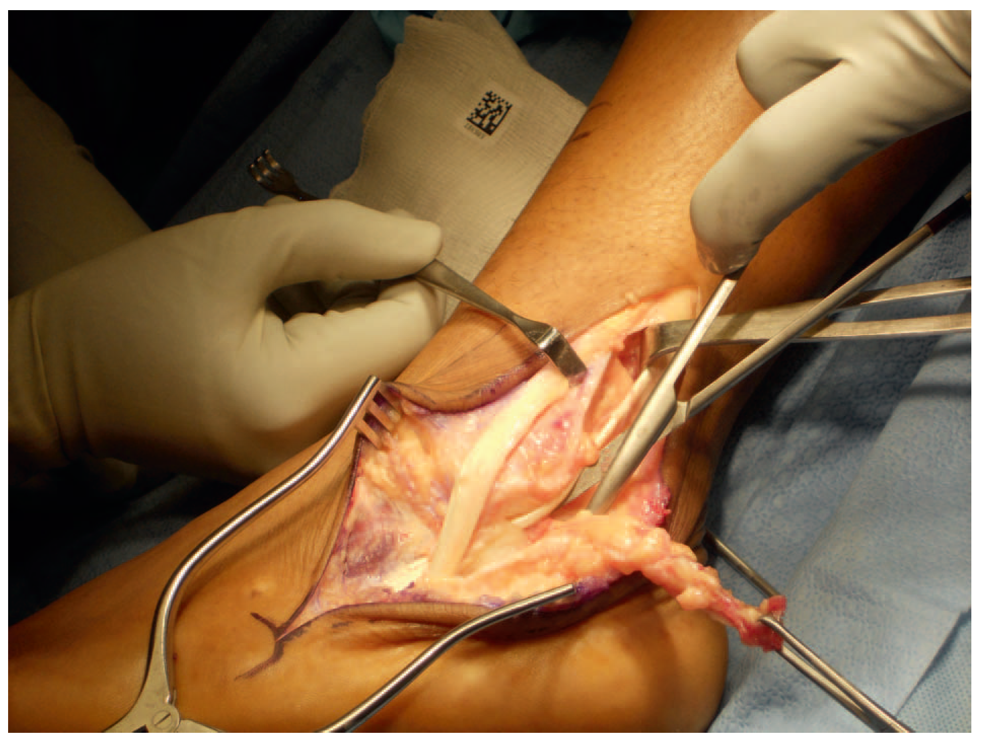
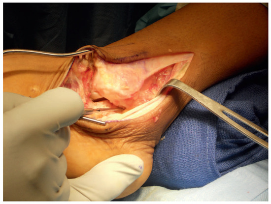
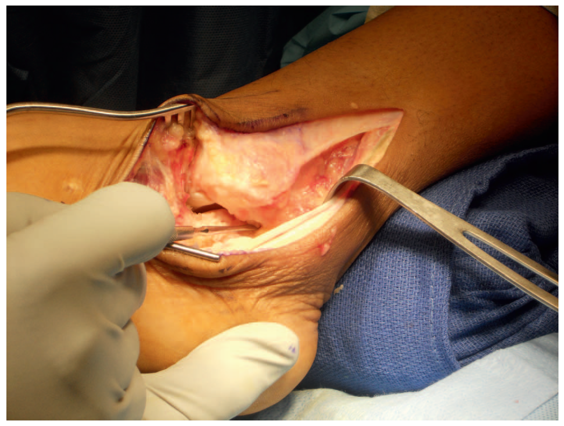
The patient was taken to the operating room for attempted complete excision of the apparent mass. A modified peroneal tendon incision was used (Figure 3). This allowed for adequate mass exposure and access to the posterior subtalar joint. The common and individual peroneal tendon sheaths were opened. The mass was found to occupy the common peroneal tendon space posterior to the lateral malleolus without any direct association with the tendons. Both tendons were intact and without injury. As seen in Figure 4, the mass was traced to originate from the posterolateral aspect of the posterior subtalar joint and was found to exhibit the expected macroscopic appearance of a synovial lipoma outlined by Jowett et al [6] (glistening, hypertrophic yellow-orange synovial tissue in the form of a lobulated mass). The mass was excised completely to include the lateral and posterolateral subtalar joint capsule (Figure 5). The specimen was sent to the pathology department for gross and microscopic analysis.
Figure 3.
Modified peroneal tendon incision.
Figure 4.
Intraoperative view of the mass extending from the posterior facet of the subtalar joint.
Figure 5.
Intraoperative view after excision of the mass.
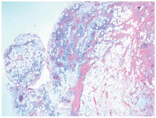
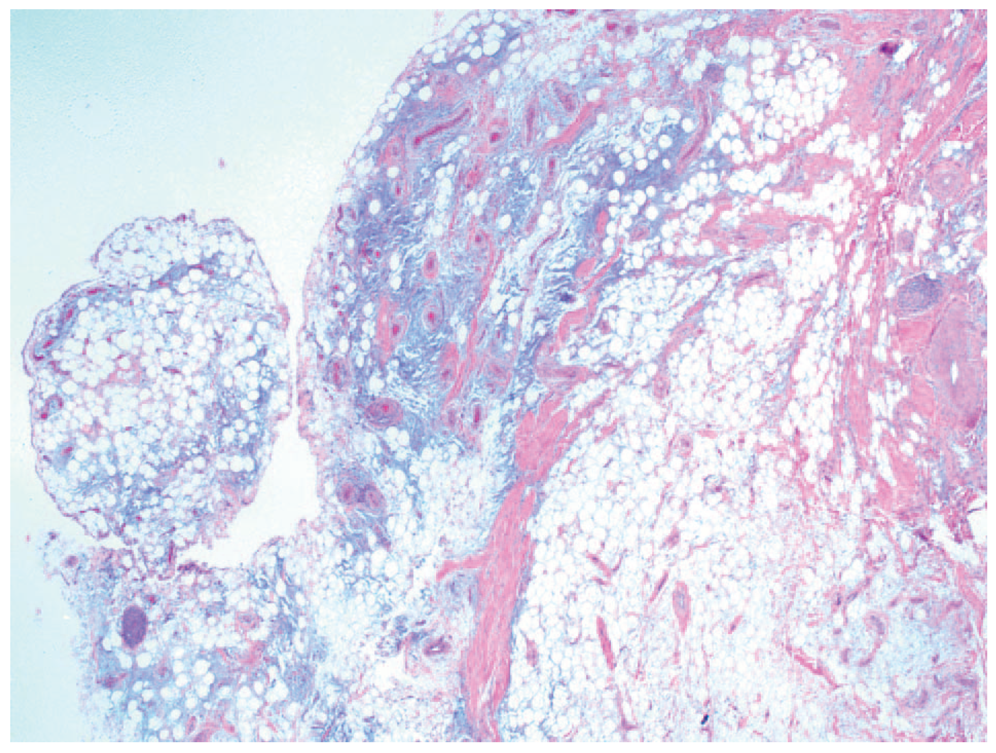
The final pathologic diagnosis included synovial lipoma in addition to mixed degenerative and inflammatory soft-tissue changes. The microscopic appearance of the patient’s specimen is shown in Figure 6 and is consistent with the synovial lipoma findings outlined in the article by Pavithra et al. [4] These findings included mature adipocytes separated by thin fibrous septae in the synovium.[4]
Figure 6.
Microscopic appearance of the mass (H&E, low magnification).
The excision was considered curative, and the patient healed uneventfully, with resolution of pain and restoration of subtalar joint motion.
Discussion
There are a variety of benign soft-tissue masses that can be associated with the intra-articular synovial membrane and, thereby, result in the restriction of joint motion, pain, and gait abnormalities. These lesions include rheumatoid pannus, pigmented villonodular synovitis, lipoma arborescens, and synovial lipoma. [4,5,7] Synovial lipoma, especially in the foot, is a very uncommon lesion in this group. Therefore, this case report describing a previously unreported presentation of this lesion in the subtalar joint will contribute to the existing body of knowledge regarding recognition and treatment of this pathologic abnormality.
Financial Disclosure
None reported.
Conflicts of Interest
None reported.
References
- KIRBY, E.J.; SHEREFF, M.J.; LEWIS, M.M. Soft-tissue tumors and tumor-like lesions of the foot. J Bone Joint Surg Am 1989, 71, 621. [Google Scholar] [CrossRef] [Scilit] [PubMed]
- PUDLOWSKI, R.; GILULA, L.; KYRIAKOS, M. Intraarticular lipoma with osseous metaplasia: radiographic-pathologic correlation. Am J Roentgenol 1979, 132, 471. [Google Scholar] [CrossRef] [Scilit] [PubMed]
- DREVELEGAS, A.; PIKAVAKI, M.; CHOURMOUZI, D. Lipamatous tumor of soft tissue: MR appearance with histological correlation. Eur J Radiol 2004, 50, 257. [Google Scholar] [CrossRef] [Scilit] [PubMed]
- PAVITHRA, P.; ARUNDHATHI, S.; KODANDASWAMY, C. Intra articular synovial lipoma of the right tarsometatarsal joint: a rare case report. J Clin Diagn Res 2014, 8, FD03. [Google Scholar]
- SZENDROI M, DEODHAR A: Synovial neoformations and tumours. Baillieres Best Pract Res Clin Rheumatol 2000, 14, 363. [CrossRef] [Scilit] [PubMed]
- JOWETT, C.; MITRA, P.; O’DONNEL, P.; et al. Synovial lipomatosis of hindfoot tendon sheaths: case reports and literature review. Foot Ankle Int 2008, 29, 752. [Google Scholar] [CrossRef] [Scilit] [PubMed]
- BABAR, S.A.; SANDISON, A.; MITCHELL, A.W. Synovial and tenosynovial lipoma arborescens of the ankle in an adult: a case report. Skeletal Radiol 2008, 37, 75. [Google Scholar] [CrossRef] [Scilit] [PubMed]
© 2017 American Podiatric Medical Association