


Journal Description
Journal of the American Podiatric Medical Association
(JAPMA) is an international, peer-reviewed, open access journal in the profession of foot and ankle medicine. It is the official journal of the American Podiatric Medical Association (APMA) and is published bimonthly online by MDPI (since Volume 116, Issue 1 - 2026). Association members receive discounts on the article processing charges.
- Open Access— free for readers, with article processing charges (APC) paid by authors or their institutions.
- High Visibility: indexed within Scopus, SCIE (Web of Science), PubMed, MEDLINE, and other databases.
- Rapid Publication: manuscripts are peer-reviewed and a first decision is provided to authors approximately 70 days after submission; acceptance to publication is undertaken in 15.3 days (median values for papers published in this journal in the first half of 2026).
- Recognition of Reviewers: Reviewers whose reports are timely and of high quality receive an APC discount voucher for a future publication in an MDPI journal. Become a reviewer.
Impact Factor:
0.7 (2025)
Latest Articles
Valenti Arthroplasty with a Dermal Graft Spacer in Stage III–IV Freiberg’s Disease: A Case Report
J. Am. Podiatr. Med. Assoc. 2026, 116(5), 65; https://doi.org/10.3390/japma116050065 - 10 Sep 2026
Abstract
►
Show Figures
Freiberg’s disease is a rare condition involving osteochondrosis and collapse of the metatarsal head, often leading to chronic pain and impaired mobility. Traditional surgical treatments such as osteotomies, autografts, and joint replacements can relieve symptoms but may compromise joint mechanics or cause donor-site
[...] Read more.
Freiberg’s disease is a rare condition involving osteochondrosis and collapse of the metatarsal head, often leading to chronic pain and impaired mobility. Traditional surgical treatments such as osteotomies, autografts, and joint replacements can relieve symptoms but may compromise joint mechanics or cause donor-site complications. In this case, a 23-year-old woman with Smillie stage 3–4 disease underwent a Valenti-type resectional arthroplasty combined with a folded, decellularized dermal graft spacer to preserve joint function. The procedure resulted in rapid recovery, with pain-free motion and restored within weeks. Radiographs showed remodeling of the metatarsal head, and the patient remained fully ambulatory without complications. This technique offers a promising joint-preserving alternative for young, active patients, although further research is needed to confirm its long-term efficacy.
Full article
Open AccessArticle
An Evaluation of the Short-Term Results on the Pericalcaneal Joints of the Evans Procedure Applied to Adult Patients with Pes Planovalgus
by
Sefa Erdem Karapinar, Serdar Tunc, Recep Dincer, Tolga Atay and Hanim Kumbul
J. Am. Podiatr. Med. Assoc. 2026, 116(5), 64; https://doi.org/10.3390/japma116050064 - 10 Sep 2026
Abstract
►▼
Show Figures
Background: Adult acquired flat foot deformity (AAFD) is a triplanar deformity characterised by collapse of the medial longitudinal arch. The most frequently used surgical method is the Evans calcaneal osteotomy. It is usually used in the paediatric age group and there are
[...] Read more.
Background: Adult acquired flat foot deformity (AAFD) is a triplanar deformity characterised by collapse of the medial longitudinal arch. The most frequently used surgical method is the Evans calcaneal osteotomy. It is usually used in the paediatric age group and there are very few studies related to the use of the modified calcaneal osteotomy procedure in adults. The aim of this study was to evaluate the radiographic improvements after this procedure in the adult age group and to investigate whether or not there was real probability of increased arthritis in the STJ and CCJ. Methods: The study included 20 patients who presented at our clinic between January 2020 and June 2022 with the complaint of foot pain and were diagnosed with Grade 2 AAFD. This retrospective study was based on the data in the hospital records system and the patient follow-up forms. Preoperatively and at 1, 6, 12, and 24 months postoperatively, a record was made of calcaneus length, calcaneal slope angle, talocalcaneal angle, medial longitudinal arch angle, and the time to removal of the Kirschner wire. The presence of arthritis in the joint regions was calculated according to the Kellgren–Lawrence classification. Results: According to the data recorded preoperatively and at the postoperative follow-up examinations (mean 1–6–12–24 months), the difference in calcaneal length, calcaneal slope angle, and change in medial longitudinal arch angle were statistically significant (p < 0.001). No statistical significance was found in respect of the development of arthritis according to the preoperative and postoperative measurements for the CCJ and STJ (p = 1.00). Conclusions: The Modified Evans Osteotomy procedure is beneficial and effective in the correction of pes planovalgus. The surgeon must have a good knowledge of anatomy and it must not be forgotten that there could be variations. The probability of arthritis is not as high as has been assumed.
Full article

Figure 1
Open AccessCase Report
A Combination of Nail Grinding and Nail Fold Resection for a Juvenile’s Pincer Nail Deformity
by
Jianhua Huang and Lei Shi
J. Am. Podiatr. Med. Assoc. 2026, 116(5), 63; https://doi.org/10.3390/japma116050063 - 9 Sep 2026
Abstract
►▼
Show Figures
Pincer nail is a severe variant of nail deformity characterized by excessive transverse curvature of the nail, impinging on both lateral nail folds. The distal end is the most curved and grows inward, squeezing both lateral nail folds, resulting in varying degrees of
[...] Read more.
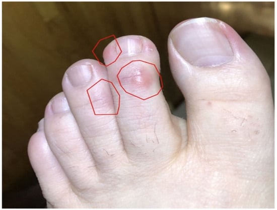
Pincer nail is a severe variant of nail deformity characterized by excessive transverse curvature of the nail, impinging on both lateral nail folds. The distal end is the most curved and grows inward, squeezing both lateral nail folds, resulting in varying degrees of pain and discomfort, making it difficult for patients to wear shoes, hindering walking, and severely impacting the quality of life. Although most previous nail surgical procedures have demonstrated good outcomes, these complicated operations result in a long recovery period and painful nail extraction. Here we present a tissue-preserving combination therapy of nail grinding followed by limited nail fold resection without nail plate extraction. By preserving the integrity of the nail bed and matrix, this approach avoids the extensive dissection and avulsion trauma inherent in conventional procedures, offering reduced postoperative pain, faster recovery, and minimal cosmetic alteration.
Full article

Figure 1
Open AccessCase Report
Calcaneal Stress Fracture Presenting with Lateral Hindfoot Pain in a Young Woman: A Case Report
by
Hongseo Hwang, Hyeong Jun Son, Chang Han Lee, Min-Kyun Oh, Eun Shin Lee and Hayoung Byun
J. Am. Podiatr. Med. Assoc. 2026, 116(5), 62; https://doi.org/10.3390/japma116050062 - 3 Sep 2026
Abstract
►▼
Show Figures
Calcaneal stress fracture is an uncommon osseous cause of heel and hindfoot pain. A 33-year-old woman with chronic underweight status (body mass index, 16.59 kg/m2) presented with a 2- to 3-week history of right lateral hindfoot pain that reportedly began during
[...] Read more.
Calcaneal stress fracture is an uncommon osseous cause of heel and hindfoot pain. A 33-year-old woman with chronic underweight status (body mass index, 16.59 kg/m2) presented with a 2- to 3-week history of right lateral hindfoot pain that reportedly began during treadmill walking in association with a perceived twisting event. The medical record documented tenderness in the region of the calcaneofibular ligament and a negative talar tilt test, but heel-specific examination was incompletely documented. Magnetic resonance imaging (MRI) obtained at the initial evaluation was formally interpreted as showing a calcaneal tuberosity stress fracture and quadratus plantae strain, with no abnormality reported in the ankle ligament complexes. On retrospective MRI review, an incomplete, vertically oriented fracture line extending to the cortex without articular extension or osseous displacement was identified, with surrounding marrow signal abnormality meeting the imaging criteria for Nattiv grade 4. Weight-bearing radiographs obtained 4 days later showed a corresponding sclerotic band. This case illustrates MRI–radiographic correlation in a calcaneal tuberosity stress fracture presenting with lateral hindfoot pain and supports inclusion of osseous injury in the differential diagnosis.
Full article

Figure 1
Open AccessCase Report
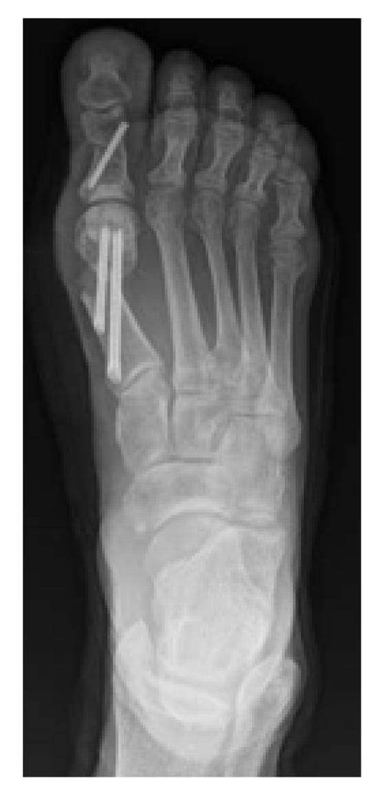
First Metatarsophalangeal Joint Arthrodesis with Calcaneal Allograft for Failed Joint Arthroplasty
by
Payaam Tavakoli, Phoram Vyas and Kevin Huntsman
J. Am. Podiatr. Med. Assoc. 2026, 116(5), 61; https://doi.org/10.3390/japma116050061 - 1 Sep 2026
Abstract
►▼
Show Figures
First metatarsophalangeal joint (MTPJ) arthrodesis is a powerful surgical option for treatment of hallux rigidus, hallux valgus, and revisional first MTPJ surgery. Joint arthroplasty serves as an alternative to arthrodesis for patients who want to preserve joint motion. However, joint arthroplasty has a
[...] Read more.
First metatarsophalangeal joint (MTPJ) arthrodesis is a powerful surgical option for treatment of hallux rigidus, hallux valgus, and revisional first MTPJ surgery. Joint arthroplasty serves as an alternative to arthrodesis for patients who want to preserve joint motion. However, joint arthroplasty has a high rate of failure and can lead to significant bone loss, which makes revisional surgery more challenging. In this study, we present the use of a calcaneal bone block allograft and bone marrow aspirate for fusion of a first MTPJ with a previously failed joint implant. Radiographic fusion was achieved after 6 weeks and ambulation has remained pain-free at 1 year follow-up.
Full article

Figure 1
Open AccessCase Report
An Anomaly of the Pes Anserinus: A Case Study Using Cadaveric Dissection
by
Samreen Mushtaheed, Stephanie Belovich, Ian Farnkopf and Kathy Siesel
J. Am. Podiatr. Med. Assoc. 2026, 116(5), 60; https://doi.org/10.3390/japma116050060 - 27 Aug 2026
Abstract
►▼
Show Figures
This case study investigates a pes anserinus anomaly in a male in his seventies who held an occupation in installation, maintenance, or repair. Using whole-body dissection, we found abnormal deposits and synovial tissue in the region of the pes anserinus, possibly suggestive of
[...] Read more.
This case study investigates a pes anserinus anomaly in a male in his seventies who held an occupation in installation, maintenance, or repair. Using whole-body dissection, we found abnormal deposits and synovial tissue in the region of the pes anserinus, possibly suggestive of chronic overuse or injury which would fit with the patient’s occupation. This study could aid healthcare providers in understanding and diagnosing variations and in managing lower limb pathologies, particularly in patients with occupational or repetitive stress-related injuries.
Full article

Figure 1
Open AccessTechnical Note
Allograft Tendon Pretensioning Technique Guide
by
Joseph R. Brown and Robert W. Mendicino
J. Am. Podiatr. Med. Assoc. 2026, 116(5), 59; https://doi.org/10.3390/japma116050059 - 26 Aug 2026
Abstract
►▼
Show Figures
In foot and ankle surgery, allograft tendons are frequently used in both primary and revision cases to enhance stability and function. However, postoperative complications, such as creep, can compromise the intended correction and stability over time. This paper presents a practical guide on
[...] Read more.
In foot and ankle surgery, allograft tendons are frequently used in both primary and revision cases to enhance stability and function. However, postoperative complications, such as creep, can compromise the intended correction and stability over time. This paper presents a practical guide on pretensioning techniques prior to implantation, aiming to reduce creep and improve long-term outcomes. Using two common allograft specimens, we demonstrate pretensioning methods with specific focus on tendon stitching, techniques for securing the allograft to the graft board, optimal tensioning and time parameters. We recommend pretensioning at 80 N for approximately 30 min to effectively decrease creep and maintain correction. This technique guide offers foot and ankle surgeons streamlined methods for tendon preparation to improve efficiency and consistency in the operating room.
Full article

Figure 1
Open AccessSystematic Review
Autologous Bone Grafting for the Treatment of Osteochondral Lesion of the Talus: A Systematic Review and Meta-Analysis
by
Katrina Hoi Kiu Law, Jin Hean Koh and Dexter Seow
J. Am. Podiatr. Med. Assoc. 2026, 116(5), 58; https://doi.org/10.3390/japma116050058 - 26 Aug 2026
Abstract
►▼
Show Figures
Treatment of osteochondral lesions of the talus (OLTs) with bone grafting is an alternative option to osteochondral transplantation for larger OLTs. The purpose of this study is to evaluate treatment of OLTs with bone grafting with continuous functional outcome scores, return-to-play data, and
[...] Read more.
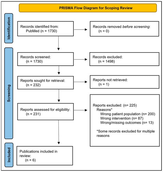
Treatment of osteochondral lesions of the talus (OLTs) with bone grafting is an alternative option to osteochondral transplantation for larger OLTs. The purpose of this study is to evaluate treatment of OLTs with bone grafting with continuous functional outcome scores, return-to-play data, and meta-analyses of overall complication rates after bone graft treatment for OLT lesions as reported and best- and worst-case scenarios. A systematic review was conducted based on PRISMA guidelines using specific search terms on the PubMed, Embase and Cochrane Library databases. Level and quality of evidence were evaluated by The Journal of Bone & Joint Surgery and the Modified Coleman Methodology Score, respectively. Meta-analysis by fixed-effects models was performed if heterogeneity was low (I2 < 25%) and by random-effects models if heterogeneity was moderate to high (I2 > 25%). Eleven studies were included in this systematic review. The Mean American Orthopaedic Foot and Ankle Society score was 56.0 ± 10.1 (range 40.3–67.3) preoperatively to 88.7 ± 5.7 (range 80.8–95.0) points postoperatively. The mean 10 mm-VAS pain score was 6.5 ± 0.89 (range 5.6–7.7) preoperatively to 1.7 ± 1.1 (range 0.4–3.7) postoperatively. The overall pooled complication rate was 3.82% (95% CI: 0.05–10.92). Limited debilitating complications were noted in these studies, with one case of deep vein thrombosis noted. Autologous bone grafting offered satisfactory outcomes for pain relief, with notable improvement of functional outcomes scores. This review proposed that autologous bone grafting should be considered as the first line of treatment for lesion sizes >150 mm2. Studies with longer follow-ups are desirable to determine the longevity of bone grafting and evaluate the durability of bone grafting for larger OLT lesions.
Full article

Figure 1
Open AccessCorrection
Correction: Turemis et al. Comparison of Single and Multiple Toe Amputations in Diabetic Patients with Long-Term Follow-Up and Evaluation of Reamputation Risk. J. Am. Podiatr. Med. Assoc. 2025, 115, 23216
by
Cihangir Turemis, Firat Erpala, Mahmud Aydin and Serkan Surucu
J. Am. Podiatr. Med. Assoc. 2026, 116(5), 57; https://doi.org/10.3390/japma116050057 - 24 Aug 2026
Abstract
Removal of co-authors post publication: [...]
Full article
Open AccessCase Report
Bidirectional Limb Salvage and Its Impact on Residual Limb Survivorship: Is a Partial Calcanectomy in Ambulatory Midfoot Amputees Worthwhile?
by
Craig J. Verdin, Nicole K. Cates, Holly D. Shan, Karen K. Evans, Christopher E. Attinger, John S. Steinberg and Jayson N. Atves
J. Am. Podiatr. Med. Assoc. 2026, 116(4), 56; https://doi.org/10.3390/japma116040056 - 21 Aug 2026
Abstract
►▼
Show Figures
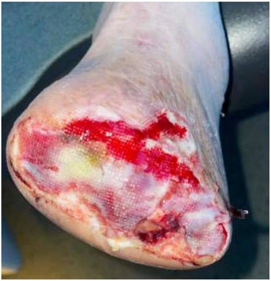
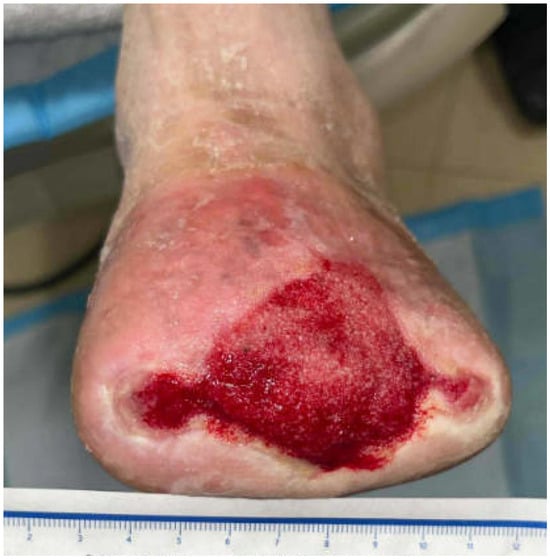
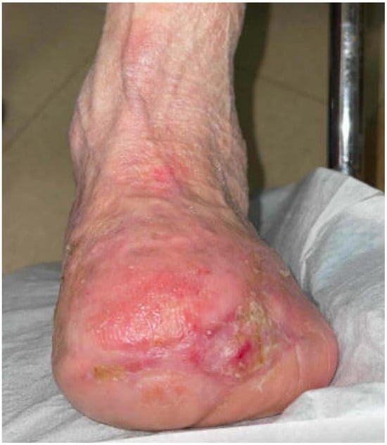
Partial calcanectomies and midfoot amputation are commonly used functional limb salvaging interventions; however, it is well documented that they independently result in global biomechanical disruption of diabetic foot, further placing the residual limb at risk for limb loss. We retrospectively identified and reviewed
[...] Read more.
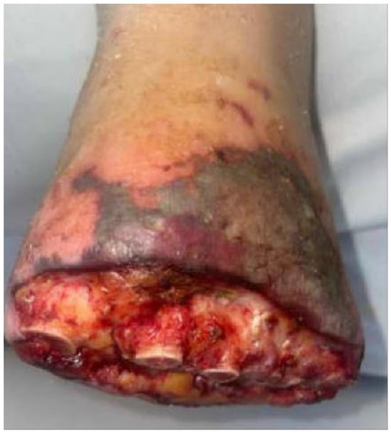
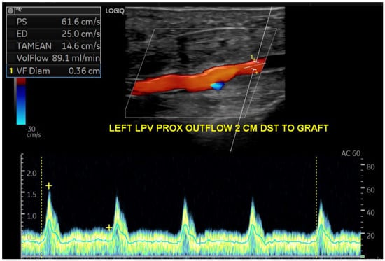
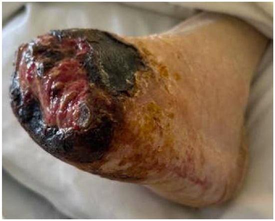
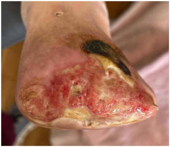
Partial calcanectomies and midfoot amputation are commonly used functional limb salvaging interventions; however, it is well documented that they independently result in global biomechanical disruption of diabetic foot, further placing the residual limb at risk for limb loss. We retrospectively identified and reviewed 9 ambulatory midfoot amputees (10 feet) who developed heel osteomyelitis, whom were treated with an ipsilateral partial calcanectomy to determine whether the combined anterior and posterior interventions result in additive dysfunction that impacts function or places the residual pedal construct at risk for limb loss.
Full article

Figure 1
Open AccessSystematic Review
Prevalence of Lower Extremity Injuries in Rodeo Athletes: A Systematic Review with Meta-Analysis
by
Bethany Robinson, Evelyn Heigh-Rosen and Charlotte Bolch
J. Am. Podiatr. Med. Assoc. 2026, 116(4), 54; https://doi.org/10.3390/japma116040054 - 20 Aug 2026
Abstract
►▼
Show Figures
Rodeo, a non-traditional sport, may pose a higher risk of injury compared to other sports due to the intense forces exerted by livestock. The purpose of this study is to investigate the prevalence of lower extremity injuries and discuss potential strategies for injury
[...] Read more.
Rodeo, a non-traditional sport, may pose a higher risk of injury compared to other sports due to the intense forces exerted by livestock. The purpose of this study is to investigate the prevalence of lower extremity injuries and discuss potential strategies for injury reduction among rodeo athletes. A systematic review with meta-analysis, following PRISMA guidelines, was performed. A comprehensive search of three electronic databases was conducted, searching for studies assessing the epidemiological trends of lower extremity injuries in rodeo athletes. Studies were included if they were published in English, reported injury frequencies by body part, involved collegiate or professional rodeo athletes, and covered more than one rodeo event type. Among the seven identified studies on rodeo injury prevalence, six focused on Canadian and American professional rodeo athletes, while one studied collegiate athletes. The order of injury prevalence of the lower extremity was found to be thigh/knee (TK) > ankle (A) > lower leg (LL) > foot/toes (FT) across all rodeo athletes. Timed event athletes exhibited a nine-percent higher prevalence of thigh/knee and ankle injuries compared to rough stock athletes. Lower extremity injuries accounted for approximately twenty-eight percent of injuries among collegiate and professional rodeo athletes.
Full article

Figure 1
Open AccessCase Report
Coexisting Onychomycosis and Subungual Malignant Melanoma: A Case Series Illustrating Diagnostic Complexity in Podiatric Practice
by
Fedan Avrumova, Erica B. Friedman and Inna Verzub
J. Am. Podiatr. Med. Assoc. 2026, 116(4), 55; https://doi.org/10.3390/japma116040055 - 19 Aug 2026
Abstract
►▼
Show Figures
Background: Subungual malignant melanoma (SMM) is an uncommon but potentially life-threatening neoplasm that frequently mimics benign nail disorders, particularly onychomycosis. Although misdiagnosis of SMM as fungal infection is well documented, laboratory-confirmed coexistence of onychomycosis and melanoma within the same nail unit is exceedingly
[...] Read more.
Background: Subungual malignant melanoma (SMM) is an uncommon but potentially life-threatening neoplasm that frequently mimics benign nail disorders, particularly onychomycosis. Although misdiagnosis of SMM as fungal infection is well documented, laboratory-confirmed coexistence of onychomycosis and melanoma within the same nail unit is exceedingly rare and remains insufficiently characterized in the literature. Methods: A two-patient case series is presented in which both individuals underwent comprehensive clinical evaluation, nail unit biopsy, histopathologic examination with special staining, and molecular and/or mycologic testing. Longitudinal follow-up was obtained to evaluate clinical course and outcomes. Results: Both patients presented with chronic hallux nail dystrophy and laboratory-confirmed onychomycosis. In each case, fungal infection was confirmed, however, persistent symptoms prompted subsequent biopsies, that revealed malignant melanoma. In Case 1, a 71-year-old man with Candida parapsilosis onychomycosis underwent an initial nail unit biopsy demonstrating at least melanoma in situ with ulceration. Repeat biopsy confirmed invasive melanoma with an approximate Breslow thickness of 1.0 mm, while final pathology following partial hallux amputation revealed residual ulcerated melanoma with a Breslow thickness of 2.1 mm, negative margins, and negative sentinel lymph nodes, consistent with Stage IIB disease. In Case 2, an 88-year-old woman with dermatophytic onychomycosis consistent with Trichophyton species had an initial fragmented biopsy demonstrating invasive subungual melanoma with an estimated Breslow thickness of 0.6 mm. Final pathology following distal hallux amputation confirmed residual acral melanoma with a Breslow thickness of 0.6 mm, no ulceration, a mitotic rate of less than 1/mm2, and negative margins, consistent with pT1a disease. Factors contributing to delayed melanoma recognition differed between cases: prior trauma, delayed intervention, and chronic ulceration complicated Case 1, whereas treatment for presumed onychomycosis without improvement delayed suspicion in Case 2. Conclusions: These findings demonstrate that positive mycologic results do not exclude concurrent melanoma and underscore the potential for diagnostic anchoring bias, supporting early biopsy consideration in refractory or atypical nail dystrophy.
Full article

Figure 1

Open AccessArticle
To Evaluate Efficacy of Intralesional Platelet-Rich Plasma in Patients with Plantar Fasciitis
by
Sanjiv Kumar, Chethan Channaveera, Satyaranjan Sethi, Ranjan Kumar Wadhwa and Vijender Anand
J. Am. Podiatr. Med. Assoc. 2026, 116(4), 24161; https://doi.org/10.7547/24-161 - 14 Aug 2026
Abstract
►▼
Show Figures
BACKGROUND: Plantar fasciitis (PF)is one of the most common causes of heel pain that affecting 10 of the general population. Platelet rich plasma(PRP)has been demonstrated to be helpful in managing PF and reducing plantar facia thickness (PFT). This study objective was to
[...] Read more.
BACKGROUND: Plantar fasciitis (PF)is one of the most common causes of heel pain that affecting 10 of the general population. Platelet rich plasma(PRP)has been demonstrated to be helpful in managing PF and reducing plantar facia thickness (PFT). This study objective was to know the effect and efficacy of ultrasound (USG) guided intralesional PRP in PF in relation to pain, PFT and foot function index (FFI). METHODS: A prospective interventional study was conducted in a tertiary care hospital on eighty-five diagnosed cases of unilateral PF. All patients had undergone pain intensity assessment by visual analogue scale (VAS), FFl, and USG examination of PFT after USG guided intralesional PRP injection was assessed at baseline (0 weeks), 2 weeks and 6 weeks. RESULTS: Following USG guided intralesional PRP injection,a statistically significant decrease was noted in plantar fascia thickness (PFT) from baseline 0 (week) 6. 592mm ± 0. 51mm to 5. 169mm ± 0. 39mm and reduced to 4. 07mm ± 0. 39mm at 2nd week and 6th week, respectively (P value 0. 001).A statistically significant reduction of VAS score was noted which reduced from 8. 647 ± 0. 55 to 5. 588 ± 1. 13 (2nd week) and further reduced to 1. 21 ± 1. 06 at the end of 6 weeks (p value 0. 001). Mean FFI in our study at baseline was 85. 494 ± 6. 55, at 2 weeks mean FFI was 49. 341 ± 7. 277 and the end of 6 week it reduced to 24. 235 ± 1. 608 (P value 0. 001) statistically significant decrease was noted. CONCLUSIONS: PRP injection helps in the reduction of heel pain (VAS), reduction in PFT, and improvement in all domains of FFI, leading to enhancement of quality of life.
Full article

Figure 1
Open AccessArticle
Correlation Analysis Between Hip Internal Rotation Range and Plantar Pressure During Standing And Walking
by
Ziyang Yang, Chen Yan, Xiaocong Yan, Chanchan Ge, Desheng Jiang, Niyuan Hu, Zhenghao Xue and Ying Qin
J. Am. Podiatr. Med. Assoc. 2026, 116(4), 24144; https://doi.org/10.7547/24-144 - 14 Aug 2026
Cited by 2
Abstract
►▼
Show Figures
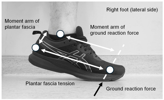
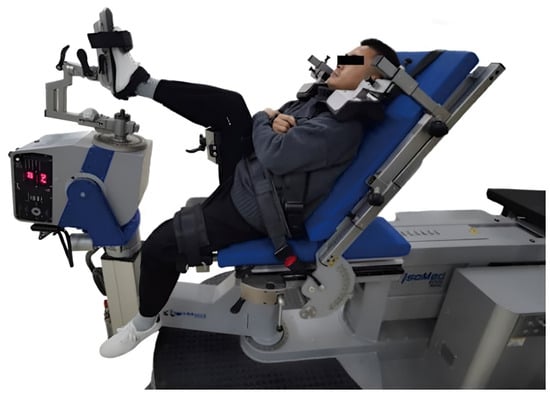
Context: Different studies on football players with fifth metatarsal stress fractures have found that hip internal rotation (HIR)range of motion is significantly reduced, while lateral forefoot pressure is significantly increased; therefore, the purpose of this study was to determine whether HIR range
[...] Read more.
Context: Different studies on football players with fifth metatarsal stress fractures have found that hip internal rotation (HIR)range of motion is significantly reduced, while lateral forefoot pressure is significantly increased; therefore, the purpose of this study was to determine whether HIR range of motion is related to lateral plantar pressure. Methods: The study included 120 college students (60 males and 60 females). HIR was measured using a smartphone inclinometer app in the prone position and a compass app in the supine position. Plantar pressure was assessed using the Gaitview AFA-50 system during standing and walking. Correlation analysis was performed using SPSS 27.0. Results: Among all subjects, HIR activity in the prone position was significantly negatively correlated with standing second through fourth metatarsophalangeal joints(MTPJ2-4), fifth metatarsophalangeal joint (MTPJ5), and midfoot (FM)pressures and significantly positively correlated with standing first toe (T1)pressure. During walking, there is a significant negative correlation between HIR in the prone position and MTPJ2-4, MTPJ5, FM, and lateral heel (LH). Similarly, in the supine position, HIR is significantly negatively correlated with MTPJ2-4, MTPJ5, FM, medial heel (MH), and LH. Conclusion: There is a significant negative correlation between HIR and lateral plantar pressure, indicating that reduced HIR is associated with increased lateral plantar pressure. Improving HIR through targeted rehabilitation may reduce lateral plantar pressure, offering new approaches for non-surgical treatment of little toe capsulitis and reducing the risk of fifth metatarsal stress fractures.
Full article

Figure 1

Open AccessArticle
Novel Approach to Plantar Fasciitis Treatment: Pulsed Electromagnetic Field Therapy
by
Christopher Bromley, Caitlyn McManus and Daniel McManus
J. Am. Podiatr. Med. Assoc. 2026, 116(4), 24136; https://doi.org/10.7547/24-136 - 13 Aug 2026
Cited by 3
Abstract
►▼
Show Figures
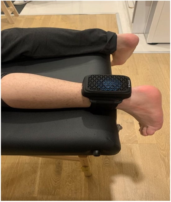
Background: Pulsed electromagnetic field (PEMF) therapy is a conservative, noninvasive, nonpharmacologic option for the treatment of plantar fasciitis that accelerates the body’s anti-inflammatory and healing responses. Methods: In this case series, adult patients presenting with more than 2 weeks of heel pain due
[...] Read more.
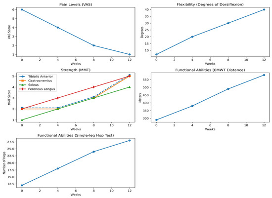
Background: Pulsed electromagnetic field (PEMF) therapy is a conservative, noninvasive, nonpharmacologic option for the treatment of plantar fasciitis that accelerates the body’s anti-inflammatory and healing responses. Methods: In this case series, adult patients presenting with more than 2 weeks of heel pain due to plantar fasciitis were treated with the OrthoCor Active System PEMF device for 12 weeks. Efficacy was measured at 0 (baseline), 4, 8, and 12 weeks of treatment. Ultrasound was used to measure maximum plantar fascia thickness and hypoechoic region width. Function was evaluated using the Foot and Ankle Disability Index (FADI) and Patient-Specific Functional Scale (PSFS) survey scores. Results: Repeated measures analysis of variance showed a statistically significant improvement in all assessments with PEMF therapy. The mean plantar fascia thickness decreased by 34%, the mean hypoechoic region width decreased by 79%, the mean FADI score improved by 46%, and the mean PSFS score improved by 166%. Compared with data from other studies, PEMF and stretching therapy reduced plantar fascia thickness by 34% compared with 16% for dexamethasone injection and 6% for placebo saline injection; the FADI improved by 46% after 12 weeks of PEMF therapy compared with 43% after 1 year of treatment with indomethacin, heat and shoe pads, plantar fascia stretching, or calf stretching; and PSFS improved by 166% after PEMF therapy compared with 96% after primal reflex release treatment. Conclusions: Pulsed electromagnetic field therapy was effective for stimulating healing and improving function in patients with plantar fasciitis, as demonstrated by significant improvements in ultrasound measures and functional scores. This study suggests that PEMF therapy is an effective conservative, nonpharmacologic treatment option for plantar fasciitis.
Full article

Figure 1

Open AccessArticle
Optimizing Ankle Fracture Outcomes with Short-Term Postoperative Immobilization
by
Zaigang Dong, Wenze Liu, Kai Lei, Xiaole Xue, Biao Ning and Xuebin Zhang
J. Am. Podiatr. Med. Assoc. 2026, 116(4), 24128; https://doi.org/10.7547/24-128 - 13 Aug 2026
Abstract
►▼
Show Figures
Background: This paper aims to investigate the effect of short-term cast immobilization on the prognosis of ankle fractures. Methods: A total of 60 patients who underwent ankle fracture surgery in our hospital from September 2021 to September 2022 were included and divided into
[...] Read more.
Background: This paper aims to investigate the effect of short-term cast immobilization on the prognosis of ankle fractures. Methods: A total of 60 patients who underwent ankle fracture surgery in our hospital from September 2021 to September 2022 were included and divided into a cast immobilization group (n = 30) and a control group (n = 30). Both groups were given open reduction and internal fixation. After the operation, the immobilization group used a plaster cast bandage to fix the ankle joint of the affected side in the functional position for 2 weeks; the control group did not receive cast immobilization and was only bandaged with routine wound dressings. The visual analog scale (VAS) score, ankle joint range of motion (ROM), lower extremity deep venous thrombosis rate, and ankle joint function score were followed up in the two groups. Results: All patients were followed up for 6 months. The VAS scores of the cast immobilization group were lower than the control group at 1, 3 and 7 days post-operation (p < 0.05). At 1, 2 and 3 months post-operation, the dorsiflexion ROM of the ankle joint in the cast immobilization group was larger than control group (p < 0.05). The ankle plantarflexion ROM at postoperative 1 and 2 months were larger than the control group (p < 0.05). The AOFAS of cast immobilization group was larger than the control group at 3 months post-operation (p = 0.002). The postoperative deep venous thrombosis rate was 1/30 (3.3%) in the cast immobilization group and 3/30 (10%) in the control group (p = 0.605). Conclusions: Short-term cast immobilization after ankle fracture surgery can significantly reduce postoperative pain in the early stage, without increasing the recovery and the incidence of deep venous thrombosis.
Full article

Figure 1
Open AccessEditorial
N95 Usage During the COVID-19 Pandemic
by
Nicole M. DeLauro, Nader Ghobrial and Dong Yu
J. Am. Podiatr. Med. Assoc. 2026, 116(4), 20101; https://doi.org/10.7547/20-101 - 13 Aug 2026
Cited by 1
Abstract
n/a
Full article
Open AccessCase Report
Angioleiomyoma of the Hindfoot: A Rare Cause of Chronic Heel Pain
by
Alec Wroblewski, Hayden Bush, Devon Niewohner, Jung Wha Kim-Shapiro and John Bonvillian
J. Am. Podiatr. Med. Assoc. 2026, 116(4), 53; https://doi.org/10.3390/japma116040053 - 4 Aug 2026
Abstract
►▼
Show Figures
Angioleiomyoma is a benign soft tissue tumor originating from the smooth muscle of vascular structures. Though relatively uncommon, it frequently occurs in the lower extremities and is often misdiagnosed due to its nonspecific clinical presentation. We report the case of a 54-year-old female
[...] Read more.
Angioleiomyoma is a benign soft tissue tumor originating from the smooth muscle of vascular structures. Though relatively uncommon, it frequently occurs in the lower extremities and is often misdiagnosed due to its nonspecific clinical presentation. We report the case of a 54-year-old female with chronic posterolateral heel pain initially attributed to insertional Achilles tendinopathy and Haglund’s deformity. After failure of conservative management, MRI revealed a well-circumscribed subcutaneous lesion with signal characteristics concerning for a soft tissue neoplasm. Surgical excision was performed, and histopathologic analysis confirmed a venous-type angioleiomyoma, characterized by thick-walled vascular channels and smooth muscle proliferation without atypia. Immunohistochemistry was positive for SMA, weakly positive for desmin, and ERG-positive in the vascular endothelium. The patient experienced complete symptom resolution postoperatively. Although not exceedingly rare, angioleiomyomas remain underrecognized in clinical practice, particularly in the foot and ankle, resulting in diagnostic delays averaging over five years. MRI demonstrated characteristic features described in recent literature, validating its utility in preoperative assessment. This case reinforces the importance of maintaining a broad differential when evaluating chronic, focal heel pain and supports the use of advanced imaging and histopathologic confirmation in atypical cases. Greater awareness of angioleiomyomas can help reduce diagnostic delays and improve outcomes in patients with chronic, unexplained soft tissue pain in the foot and ankle.
Full article

Figure 1
Open AccessArticle
Mobility of the First Ray in Feet with and Without Hallux Valgus
by
Patricia Granados-Gómez, María Reina-Bueno, Mercedes Gómez-Castro and Pedro V. Munuera-Martínez
J. Am. Podiatr. Med. Assoc. 2026, 116(4), 52; https://doi.org/10.3390/japma116040052 - 24 Jul 2026
Abstract
►▼
Show Figures
Background: This study aimed to describe the dorsiflexion and plantarflexion motion of the first ray in feet with and without hallux valgus. Methods: Eight hundred feet were examined and classified into normal feet without hallux valgus (n = 227) and feet with
[...] Read more.
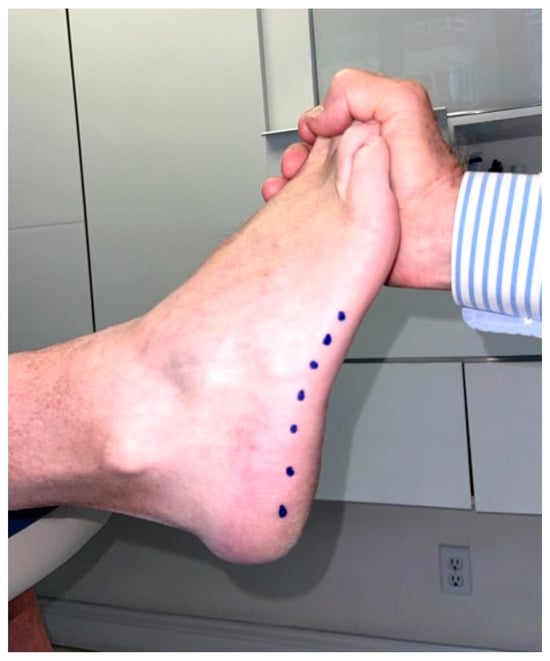
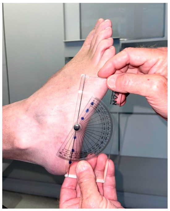
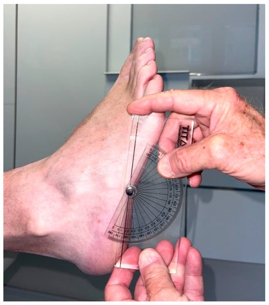
Background: This study aimed to describe the dorsiflexion and plantarflexion motion of the first ray in feet with and without hallux valgus. Methods: Eight hundred feet were examined and classified into normal feet without hallux valgus (n = 227) and feet with hallux valgus (n = 387). Dorsiflexion and plantarflexion of the first ray were quantified with a handheld ruler. Univariate comparisons of the first ray mobility were made between feet with and without hallux valgus. A two-stage cluster analysis was performed to define the range of dorsiflexion and plantarflexion values in both groups. Odds Ratios were obtained to determine whether dorsiflexion or plantarflexion showed more influence on hallux valgus. Multinomial logistic regression was used to predict the probability of having or not having hallux valgus, given dorsiflexion and plantarflexion of the first ray as independent variables. Results: Dorsiflexion showed statistically significant differences between groups (p = 0.011) but with a small effect size (Rosenthal’s r = 0.10). Plantarflexion showed statistically significant differences between groups (p < 0.001) with a larger effect size (r = 0.71). Range of dorsiflexion was between 6 and 7 mm in both normal and hallux valgus feet. Range of plantarflexion in normal feet was 6–7 mm, and in hallux valgus feet 4–5 mm. Logistic regression showed that plantarflexion of the first ray had higher influence on HV than dorsiflexion. Conclusions: In hallux valgus feet, plantarflexion of the first ray was significantly lower than in normal feet, suggesting that a decreased range of plantarflexion is strongly related to hallux valgus.
Full article

Figure 1

Open AccessArticle
Extracorporeal Magnetotransduction Therapy (EMTT) for Midfoot Osteoarthritis: A Prospective Pilot Study of Pain and Functional Outcomes
by
Rohan Charles, Alexander Vlasak and Elizabeth Bondi
J. Am. Podiatr. Med. Assoc. 2026, 116(4), 51; https://doi.org/10.3390/japma116040051 - 10 Jul 2026
Abstract
►▼
Show Figures
Extracorporeal magnetotransduction therapy (EMTT) is a high-energy electromagnetic modality that delivers field strengths of 80–150 mT, effective transduction power > 60 kT/s, and dual-frequency architecture with pulse repetition near 8 Hz and oscillations of 100–300 kHz. This prospective pilot study evaluated pain and
[...] Read more.
Extracorporeal magnetotransduction therapy (EMTT) is a high-energy electromagnetic modality that delivers field strengths of 80–150 mT, effective transduction power > 60 kT/s, and dual-frequency architecture with pulse repetition near 8 Hz and oscillations of 100–300 kHz. This prospective pilot study evaluated pain and functional outcomes in patients with symptomatic midfoot osteoarthritis (OA). Eight patients (13 feet; five bilateral and three unilateral cases) with radiographic Kellgren–Lawrence (KL) grade 3 or 4 midfoot OA received eight EMTT sessions over four weeks. Outcomes were measured at baseline, after the eighth treatment at four weeks, and at 3-month and 6-month follow-up using the 0–10 Visual Analog Scale (VAS) and Foot and Ankle Ability Measure—Activities of Daily Living (FAAM-ADL). VAS demonstrated a significant overall effect of time (F(3,36) = 3.93, p = 0.016), with mean scores decreasing from 4.15 ± 1.63 at baseline to 2.62 ± 1.98 at 3 months and 3.00 ± 2.08 at 6 months. Bonferroni-adjusted baseline-to-follow-up contrasts approached significance at 3 months (p = 0.067) and were not significant at 6 months (p = 0.150). FAAM-ADL improved significantly over time (F(3,30) = 13.76, p < 0.001), increasing from 61.7% ± 9.9% at baseline to 81.8% ± 11.1% at 6 months. No treatment-related adverse events were observed; one participant proceeded to surgery by 6 months and was classified as a treatment failure. These preliminary findings suggest that EMTT is safe and may provide clinically meaningful functional improvement in selected patients with midfoot OA; larger sham-controlled trials are needed.
Full article

Figure 1
Journal Menu
► ▼ Journal MenuJournal Browser
► ▼ Journal Browser-
arrow_forward_ios
Forthcoming issue
arrow_forward_ios Current issue - Volumes not published by MDPI
- Vol. 115 (2025)
- Vol. 114 (2024)
- Vol. 113 (2023)
- Vol. 112 (2022)
- Vol. 111 (2021)
- Vol. 110 (2020)
- Vol. 109 (2019)
- Vol. 108 (2018)
- Vol. 107 (2017)
- Vol. 106 (2016)
- Vol. 105 (2015)
- Vol. 104 (2014)
- Vol. 103 (2013)
- Vol. 102 (2012)
- Vol. 101 (2011)
- Vol. 100 (2010)
- Vol. 99 (2009)
- Vol. 98 (2008)
- Vol. 97 (2007)
- Vol. 96 (2006)
- Vol. 95 (2005)
- Vol. 94 (2004)
- Vol. 93 (2003)
- Vol. 92 (2002)
- Vol. 91 (2001)
- Vol. 90 (2000)
- Vol. 89 (1999)
- Vol. 88 (1998)
- Vol. 87 (1997)
- Vol. 86 (1996)
Highly Accessed Articles
Latest Books
E-Mail Alert
News
14 January 2026
Journal of the American Podiatric Medical Association (JAPMA) Announces Move to Open Access with MDPI
1 September 2026
MDPI INSIGHTS: The CEO’s Letter #38 – 2 Million Published Articles, Outstanding Reviewers, Michele Parrinello Award, AIS 2026 & WSF-12
Topics
Topic in
Antibiotics, Diabetology, JAPMA, JCM, Medicina
Diabetic Foot Disease: Current Challenges, Emerging Concepts and Future Directions
Topic Editors: José Luis Lázaro-Martínez, Marco Meloni, Prashanth VasDeadline: 31 March 2028