


Simple Summary
Lately, investigations of artificial intelligence as an assisting tool for analyzing and identifying stem cells have increased. In this systematic scoping review, we aimed to identify and map the available artificial-intelligence-based techniques for imaging analysis, the characterization of stem cell differentiation, and trans-differentiation pathways. After an extensive search for the literature following a structured methodology, we included 27 studies in our systematic scoping review that we extracted the relevant data from. Based on the results of the included studies, artificial intelligence has the potential to serve as an assisting tool in stem cell imaging. However, it is still considered relatively new and under maturation. The goal of our review is to guide and help researchers while planning for future investigations.
Abstract
This systematic scoping review aims to map and identify the available artificial-intelligence-based techniques for imaging analysis, the characterization of stem cell differentiation, and trans-differentiation pathways. On the ninth of March 2022, data were collected from five electronic databases (PubMed, Medline, Web of Science, Cochrane, and Scopus) and manual citation searching; all data were gathered in Zotero 5.0. A total of 4422 articles were collected after deduplication; only twenty-seven studies were included in this systematic scoping review after a two-phase screening against inclusion criteria by two independent reviewers. The amount of research in this field is significantly increasing over the years. While the current state of artificial intelligence (AI) can tackle a multitude of medical problems, the consensus amongst researchers remains that AI still falls short in multiple ways that investigators should examine, ranging from the quality of images used in training sets and appropriate sample size, as well as the unexpected events that may occur which the algorithm cannot predict.
1. Introduction
The emergence of stem cell research began in the year 1961, which marked the discovery of bone multipotent stem cells [1]. After several decades, the human pluripotent stem cells were first used in the preclinical stages of research which included isolating cells, implementing their functions, identifying their roles, and applying animal trials (such as injecting human-derived cardiomyocytes in damaged rodent hearts and witnessing their improvements) [2,3]. In the past few years, a trend in stem cell research has exponentially increased as it passed to the clinical stages, where the advancement of technology has enabled such innovation to potentially transition into human clinical trials in the future [2,3]. Stem cells are unspecialized human cells capable of self-renewal through mitosis, eventually forming more cells. Such a division generates two types of cells in which the first differentiate into a specific type of cell, whereas the second sustains self-renewal ability [4]. Three categories make up the stem cell division types which consist of induced pluripotent stem cells (iPSCs), embryonic stem cells (ECSs), and adult stem cells (ACSs) [5]. Pluripotent stem cells (PSCs) are defined by their ability to differentiate into the three layers of germ cells (ectoderm, mesoderm, and endoderm). Both iPSCs and ESCs are considered to be PSCs due to their ability to differentiate into the three germ layer derivatives, but a distinguishing feature between iPSCs and ESCs is that IPSCs are special reprogrammed somatic cells, generating pluripotent patient-specific cell lineages capable of aiding model human diseases [6]. Unlike iPSCs and ECSs, ACSs have a lower differentiation level, termed multipotent, and hence can differentiate into more tissue-specific stem cells [7]. ACSs are rare undifferentiated cells that spread throughout the entire body and transform into a proliferative state from their quiescent one in order to divide into new cells that would replace the naturally dying ones [7].
The future of stem cell-based therapy is becoming the precedent in advanced medicine. The potential for stem cell implementation grows with every experiment, bringing a new look at the possibilities of transplantology and regenerative medicine [8]. These therapies have targeted multiple medically severe conditions underscored by defective cell division or differentiation, such as cancer or congenital disabilities [8]. A wide variety of diseases are under the scope of stem-cell-based therapy in various fields of medicine, including cardiology (heart failure) [9] and ophthalmology (retinal and macular degeneration) [10]. Stem cells transformed the idea of treating what was once considered untreatable, for example certain neurodegeneration diseases, including Alzheimer’s and Parkinson’s [11]. In addition, arthroplasty has seen the impact of stem cells in various forms, such as healing tendon injuries, as well as in the cases of osteoarthritis, highlighting stem cell use in cartilage repair [12,13]. Stem cell research has also made its way to fertility disease, where the ability to produce sperm cells from iPSCs proved not only to be successful but also produced healthy and fertile mice [14,15]. Another field of medicine that has the potential to become a revolutionary step in stem cell research is diabetes, where induced stem cells differentiate into the missing pancreatic beta cells instead of transplanting them for a donor [16]. The most significant advancements are highlighted in hematopoietic stem cell research, earning them the title of being the most popular stem cell due to extensive experimentation and studies over the last fifty years, laying numerous foundations that have guided other medical fields in stem cell research and development [8]. As time progresses, research on stem cells is expanding beyond fields in medicine, reaching disciplines of dentistry and pharmacology.
The field of pharmacology has also been infiltrated by stem cell research. Notably, human-induced pluripotent stem cells (hiPSCs) saw their upbringing in 2009 when they were screened to model a type of neuropathic disease called familial dysautonomia, where multiple model features were discovered, and drugs targeting these features were manufactured and later tested [17]. Other examples include screening an anticancer drug, Bosutinib, which inhibits Src/c-Abl receptors, on hiPSCs extracted from amyotrophic lateral sclerosis (ALS) patients [18]. Now, the state of pharmacologic stem cell research has advanced tremendously, reaching the forefront of gene editing, CRISPR/cas9, and when combined with the stem cell application, can provide revolutionary input to the development of drug therapies that occurs through the integration of genes into hiPSCs, ultimately leading to both the development of therapeutic drug candidates and also the selection of the best drug out of these candidates [18]. Hence, such an approach to stem cells allows for systemic compound and drug evaluation regarding their safety, tolerability, and efficacy when applied for certain severe diseases due to drug screening on iPSCs [19].
The revolutionary advancements in technology, underlined by artificial intelligence (AI), have made stem cells available in terms of selecting the most suitable medication, establishing a diagnosis, and formulating risks and benefits when it comes to therapy [20]. In particular, emerging techniques of machine learning, deep learning, and convolutional neural networks (CNN) have assisted the framework of the reliable detection for various functions, including iPSC colony classifications [2], non-invasive cell therapy characterizations of normal versus abnormal cells [2], and image-based cellular morphology [2]. The accessibility of a wide variety of medical images combined with the continuously developing technology in the field of AI will take medicine to a whole different level [2]. The goal of the current research is to map and identify the available AI-based techniques for the imaging analysis of stem cells, the characterization of stem cell differentiation, and trans-differentiation pathways.
2. Materials and Methods
Guided by the Joanna Briggs Institute (JBI) methodology for a scoping review [21] and PRISMA-ScR (Transparent Reporting of Systematic Reviews and Meta-analyses Extension for Scoping Reviews) checklist [22], this systematic scoping review was conducted, as defined by its protocol that was developed previously by the research team [23].
2.1. Identifying the Research Question
Following the identification of the population, concept, and context (PCC) [21] components, a research question was developed. What are the available applications of AI-based imaging analysis for various types of stem cells?
The PCC component is established as follows:
- Population: stem cells;
- Concept: AI-based technique;
- Context: imaging analysis.
2.2. Searching Strategy
On 9 March 2022, we searched five electronic databases (PubMed, Medline, Web of Science, Cochrane, and Scopus) to identify inclusive studies based on a research strategy answering the research question (Table S1). Following a primary search, the complete searching strategy was then developed and customized for each database using specific queries. Additionally, we carried out a manual citation search of the retrieved data by screening the reference section of all studies to look for potential studies answering our research question.
The keywords included in the queries are as follows: algorithm, algorithm*, artificial intelligence, AI, automated, automatic, semi-automated, semi-automatic, deep learning, convolutional neural network, CNN, machine learning, stem cells, stem cell*, and imaging.
All the studies found were imported and stored in the Zotero 5.0 (Corporation for Digital Scholarship, Vienna, VA, USA) library. The library was deduplicated using web-based software and an SR accelerator [24], and a manual review by J.I was performed to confirm the removal of duplicates.
2.3. Eligibility Criteria
We generated the inclusion and exclusion criteria based on the PCC mnemonics [21].
Quantitative studies testing AI-based imaging analysis on any type of stem cell (iPSCs, ECSs, ACSs, and PSCs) of animals or humans were included without any language or date restrictions due to the novelty of this field. We excluded preprints and conference papers, as well as qualitative studies and quantitative studies investigating the use of AI technology for other purposes, including the imaging analysis of any type of cells rather than of stem cells. The studies where the full text was inaccessible were excluded. The inclusion and exclusion criteria are presented in Table 1.
Table 1.
Table of Inclusion and Exclusion Criteria.
2.4. Study Selection and Data Extraction
In the first phase of study selection, the title and abstract of all studies after deduplication were screened against the inclusion criteria by two independent reviewers (J.I. and M.A.C.). The reviewers met several times during this process; the first session was based on testing the study selection method and ensuring the understanding of the inclusion criteria by both reviewers. During the second phase, the reviewers again independently assessed the full text of the held studies following the first screening phase based on inclusion criteria. In case of any disagreements between reviewers at any stage, the opinion of the third reviewer (M.D.-K.) was taken, or the conflict was solved by discussion between the two reviewers.
Data were extracted from the included studies by one reviewer (M.A.C.) and evaluated independently by the second reviewer (J.I.), and any disagreement between them was resolved by discussion or by the opinion of the third review (M.D.-K.). The data extraction tools were developed based on the JBI methodology for scoping review [21].
3. Results
3.1. Search Result
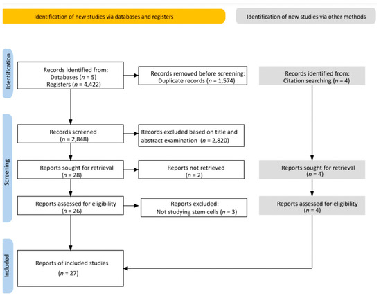
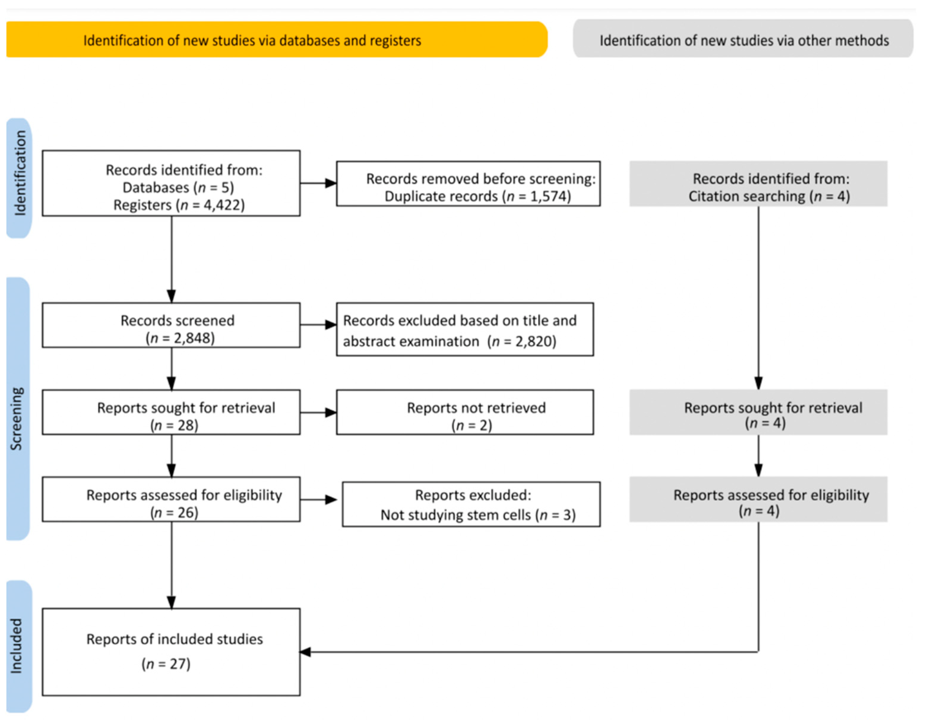
In total, 4422 articles were collected from the five electronic databases. Subsequently, 1574 articles were eliminated after deduplication of the library, progressing our screening for titles and abstracts against the inclusion criteria of 2848 studies. After the first screening phase, 28 articles were eligible for full-text analysis. While collecting the full text, two studies were eliminated due to the inaccessibility of the full text, and three other studies were excluded after the full-text screening as they were not studying stem cells. Finally, four additional articles were added through manual citation searching. In total, 27 studies were included in this systematic review (Figure 1). The level of the reviewer agreement was calculated using kappa statistics, K = 0.862, indicating a significant agreement between both reviewers.
Figure 1.
PRISMA flow diagram.
3.2. Extracted Data
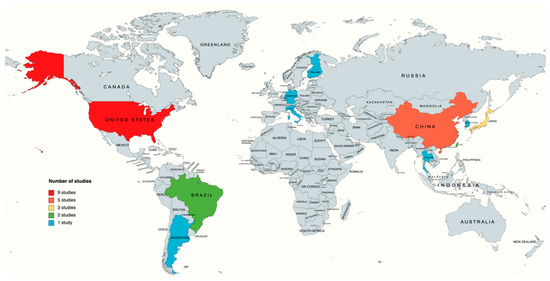
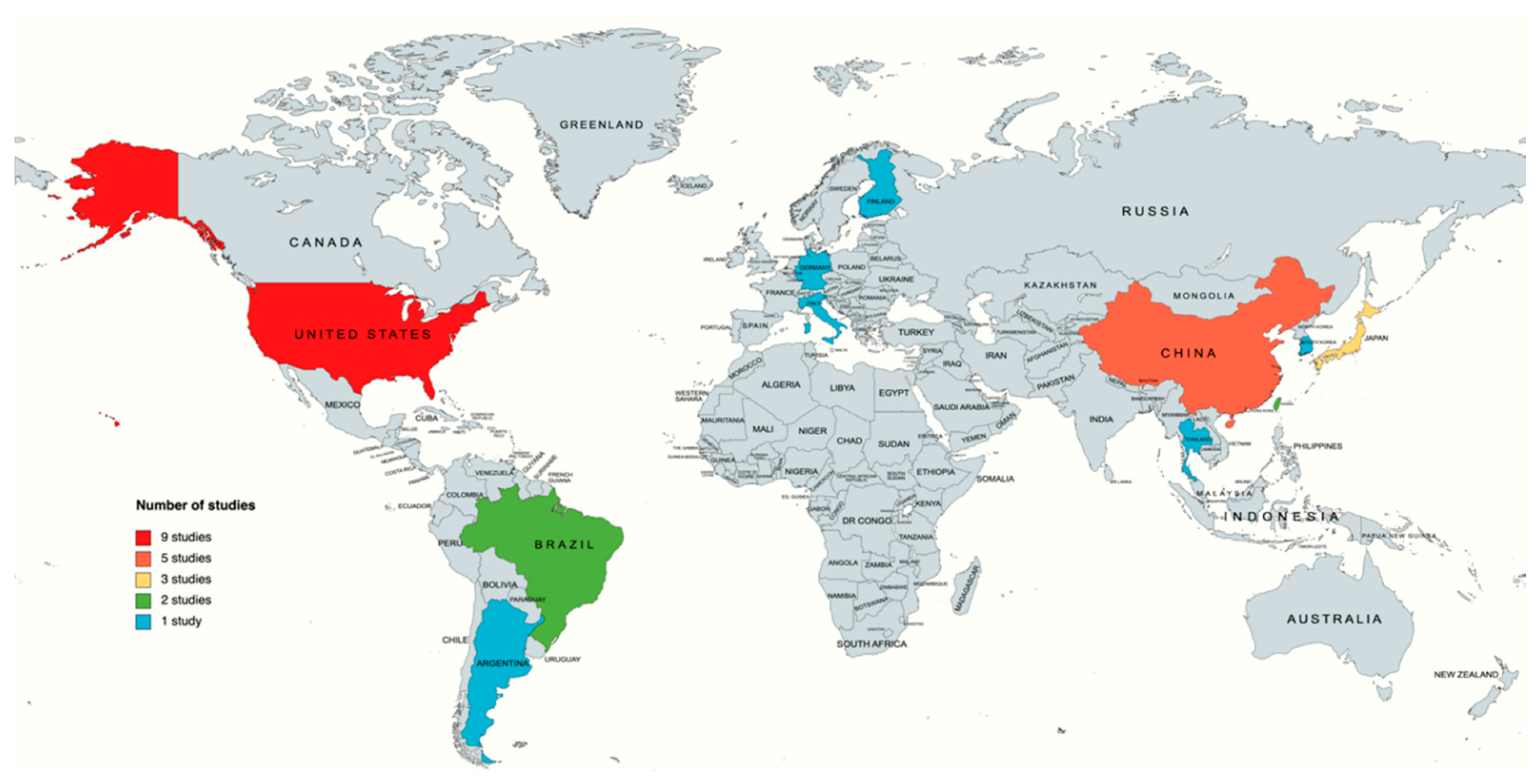
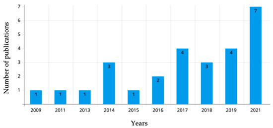
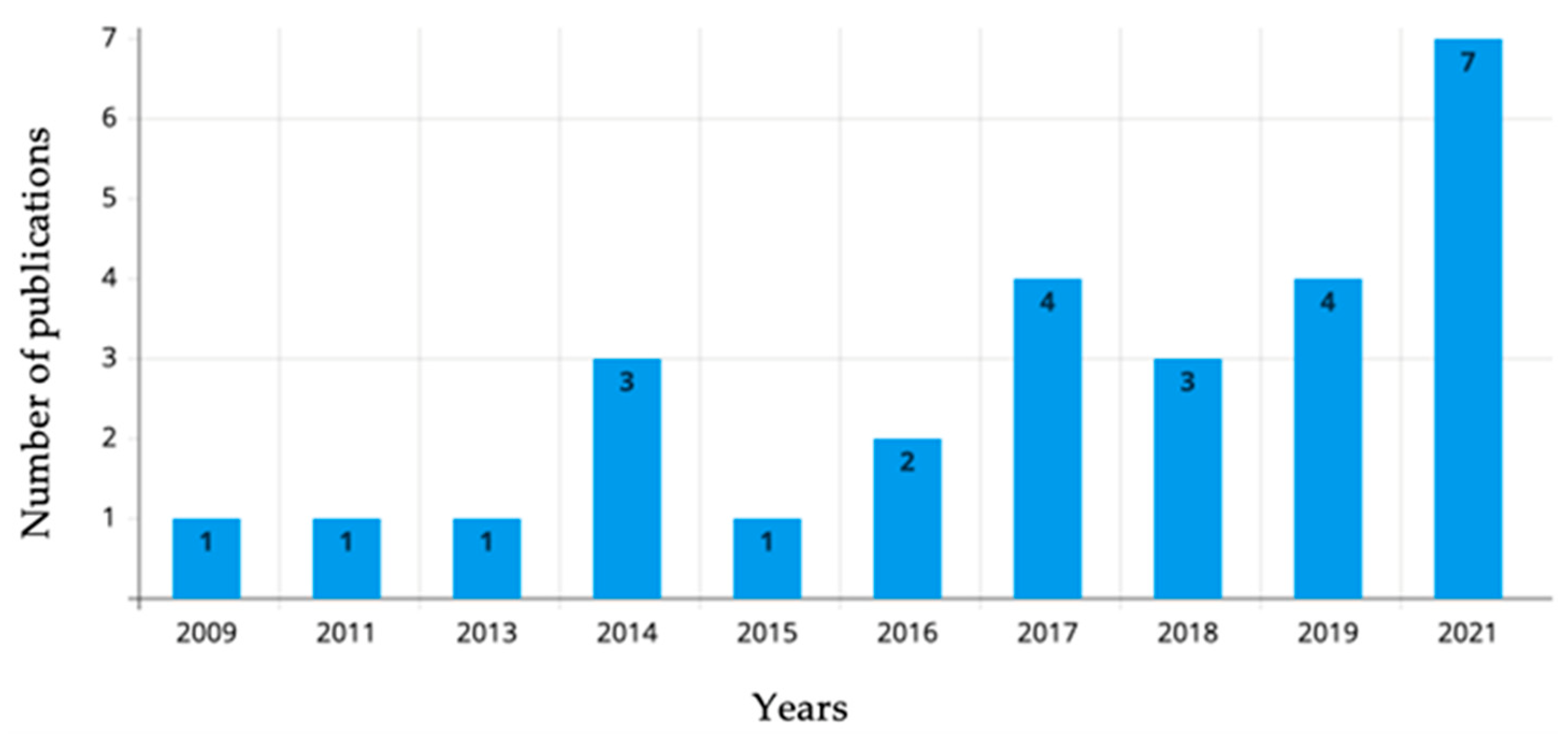
The extracted data will include the study author(s), the year of publication, the study location, the study aim, the type of cell, the sample size, the used algorithm, and the findings (Table 2). Additionally, we performed a demographic analysis to visualize the distribution of included studies on the world map with the number of publications per country (Figure 2) and a bar chart of the number of publications per year (Figure 3).
Table 2.
Data extracted from included studies.
Figure 2.
Geographic distribution of the retrieved publication studying stem cell image analysis using artificial intelligence.
Figure 3.
Bar chart showing the number of publications per year.
Upon a demographical analysis of the collected data, the United States was the highest contributing country in this field with 9 studies out of 27 (33.3%) [25,26,29,30,38,45,46,47,48], followed by China (13.5%) [27,32,41,42], Japan (11.1%) [28,33,37], and Brazil [50,51] and Taiwan [32,40] (both 7.4%). Argentina [34], Finland [43], Germany [39], Italy [49], South Korea [36], and Thailand [44] each had one study each (3.7%), as shown in Figure 2.
The number of publications studying stem cell image analysis using artificial intelligence increased throughout the years. Based on the included publications, the majority of published studies were in 2021 (seven studies) [25,26,27,28,29,30,31].
4. Discussion
A microscopic evaluation of all types of cultured cells is a routinely performed task in the laboratory setting. The contrast microscope is commonly used at various magnifications for cell analysis. The general criteria for cultured cells are confluence, dead cells, or severely atypical morphology. Therefore, operators must be trained in cell morphology and their ongoing relationship in all cases. The advanced approach to this task uses automatically extracted data that should include the most important parameters, including the cells’ confluence, cell-free areas, dead cells, and cell morphology changes. When performed manually, it suffers from low reproducibility and, on top of that, must be assessed quickly to ensure sufficient throughput. Some of the available software requires an invasive approach using either reporter genes or immunofluorescence labeling, which may lead to the irreversible modification of the cell sample or cell line death. Both systems require either cell modifications or cell sample sacrifice. Only methods that are non-invasive and capable of processing large extensive image data within a short time frame are applicable. They also need to be able to perform multi-class segmentation to assess all required parameters. Deep-learning-based algorithms are at the forefront of such complex tasks.
This systematic scoping review aimed to identify the currently available AI methods which support the laboratory evaluation of stem cells. An analysis of the included studies revealed that the visual assessment of stem cells was mainly based on the morphological features of cells in colonies and the attempt to indicate their expected features. The potential of several algorithms was tested by authors to assess the level of differentiation and morphological changes during stem cell culture and an attempt to distinguish between different types of cells during their development stages based on the morphological features. An assessment made with the help of algorithms has a chance to be more effective and also less time-consuming. The key to developing successful algorithms is the correct selection of images, the use of appropriate data sets, and enough training images. Some of the studies described above boast a large sample size for training purposes; this significantly increases the reliability of the results.
4.1. iPSC
iPSCs have the potential to differentiate into all cell types, except extraembryonic cells, and possess a high proliferative capacity, and can be cultured on an industrial scale. These features make them an area of great interest for potential application in advanced therapies.
In the following section, we present the 16 studies that test the power of artificial intelligence in analyzing, identifying, and classifying human or animal iPSC.
Fischbacher et al. [25] tested the power of three algorithms (Monoqlo, RetinaNet, and ResNet) in the automatic detection of colony presence and the identification of clonality on approximately 30,000 images. Developed using the PerkinElmer HCS system, Guo et al. [27] tested the ability of the algorithm on high-content analysis to study embryo-like structures derived from several mice-induced PSC lines, TSCs and ESCs. Imamura et al. [28] built an ALS prediction model using a CNN-based deep learning algorithm where 4500, 1350, and 900 images were used for training, validation, and testing, respectively. Joy et al. [29] trained five different neural networks (FCRN-A, FCRN-B, U-Net, Residual U-Net, and Count-ception) to localize each cell nucleus individually in a human-induced pluripotent stem cell (hiPSC) colony, generating longitudinal measures of cell and cellular neighborhood properties.
Chang et al. [32] and Chang et al. [40] tested CNN and human iPSCs. Chang et al. [32] tested CNN’s ability to trace human iPS cell formation from CD34+ cord blood cells on 144 images. In contrast, Chang et al. [40] applied the CNN on 132 images for the automatic detection and localization of human iPSC regions in brightfield microscopy images.
Orita et al. [33] trained VGG16 using bright-field images of cultured human-induced pluripotent stem cell-derived cardiomyocytes (hiPSC-CMs). The sample size was divided into 14,000 images for training, 2000 for validation, and 2000 for testing.
Zhang et al. [35] used XGBoost to model an algorithm for iPS cell identification against MEFs in the same stage by using live-cell images during the early stages of iPSC reprogramming
Kavitha et al. [36] used a set of 169 phase-contrast microscopic images of iPSC colonies and five different machine learning algorithms (support vector machine (SVM), random forest (RF), multilayer perceptron (MLP), decision tree (DT), and adaptive boosting (Adaboost) classifier models) to evaluate the selected iPSC colony features (shape, statistics, spectrum, etc.) and to eventually characterize stem cells where SVM, RF, and Adaboost were significant outperformers amongst the algorithms used. With the utilization of LeNET and AlexNET, Kusumoto et al. [37] tested a morphology-based automatic method for identifying endothelial cells derived from iPSCs which required 640 images for training and 160 for validation. Buggenthin et al. [39] implemented RNNs and CNNs to evaluate how accurately a deep learning method can predict lineage choice in differentiating primary hematopoietic cells.
Fan et al. [41] utilized a modified version of AlexNET to quantitatively test the automatic and label-free classification and segmentation of iPSC colonies derived from humans and animals. Using the improved supervised normalized cut (ISNC) segmentation and k-means clustering algorithm, Li et al. [42] proposed a multi-stage framework system, CBMIA, which was tested on 81 microscopic human iPSC images.
Joutsijoki et al. [43] assessed the automated quality identification of iPSC colony images using multiclass support vector machines and scaled invariant feature transformation (SIFT), where feeder cells were included and not included, using 173 images.
Finally, two studies by Maddah et al. [47] and Maddah et al. [48] used recorded videos to test AI. Maddah et al. [47] successfully identified iPSC-derived cardiomyocytes in the analyzed cell culture based on an algorithm that captures the beating signals, implementing a hierarchical clustering algorithm trained using 500 recorded videos. Utilizing more than 500 time-lapse sequence images of iPSCs, Maddah et al. [48] tested and presented a framework for automated analysis of phase-contrast images of iPSCs to capture and quantify morphological changes during colony growth.
4.2. ESC
There is a high demand for the precise and automatic quantification of ESC pluripotency in challenging to evaluate the environment of mixed colonies with undifferentiated and differentiated cells. Seven analyzed studies classified the human and animal ESC using a different algorithm, presented in the following.
Guan et al. [26] used 27,603 unlabeled grayscale images for fine-tuning and 3559 labeled ones for training the model, aiming to develop a deep learning method for hESC classification on a dataset of videos. Random network (RandNet) achieved a classification accuracy of 97.23 ± 0.94%. Waisman et al. [34] focused on designing an algorithm capable of distinguishing an early-differentiating cell from pluripotent cells by utilizing the ResNet50 and DenseNET architecture. In total, 1116 images under various differentiation-inducing conditions of mouse ESCs were used.
Theagarajan et al. [38] proposed a system for classifying hESCs into six categories using the CNN approach alone or in combination with Triplet CNN, achieving more than 94% accuracy. A total of 784 realistic unlabeled images were used to train, validate, and test the CNN for classifying hESCs images in a hierarchical system, allowing for their classifications into six categories.
Lou et al. [46] also addressed the segmentation issue, focusing mainly on nuclear segmentation by procuring MINS, which was developed on a MATLAB/C++-based segmentation tool on murine extraembryonic endoderm stem cells and ESCs. Paduano et al. [49] applied a MATLAB script pack and an orientation matching algorithm to mESCs. The developed algorithm was trained using 57 images and was able to properly process images. Faustino et al. [50] tested a developed algorithm for counting and detecting fluorescence microscopy images, where a total of 234 images of murine embryonic stem cells were used. Using a database of 92 images, Faustino et al. [51] implemented Java language 6.0 and Clipse 3.2 (development tool E) to shed light on fluorescent microscopy image use in automated embryonic stem counting and cell detection.
4.3. Other Stem cells
The presented studies in this section analyzed and identified different types of stem cells. Two studies classified hBMSCs, and the others screened NSCs and MAPCs using AI.
In their study, Mota et al. [30] proposed an objective approach for automatically classifying mesenchymal stem cell (MSC) efficacy using a training dataset of 71 images and a dataset of 36 images for validation. Bajcsy et al. [45] focused on three-dimensional (3D) focal segmentation in their algorithm process, where they managed to construct six new 3D segmentation algorithms using over a thousand z-stacks (3D images). Zhu et al. [31] trained and tested Xception, ResNet, VGGNet, and Inception-v730 to recognize the features of differentiated NSCs via un-labelled brightfield single-cell images. The number of NSC images used for training and testing was 19,533 and 29,895, respectively. Wuttisarnwattana et al. [44] delved deeper into fluorescent imaging and focused on cryo-imaging, specifically creating fluorescent images of MAPCs, from which 700 images were taken.
5. Conclusions
CNN is used to address several tasks and issues in the medical field and is extensively studied to implement its potential into the domain of stem cell biology in the form of automatic identification of cell types, their features, and development stages with the use of microscopic images without the necessity for molecular labeling. Deep learning technology has significantly improved, and the accuracy of the CNN image classification task has vastly exceeded that of humans. The segmentation process assigns each pixel in the image to an object class, making the classification of the image on the pixel level achievable within the boundary area.
Automatic algorithms outperform human-labeling skills in multiple ways, but proper annotation is still required to achieve this level of effectiveness. AI is at the forefront of accelerated progress in biomedical research and will majorly influence each stage of stem cell studies, consequently impacting the transfer of its research results to clinical practice. The amount of research in this field is significantly increasing with time. However, there are important factors affecting the final results that investigators should consider: the quality of images used as a training set, the sample size, and the elimination of unexpected events that the algorithm cannot predict.
Supplementary Materials
The following supporting information can be downloaded at: https://www.mdpi.com/article/10.3390/biology11101412/s1. Table S1: Searching stagey.
Author Contributions
Conceptualization, M.D.-K. and J.I.; methodology, J.I.; validation, M.D.-K., B.K. and J.I.; formal analysis, J.I. and M.A.C.; investigation, J.I. and M.A.C.; data curation, J.I. and M.A.C.; writing—original draft preparation, J.I., M.A.C. and M.D.-K.; writing—review and editing, M.D.-K., B.K., L.G., P.M. and R.O.; supervision, M.D.-K.; funding acquisition, P.M. and J.I. All authors have read and agreed to the published version of the manuscript.
Funding
This research was funded by the National Institute of Food and Agriculture, United States Department of Agriculture Animal Health NC07082. Julien Issa is a participant of the STER Internationalization of Doctoral Schools Program from NAWA Polish National Agency for Academic Exchange No. PPI/STE/2020/1/00014/DEC/02.
Institutional Review Board Statement
Not applicable.
Informed Consent Statement
Not applicable.
Data Availability Statement
The data that support the findings of this study are available from the corresponding author upon reasonable request.
Conflicts of Interest
The authors declare no conflict of interest.
References
- Liu, G.; David, B.T.; Trawczynski, M.; Fessler, R.G. Advances in Pluripotent Stem Cells: History, Mechanisms, Technologies, and Applications. Stem Cell Rev. Rep. 2020, 16, 3–32. [Google Scholar] [CrossRef] [Scilit] [PubMed]
- Srinivasan, M.; Thangaraj, S.R.; Ramasubramanian, K.; Thangaraj, P.P.; Ramasubramanian, K.V. Exploring the Current Trends of Artificial Intelligence in Stem Cell Therapy: A Systematic Review. Cureus 2021, 12, e20083. [Google Scholar] [CrossRef] [Scilit] [PubMed]
- Jankowski, M.; Dompe, C.; Sibiak, R.; Wąsiatycz, G.; Mozdziak, P.; Jaśkowski, J.M.; Antosik, P.; Kempisty, B.; Dyszkiewicz-Konwińska, M. In Vitro Cultures of Adipose-Derived Stem Cells: An Overview of Methods, Molecular Analyses, and Clinical Applications. Cells 2020, 9, 1783. [Google Scholar] [CrossRef] [Scilit] [PubMed]
- Ramakrishna, R.R.; Abd Hamid, Z.; Wan Zaki, W.; Huddin, A.B.; Mathialagan, R. Stem cell imaging through convolutional neural networks: Current issues and future directions in artificial intelligence technology. PeerJ 2020, 8, e10346. [Google Scholar] [CrossRef] [Scilit] [PubMed]
- Cable, J.; Fuchs, E.; Weissman, I.; Jasper, H.; Glass, D.; Rando, T.A.; Blau, H.; Debnath, S.; Oliva, A.; Park, S.; et al. Adult stem cells and regenerative medicine-a symposium report. Ann. N. Y. Acad. Sci. 2020, 1462, 27–36. [Google Scholar] [CrossRef] [Scilit]
- Malik, N.; Rao, M.S. A review of the methods for human iPSC derivation. Methods Mol. Biol. 2013, 997, 23–33. [Google Scholar] [CrossRef] [Scilit]
- Bacakova, L.; Zarubova, J.; Travnickova, M.; Musilkova, J.; Pajorova, J.; Slepicka, P.; Kasalkova, N.S.; Svorcik, V.; Kolska, Z.; Motarjemi, H.; et al. Stem cells: Their source, potency and use in regenerative therapies with focus on adipose-derived stem cells—A review. Biotechnol. Adv. 2018, 36, 1111–1126. [Google Scholar] [CrossRef] [Scilit] [PubMed]
- Zakrzewski, W.; Dobrzyński, M.; Szymonowicz, M.; Rybak, Z. Stem cells: Past, present, and future. Stem Cell Res. Ther. 2019, 10, 68. [Google Scholar] [CrossRef] [Scilit]
- Menasche, P.; Vanneaux, V.; Hagege, A.; Bel, A.; Cholley, B.; Cacciapuoti, I.; Parouchev, A.; Benhamouda, N.; Tachdjian, G.; Tosca, L.; et al. Human embryonic stem cell-derived cardiac progenitors for severe heart failure treatment: First clinical case report. Eur. Heart J. 2015, 36, 2011–2017. [Google Scholar] [CrossRef] [Scilit]
- Schwartz, S.D.; Regillo, C.D.; Lam, B.L.; Eliott, D.; Rosenfeld, P.J.; Gregori, N.Z.; Hubschman, J.P.; Davis, J.L.; Heilwell, G.; Spirn, M.; et al. Human embryonic stem cell-derived retinal pigment epithelium in patients with age-related macular degeneration and Stargardt’s macular dystrophy: Follow-up of two open-label phase 1/2 studies. Lancet 2015, 385, 509–516. [Google Scholar] [CrossRef] [Scilit]
- Dantuma, E.; Merchant, S.; Sugaya, K. Stem cells for the treatment of neurodegenerative diseases. Stem Cell Res. Ther. 2010, 1, 37. [Google Scholar] [CrossRef] [Scilit] [PubMed]
- Longo, U.G.; Ronga, M.; Maffulli, N. Achilles tendinopathy. Sports Med. Arthrosc. 2009, 17, 112–126. [Google Scholar] [CrossRef] [Scilit] [PubMed]
- Widuchowski, W.; Widuchowsk, J.; Trzaska, T. Articular cartilage defects: Study of 25,124 knee arthroscopies. Knee 2007, 14, 177–182. [Google Scholar] [CrossRef] [Scilit] [PubMed]
- Hayashi, K.; Ohta, H.; Kurimoto, K.; Aramaki, S.; Saitou, M. Reconstitution of the mouse germ cell specification pathway in culture by pluripotent stem cells. Cell 2011, 146, 519–532. [Google Scholar] [CrossRef] [Scilit] [PubMed]
- Sadri-Ardekani, H.; Atala, A. Testicular tissue cryopreservation and spermatogonial stem cell transplantation to restore fertility: From bench to bedside. Stem Cell Res. Ther. 2014, 5, 68. [Google Scholar] [CrossRef] [Scilit]
- Kroon, E.; Martinson, L.A.; Kadoya, K.; Bang, A.G.; Kelly, O.G.; Eliazer, S.; Young, H.; Richardson, M.; Smart, N.G.; Cunningham, J.; et al. Pancreatic endoderm derived from human embryonic stem cells generates glucose-responsive insulin-secreting cells in vivo. Nat. Biotechnol. 2008, 26, 443–452. [Google Scholar] [CrossRef] [Scilit]
- Lee, G.; Papapetrou, E.P.; Kim, H.; Chambers, S.M.; Tomishima, M.J.; Fasano, C.A.; Ganat, Y.M.; Menon, J.; Shimizu, F.; Viale, A.; et al. Modelling pathogenesis and treatment of familial dysautonomia using patient-specific iPSCs. Nature 2009, 461, 402–406. [Google Scholar] [CrossRef] [Scilit]
- Imamura, K.; Izumi, Y.; Banno, H.; Uozumi, R.; Morita, S.; Egawa, N.; Ayaki, T.; Nagai, M.; Nishiyama, K.; Watanabe, Y.; et al. Induced pluripotent stem cell-based Drug Repurposing for Amyotrophic lateral sclerosis Medicine (iDReAM) study: Protocol for a phase I dose escalation study of bosutinib for amyotrophic lateral sclerosis patients. BMJ 2019, 9, e033131. [Google Scholar] [CrossRef] [Scilit]
- De Masi, C.; Spitalieri, P.; Murdocca, M.; Novelli, G.; Sangiuolo, F. Application of CRISPR/Cas9 to human-induced pluripotent stem cells: From gene editing to drug discovery. Hum. Genom. 2020, 14, 25. [Google Scholar] [CrossRef] [Scilit]
- Amisha; Malik, P.; Pathania, M.; Rathaur, V.K. Overview of artificial intelligence in medicine. J. Fam. Med. Prim. Care 2019, 8, 2328–2331. [Google Scholar] [CrossRef] [Scilit]
- Peters, M.D.; Godfrey, C.M.; Khalil, H.; McInerney, P.; Parker, D.; Soares, C.B. Guidance for conducting systematic scoping reviews. Int. J. Evid.-Based Healthc. 2015, 13, 141–146. [Google Scholar] [CrossRef] [Scilit] [PubMed]
- Tricco, A.C.; Lillie, E.; Zarin, W.; O’Brien, K.K.; Colquhoun, H.; Levac, D.; Moher, D.; Peters, M.; Horsley, T.; Weeks, L.; et al. PRISMA Extension for Scoping Reviews (PRISMA-ScR): Checklist and Explanation. Ann. Intern. Med. 2018, 169, 467–473. [Google Scholar] [CrossRef] [Scilit] [PubMed]
- Issa, J.; Abou Chaar, M.; Kempisty, B.; Mozdziak, P.; Dyszkiewicz-Konwińska, M. Artificial intelligence-based imaging analysis of stem cells: A systematic scoping review protocol. Res. Sq. 2022, preprint. [Google Scholar] [CrossRef] [Scilit]
- Clark, J.; Glasziou, P.; Del Mar, C.; Bannach-Brown, A.; Stehlik, P.; Scott, A.M. A full systematic review was completed in 2 weeks using automation tools: A case study. J. Clin. Epidemiol. 2020, 121, 81–90. [Google Scholar] [CrossRef] [Scilit] [PubMed]
- Fischbacher, B.; Hedaya, S.; Hartley, B.J.; Wang, W.; Lallos, G.; Hutson, D.; Zimmer, M.; Brammer, J.; The NYSCF Global Stem Cell Array Team; Paull, D. Modular deep learning enables automated identification of monoclonal cell lines. Nat. Mach. Intell. 2021, 3, 632–640. [Google Scholar] [CrossRef] [Scilit]
- Guan, B.X.; Bhanu, B.; Theagarajan, R.; Liu, H.; Talbot, P.; Weng, N. Human embryonic stem cell classification: Random network with autoencoded feature extractor. J. Biomed. Opt. 2021, 26, 052913. [Google Scholar] [CrossRef] [Scilit]
- Guo, J.; Wang, P.; Sozen, B.; Qiu, H.; Zhu, Y.; Zhang, X.; Ming, J.; Zernicka-Goetz, M.; Na, J. Machine learning-assisted high-content analysis of pluripotent stem cell-derived embryos in vitro. Stem Cell Rep. 2021, 16, 1331–1346. [Google Scholar] [CrossRef] [Scilit]
- Imamura, K.; Yada, Y.; Izumi, Y.; Morita, M.; Kawata, A.; Arisato, T.; Nagahashi, A.; Enami, T.; Tsukita, K.; Kawakami, H.; et al. Prediction Model of Amyotrophic Lateral Sclerosis by Deep Learning with Patient Induced Pluripotent Stem Cells. Ann. Neurol. 2021, 89, 1226–1233. [Google Scholar] [CrossRef] [Scilit]
- Joy, D.A.; Libby, A.; McDevitt, T.C. Deep neural net tracking of human pluripotent stem cells reveals intrinsic behaviors directing morphogenesis. Stem Cell Rep. 2021, 16, 1317–1330. [Google Scholar] [CrossRef] [Scilit]
- Mota, S.M.; Rogers, R.E.; Haskell, A.W.; McNeill, E.P.; Kaunas, R.; Gregory, C.A.; Giger, M.L.; Maitland, K.C. Automated mesenchymal stem cell segmentation and machine learning-based phenotype classification using morphometric and textural analysis. J. Med. Imaging 2021, 8, 014503. [Google Scholar] [CrossRef] [Scilit]
- Zhu, Y.; Huang, R.; Wu, Z.; Song, S.; Cheng, L.; Zhu, R. Deep learning-based predictive identification of neural stem cell differentiation. Nat. Commun. 2021, 12, 2614. [Google Scholar] [CrossRef] [Scilit] [PubMed]
- Chang, Y.H.; Abe, K.; Yokota, H.; Sudo, K.; Nakamura, Y.; Chu, S.L.; Hsu, C.Y.; Tsai, M.D. Human Induced Pluripotent Stem Cell Reprogramming Prediction in Microscopy Images using LSTM based RNN. In Proceedings of the Annual International Conference of the IEEE Engineering in Medicine and Biology Society, Berlin, Germany, 23–27 July 2019; pp. 2416–2419. [Google Scholar] [CrossRef] [Scilit]
- Orita, K.; Sawada, K.; Koyama, R.; Ikegaya, Y. Deep learning-based quality control of cultured human-induced pluripotent stem cell-derived cardiomyocytes. J. Pharmacol. Sci. 2019, 140, 313–316. [Google Scholar] [CrossRef] [Scilit] [PubMed]
- Waisman, A.; La Greca, A.; Möbbs, A.M.; Scarafía, M.A.; Santín Velazque, N.L.; Neiman, G.; Moro, L.N.; Luzzani, C.; Sevlever, G.E.; Guberman, A.S.; et al. Deep Learning Neural Networks Highly Predict Very Early Onset of Pluripotent Stem Cell Differentiation. Stem Cell Rep. 2019, 12, 845–859. [Google Scholar] [CrossRef] [Scilit] [PubMed]
- Zhang, H.; Shao, X.; Peng, Y.; Teng, Y.; Saravanan, K.M.; Zhang, H.; Li, H.; Wei, Y. A novel machine learning based approach for iPS progenitor cell identification. PLoS Comput. Biol. 2019, 15, e1007351. [Google Scholar] [CrossRef] [Scilit] [PubMed]
- Kavitha, M.S.; Kurita, T.; Ahn, B.C. Critical texture pattern feature assessment for characterizing colonies of induced pluripotent stem cells through machine learning techniques. Comput. Biol. Med. 2018, 94, 55–64. [Google Scholar] [CrossRef] [Scilit] [PubMed]
- Kusumoto, D.; Lachmann, M.; Kunihiro, T.; Yuasa, S.; Kishino, Y.; Kimura, M.; Katsuki, T.; Itoh, S.; Seki, T.; Fukuda, K. Automated Deep Learning-Based System to Identify Endothelial Cells Derived from Induced Pluripotent Stem Cells. Stem Cell Rep. 2018, 10, 1687–1695. [Google Scholar] [CrossRef] [Scilit]
- Theagarajan, R.; Guan, B.X.; Bhanu, B. DeephESC: An Automated System for Generating and Classification of Human Embryonic Stem Cells. In Proceedings of the 2018 24th International Conference on Pattern Recognition (ICPR), Beijing, China, 20–24 August 2018; pp. 3826–3831. [Google Scholar] [CrossRef] [Scilit]
- Buggenthin, F.; Buettner, F.; Hoppe, P.S.; Endele, M.; Kroiss, M.; Strasser, M.; Schwarzfischer, M.; Loeffler, D.; Kokkaliaris, K.D.; Hilsenbeck, O.; et al. Prospective identification of hematopoietic lineage choice by deep learning. Nat. Methods 2017, 14, 403–406. [Google Scholar] [CrossRef] [Scilit]
- Chang, Y.H.; Abe, K.; Yokota, H.; Sudo, K.; Nakamura, Y.; Lin, C.-Y.; Tsai, M.D. Human induced pluripotent stem cell region recognition in microscopy images using Convolutional Neural Networks. In Proceedings of the Annual International Conference of the IEEE Engineering in Medicine and Biology Society, Jeju, Korea, 11–15 July 2017; pp. 4058–4061. [Google Scholar] [CrossRef] [Scilit]
- Fan, K.; Zhang, S.; Zhang, Y.; Lu, J.; Holcombe, M.; Zhang, X. A Machine Learning Assisted, Label-free, Non-invasive Approach for Somatic Reprogramming in Induced Pluripotent Stem Cell Colony Formation Detection and Prediction. Sci. Rep. 2017, 7, 13496. [Google Scholar] [CrossRef] [Scilit]
- Li, C.; Huang, X.; Jiang, T.; Xu, N. Full-automatic computer aided system for stem cell clustering using content-based microscopic image analysis. Biocybern. Biomed. Eng. 2017, 37, 540–558. [Google Scholar] [CrossRef] [Scilit]
- Joutsijoki, H.; Haponen, M.; Rasku, J.; Aalto-Setälä, K.; Juhola, M. Machine Learning Approach to Automated Quality Identification of Human Induced Pluripotent Stem Cell Colony Images. Comput. Math. Methods Med. 2016, 2016, 3091039. [Google Scholar] [CrossRef] [Scilit]
- Wuttisarnwattana, P.; Gargesha, M.; van’t Hof, W.; Cooke, K.R.; Wilson, D.L. Automatic Stem Cell Detection in Microscopic Whole Mouse Cryo-Imaging. IEEE Trans. Med. Imaging 2016, 35, 819–829. [Google Scholar] [CrossRef] [Scilit] [PubMed]
- Bajcsy, P.; Simon, M.; Florczyk, S.J.; Simon, C.G., Jr.; Juba, D.; Brady, M.C. A method for the evaluation of thousands of automated 3D stem cell segmentations. J. Microsc. 2015, 260, 363–376. [Google Scholar] [CrossRef] [Scilit] [PubMed]
- Lou, X.; Kang, M.; Xenopoulos, P.; Muñoz-Descalzo, S.; Hadjantonakis, A.K. A rapid and efficient 2D/3D nuclear segmentation method for analysis of early mouse embryo and stem cell image data. Stem Cell Rep. 2014, 2, 382–397. [Google Scholar] [CrossRef] [Scilit] [PubMed]
- Maddah, M.; Loewke, K. Automated, non-invasive characterization of stem cell-derived cardiomyocytes from phase-contrast microscopy. In Medical Image Computing and Computer-Assisted Intervention: MICCAI, Proceedings of the International Conference on Medical Image Computing and Computer-Assisted Intervention, Boston, MA, USA, 14–18 September 2014; Springer: Cham, Switzerland, 2014; Volume 17, pp. 57–64. [Google Scholar] [CrossRef] [Scilit]
- Maddah, M.; Loewke, K. Dynamic morphology-based characterization of stem cells enabled by texture-based pattern recognition from phase-contrast images. In Proceedings of the 2014 IEEE 11th International Symposium on Biomedical Imaging (ISBI), Beijing, China, 29 April–2 May 2014; pp. 77–80. [Google Scholar] [CrossRef] [Scilit]
- Paduano, V.; Tagliaferri, D.; Falco, G.; Ceccarelli, M. Automated Identification and Location Analysis of Marked Stem Cells Colonies in Optical Microscopy Images. PLoS ONE 2013, 8, e80776. [Google Scholar] [CrossRef] [Scilit] [PubMed]
- Faustino, G.M.; Gattass, M.; de Lucenaa, C.J.P.; Camposb, P.B.; Rehenb, S.K. A Graph-mining Algorithm for Automatic Detection and Counting of Embryonic Stem Cells in Fluorescence Microscopy Images. Integr. Comput. Aided Eng. 2011, 18, 91–106. [Google Scholar] [CrossRef] [Scilit]
- Faustino, G.M.; Gattass, M.; Rehen, S.; de Lucena, C.J.P. Automatic embryonic stem cells detection and counting method in fluorescence microscopy images. In Proceedings of the 2009 IEEE International Symposium on Biomedical Imaging: From Nano to Macro, Boston, MA, USA, 28 June–1 July 2009; pp. 799–802. [Google Scholar] [CrossRef] [Scilit]

Publisher’s Note: MDPI stays neutral with regard to jurisdictional claims in published maps and institutional affiliations. |
© 2022 by the authors. Licensee MDPI, Basel, Switzerland. This article is an open access article distributed under the terms and conditions of the Creative Commons Attribution (CC BY) license (https://creativecommons.org/licenses/by/4.0/).