


Abstract
In the last decades, the interest in seaweed has significantly increased. Bioactive compounds from seaweed’s currently receive major attention from pharmaceutical companies as they express several interesting biological activities which are beneficial for humans. The structural diversity of seaweed metabolites provides diverse biological activities which are expressed through diverse mechanisms of actions. This review mainly focuses on the antiviral activity of seaweed’s extracts, highlighting the mechanisms of actions of some seaweed molecules against infection caused by different types of enveloped viruses: influenza, Lentivirus (HIV-1), Herpes viruses, and coronaviruses. Seaweed metabolites with antiviral properties can act trough different pathways by increasing the host’s defense system or through targeting and blocking virus replication before it enters host cells. Several studies have already established the large antiviral spectrum of seaweed’s bioactive compounds. Throughout this review, antiviral mechanisms and medical applications of seaweed’s bioactive compounds are analyzed, suggesting seaweed’s potential source of antiviral compounds for the formulation of novel and natural antiviral drugs.
1. Introduction
Viral infections are common and recurrent across the globe, and the lack of effective treatments for diseases of viral origin puts a serious burden on public healthcare systems. Virology and pharmaceutics are focused on the development of broad-spectrum antiviral drugs, which are effective, present low toxicity, and are inexpensive. The first class of antiviral agents are identified with nucleosides, synthetic drugs that act faster and provide the maximum therapeutic effect. However, the side-effects associated with these drugs could lead to health issues, such as acute renal failure, as the drug doses increase, as well as addiction, with a consequential absence of the desired effect in the future [1]. To reduce the collateral effects, new and effective antiviral agents are therefore urgently required. In the last decades, the activity of herbal antiviral drugs with a wide spectrum of action for viruses has been reported. Natural drugs developed with natural compounds have been shown to help alleviate the symptoms of specific viral diseases and shorten the disease period, are less toxic, and the side effects are minimized [2]. The development of an effective antiviral agent depends on the type of virus to defeat, its mechanisms of infection and replication. Viruses are classified as simple and complex; simple (or non-enveloped) viruses are composed of a nucleic acid and protein envelope (capsid); complex (or enveloped) viruses are surrounded by a lipoprotein envelope (supercapsid) over the capsid, which makes them more vulnerable to adverse environmental factors [3]. The presence/absence of the envelope may condition their susceptibility to different disinfectants or antiviral agents.
Generally, the virus mechanism of infection includes three steps: (1) the virus is absorbed by the host cell through a specific interaction between surface proteins of the virus and the receptors located on the surface of host cells. The virus undergoes a modification and releases its internal structure into a state which will cause infection; (2) during the second phase, complex processes allow the expression of the viral genome to replication; (3) in the third and last step, the viral offspring obtained is released out of the host cell by the budding of lysis, going on to infect other host cells. Currently, pharmacology is researching and developing novel antiviral agents that are able to block the virus entry or interfere at each stage of virus replication, reducing the incubation time and disease [4,5,6,7,8].
In the present work, seaweed compounds with antiviral properties are discussed, as they could have potential as a prophylactic agent and could be involved in the development of natural therapeutic agents for patients with viral infections.
The mechanisms of action of seaweed’s bioactive compounds against enveloped viruses such as influenza, Lentivirus, Herpes viruses and coronaviruses are analysed throughout the manuscript. Seaweed-derived antiviral drugs possess a broad-spectrum against viral infection, they are less toxic or non-toxic at all, thus the side effects are not as harmful as for synthetic compounds. The aim of the present review is to show the importance of seaweed’s’ compounds in the pharmaceutical field, with a particular focus on virology.
2. Mechanisms of Infection for Enveloped Viruses
To investigate the antiviral mechanism of action of seaweed’s bioactive compounds it is important to acknowledge the mechanism of infection of interested viruses. In the following sections are described cycles of infection of the influenza virus, Lentivirus, Herpes viruses and coronaviruses, to make the reader understand the antiviral pathway performed by seaweed bioactive compounds against enveloped viruses.
2.1. Influenza Viruses
Influenza A virus (Alphainfluenzavirus) and Influenza B virus (Betainfluenzavirus) both belong both to fam. Orthomyxoviridae, composed by RNA viruses.
Influenza epidemics are mainly caused by Influenza A, the most common influenza infection during the flu season, which causes mild to severe illness and affects humans and animals [9,10], and type B influenza infection, which is highly contagious and can sometimes cause serious illness. Influenza B virus is reported to be less common during flu season and causes minor localized outbreaks [11,12]. Influenza A and B viruses symptoms include cough, sore throat, nasal discharge, fever, headache, and muscle pain. However, the symptoms can be more severe and lead to serious complications like bronchitis and pneumonia [13].
Influenza C viruses primarily infect humans and cause illness in some animals, such as swine. It causes mild upper respiratory symptoms, and is found in minor localized outbreaks [12,14]. The Type D viruses mainly affect pigs and cattle and are not known to cause humans infection [12,15].
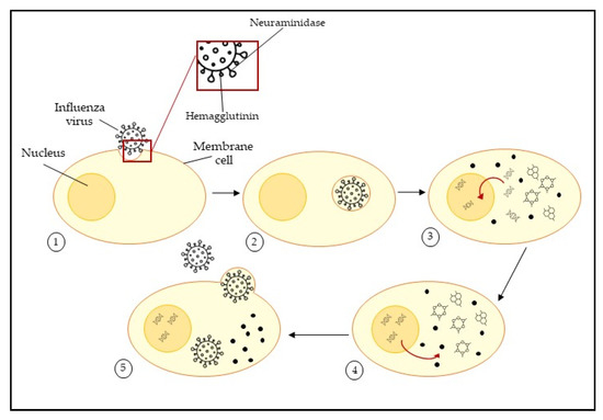
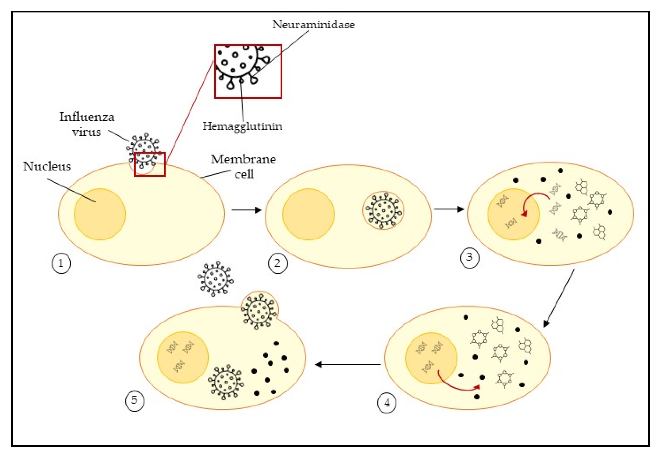
Influenza viruses are enveloped viruses that possess surface glycoproteins, mainly hemagglutinin (HA) and neuraminidase (NA). These glycoproteins play a key role in cycle infection (Figure 1); hemagglutinin mediates cell attachment and infection trough release of the viral genome into the cell, while neuraminidase mediates the release of progeny virions. These two protein spikes are very exposed; therefore, they are ideal targets for the development of vaccines, antibodies, and antiviral drugs.
Figure 1.
Schematic representation of influenza virus mechanism of cell infection. ① the spike protein hemagglutinin bond with the receptor on the host cell membrane; ② the virus enters into the cell by endocytosis; ③ the spike protein neuraminidase mediates the viral RNA and release, which enters the nucleus where it is replicated by the viral RNA polymerase; ④ viral mRNA is used to make viral proteins; ⑤ new viral particles are released into the extracellular matrix and the host cell continues to make new virus particles.
Hemagglutinin inhibitors act during the first infection phase, preventing the attachment of the glycoprotein to target cells. Neuraminidase inhibitors prevent the release of the virus progeny into host cells.
The HA bind to sialic acids on the surface of epithelial cells of the respiratory tract, dendritic cells, type II pneumocytes, alveolar macrophages, or retinal epithelial cells [16,17]. Viruses enter by endocytosis, and follow the degradation of M1 and M2 proteins and release vRNPs, which transcribe viral mRNAs for the production of viral proteins in the cytoplasm and successively it enters the nucleus [18,19,20]. In the nucleus, negative-sense vRNA is transcribed into positive-sense mRNA using viral polymerase [21]. The viral proteins are translated from mRNA in the cytoplasm by ribosomes. IAVs complete successful replication by relying on multiple cellular proteins. Cellular clathrin, epsin-1 Ras-related GTPases, and COPI are important proteins for virus dynamin-dependent endocytic uptake. They degrade the M1 shell and uncoat vRNPs. Subsequently, cytoplasmic importins mediate the nuclear import of vRNPs through the nuclear pore complex (NPC). In the cytoplasm, a translation apparatus translates viral mRNAs into proteins and GRSF1 stimulates this process. Newly synthesized M1, M2, HA, and NA are also transported to the plasma membrane through the trans-Golgi network with the help of COPI and Rab8. β-actin, CK2 and Rab11 are cellular proteins required for the budding and release of new virions [13].
Early studies on HA revealed that HA-mediated fusion involves the insertion of a key segment into the host membrane and that segment is known as the influenza fusion peptide (IFP). It has been shown that mutations within the FP region of HA can either maintain its ability to induce complete fusion or only promote hemifusion (fusion of the outer leaflets of two membranes), depending on the residue which is mutated [22]. Therefore, more studies focusing on characterizing its structure and effect on membranes of host cells can help to find new anti-influenza treatments that block the fusion of the virus into the host cell [23].
Mutation in amino acids in IAV proteins can cause antigenic drift, which allows emerging viruses to evade host immunity developed from previous IAV infections or vaccinations. One of the causes of mutation can be due to the error-prone nature of viral polymerase [24]. The viruses can also undergo rearrangement of genetic segments to generate different variations and sometimes antigenic shift. Genetic shifts and drifts are potential causes of epidemic and pandemic outbreaks, and are sometimes even dangerous [25].
Among antivirals, oseltamivir represents the most used agent against influenza viruses, which inhibit neuraminidase action. Oseltamivir proved potential clinical utility against seasonal and emerging influenza viruses [26].
2.2. Lentivirus
HIV belongs to the genus Lentivirus, fam. Retroviridae. It is an RNA enveloped virus with an unusual method of replication of genetic material. Lentiviruses can cause diseases with a long incubation period and a slow but steady progressive course that involves the central nervous system [27]. HIVs are usually grouped into two types: HIV-type 1 (HIV-1), also known as the main acquired immunodeficiency syndrome (AIDS) agent, and HIV-type 2 (HIV-2), present mainly in some regions of Western and Central Africa [28]. The structure of Lentivirus is made of the retrovirus genome, composed of two identical copies of single-stranded RNA molecules, major proteins such as glycoproteins, group antigens polyproteins (GAg), reverse transcriptase (pol), and envelope proteins (Env). Reverse transcriptase is the essential enzyme that carries out the reverse transcription process to produce molecules of complementary double-stranded DNA pre-formed from viral RNA. Additionally, HIV-1 and HIV-2 viruses possess other regulatory genes which can cause immunodeficiency disorders such as AIDS. It appears that AIDS is more frequent in HIV-2 infection [29], but it appears less virulent than HIV-1 and the infection course takes longer to develop into AIDS compared to HIV-1 [30,31,32].
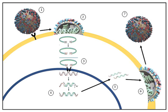
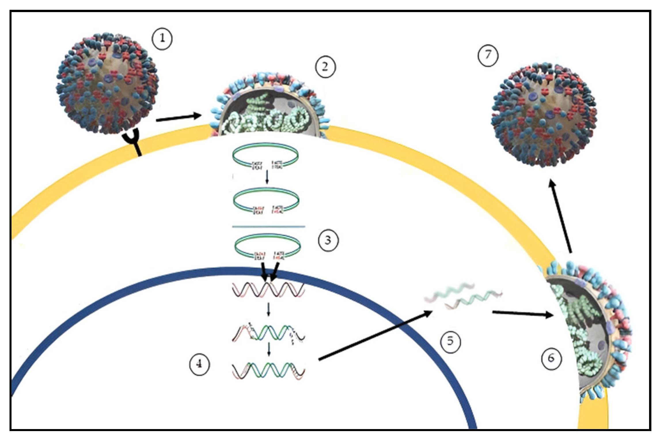
The HIV replication cycle can be summarised in six steps (Figure 2): (1) binding and entry, HIV-1 and HIV-2 possess heterodimer proteins gp120 and gp41 that compose the viral envelope, which are essential for virus recognition and entry into target cellular membranes; (2) uncoating, the virus core uncoats into the cytoplasm of the target cell freeing the viral RNA; (3) reverse transcription, the viral RNA is transcribed into an RNA/DNA hybrid double helix. The ribonuclease enzyme breaks down the RNA strand and the polymerase active site of the reverse transcriptase completes a complementary DNA strand to form a double helix DNA molecule which is integrated within the cell genome through integrase enzyme; (4) provirus integration, where the viral messenger RNA coding for long fragments migrates into the cytoplasm, where (5) new virus proteins are synthesized, such as GAg, pol, Env. The formation of new HIV virus derives from the association of two viral RNA strands incorporated together with replication enzymes, while core proteins assemble over them, forming the virus capsid. This immature and infective particle migrates towards the cell surface, where they bud through the host cell membrane, acquiring a new envelope. During the last step, the (6) budding process, the virus lipid membranes may incorporate various host cell proteins and become enriched with phospholipids and cholesterol and the virus goes to infect other cells [28].
Figure 2.
Schematic representation of HIV infection. ① binding of the virus to the host cell membrane; ② fusion of the virus and uncoating: the virus core uncoats into the cytoplasm of the target cell freeing the viral RNA; ③ reverse transcription: the viral RNA is transcribed into an RNA/DNA hybrid double helix; ④ integration of the viral gene to human DNA; ⑤ replication; ⑥ assembly of the new viral particles and proteins; ⑦ budding process and release of the new virus.
HIV attachment and fusion with the cell membrane is mediated by the viral glycoprotein (Env) and the CD4 receptor and coreceptor (CXCR4 or CCR5) [33]. The viral capsid containing the HIV-1 genome and replicative enzymes is then released into the cytoplasm where the viral reverse transcriptase (RT) enzyme transcribes the viral RNA genome into a double-stranded DNA copy.
In the nucleus, the viral integrase (IN) enzyme directs the integration of the viral DNA into transcriptionally active sites within the host chromatin. The integrated proviral DNA is then transcribed into viral mRNAs and full-length genomic RNA, and the GAg and Pol polyproteins are targeted to the inner leaflet of the plasma membrane where they assemble into an immature hexameric lattice. The Env glycoprotein precursor, gp160, is cleaved by host furin or furin-like proteases to generate the surface glycoprotein subunit gp120 and the transmembrane glycoprotein gp41. Viral genomic RNA dimers are packaged by the assembling Gag lattice and heterotrimeric gp120/gp41 Env complexes are incorporated into the membrane at the sites of assembly [34]. Immature Pol polyprotein are arranged into mature proteins by the viral protease (PR). PR mediated Gag cleavage triggers the disassembly of the membrane-bound immature Gag lattice, resulting in the assembly of the mature conical capsid from the fully processed capsid protein [35]. The virion assumes its proper infectivity when it reaches maturation that requires the packaging of two single-stranded copies of the viral RNA genome, together with RT and IN, into the nascent capsid and then is ready to bud and infect the organism [36].
2.3. Herpes Viruses
Herpes viruses (fam. Herpesviridae) are extremely successful parasites developed for millions of years, evolving action mechanisms to coexist with their hosts and to maintain host-to-host transmission and lifelong infection by regulating their life cycles. Human Herpes viruses are the causative agents of many common diseases, including chickenpox, shingles, mononucleosis, cold sores, and genital Herpes [37]. Eight Herpes viruses are known pathogens of humans: α-herpesviruses (Herpes simplex virus-HSV type 1 and type 2, Varicella zoster virus -VZV), cytomegalovirus, β-herpesviruses (Human Herpes virus-HHV 6 and 7) and γ-herpesviruses (Epstein–Barr virus, HHV8) [38].
Herpes viruses have a unique four-layered structure: a core containing the double-stranded DNA genome, which is enclosed by an icosapentahedral capsid composed of capsomers. The capsid is surrounded by an amorphous protein coat called the tegument. It is encased in a glycoprotein-bearing lipid bilayer envelope.
After the entry into the host cell, the life cycle of HSVs consists of two phases: lytic infection and latent infection [39,40,41,42,43]. During lytic infection, virions into host cell progress through uncoating, gene transcription, DNA replication, protein translation, assembly, release, etc., to produce progeny virions [43,44,45]. During this stage, the replication of HSVs is limited by the immune system, leading to the stage of latent infection.
HSV-1 and HSV-2 contain a large, linear double stranded DNA genome protected by an icosahedral capsid surrounded by a proteinaceous layer termed the tegument and are wrapped in an envelope containing viral glycoproteins. Initial attachment to the plasma membrane occurs through the binding of glycoprotein B (gB) and gC to glycosaminoglycans (GAG), attached to the outer surface of the cell membrane [46]. The interaction of HSV-1 gH/gL with specific integrins leads to HSV-1 entry through endocytosis [47]. Fusion can take place at the plasma membrane or within vesicles following viral internalization. Following fusion, some tegument proteins, like VP16, dissociate from the capsid and travel to the nucleus independently [48], while others remain bound. Inner tegument proteins mediate interaction with dynein, dynactin and kinesin motor proteins and facilitate capsid transport on microtubules toward the nucleus. The viral linear DNA genome enters the nucleus through a nuclear pore [49], and the RNA polymerase II and viral proteins transcribe HSV genes. Gene expression follows an ordered cascade during lytic replication. Immediate early genes are expressed in the absence of de novo viral protein synthesis. The tegument protein VP16 forms a complex with host cell factor 1 (HCF-1) and octamer binding protein-1 (Oct-1) that binds to the promoter of ICP, driving their expression [50]. Once DNA replication occurs, late genes which involved virus assembly are expressed. Viral transcription, DNA replication, capsid assembly and DNA encapsulation occur exclusively in the nucleus. Mature capsids containing viral DNA, leave the nucleus through a process mediated by pUL31 and pUL34. Following exit from the nucleus, cytosolic capsids acquire more inner tegument proteins, while outer tegument proteins and viral membrane proteins are incorporated at the membrane compartments of trans Golgi network vesicles and endosomes [51,52].
The mechanisms leading to transport and incorporation of viral glycoproteins are not completely understood. Data showed that pUL36 and pUL37 mediate motility in the neuronal cell body but cannot direct the non-enveloped capsids to the axons, contrary to vesicles containing gD that efficiently employed axonal transport [53]. These results suggest that only fully assembled viral particles can travel from the cell body to the axon termini. Vesicles transport HSV particles to the plasma membrane and enveloped HSV exits the cell upon fusion of the vesicle with the plasma membrane [54].
During latent infection, the viral genome remains in an inactive state in the host cell, but it can re-start the lytic cycle under immune suppression or environmental stimuli [55,56]. HSVs start the active replication to produce large numbers of infectious virions that can be transmitted to new hosts [57]. For example, HSV latency has been detected in neurons. Infection of susceptible non-neuronal cells normally leads to lytic replication, although a recent report suggested the existence of latency in a proportion of non-neuronal cells [58], even though latency mechanisms in neurons are not yet completely clear [59,60,61].
2.4. Coronaviruses
Human coronaviruses such as HCoV-229E and HCoV-OC43 have long been known to circulate in the population; all strains are identified to cause seasonal and usually mild respiratory tract infections associated with symptoms of the ‘common cold’. Over the last years, Severe Acute Respiratory Syndrome Coronavirus (SARS-CoV), Middle East Respiratory Syndrome Coronavirus (MERS-CoV) and SARS-CoV-2 have emerged in the human population. These strains are highly pathogenic and can infecting bronchial epithelial cells, pneumocytes and upper respiratory tract cells in humans, developing severe life-threatening respiratory pathologies and lung injuries. Infected patients with SARS-CoV-2 reported a high rate of morbidity and elevated mortality, making this new coronavirus one of the most important threats to humankind in the last few centuries [62].
Coronaviruses are mainly composed of four main structural proteins: spike (S), envelope (E), membrane (M), and nucleocapsid (N) [63].
S protein is composed of two functional subunits, S1 and S2. S1 binds with the receptor on host cell, while the S2 subunit fuses the membranes of viruses and host cells [64]. The positive-sense, single-stranded RNA viral genome is protected by nucleocapsid proteins, whereas membrane proteins and envelope proteins ensure its incorporation in the viral particle during the assembly process. Coronavirus infection involves the specific binding of the spike protein to the cellular entry receptors, which have been identified for several coronaviruses and include human aminopeptidase N (APN) for HCoV-229E, angiotensin-converting enzyme 2 (ACE2) for HCoV-NL63, SARS-CoV and SARS-CoV-2, and dipeptidyl peptidase 4 (DPP4), for MERS-CoV. The interaction between spike protein and cell receptor is aided by host factors, such as the cell surface serine protease TMPRSS2, which promotes the viral uptake and fusion at the cellular or endosomal membrane. Viral RNA is released and uncoated and then translates into two large open reading frames, ORF1a and ORF1b, that encode polyproteins pp1a and pp1ab involved in the viral replication and transcription complex. Translated structural proteins translocate into endoplasmic reticulum membranes and transit through the ER-to-Golgi intermediate compartment, where interaction with N-encapsidated, newly produced genomic RNA results in budding into the lumen of secretory vesicular compartments. Finally, virions are secreted from the infected cell by exocytosis [63]. The route of transmission for SARS-CoV-2 is primarily through respiratory droplets, aerosol, direct contact with contaminated surfaces, and faecal–oral transmission [65,66,67].
The adaptative mutations in the SARS-CoV-2 genome could alter its pathogenic potential, thus the difficulty of finding drugs or vaccines to block the transmission of the virus increases. Therefore, it is important to obtain further information regarding the mechanisms of action of SARS-CoV-2 in order to find treatments to reduce symptoms and reduce mortality rates [62].
5. Conclusions
The present review highlighted pre-clinical studies that involved seaweed’s bioactive compounds with the aim of investigating their antiviral activity. As several studies undertaken have reported, seaweeds are potential sources for natural antiviral molecules that might be integrated into new natural antiviral treatments, even though they possess pros and cons that need to be considered (Table 3). Each compound acts in a different way depending on its characteristics and those of the target virus, therefore future investigations with regard to seaweeds need to be undertaken to better know the potentiality of their bioactive compounds. It is clear from the pre-clinical and clinical tests performed that fucoidans possess a broad antiviral spectrum, making brown algae interesting candidates for pharmaceutical applications. Red algae also possess interesting antiviral properties, especially for Herpes viruses, and they could be used in combination with antiherpetic drugs as well to prevent sexually transmitted diseases, in addition to the incorporation of the griffithsin protein.
Table 3.
Clinical studies on antiviral activity of seaweed’s bioactive compounds against influenza virus, HSVs, SARS-CoV-2.
Research focused on the antiviral properties of seaweed’s active compounds is happening rapidly, as investigations carried out until now gave optimal results and described seaweed’s compounds as potential antiviral coadjutants. Clinical trials have been carried out to test the antiviral efficacy of seaweed’s bioactive compounds [164,166,167,169,191]. Indeed, carrageenan-based treatments have been approved for marketing in the EU, Asia and Australia as part of prophylaxis products to treat the common cold and related diseases [192], but they have not been approved in Europe.
The use of natural compounds as a replacement for synthetic ones or in combination with pre-existing antiviral treatments could be a way to optimize the production cost of drugs, to reduce side effects in patients as seaweed’s compounds present low-cytotoxicity, and to shift towards more natural, sustainable, inexpensive pharmaceutical products that could also be accessible in underdeveloped countries, where populations are often affected by viral epidemics, and they cannot access the necessary treatments to ensure a better quality of life due to their challenging economic situations.
Author Contributions
Conception and design of the idea: S.L. and A.M.M.G.; writing and bibliographic research: S.L. and A.M.M.G.; supervision and manuscript revision: A.M.M.G. All authors have read and agreed to the published version of the manuscript.
Funding
This work is financed by national funds through FCT—Foundation for Science and Technology, I.P., within the scope of the projects LA/P/0069/2020 granted to the Associate Laboratory ARNET, UIDB/04292/2020—granted to MARE—Marine and Environmental Sciences Centre and UIDP/50017/2020+UIDB/50017/2020 (by FCT/MTCES) granted to CESAM—Centre for Environmental and Marine Studies. This research was co-financed by the project MENU—Marine Macroalgae: Alternative recipes for a daily nutritional diet (FA_05_2017_011), funded by the Blue Fund under Public Notice No. 5-Blue Biotechnology and by the project AlgaMar4antivirus—Marine MacroalgaE in the prevention and treatment of ANTIviral diseases (Coronavirus SARS-CoV-2) (FA_07_2018_009), funded by the Blue Fund under Public Notice No. 7 /2018—Scientific Research and Marine Technology. S.L. thanks Foundation for Science and Technology (FCT) for the financial support granted through the doctoral grant 2021.05005.BD. A.M.M.G. acknowledges University of Coimbra for the contract IT057-18-7253.
Institutional Review Board Statement
Not applicable.
Informed Consent Statement
Not applicable.
Data Availability Statement
Not applicable.
Conflicts of Interest
The authors declare no conflict of interest.
References
- Jabłonowska, E.; Pulik, P.; Kalinowska, A.; Gąsiorowski, J.; Parczewski, M.; Bociąga-Jasik, M.; Pulik, Ł.; Siwak, E.; Wójcik, K. Efficacy and safety of nucleoside-sparing regimen based on raltegravir and ritonavir-boosted darunavir in HIV-1-infected treatment-experienced patients. J. Med. Virol. 2017, 89, 2122–2129. [Google Scholar] [CrossRef] [Scilit]
- Marchetti, M.; Pisani, S.; Pietropaolo, V.; Seganti, L.; Nicoletti, R.; Orsi, N. Inhibition of Herpes Simplex Virus Infection by Negatively Charged and Neutral Carbohydrate Polymers. J. Chemother. 1995, 7, 90–96. [Google Scholar] [CrossRef] [Scilit] [PubMed]
- Firquet, S.; Beaujard, S.; Lobert, P.E.; Sané, F.; Caloone, D.; Izard, D.; Hober, D. Survival of enveloped and non-enveloped viruses on inanimate surfaces. Microbes Environ. 2015, 30, 140–144. [Google Scholar] [CrossRef] [Scilit] [PubMed]
- Abdullah, A.A.; Abdullah, R.; Nazariah, Z.A.; Balakrishnan, K.N.; Abdullah, F.F.J.; Bala, J.A.; Mohd-Lila, M.A. Cyclophilin a as a target in the treatment of cytomegalovirus infections. Antivir. Chem. Chemother. 2018, 26, 2040206618811413. [Google Scholar] [CrossRef] [Scilit] [PubMed]
- Andersen, P.I.; Ianevski, A.; Lysvand, H.; Vitkauskiene, A.; Oksenych, V.; Bjørås, M.; Telling, K.; Lutsar, I.; Dumpis, U.; Irie, Y.; et al. Discovery and development of safe-in-man broad-spectrum antiviral agents. Int. J. Infect. Dis. 2020, 93, 268–276. [Google Scholar] [CrossRef] [Scilit]
- Mishra, S.; Pandey, A.; Manvati, S. Coumarin: An emerging antiviral agent. Heliyon 2020, 6, e03217. [Google Scholar] [CrossRef] [Scilit]
- Ianevski, A.; Zusinaite, E.; Kuivanen, S.; Strand, M.; Lysvand, H.; Teppor, M.; Kakkola, L.; Paavilainen, H.; Laajala, M.; Kallio-Kokko, H.; et al. Novel activities of safe-in-human broad-spectrum antiviral agents. Antivir. Res. 2018, 154, 174–182. [Google Scholar] [CrossRef] [Scilit]
- Gutierrez-Chamorro, L.; Felip, E.; Ezeonwumelu, I.J.; Margelí, M.; Ballana, E. Cyclin-dependent Kinases as Emerging Targets for Developing Novel Antiviral Therapeutics. Trends Microbiol. 2021, 29, 836–848. [Google Scholar] [CrossRef] [Scilit]
- Yoo, S.J.; Kwon, T.; Lyoo, Y.S. Challenges of influenza A viruses in humans and animals and current animal vaccines as an effective control measure. Clin. Exp. Vaccine Res. 2018, 7, 1–15. [Google Scholar]
- Yu, X.; Wang, C.; Chen, T.; Zhang, W.; Yu, H.; Shu, Y.; Hu, W.; Wang, X. Excess pneumonia and influenza mortality attributable to seasonal influenza in subtropical Shanghai, China. Infect. Dis. 2017, 17, 756. [Google Scholar] [CrossRef] [Scilit]
- Koutsakos, M.; Nguyen, T.H.O.; Barclay, W.S.; Kedzierska, K. Knowns and unknowns of influenza B viruses. Future Microbiol. 2015, 11, 119–135. [Google Scholar] [CrossRef] [Scilit] [PubMed]
- Chen, G.; Shih, S.; Hsiao, M.; Chang, S.; Lin, S.; Sun, C.; Tsao, K. Multiple Genotypes of Influenza B Viruses Cocirculated in Taiwan in 2004 and 2005. J. Clin. Microbiol. 2007, 45, 1515–1522. [Google Scholar] [CrossRef] [Scilit] [PubMed]
- Shim, J.M.; Kim, J.; Tenson, T.; Min, J.Y.; Kainov, D.E. Influenza virus infection, interferon response, viral counter-response and apoptosis. Viruses 2017, 9, 223. [Google Scholar] [CrossRef] [Scilit] [PubMed]
- Njouom, R.; Monamele, G.C.; Ermetal, B.; Tchatchouang, S.; Moyo-tetang, S.; Mccauley, J.W.; Daniels, R.S. Detection of Influenza C Virus Infection among Hospitalized Patients, Cameroon. Emerg. Infect. Dis. 2019, 25, 607–609. [Google Scholar] [CrossRef] [Scilit]
- Foni, E.; Chiapponi, C.; Baioni, L.; Zanni, I.; Merenda, M.; Rosignoli, C.; Kyriakis, C.S.; Luini, M.V.; Mandola, M.L.; Nigrelli, A.D.; et al. Influenza D in Italy: Towards a better understanding of an emerging viral infection in swine. Sci. Rep. 2017, 7, 11660. [Google Scholar] [CrossRef] [Scilit]
- Werner, J.L.; Steele, C.; Werner, J.L.; Steele, C. Innate Receptors and Cellular Defense against Pulmonary Infections. J. Immunol. 2022, 193, 3842–3850. [Google Scholar] [CrossRef] [Scilit]
- Ezzat, D.; Aziz, A.; Amany, M.; Elawamry, A.E.A.I. Comparison of Ocular Findings in Patients with H1N1 Influenza Infection versus Patients Receiving Influenza Vaccine during a Pandemic. Ophthalmic Res. 2012, 48, 134–138. [Google Scholar] [CrossRef] [Scilit]
- White, J.M.; Whittaker, G.R. Fusion of Enveloped Viruses in Endosomes. Traffic 2016, 17, 593–614. [Google Scholar] [CrossRef] [Scilit]
- Banerjee, I.; Miyake, Y.; Philip, S.N.; Schneider, C.; Horvath, P.; Kopf, M.; Patrick, M.; Ari, H.; Yamauchi, Y. During Influenza A virus uses the aggresome processing machinery for host cell entry. Science 2014, 346, 473–477. [Google Scholar] [CrossRef] [Scilit]
- Pumroy, R.A.; Ke, S.; Zachariae, U.; Cingolani, G.; Pumroy, R.A.; Ke, S.; Hart, D.J.; Zachariae, U.; Cingolani, G. Molecular Determinants for Nuclear Import of Influenza A PB2 by Importin a Isoforms 3 and 7 Article Molecular Determinants for Nuclear Import of Influenza A PB2 by Importin a Isoforms 3 and 7. Structure 2015, 23, 374–384. [Google Scholar] [CrossRef] [Scilit]
- Reguera, J.; Gerlach, P.; Cusack, S. Towards a structural understanding of RNA synthesis by negative strand RNA viral polymerases. Curr. Opin. Struct. Biol. 2016, 36, 75–84. [Google Scholar] [CrossRef] [Scilit] [PubMed]
- Lai, A.L.; Park, H.; White, J.M.; Tamm, L.K. Fusion Peptide of Influenza Hemagglutinin Requires a Fixed Angle Boomerang Structure for Activity. J. Biol. Chem. 2006, 281, 5760–5770. [Google Scholar] [CrossRef] [Scilit] [PubMed]
- Lousa, D.; Soares, C.M. Molecular mechanisms of the influenza fusion peptide: Insights from experimental and simulation studies. FEBS Open Bio. 2021, 11, 3253–3261. [Google Scholar] [CrossRef] [Scilit] [PubMed]
- Belanov, S.S.; Bychkov, D.; Benner, C.; Ripatti, S.; Ojala, T.; Kankainen, M.; Lee, H.K.; Tang, J.W.; Kainov, D.E. Genome-Wide Analysis of Evolutionary Markers of Human Influenza A(H1N1)pdm09 and A(H3N2) Viruses May Guide Selection of Vaccine Strain Candidates. Genome Biol. Evol. 2015, 7, 3472–3483. [Google Scholar] [CrossRef] [Scilit] [PubMed]
- Bouvier, N.M.; Palese, P. The biology of influenza viruses. Vaccine 2008, 26, 49–53. [Google Scholar] [CrossRef] [Scilit] [PubMed]
- Bai, Y.; Jones, J.C.; Wong, S.S.; Zanin, M. Antivirals targeting the surface glycoproteins of influenza virus: Mechanisms of action and resistance. Viruses 2021, 13, 624. [Google Scholar] [CrossRef] [Scilit]
- Vo, T.S.; Kim, S.K. Potential anti-HIV agents from marine resources: An overview. Mar. Drugs 2010, 8, 2871–2892. [Google Scholar] [CrossRef] [Scilit]
- Fanales-Belasio, E.; Raimondo, M.; Suligoi, B.; Buttò, S. HIV virology and pathogenetic mechanisms of infection: A brief overview. Ann. Ist. Super. Sanita 2010, 46, 5–14. [Google Scholar] [CrossRef] [Scilit]
- Lucas, S.B.; Hounnoun, A.; Peacock, C.; Beaumel, A.; Djomand, G.; N’Gbichi, J.-M.; Yeboeu, K.; Hondé, M.; Diomande, M.; Giordano, C.; et al. The mortality and pathology of HIV infection in a West African city. AIDS 1993, 7, 1569–1579. [Google Scholar] [CrossRef] [Scilit]
- Vijayan, K.V.; Karthigeyan, K.P.; Tripathi, S.P.; Hanna, L.E.; de Mendoza, C.; Lozano, A.B.; Caballero, E.; Cabezas, T.; Ramos, J.M.; Soriano, V.; et al. Pathophysiology of CD4+ T-Cell depletion in HIV-1 and HIV-2 infections. Front. Immunol. 2017, 8, 580. [Google Scholar] [CrossRef] [Scilit]
- De Mendoza, C.; Lozano, A.B.; Caballero, E.; Cabezas, T.; Ramos, J.M.; Soriano, V. Antiretroviral therapy for HIV-2 infection in non-endemic regions. AIDS Rev. 2020, 22, 44–56. [Google Scholar] [CrossRef] [Scilit] [PubMed]
- Hilton, W.; Joanne, M.; Jim, T.; Tumani, C.; Sehu, S.; Joe, B.; Ngom, P.T.; Rolfe, M.; Wilkins, A. THIV-2 infected patients surviver longer than HIV-1 infected patients. AIDS 1994, 8, 1617–1620. [Google Scholar]
- Wilen, C.B.; Tilton, J.C.; Doms, R.W. HIV: Cell Binding and Entry. Cold Spring Harb. Perspect. Med. 2012, 10, a006866. [Google Scholar] [CrossRef] [Scilit] [PubMed]
- Rein, A. RNA Packaging in HIV. Trends Microbiol. 2019, 27, 715–723. [Google Scholar] [CrossRef] [Scilit]
- Pornillos, O.; Ganser-pornillos, B.K.; Yeager, M. Atomic-level modelling of the HIV capsid. Nature 2011, 469, 424–427. [Google Scholar] [CrossRef] [Scilit]
- Kleinpeter, A.B.; Freed, E.O. HIV-1 maturation: Lessons learned from inhibitors. Viruses 2020, 12, 940. [Google Scholar] [CrossRef] [Scilit]
- Kimberlin, D.W.; Rouse, D.J. Genital herpes. N. Engl. J. Med. 2004, 350, 1970–1977. [Google Scholar] [CrossRef] [Scilit]
- Wu, Y.; Yang, Q.; Wang, M.; Chen, S.; Jia, R.; Yang, Q.; Zhu, D.; Liu, M.; Zhao, X.; Zhang, S.; et al. Multifaceted Roles of ICP22/ORF63 Proteins in the Life Cycle of Human Herpesviruses. Front. Microbiol. 2021, 12, 1370. [Google Scholar] [CrossRef] [Scilit]
- Knipe, D.M.; Cliffe, A. Chromatin control of herpes simplex virus lytic and latent infection. Nat. Rev. Microbiol. 2008, 6, 211–221. [Google Scholar] [CrossRef] [Scilit]
- Pires de Mello, C.P.; Bloom, D.C.; Paixão, I.C. Herpes simplex virus type-1: Replication, latency, reactivation and its antiviral targets. Antivir Ther. 2016, 21, 277–286. [Google Scholar] [CrossRef] [Scilit] [PubMed]
- Riaz, A. Recent Understanding of the Classification and Life Cycle of Herpesviruses—A Review. Sci. Lett. 2017, 5, 195–207. [Google Scholar]
- Münz, C. Latency and lytic replication in Epstein–Barr virus-associated oncogenesis. Nat. Rev. Microbiol. 2019, 17, 691–700. [Google Scholar] [CrossRef] [Scilit] [PubMed]
- Cohen, J.I. Herpesvirus latency. J. Clin. Investig. 2020, 130, 3361–3369. [Google Scholar] [CrossRef] [Scilit] [PubMed]
- Glaunsinger, B.A. Modulation of the Translational Landscape during Herpesvirus Infection. Annu. Rev. Virol. 2015, 2, 311–333. [Google Scholar] [CrossRef] [Scilit]
- Lv, Y.; Zhou, S.; Gao, S.; Deng, H. Remodeling of host membranes during herpesvirus assembly and egress. Protein Cell 2019, 10, 315–326. [Google Scholar] [CrossRef] [Scilit]
- Heldwein, E.E.; Krummenacher, C. Review Entry of herpesviruses into mammalian cells. Cell. Mol. Life Sci. 2008, 65, 1653–1668. [Google Scholar] [CrossRef] [Scilit]
- Gianni, T.; Salvioli, S.; Chesnokova, L.S.; Hutt-fletcher, L.M.; Campadelli-fiume, G. αvβ6-and αvβ8-integrins serve as interchangeable receptors for HSV gH/gL to promote endocytosis and activation of membrane fusion. PLoS Pathog. 2013, 9, e1003806. [Google Scholar] [CrossRef] [Scilit]
- Aggarwal, A.; Miranda-Saksena, M.; Boadle, R.A.; Kelly, B.J.; Diefenbach, R.J.; Alam, W.; Cunningham, A.L. Ultrastructural Visualization of Individual Tegument Protein Dissociation during Entry of Herpes Simplex Virus 1 into Human and Rat Dorsal Root Ganglion Neurons. J. Virol. 2012, 86, 6123–6137. [Google Scholar] [CrossRef] [Scilit]
- Liu, T.; Du, T.; Evilevitch, A.; Brandariz-Nuñez, A. Pressure-driven release of viral genome into a host nucleus is a mechanism leading to herpes infection. eLife 2019, 8, e47212. [Google Scholar]
- Wysocka, J.; Herr, W. The herpes simplex virus VP16-induced complex: The makings of a regulatory switch. Trends Biochem. Sci. 2003, 28, 294–304. [Google Scholar] [CrossRef] [Scilit]
- Turcotte, S.; Lippe, R. Herpes Simplex Virus Type 1 Capsids Transit by the Trans-Golgi Network, Where Viral Glycoproteins Accumulate Independently of Capsid Egress. J. Virol. 2005, 79, 8847–8860. [Google Scholar] [CrossRef] [Scilit] [PubMed]
- Hollinshead, M.; Johns, H.L.; Sayers, C.L.; Gonzalez-lopez, C.; Smith, G.L.; Elliott, G. Endocytic tubules regulated by Rab GTPases 5 and 11 are used for envelopment of herpes simplex virus. EMBO J. 2012, 31, 4204–4220. [Google Scholar] [CrossRef] [Scilit] [PubMed]
- Bialy, D.; Buch, A.; Mu, O.; Ivanova, L.; Do, K.; Bosse, J.B.; Pohlmann, A.; Binz, A.; Hegemann, M.; Nagel, H.; et al. Inner tegument proteins of Herpes Simplex Virus are sufficient for intracellular capsid motility in neurons but not for axonal targeting. PLoS Pathog. 2017, 13, e1006813. [Google Scholar]
- Zhu, S.; Viejo-Borbolla, A. Pathogenesis and virulence of herpes simplex virus. Virulence 2021, 12, 2670–2702. [Google Scholar] [CrossRef] [Scilit] [PubMed]
- Aneja, K.K.; Yuan, Y. Reactivation and lytic replication of Kaposi’s sarcoma-associated herpesvirus: An update. Front. Microbiol. 2017, 8, 613. [Google Scholar] [CrossRef] [Scilit] [PubMed]
- Bloom, D.C. Alphaherpesvirus Latency: A Dynamic State of Transcription and Reactivation, 1st ed.; Elsevier Inc.: Amsterdam, The Netherlands, 2016; Volume 94. [Google Scholar]
- Broussard, G.; Damania, B. Regulation of KSHV Latency and Lytic Reactivation. Viruses 2020, 12, 1034. [Google Scholar] [CrossRef] [Scilit]
- Cohen, E.M.; Avital, N.; Shamay, M.; Kobiler, O. Abortive herpes simplex virus infection of nonneuronal cells results in quiescent viral genomes that can reactivate. Proc. Natl. Acad. Sci. USA 2020, 117, 635–640. [Google Scholar] [CrossRef] [Scilit]
- Hafezi, W.; Lorentzen, E.U.; Eing, B.R.; Muller, M.; King, N.J.C.; Klupp, B.; Mettenleiter, T.C.; Kuhn, J. Entry of Herpes Simplex Virus Type 1 (HSV-1) into the Distal Axons of Trigeminal Neurons Favors the Onset of Nonproductive, Silent Infection. PLoS Pathog. 2012, 8, e1002679. [Google Scholar] [CrossRef] [Scilit]
- Ren, S.; Koyuncu, O.O.; Greco, T.M.; Diner, B.A.; Cristea, I.M.; Enquist, L.W. Two Modes of the Axonal Interferon Response Limit Alphaherpesvirus Neuroinvasion. Am. Soc. Microbiol. 2016, 7, e02145-15. [Google Scholar] [CrossRef]
- Koyuncu, O.O.; Song, R.; Greco, T.M.; Cristea, I.M.; Enquist, L.W. The Number of Alphaherpesvirus Particles Infecting Axons and the Axonal Protein Repertoire Determines the Outcome of Neuronal. ASM J. 2015, 6, e00276-15. [Google Scholar] [CrossRef] [Scilit]
- Giovanetti, M.; Benedetti, F.; Campisi, G.; Ciccozzi, A.; Fabris, S.; Ceccarelli, G.; Tambone, V.; Caruso, A.; Angeletti, S.; Zella, D.; et al. Evolution patterns of SARS-CoV-2: Snapshot on its genome variants. Biochem. Biophys. Res. Commun. 2021, 538, 88–91. [Google Scholar] [CrossRef] [Scilit] [PubMed]
- V’kovski, P.; Kratzel, A.; Steiner, S.; Stalder, H.; Thiel, V. Coronavirus biology and replication: Implications for SARS-CoV-2. Nat. Rev. Microbiol. 2021, 19, 155–170. [Google Scholar] [CrossRef] [Scilit] [PubMed]
- Wang, M.Y.; Zhao, R.; Gao, L.J.; Gao, X.F.; Wang, D.P.; Cao, J.M. SARS-CoV-2: Structure, Biology and Structure-Based Therapeutics Development. Front. Cell. Infect. Microbiol. 2020, 10, 1–17. [Google Scholar] [CrossRef] [Scilit] [PubMed]
- Ignatius, T.S.; Yu, M.B.; Yuguo, L.; Tze, W.W.; Chan, A.T.; Lee, J.H.W.; Leung, D.Y.C.; Ho, T. Evidence of Airborne Transmission of the Severe Acute Respiratory Syndrome Virus. N. Engl. J. Med. 2004, 350, 1731–1739. [Google Scholar]
- Otter, J.A.; Donskey, C.; Yezli, S.; Douthwaite, S.; Goldenberg, S.D.; Weber, D.J. Transmission of SARS and MERS coronaviruses and influenza virus in healthcare settings: The possible role of dry surface contamination. J. Hosp. Infect. 2016, 92, 235–250. [Google Scholar] [CrossRef] [Scilit]
- Li, Y.; Huang, X.; Yu, I.T.S.; Wong, T.W.; Qian, H. Role of air distribution in SARS transmission during the largest nosocomial outbreak in Hong Kong. Indoor Air 2004, 15, 83–95. [Google Scholar] [CrossRef] [Scilit]
- Callahan, L.N.; Phelan, M.; Mallinson, M.; Norcross, M.A. Dextran sulfate blocks antibody binding to the principal neutralizing domain of human immunodeficiency virus type 1 without interfering with gp120-CD4 interactions. J. Virol. 1991, 65, 1543–1550. [Google Scholar] [CrossRef] [Scilit]
- Mandal, P.; Mateu, C.G.; Chattopadhyay, K.; Pujol, C.A.; Damonte, E.B.; Ray, B. Structural features and antiviral activity of sulphated fucans from the brown seaweed Cystoseira indica. Antivir. Chem. Chemother. 2007, 18, 153–162. [Google Scholar] [CrossRef] [Scilit]
- Damonte, E.; Matulewicz, M.; Cerezo, A. Sulfated Seaweed Polysaccharides as Antiviral Agents. Curr. Med. Chem. 2004, 11, 2399–2419. [Google Scholar] [CrossRef] [Scilit]
- Dinesh, S.; Menon, T.; Hanna, L.E.; Suresh, V.; Sathuvan, M.; Manikannan, M. In vitro anti-HIV-1 activity of fucoidan from Sargassum swartzii. Int. J. Biol. Macromol. 2016, 82, 83–88. [Google Scholar] [CrossRef] [Scilit]
- Iqbal, M.; Flick-Smith, H.; McCauley, J.W. Interactions of bovine viral diarrhoea virus glycoprotein E(rns) with cell surface glycosaminoglycans. J. Gen. Virol. 2000, 81, 451–459. [Google Scholar] [CrossRef] [Scilit] [PubMed]
- Jiao, G.; Yu, G.; Wang, W.; Zhao, X.; Zhang, J.; Ewart, S.H. Properties of polysaccharides in several seaweeds from Atlantic Canada and their potential anti-influenza viral activities. J. Ocean Univ. China 2012, 11, 205–212. [Google Scholar] [CrossRef] [Scilit]
- Witvrouw, M.; De Clercq, E. Sulfated polysaccharides extracted from sea algae as potential antiviral drugs. Gen. Pharmacol. 1997, 29, 497–511. [Google Scholar] [CrossRef] [Scilit]
- Zhao, Y.; Zheng, Y.; Wang, J.; Ma, S.; Yu, Y.; White, W.L.; Yang, S.; Yang, F.; Lu, J. Fucoidan extracted from Undaria pinnatifida: Source for nutraceuticals/functional foods. Mar. Drugs 2018, 16, 321. [Google Scholar] [CrossRef] [Scilit]
- Lee, J.B.; Hayashi, K.; Hashimoto, M.; Nakano, T.; Hayashi, T. Novel antiviral fucoidan from sporophyll of Undaria pinnatifida (Mekabu). Chem. Pharm. Bull. 2004, 52, 1091–1094. [Google Scholar] [CrossRef] [Scilit] [PubMed]
- Harden, E.A.; Falshaw, R.; Carnachan, S.M.; Kern, E.R.; Prichard, M.N. Virucidal activity of polysaccharide extracts from four algal species against herpes simplex virus. Antivir. Res. 2009, 83, 282–289. [Google Scholar] [CrossRef] [Scilit] [PubMed]
- Sun, T.; Zhang, X.; Miao, Y.; Zhou, Y.; Shi, J.; Yan, M.; Chen, A. Studies on Antiviral and Immuno-Regulation Activity of Low Molecular Weight Fucoidan from Laminaria japonica. J. Ocean Univ. China 2018, 17, 705–711. [Google Scholar] [CrossRef] [Scilit]
- Baqai, F.P.; Gridley, D.S.; Slater, J.M.; Luo-Owen, X.; Stodieck, L.S.; Ferguson, V.; Chapes, S.K.; Pecaut, M.J. Effects of spaceflight on innate immune function and antioxidant gene expression. J. Appl. Physiol. 2009, 106, 1935–1942. [Google Scholar] [CrossRef] [Scilit]
- Queiroz, K.C.S.; Medeiros, V.P.; Queiroz, L.S.; Abreu, L.R.D.; Rocha, H.A.O.; Ferreira, C.V.; Jucá, M.B.; Aoyama, H.; Leite, E.L. Inhibition of reverse transcriptase activity of HIV by polysaccharides of brown algae. Biomed. Pharmacother. 2008, 62, 303–307. [Google Scholar] [CrossRef] [Scilit]
- Ahmadi, A.; Zorofchian Moghadamtousi, S.; Abubakar, S.; Zandi, K. Antiviral Potential of Algae Polysaccharides Isolated from Marine Sources: A Review. BioMed Res. Int. 2015, 2015, 825203. [Google Scholar] [CrossRef] [Scilit]
- Muto, S.; Niimura, K.; Oohara, M.; Oguchi, Y.; Matsunaga, K.; Hirose, K.; Kakuchi, J.; Sugita, N.; Furusho, T.; Et, A. Polysaccharides from Marine Algae and Antiviral Drugs Containing the Same as Active Ingredient. European Patent Application No. EP295956, 21 December 1988. p. 13. [Google Scholar]
- Fabra, M.J.; Falcó, I.; Randazzo, W.; Sánchez, G.; López-Rubio, A. Antiviral and antioxidant properties of active alginate edible films containing phenolic extracts. Food Hydrocoll. 2018, 81, 96–103. [Google Scholar] [CrossRef] [Scilit]
- Serrano-Aroca, Á.; Ferrandis-Montesinos, M.; Wang, R. Antiviral Properties of Alginate-Based Biomaterials: Promising Antiviral Agents against SARS-CoV-2. ACS Appl. Bio Mater. 2021, 4, 5897–5907. [Google Scholar] [CrossRef] [Scilit] [PubMed]
- Sano, Y. Antiviral activity of alginate against infection by tobacco mosaic virus. Carbohydr. Polym. 1999, 38, 183–186. [Google Scholar] [CrossRef] [Scilit]
- Cano-Vicent, A.; Hashimoto, R.; Takayama, K.; Serrano-Aroca, Á. Biocompatible Films of Calcium Alginate Inactivate Enveloped Viruses such as SARS-CoV-2. Polymers 2022, 14, 1483. [Google Scholar] [CrossRef] [Scilit]
- Vera, J.; Castro, J.; Gonzalez, A.; Moenne, A. Seaweed polysaccharides and derived oligosaccharides stimulate defense responses and protection against pathogens in plants. Mar. Drugs 2011, 9, 2514–2525. [Google Scholar] [CrossRef] [Scilit]
- Qin, Y. Alginate fibres: An overview of the production processes and applications in wound management. Polym. Int. 2008, 57, 171–180. [Google Scholar] [CrossRef] [Scilit]
- Gao, Y.; Zhang, L.; Jiao, W. Marine glycan-derived therapeutics in China. In Progress in Molecular Biology and Translational Science; Elsevier Inc.: Amsterdam, The Netherlands, 2019; Chapter 7; Volume 163, pp. 113–134. ISBN 9780128177402. [Google Scholar]
- Szekalska, M.; Puciłowska, A.; Szymańska, E.; Ciosek, P.; Winnicka, K. Alginate: Current Use and Future Perspectives in Pharmaceutical and Biomedical Applications. Int. J. Polym. Sci. 2016, 2016, 7697031. [Google Scholar] [CrossRef] [Scilit]
- Gaikwad, M.; Pawar, Y.; Nagle, V.; Santanu, D. Marine Red Alga Porphyridium sp. as a Source of Sulfated Polysaccharides (SPs) for Combating against COVID-19. Preprints. 2020. Available online: https://www.preprints.org/manuscript/202004.0168/v1 (accessed on 22 April 2022).
- Jabeen, M.; Dutot, M.; Fagon, R.; Verrier, B.; Monge, C. Seaweed sulfated polysaccharides against respiratory viral infections. Pharmaceutics 2021, 13, 733. [Google Scholar] [CrossRef] [Scilit]
- Krylova, N.V.; Kravchenko, A.O.; Iunikhina, O.V.; Pott, A.B.; Likhatskaya, G.N.; Volod’ko, A.V.; Zaporozhets, T.S.; Shchelkanov, M.Y.; Yermak, I.M. Influence of the Structural Features of Carrageenans from Red Algae of the Far Eastern Seas on Their Antiviral Properties. Mar. Drugs 2022, 20, 60. [Google Scholar] [CrossRef] [Scilit]
- McCandless, E.L.; Craigie, J.S. Sulfated Polysaccharides in Red and Brown Algae. Annu. Rev. Plant Physiol. 1979, 30, 41–53. [Google Scholar] [CrossRef] [Scilit]
- Therkelsen, G.H. Carrageenan. In Industrial Gums: Polysaccharides and Their Derivatives, 3rd ed.; Academic Press, INC.: Cambridge, MA, USA, 2012; pp. 145–180. ISBN 9780127462530. [Google Scholar]
- Zhong, H.; Gao, X.; Cheng, C.; Liu, C.; Wang, Q.; Han, X. The Structural Characteristics of Seaweed Polysaccharides and Their Application in Gel Drug Delivery Systems. Mar. Drugs 2020, 18, 658. [Google Scholar] [CrossRef] [Scilit] [PubMed]
- Necas, J.; Bartosikova, L. Carrageenan: A review. Vet. Med. 2013, 58, 187–205. [Google Scholar] [CrossRef] [Scilit]
- Carlucci, M.J.; Ciancia, M.; Matulewicz, M.C.; Cerezo, A.S.; Damonte, E.B. Antiherpetic activity and mode of action of natural carrageenans of diverse structural types. Antivir. Res. 1999, 43, 93–102. [Google Scholar] [CrossRef] [Scilit]
- Girond, S.; Crance, J.M.; Van Cuyck-Gandre, H.; Renaudet, J.; Deloince, R. Antiviral activity of carrageenan on hepatitis A virus replication in cell culture. Res. Virol. 1991, 142, 261–270. [Google Scholar] [CrossRef] [Scilit]
- Talarico, L.B.; Pujol, C.A.; Zibetti, R.G.M.; Faría, P.C.S.; Noseda, M.D.; Duarte, M.E.R.; Damonte, E.B. The antiviral activity of sulfated polysaccharides against dengue virus is dependent on virus serotype and host cell. Antivir. Res. 2005, 66, 103–110. [Google Scholar] [CrossRef] [Scilit]
- Buck, C.B.; Thompson, C.D.; Roberts, J.N.; Müller, M.; Lowy, D.R.; Schiller, J.T. Carrageenan is a potent inhibitor of papillomavirus infection. PLoS Pathog. 2006, 2, 671–680. [Google Scholar] [CrossRef] [Scilit]
- Grassauer, A.; Weinmuellner, R.; Meier, C.; Pretsch, A.; Prieschl-Grassauer, E.; Unger, H. Iota-Carrageenan is a potent inhibitor of rhinovirus infection. Virol. J. 2008, 5, 107. [Google Scholar] [CrossRef] [Scilit]
- Zhou, G.; Sun, Y.P.; Xin, H.; Zhang, Y.; Li, Z.; Xu, Z. In vivo antitumor and immunomodulation activities of different molecular weight lambda-carrageenans from Chondrus ocellatus. Pharmacol. Res. 2004, 50, 47–53. [Google Scholar] [CrossRef] [Scilit]
- Yuan, H.; Song, J.; Li, X.; Li, N.; Dai, J. Immunomodulation and antitumor activity of κ-carrageenan oligosaccharides. Cancer Lett. 2006, 243, 228–234. [Google Scholar] [CrossRef] [Scilit]
- Besednova, N.N.; Andryukov, B.G.; Zaporozhets, T.S.; Kryzhanovsky, S.P.; Fedyanina, L.N.; Kuznetsova, T.A.; Zvyagintseva, T.N.; Shchelkanov, M.Y. Antiviral effects of polyphenols from marine algae. Biomedicines 2021, 9, 200. [Google Scholar] [CrossRef] [Scilit]
- Wink, M. Potential of DNA intercalating alkaloids and other plant secondary metabolites against SARS-CoV-2 causing COVID-19. Diversity 2020, 12, 175. [Google Scholar] [CrossRef] [Scilit]
- Wink, M. Modes of Action of Herbal Medicines and Plant Secondary Metabolites. Medicines 2015, 2, 251–286. [Google Scholar] [CrossRef] [Scilit] [PubMed]
- Venkatesan, J.; Keekan, K.K.; Anil, S.; Bhatnagar, I.; Kim, S.K. Phlorotannins. Encycl. Food Chem. 2018, 3, 515–527. [Google Scholar] [CrossRef] [Scilit]
- Yang, H.; Zeng, M.; Dong, S.; Liu, Z.; Li, R. Anti-proliferative activity of phlorotannin extracts from brown algae Laminaria japonica Aresch. Chinese J. Oceanol. Limnol. 2010, 28, 122–130. [Google Scholar] [CrossRef] [Scilit]
- Cotas, J.; Leandro, A.; Monteiro, P.; Pacheco, D.; Figueirinha, A.; Goncąlves, A.M.M.; Da Silva, G.J.; Pereira, L. Seaweed phenolics: From extraction to applications. Mar. Drugs 2020, 18, 384. [Google Scholar] [CrossRef] [Scilit]
- Freile-Pelegrín, Y.; Robledo, D. Bioactive Phenolic Compounds from Algae. In Bioactive Compounds from Marine Foods: Plant and Animal Sources; Wiley: Hoboken, NJ, USA, 2013; pp. 113–129. [Google Scholar] [CrossRef] [Scilit]
- Rengasamy, K.R.R.; Aderogba, M.A.; Amoo, S.O.; Stirk, W.A.; Van Staden, J. Potential antiradical and alpha-glucosidase inhibitors from Ecklonia maxima (Osbeck) Papenfuss. Food Chem. 2013, 141, 1412–1415. [Google Scholar] [CrossRef] [Scilit]
- Kwon, H.-J.; Ryu, Y.B.; Kim, Y.-M.; Song, N.; Kim, C.Y.; Rho, M.-C.; Jeong, J.-H.; Cho, K.-O.; Lee, W.S.; Park, S.-J. In vitro antiviral activity of phlorotannins isolated from Ecklonia cava against porcine epidemic diarrhea coronavirus infection and hemagglutination. Bioorg. Med. Chem. 2013, 21, 4706–4713. [Google Scholar] [CrossRef] [Scilit]
- Park, J.Y.; Yuk, H.J.; Ryu, H.W.; Lim, S.H.; Kim, K.S.; Park, K.H.; Ryu, Y.B.; Lee, W.S. Evaluation of polyphenols from Broussonetia papyrifera as coronavirus protease inhibitors. J. Enzyme Inhib. Med. Chem. 2017, 32, 504–512. [Google Scholar] [CrossRef] [Scilit]
- Hayashi, K.; Lee, J.-B.; Nakano, T.; Hayashi, T. Anti-influenza A virus characteristics of a fucoidan from sporophyll of Undaria pinnatifida in mice with normal and compromised immunity. Microbes Infect. 2013, 15, 302–309. [Google Scholar] [CrossRef] [Scilit]
- Hashem, A.M.; Flaman, A.S.; Farnsworth, A.; Brown, E.G.; Van Domselaar, G.; He, R.; Li, X. Aurintricarboxylic Acid Is a Potent Inhibitor of Influenza A and B Virus Neuraminidases. PloS ONE 2009, 4, e8350. [Google Scholar] [CrossRef] [Scilit]
- Richards, C.; Williams, N.A.; Fitton, J.H.; Stringer, D.N.; Karpiniec, S.S.; Park, A.Y. Oral fucoidan attenuates lung pathology and clinical signs in a severe influenza a mouse model. Mar. Drugs 2020, 18, 246. [Google Scholar] [CrossRef] [Scilit] [PubMed]
- Kim, M.; Han, J.; Kim, S.; Soo, H.; Ghil, W.; Jin, S.; Kang, P.; Lee, C. In vitro inhibition of influenza A virus infection by marine microalga-derived sulfated polysaccharide p-KG03. Antivir. Res. 2012, 93, 253–259. [Google Scholar] [CrossRef] [Scilit] [PubMed]
- Ryu, Y.B.; Jeong, H.J.; Yoon, S.Y.; Park, J.-Y.; Kim, Y.M.; Park, S.-J.; Rho, M.-C.; Kim, S.-J.; Lee, W.S. Influenza Virus Neuraminidase Inhibitory Activity of Phlorotannins from the Edible Brown Alga Ecklonia cava. J. Agric. Food Chem. 2011, 59, 6467–6473. [Google Scholar] [CrossRef] [Scilit] [PubMed]
- Cho, H.M.; Doan, T.P.; Quy Ha, T.K.; Kim, H.W.; Lee, B.W.; Tung Pham, H.T.; Cho, T.O.; Oh, W.K. Dereplication by High-Performance Liquid Chromatography (HPLC) with Quadrupole-Time-of-Flight Mass Spectroscopy (qTOF-MS) and Antiviral Activities of Phlorotannins from Ecklonia cava. Mar. Drugs 2019, 17, 149. [Google Scholar] [CrossRef] [Scilit]
- Yu, G.; Li, M.; Wang, W.; Liu, X.; Zhao, X.; Lv, Y.; Li, G.; Jiao, G.; Zhao, X. Structure and anti-influenza A (H1N1) virus activity of three polysaccharides from Eucheuma denticulatum. J. Ocean Univ. China 2012, 11, 527–532. [Google Scholar] [CrossRef] [Scilit]
- Tang, F.; Chen, F.; Li, F. Preparation and potential in vivo anti-influenza virus activity of low molecular-weight κ-carrageenans and their derivatives. J. Appl. Polym. Sci. 2013, 127, 2110–2115. [Google Scholar] [CrossRef] [Scilit]
- Morokutti-Kurz, M.; König-Schuster, M.; Koller, C.; Graf, C.; Graf, P.; Kirchoff, N.; Reutterer, B.; Seifert, J.-M.; Unger, H.; Grassauer, A.; et al. The Intranasal Application of Zanamivir and Carrageenan Is Synergistically Active against Influenza a Virus in the Murine Model. PLoS ONE 2015, 10, e0128794. [Google Scholar] [CrossRef] [Scilit]
- Leibbrandt, A.; Meier, C.; König-Schuster, M.; Weinmüllner, R.; Kalthoff, D.; Pflugfelder, B.; Graf, P.; Frank-Gehrke, B.; Beer, M.; Fazekas, T.; et al. Iota-carrageenan is a potent inhibitor of influenza a virus infection. PLoS ONE 2010, 5, e14320. [Google Scholar] [CrossRef] [Scilit]
- Shao, Q.; Guo, Q.; Xu, W.P.; Li, Z.; Zhao, T.T. Specific inhibitory effect of κ-carrageenan polysaccharide on swine pandemic 2009 H1N1 influenza virus. PLoS ONE 2015, 10, e0126577. [Google Scholar] [CrossRef] [Scilit]
- Jang, Y.; Shin, H.; Lee, M.K.; Kwon, O.S.; Shin, J.S.; Kim, Y.; Kim, C.W.; Lee, H.-R.; Kim, M. Antiviral activity of lambda-carrageenan against influenza viruses and severe acute respiratory syndrome coronavirus 2. Sci. Rep. 2021, 11, 821. [Google Scholar] [CrossRef] [Scilit]
- Synytsya, A.; Bleha, R.; Synytsya, A.; Pohl, R.; Hayashi, K.; Yoshinaga, K.; Nakano, T.; Hayashi, T. Mekabu fucoidan: Structural complexity and defensive effects against avian influenza A viruses. Carbohydr. Polym. 2014, 111, 633–644. [Google Scholar] [CrossRef] [Scilit]
- Thuy, T.T.T.; Ly, B.M.; Van, T.T.T.; Van Quang, N.; Tu, H.C.; Zheng, Y.; Seguin-Devaux, C.; Mi, B.; Ai, U. Anti-HIV activity of fucoidans from three brown seaweed species. Carbohydr. Polym. 2015, 115, 122–128. [Google Scholar] [CrossRef] [Scilit]
- Sanniyasi, E.; Venkatasubramanian, G.; Anbalagan, M.M.; Raj, P.P.; Gopal, R.K. In vitro anti-HIV-1 activity of the bioactive compound extracted and purified from two different marine macroalgae (seaweeds) (Dictyota bartayesiana J.V. Lamouroux and Turbinaria decurrens Bory). Sci. Rep. 2019, 9, 12185. [Google Scholar] [CrossRef] [Scilit] [PubMed]
- Prokofjeva, M.M.; Imbs, T.I.; Shevchenko, N.M.; Spirin, P.V.; Horn, S.; Fehse, B.; Zvyagintseva, T.N.; Prassolov, V.S. Fucoidans as potential inhibitors of HIV-1. Mar. Drugs 2013, 11, 3000–3014. [Google Scholar] [CrossRef] [Scilit] [PubMed]
- Trinchero, J.; Ponce, N.M.A.; Córdoba, O.L.; Flores, M.L.; Pampuro, S.; Stortz, C.A.; Salomón, H.; Turk, G. Antiretroviral Activity of Fucoidans Extracted from the Brown Seaweed Adenocystis utricularis. Phyther. Res. 2009, 23, 707–712. [Google Scholar] [CrossRef] [Scilit]
- Santos, J.P.; Torres, P.B.; dos Santos, D.Y.A.C.; Motta, L.B.; Chow, F. Seasonal effects on antioxidant and anti-HIV activities of Brazilian seaweeds. J. Appl. Phycol. 2019, 31, 1333–1341. [Google Scholar] [CrossRef] [Scilit]
- Harb, T.B.; Chow, F. Anti-HIV activity of methanolic and aqueous extracts of fifteen materials of beach-cast macroalgae: Valorization of underused waste biomass. Appl. Phycol. 2022, 1–11. [Google Scholar] [CrossRef] [Scilit]
- Polo, L.K.; Chow, F. Variation of antioxidant capacity and antiviral activity of the brown seaweed Sargassum filipendula (Fucales, Ochrophyta) under UV radiation treatments. Appl. Phycol. 2022, 1–14. [Google Scholar] [CrossRef] [Scilit]
- Artan, M.; Li, Y.; Karadeniz, F.; Lee, S.H.; Kim, M.M.; Kim, S.K. Anti-HIV-1 activity of phloroglucinol derivative, 6,6′-bieckol, from Ecklonia cava. Bioorganic Med. Chem. 2008, 16, 7921–7926. [Google Scholar] [CrossRef] [Scilit]
- Ahn, M.J.; Yoon, K.D.; Min, S.Y.; Lee, J.S.; Kim, J.H.; Kim, T.G.; Kim, S.H.; Kim, N.G.; Huh, H.; Kim, J. Inhibition of HIV-1 reverse transcriptase and protease by phlorotannins from the brown alga Ecklonia cava. Biol. Pharm. Bull. 2004, 27, 544–547. [Google Scholar] [CrossRef] [Scilit]
- Santos, M.G.M.; Lagrota, M.H.C.; Miranda, M.M.F.S.; Yoneshigue-Valentin, Y.; Wigg, M.D. A screening for the antiviral effect of extracts from Brazilian marine algae against acyclovir resistant herpes simplex virus type 1. Bot. Mar. 1999, 42, 227–230. [Google Scholar] [CrossRef] [Scilit]
- Wang, H.; Ooi, E.V.; Ang, P.O. Antiviral activities of extracts from Hong Kong seaweeds. J. Zhejiang Univ. Sci. B 2008, 9, 969–976. [Google Scholar] [CrossRef] [Scilit]
- Hayashi, K.; Nakano, T.; Hashimoto, M.; Kanekiyo, K.; Hayashi, T. Defensive effects of a fucoidan from brown alga Undaria pinnatifida against herpes simplex virus infection. Int. Immunopharmacol. 2008, 8, 109–116. [Google Scholar] [CrossRef] [Scilit]
- Bouhlal, R.; Haslin, C.; Chermann, J.C.; Colliec-Jouault, S.; Sinquin, C.; Simon, G.; Cerantola, S.; Riadi, H.; Bourgougnon, N. Antiviral activities of sulfated polysaccharides isolated from Sphaerococcus coronopifolius (Rhodophytha, Gigartinales) and Boergeseniella thuyoides (Rhodophyta, Ceramiales). Mar. Drugs 2011, 9, 1187–1209. [Google Scholar] [CrossRef] [Scilit]
- Boulho, R.; Marty, C.; Freile-Pelegrín, Y.; Robledo, D.; Bourgougnon, N.; Bedoux, G. Antiherpetic (HSV-1) activity of carrageenans from the red seaweed Solieria chordalis (Rhodophyta, Gigartinales) extracted by microwave-assisted extraction (MAE). J. Appl. Phycol. 2017, 29, 2219–2228. [Google Scholar] [CrossRef] [Scilit]
- Bedoux, G.; Caamal-Fuentes, E.; Boulho, R.; Marty, C.; Bourgougnon, N.; Freile-Pelegrín, Y.; Robledo, D. Antiviral and cytotoxic activities of polysaccharides extracted from four tropical seaweed species. Nat. Prod. Commun. 2017, 12, 807–811. [Google Scholar] [CrossRef] [Scilit]
- Kulshreshtha, G.; Burlot, A.S.; Marty, C.; Critchley, A.; Hafting, J.; Bedoux, G.; Bourgougnon, N.; Prithiviraj, B. Enzyme-assisted extraction of bioactive material from Chondrus crispus and Codium fragile and its effect on Herpes simplex virus (HSV-1). Mar. Drugs 2015, 13, 558–580. [Google Scholar] [CrossRef] [Scilit]
- Soares, A.R.; Robaina, M.C.S.; Mendes, G.S.; Silva, T.S.L.; Gestinari, L.M.S.; Pamplona, O.S.; Yoneshigue-Valentin, Y.; Kaiser, C.R.; Romanos, M.T.V. Antiviral activity of extracts from Brazilian seaweeds against herpes simplex virus. Braz. J. Pharmacogn. 2012, 22, 714–723. [Google Scholar] [CrossRef] [Scilit]
- Pujol, C.A.; Errea, M.I.; Matulewicz, M.C.; Damonte, E.B. Antiherpetic activity of S1, an algal derived sulphated galactan. Phyther. Res. 1996, 10, 410–413. [Google Scholar] [CrossRef]
- Rhimou, B.; Hassane, R.; Nathalie, B. Antiviral activity of the extracts of Rhodophyceae from Morocco. Afr. J. Biotechnol. 2010, 9, 7968–7975. [Google Scholar] [CrossRef] [Scilit]
- Pliego-Cortés, H.; Hardouin, K.; Bedoux, G.; Marty, C.; Cérantola, S.; Freile-Pelegrín, Y.; Robledo, D.; Bourgougnon, N. Sulfated Polysaccharides from Seaweed Strandings as Renewable Source for Potential Antivirals against Herpes simplex Virus 1. Mar. Drugs 2022, 20, 116. [Google Scholar] [CrossRef] [Scilit] [PubMed]
- Talarico, L.B.; Zibetti, R.G.M.; Faria, P.C.S.; Scolaro, L.A.; Duarte, M.E.R.; Noseda, M.D.; Pujol, C.A.; Damonte, E.B. Anti-herpes simplex virus activity of sulfated galactans from the red seaweeds Gymnogongrus griffithsiae and Cryptonemia crenulata. Int. J. Biol. Macromol. 2004, 34, 63–71. [Google Scholar] [CrossRef] [Scilit] [PubMed]
- Sun, Q.L.; Li, Y.; Ni, L.Q.; Li, Y.X.; Cui, Y.S.; Jiang, S.L.; Xie, E.Y.; Du, J.; Deng, F.; Dong, C.X. Structural characterization and antiviral activity of two fucoidans from the brown algae Sargassum henslowianum. Carbohydr. Polym. 2020, 229, 115487. [Google Scholar] [CrossRef] [Scilit] [PubMed]
- Mendes, S.; Bravin, I.C.; Yokoya, N.S.; Villela, M.T. Anti-HSV activity of Hypnea musciformis cultured with different phytohormones. Braz. J. Pharmacogn. 2012, 22, 789–794. [Google Scholar] [CrossRef] [Scilit]
- Carlucci, M.J.; Pujol, C.A.; Ciancia, M.; Noseda, M.D.; Matulewicz, M.C.; Damonte, E.B.; Cerezo, A.S. Antiherpetic and anticoagulant properties of carrageenans from the red seaweed Gigartina skottsbergii and their cyclized derivatives: Correlation between structure and biological activity. Int. J. Biol. Macromol. 1997, 20, 97–105. [Google Scholar] [CrossRef] [Scilit]
- Dhanasezhian, A.; Srivani, S.; Govindaraju, K.; Parija, P.; Sasikala, S.; Ramesh Kumar, M.R. Anti-herpes simplex virus (HSV-1 and HSV-2) activity of biogenic gold and silver nanoparticles using seaweed Sargassum wightii. Indian J. Geo-Mar. Sci. 2019, 48, 1252–1257. [Google Scholar]
- Vissani, M.A.; Galdo Novo, S.; Ciancia, M.; Zabal, O.; Thiry, E.; Bratanich, A.; Barrandeguy, M. Effects of lambda-carrageenan on equid herpesvirus 3 in vitro. J. Equine Vet. Sci. 2016, 39, S61–S62. [Google Scholar] [CrossRef] [Scilit]
- Carlucci, M.J.; Scolaro, L.A.; Noseda, M.D.; Cerezo, A.S.; Damonte, E.B. Protective effect of a natural carrageenan on genital Herpes simplex virus infection in mice. Antivir. Res. 2004, 64, 137–141. [Google Scholar] [CrossRef] [Scilit]
- Nixon, B.; Stefanidou, M.; Mesquita, P.M.M.; Fakioglu, E.; Segarra, T.; Rohan, L.; Halford, W.; Palmer, K.E.; Herold, B.C. Griffithsin Protects Mice from Genital Herpes by Preventing Cell-to-Cell Spread. J. Virol. 2013, 87, 6257–6269. [Google Scholar] [CrossRef] [Scilit]
- Derby, N.; Lal, M.; Aravantinou, M.; Kizima, L.; Barnable, P.; Rodriguez, A.; Lai, M.; Wesenberg, A.; Ugaonkar, S.; Levendosky, K.; et al. Griffithsin carrageenan fast dissolving inserts prevent SHIV HSV-2 and HPV infections in vivo. Nat. Commun. 2018, 9, 3881. [Google Scholar] [CrossRef] [Scilit]
- Levendosky, K.; Mizenina, O.; Martinelli, E.; Jean-Pierre, N.; Kizima, L.; Rodriguez, A.; Kleinbeck, K.; Bonnaire, T.; Robbiani, M.; Zydowsky, T.M.; et al. Griffithsin and carrageenan combination to target herpes simplex virus 2 and human papillomavirus. Antimicrob. Agents Chemother. 2015, 59, 7290–7298. [Google Scholar] [CrossRef] [Scilit] [PubMed]
- Tyo, K.M.; Lasnik, A.B.; Zhang, L.; Mahmoud, M.; Jenson, A.B.; Fuqua, J.L.; Palmer, K.E.; Steinbach-Rankins, J.M. Sustained-release Griffithsin nanoparticle-fiber composites against HIV-1 and HSV-2 infections. J. Control. Release 2020, 321, 84–99. [Google Scholar] [CrossRef] [Scilit] [PubMed]
- Kwon, P.S.; Oh, H.; Kwon, S.-J.; Jin, W.; Zhang, F.; Fraser, K.; Hong, J.J.; Linhardt, R.J.; Dordick, J.S. Sulfated polysaccharides effectively inhibit SARS-CoV-2 in vitro. Cell Discov. 2020, 6, 50. [Google Scholar] [CrossRef] [Scilit] [PubMed]
- Song, S.; Peng, H.; Wang, Q.; Liu, Z.; Dong, X.; Wen, C.; Ai, C.; Zhang, Y.; Wang, Z.; Zhu, B. Inhibitory activities of marine sulfated polysaccharides against SARS-CoV-2. Food Funct. 2020, 11, 7415–7420. [Google Scholar] [CrossRef] [Scilit] [PubMed]
- Morokutti-Kurz, M.; Fröba, M.; Graf, P.; Große, M.; Grassauer, A.; Auth, J.; Schubert, U.; Prieschl-Grassauer, E. Iota-carrageenan neutralizes SARS-CoV-2 and inhibits viral replication in vitro. PLoS ONE 2021, 16, e0237480. [Google Scholar] [CrossRef] [Scilit]
- Varese, A.; Paletta, A.; Ceballos, A.; Palacios, C.A.; Figueroa, J.M.; Dugour, A.V. Iota-Carrageenan Prevents the Replication of SARS-CoV-2 in a Human Respiratory Epithelium Cell Line In Vitro. Front. Virol. 2021, 1, 1–7. [Google Scholar] [CrossRef] [Scilit]
- Fröba, M.; Große, M.; Setz, C.; Rauch, P.; Auth, J.; Spanaus, L.; Münch, J.; Ruetalo, N.; Schindler, M.; Morokutti-Kurz, M.; et al. Iota-carrageenan inhibits replication of sars-cov-2 and the respective variants of concern alpha, beta, gamma and delta. Int. J. Mol. Sci. 2021, 22, 13202. [Google Scholar] [CrossRef] [Scilit] [PubMed]
- Koenighofer, M.; Lion, T.; Bodenteich, A.; Prieschl-Grassauer, E.; Grassauer, A.; Unger, H.; Mueller, C.A.; Fazekas, T. Carrageenan nasal spray in virus confirmed common cold: Individual patient data analysis of two randomized controlled trials. Multidiscip. Respir. Med. 2014, 9, 57. [Google Scholar] [CrossRef] [Scilit]
- Negishi, H.; Mori, M.; Mori, H.; Yamori, Y. Supplementation of elderly Japanese men and women with fucoidan from seaweed increases immune responses to seasonal influenza vaccination. J. Nutr. 2013, 143, 1794–1798. [Google Scholar] [CrossRef] [Scilit]
- Eccles, R.; Meier, C.; Jawad, M.; Weinmüllner, R.; Grassauer, A.; Prieschl-Grassauer, E. Efficacy and safety of an antiviral Iota-Carrageenan nasal spray: A randomized, double-blind, placebo-controlled exploratory study in volunteers with early symptoms of the common cold. Respir. Res. 2010, 11, 108. [Google Scholar] [CrossRef] [Scilit]
- Ludwig, M.; Enzenhofer, E.; Schneider, S.; Rauch, M.; Bodenteich, A.; Neumann, K.; Prieschl-Grassauer, E.; Grassauer, A.; Lion, T.; Mueller, C.A. Efficacy of a Carrageenan nasal spray in patients with common cold: A randomized controlled trial. Respir. Res. 2013, 14, 124. [Google Scholar] [CrossRef] [Scilit] [PubMed]
- Tsubura, S.; Suzuki, A. Case Report Using 4% Fucoidan Cream for Recurrent Oral Herpes Labialis: Patient Symptoms Markedly Improved in Terms of Time to Healing and Time to Loss of Discomfort. Dent.-Open J. 2018, 4, 19–23. [Google Scholar] [CrossRef] [Scilit]
- Figueroa, J.M.; Lombardo, M.E.; Dogliotti, A.; Flynn, L.P.; Giugliano, R.; Simonelli, G.; Valentini, R.; Ramos, A.; Romano, P.; Marcote, M.; et al. Efficacy of a nasal spray containing iota-carrageenan in the postexposure prophylaxis of COVID-19 in hospital personnel dedicated to patients care with COVID-19 disease. Int. J. Gen. Med. 2021, 14, 6277–6286. [Google Scholar] [CrossRef] [Scilit] [PubMed]
- Álvarez-Viñas, M.; Souto, S.; Flórez-Fernández, N.; Torres, M.D.; Bandín, I.; Domínguez, H. Antiviral activity of carrageenans and processing implications. Mar. Drugs 2021, 19, 437. [Google Scholar] [CrossRef] [Scilit] [PubMed]
- Wang, W.; Zhang, P.; Hao, C.; Zhang, X.-E.; Cui, Z.-Q.; Guan, H.-S. In vitro inhibitory effect of carrageenan oligosaccharide on influenza A H1N1 virus. Antivir. Res. 2011, 92, 237–246. [Google Scholar] [CrossRef] [Scilit] [PubMed]
- De Clercq, E. Antiviral drug discovery and development: Where chemistry meets with biomedicine. Antivir. Res. 2005, 67, 56–75. [Google Scholar] [CrossRef] [Scilit]
- Skoler-Karpoff, S.; Ramjee, G.; Ahmed, K.; Altini, L.; Plagianos, M.G.; Friedland, B.; Govender, S.; De Kock, A.; Cassim, N.; Palanee, T.; et al. Efficacy of Carraguard for prevention of HIV infection in women in South Africa: A randomised, double-blind, placebo-controlled trial. Lancet 2008, 372, 1977–1987. [Google Scholar] [CrossRef] [Scilit]
- Lynch, G.; Low, L.; Li, S.; Sloane, A.; Adams, S.; Parish, C.; Kemp, B.; Cunningham, A.L. Sulfated polyanions prevent HIV infection of lymphocytes by disruption of the CD4-gp120 interaction, but do not inhibit monocyte infection. J. Leukoc. Biol. 1994, 56, 266–272. [Google Scholar] [CrossRef] [Scilit]
- Yamada, T.; Ogamo, A.; Saito, T.; Watanabe, J.; Uchiyama, H.; Nakagawa, Y. Preparation and anti-HIV activity of low-molecular-weight carrageenans and their sulfated derivatives. Carbohydr. Polym. 1997, 32, 51–55. [Google Scholar] [CrossRef] [Scilit]
- Emau, P.; Tian, B.; O’keefe, B.R.; Mori, T.; McMahon, J.B.; Palmer, K.E.; Jiang, Y.; Bekele, G.; Tsai, C.C. Griffithsin, a potent HIV entry inhibitor, is an excellent candidate for anti-HIV microbicide. J. Med. Primatol. 2007, 36, 244–253. [Google Scholar] [CrossRef] [Scilit]
- O’Brien, J.J.; Campoli-Richards, D.M. Acyclovir: An Updated Review of Its Antiviral Activity, Pharmacokinetic Properties and Therapeutic Efficacy. Drugs 1989, 37, 233–309. [Google Scholar] [CrossRef] [Scilit] [PubMed]
- Corey, L.; Benedetti, J.K.; Critchlow, C.W.; Remington, M.R.; Winter, C.A.; Fahnlander, A.L.; Smith, K.; Salter, D.L.; Keeney, R.E.; Davis, L.G.; et al. Double-blind controlled trial of topical acyclovir in genital herpes simplex virus infections. Am. J. Med. 1982, 73, 326–334. [Google Scholar] [CrossRef] [Scilit]
- Galdiero, S.; Falanga, A.; Vitiello, M.; Cantisani, M.; Marra, V.; Galdiero, M. Silver Nanoparticles as Potential Antiviral Agents. Molecules 2011, 16, 8894–8918. [Google Scholar] [CrossRef] [Scilit] [PubMed]
- Russo, L.; Galdiero, S.; Galdiero, M. Antiviral activity of mycosynthesized silver nanoparticles against herpes simplex virus and human parainfluenza virus type 3. Int. J. Nanomed. 2013, 8, 4303–4314. [Google Scholar]
- Munazza, F.; Zaidi, N.-S.S.; Amraiz, D.; Afzal, F. In Vitro Antiviral Activity of Cinnamomum cassia and Its Nanoparticles Against H7N3 Influenza A Virus. J. Microbiol. Biotechnol. 2016, 26, 151–159. [Google Scholar]
- Broglie, J.J.; Alston, B.; Yang, C.; Ma, L.; Adcock, A.F. Antiviral Activity of Gold / Copper Sulfide Core/Shell Nanoparticles against Human Norovirus Virus-Like Particles. PLoS ONE 2015, 10, e0141050. [Google Scholar] [CrossRef] [Scilit]
- Elechiguerra, J.L.; Burt, J.L.; Morones, J.R.; Camacho-Bragado, A.; Gao, X.; Lara, H.H.; Yacaman, M.J. Interaction of silver nanoparticles with HIV-1. J. Nanobiotechnol. 2005, 10, 6. [Google Scholar] [CrossRef] [Scilit]
- Maneesh, A.; Chakraborty, K.; Makkar, F. Pharmacological activities of brown seaweed Sargassum wightii (family Sargassaceae) using different in vitro models. Int. J. Food Prop. 2017, 20, 931–945. [Google Scholar] [CrossRef] [Scilit]
- Pereira, L.; Critchley, A.T. The COVID 19 novel coronavirus pandemic 2020: Seaweeds to the rescue? Why does substantial, supporting research about the antiviral properties of seaweed polysaccharides seem to go unrecognized by the pharmaceutical community in these desperate times? J. Appl. Phycol. 2020, 32, 1875–1877. [Google Scholar] [CrossRef] [Scilit]
- Li, H.; Liu, L.; Zhang, D.; Xu, J.; Dai, H.; Tang, N.; Su, X.; Cao, B. Hypothesis SARS-CoV-2 and viral sepsis: Observations and hypotheses. Lancet 2020, 395, 1517–1520. [Google Scholar] [CrossRef] [Scilit]
- Yang, Y.; Tang, H. Aberrant coagulation causes a hyper-inflammatory response in severe influenza pneumonia. Cell. Mol. Immunol. 2016, 13, 432–442. [Google Scholar] [CrossRef] [Scilit] [PubMed]
- Zuo, T.; Zhang, F.; Lui, G.C.Y.; Yeoh, Y.K.; Li, A.Y.L.; Zhan, H.; Wan, Y.; Chung, A.C.K.; Cheung, C.P.; Chen, N.; et al. Alterations in Gut Microbiota of Patients with COVID-19 during Time of Hospitalization. Gastroenterology 2020, 159, 944–955.e8. [Google Scholar] [CrossRef] [Scilit] [PubMed]
- He, Y.; Wang, J.; Li, F.; Shi, Y. Main Clinical Features of COVID-19 and Potential Prognostic and Therapeutic Value of the Microbiota in SARS-CoV-2 Infections. Front. Microbiol. 2020, 11, 1302. [Google Scholar] [CrossRef] [Scilit]
- Pradhan, B.; Nayak, R.; Patra, S.; Jit, B.P.; Ragusa, A.; Jena, M. Bioactive Metabolites from Marine Algae as Potent Pharmacophores against Oxidative Stress-Associated Human Diseases: A Comprehensive Review. Molecules 2020, 26, 37. [Google Scholar] [CrossRef] [Scilit] [PubMed]
- Fazekas, T.; Eickhoff, P.; Pruckner, N.; Vollnhofer, G.; Fischmeister, G.; Diakos, C.; Rauch, M.; Verdianz, M.; Zoubek, A.; Gadner, H.; et al. Lessons learned from a double-blind randomised placebo-controlled study with a iota-carrageenan nasal spray as medical device in children with acute symptoms of common cold. BMC Complement. Altern. Med. 2012, 12, 1262. [Google Scholar] [CrossRef] [Scilit]
- Graf, C.; Bernkop-Schnürch, A.; Egyed, A.; Koller, C.; Prieschl-Grassauer, E.; Morokutti-Kurz, M. Development of a nasal spray containing xylometazoline hydrochloride and iota-carrageenan for the symptomatic relief of nasal congestion caused by rhinitis and sinusitis. Int. J. Gen. Med. 2018, 11, 275–283. [Google Scholar] [CrossRef] [Scilit]

Publisher’s Note: MDPI stays neutral with regard to jurisdictional claims in published maps and institutional affiliations. |
© 2022 by the authors. Licensee MDPI, Basel, Switzerland. This article is an open access article distributed under the terms and conditions of the Creative Commons Attribution (CC BY) license (https://creativecommons.org/licenses/by/4.0/).