


Abstract
NRF2 is a key regulatory transcription factor that protects cells against oxidative stress and has been linked to the pathology of disorders such as Alzheimer’s disease. However, traditional measurement techniques for quantification of NRF2 lack the sensitivity required to detect endogenous levels of NRF2. Here, we present a highly sensitive SiMoA-(single molecule array) based assay for accurately detecting low levels of NRF2. The affinity and stability of the antibodies were analyzed using surface plasmon resonance (SPR). An antibody pair with a high affinity and binding stability for NRF2 was used to develop a SiMoA-based NRF2 assay. The developed assay was validated by analyzing its variability, reproducibility, detection limit and lower limit of quantification. The findings revealed that the SiMoA-based NRF2 assay can precisely quantify lower levels of NRF2 in cell culture samples, with a detection limit of 2.356 pg/mL. In addition, we assessed NRF2 levels in patients with Alzheimer’s disease and healthy controls. While the SiMoA-based NRF2 assay can quantify NRF2 levels in PBMC samples from patients, conventional ELISA could not measure NRF2 levels. Due to the high precision of the assay and its use with clinical samples, the assay has strong potential for use in clinical studies for diagnosis, evaluation of drug efficacy and patient monitoring.
1. Introduction
Homeostasis is the key biological process for health and survival through which organisms maintain internal stability and adopt external changes. During the life of an organism, reactive oxidants including reactive oxygen species (ROS) are produced by internal mechanisms and by exposure to environmental toxins [1]. Nuclear factor erythroid 2-related factor 2 (NRF2) is the key transcriptional factor for maintaining cellular homeostasis by inducing the expression of cryoprotective and antioxidant genes [2]. Under normal conditions, KEAP1 binds to NRF2 and the CUL3-RBX1 ubiquitin ligase ubiquitylates NRF2 and inhibits its translocation to the nucleus, leading to proteasomal degradation of NRF2 [3]. However, under oxidative stress, Keap1 has several reactive cysteine residues that sense electrophilic and oxidative stress signals [4], ROS and electrophiles interact with the cysteine residues of KEAP1, leading to dissociation from NRF2. This allows NRF2 to escape KEAP-1-dependent degradation and translocate to the nucleus, where it induces cytoprotective genes by binding to the antioxidant response element (ARE) sequence with the involvement of sMaf proteins [5]. Accurate quantification of NRF2 level is significant due to its crucial role in cellular defense against oxidative stress, particularly in disease-specific mechanisms and therapeutic approaches.
NRF2 levels can be determined using commercial ELISA kits or by Western blotting with NRF2-specific antibodies [6]. However, Western-blot techniques have limitations in sensitivity, especially for low levels of analyte concentration. Commercial ELISA kits also have the same challenge in sensitivity and which is not sufficient for detection in biological samples, especially clinical samples [7]. Due to a lack of ultrasensitive measurement techniques for detecting NRF2 levels, there is a need to develop novel detection strategies.
In recent years, traditional immunoassays have become insufficient and limited to determine patient diagnosis and treatment strategies. As technology advances, new ultra-sensitive detection biomarker platforms are being developed to meet critical needs in diagnostic and prognostic studies. Single Molecule Arrays (SiMoA) is a new emerging technology for biomarker studies and was developed by scientists at Quanterix company in 2010 [8]. SiMoA is a digital immunoassay technology that allows the measurement of low levels of analyte in the femtomolar range by utilizing the detection of a single molecule [9]. The working principle of SiMoA is very similar to that of sandwich ELISA, which is why it is also called digital ELISA. SiMoA uses the traditional immunoassay methodology, capturing the target analyte with a capture and detection antibody and generating a digital signal using an enzyme substrate reaction. Unlike traditional immunoassays, SiMoA captures the target analyte at the single molecule level, resulting in a 1000-fold increase in sensitivity compared to traditional ELISA [10]. The working principle of the SiMoA technology is based on two main steps: (i) capture and enzyme labelling of the target analyte on paramagnetic beads and (ii) isolation of each individual bead on microwells for quantification of the target analyte concentration. In this way, each individual target analyte generates a signal that can be counted using Poisson statistics.
To the best of our knowledge, no ultra-sensitive quantitative assay for NRF2 detection has been described to date. As alterations in NRF2 levels have been associated with various diseases, detecting low levels of NRF2 in blood-based samples could provide an efficient and straightforward approach to clinical studies focusing on disease diagnosis, drug efficacy evaluation and patient monitoring. Thus, this study aims to fill this gap by developing a novel, sensitive SiMoA-based assay for the detection of NRF2 levels.
2. Materials and Methods
2.1. Selection of Anti-NRF2 Antibodies
Four commercial anti-NRF2 monoclonal antibodies were selected to determine their affinity for the NRF2 protein. For the selection of monoclonal anti-NRF2 antibodies, the following were evaluated to minimize non-specific interactions and background noise: clonality; validation using NRF2 knockout models; host species; and reported binding regions to NRF2. Selected antibodies were listed in Table 1.
Table 1.
Features of commercial anti-NRF2 antibodies.
2.2. Determination of the Affinity of Anti-NRF2 Antibodies to NRF2 by Surface Plasmon Resonance (SPR)
The affinities of anti-NRF2 antibodies were measured using a Biacore™ T200 SPR instrument (Cytiva, Uppsala, Sweden) and evaluated by Biacore T200 Evaluation Software version 3.2.1 (Cytiva, Uppsala, Sweden). 50 μg/mL recombinant NRF2 protein was immobilized onto a Series S Sensor Chip CM5 in Acetate buffer pH 5.0). After NRF2 immobilization onto CM5 chip, anti-NRF2 antibodies were run over the NRF2-immobilised CM5 chips at dilutions of 4, 32 nM with a flow rate of 30 µL/min. The surface was regenerated with Glycine 2.0 after each sample injection. The association rate constant (kon), dissociation rate constant (koff) and equilibrium dissociation constant (KD; KD = koff/kon) were calculated from the data using Biacore T200 Evaluation Software. The standard error (SE) of KD was calculated using the error propagation formula:
SEKD = KD × √(SEkon/kon)2 + (SEkoff/koff)2
2.3. Competitive Binding Analysis of Antibodies Binding to NRF2 by SPR
The Protein A sensor chip was used for competitive binding analysis as the Fc region of the antibodies binds to the Protein A [11]. 25 nM of the NRF2 antibody (Abcam, Cambridge, UK) was immobilized onto a Protein A chip. After NRF2 Ab immobilization onto Protein A chip, the surface was blocked with Bevacuzimab antibody at a saturating concentration. Four NRF2 antibodies at concentrations of 0, 2, 20, 40 nM were pre-mixed with 5 nM NRF2 separately and incubated for 1 h. Then, mixtures were passed over the NRF2 antibody (Abcam) immobilized chip and binding and stability levels were recorded. As negative controls, NRF2 antibodies without NRF2 protein were injected onto the surface to check whether the antibodies bind to themselves or to other antibodies.
2.4. Competitive Binding Analysis of Antibodies Binding to NRF2 by ELISA
Three antibodies (Invitrogen Waltham, MA, USA; Cell Signaling; Danvers, MA, USA and Abcam) were tested. First, 10 ng/well NRF2 Ab (Invitrogen) was immobilized on the plate and the wells were blocked with 1% BSA in PBS. Next, 5 nM NRF2 was incubated for 2 h at RT, then washed 5 times with PBST and 2 ng/well NRF2 Ab (Cell signaling and Abcam) were added and incubated overnight at +4 °C. Finally, the wells were washed and anti-rabbit HRP conjugated antibody and TMB substrate were added, consecutively. The reaction was stopped with stop solution and the plate was read at 450 and 570 nm.
2.5. Development and Validation of a SiMoA Technology-Based Assay for the Detection of NRF2 Level
Two commercial anti-NRF2 antibodies selected through SPR analyses were used as capture and detection antibodies. The NRF2 antibody (Abcam) was immobilized on magnetic beads as the capture antibody, while the NRF2 antibody (Invitrogen) was biotinylated as the detection antibody.
2.5.1. Biotinylation of Detection Antibody
NRF2 antibody (Invitrogen) was used as detection antibody. First, the antibody buffer was exchanged for the biotinylation reaction buffer. For the biotinylation reaction, a 2 mg vial of NHS-PEG4-Biotin was removed from the freezer and dissolved in 383 µL of deionized water to prepare an 8.9 mM stock solution. Then, 3 µL of the biotin solution was added to the buffer-exchanged antibody. The mixture was incubated at RT for 30 min. After the biotinylation reaction, any excess biotin was removed by washing. Finally, the biotinylated antibody was collected by centrifugation at 1000× g for 2 min at RT. The final antibody concentration was measured using a NanoDrop spectrophotometer (Thermo Fischer, Waltham, MA, USA) and stored at 4 °C until use.
2.5.2. Preparation of Antibody-Coated Capture Beads
NRF2 antibody (Abcam) was used as capture antibody. Prior to antibody conjugation to the beads, the Capture antibody buffer was exchanged with the bead conjugation buffer using an Amicon filter. For bead activation, EDC was added to the beads at a final concentration of 0.3 mg/mL and incubated at 4 °C for 30 min on a rotator. The beads were washed once with 300 µL of bead conjugation buffer. Next, the ice-cold buffer-exchanged capture antibody was added. The tube was then incubated at 4 °C for 2 h on a rotator. After washing, the bead–antibody complex was blocked with 300 µL of bead blocking buffer and incubated at 4 °C for 45 min on a rotator. Following blocking, the complex was washed once with bead wash buffer and once with bead diluent. Finally, the bead–antibody complex was resuspended in 300 µL of bead diluent and stored at 4 °C until use.
2.5.3. SiMoA Assay
Once all the reagents had been prepared, the assay was performed in three steps according to the manufacturer’s instructions. Recombinant NRF2 protein (Novus Biologicals, Centennial, CO, USA, NBP2-51955) was diluted with calibrator diluent to achieve the following concentrations: 250 pg/mL, 125 pg/mL, 25 pg/mL, 5 pg/mL, 1 pg/mL and 0 pg/mL. A volume of 100 µL of each calibrator and sample was loaded into a 96-well plate in duplicate. The bead–antibody complex was diluted to a concentration of 2 × 10⁷ beads/mL and 50 µL of the suspension was added to the calibrators and samples within 2 min. The plate was then incubated at 25 °C with shaking at 800 rpm for 2 h. Following incubation, the plate was washed and 0.1 µg/mL biotinylated detection antibody was added. The plate was then incubated for a further 1 h under the same conditions. After a second wash step, 50 pM SBG was added, followed by a third incubation for 30 min. After the final incubation, the plate was air-dried for 10 min before being inserted into the SiMoA SR-X instrument (Quanterix, Lexington, MA, USA) using the homebrew assay protocol.
2.5.4. Validation Analyses
Sensitivity was determined by analyzing sixteen aliquots of blank samples, and the limit of detection (LOD) and lower limit of quantification (LLOQ) were calculated as 3 and 10 standard deviations (SD) above the mean blank signal, respectively. Cell culture samples isolated from THP-1 cells were used for intra-assay and inter-assay validations. Intra-assay precision was calculated from the triplicate measurements of the five cell culture samples. The inter-assay precision was evaluated using triplicate measurements of three cell culture samples conducted in three independent runs. The percentage coefficient of variation (%CV) was calculated by dividing the standard deviation by the means of the samples and then multiplying by 100.
To assess the stability of the samples, samples were left at room temperature (RT), at +4 °C, and at −20 °C for one hour, then the concentration of the samples was measured.
2.6. Sample Preparation
2.6.1. Cell Culture and Protein Isolation
THP-1 monocytes were purchased from ATCC (Manassas, VA, USA). RPMI-1640 medium containing 10% fetal bovine serum (FBS) and 100 µg/ml penicillin-streptomycin (PS) was used to culture THP-1 cells. The cells were maintained at 37 °C in an incubator under conditions of %5 CO2 and 95% humidity. 5 mL of complete RPMI medium was added to the cell suspension twice a week and the cells were passaged once a week. The cells were lysed using RIPA buffer prior to being used in the validation analyses.
2.6.2. Collection of Alzheimer’s Disease Patients and Aged-Matched Healthy Controls
This study included 11 patients with AD (7 female, 4 male) and six healthy controls (2 female, 4 male) who were enrolled in the Neurology Clinics at Dokuz Eylul University in Izmir, Turkey. Patients with AD were diagnosed according to the criteria specified by the National Institute on Aging and the Alzheimer’s Association [12]. Mean MMSE were 21.9 ± 6.2 in patients with AD. The inclusion criterion for healthy controls was a Mini-Mental State Examination (MMSE) score of >27. Ethical approval for this study was provided by the Ethics Committee of the Izmir Biomedicine and Genome Center on 11 November 2011 (protocol number 2022-038). Additionally, written informed consent was taken from all participants in the study and their family members, in accordance with the Helsinki Declaration.
2.6.3. Protein Isolation from Peripheral Blood Mononuclear Cell (PBMC)
Lymphosep (Biowest, Nuaillé, France) was used for PBMC isolation by density gradient centrifugation according to the manufacturer’s instructions. After centrifugation, PBMC separated in the middle layer was carefully collected and washed once with an equal volume of PBS. PBMC samples were lysed in RIPA buffer prior to validation analyses and two million cells were used to measure NRF2 levels using the developed assay.
3. Results
3.1. Invitrogen and Abcam NRF2 Antibodies Have the Highest Affinity for NRF2
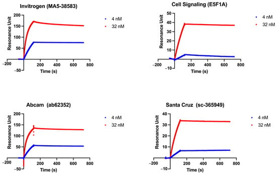
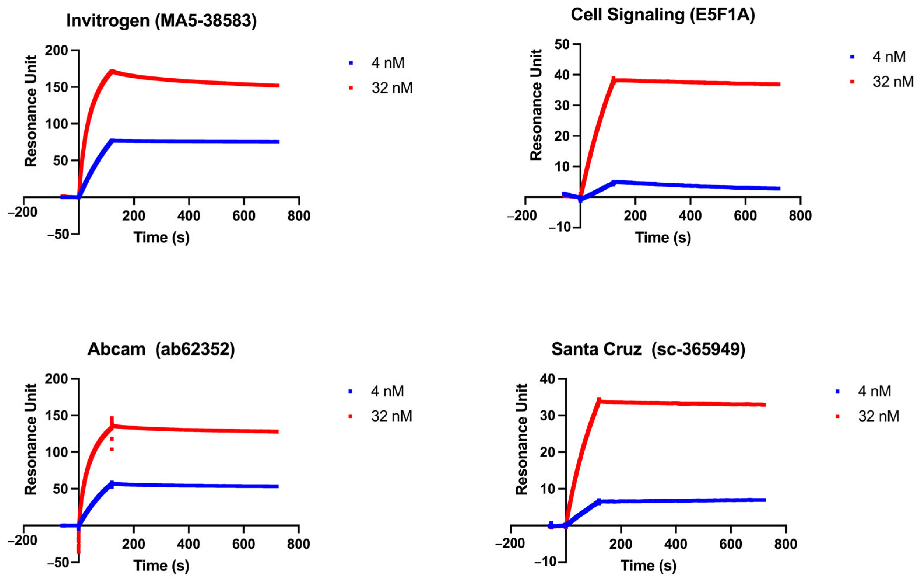
To develop a SiMoA-based assay for the detection of NRF2 levels, two commercial anti-NRF2 antibodies were required that would bind to different epitopes of the NRF2 protein with high affinity and stability. First, affinities of commercial anti-NRF2 antibodies were measured using a SPR Biacore™ T200 instrument. Among four antibodies, Invitrogen, Abcam and Santa Cruz (Dallas, TX, USA) NRF2 antibodies have the high affinity for NRF2 with KD in the low picomolar range (Table 2), whereas Cell Signaling NRF2 antibody showed the lowest affinity for NRF2. According to the sensorgrams of four commercial anti-NRF2 antibodies, Invitrogen and Abcam NRF2 antibodies showed higher resonance unit (RU) values (nearly 150 RU at 32 nM) than Cell Signaling and Santa Cruz NRF2 antibodies (nearly 30 RU at 32 nM) at the end of binding phase. These results indicate that Invitrogen and Abcam NRF2 antibodies bind more strongly to the NRF2-immobilized CM5 chip compared to Cell Signaling and Santa Cruz NRF2 antibodies (Figure 1). Based on these findings, Invitrogen and Abcam NRF2 antibodies were selected for subsequent competitive binding analyses.
Table 2.
Association, dissociation, and equilibrium dissociation constants of the NRF2 antibodies. Lower KD value indicates binding with higher affinity.
Figure 1.
SPR Sensorgrams of Commercial NRF2 Antibodies on Nrf2-bound CM5 chip: The binding affinities of various concentrations of four commercial anti-Nrf2 antibodies (Invitrogen, Cell Signaling, Abcam, Santa Cruz) on an Nrf2-immobilised CM5 chip were recorded using a Biacore™ T200 SPR instrument. The data were evaluated using BIAevaluation software 3.1. Blue and red indicate 4 nM and 32 nM antibody concentrations, respectively.
3.2. Invitrogen and Abcam NRF2 Antibodies Target Different Epitopes of NRF2
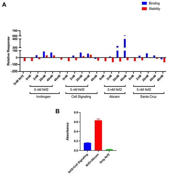
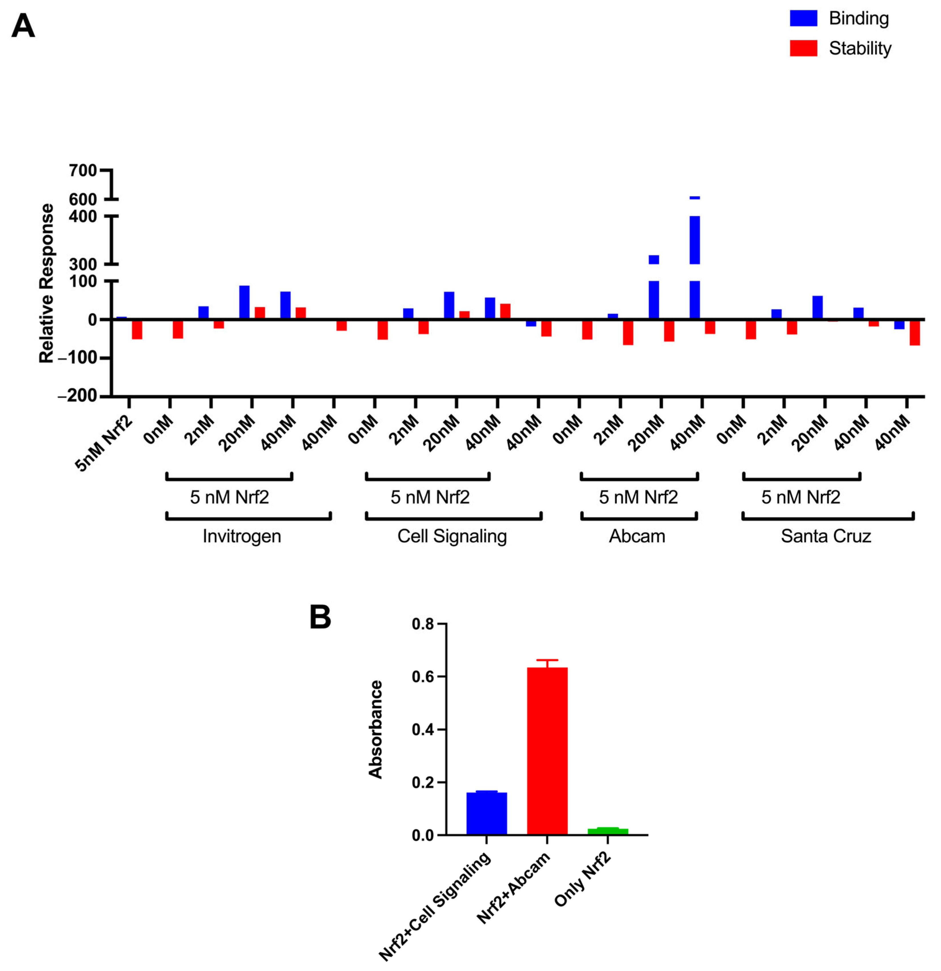
Competitive binding analyses using SPR were performed to check whether the antibodies bind to the same or different epitopes of NRF2. A concentration of 25 nM Abcam NRF2 antibody was used to coat the Protein A sensor chip, and various concentrations of NRF2 antibodies, pre-mixed with stable NRF2 protein, flowed through the chip. According to our results, all antibodies gave a significant binding signal compared to their single antibody control (without NRF2) (Figure 2A). Interestingly, Abcam NRF2 antibody gave the highest binding signal even though it was the antibody immobilized on the chip. The stability of the binding was also checked, as a highly stable binding is also required for an efficient SiMoA-based assay. Only Invitrogen and Cell Signaling NRF2 antibodies gave a stable binding pattern (Figure 2A).
Figure 2.
Competitive Binding Analysis of Commercial NRF2 Antibodies. (A) The competitive binding (blue) and stability (red) responses of anti-Nrf2 antibodies on Abcam-immobilized Protein A chip. After immobilization of Abcam NRF2 antibody, different concentrations of anti-Nrf2 antibodies with or without 5 nM Nrf2 were flowed through the chip. Data were recorded using a Biacore™ T200 SPR instrument. (B) Competitive binding analysis using home-made ELISA on an Invitrogen NRF2 antibody-coated plate. Invitrogen-Cell Signaling and Invitrogen-Abcam antibody combinations were evaluated. Data was presented as mean ± SEM.
The results of the competitive binding analyses were confirmed by a home-made ELISA using Invitrogen, Cell Signaling and Abcam NRF2 antibodies. Cell Signaling, Abcam NRF2 antibodies or no antibodies were added to the plate immobilized with Invitrogen NRF2 antibody and incubated with NRF2 antigen. According to our results, Abcam NRF2 antibody was bound almost 4 times more than Cell Signaling NRF2 antibody (Figure 2B).
Overall, both SPR-based and ELISA-based competition assays confirmed that the Invitrogen-Abcam NRF2 antibody combination gives the most favorable performance for application in the sandwich-based immunoassay. The Invitrogen NRF2 antibody was selected for use as the detection antibody since it produced the most stable binding compared with the Abcam NRF2 antibody. These antibodies were selected for the development of a SiMoA-based NRF2 level assay.
3.3. Developed SiMoA-Based Assay Detects NRF2 with High Precision
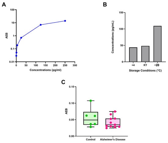
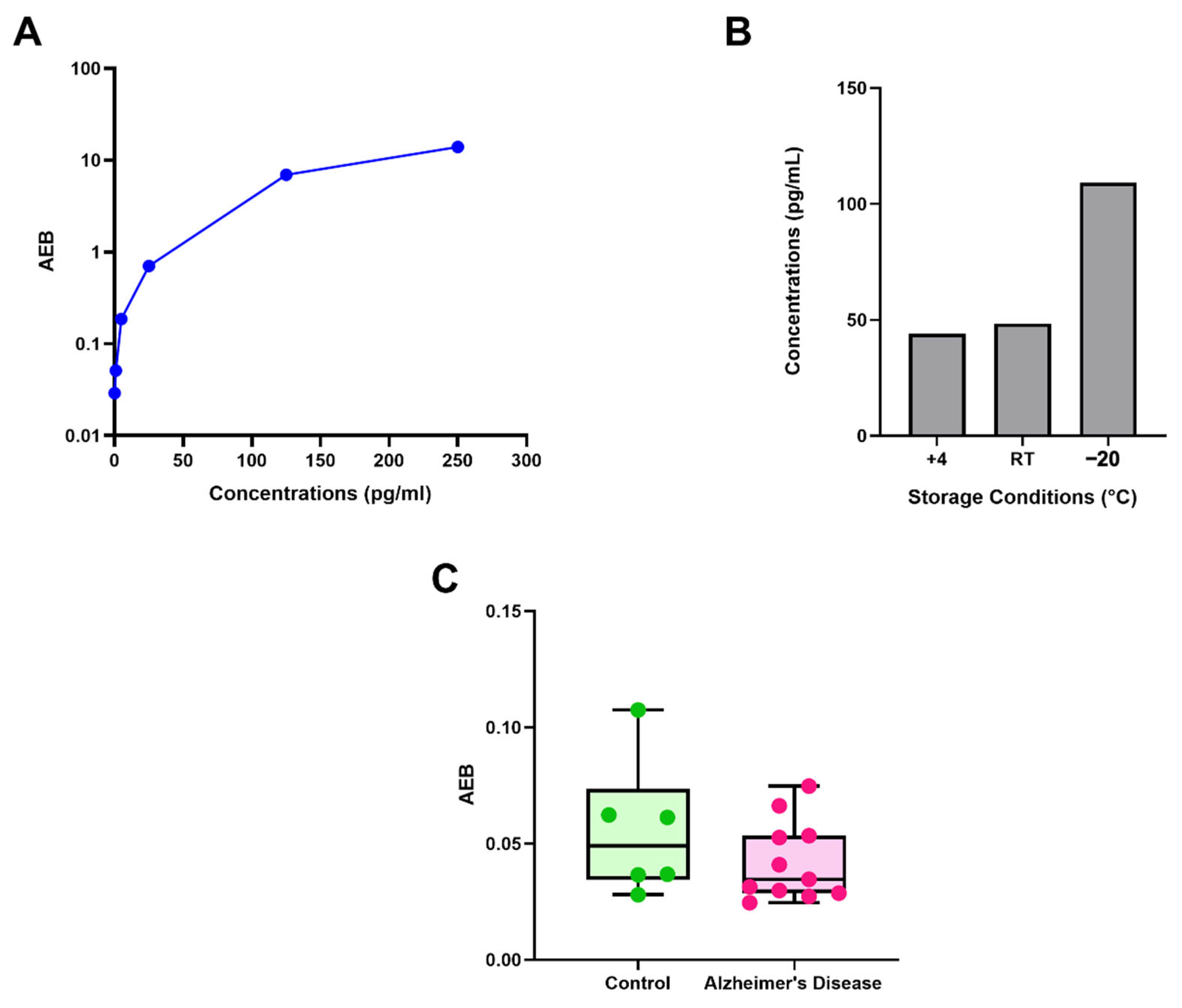
SiMoA-based NRF2 assay was developed by using the SiMoA Homebrew Assay Starter kit with Invitrogen and Abcam NRF2 antibody combination optimizing antibody concentration, incubation time, and temperature. While the Abcam anti-NRF2 antibody is immobilized onto the beads, the Invitrogen anti-NRF2 antibody is biotinylated and used as the detection antibody. The standard curve is generated using a range of known NRF2 protein concentrations. The results showed that, as the NRF2 concentration increased from 0 pg/mL to 250 pg/mL, the signals increased in a concentration-dependent manner, ranging from 0.029 to 13.9274 AEB, confirming that the developed assay detected recombinant NRF2 protein (Figure 3A).
Figure 3.
SiMoA-Based NRF2 Assay Development and Validation. (A) Concentration-dependent response of SiMoA-based NRF2 assay using Abcam and Invitrogen antibodies. (B) Short-term stability analysis at different storage temperatures (+4 °C, RT, −20 °C) of SiMoA-based NRF2 assay. (C) AEB values of PBMC samples from healthy controls and Alzheimer’s disease patients using the developed SiMoA assay.
The SiMoA-based NRF2 assay was validated by analyzing calibrators and samples across multiple independent runs. LOD and LLOQ were determined by analyzing sixteen blank samples. The LOD was calculated as three standard deviations above the mean blank signal and was determined to be 2.356 pg/mL. The LLOQ was calculated as ten SD above the mean blank signal and was determined to be 5.989 pg/mL. In the case with AEB values, we determined LOD and LLOQ as 0.0333 AEB and 0.0514 AEB, respectively. In addition, the short-term stability of the samples was evaluated by storing them under different temperature conditions for one hour. The measured NRF2 concentrations were comparable between samples stored at 4 °C and RT, with concentrations of 43.99 pg/mL and 48.42 pg/mL, respectively. However, the NRF2 concentration was slightly higher in samples stored at −20 °C, with concentrations of 109.21 pg/mL, respectively (Figure 3B).
The precision of the assay was validated using samples with varying levels of NRF2. Intra-assay precision was assessed by analyzing each sample in triplicate within the same run. The intra-assay precision of the assay ranged from 2.2753% to 7.0159%, with a mean CV of 4.4522% (Table 3). The inter-assay precision of the assay was evaluated across three independent runs conducted over three days. The inter-assay precision of the assay ranged from 16.4684% to 20.5280%, with a mean CV of 17.9012% (Table 4). Overall, the findings indicate that the SiMoA-based NRF2 assay can detect low levels of NRF2 with high precision.
Table 3.
Intra-Assay Precision of the SiMoA-Based NRF2 Assay Using Cell Culture Samples.
Table 4.
Inter-Assay Precision of the SiMoA-Based NRF2 Assay Using Cell Culture Samples.
3.4. Detection of NRF2 Levels in Patients with AD
The performance of the developed assay was assessed in a small clinical cohort. According to the results, no significant difference was observed in the AEB values of the control group and patients with AD. However, a slight decrease in AEB values was detected in patients with AD compared to the control group (Figure 3C).
4. Discussion
Current research introduces a new methodology for sensitive quantification of NRF2 using SiMoA. NRF2 is a master transcription factor that plays a significant role in protecting cells against oxidative stress by activating cytoprotective and antioxidant genes in the nucleus [3]. Impaired NRF2 signaling has been reported in various diseases [13,14,15,16,17,18], including neurodegenerative disorders [13,14], cancer [15,16], and cardiovascular diseases [18]. Both hyperactivation and insufficient activation of NRF2 have been revealed to contribute to disease progression [19]. Previous studies have revealed the significance of NRF2 in different disease contexts. Therefore, quantifying NRF2 level plays a critical role in biomarker studies, testing the efficiency of drugs and evaluating disease progression.
Traditional immunoassays have limitations on sensitivity [20]. However, most of the proteins that may take part as biomarkers exist in biological fluids at extremely low concentrations. The emergence of highly sensitive new technologies, such as SiMoA, holds great potential for the quantification of low-abundance proteins in clinical samples. SiMoA technology overcomes the limitations of traditional immunoassays in detecting low levels of analytes by capturing individual analyte molecules on single beads [21]. In our study, we achieved a 5.9-fold increase in sensitivity, with an LOD of 2.356 pg/mL, compared with conventional NRF2 ELISA assays, which can quantify NRF2 at levels around 14 pg/mL. Similar SiMoA-based immunoassays targeting different analytes have been developed to increase the sensitivity of traditional methods [22,23,24]. The prostate-specific antigen (PSA) immunoassay was developed using SiMoA technology, achieving a sensitivity of 0.05 pg/mL and enabling the detection of PSA in patients with prostate cancer [24]. Furthermore, Kuang, Wei [25] developed an anti-Müllerian hormone (AMH) immunoassay with 100-fold greater sensitivity compared to the conventional AMH assay. Another study by Sperinde, Hokom [26] developed an ultrasensitive SiMoA-based assay for IL-13 that achieved 10-fold greater sensitivity than the commercially available Quanterix IL-13 assay through optimization of the assay buffers. Overall, these studies highlight the need for ultrasensitive immunoassay development for low-abundance analytes. To date, no study has reported the ultra-sensitive detection of NRF2 levels.
This study reported the sensitive and highly precise detection of NRF2 levels using cutting-edge SiMoA technology. Reproducibility of the immunoassays was evaluated by calculating the intra-assay CV. According to FDA guidelines and immunoassay validation studies, a CV below 20% is considered acceptable for ligand-binding assays [27]. The SiMoA-based NRF2 assay demonstrated high precision with a CV below 20% for both intra-assay and inter-assay variation, confirming its acceptability among immunoassays. The assay’s high precision and low variability are especially important for analytes found at low levels, such as NRF2 which has a very short half-life [28]. Although the assay is highly precise and has a low detection limit compared to conventional NRF2 immunoassays, our study revealed that the storage conditions of the samples were highly important, even when exposed to different storage conditions for a short time. Studies have shown that the stability and detectability of proteins can be affected by different storage conditions of biological fluids [29,30].
To further test the usage of the developed assay in clinical samples and its ability to detect low levels of NRF2, we measured NRF2 levels in PBMC samples isolated from patients with AD and healthy controls. AEB values were obtained for all participants, including patients and controls. However, only the AEB values of one healthy control and five Alzheimer’s patients were below the LOD of the developed assay. Although the low variability of the assay proves its ability to detect minor changes in cell culture samples, further optimization of the assay’s sensitivity is necessary for its use with clinical samples.
Although the present study provides high sensitivity and accuracy, several limitations should be considered. One of the major limitations of this study is the small number of participants, which may have contributed to the difference in NRF2 detection between AD patients and healthy controls. Additionally, undetectable NRF2 levels in some participants might raise concerns about the use of the developed kit in clinical applications. Therefore, the sensitivity of the developed kit should be improved, especially for clinical utilization. Previous research has shown that low levels of analytes that cannot be detected in ELISA can be successfully detected by optimizing background noise and enhancing assay sensitivity in SiMoA-based immunoassays [26,27]. Moreover, it has been demonstrated that the yield of isolated PBMCs can vary between individuals [8], which may also affect the undetectable NRF2 signals observed in some clinical samples. Furthermore, although this study quantified total NRF2 levels in both cell cultures and PBMC samples, future studies should investigate nuclear NRF2 levels. Separating the nuclear and cytoplasmic fractions of PBMCs could provide a clearer insight into NRF2 levels, since NRF2 is primarily found in the nucleus. To address these limitations, the evaluation of different buffers to improve the sensitivity of the assay and the separation of nuclear NRF2 should be a focus of future studies. It should also be noted that the detectability of NRF2 was affected by the short-term storage conditions of the samples. Exposure to different temperatures over a short period of time can lead to incorrect quantifications. In this study, all samples were stored at −80 °C until further use and freezing and thawing were avoided. Moreover, the present results should be compared with those obtained using other experimental methodologies and samples with known NRF2 concentrations. Since NRF2 levels in clinical samples cannot be detected using conventional NRF2 ELISA, this should also be evaluated as a limitation of the present study.
This study presents the first report on the development of a highly sensitive SiMoA-based assay for the quantification of NRF2 levels. Developed SiMoA-based NRF2 assay demonstrated a 5.9-fold improvement in sensitivity compared to conventional NRF2 immunoassays. Since impaired NRF2 signaling has been associated with various diseases including COVID-19 [30], chronic kidney disease [31], cancer [32,33], Alzheimer’s disease [34,35], and Parkinson’s disease [36,37], sensitive quantification of NRF2 is crucial for evaluating its potential as a biomarker, investigating disease progression and for diagnostic and therapeutic drug testing.
Author Contributions
S.G., S.K. and B.I.A. developed the study. A.C., B.O., B.I.A. and M.E.A. carried out the experiments. A.C., B.O. and B.I.A. were responsible for managing ethical issues and collecting human samples. E.C.Y., G.Y. and D.O. evaluated and included participants in the study. All authors have read and agreed to the published version of the manuscript.
Funding
The study was funded by The Scientific and Technological Research Council of Türkiye and COST Action CA20121 (TUBITAK, Project ID: 122Z205).
Institutional Review Board Statement
The study was conducted in accordance with the Declara-tion of Helsinki, and approved by the Ethics Committee of the Izmir Biomedicine and Genome Center (protocol number 2022-038, 11 November 2022).
Informed Consent Statement
Informed consent was obtained from all subjects involved in the study.
Data Availability Statement
The original contributions presented in this study are included in the article. Further reasonable inquiries can be directed to the corresponding author.
Acknowledgments
We thank the IBG-Pharma facility for their support in SPR analysis.
Conflicts of Interest
The authors declare no conflicts of interest.
Abbreviations
The following abbreviations are used in this manuscript:
| AD | Alzheimer’s disease |
| AEB | Average number of enzymes per bead |
| AMH | Anti-Müllerian hormone |
| ARE | Antioxidant response element |
| CV | Coefficient of variation |
| DMF | Dimethyl fumarate |
| EDC | 1-Ethyl-3-(3-dimethylaminopropyl) carbodiimide |
| FDA | U.S. Food and Drug Administration |
| HBS | Hepes Buffered Saline buffer |
| Kd | Equilibrium dissociation constant |
| koff | Dissociation rate constant |
| kon | Association rate constant |
| LLOQ | Lower limit of quantification |
| LOD | Limit of detection |
| NRF2 | Nuclear factor erythroid 2-related factor 2 |
| PBMC | Peripheral Blood Mononuclear Cell |
| PSA | Prostate-specific antigen |
| ROS | Reactive oxygen species |
| RT | Room temperature |
| RU | Resonance unit |
| SD | Standard deviation |
| SE | Standard error |
| SiMoA | Single Molecule Array |
| SPR | Surface Plasmon Resonance |
References
- Yang, J.; Luo, J.; Tian, X.; Zhao, Y.; Li, Y.; Wu, X. Progress in Understanding Oxidative Stress, Aging, and Aging-Related Diseases. Antioxidants 2024, 13, 394. [Google Scholar] [CrossRef] [Scilit] [PubMed]
- Tastan, B.; Arioz, B.I.; Genc, S. Targeting NLRP3 Inflammasome With Nrf2 Inducers in Central Nervous System Disorders. Front. Immunol. 2022, 13, 865772. [Google Scholar] [CrossRef] [Scilit] [PubMed]
- Canning, P.; Sorrell, F.J.; Bullock, A.N. Structural basis of Keap1 interactions with Nrf2. Free Radic. Biol. Med. 2015, 88, 101–107. [Google Scholar] [CrossRef] [Scilit] [PubMed]
- Naidu, S.D.; Muramatsu, A.; Saito, R.; Asami, S.; Honda, T.; Hosoya, T.; Itoh, K.; Yamamoto, M.; Suzuki, T.; Dinkova-Kostova, A.T. C151 in KEAP1 is the main cysteine sensor for the cyanoenone class of NRF2 activators, irrespective of molecular size or shape. Sci. Rep. 2018, 8, 8037, Correction in Sci. Rep. 204, 14, 4774. [Google Scholar] [CrossRef] [Scilit] [PubMed]
- Cuadrado, A.; Rojo, A.I.; Wells, G.; Hayes, J.D.; Cousin, S.P.; Rumsey, W.L.; Attucks, O.C.; Franklin, S.; Levonen, A.-L.; Kensler, T.W.; et al. Therapeutic targeting of the NRF2 and KEAP1 partnership in chronic diseases. Nat. Rev. Drug Discov. 2019, 18, 295–317. [Google Scholar] [CrossRef] [Scilit] [PubMed]
- Morgenstern, C.; Lastres-Becker, I.; Demirdöğen, B.C.; Costa, V.M.; Daiber, A.; Foresti, R.; Motterlini, R.; Kalyoncu, S.; Arioz, B.I.; Genc, S.; et al. Biomarkers of NRF2 signalling: Current status and future challenges. Redox Biol. 2024, 72, 103134. [Google Scholar] [CrossRef] [Scilit] [PubMed]
- Rasmussen, M.; Hansen, K.H.; Scholze, A. Nrf2 Protein Serum Concentration in Human CKD Shows a Biphasic Behavior. Antioxidants 2023, 12, 932. [Google Scholar] [CrossRef] [Scilit] [PubMed]
- Rissin, D.M.; Kan, C.W.; Campbell, T.G.; Howes, S.C.; Fournier, D.R.; Song, L.; Piech, T.; Patel, P.P.; Chang, L.; Rivnak, A.J.; et al. Single-molecule enzyme-linked immunosorbent assay detects serum proteins at subfemtomolar concentrations. Nat. Biotechnol. 2010, 28, 595–599. [Google Scholar] [CrossRef] [Scilit] [PubMed]
- Zhang, J.; Wiener, A.D.; Meyer, R.E.; Kan, C.W.; Rissin, D.M.; Kolluru, B.; George, C.; Tobos, C.I.; Shan, D.; Duffy, D.C. Improving the Accuracy, Robustness, and Dynamic Range of Digital Bead Assays. Anal. Chem. 2023, 95, 8613–8620. [Google Scholar] [CrossRef] [Scilit] [PubMed]
- Malhotra, K.; Hansen, L.L.; Han, S.; Roth, S.; Walt, D.R. Ultrasensitive Enzyme-Free Detection of Proteins on Magnetic Beads. J. Am. Chem. Soc. 2026, 148, 11641–11653. [Google Scholar] [CrossRef] [Scilit] [PubMed]
- Junter, G.A.; Lebrun, L. Polysaccharide-based chromatographic adsorbents for virus purification and viral clearance. J. Pharm. Anal. 2020, 10, 291–312. [Google Scholar] [CrossRef] [Scilit] [PubMed]
- McKhann, G.M.; Knopman, D.S.; Chertkow, H.; Hyman, B.T.; Jack, C.R., Jr.; Kawas, C.H.; Klunk, W.E.; Koroshetz, W.J.; Manly, J.J.; Mayeux, R.; et al. The diagnosis of dementia due to Alzheimer’s disease: Recommendations from the National Institute on Aging-Alzheimer’s Association workgroups on diagnostic guidelines for Alzheimer’s disease. Alzheimer’s Dement. 2011, 7, 263–269. [Google Scholar] [CrossRef] [Scilit] [PubMed]
- Ngo, V.; Duennwald, M.L. Nrf2 and Oxidative Stress: A General Overview of Mechanisms and Implications in Human Disease. Antioxidants 2022, 11, 2345. [Google Scholar] [CrossRef] [Scilit] [PubMed]
- Ramsey, C.P.; Glass, C.A.; Montgomery, M.B.; Lindl, K.A.; Ritson, G.P.; Chia, L.A.; Hamilton, R.L.; Chu, C.T.; Jordan-Sciutto, K.L. Expression of Nrf2 in neurodegenerative diseases. J. Neuropathol. Exp. Neurol. 2007, 66, 75–85. [Google Scholar] [CrossRef] [Scilit] [PubMed]
- Aoki, Y.; Sato, H.; Nishimura, N.; Takahashi, S.; Itoh, K.; Yamamoto, M. Accelerated DNA adduct formation in the lung of the Nrf2 knockout mouse exposed to diesel exhaust. Toxicol. Appl. Pharmacol. 2001, 173, 154–160. [Google Scholar] [CrossRef] [Scilit] [PubMed]
- Arfin, S.; Jha, N.K.; Jha, S.K.; Kesari, K.K.; Ruokolainen, J.; Roychoudhury, S.; Rathi, B.; Kumar, D. Oxidative Stress in Cancer Cell Metabolism. Antioxidants 2021, 10, 642. [Google Scholar] [CrossRef] [Scilit] [PubMed]
- Jiang, T.; Huang, Z.; Lin, Y.; Zhang, Z.; Fang, D.; Zhang, D.D. The protective role of Nrf2 in streptozotocin-induced diabetic nephropathy. Diabetes 2010, 59, 850–860. [Google Scholar] [CrossRef] [Scilit] [PubMed]
- He, X.; Kan, H.; Cai, L.; Ma, Q. Nrf2 is critical in defense against high glucose-induced oxidative damage in cardiomyocytes. J. Mol. Cell Cardiol. 2009, 46, 47–58. [Google Scholar] [CrossRef] [Scilit] [PubMed]
- Cuadrado, A.; Cazalla, E.; Bach, A.; Bathish, B.; Naidu, S.D.; DeNicola, G.M.; Dinkova-Kostova, A.T.; Fernández-Ginés, R.; Grochot-Przeczek, A.; Hayes, J.D.; et al. Health position paper and redox perspectives—Bench to bedside transition for pharmacological regulation of NRF2 in noncommunicable diseases. Redox Biol. 2025, 81, 103569. [Google Scholar] [CrossRef] [Scilit] [PubMed]
- Rissin, D.M.; Wilson, D.H.; Duffy, D.C. Chapter 2.13—Measurement of Single Protein Molecules Using Digital ELISA. In The Immunoassay Handbook, 4th ed.; Wild, D., Ed.; Elsevier: Oxford, UK, 2013; pp. 223–242. [Google Scholar]
- Gupta, V.; Kalia, N.; Yadav, M.; Noyes, A.; Good, J.; Hendricks, R.; Xu, W. Optimization and qualification of the single molecule array digital immunoassay for IL-12p70 in plasma of cancer patients. Bioanalysis 2018, 10, 1413–1425. [Google Scholar] [CrossRef] [Scilit] [PubMed]
- Song, L.; Hanlon, D.W.; Chang, L.; Provuncher, G.K.; Kan, C.W.; Campbell, T.G.; Fournier, D.R.; Ferrell, E.P.; Rivnak, A.J.; Pink, B.A.; et al. Single molecule measurements of tumor necrosis factor alpha and interleukin-6 in the plasma of patients with Crohn’s disease. J. Immunol. Methods 2011, 372, 177–186. [Google Scholar] [CrossRef] [Scilit] [PubMed]
- Thangavelu, B.; Boutté, A.M. Single Molecule Assay for Ultrasensitive Detection of Cathepsin B in Human Blood. ACS Omega 2021, 6, 9609–9616. [Google Scholar] [CrossRef] [Scilit] [PubMed]
- Wilson, D.H.; Hanlon, D.W.; Provuncher, G.K.; Chang, L.; Song, L.; Patel, P.P.; Ferrell, E.P.; Lepor, H.; Partin, A.W.; Chan, D.W.; et al. Fifth-generation digital immunoassay for prostate-specific antigen by single molecule array technology. Clin. Chem. 2011, 57, 1712–1721. [Google Scholar] [CrossRef] [Scilit] [PubMed]
- Kuang, X.; Wei, L.; Huang, Y.; Ji, M.; Tang, Y.; Wei, B.; Yang, S.; Lai, D.; Xu, H. Development of a digital anti-Mullerian hormone immunoassay: Ultrasensitive, accurate and practical strategy for reduced ovarian reserve monitoring and assessment. Talanta 2023, 253, 123970. [Google Scholar] [CrossRef] [Scilit] [PubMed]
- Sperinde, G.; Hokom, M.; Peng, K.; Fischer, S.K. Bioanalytical challenges in development of ultrasensitive Home Brew assays: A case study using IL-13. Bioanalysis 2019, 11, 1045–1054. [Google Scholar] [CrossRef] [Scilit] [PubMed]
- Andreasson, U.; Perret-Liaudet, A.; van Waalwijk van Doorn, L.J.C.; Blennow, K.; Chiasserini, D.; Engelborghs, S.; Fladby, T.; Genc, S.; Kruse, N.; Kuiperij, H.B.; et al. A Practical Guide to Immunoassay Method Validation. Front. Neurol. 2015, 6, 179. [Google Scholar] [CrossRef] [Scilit] [PubMed]
- Bhatnagar, B.S.; Bogner, R.H.; Pikal, M.J. Protein stability during freezing: Separation of stresses and mechanisms of protein stabilization. Pharm. Dev. Technol. 2007, 12, 505–523. [Google Scholar] [CrossRef] [Scilit] [PubMed]
- Gegner, H.M.; Naake, T.; Dugourd, A.; Müller, T.; Czernilofsky, F.; Kliewer, G.; Jäger, E.; Helm, B.; Kunze-Rohrbach, N.; Klingmüller, U.; et al. Pre-analytical processing of plasma and serum samples for combined proteome and metabolome analysis. Front. Mol. Biosci. 2022, 9, 961448. [Google Scholar] [CrossRef] [Scilit] [PubMed]
- Gümüş, H.; Erat, T.; Öztürk, I.; Demir, A.; Koyuncu, I. Oxidative stress and decreased Nrf2 level in pediatric patients with COVID-19. J. Med. Virol. 2022, 94, 2259–2264. [Google Scholar] [CrossRef] [Scilit] [PubMed]
- Juul-Nielsen, C.; Shen, J.; Stenvinkel, P.; Scholze, A. Systematic review of the nuclear factor erythroid 2-related factor 2 (NRF2) system in human chronic kidney disease: Alterations, interventions and relation to morbidity. Nephrol. Dial. Transplant. 2022, 37, 904–916. [Google Scholar] [CrossRef] [Scilit] [PubMed]
- Kerins, M.J.; Ooi, A. A catalogue of somatic NRF2 gain-of-function mutations in cancer. Sci. Rep. 2018, 8, 12846. [Google Scholar] [CrossRef] [Scilit] [PubMed]
- Panda, H.; Rowland, N.G.; Krall, C.M.; Bowman, B.M.; Major, M.B.; Zolkind, P. NRF2 immunobiology in cancer: Implications for immunotherapy and therapeutic targeting. Oncogene 2025, 44, 3641–3651. [Google Scholar] [CrossRef] [Scilit] [PubMed]
- Branca, C.; Ferreira, E.; Nguyen, T.-V.; Doyle, K.; Caccamo, A.; Oddo, S. Genetic reduction of Nrf2 exacerbates cognitive deficits in a mouse model of Alzheimer’s disease. Hum. Mol. Genet. 2017, 26, 4823–4835. [Google Scholar] [CrossRef] [Scilit] [PubMed]
- Rojo, A.I.; Pajares, M.; García-Yagüe, A.J.; Buendia, I.; Van Leuven, F.; Yamamoto, M.; López, M.G.; Cuadrado, A. Deficiency in the transcription factor NRF2 worsens inflammatory parameters in a mouse model with combined tauopathy and amyloidopathy. Redox Biol. 2018, 18, 173–180. [Google Scholar] [CrossRef] [Scilit] [PubMed]
- Lastres-Becker, I.; García-Yagüe, Á.J.; Scannevin, R.H.; Casarejos, M.J.; Kügler, S.; Rábano, A.; Cuadrado, A. Repurposing the NRF2 Activator Dimethyl Fumarate as Therapy Against Synucleinopathy in Parkinson’s Disease. Antioxid. Redox Signal 2016, 25, 61–77. [Google Scholar] [CrossRef] [Scilit] [PubMed]
- Wang, Q.; Li, W.-X.; Dai, S.-X.; Guo, Y.; Han, F.-F.; Zheng, J.-J.; Li, G.-H.; Huang, J.-F. Meta-Analysis of Parkinson’s Disease and Alzheimer’s Disease Revealed Commonly Impaired Pathways and Dysregulation of NRF2-Dependent Genes. J. Alzheimer’s Dis. 2017, 56, 1525–1539. [Google Scholar] [CrossRef] [Scilit] [PubMed]

Disclaimer/Publisher’s Note: The statements, opinions and data contained in all publications are solely those of the individual author(s) and contributor(s) and not of MDPI and/or the editor(s). MDPI and/or the editor(s) disclaim responsibility for any injury to people or property resulting from any ideas, methods, instructions or products referred to in the content. |
© 2026 by the authors. Licensee MDPI, Basel, Switzerland. This article is an open access article distributed under the terms and conditions of the Creative Commons Attribution (CC BY) license.