


Abstract
Stem cell research has reshaped human biology and opened new frontiers for regenerative medicine. Yet public and commercial enthusiasm has at times outpaced the evidence, leaving room for unproven and occasionally harmful interventions. This narrative review examines the roots of that gap. Stem cells are not autonomous repair kits: their behavior depends substantially on their microenvironment, and directing it reliably remains difficult. We contrast established therapy, notably hematopoietic stem cell transplantation, and emerging clinical successes, including pluripotent stem cell-derived retinal and dopaminergic grafts and gene-edited hematopoietic stem cells, with applications whose benefit remains unproven. We also consider how direct-to-consumer markets reach patients with few treatment options. Successful translation depends on the specific cell product, indication, manufacturing process and clinical context; lasting progress therefore rests on rigorous standards and transparent oversight rather than hype.
1. Introduction
Stem cell biology has reshaped the life sciences, changing how we understand development, tissue homeostasis and disease. The path from the isolation of embryonic stem cells to the derivation of induced pluripotent stem cells (iPSCs) has provided powerful frameworks for studying cellular identity, lineage plasticity and regeneration [1].
Alongside these advances, a simplified narrative has developed in public, commercial and media discourse [2]. In this narrative, stem cells are often portrayed as autonomous, universal remedies rather than as biologically constrained, context-dependent cells. This framing has been linked to premature clinical use, the direct-to-consumer marketing of unproven interventions, and public expectations that exceed the available evidence [2].
This narrative review examines why stem cell therapies are biologically difficult to translate, and how simplified accounts of stem cell biology may affect clinical practice, research incentives, regulation and public trust. We distinguish established therapies, emerging applications with early clinical success, and experimental approaches, and argue that successful translation depends on the specific cell product, indication, manufacturing process and clinical context rather than on the properties of stem cells in general.
2. Methods
This narrative review was based on a non-systematic search of PubMed/MEDLINE, Scopus and Google Scholar conducted between January and September 2026, supplemented by hand-searching the reference lists of key articles and by documents from regulatory agencies and professional societies. Search terms combined stem cell types (e.g., embryonic, induced pluripotent and mesenchymal stromal cells, hematopoietic stem cell transplantation and organoids) with terms for biological mechanisms, clinical translation and the commercial and regulatory context. Only English-language publications were included, without date restriction.
The four first authors selected sources by consensus according to their relevance to the themes of the review, giving priority to systematic reviews and meta-analyses, landmark clinical studies, recent reviews and marketplace-surveillance studies. News reports were used only for recent events not yet covered in the peer-reviewed literature. Because this is a narrative review, no formal inclusion criteria, quality appraisal or Preferred Reporting Items for Systematic Reviews and Meta-Analyses (PRISMA) procedures were applied; the resulting limitations are discussed in Section 8. The 114 cited sources were published between 2006 and 2026, approximately 85% of them in 2015 or later.
Claude (Claude Sonnet 4.5, Anthropic) was used only for language editing: correcting grammar and spelling, improving clarity, and refining the organization of the text. It was not used to search or select the literature, generate scientific content, analyze data, create figures, or interpret the cited studies. All output was reviewed and edited by the authors, who take full responsibility for the content.
3. Biological Foundations
Stem cells are defined by two capacities: self-renewal, the maintenance of an undifferentiated state through successive divisions; and differentiation into specialized progeny [3]. Their potency describes the range of cell fates they can adopt within a developmental hierarchy [4].
At the top of this hierarchy are totipotent cells, such as the zygote, which can generate a complete organism including extraembryonic tissues. Pluripotent cells can form any lineage derived from the three embryonic germ layers [5]. As development proceeds, potential becomes progressively restricted, giving rise to multipotent stem cells committed to particular tissues, such as hematopoietic or neural stem cells, and ultimately to unipotent progenitors [6].
This restriction was long regarded as irreversible. It is now understood to depend largely on epigenetic and molecular states rather than permanent genetic changes, which is why cellular potency can be reversed experimentally by reprogramming [7].
4. Types of Stem Cells and Their Biological Limits
Stem cells are classified by potency, tissue of origin and functional context. The three major categories—embryonic, induced pluripotent and adult tissue-specific stem cells—differ in therapeutic potential, limitations and clinical maturity (Section 6).
4.1. Embryonic Stem Cells (ESCs)
ESCs are derived from the inner cell mass of the blastocyst, approximately four to five days after fertilization [8]. They can proliferate indefinitely in vitro and differentiate into virtually any somatic cell type of the three germ layers, although they cannot independently form extraembryonic structures [9].
Their clinical use faces ethical and practical constraints. Derivation requires the destruction of a human embryo, which has generated sustained ethical debate and widely varying legal restrictions [10]. As allogeneic products, ESC derivatives generally require immunosuppression to prevent rejection, and any residual undifferentiated cells carry a risk of teratoma formation [9]. ESCs therefore remain an important research tool; clinically, they serve as a source of differentiated derivatives, such as retinal pigment epithelial cells, that are being tested in early-phase trials (Section 6.1), rather than being transplanted in an undifferentiated state [11].
4.2. Induced Pluripotent Stem Cells (iPSCs)
iPSCs are generated by reprogramming differentiated somatic cells to a pluripotent state, which avoids the destruction of embryos and, in principle, allows autologous use [12]. First derived from mouse cells by Takahashi and Yamanaka in 2006, and from human cells in 2007, they showed that cellular identity can be reset by a defined set of transcription factors (OCT4, SOX2, KLF4 and c-MYC) [13].
iPSCs share core pluripotency networks with ESCs and enable patient-specific disease models and, potentially, autologous therapies [3]. They also have recognized limitations. Reprogramming can be incomplete, leaving an “epigenetic memory” that biases differentiation towards the tissue of origin [14], and prolonged ex vivo expansion can introduce mutations and copy-number variations with potential oncogenic consequences [15]. Non-integrating reprogramming methods and stringent genomic quality control have reduced these risks, and iPSC-derived products, including retinal and dopaminergic cells, have entered early-phase clinical trials (Section 6.1) [15,16]. Their wider clinical use will depend on long-term safety data from such trials.
4.3. Adult/Tissue-Specific Stem Cells
Adult stem cells have a more restricted differentiation potential than pluripotent cells. These multipotent or unipotent cells reside in specialized niches within adult tissues, where they maintain and repair specific lineages [12]. Under physiological conditions they rarely cross lineage boundaries; hematopoietic stem cells (HSCs), for example, replenish blood cells but do not normally give rise to neural or muscle cells [12].
In response to injury or physiological cues, adult stem cells can divide asymmetrically to self-renew and generate transit-amplifying progenitors [17]. This regenerative capacity is substantial in tissues with high turnover, such as the intestinal epithelium and bone marrow, but limited in organs such as the myocardium and central nervous system.
Clinically, adult stem cells raise fewer ethical concerns, and autologous use reduces the risk of rejection [10]. Hematopoietic stem cell transplantation (HSCT), first performed in the late 1950s and now a standard of care for many hematologic malignancies and bone marrow disorders, is the clearest example (Section 6.1). Broader use is limited by the low frequency of many adult stem cell populations in native tissues, difficulties in isolating them, and their tendency to senesce, drift phenotypically or differentiate during ex vivo expansion [10].
5. Biological Complexity and Multilevel Barriers
Promotional descriptions sometimes portray transplanted stem cells as autonomous repair units capable of homing to injuries, differentiating and restoring organ function. Such accounts may overlook the product-specific biological constraints that influence cell survival, integration and clinical performance.
Stem cell behavior reflects interactions among intrinsic molecular programs, microenvironmental signals, immune surveillance, mechanical forces and metabolic gradients. Regenerative capacity is influenced by surrounding tissue architecture; isolation, ex vivo manipulation and transplantation into diseased tissues can disrupt these interactions.
5.1. Intrinsic and Niche Regulation of Stemness
Stemness reflects interactions between intrinsic transcriptional and epigenetic programs and cues from the surrounding microenvironment. Rather than being a fixed property encoded solely in the genome, it represents a dynamically regulated cellular state. The balance between cell-intrinsic and niche-derived signals differs across stem cell types, tissue compartments and physiological or pathological conditions [18]. This balance may also shift during injury, aging or experimental manipulation.
Isolation and ex vivo expansion can disrupt niche-derived signals and alter self-renewal capacity, differentiation potential and cellular phenotype. Their magnitude depends on the cell type, culture system, expansion duration and manufacturing controls. The translational challenge is therefore to reproduce relevant regulatory conditions where needed and to verify that the final cell product retains adequate potency, identity and stability for its intended clinical application [18]. Validated potency assays, where available, are central to interpreting expansion success.
5.2. The Stem Cell Niche as a Regulator of Fate
The stem cell niche is a specialized microenvironment that integrates biochemical gradients, biophysical forces, metabolic conditions and spatial organization. Together with cell-intrinsic molecular pathways, these signals influence quiescence, self-renewal and differentiation in a tissue-dependent manner [18].
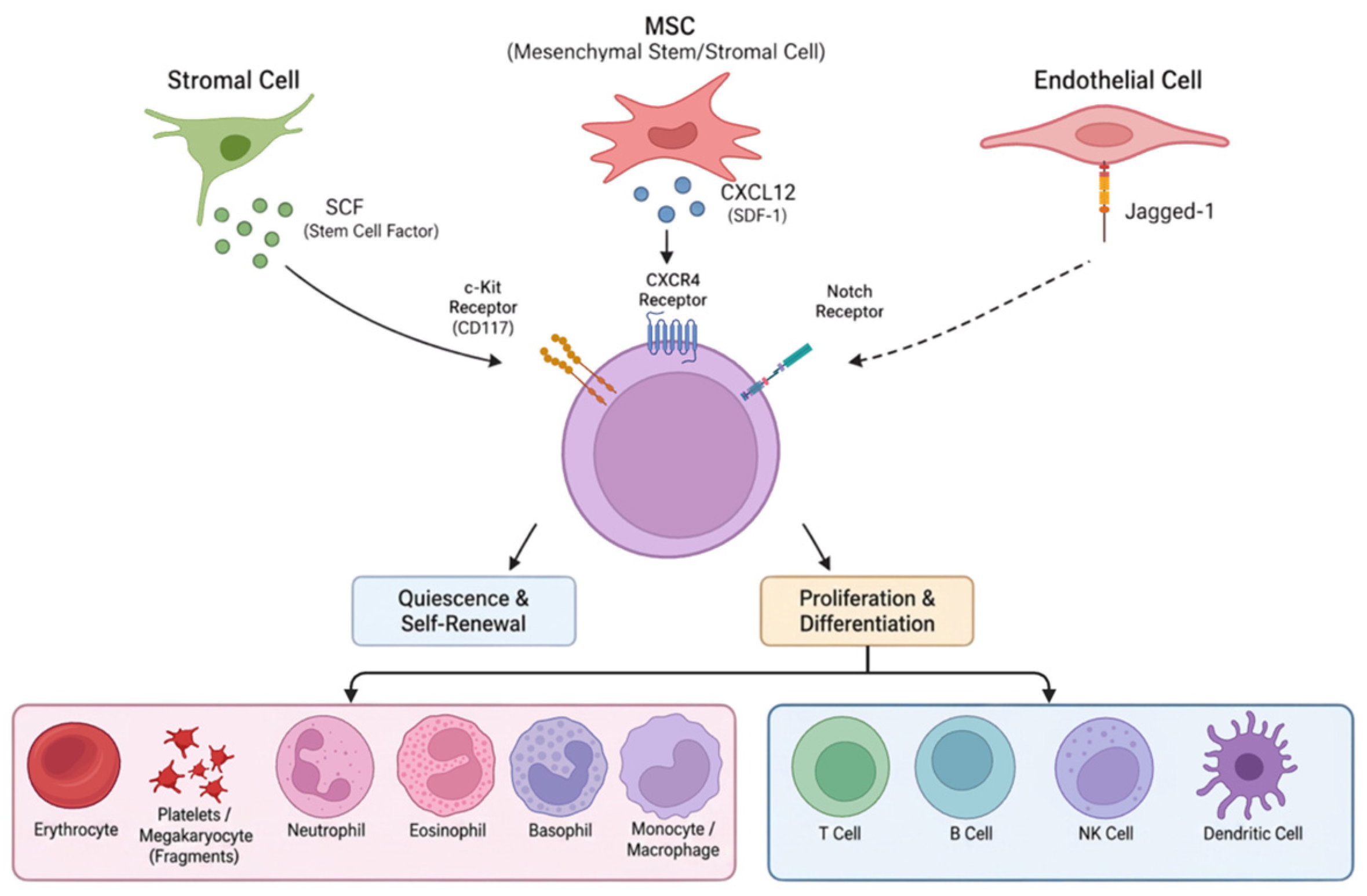
In the bone marrow, for example, HSCs inhabit perivascular niches where endothelial cells and mesenchymal stromal cells (MSCs) provide regulatory signals, including stem cell factor (SCF) and the chemokine C-X-C motif chemokine ligand 12 (CXCL12). SCF binding to its receptor c-Kit supports cell survival and self-renewal, whereas signaling through CXCL12 and its receptor C-X-C chemokine receptor type 4 (CXCR4) contributes to retention and quiescence. Endothelial Notch ligands, including Jagged-1, may also influence HSC maintenance. These pathways operate alongside extracellular matrix (ECM) interactions, oxygen tension and mechanical cues (Figure 1). Throughout this review, MSC denotes mesenchymal stromal cells; “mesenchymal stem cells” is reserved for populations supported by evidence of stemness, consistent with International Society for Cell & Gene Therapy (ISCT) nomenclature [19].
Across hematopoietic, intestinal and epithelial tissues, fate decisions reflect both intracellular regulatory programs and niche cues. In certain settings, differentiated cells may reacquire stem-like properties in response to specific signals.
Disruption of niche signaling can alter stem cell behavior. Inflammatory cytokines may weaken quiescence signals; fibrotic remodeling changes ECM stiffness and can influence lineage bias; and vascular injury may modify HSC retention or mobilization [20]. These changes can contribute to dysfunction, although cell-intrinsic alterations and disease-specific factors may also be important. Similar challenges arise during ex vivo expansion, when increased cell numbers do not necessarily indicate maintenance of the long-term regenerative potential or functional identity required for clinical use [21]. Functional assessment after culture may be more informative than cell yield alone.
Figure 1.
The stem cell niche as a molecular regulator of hematopoietic stem cell fate. A schematic representation of reciprocal interactions between hematopoietic stem cells (HSCs), endothelial cells and mesenchymal stromal cells (MSCs). Stem cell factor (SCF) and C-X-C motif chemokine ligand 12 (CXCL12) contribute to HSC survival, retention and quiescence through c-Kit and C-X-C chemokine receptor type 4 (CXCR4) signaling, respectively. Endothelial Notch ligands (e.g., Jagged-1), extracellular matrix interactions and local biophysical cues may further influence cell fate. Pathway changes can shift the balance among quiescence, proliferation and differentiation. Solid arrows indicate established signaling interactions; dashed arrows indicate modulatory or less well-established influences. Created in BioRender. Nguyen, H. (2026) https://BioRender.com/1pume0s.
5.3. Differentiation and Functional Integration
Even after transplanted stem or progenitor cells survive and differentiate toward a desired lineage, the restoration of organ-level function is not guaranteed. Differentiation establishes cellular identity but does not demonstrate the electrical, mechanical and metabolic coordination required for tissue repair. Mature organs comprise organized networks of interacting cell types, making structural and functional graft integration an additional therapeutic requirement beyond the expression of lineage-specific markers.
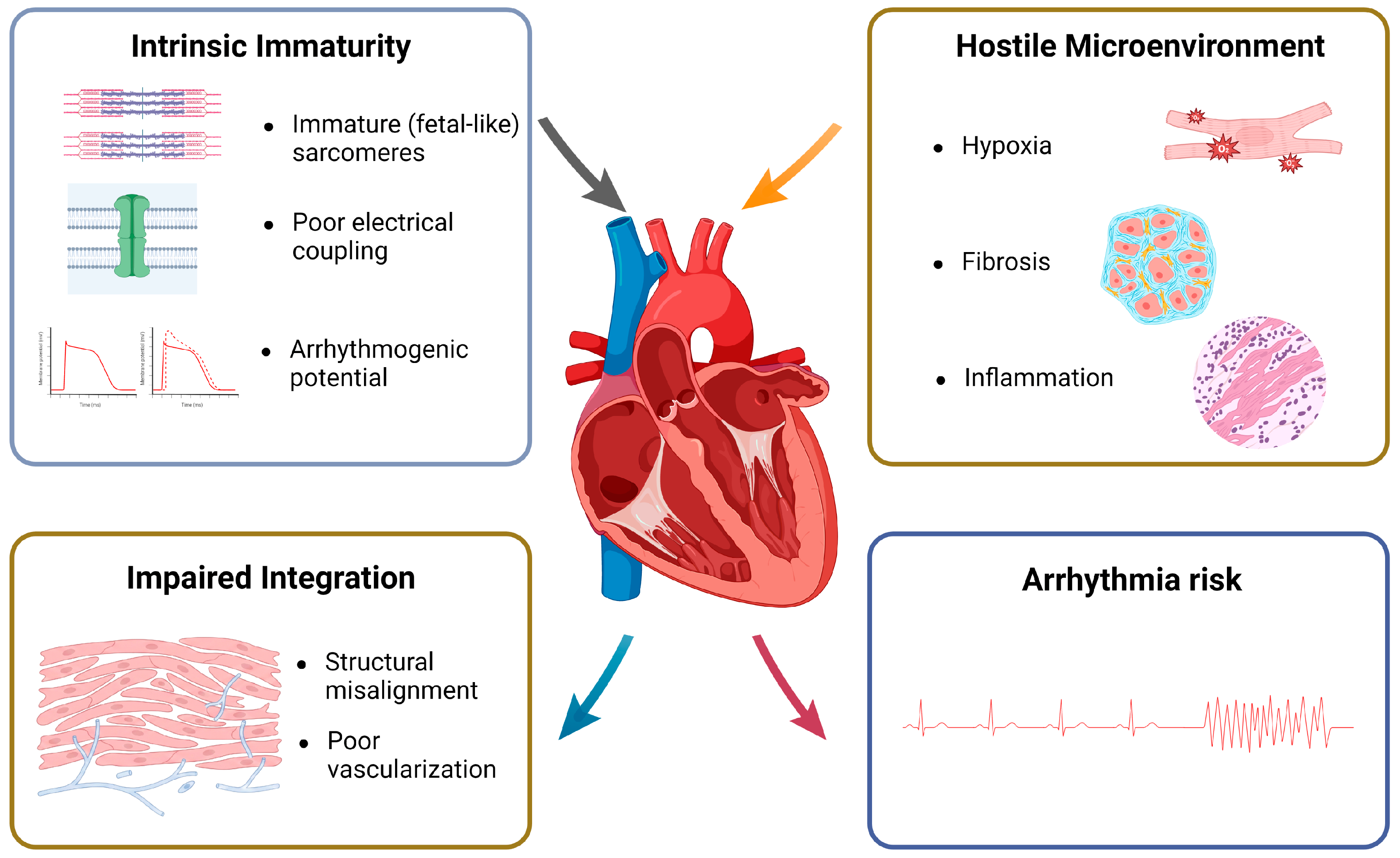
Cardiac regeneration illustrates these challenges. Pluripotent stem cell-derived cardiomyocytes (PSC-CMs) must establish synchronized electrical coupling, integrate into the host myocardium and connect with vascular networks. However, current differentiation protocols can yield cells with immature, fetal-like sarcomeres and atypical electrophysiological properties [22]. Graft-associated ventricular arrhythmias have been observed in large-animal preclinical studies, where graft composition, maturation and transplantation conditions influence the risk [23,24].
The injured myocardial microenvironment, including fibrosis, ischemia, hypoxia and oxidative stress [25], may further impair differentiation, structural maturation and vascular support for transplanted cardiomyocytes [26] (Figure 2).
5.4. Immune and Inflammatory Influences on Graft Function
Host immunity and local inflammation can affect graft survival, lineage fate and integration. Transplanted cells encounter innate and adaptive immune surveillance, cytokine gradients and damage-associated molecular patterns (DAMPs). These effects vary with cell source, manufacturing and recipient tissue status.
Autologous transplantation is not necessarily immunologically neutral: prolonged ex vivo manipulation may alter metabolic state, cell-surface antigen expression or other features relevant to immune recognition [27]. In allogeneic settings, histocompatibility differences can lead to alloimmune responses [28]. Even without overt rejection, sustained exposure to inflammatory mediators such as tumor necrosis factor-α (TNF-α) and interferon-γ (IFN-γ) may influence differentiation and compromise graft stability in certain settings [29].
Figure 2.
Multilevel biological barriers to functional cardiac tissue regeneration. Two sets of barriers limit repair. The first is the intrinsic immaturity of pluripotent stem cell (PSC)-derived cardiomyocytes, which have fetal-like sarcomeres, poor electrical coupling and arrhythmogenic potential. The second is a hostile host microenvironment of hypoxia, fibrosis and inflammation. Together, these barriers may impair the integration of the graft, through structural misalignment and poor vascularization, and may increase arrhythmia risk. Created in BioRender. Nguyen, H. (2026) https://BioRender.com/4sjr3f2.
Inflammatory mediators, including TNF-α and interleukin-1β (IL-1β), may alter lineage commitment, differentiation or apoptosis under certain conditions [30,31,32]. After myocardial infarction, inflammation can contribute to fibroblast activation, ECM remodeling and matrix stiffening, potentially impairing graft maturation [26]. Immune effects therefore interact with structural barriers.
Strategies to limit host immune recognition include editing human leukocyte antigen (HLA) class I/II expression, introducing immunomodulatory molecules such as cluster of differentiation 47 (CD47), and developing hypoimmunogenic induced pluripotent stem cell lines. These approaches have shown preclinical promise but require evaluation of immune surveillance, graft function and long-term safety [33,34,35].
5.5. Tumorigenic Risks of Pluripotent Stem Cell-Derived Products
ESCs and iPSCs have extensive self-renewal and differentiation potential, making them attractive starting materials for regenerative therapies. Nevertheless, undifferentiated cells remaining within a final product can form teratomas. Differentiation protocols, purification and release testing are therefore important safeguards. The relevant risk depends on the final cellular composition, manufacturing process and clinical application, rather than pluripotent origin alone.
Some regulators involved in pluripotency or somatic reprogramming, including OCT4, SOX2 and c-MYC, can also contribute to oncogenic programs when dysregulated [36]. Reprogramming and prolonged in vitro culture may favor the expansion of clones with genetic or epigenetic changes, including alterations affecting p53-related pathways [37]. Consequently, product safety assessment should consider both residual undifferentiated pluripotent cells and possible culture-acquired abnormalities in differentiated derivatives [38].
Importantly, tumorigenic risk is not uniform across cell-based therapies. Pluripotent-derived products have distinct concerns related to residual undifferentiated cells and culture-acquired variants; adult tissue-specific stem cells, HSCs and MSC preparations have different risk profiles. Risk also depends on differentiation status, manufacturing, transplantation site and quality controls. Inflammation and hypoxia in the host tissue may influence the growth of aberrant clones through pathways involving NF-κB, STAT3 and VEGF [39].
5.6. Biological Variability and Standardization
Living-cell therapies differ from conventional molecular drugs because their characteristics can evolve during processing and storage. Consequently, characterization and quality control must evaluate product-specific identity, viability, potency and stability, rather than assuming that shared surface markers imply equivalent biological function.
Standardized preparations can still differ in proliferation, differentiation efficiency, epigenetic state and stress responses. Donor age, genetic background and previous inflammatory exposures may contribute to variability in adult stem cells and iPSC lines [40,41].
Culture conditions, including oxygen tension, media composition, mechanical forces and passaging procedures, can further influence cell state. Such variation may contribute to differences in functional potency or safety between batches, even when standard release markers appear comparable [42,43]. Functional assays tailored to the intended mechanism of action may better capture clinically relevant variation than surface markers alone.
Manufacturing advances, including closed-system bioreactors, automated image-based assessment, single-cell quality control and digital process monitoring, may improve consistency and reduce operator-dependent variation. Although these methods cannot fully eliminate heterogeneity within living-cell products, they can strengthen reproducibility and quality assessment [44,45].
5.7. Interactions Among Biological Constraints
These six biological constraints are interconnected and may jointly influence therapeutic outcomes rather than acting as independent barriers. Table 1 summarizes their potential translational consequences.
Table 1.
Interconnected biological constraints and potential translational consequences. A summary of context-dependent stemness, niche regulation, functional integration, immune interference, tumorigenic concerns mainly relevant to pluripotent-derived products, and manufacturing variability. The listed outcomes are possible limitations rather than inevitable consequences and require interpretation within each product and indication.
Clinical translation requires product-specific evaluation of cellular potency, host–graft interactions, manufacturing consistency, genomic stability and long-term safety [40].
6. Clinical Translation Across the Evidence Spectrum
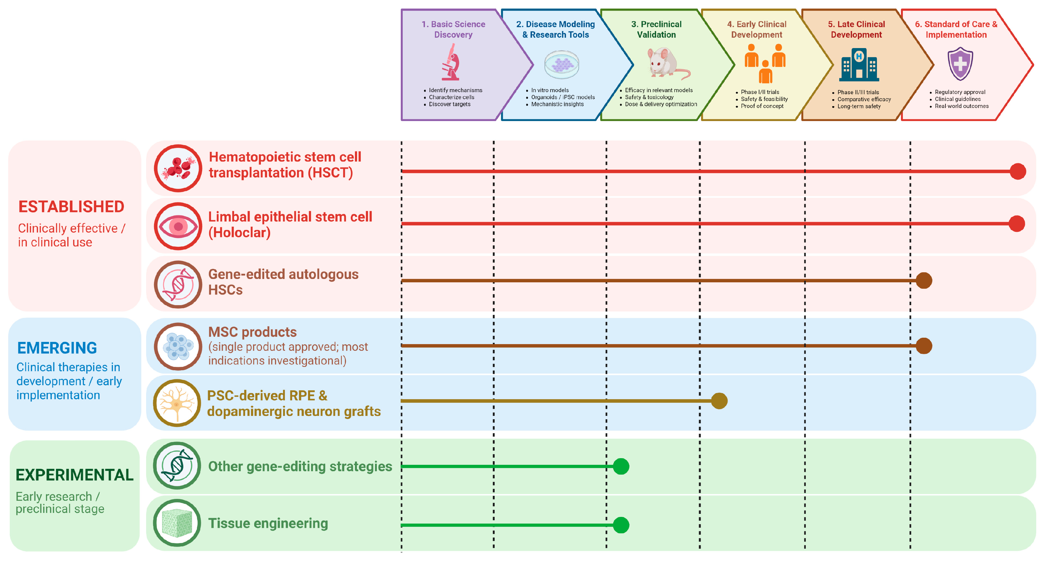
Stem cell-based applications span a wide spectrum of clinical validation, from proven standards of care to interventions offered without supporting evidence. In this review, we group them into three tiers (Figure 3):
Established applications have regulatory approval or are a standard of care, with long-term outcome data or mandated long-term follow-up. Examples are HSCT, limbal stem cell transplantation for limbal stem cell deficiency and, more recently, gene-edited autologous HSCs for sickle cell disease and transfusion-dependent β-thalassemia.
Emerging applications have shown encouraging safety and preliminary efficacy in early-phase clinical trials but are not yet approved for routine use. Examples are PSC-derived retinal pigment epithelial cells and dopaminergic neurons.
Experimental applications remain preclinical, or have been used in patients without adequate evidence. Examples are the transplantation of organoids or engineered tissues, most genome-editing strategies beyond HSCs, and many interventions marketed directly to consumers.
These tiers are not fixed. Products move between them as evidence accumulates, and a single cell type can span several tiers depending on the product, indication and manufacturing process; mesenchymal stromal cells illustrate this most clearly (Section 6.2).
Figure 3.
The translational maturity of stem cell-based applications. Representative modalities are mapped across six translational stages, from basic discovery to standard-of-care implementation, and line length indicates the furthest stage reached. Established modalities are hematopoietic stem cell transplantation (HSCT), limbal epithelial stem cell transplantation and gene-edited autologous HSCs for sickle cell disease and β-thalassemia. Emerging modalities are PSC-derived retinal pigment epithelium and dopaminergic neuron grafts in early clinical trials. MSC products span tiers: remestemcel-L is established for a single indication, most other MSC products remain emerging, and poorly characterized preparations marketed directly to consumers are experimental (Section 6.2). Experimental modalities are other gene-editing strategies and tissue engineering, which remain at the preclinical stage. Organoid and iPSC models serve as research tools rather than therapies. Colors indicate the evidence tier: established, emerging and experimental. Created in BioRender. Nguyen, H. (2026) https://BioRender.com/cy190lp.
6.1. Established and Emerging Clinical Applications
HSCT is the most mature stem cell therapy. It is a potentially curative standard of care for a defined range of hematologic malignancies, including acute leukemias, lymphomas and multiple myeloma, and for non-malignant disorders such as aplastic anemia, sickle cell disease, thalassemia and severe primary immunodeficiencies. In selected pediatric and congenital populations, long-term overall survival after HSCT exceeds 90%, and event-free survival in thalassemia approaches 86% [46,47,48].
This success took time. Roughly two decades of experimental, often fatal, transplantation separated the first bone marrow transplants of the late 1950s from routine clinical use [46]. Progress came from incremental refinement of HLA matching, conditioning regimens, infection control and graft-versus-host disease (GVHD) prophylaxis, rather than from a single breakthrough [46]. HSCT also benefits from standardized endpoints, such as overall, relapse-free and GVHD-free survival, which allow meta-analyses and comparison across international registries [46]. Limbal epithelial stem cell transplantation is a second established application, restoring the ocular surface in 60–80% of patients with severe limbal stem cell deficiency [49].
The same principles underlie more recent successes. Autologous HSCs edited with clustered regularly interspaced short palindromic repeats (CRISPR)-associated protein 9 (CRISPR-Cas9) to reactivate fetal hemoglobin have produced durable benefit in sickle cell disease and transfusion-dependent β-thalassemia, leading to regulatory approval in the United Kingdom and the United States in 2023–2024 [50,51,52]. Among emerging applications, PSC-derived retinal pigment epithelial cells have shown long-term survival and acceptable safety in patients with age-related macular degeneration and inherited retinal disorders [53], and early clinical studies of PSC-derived dopaminergic neurons for Parkinson’s disease have reported graft survival with preliminary signs of functional improvement [16,54].
These successes do not mean that the biological constraints described in Section 5 have been overcome. Each was achieved for a specific cell product, in a well-defined indication, with standardized manufacturing, rigorous preclinical validation and carefully designed trials with long-term follow-up. Successful translation therefore depends on the product, indication, manufacturing process and clinical context, rather than on the general properties of stem cells.
6.2. Product-Specific Evidence for MSC Therapies
MSC preparations differ by tissue source, donor, expansion and manufacturing, and should not be treated as interchangeable products [40]. Trials in inflammatory and autoimmune conditions have produced heterogeneous outcomes [55,56]. Paracrine signaling, direct cellular interactions and recipient-mediated responses to apoptotic MSCs are proposed mechanisms, supported by experimental work and selected human translational observations; their relative contributions are not established across indications [57].
The MSC secretome includes soluble mediators and extracellular vesicles whose composition changes with cellular source and culture conditions [58]. The limited persistence of administered cells does not necessarily mean short-lived clinical benefit. Surface-marker identity alone does not establish therapeutic potency. Mechanism-relevant functional assays are therefore needed alongside identity and manufacturing controls, although no single assay predicts clinical efficacy across all products [59].
Clinical maturity therefore varies within this category, and individual products can occupy different tiers. In December 2024, the US Food and Drug Administration (FDA) approved the bone-marrow-derived MSC product remestemcel-L-rknd (Ryoncil) for steroid-refractory acute graft-versus-host disease in pediatric patients aged two months and older [60]; for this single indication, it meets our definition of an established application. Conversely, darvadstrocel (Alofisel) was withdrawn from the European Union market in December 2024 after its confirmatory trial failed to demonstrate the required clinical benefit [61]. Most other MSC products, in inflammatory and autoimmune indications, remain emerging, while poorly characterized preparations marketed directly to consumers are experimental (Section 6.4). These outcomes concern particular products and indications, not MSC therapy as a class.
Controlled trials of characterized MSC products have generally shown acceptable tolerability, with an increased risk of fever; available studies do not exclude uncommon or delayed harms [62]. These data apply to the products tested and cannot be extended to poorly characterized commercial preparations (Section 6.3).
6.3. Reported Harms of Unproven Interventions
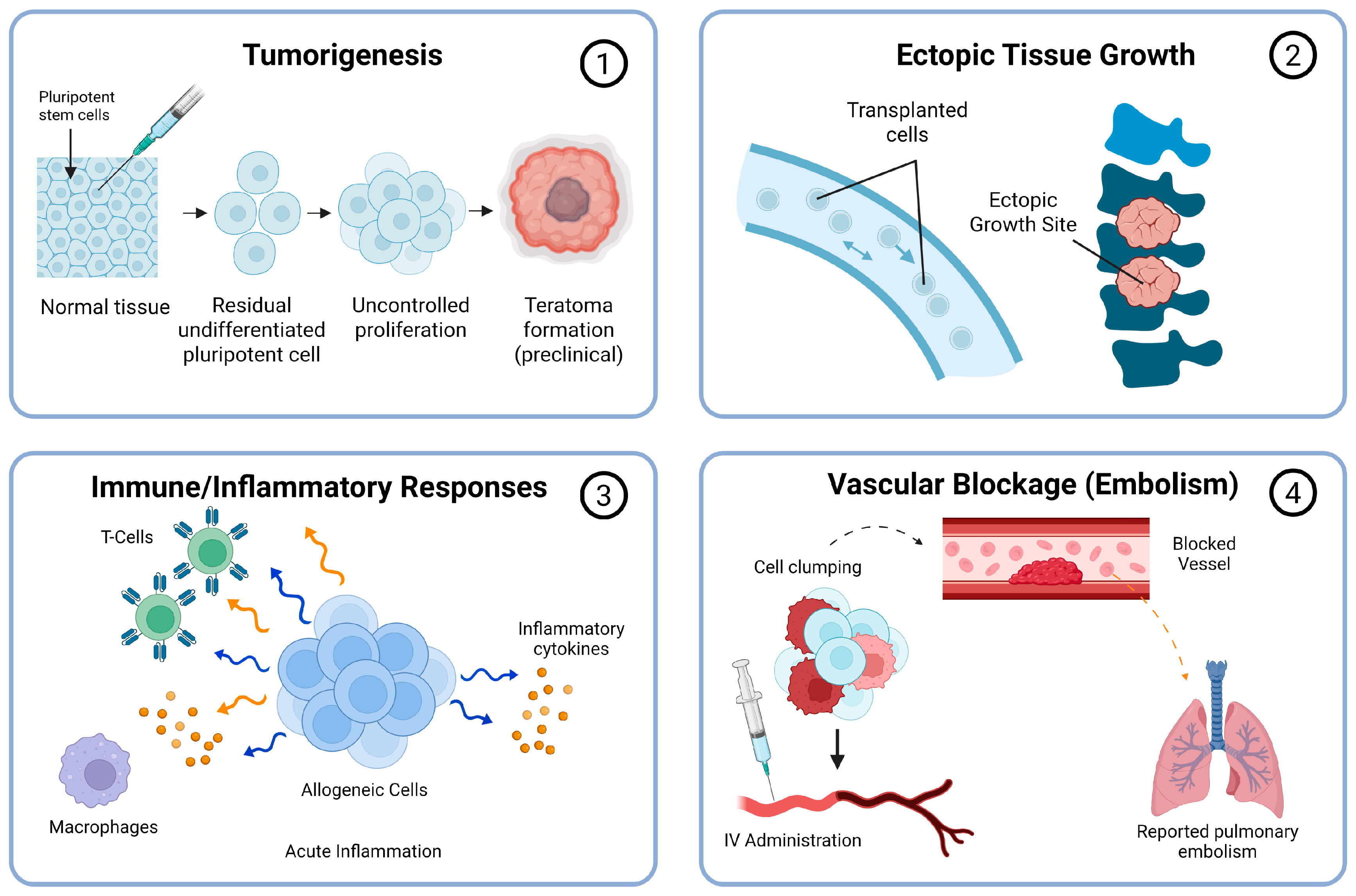
Serious adverse events have been reported after specific unproven cell interventions. Case reports and series can establish occurrence and suggest mechanisms, but without reliable exposure denominators and systematic follow-up they cannot estimate incidence. Risks vary with the cell product, route of administration, recipient and quality controls; they should not be generalized to all stem cell therapies (Figure 4).
A glioneuronal tumor developed after fetal neural-cell injections in a child with ataxia telangiectasia; molecular testing identified donor-derived cells [63]. A delayed spinal mass containing respiratory epithelium and glands was reported after olfactory mucosal autografting [64]. These observations concern specific grafts, not MSC products. Experimental teratoma formation by undifferentiated iPSCs is a separate, preclinical safety finding [65].
Pulmonary emboli were identified in three relatives following repeated intravenous autologous adipose-derived cell preparations, with pulmonary infarction in the index patient [66]. This single family report cannot establish incidence or prove that cell aggregates caused the emboli.
Figure 4.
Potential mechanisms and reported categories of harm in cell interventions. (1) Tumorigenesis: teratoma formation by residual undifferentiated pluripotent cells has been shown in preclinical studies; a donor-derived tumor after fetal neural-cell injection has been reported clinically in a single patient. (2) Ectopic tissue growth: a single clinical report after olfactory mucosal autografting. (3) Immune and inflammatory responses: a clinical report of demyelinating encephalomyelitis after cell treatment, with uncertain causality; product contamination and infection are a separate category. (4) Pulmonary vascular obstruction after intravenous infusion: pulmonary emboli reported clinically in one family, without proof that cell aggregates were the cause. Clinical reports show that these events can occur but not how often; preclinical findings indicate potential mechanisms only. Dashed arrows indicate proposed rather than demonstrated causal links. Created in BioRender. Nguyen, H. (2026) https://BioRender.com/16zcpd7.
Fulminant demyelinating encephalomyelitis followed intrathecal and intravenous cell treatment for multiple sclerosis, although causality remained uncertain [67]. Separately, a US investigation identified bacterial infections in 20 recipients of unapproved cord-blood-derived products, with microbiological evidence supporting product contamination [68]. These reports support careful product characterization, informed consent and adverse-event surveillance rather than a single risk estimate for all interventions.
6.4. Ethical Concerns in Commercial Cell Interventions
Direct-to-consumer (DTC) cell interventions raise concerns about the quality of evidence communicated to patients, financial exposure and accountability. These concerns depend on the intervention and setting, rather than commercial provision alone [69]. Here, “unproven” denotes insufficient supporting evidence, whereas “unregulated” denotes provision outside applicable regulatory oversight; the terms are not interchangeable.
6.4.1. Therapeutic Misconception and Misestimation
Therapeutic misconception concerns misunderstanding the distinction between research and individualized clinical care; therapeutic misestimation concerns inaccurate expectations of benefit or harm. In the DTC setting, patients may also mistake an unproven intervention for an established treatment [69]. Such misunderstandings may be reinforced by marketing practices that blur research, clinical care and commercial promotion:
- •
- Research registration: Listing a paid intervention on ClinicalTrials.gov does not itself establish regulatory approval, independent ethical oversight or therapeutic efficacy [70].
- •
- Scientific terminology: References to exosomes, secretomes or paracrine signaling can convey scientific credibility without supplying evidence for the specific product and indication being offered [71,72].
- •
- Cross-border availability: Differences in regulatory requirements and enforcement can complicate patients’ assessment of treatment status and accountability [73].
6.4.2. Direct-Payment Models and Financial Exposure
Two published website surveys, conducted in 2022 and 2023, report substantial variation in advertised prices for DTC cell and cell-derived interventions (Table 2). These are advertised prices from previous surveys, not current or standardized treatment costs, and the services advertised may differ in dose, route, number of administrations and ancillary care.
Table 2.
Illustrative advertised prices for unproven DTC cell and cell-derived interventions. The prices are website-advertised ranges from distinct surveys, not pooled averages, verified payments or current standardized fees. Units and treatment packages were not harmonized across providers. The year column gives the survey period rather than publication year. Cell-free secretome/exosome services are shown separately from cell-containing products.
Charging for an unproven intervention creates financial exposure without an established expectation of benefit. Pay-to-participate arrangements warrant scrutiny of scientific validity, independent review, consent and financial conflicts [76]. Some patients have used crowdfunding to pay for unproven stem cell interventions [77].
Counseling should distinguish advertised procedure prices from total expected expenditure, including travel, repeat treatment, follow-up and possible management of complications. Providers should disclose financial interests and the limits of supporting evidence so that patients can assess these commitments alongside conventional treatment and legitimate research options [78].
6.4.3. Vulnerability: Chronic, Terminal and Pediatric Populations
Patients with progressive or life-limiting conditions and few effective options may attach substantial value to uncertain potential benefit, which does not in itself indicate impaired decision-making. Good counseling acknowledges patients’ priorities while explaining the intervention’s evidence, alternatives, foreseeable burdens and uncertainties in accessible language [69].
For children, parental permission should be accompanied by age- and capacity-appropriate assent where possible. Families considering overseas interventions need clear information about oversight, follow-up and avenues for redress [73]. Vulnerability strengthens the need for independent review and proportionate risk assessment.
6.4.4. Geographical Evidence and Regulatory Context
Geographical descriptions require dates and source-specific interpretation. The 2016 global mapping study documents a historical distribution of businesses, not present-day regulatory performance [79]. India’s 2017 Indian Council of Medical Research (ICMR) guidelines and 2019 New Drugs and Clinical Trials Rules provide oversight frameworks for stem cell research and products [80], although evidence on their enforcement is more limited [81]. China’s 2015 clinical research measures introduced institutional and project filing, ethical review and restrictions on research-related charging and advertising [82]. The 2008 account of Chinese providers is historical journalism, not a current national surveillance estimate [83]. In Vietnam, the cited 2025 news report describes concerns about advertising and compliance [84]; it does not quantify the national market or establish absence of regulation. The Ministry of Health’s Circular 43/2024/TT-BYT, effective February 2025, governs biomedical research ethics councils [85], although ethics review alone does not authorize a particular cell product or clinical use. These sources describe different periods and regulatory functions and do not support ranking countries.
6.5. Emerging Preclinical Tools: Organoids and iPSCs
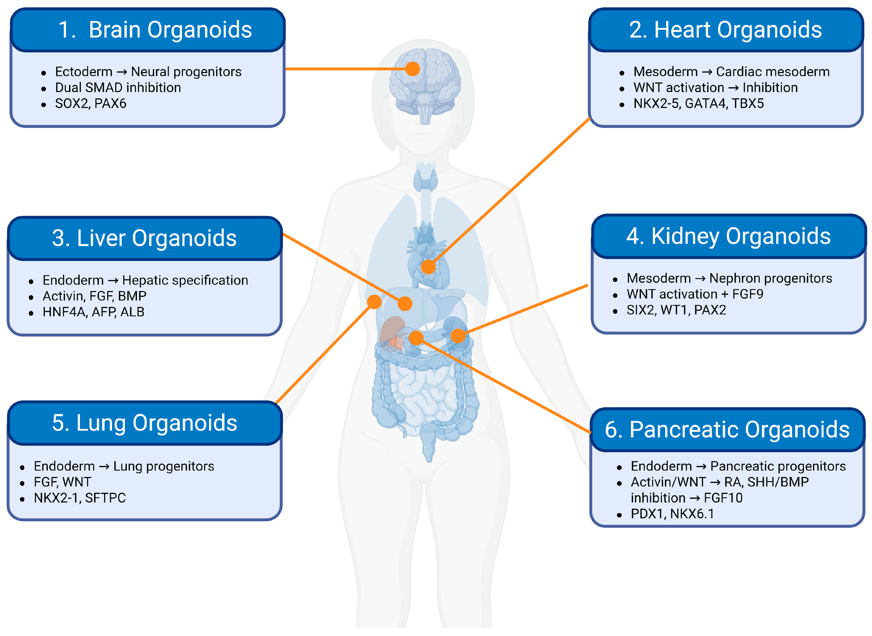
Although direct therapeutic use of pluripotent stem cells remains limited, PSC-based systems have become valuable research tools for drug discovery, toxicology and disease modeling. Human organoids, three-dimensional self-organizing structures derived from ESCs, iPSCs or adult stem cells, reproduce key aspects of the architecture, cellular composition and gene expression of native organs in vitro [86].
Patient-derived organoids are used to model genetic, infectious and malignant diseases and to test drug responses in individual patients, for example in colorectal cancer, pancreatic adenocarcinoma and cystic fibrosis [87,88,89]. iPSC-derived cardiac, hepatic and neural systems support mechanistic studies and target validation where animal models predict human responses poorly (Figure 5) [86]. Combined with CRISPR-Cas9 editing, iPSCs allow the generation of isogenic cell lines that differ only at a disease-causing mutation, providing well-matched genetic controls [90], and regulators have begun to accept data from organoids and other microphysiological systems as complements or alternatives to animal testing [91].
These are research applications, not therapies. Transplantation of organoids or engineered tissues remains experimental, limited by inadequate vascularization, structural immaturity and long-term immunogenicity [90].
Figure 5.
Directed differentiation of human pluripotent stem cells into organoids. Key signaling cues and markers: (1) brain: dual SMAD inhibition; SOX2, PAX6 [92]; (2) heart: WNT activation → inhibition; NKX2-5, GATA4, TBX5 [93]; (3) liver: Activin, fibroblast growth factor (FGF), bone morphogenetic protein (BMP); HNF4A, AFP, ALB [94]; (4) kidney: WNT activation, FGF9; SIX2, WT1, PAX2 [95]; (5) lung: FGF, WNT; NKX2-1, SFTPC [96]; (6) pancreas: Activin/WNT → retinoic acid (RA), sonic hedgehog (SHH)/BMP inhibition → FGF10; PDX1, NKX6 [97]. Created in BioRender. Nguyen, H. (2026) https://BioRender.com/ukgz9ny.
6.6. Genome Editing as a Complementary Strategy
Genome editing extends stem cell therapy in two ways. CRISPR-Cas systems allow pathogenic mutations to be corrected in patient-derived stem cells before transplantation [98,99]; this approach has already reached approval for hemoglobinopathies (Section 6.1) and is in clinical trials for several primary immunodeficiencies [52]. Editing can also be used to engineer universal donor pluripotent stem cell lines with reduced immunogenicity, which could widen access to allogeneic therapies [100,101]. Beyond hemoglobinopathies, however, most genome-editing strategies remain experimental, and off-target editing, genomic stability and long-term safety still require careful evaluation.
6.7. Long-Term Horizons and Incremental Progress
Biomedical innovation has typically advanced through incremental refinement rather than sudden breakthroughs. Monoclonal antibodies, first described in 1975, required roughly 25 to 30 years of optimization, including chimerization, humanization and large-scale manufacturing, before achieving broad clinical impact [102]. HSCT likewise took decades of multicenter research to become a reliable standard of care (Section 6.1) [103].
Many stem cell-based products face additional hurdles. Living cells must survive transplantation, engraft, integrate functionally with host tissue, avoid immune clearance and maintain lineage fidelity without malignant transformation, while manufacturing must control batch variability, potency testing and cold-chain logistics [104]. Development timelines will therefore vary widely between products and indications: gene-edited HSC therapies are built on decades of accumulated HSCT experience (Section 6.6), whereas complex tissue replacement remains at an early stage. What these products share is the need for long-term, rigorous validation, rather than the short horizons often implied in promotional materials.
Organoid technology shows the same pattern in preclinical research, having developed incrementally over nearly two decades into current multi-tissue platforms [105]. Further progress will depend on resolving bottlenecks in vascularization, structural maturation and immune tolerance, and on reporting failures alongside successes. This is slower than the timelines claimed by some commercial providers, but remains the most reliable route to safe and effective cell therapies.
7. A Roadmap for Clinical Integrity
Beyond direct clinical harms, premature commercialization may carry opportunity costs for the field. It has been argued that inflated public expectations and academic reward structures can create pressure to demonstrate rapid clinical impact, potentially favoring early clinical deployment over the slower basic and safety research that the field still requires [106]. The availability of unproven interventions outside clinical trials has also been identified as a possible barrier to trial recruitment, which could slow the generation of reliable evidence [2], although the size of this effect has not been quantified.
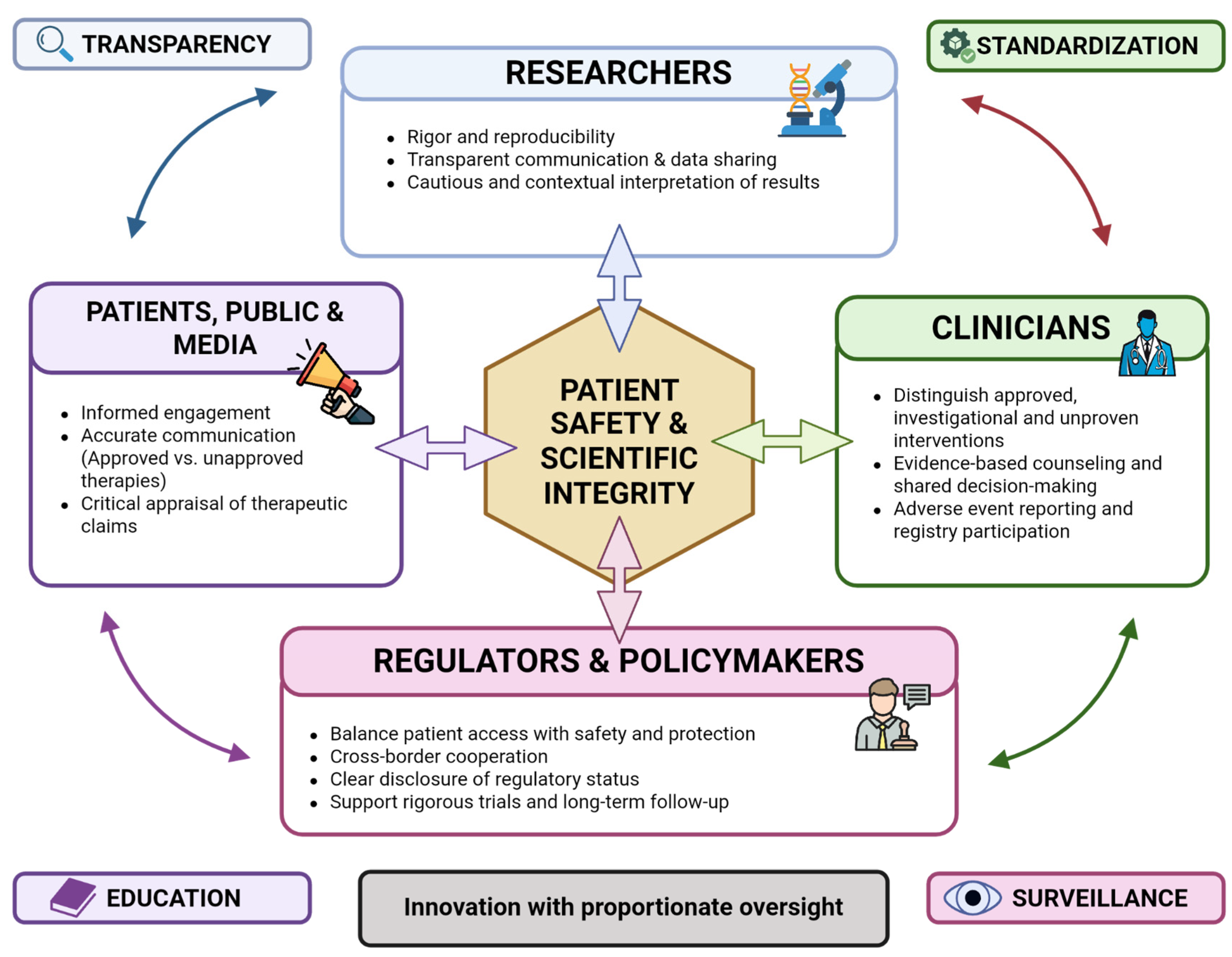
Safe progress therefore requires closer collaboration across research, clinical, regulatory and public domains (Figure 6), built on four pillars: transparency, standardization, education and surveillance. In this framework, innovation and regulation are complementary rather than opposing forces.
7.1. Ensuring Translational Rigor Through Standardization and Surveillance
Regenerative medicine is a competitive field, and an academic culture that rewards novelty over reproducibility may increase susceptibility to exaggeration and, in some cases, misconduct [107]. High-profile cases of data fabrication and image manipulation in stem cell research have damaged public trust and delayed legitimate progress [108,109]. Scientific rigor is therefore an ethical obligation as well as a methodological one.
Researchers can help by prioritizing reproducibility over rapid publication. For MSC products in particular, manufacturing variability (Section 5.6) makes standardized cell sourcing, identity panels and consensus potency assays a prerequisite for meaningful comparison between studies [42,43]. Publication bias should also be addressed: preregistering preclinical and clinical protocols and publishing negative or inconclusive results would reduce duplication of unproductive approaches.
Figure 6.
A multi-stakeholder framework for ensuring safe and evidence-based translation of stem cell therapies. Patient safety and scientific integrity are supported by four groups: researchers (rigor, reproducibility, transparent communication); clinicians (evidence-based counseling, adverse-event reporting and registries); regulators and policymakers (balancing access with protection, cross-border cooperation, disclosure of regulatory status); and patients, the public and media (informed engagement, accurate communication, critical appraisal of claims). These responsibilities are linked by transparency, standardization, education and surveillance. Created in BioRender. Nguyen, H. (2026) https://BioRender.com/udhq6d4.
Clinicians can contribute through multicenter registries that follow all patients receiving cellular interventions in routine practice, which can detect rare, late complications and durability outcomes that short, small trials cannot capture. Because some cell-based interventions produce biological changes that are difficult to reverse, with potential for delayed effects such as late-onset tumorigenesis, insertional mutagenesis or ectopic tissue formation [36,110], regulators should consider long-term follow-up and registry participation as conditions of approval where these risks apply. For gene therapy products with such risks, for example, the FDA recommends follow-up of up to 15 years [111].
7.2. Ethical Gatekeeping and Market Oversight
Clinicians can support informed decisions by distinguishing approved treatments for specified indications, regulated investigational interventions and commercial offerings without adequate evidence. Counseling should explain the relevant product, expected benefits, uncertainties and alternatives, and why testimonials or registry listings do not establish efficacy [69,70]. Regulatory approval applies to a specific indication and jurisdiction and is not a guarantee of benefit or absence of risk. Patients considering unproven interventions need clear information on product characterization, oversight, follow-up and reporting of adverse events.
Ethical care also includes psychological and social support [112]. Adjustment after a major improvement in health, described as the “burden of normality” in other clinical settings, may merit discussion [113]. Such support is best tailored to individual needs and expectations.
Policymakers should consider proportionate measures addressing misleading efficacy claims and unclear regulatory status, informed by studies of DTC marketing [72,114]. Options include standardized disclosure of authorization and evidence, scrutiny of testimonials used to imply efficacy, transparent financial information, and mechanisms for complaints and redress. Advertising platforms could require verifiable regulatory information. Such proposals require jurisdiction-specific legal assessment and evaluation of their effects on patients and access to legitimate research.
Cross-border care adds difficulties in verifying treatment status, maintaining follow-up and pursuing complaints [73]. Cooperation between regulators, professional bodies and clinicians could improve information sharing and referral accountability. Patients considering treatment abroad need to be able to identify the responsible provider, applicable oversight and arrangements for subsequent care. These measures complement the surveillance proposals in Section 7.1.
8. Limitations
This review is narrative rather than systematic. We did not follow a prespecified protocol or apply formal criteria for study selection and quality appraisal; source selection reflected the authors’ judgment of relevance (Section 2). Some of our interpretations, particularly regarding the drivers of premature clinical translation and the effects of commercial activity on research and patients, are therefore presented as arguments supported by the cited literature rather than as established causal findings. For quantitative estimates of efficacy, readers are referred to the systematic reviews and meta-analyses cited.
Evidence on harms and markets has particular limitations. The adverse-event literature combines product-specific case reports, case series and outbreak investigations with preclinical safety findings. Without reliable exposure denominators, systematic ascertainment or adequate follow-up, these sources show that serious harm can occur but cannot establish its incidence, and findings from fetal neural or olfactory grafts, adipose- or cord-blood-derived products and pluripotent cells should not be treated as interchangeable. The geographical discussion is selective rather than a comprehensive assessment of current regulation or enforcement: the cited frameworks address different regulatory functions, their existence does not establish compliance in practice, and news reports were used as dated context, with less weight than peer-reviewed surveillance studies and official documents. Finally, Table 2 presents illustrative website-advertised prices from two surveys conducted in 2022 and 2023; these are not verified payments, pooled averages or current standardized fees.
9. Conclusions
The clinical maturity of stem cell-based applications differs widely. Established therapies, such as HSCT, limbal stem cell transplantation, gene-edited HSCs for hemoglobinopathies and one MSC product for a single indication, show what rigorous translation can achieve; emerging applications, including PSC-derived retinal and dopaminergic grafts and most other MSC products, have shown encouraging early clinical results; and many other approaches, including most interventions marketed directly to consumers, remain experimental. The evidence reviewed here suggests that success depends less on the properties of stem cells in general than on the specific cell product, indication, manufacturing process and clinical context. Differentiation alone is generally insufficient to restore tissue function. For pluripotent stem cell-derived products, the properties that confer regenerative potential are closely linked to tumorigenic risk; adult tissue-specific stem cells, HSCs and MSC preparations have different risk profiles that depend on the product (Section 5.5). As the history of HSCT shows, durable success requires sustained optimization of immune compatibility, conditioning and complication management, an experience at odds with simplified accounts that present unproven interventions as ready-made cures.
Future progress is likely to depend less on discovering new stem cell types than on integrating advances in developmental biology, biomaterials, genome editing, artificial intelligence and systems immunology. By matching ambition with biological realism and transparent evidence, regenerative medicine can realize its promise while retaining the trust of patients and society.
Author Contributions
H.M.L., H.H.K.N., T.Q.L. and T.T.N. contributed equally to this work and share first authorship. Conceptualization: C.D.H.; literature review and data curation: H.M.L., H.H.K.N., T.Q.L. and T.T.N.; writing—original draft: H.M.L., H.H.K.N., T.Q.L. and T.T.N.; writing—review and editing: D.M.H. and C.D.H.; visualization: H.H.K.N.; supervision: C.D.H.; project administration: H.M.L. All authors have read and agreed to the published version of the manuscript.
Funding
This research received no external funding.
Institutional Review Board Statement
Not applicable.
Informed Consent Statement
Not applicable.
Data Availability Statement
No new data were created or analyzed in this study. Data sharing is not applicable to this article.
Acknowledgments
During the preparation of this manuscript, the authors used Claude (Claude Sonnet 4.5, Anthropic) to correct grammar and spelling, improve language clarity, and refine the structure and organization of the text. The tool was not used to generate scientific content, produce or analyze data, create figures, or interpret the cited literature. All figures were prepared by the authors without the use of generative artificial intelligence. All content was subsequently reviewed and edited by the authors, who take full responsibility for the accuracy and integrity of the published work. Figure 1, Figure 2, Figure 3, Figure 4, Figure 5 and Figure 6 were created by the authors in BioRender (BioRender.com) under BioRender Academic Publication Licenses.
Conflicts of Interest
The authors declare no competing interests.
References
- Della Rocca, Y.; Mazzone, A.; Marconi, G.D.; Trubiani, O.; Pizzicannella, J.; Diomede, F. Stem Cells in Regenerative Medicine: A Journey from Adult Stem Cells to Induced Pluripotent Cells. Int. J. Mol. Sci. 2025, 26, 8255. [Google Scholar] [CrossRef] [Scilit]
- Master, Z.; Matthews, K.R.W.; Abou-el-Enein, M. Unproven stem cell interventions: A global public health problem requiring global deliberation. Stem Cell Rep. 2021, 16, 1435–1445. [Google Scholar] [CrossRef] [Scilit]
- Blau, H.M.; Daley, G.Q. Stem Cells in the Treatment of Disease. N. Engl. J. Med. 2019, 380, 1748–1760. [Google Scholar] [CrossRef] [Scilit]
- Rebuzzini, P.; Zuccotti, M.; Garagna, S. Building Pluripotency Identity in the Early Embryo and Derived Stem Cells. Cells 2021, 10, 2049. [Google Scholar] [CrossRef] [Scilit]
- Kolios, G.; Moodley, Y. Introduction to stem cells and regenerative medicine. Respir. Int. Rev. Thorac. Dis. 2013, 85, 3–10. [Google Scholar] [CrossRef] [Scilit]
- Hematopoietic Stem Cells. In Methods in Enzymology; Academic Press: Cambridge, MA, USA, 2006; Volume 419, pp. 149–179.
- Zhu, F.; Nie, G. Cell reprogramming: Methods, mechanisms and applications. Cell Regen. 2025, 14, 12. [Google Scholar] [CrossRef] [Scilit]
- Dupont, G.; Yilmaz, E.; Loukas, M.; Macchi, V.; De Caro, R.; Tubbs, R.S. Human embryonic stem cells: Distinct molecular personalities and applications in regenerative medicine. Clin. Anat. 2019, 32, 354–360. [Google Scholar] [CrossRef] [Scilit]
- Eguizabal, C.; Aran, B.; Chuva de Sousa Lopes, S.M.; Geens, M.; Heindryckx, B.; Panula, S.; Popovic, M.; Vassena, R.; Veiga, A. Two decades of embryonic stem cells: A historical overview. Hum. Reprod. Open 2019, 2019, hoy024. [Google Scholar] [CrossRef] [Scilit]
- Charitos, I.A.; Ballini, A.; Cantore, S.; Boccellino, M.; Di Domenico, M.; Borsani, E.; Nocini, R.; Di Cosola, M.; Santacroce, L.; Bottalico, L. Stem Cells: A Historical Review about Biological, Religious, and Ethical Issues. Stem Cells Int. 2021, 2021, 9978837. [Google Scholar] [CrossRef] [Scilit]
- Okita, K.; Yamanaka, S. Induction of pluripotency by defined factors. Exp. Cell Res. 2010, 316, 2565–2570. [Google Scholar] [CrossRef] [Scilit]
- Poliwoda, S.; Noor, N.; Downs, E.; Schaaf, A.; Cantwell, A.; Ganti, L.; Kaye, A.D.; Mosel, L.I.; Carroll, C.B.; Viswanath, O.; et al. Stem cells: A comprehensive review of origins and emerging clinical roles in medical practice. Orthop. Rev. 2022, 14, 37498. [Google Scholar] [CrossRef] [Scilit]
- Takahashi, K.; Yamanaka, S. Induction of pluripotent stem cells from mouse embryonic and adult fibroblast cultures by defined factors. Cell 2006, 126, 663–676. [Google Scholar] [CrossRef] [Scilit]
- Kim, K.; Doi, A.; Wen, B.; Ng, K.; Zhao, R.; Cahan, P.; Kim, J.; Aryee, M.J.; Ji, H.; Ehrlich, L.I.; et al. Epigenetic memory in induced pluripotent stem cells. Nature 2010, 467, 285–290. [Google Scholar] [CrossRef] [Scilit]
- Cerneckis, J.; Cai, H.; Shi, Y. Induced pluripotent stem cells (iPSCs): Molecular mechanisms of induction and applications. Signal Transduct. Target. Ther. 2024, 9, 112. [Google Scholar] [CrossRef] [Scilit]
- Sawamoto, N.; Doi, D.; Nakanishi, E.; Sawamura, M.; Kikuchi, T.; Yamakado, H.; Taruno, Y.; Shima, A.; Fushimi, Y.; Okada, T.; et al. Phase I/II trial of iPS-cell-derived dopaminergic cells for Parkinson’s disease. Nature 2025, 641, 971–977. [Google Scholar] [CrossRef] [Scilit]
- Rando, T.A.; Brunet, A.; Goodell, M.A. Hallmarks of stem cell aging. Cell Stem Cell 2025, 32, 1038–1054. [Google Scholar] [CrossRef] [Scilit]
- Chacón-Martínez, C.A.; Koester, J.; Wickström, S.A. Signaling in the stem cell niche: Regulating cell fate, function and plasticity. Development 2018, 145, dev165399. [Google Scholar] [CrossRef] [Scilit]
- Viswanathan, S.; Shi, Y.; Galipeau, J.; Krampera, M.; Leblanc, K.; Martin, I.; Nolta, J.; Phinney, D.G.; Sensebe, L. Mesenchymal stem versus stromal cells: International Society for Cell & Gene Therapy (ISCT®) Mesenchymal Stromal Cell committee position statement on nomenclature. Cytotherapy 2019, 21, 1019–1024. [Google Scholar] [CrossRef] [Scilit]
- Miller, L.H.; Qu, C.-K.; Pauly, M. Germline mutations in the bone marrow microenvironment and dysregulated hematopoiesis. Exp. Hematol. 2018, 66, 17–26. [Google Scholar] [CrossRef] [Scilit]
- Comazzetto, S.; Shen, B.; Morrison, S.J. Niches that regulate stem cells and hematopoiesis in adult bone marrow. Dev. Cell 2021, 56, 1848–1860. [Google Scholar] [CrossRef] [Scilit]
- Martyniak, A.; Jeż, M.; Dulak, J.; Stępniewski, J. Adaptation of cardiomyogenesis to the generation and maturation of cardiomyocytes from human pluripotent stem cells. IUBMB Life 2023, 75, 8–29. [Google Scholar] [CrossRef] [Scilit]
- Gepstein, L.; Ding, C.; Rehemedula, D.; Wilson, E.E.; Yankelson, L.; Caspi, O.; Gepstein, A.; Huber, I.; Olgin, J.E. In vivo assessment of the electrophysiological integration and arrhythmogenic risk of myocardial cell transplantation strategies. Stem Cells 2010, 28, 2151–2161, Erratum in Stem Cells 2011, 29, 1475. [Google Scholar] [CrossRef] [Scilit]
- Selvakumar, D.; Clayton, Z.E.; Prowse, A.; Dingwall, S.; Kim, S.K.; Reyes, L.; George, J.; Shah, H.; Chen, S.; Leung, H.H.; et al. Cellular heterogeneity of pluripotent stem cell-derived cardiomyocyte grafts is mechanistically linked to treatable arrhythmias. Nat. Cardiovasc. Res. 2024, 3, 145–165. [Google Scholar] [CrossRef] [Scilit]
- Smith, L.R.; Cho, S.; Discher, D.E. Stem Cell Differentiation is Regulated by Extracellular Matrix Mechanics. Physiology 2018, 33, 16–25. [Google Scholar] [CrossRef] [Scilit]
- Yanamandala, M.; Zhu, W.; Garry, D.J.; Kamp, T.J.; Hare, J.M.; Jun, H.W.; Yoon, Y.S.; Bursac, N.; Prabhu, S.D.; Dorn, G.W., II; et al. Overcoming the Roadblocks to Cardiac Cell Therapy Using Tissue Engineering. J. Am. Coll. Cardiol. 2017, 70, 766–775. [Google Scholar] [CrossRef] [Scilit]
- Richter, S.; Böttcher, M.; Völkl, S.; Mackensen, A.; Ullrich, E.; Jacobs, B.; Mougiakakos, D. The metabolic profile of reconstituting T-cells, NK-cells, and monocytes following autologous stem cell transplantation and its impact on outcome. Sci. Rep. 2022, 12, 11406. [Google Scholar] [CrossRef] [Scilit]
- Martin, P.J.; Levine, D.M.; Storer, B.E.; Zheng, X.; Jain, D.; Heavner, B.; Norris, B.M.; Geraghty, D.E.; Spellman, S.R.; Sather, C.L.; et al. A Model of Minor Histocompatibility Antigens in Allogeneic Hematopoietic Cell Transplantation. Front. Immunol. 2021, 12, 782152. [Google Scholar] [CrossRef] [Scilit]
- Hill, G.R.; Koyama, M. Cytokines and costimulation in acute graft-versus-host disease. Blood 2020, 136, 418–428. [Google Scholar] [CrossRef] [Scilit]
- Khodayari, S.; Khodayari, H.; Amiri, A.Z.; Eslami, M.; Farhud, D.; Hescheler, J.; Nayernia, K. Inflammatory Microenvironment of Acute Myocardial Infarction Prevents Regeneration of Heart with Stem Cells Therapy. Cell. Physiol. Biochem. Int. J. Exp. Cell. Physiol. Biochem. Pharmacol. 2019, 53, 887–909. [Google Scholar] [CrossRef] [Scilit]
- Hamid, T.; Xu, Y.; Ismahil, M.A.; Li, Q.; Jones, S.P.; Bhatnagar, A.; Bolli, R.; Prabhu, S.D. TNF receptor signaling inhibits cardiomyogenic differentiation of cardiac stem cells and promotes a neuroadrenergic-like fate. Am. J. Physiol. Heart Circ. Physiol. 2016, 311, H1189–H1201. [Google Scholar] [CrossRef] [Scilit]
- Joshi, J.; Kothapalli, C.R. Role of Inflammatory Niche and Adult Cardiomyocyte Coculture on Differentiation, Matrix Synthesis, and Secretome Release by Human Bone Marrow Mesenchymal Stem Cells. Appl. Biochem. Biotechnol. 2022, 194, 1938–1954. [Google Scholar] [CrossRef] [Scilit]
- Zhao, W.; Lei, A.; Tian, L.; Wang, X.; Correia, C.; Weiskittel, T.; Li, H.; Trounson, A.; Fu, Q.; Yao, K.; et al. Strategies for Genetically Engineering Hypoimmunogenic Universal Pluripotent Stem Cells. iScience 2020, 23, 101162. [Google Scholar] [CrossRef] [Scilit]
- Wang, X.; Brown, N.K.; Wang, B.; Shariati, K.; Wang, K.; Fuchs, S.; Melero-Martin, J.M.; Ma, M. Local Immunomodulatory Strategies to Prevent Allo-Rejection in Transplantation of Insulin-Producing Cells. Adv. Sci. 2021, 8, e2003708. [Google Scholar] [CrossRef] [Scilit]
- Meissner, T.B.; Schulze, H.S.; Dale, S.M. Immune Editing: Overcoming Immune Barriers in Stem Cell Transplantation. Curr. Stem Cell Rep. 2022, 8, 206–218. [Google Scholar] [CrossRef] [Scilit]
- Ben-David, U.; Benvenisty, N. The tumorigenicity of human embryonic and induced pluripotent stem cells. Nat. Rev. Cancer 2011, 11, 268–277. [Google Scholar] [CrossRef] [Scilit]
- Poetsch, M.S.; Strano, A.; Guan, K. Human Induced Pluripotent Stem Cells: From Cell Origin, Genomic Stability, and Epigenetic Memory to Translational Medicine. Stem Cells 2022, 40, 546–555. [Google Scholar] [CrossRef] [Scilit]
- Riggs, J.W.; Barrilleaux, B.L.; Varlakhanova, N.; Bush, K.M.; Chan, V.; Knoepfler, P.S. Induced pluripotency and oncogenic transformation are related processes. Stem Cells Dev. 2013, 22, 37–50. [Google Scholar] [CrossRef] [Scilit]
- Wang, L.; Zhao, Y.; Liu, Y.; Akiyama, K.; Chen, C.; Qu, C.; Jin, Y.; Shi, S. IFN-γ and TNF-α synergistically induce mesenchymal stem cell impairment and tumorigenesis via NFκB signaling. Stem Cells 2013, 31, 1383–1395. [Google Scholar] [CrossRef] [Scilit]
- Česnik, A.B.; Švajger, U. The issue of heterogeneity of MSC-based advanced therapy medicinal products-a review. Front. Cell Dev. Biol. 2024, 12, 1400347. [Google Scholar] [CrossRef] [Scilit]
- Kim, M.-H.; Kino-Oka, M. Epigenetic memory as a readout of environmental susceptibility defines cell manufacturability in mesenchymal stem cell production. Stem Cell Res. Ther. 2025, 17, 60. [Google Scholar] [CrossRef] [Scilit]
- Stroncek, D.F.; Jin, P.; McKenna, D.H.; Takanashi, M.; Fontaine, M.J.; Pati, S.; Schäfer, R.; Peterson, E.; Benedetti, E.; Reems, J.A. Human Mesenchymal Stromal Cell (MSC) Characteristics Vary Among Laboratories When Manufactured From the Same Source Material: A Report by the Cellular Therapy Team of the Biomedical Excellence for Safer Transfusion (BEST) Collaborative. Front. Cell Dev. Biol. 2020, 8, 458. [Google Scholar] [CrossRef] [Scilit]
- Fitzgerald, J.C.; Shaw, G.; Murphy, J.M.; Barry, F. Media matters: Culture medium-dependent hypervariable phenotype of mesenchymal stromal cells. Stem Cell Res. Ther. 2023, 14, 363. [Google Scholar] [CrossRef] [Scilit]
- Melocchi, A.; Schmittlein, B.; Sadhu, S.; Nayak, S.; Lares, A.; Uboldi, M.; Zema, L.; di Robilant, B.N.; Feldman, S.A.; Esensten, J.H. Automated manufacturing of cell therapies. J. Control. Release Off. J. Control. Release Soc. 2025, 381, 113561. [Google Scholar] [CrossRef] [Scilit]
- Foti, R.; Storti, G.; Palmesano, M.; Calicchia, A.; Foti, R.; Ciprandi, G.; Cervelli, G.; Scioli, M.G.; Orlandi, A.; Cervelli, V. AI-Driven Innovations for Quality Control and Standardization: Future Strategies in Adipose-Derived Stem Cell Manufacturing. Int. J. Mol. Sci. 2026, 27, 2388. [Google Scholar] [CrossRef] [Scilit]
- Copelan, E.A. Hematopoietic Stem-Cell Transplantation. N. Engl. J. Med. 2006, 354, 1813–1826. [Google Scholar] [CrossRef] [Scilit]
- Kattamis, A.; Kwiatkowski, J.L.; Aydinok, Y. Thalassaemia. Lancet 2022, 399, 2310–2324. [Google Scholar] [CrossRef] [Scilit]
- Alsaati, N.; Grier, A.; Ochfeld, E.; McClory, S.; Heimall, J. Hematopoietic stem cell transplantation for primary immunodeficiency. Allergy Asthma Proc. 2024, 45, 371–383. [Google Scholar] [CrossRef] [Scilit]
- Le, Q.; Chauhan, T.; Yung, M.; Tseng, C.-H.; Deng, S.X. Outcomes of Limbal Stem Cell Transplant: A Meta-analysis. JAMA Ophthalmol. 2020, 138, 660–670. [Google Scholar] [CrossRef] [Scilit]
- Li, L.; Mandal, P.K. Recent advancements in gene therapy for sickle cell disease and β-thalassemia. Front. Hematol. 2024, 3, 1468952. [Google Scholar] [CrossRef] [Scilit]
- Aprile, A.; Lidonnici, M.R.; Ferrari, G. Gene therapy for hemoglobinopathies: Clinical trial results and biology of hematopoietic stem cell and the bone marrow niche. Cell Rep. Med. 2025, 6, 102419. [Google Scholar] [CrossRef] [Scilit]
- O’Hanlon Cohrt, K.; O’Dea, S. Clinical Trial Landscape of Gene-Edited Autologous Hematopoietic Stem Cells for Hemoglobinopathies and Immunodeficiencies. Int. J. Mol. Sci. 2026, 27, 3384. [Google Scholar] [CrossRef] [Scilit]
- Ahmed, I.; Johnston, R.J.; Singh, M.S. Pluripotent stem cell therapy for retinal diseases. Ann. Transl. Med. 2021, 9, 1279. [Google Scholar] [CrossRef] [Scilit]
- Nakanishi, E.; Yamakado, H.; Sawamoto, N.; Takahashi, J.; Takahashi, R. Parkinson’s Disease Cell Transplantation Therapy: A New Dawn With Pluripotent Stem Cell-Based Therapy. J. Neurochem. 2026, 170, e70500. [Google Scholar] [CrossRef] [Scilit]
- Zeng, L.; Liu, C.; Wu, Y.; Liu, S.; Zheng, Y.; Hao, W.; Wang, D.; Sun, L. Efficacy and safety of mesenchymal stromal cell transplantation in the treatment of autoimmune and rheumatic immune diseases: A systematic review and meta-analysis of randomized controlled trials. Stem Cell Res. Ther. 2025, 16, 65. [Google Scholar] [CrossRef] [Scilit]
- Gilkeson, G.S. Safety and Efficacy of Mesenchymal Stromal Cells and Other Cellular Therapeutics in Rheumatic Diseases in 2022: A Review of What We Know So Far. Arthritis Rheumatol. 2022, 74, 752–765. [Google Scholar] [CrossRef] [Scilit]
- Galleu, A.; Riffo-Vasquez, Y.; Trento, C.; Lomas, C.; Dolcetti, L.; Cheung, T.S.; Von Bonin, M.; Barbieri, L.; Halai, K.; Ward, S.; et al. Apoptosis in mesenchymal stromal cells induces in vivo recipient-mediated immunomodulation. Sci. Transl. Med. 2017, 9, eaam7828. [Google Scholar] [CrossRef] [Scilit]
- Maacha, S.; Sidahmed, H.; Jacob, S.; Gentilcore, G.; Calzone, R.; Grivel, J.C.; Cugno, C. Paracrine Mechanisms of Mesenchymal Stromal Cells in Angiogenesis. Stem Cells Int. 2020, 4356359. [Google Scholar] [CrossRef] [Scilit]
- Galipeau, J.; Krampera, M.; Barrett, J.; Dazzi, F.; Deans, R.J.; DeBruijn, J.; Dominici, M.; Fibbe, W.E.; Gee, A.P.; Gimble, J.M.; et al. International Society for Cellular Therapy perspective on immune functional assays for mesenchymal stromal cells as potency release criterion for advanced phase clinical trials. Cytotherapy 2016, 18, 151–159. [Google Scholar] [CrossRef] [Scilit]
- Center for Biologics Evaluation and Research. FDA Approves Remestemcel-L-rknd for Steroid-Refractory Acute Graft Versus Host Disease in Pediatric Patients; FDA: Silver Spring, MD, USA, 2024. Available online: https://www.fda.gov/drugs/resources-information-approved-drugs/fda-approves-remestemcel-l-rknd-steroid-refractory-acute-graft-versus-host-disease-pediatric (accessed on 2 November 2026).
- Alofisel|European Medicines Agency (EMA). 2018. Available online: https://www.ema.europa.eu/en/medicines/human/EPAR/alofisel (accessed on 2 November 2026).
- Thompson, M.; Mei, S.H.; Wolfe, D.; Champagne, J.; Fergusson, D.; Stewart, D.J.; Sullivan, K.J.; Doxtator, E.; Lalu, M.; English, S.W.; et al. Cell therapy with intravascular administration of mesenchymal stromal cells continues to appear safe: An updated systematic review and meta-analysis. EClinicalMedicine 2020, 19, 100249. [Google Scholar] [CrossRef] [Scilit]
- Amariglio, N.; Hirshberg, A.; Scheithauer, B.W.; Cohen, Y.; Loewenthal, R.; Trakhtenbrot, L.; Paz, N.; Koren-Michowitz, M.; Waldman, D.; Leider-Trejo, L.; et al. Donor-Derived Brain Tumor Following Neural Stem Cell Transplantation in an Ataxia Telangiectasia Patient. PLoS Med. 2009, 6, e1000029. [Google Scholar] [CrossRef] [Scilit]
- Dlouhy, B.J.; Awe, O.; Rao, R.C.; Kirby, P.A.; Hitchon, P.W. Autograft-derived spinal cord mass following olfactory mucosal cell transplantation in a spinal cord injury patient: Case report. J. Neurosurg. Spine 2014, 21, 618–622. [Google Scholar] [CrossRef] [Scilit]
- Yasuda, S.; Kusakawa, S.; Kuroda, T.; Miura, T.; Tano, K.; Takada, N.; Matsuyama, S.; Matsuyama, A.; Nasu, M.; Umezawa, A.; et al. Tumorigenicity-associated characteristics of human iPS cell lines. PLoS ONE 2018, 13, e0205022. [Google Scholar] [CrossRef] [Scilit]
- Jung, J.W.; Kwon, M.; Choi, J.C.; Shin, J.W.; Park, I.W.; Choi, B.W.; Kim, J.Y. Familial Occurrence of Pulmonary Embolism after Intravenous, Adipose Tissue-Derived Stem Cell Therapy. Yonsei Med. J. 2013, 54, 1293–1296. [Google Scholar] [CrossRef] [Scilit]
- Alderazi, Y.J.; Coons, S.W.; Chapman, K. Catastrophic demyelinating encephalomyelitis after intrathecal and intravenous stem cell transplantation in a patient with multiple sclerosis. J. Child Neurol. 2012, 27, 632–635. [Google Scholar] [CrossRef] [Scilit]
- Hartnett, K.P.; Powell, K.M.; Rankin, D.; Gable, P.; Kim, J.J.; Spoto, S.; Breaker, E.; Hunter, R.; Dotson, N.; McAllister, G.; et al. Investigation of Bacterial Infections Among Patients Treated With Umbilical Cord Blood-Derived Products Marketed as Stem Cell Therapies. JAMA Netw. Open 2021, 4, e2128615. [Google Scholar] [CrossRef] [Scilit]
- Arthurs, J.R.; Martin Lillie, C.M.; Master, Z.; Shapiro, S.A. The Direct to Consumer Stem Cell Market and the Role of Primary Care Providers in Correcting Misinformation. J. Prim. Care Community Health 2022, 13, 21501319221121460. [Google Scholar] [CrossRef] [Scilit]
- Turner, L. ClinicalTrials.gov, stem cells and ‘pay-to-participate’ clinical studies. Regen. Med. 2017, 12, 705–719. [Google Scholar] [CrossRef] [Scilit]
- Cook, M.; Richey, A.; Brafman, D.A.; Frow, E.K. Weighing up the evidence used by direct-to-consumer stem cell businesses. Stem Cell Rep. 2021, 16, 2852–2860. [Google Scholar] [CrossRef] [Scilit]
- Sipp, D.; Caulfield, T.; Kaye, J.; Barfoot, J.; Blackburn, C.; Chan, S.; De Luca, M.; Kent, A.; McCabe, C.; Munsie, M.; et al. Marketing of unproven stem cell-based interventions: A call to action. Sci. Transl. Med. 2017, 9, eaag0426. [Google Scholar] [CrossRef] [Scilit]
- Lyons, S.; Salgaonkar, S.; Flaherty, G.T. International stem cell tourism: A critical literature review and evidence-based recommendations. Int. Health 2021, 14, 132–141. [Google Scholar] [CrossRef] [Scilit]
- Turner, L.; Wang, J.C.; Martinez, J.R., Jr.; Najjar, S.; Arachchilage, T.R.; Sahrai, V. US businesses engaged in direct-to-consumer marketing of perinatal stem cell interventions following the Food and Drug Administration’s enforcement discretion era. Cytotherapy 2024, 26, 393–403. [Google Scholar] [CrossRef] [Scilit]
- Asadpour, A.; Yahaya, B.H.; Bicknell, K.; Cottrell, G.S.; Widera, D. Uncovering the gray zone: Mapping the global landscape of direct-to-consumer businesses offering interventions based on secretomes, extracellular vesicles, and exosomes. Stem Cell Res. Ther. 2023, 14, 111. [Google Scholar] [CrossRef] [Scilit]
- Turner, L.; Snyder, J. Ethical issues concerning a pay-to-participate stem cell study. Stem Cells Transl. Med. 2021, 10, 815–819. [Google Scholar] [CrossRef] [Scilit]
- Snyder, J.; Turner, L.; Crooks, V.A. Crowdfunding for Unproven Stem Cell-Based Interventions. JAMA 2018, 319, 1935–1936. [Google Scholar] [CrossRef] [Scilit]
- Ikonomou, L.; Cuende, N.; Forte, M.; Grilley, B.J.; Levine, A.D.; Munsie, M.; Rasko, J.E.J.; Turner, L.; Bidkhori, H.R.; Ciccocioppo, R.; et al. International Society for Cell & Gene Therapy Position Paper: Key considerations to support evidence-based cell and gene therapies and oppose marketing of unproven products. Cytotherapy 2023, 25, 920–929. [Google Scholar] [CrossRef] [Scilit]
- Berger, I.; Ahmad, A.; Bansal, A.; Kapoor, T.; Sipp, D.; Rasko, J.E. Global Distribution of Businesses Marketing Stem Cell-Based Interventions. Cell Stem Cell 2016, 19, 158–162. [Google Scholar] [CrossRef] [Scilit]
- Indian Council of Medical Research; Department of Biotechnology. National Guidelines for Stem Cell Research; Indian Council of Medical Research: New Delhi, India, 2017. [Google Scholar]
- Vaishnav, M. The Indian regulatory framework and the surge of unproven stem cell therapies—A call for diagnosis. J. Law Biosci. 2025, 12, lsaf027. [Google Scholar] [CrossRef] [Scilit]
- The ‘Administrative Measures for Stem Cell Clinical Research (Trial)’ Were Released. Available online: https://www.nhc.gov.cn/qjjys/c100015/201508/43d7f4c288e04fb9950f352e33d6d46d.shtml (accessed on 2 November 2026).
- Qiu, J. Injection of hope through China’s stem-cell therapies. Lancet Neurol. 2008, 7, 122–123. [Google Scholar] [CrossRef] [Scilit]
- VietNamNet News. Báo VietnamNet. Available online: https://vietnamnet.vn/en/health-ministry-to-probe-stem-cell-ads-after-supplement-violations-2454504.html (accessed on 2 November 2026).
- Ministry of Health of Vietnam. Circular No. 43/2024/TT-BYT Regulating the Establishment, Organization and Operation of Biomedical Research Ethics Councils; Ministry of Health: Hanoi, Vietnam, 2024; Available online: https://vbpl.vn/van-ban/chi-tiet/thong-tu-so-thong-tu-43-2024-tt-byt-quy-dinh-viec-thanh-lap-to-chuc-va-hoat-dong-cua-hoi-dong-dao-duc-trong-nghien-cuu-y-sinh-hoc-do-bo-truong-bo-y-te-ban-hanh--172919 (accessed on 2 November 2026).
- Yao, Q.; Cheng, S.; Pan, Q.; Yu, J.; Cao, G.; Li, L.; Cao, H. Organoids: Development and applications in disease models, drug discovery, precision medicine, and regenerative medicine. MedComm 2024, 5, e735. [Google Scholar] [CrossRef] [Scilit]
- van de Wetering, M.; Francies, H.E.; Francis, J.M.; Bounova, G.; Iorio, F.; Pronk, A.; van Houdt, W.; van Gorp, J.; Taylor-Weiner, A.; Kester, L.; et al. Prospective derivation of a living organoid biobank of colorectal cancer patients. Cell 2015, 161, 933–945. [Google Scholar] [CrossRef] [Scilit]
- Boilève, A.; Cartry, J.; Goudarzi, N.; Bedja, S.; Mathieu, J.R.; Bani, M.A.; Nicolle, R.; Mouawia, A.; Bouyakoub, R.; Nicotra, C.; et al. Organoids for Functional Precision Medicine in Advanced Pancreatic Cancer. Gastroenterology 2024, 167, 961–976.e13. [Google Scholar] [CrossRef] [Scilit]
- Dekkers, J.F.; Berkers, G.; Kruisselbrink, E.; Vonk, A.; De Jonge, H.R.; Janssens, H.M.; Bronsveld, I.; van de Graaf, E.A.; Nieuwenhuis, E.E.; Houwen, R.H.; et al. Characterizing responses to CFTR-modulating drugs using rectal organoids derived from subjects with cystic fibrosis. Sci. Transl. Med. 2016, 8, 344ra84. [Google Scholar] [CrossRef] [Scilit]
- McTague, A.; Rossignoli, G.; Ferrini, A.; Barral, S.; Kurian, M.A. Genome Editing in iPSC-Based Neural Systems: From Disease Models to Future Therapeutic Strategies. Front. Genome Ed. 2021, 3, 630600. [Google Scholar] [CrossRef] [Scilit]
- Zushin, P.-J.H.; Mukherjee, S.; Wu, J.C. FDA Modernization Act 2.0: Transitioning beyond animal models with human cells, organoids, and AI/ML-based approaches. J. Clin. Investig. 2023, 133, e175824. [Google Scholar] [CrossRef] [Scilit]
- Lancaster, M.A.; Renner, M.; Martin, C.A.; Wenzel, D.; Bicknell, L.S.; Hurles, M.E.; Homfray, T.; Penninger, J.M.; Jackson, A.P.; Knoblich, J.A. Cerebral organoids model human brain development and microcephaly. Nature 2013, 501, 373–379. [Google Scholar] [CrossRef] [Scilit]
- Lewis-Israeli, Y.R.; Wasserman, A.H.; Gabalski, M.A.; Volmert, B.D.; Ming, Y.; Ball, K.A.; Yang, W.; Zou, J.; Ni, G.; Pajares, N.; et al. Self-assembling human heart organoids for the modeling of cardiac development and congenital heart disease. Nat. Commun. 2021, 12, 5142. [Google Scholar] [CrossRef] [Scilit]
- Takebe, T.; Sekine, K.; Enomura, M.; Koike, H.; Kimura, M.; Ogaeri, T.; Zhang, R.R.; Ueno, Y.; Zheng, Y.W.; Koike, N.; et al. Vascularized and functional human liver from an iPSC-derived organ bud transplant. Nature 2013, 499, 481–484. [Google Scholar] [CrossRef] [Scilit]
- Takasato, M.; Er, P.X.; Chiu, H.S.; Maier, B.; Baillie, G.J.; Ferguson, C.; Parton, R.G.; Wolvetang, E.J.; Roost, M.S.; Chuva de Sousa Lopes, S.M.; et al. Kidney organoids from human iPS cells contain multiple lineages and model human nephrogenesis. Nature 2015, 526, 564–568, Erratum in Nature 2016, 536, 238. [Google Scholar] [CrossRef] [Scilit]
- Dye, B.R.; Hill, D.R.; Ferguson, M.A.; Tsai, Y.H.; Nagy, M.S.; Dyal, R.; Wells, J.M.; Mayhew, C.N.; Nattiv, R.; Klein, O.D.; et al. In vitro generation of human pluripotent stem cell derived lung organoids. eLife 2015, 4, e05098. [Google Scholar] [CrossRef]
- Hohwieler, M.; Illing, A.; Hermann, P.C.; Mayer, T.; Stockmann, M.; Perkhofer, L.; Eiseler, T.; Antony, J.S.; Müller, M.; Renz, S.; et al. Human pluripotent stem cell-derived acinar/ductal organoids generate human pancreas upon orthotopic transplantation and allow disease modelling. Gut 2017, 66, 473–486. [Google Scholar] [CrossRef] [Scilit]
- Xu, Y.; Li, Z. CRISPR-Cas systems: Overview, innovations and applications in human disease research and gene therapy. Comput. Struct. Biotechnol. J. 2020, 18, 2401–2415. [Google Scholar] [CrossRef] [Scilit]
- Chakraborty, C.; Bhattacharya, M.; Das, A.; Agoramoorthy, G.; Lee, S.-S. CRISPR-Cas9-mediated therapeutics: Current clinical trials and therapy approval landscape to treat human diseases. Mol. Ther. Nucleic Acids 2026, 37, 102859. [Google Scholar] [CrossRef] [Scilit]
- Konstantinov, I.E.; King, G.; Porrello, E.R. From genome editing to blastocyst complementation: A new horizon in heart transplantation? JTCVS Tech 2022, 12, 177–184. [Google Scholar] [CrossRef] [Scilit]
- Han, S.; Kim, S.-W. Gene-Edited Stem Cells for Ischemic Vascular Disease: Current Advances and Future Perspectives. Curr. Issues Mol. Biol. 2026, 48, 681. [Google Scholar] [CrossRef] [Scilit]
- Chan, A.C.; Martyn, G.D.; Carter, P.J. Fifty years of monoclonals: The past, present and future of antibody therapeutics. Nat. Rev. Immunol. 2025, 25, 745–765, Correction in Nat. Rev. Immunol. 2025. https://doi.org/10.1038/s41577-025-01261-3. [Google Scholar] [CrossRef] [Scilit]
- Juric, M.K.; Ghimire, S.; Ogonek, J.; Weissinger, E.M.; Holler, E.; Van Rood, J.J.; Oudshoorn, M.; Dickinson, A.; Greinix, H.T. Milestones of Hematopoietic Stem Cell Transplantation - From First Human Studies to Current Developments. Front. Immunol. 2016, 7, 470. [Google Scholar] [CrossRef] [Scilit]
- Patel, J.C.; Shukla, M.; Shukla, M. From bench to bedside: Translating mesenchymal stem cell therapies through preclinical and clinical evidence. Front. Bioeng. Biotechnol. 2025, 13, 1639439. [Google Scholar] [CrossRef] [Scilit]
- Qu, B.; Mu, Q.; Bi, H.; Chen, Y.; Wang, Q.; Ma, X.; Lu, L. Interpretation of the past, present, and future of organoid technology: An updated bibliometric analysis from 2009 to 2024. Front. Cell Dev. Biol. 2024, 12, 1433111. [Google Scholar] [CrossRef] [Scilit]
- Trinh, A.; Turner, L. Challenging Reward Structures and Organizational Cultures that Propagate Stem Cell Hyperbole. Stem Cell Rev. Rep. 2025, 21, 2810–2819. [Google Scholar] [CrossRef] [Scilit]
- Grimes, D.R.; Bauch, C.T.; Ioannidis, J.P.A. Modelling science trustworthiness under publish or perish pressure. R. Soc. Open Sci. 2018, 5, 171511. [Google Scholar] [CrossRef] [Scilit]
- Resnik, D.B.; Shamoo, A.; Krimsky, S. Fraudulent Human Embryonic Stem Cell Research in South Korea: Lessons Learned. Account. Res. 2006, 13, 101–109. [Google Scholar] [CrossRef] [Scilit]
- Chien, K.R.; Frisén, J.; Fritsche-Danielson, R.; Melton, D.A.; Murry, C.E.; Weissman, I.L. Regenerating the field of cardiovascular cell therapy. Nat. Biotechnol. 2019, 37, 232–237. [Google Scholar] [CrossRef] [Scilit]
- Wu, C.; Dunbar, C.E. Stem cell gene therapy: The risks of insertional mutagenesis and approaches to minimize genotoxicity. Front. Med. 2011, 5, 356–371. [Google Scholar] [CrossRef] [Scilit]
- Center for Biologics Evaluation and Research. Long Term Follow-up After Administration of Human Gene Therapy Products. 2020. Available online: https://www.fda.gov/regulatory-information/search-fda-guidance-documents/long-term-follow-after-administration-human-gene-therapy-products (accessed on 2 November 2026).
- Assen, L.S.; Jongsma, K.R.; Isasi, R.; Tryfonidou, M.A.; Bredenoord, A.L. Recognizing the ethical implications of stem cell research: A call for broadening the scope. Stem Cell Rep. 2021, 16, 1656–1661. [Google Scholar] [CrossRef] [Scilit]
- Brown, S.; Coleman, H.; Shella, T.; Wilson, S.J. The lived experience of the burden of normality: Explored through the lens of art therapy. Epilepsy Behav. 2025, 165, 110297. [Google Scholar] [CrossRef] [Scilit]
- Turner, L.; Knoepfler, P. Selling Stem Cells in the USA: Assessing the Direct-to-Consumer Industry. Cell Stem Cell 2016, 19, 154–157. [Google Scholar] [CrossRef] [Scilit]

Disclaimer/Publisher’s Note: The statements, opinions and data contained in all publications are solely those of the individual author(s) and contributor(s) and not of MDPI and/or the editor(s). MDPI and/or the editor(s) disclaim responsibility for any injury to people or property resulting from any ideas, methods, instructions or products referred to in the content. |
© 2026 by the authors. Licensee MDPI, Basel, Switzerland. This article is an open access article distributed under the terms and conditions of the Creative Commons Attribution (CC BY) license.