


Abstract
Liver fibrosis represents a progressive pathological process characterized by the excessive accumulation of extracellular matrix (ECM), ultimately disrupting hepatic architecture and function. MicroRNAs (miRNAs) have emerged as critical post-transcriptional regulators in fibrogenesis, functioning both independently and through exosome-mediated intercellular communication pathways. This review synthesizes recent advancements regarding the regulatory roles of intracellular and exosome-mediated miRNA signaling in liver fibrogenesis. We examine how specific intracellular miRNAs modulate critical fibrotic pathways, including oxidative stress, inflammatory signaling, hepatic stellate cell (HSC) activation, and lipid metabolism. We further discuss intercellular communication mediated by exosomal miRNAs derived from hepatocytes, HSCs, macrophages, and mesenchymal stem cells (MSCs) and other cell types. Finally, we evaluate the translational potential of miRNA-based biomarkers and exosome-based therapeutics, with emphasis on technical limitations, targeting efficiency, safety, and other barriers to clinical application. This review aims to provide a clearer understanding of miRNA–exosome networks that may facilitate the development of novel diagnostic and therapeutic approaches for liver fibrosis.
1. Introduction
Liver fibrosis represents a maladaptive response to chronic hepatic injury, with a hallmark of excessive accumulation of extracellular matrix (ECM). The etiology of liver fibrosis mainly encompasses metabolic dysfunction-associated steatotic liver disease (MASLD), chronic viral infections, alcoholic liver disease (ALD), and autoimmune liver diseases (AILDs). Regardless of the primary trigger, the fibrosis process is initiated by hepatocyte apoptosis and the activation of hepatic stellate cells (HSCs), the principal ECM-producing cells in the liver [1]. Although liver fibrosis is potentially reversible following the elimination of the underlying causative agent, persistent injury can result in irreversible architectural remodeling, cirrhosis, portal hypertension, hepatocellular carcinoma and hepatic failure [2,3].
Recent studies have highlighted the critical involvement of microRNAs (miRNAs) and exosomes in the pathogenesis of liver fibrosis. MiRNAs, as small non-coding RNAs, participate in many cellular processes such as oxidative stress response, inflammation, lipid metabolism, cell activation and apoptosis through regulating gene expression post-transcriptionally. During fibrogenesis, both intracellular and exosome-encapsulated miRNAs modulate the fibrotic response through complex signal networks [4,5]. Exosomes—small extracellular vesicles (EVs) ranging from 30 to 200 nm in diameter—serve as carriers of miRNAs, proteins, lipids, and other bioactive molecules, facilitating intercellular communication [6]. Exosomes secreted by intrahepatic cells and extrahepatic cells such as mesenchymal stem cells (MSCs) influence fibrotic progression by transferring regulatory miRNAs to recipient cells, thereby modulating fibrogenic signals [7].
This review comprehensively summarizes current advances in the miRNA-mediated regulatory mechanisms in liver fibrosis. We categorize these effects into exosome-independent intracellular regulation and exosome-mediated intercellular crosstalk. We further discuss their potential as biomarkers and therapeutic targets and address the challenges remaining for clinical translation. By integrating recent findings, this review aims to provide an update on the role of miRNAs and exosomes during liver fibrosis and highlight promising avenues for its diagnosis and treatment.
2. Pathophysiology of Liver Fibrosis Arising from Diverse Etiologies
Liver fibrosis is not an independent disease, but rather a common terminal pathological outcome shared by various chronic liver diseases such as MASLD, ALD, hepatitis and AILDs. Despite the distinct pathogenic mechanisms triggered by different etiologies, they ultimately converge into a shared terminal pathway characterized by activation of HSCs, culminating in excess ECM production and fibrotic tissue remodeling. MASLD is currently one of the most prevalent chronic liver diseases worldwide, driven largely by dietary excess and metabolic dysfunction. During its progression, excessive lipid accumulation in hepatocytes induces mitochondrial and endoplasmic reticulum stress, promoting lipotoxicity, oxidative stress, and hepatocyte apoptosis. These factors stimulate inflammatory cytokine release and HSC activation [7].
In contrast to metabolic etiologies, the profibrotic mechanisms of viral infections caused by hepatitis B or C virus (HBV or HCV) rely on the persistent interplay between viral replication and host immune responses, which promotes chronic hepatic inflammation, necrosis, and regenerative attempts, thereby stimulating fibrogenic pathways and aggravating liver fibrosis [8].
ALD represents a third common etiology for liver fibrosis because ethanol not only damages the liver through direct toxicity and oxidative stress but also disrupts intestinal permeability, promoting the translocation of endotoxins, particularly lipopolysaccharides (LPS), to the liver. Therefore, a Kupffer cell-mediated inflammatory cascade drives HSC activation and collagen deposition [9,10].
AILDs represent a fourth major etiologic category and result from the aberrant immune-mediated attacks on hepatocytes or cholangiocytes. Major AILDs include autoimmune hepatitis (AIH), primary biliary cholangitis (PBC), and primary sclerosing cholangitis (PSC), which may progress to liver fibrosis if left untreated [11].
In addition, other pathogenic factors like chemical and drug-induced liver injury, inherited metabolic disorders, and parasitic infection may also trigger liver fibrosis. All of these diverse etiologies may trigger liver fibrosis through metabolic overload, viral replication, gut-derived endotoxins, and autoimmune injury, thereby enhancing oxidative stress, inflammatory reactions, and profibrotic factor secretion, ultimately culminating in pathological convergence through HSC activation.
3. Functional Roles of Intracellular MiRNAs in Liver Fibrosis
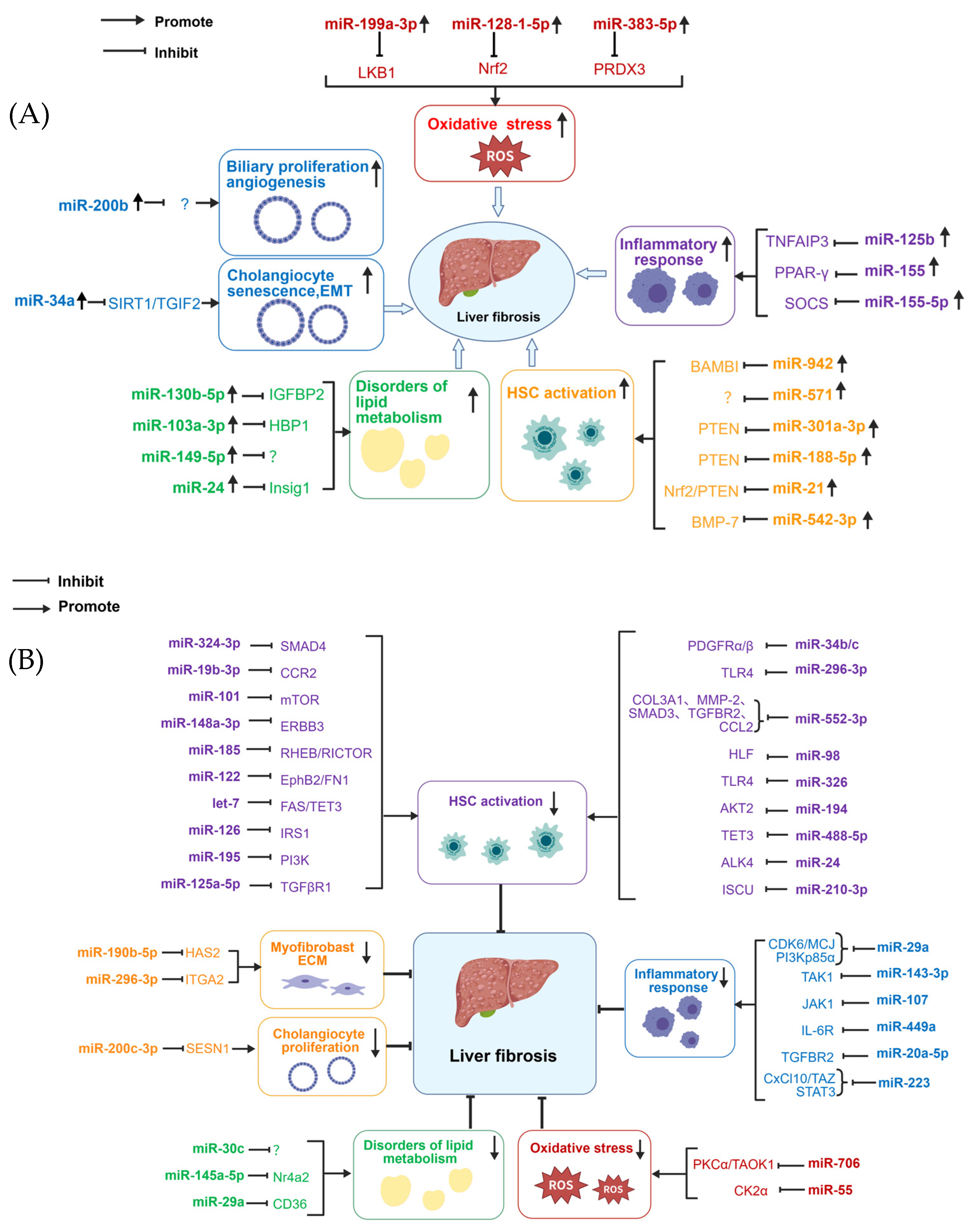
Intracellular miRNAs exert multifaceted roles by regulating gene expression during fibrosis. This section summarizes the diverse functions of these miRNAs involved in liver fibrosis progression (Figure 1, Table 1). The specific quantitative methods used for miRNA expression profiling in this table include real-time PCR, miRNA chips/microarrays, and high-throughput small RNA sequencing. Among these, quantitative real-time PCR consistently employs the comparative cycle threshold (Ct) method, using U6 as the internal standard, to quantify the levels of the indicated miRNAs in each sample (Table 1).
Table 1.
Functional Roles of Intracellular miRNAs in Liver Fibrosis.
3.1. Regulation of Oxidative Stress
Oxidative stress imbalance is a core upstream event driving the pathological progression of liver fibrosis. Excessive accumulation of ROS can induce oxidative damage to hepatocytes, trigger intrahepatic inflammatory cascades and HSC activation, and ultimately lead to excessive ECM deposition and the progression of liver fibrosis. Oxidative stress can interact with canonical pathways such as transforming growth factor-beta (TGF-β), nuclear factor kappa B (NF-κB), nuclear factor erythroid 2-related factor 2 (Nrf2) and various miRNA regulatory networks, contributing to hepatocyte injury, macrophage-mediated inflammation and HSC activation. Promoting ROS clearance, activating antioxidant pathways or correcting abnormal miRNA expression represents a core strategy for preventing or even reversing liver fibrosis.
Some miRNAs can further exacerbate the pathological progression of liver fibrosis by amplifying intrahepatic oxidative stress and disrupting endogenous antioxidant balance. MiR-199a-3p is a key profibrotic molecule mediating oxidative stress. Existing studies have confirmed that abnormally high expression of hepatocyte-derived miR-199a-3p can significantly exacerbate liver oxidative stress and fibrosis progression. Clinical studies have shown that farnesoid X receptor (FXR) expression in liver tissue of patients with liver fibrosis progressively decreases with disease progression, significantly weakening its transcriptional inhibitory effect on miR-199a-3p, ultimately leading to abnormal upregulation of miR-199a-3p in the liver. MiR-199a-3p can target liver kinase B1 (LKB1) and inhibit its expression. LKB1 is a key regulator maintaining mitochondrial function stability and exerts antioxidant effects in hepatocytes by activating the downstream adenosine monophosphate-activated protein kinase (AMPK) pathway. Therefore, miR-199a-3p may exacerbate oxidative stress and fibrosis by inhibiting the LKB1/AMPK axis [12].
The kelch-like ECH-associated protein 1 (Keap1)/Nrf2 antioxidative pathway represents a central defensive system against intrahepatic oxidative injury, and its homeostatic balance is precisely modulated by multiple miRNAs. Dysregulation of this regulatory network serves as a critical driver of oxidative stress-mediated liver fibrosis. Upon activation, Nrf2 binds to antioxidant response elements (ARE) sequences to upregulate a panel of antioxidant enzymes including heme oxygenase-1(HO-1), NAD(P)H:Quinone Oxidoreductase 1 (NQO1), glutathione S-transferases (GSTs) and thioredoxin reductase 1 (TrxR1), thereby eliminating excess ROS and mitigating liver damage. By contrast, Keap1 functions as a major negative regulator that mediates persistent ubiquitination and degradation of Nrf2. Accumulating evidence has revealed that miR-128-1-5p is markedly upregulated in carbon tetrachloride (CCl4)-induced liver fibrosis models, where it represses Nrf2 pathway activation and impairs endogenous antioxidant responses. Notably, reserpine treatment substantially downregulates miR-128-1-5p expression, restores Nrf2-dependent antioxidant capacity, and ameliorates fibrotic liver injury. These findings support the notion that miR-128-1-5p participates in the progression of liver fibrosis via modulating the Keap1/Nrf2 axis to enhance oxidative stress [13].
Schistosomiasis-induced hepatic fibrosis, a major complication of the infection, is primarily caused by the host’s immune response to schistosome eggs. In the livers of mice with schistosomiasis-induced liver fibrosis, the expression of miR-383-5p is upregulated. Functional experiments in LX-2 cells verified that miR-383-5p suppresses the expression of peroxiredoxin-3 (PRDX3), a mitochondrial peroxidase responsible for scavenging reactive oxygen species. This effect facilitates ROS accumulation and aggravates oxidative stress, thereby aggravating schistosomiasis-induced liver fibrosis [14].
Conversely, certain miRNAs exhibit protective effects against liver fibrosis through alleviating oxidative stress and reversing fibrotic pathological changes. In CCl4-induced fibrotic livers, the expression of miR-706 in hepatocytes was downregulated, leading to enhanced epithelial–mesenchymal transition (EMT) of hepatocytes induced by oxidative stress. Mechanistically, miR-706 can alleviate EMT in hepatocytes by reducing oxidative stress-induced expression of protein kinase Cα (PKCα) and TAO kinase 1 (TAOK1) in hepatocytes, thereby attenuating liver fibrosis; thus, miR-706 may serve as an antifibrotic target for the treatment of liver fibrosis [15]. Interestingly, plant-derived miRNAs also exert beneficial effects on redox balance and fibrotic improvement. Plant-origin miR55 may alleviate lipid accumulation, lipotoxicity, and oxidative stress in MAFLD rats by targeting casein kinase II subunit alpha (CK2α). Inhibition of CK2α eventually ameliorates liver fibrosis, which suggests plant-derived miR55 may act as a natural therapeutic candidate [16]. However, cross-kingdom miRNA transfer remains controversial; the species, sequence identity, dose, intact uptake, achievable physiological exposure, and independent replication of this study require verification. Activity after experimental administration does not demonstrate functional delivery through an ordinary diet.
In fact, oxidative injury is both a driver of fibrosis and an intrinsic feature of several experimental models like the CCl4 model. An intervention that reduces CCl4-associated injury may secondarily reduce collagen accumulation without directly modifying established scar. These findings therefore support candidate miRNAs’ involvement in redox-regulatory mechanisms, but their relevance to metabolically driven human fibrosis requires confirmation in complementary disease models and human cellular systems.
3.2. Modulation of Inflammatory Responses
Inflammation is a central pathological driver of the initiation and progression of liver fibrosis. Chronic liver injury can activate Kupffer cells and macrophages that secrete plenty of pro-inflammatory and profibrotic cytokines, which further promote HSC proliferation, activation, and extensive synthesis of ECM, ultimately resulting in abnormal collagen deposition and fibrogenesis. Inhibition of the inflammatory response could effectively limit the initiation and development of liver fibrosis. Emerging evidence indicates that miRNAs participate in fibrotic pathogenesis by fine-tuning hepatic inflammatory signaling. MiR-125b is a vital inflammation-related miRNA closely associated with NAFLD progression, liver fibrosis and hepatocellular carcinoma. Clinical and experimental data have confirmed that hepatic miR-125b expression is upregulated in the livers of NAFLD patients. Elevated miR-125b expression exacerbates intrahepatic inflammatory cascades by targeting and repressing tumor necrosis factor α inducible protein 3 (TNFAIP3), a key negative regulator of NF-κB signaling. The suppression of TNFAIP3 further amplifies NF-κB transcriptional activities, thereby markedly increasing the release of inflammatory cytokines, IL-1β, IL-6 and TNF-α, which ultimately drives HSC activation and ECM accumulation, promoting liver fibrosis progression [17].
As a pivotal pathogenic miRNA in hepatic fibrosis, miR-155-5p acts as a core inflammatory regulator that is markedly upregulated in multiple liver injury models, including NASH and cholestatic liver disease. Its upregulation sustains pro-inflammatory signaling cascades to facilitate fibrotic progression, whereas specific inhibition of miR-155-5p can significantly reverse hepatic inflammatory disorders and alleviate fibrotic lesions. Beyond direct modulation of inflammatory pathways, miR-155-5p participates in fibrogenesis by remodeling hepatic macrophage polarization, a key determinant of inflammatory microenvironment homeostasis. In CCl4-induced liver fibrosis and ALD animal models, miR-155-5p directly targets and inhibits peroxisome proliferator-activated receptor γ (PPAR-γ), an essential anti-inflammatory transcription factor that governs macrophage phenotypic transformation. Downregulation of PPAR-γ abolishes its inhibitory effect on the profibrotic M1 phenotype and induces robust secretion of pro-inflammatory mediators. This sustained M1-type inflammatory response further exacerbates hepatic steatohepatitis in ALD and amplifies HSC-mediated ECM deposition, ultimately aggravating liver fibrosis [18]. In addition, miR-155-5p enhances pro-inflammatory macrophage polarization and fibrotic progression through an alternative signaling mechanism. MiR-155-5p suppresses the expression of the suppressor of cytokine signaling (SOCS), a critical negative feedback regulator of inflammatory signaling. SOCS inhibition subsequently triggers persistent activation of the Janus kinase 1/signal transducer and activator of transcription 1 (JAK1/STAT1) signaling axis, which serves as a dominant driver of M1 macrophage phenotypic transformation. Collectively, the miR-155-5p/SOCS/JAK1/STAT1 cascade sustains intrahepatic pro-inflammatory microenvironment formation, promotes excessive inflammatory infiltration, and ultimately aggravates liver fibrosis [19].
However, certain miRNAs exert protective effects against hepatic fibrosis by mitigating intrahepatic inflammatory responses and blocking pathological cascade amplification. MiR-29a is a typical anti-inflammatory and antifibrotic miRNA that is significantly downregulated in fibrotic liver tissues from BDL mice. Mechanistically, miR-29a directly targets and represses phosphoinositide 3-kinase (PI3K)- p85 alpha (PI3Kp85α) to modulate hepatic inflammatory homeostasis. Restoration of miR-29a expression via miRNA mimics effectively attenuates hepatic inflammation and alleviates fibrotic lesions in BDL-induced liver injury models [20]. In addition, miR-29a exhibits prominent protective functions in NASH-related liver damage through multiple regulatory pathways. It ameliorates methylation-controlled J protein (MCJ) and simultaneously suppresses cyclin-dependent kinase 6 (CDK6)-driven inflammation activation and cellular pyroptosis. The combined improvement of mitochondrial function, inflammatory inhibition and pyroptosis suppression collectively stabilizes the hepatic microenvironment and restrains the progression of NASH and subsequent liver fibrosis [21].
Additionally, immune cell-derived miRNAs constitute an essential regulatory layer for limiting hepatic inflammation and MASH-related fibrosis. Neutrophil-originated miR-223 derived from bone marrow has been identified as a critical protective regulator against steatohepatitis and fibrogenesis. MiR-223 directly targets and suppresses STAT3, which further inhibits the expression of downstream pro-inflammatory proteases, including neutrophil elastase (NE) and proteinase 3 (PR3). The suppression of NE and PR3 alleviates excessive neutrophil-mediated inflammatory injury, relieves intrahepatic inflammatory infiltration and tissue damage, and ultimately hinders the pathological progression of inflammatory and MASH-associated liver fibrosis. This cell-specific miRNA regulatory mechanism further illustrates the multi-source and multi-pathway characteristics of miRNA-mediated inflammatory homeostasis in fibrotic liver injury [22].
Another miRNA, miR-143-3p, also serves as a protective miRNA against autoimmune AIH-related liver damage and fibrogenesis. Hepatic miR-143-3p expression is markedly decreased in AIH mouse models, whereas forced overexpression of miR-143-3p efficiently alleviates hepatic inflammatory responses and fibrotic progression. Mechanistically, miR-143-3p directly targets and inhibits TGF-β-activated kinase 1 (TAK1), a central upstream regulator of pro-inflammatory and profibrotic signaling cascades. Consistent with its protective function, miR-143-3p supplementation significantly reduces the expression of fibrotic markers, including collagen, TGF-β and α-smooth muscle actin (α-SMA) in AML12 hepatocytes stimulated by LPS. Collectively, these results indicate that miR-143-3p restrains liver inflammation and ECM accumulation via suppressing the TAK1-mediated profibrotic cascade, providing another potential miRNA-based candidate for antifibrotic therapy [23].
Distinct miRNA expression signatures are also observed in virus-triggered liver fibrosis. In patients with HCV-induced liver fibrosis, miR-107 and miR-449a are down-regulated, and their target genes JAK1 and interleukin-6 receptor (IL-6R) are upregulated, thereby contributing to upregulation of C-C motif ligand 2 (CCL2) and liver fibrosis [24].
MiR-20a-5p is another essential protective miRNA implicated in inflammatory and fibrotic liver pathogenesis. Consistent downregulation of miR-20a-5p has been validated in clinical fibrotic liver tissues, CCl4-treated immortalized mouse hepatocytes and animal fibrotic livers. Functionally, miR-20a-5p directly targets and inhibits TGF-β receptor 2 (TGFBR2) and modulates TGF-β signaling. Overexpression of miR-20a-5p can suppress the release of pro-inflammatory factors and inhibit macrophage polarization toward M1 phenotype. This dual anti-inflammatory and antifibrotic function effectively ameliorates liver injury and halts fibrotic progression [25].
The inflammation-related miRNA expression has been identified in human fibrotic liver tissue from patients with NASH or HCV infection (Table 1), which enhances the value of miRNAs in the regulation of hepatic inflammation and fibrosis. Moreover, the M1/M2 terminology used in several experimental studies provides a simplified description rather than a complete representation of macrophage states in chronic disease, which needs further exploration.
3.3. Regulation of HSC Activation
HSCs are the core effector cells in the development of liver fibrosis. When the liver is injured due to various causes, numerous inflammatory factors are released and quiescent HSCs are activated, leading to increased expression of fibrosis-related genes and abnormal deposition of ECM. Persistently activated HSCs maintain their activated state through autocrine positive feedback, promoting the progression of liver fibrosis and gradually causing irreversible pathological changes. Therefore, inhibition of HSC activation is a key strategy for halting and reversing liver fibrosis.
MiR-21 is a crucial profibrotic miRNA that predominantly facilitates liver fibrosis progression by driving persistent HSC activation and amplifying intrahepatic inflammatory and oxidative injury. Increasing evidence has verified that miR-21 is significantly upregulated in the liver tissues of mice with liver fibrosis induced by cadmium chloride (CdCl2) and CCl4. As a multifunctional regulatory molecule, upregulated miR-21 sustains the continuous activation of core profibrotic signaling cascades including TGF-β/small mother against decapentaplegic family (Smad) and PI3K/protein kinase B (AKT) which are essential for maintaining the activated phenotype of HSCs. Mechanistically, miR-21 targets and represses PTEN to hyperactivate the PI3K/AKT pathway, while simultaneously inhibiting the Nrf2-dependent antioxidant system. This dual regulatory mechanism synergistically promotes robust HSC activation, aggravates hepatic inflammatory infiltration, and accelerates excessive ECM deposition, ultimately forming a self-perpetuating pathological loop to exacerbate liver fibrosis. Therefore, inhibition of miR-21 can effectively block HSC activation and interrupt the cascade of inflammation and oxidative injury, providing a promising therapeutic strategy for restraining liver fibrosis progression [26,27].
In addition, miR-301a-3p acts as a novel profibrotic mediator closely linked to HSC activation and clinical fibrotic severity. Elevated miR-301a-3p expression has been detected in serum samples of liver fibrosis patients and in activated HSCs and CCl4-induced mouse fibrotic livers. MiR-301a-3p directly targets and suppresses PTEN, thereby indirectly upregulating the expression of platelet-derived growth factor receptor-β (PDGFR-β). As a key fibrotic gene, PDGFR-β potently triggers HSC activation and accelerates liver fibrosis progression. At the same time, the expression of miR-301a-3p is correlated with the pathological degree of liver fibrosis, suggesting its value as a promising diagnostic and prognostic biomarker for liver fibrosis [28]. Similarly, miR-188-5p is another PTEN-targeted profibrotic miRNA that drives HSC activation and liver fibrogenesis. Significant upregulation of miR-188-5p has been observed in fibrotic liver biopsy of human subjects and in TGF-β1-stimulated HSCs. MiR-188-5p can directly suppress PTEN, thereby promoting the activation of the PI3K/AKT pathway. The hyperactivated PI3K/AKT pathway further promotes excessive proliferation and persistent activation of HSCs, facilitates massive ECM accumulation, and contributes to fibrosis progression [29].
MiR-542-3p also functions as a novel profibrotic regulator involved in CCl4-induced liver fibrosis. MiR-542-3p is markedly upregulated in fibrotic liver tissues from CCl4-treated mice. Mechanistically, elevated miR-542-3p directly downregulates bone morphogenetic protein 7 (BMP-7), an endogenous antagonist of the TGF-β/Smad pathway. Downregulation of BMP-7 removes its inhibitory effect on the TGF-β/Smad cascade, leading to persistent pathway activation. This molecular event further induces robust HSC activation, promotes abnormal ECM deposition, and ultimately accelerates the progression of liver fibrosis [30].
Clinical evidence reveals that miR-942 is upregulated in HBV-induced fibrosis and is positively correlated with fibrosis staging. Elevated miR-942 promotes the activation of the TGF-β/Smad pathway by downregulating the BMP membrane-binding inhibitors (BAMBI), a key membrane-bound inhibitory factor of TGF-β signaling. The repression of BAMBI eliminates endogenous feedback restriction on TGF-β/Smad signaling, thereby forming a sustained positive regulatory loop. This pathological loop continuously drives the proliferation and persistent activation of HSCs, enhances excessive ECM synthesis and deposition, and ultimately aggravates the progression of HBV-associated liver fibrosis [31]. MiR-571 is a newly identified profibrotic miRNA that participates in hepatic fibrogenesis by modulating Notch signaling. Clinical studies have confirmed that miR-571 expression is significantly elevated in liver tissues of patients with liver fibrosis. Functionally, excessive miR-571 expression can trigger the activation of the Notch3 signaling pathway, a crucial profibrotic cascade driving HSC phenotypic transformation. Aberrant Notch3 activation robustly promotes HSC proliferation and stimulates the secretion of fibrotic factors, thereby facilitating the progression of liver fibrosis [32].
Beyond these well-characterized profibrotic miRNAs, several miRNAs exhibit prominent antifibrotic effects by repressing aberrant HSC activation, among which the let-7 family plays an important role in combating liver fibrosis. During liver injury, the expression of let-7 is significantly suppressed, and the loss of let-7 family members leads to excessive activation of the TGF-β/Smad pathway, subsequent hyperactivation of HSCs, and massive collagen deposition in the liver. Restoring let-7 expression can block HSC activation and inhibit collagen synthesis, thereby effectively slowing down or even reversing liver fibrosis. Mechanistically, let-7 ameliorates liver fibrosis through dual protective mechanisms in hepatocytes. It directly targets and downregulates methylcytosine dioxygenase 3 (TET3) to reduce hepatic TGF-β expression and simultaneously inhibits FAS-mediated apoptotic signaling to attenuate hepatocyte apoptosis in BDL and CCl4 mouse models. Owing to its stable dysregulated expression and robust multi-target antifibrotic function, the let-7 family holds great promise as an ideal diagnostic biomarker and a high-potential therapeutic candidate for antifibrotic intervention [33]. MiR-552-3p and miR-98 serve as two representative miRNAs that inhibit HSC activation. Both are significantly downregulated in activated HSCs and fibrotic liver tissues, suggesting that their loss may contribute to fibrotic progression. MiR-552-3p exerts its antifibrotic effects through dual pathways, suppressing the TGF-β1/Smad3 signaling axis and reducing LPS-induced inflammatory responses to synergistically block HSC activation, whereas miR-98 acts through inhibition of hepatic leukemia factor (HLF) and modulation of the hypoxia-inducible factor-1 (HIF-1α)/TGF-β/Smad2/3 signaling pathway. These findings suggest that restoring the function of endogenous protective miRNAs may represent a novel antifibrotic intervention paradigm distinct from direct HSC targeting [34,35].
In addition, a growing number of miRNAs have been identified as negative regulators of HSC activation and liver fibrosis, most of which are consistently downregulated in CCl4-injured liver tissues and activated HSCs. MiR-326 suppresses HSC activation and liver fibrosis by targeting TLR4 and further blocking the TLR4/MyD88/NF-κB inflammatory signaling cascade [36]. MiR-194 can inhibit HSC proliferation via negative modulation of the AKT beta (AKT2) pathway [37]. MiR-488-5p directly targets TET3 to suppress the TGF-β/Smad2/3 pathway, thereby decreasing fibrosis marker expression and ECM deposition [38]. In the regression phase of liver fibrosis, the restored expression of miR-324-3p contributes to the alleviation of hepatic injury; overexpression of miR-324-3p directly targets Smad4 and downregulates fibrotic markers includingα-SMA and vimentin in HSCs [39]. Other findings showed that miR-19b-3p could alleviate liver fibrosis by downregulating C-C motif chemokine receptor 2 (CCR2) and suppressing HSC activation [40].
MiR-148a-3p, miR-101 and miR-185 exert comprehensive antifibrotic effects by collectively suppressing the PI3K/AKT/mTOR pathway through targeting receptor tyrosine-protein kinase erbB-3 (ERBB3) and mechanistic target of rapamycin (mTOR), while miR-148a-3p and miR-185 additionally target Ras homolog enriched in brain (RHEB) and rapamycin-insensitive companion of mTOR (RICTOR), respectively, to constrain profibrotic signaling [41,42,43]. Hepatic expression of miR-122 is also downregulated during liver fibrosis and its restoration inhibits HSC activation by directly repressing Ephrin type-B receptor 2 (EphB2) and fibronectin 1 (FN1) [44,45]. Studies have shown that several other miRNAs such as miR-24, miR-34b/c, miR-125a-5p, miR-126-3p, miR-195-5p, miR-210-3p and miR-296-3p have been reported to suppress HSC activation and alleviate liver fibrosis [46,47,48,49,50,51].
In fact, the evidence for individual HSC-regulatory miRNAs is not uniformly consistent. For miR-21, some intervention studies reported reduced experimental fibrosis, whereas other researchers found that genetic deletion or antisense inhibition did not suppress HSC activation or fibrosis in toxic and biliary injury models [64]. This disagreement remains unresolved and precludes describing miR-21 inhibition as an established antifibrotic strategy. Furthermore, the regulation of miRNAs in different statuses of HSCs, except activation, such as apoptosis of activated HSCs and senescent HSCs, should be included in future studies, which may be valuable for the treatment of liver fibrosis.
3.4. Influence on Lipid Metabolism
Beyond direct inflammatory and HSC regulatory mechanisms, aberrant lipid metabolism is a hallmark of metabolic liver diseases and acts as a powerful driver of fibrotic progression. Multiple dysregulated miRNAs exacerbate metabolic liver injury and subsequent fibrogenesis by disrupting hepatic lipid homeostasis, thereby accelerating lipid accumulation, steatosis, inflammatory infiltration and fibrotic lesion formation. In the context of MASLD/NAFLD, a set of miRNAs promotes the fibrotic microenvironment indirectly by amplifying hepatic lipid metabolic dysregulation. Due to differences in their functional nodes, these metabolism-related profibrotic miRNAs can be categorized into two classes. The first class directly targets lipid metabolic regulators. MiR-130b-5p sustains AKT pathway activation by suppressing insulin-like growth factor-binding protein 2 (IGFBP2), driving hepatic lipid accumulation [52]; miR-24 targets insulin-induced gene 1 (Insig1), relieving its repression of lipogenic genes and accelerating lipid synthesis. These two miRNAs increase hepatic lipid burden via distinct regulatory mechanisms [55]. The second class of miRNAs indirectly promotes lipid accumulation by disrupting broader metabolic homeostasis. MiR-103a-3p disturbs metabolic balance by suppressing HMG-box transcription factor 1 (HBP1) expression; miR-149-5p not only promotes steatosis but also simultaneously drives inflammation and fibrotic progression, suggesting that its functional scope extends beyond pure metabolic regulation [53,54,55].
Conversely, some miRNAs alleviate NASH/MASLD-associated liver fibrosis by rectifying hepatic lipid metabolic disorders, disrupting the pathological loop of steatosis, metabolic dysfunction, and inflammation. MiR-145a-5p serves as a key metabolic protective miRNA, whose expression is negatively correlated with the severity of steatosis, inflammation, and fibrosis in NASH patients. Mechanistically, miR-145a-5p inhibits the nuclear receptor subfamily 4 group A member 2 (Nr4a2) signaling axis and further downregulates downstream pathogenic NADPH oxidase 4 (Nox4) and pyruvate dehydrogenase kinase 4 (Pdk4), thereby ameliorating hepatic steatosis and suppressing fibrosis. Restoring the expression of miR-145a-5p can effectively reverse NASH progression [56].
Similarly, miR-29a exerts prominent metabolic antifibrotic effects by directly targeting the 3′UTR of lipid transporter CD36 to block excessive hepatic lipid accumulation. This suppression further inhibits hepatocyte EMT and intrahepatic inflammatory responses, ultimately restraining steatohepatitis and progression in HFD-induced models [57]. In addition, miR-30c is indispensable for maintaining hepatic metabolic homeostasis. Loss of miR-30c exacerbates lipid disorder, inflammation and liver fibrosis in TAA-treated mice, suggesting its protective effects against liver fibrosis progression [58].
For metabolism-associated miRNAs, improvement in steatosis must be distinguished from an independent antifibrotic effect. In metabolism dysfunction-related liver fibrosis like MASH, reduced caloric intake, altered body weight, or improved insulin sensitivity can modify lipid burden and downstream injury, which are confounding factors that should be considered when exploring the role of miRNAs in fibrosis development. Human expression correlations strengthen disease relevance but do not establish that miRNA manipulation will reverse fibrosis independently of its metabolic effects.
3.5. Other Regulatory Functions
In addition to above regulatory mechanisms, miRNAs exert unique context-specific profibrotic roles in cholestatic and biliary liver fibrosis. MiR-34a is an important driving factor of liver fibrosis. During cholestatic liver injury, miR-34a is significantly upregulated during obstructive bile duct injury and in the circulation of PBC patients, acting as a core driver of biliary fibrogenesis. It promotes the TGF-β/Smad pathway and p53-mediated inflammation to trigger HSC activation and extensive ECM deposition while targeting sirtuin 1(SIRT1) to induce cholangiocyte senescence. In human intrahepatic biliary epithelial cells, miR-34a suppresses the TGF-β/Smad antagonist TGF-β-induced factor homeobox 2 (TGIF2), thereby facilitating biliary epithelial EMT and amplifying fibrotic responses [59,60]. MiR-200b is upregulated in cholangiocytes and HSCs in a PSC animal model, the multidrug resistance gene 2-knockout (Mdr2−/−) mouse. Inhibition of miR-200b alleviates biliary fibrosis by repressing pathological biliary proliferation and angiogenesis [61]. Collectively, both miR-34a and miR-200b serve as biliary-specific profibrotic regulators, revealing distinct miRNA-dependent mechanisms underlying cholestatic liver injury and representing promising biomarkers and therapeutic targets for biliary fibrotic diseases.
Moreover, multiple newly identified miRNAs exert prominent antifibrotic functions by targeting ECM synthesis and biliary pathological proliferation during liver fibrogenesis. MiR-190b-5p and miR-296-3p expression are downregulated during liver fibrosis. These two miRNAs attenuate liver fibrosis via distinct target regulation: miR-190b-5p directly targets hyaluronate synthase 2 (HAS2), while miR-296-3p inhibits integrin α6 (ITGA6) expression. Both molecules effectively restrain excessive ECM production in activated HSCs, thereby exerting robust antifibrotic effects [62]. In addition, miR-200c-3p serves as a protective regulator specifically against cholestatic liver fibrosis by targeting sestrin 1(SESN1) and inhibiting activation of the IL-6/AKT signaling pathway to suppress cholangiocyte proliferation and ductular reaction, ultimately alleviating cholestatic liver injury and fibrotic progression [63].
For the roles of miRNAs in the cholestatic and biliary settings, changes in cholangiocyte proliferation, senescence, or EMT-associated markers can identify injury-related phenotypes but do not alone establish conversion into matrix-producing myofibroblasts in vivo. Distinguishing indirect effects on HSCs from direct matrix production therefore remains important. Validation across etiologies and in human biliary systems is needed before assigning broadly applicable therapeutic or diagnostic roles to these miRNAs.
Figure 1.
Regulatory roles of intracellular microRNAs in liver fibrosis. Intracellular miRNAs modulate liver fibrosis by regulating oxidative stress, inflammation, hepatic stellate cell (HSC) activation, and lipid metabolism and related pathways. (A) Profibrotic miRNAs promote oxidative stress, hepatic inflammation, HSC activation and lipid metabolism dysfunction. (B) Antifibrotic miRNAs counter these processes and attenuate fibrosis. Abbreviations used in the figure are defined in the Abbreviation section. The figures are created by the authors with (BioGDP; https://BioGDP.com) [65].
4. Exosomal miRNAs in Liver Fibrosis
Exosomes are nano-sized EVs that originate from multivesicular bodies and are secreted by nearly all eukaryotic cells. As carriers of proteins, lipids, DNA, and RNA, including miRNAs, they mediate intercellular communication under both physiological and pathological conditions [66,67]. According to the 2018 MISEV guideline, exosomes can be isolated by several methods, such as differential ultracentrifugation (dUC) [68,69,70,71,72,73,74,75,76,77,78,79,80,81,82,83,84,85,86,87], size-exclusion chromatography (SEC), tangential flow filtration (TFF), polyethylene glycol (PEG)-based precipitation, immunoaffinity capture and Microfluidic isolation. PEG precipitation) is already available in commercial kits [88,89,90,91,92,93]. The morphology of isolated exosomes was identified by transmission electron microscopy. The surface markers of exosomes were examined using Western blot analysis, including CD63, CD81, CD9 or TSG101.
Exosomal miRNAs are particularly important regulators of gene expression in recipient cells. Through horizontal transfer, they modulate transcriptional networks and influence various signaling pathways [94]. Depending on their source and cargo, exosomes may promote or suppress liver fibrosis by regulating immune responses, metabolic balance, and fibrogenic signaling (Table 2).
Table 2.
Functions of exosomal miRNAs from different cell sources in liver fibrosis.
4.1. Hepatocyte-Derived Exosomal miRNAs
Stressed or injured hepatocytes release exosomes enriched with specific miRNAs that significantly affect the liver microenvironment (Figure 2). In alcoholic liver fibrosis, hepatocyte-derived exosomes rich in miR-192-5p promote M1 macrophage polarization by regulating the Rictor/Akt/forkhead box protein O1(FoxO1) signaling pathway, thereby enhancing the pro-inflammatory response [91]. In NAFLD-related fibrosis, hepatocyte-derived exosomes loaded with miR-107 (induced by palmitic acid) activated HSCs through dual mechanisms. MiR-107 targets Dickkopf-1 (DKK1) (an antagonist in the Wnt pathway) to activate Wnt signaling. Meanwhile, these miR-107-rich exosomes induce Th9 differentiation and IL-9 expression in CD4+ T cells via inhibiting forkhead box P1(Foxp1) expression. IL-9 enhances HSC activation by activating the rapidly accelerated fibrosarcoma (Raf)/mitogen-activated protein kinase (MEK)/extracellular signal-regulated kinase (ERK) signaling pathway [85]. Hepatocyte-derived exosomal miR-107 has a profibrotic effect by targeting DKK1 and Foxp1 in HSCs [85], which is different from the effect of intracellular miR-107 in hepatocytes by targeting JAK1 and inhibiting IL-6/STAT3-CCL2 inflammatory signaling, thereby playing a protective role [24]. These results indicate that miR-107 exhibits different effects in liver fibrosis that depend on different target genes and effector cells. Hepatocyte-derived exosomal miR-1297 may downregulate PTEN and activate the PI3K/AKT pathway in HSCs, promoting their proliferation and ECM production [76]. In MAFLD, miR-27a carried by hepatocyte-derived exosomes targets PTEN-induced putative kinase 1 (PINK1), disrupting mitochondrial function in HSCs and accelerating fibrogenesis [77].
Figure 2.
Intercellular communication mediated by exosomal miRNAs in liver fibrosis. Exosomal miRNAs from hepatocytes, macrophages, hepatic stellate cells, mesenchymal stem cells, NK cells, adipocytes, and other cell sources mediate intercellular crosstalk that can either promote or suppress fibrogenesis. Abbreviations used in the figure are defined in the Abbreviation section. The figure was created by the authors with the assistance of (BioGDP; https://BioGDP.com) [65].
Conversely, exosomal miR-146a-5p from Salidroside-treated hepatocytes exhibits antifibrotic effects. It targets eukaryotic translation initiation factor 5A2 (EIF5A2), suppressing the activation and EMT of HSCs [95]. Exosomes derived from HepG2 cells carrying miR-423-5p can inhibit CCl4-induced liver fibrosis by targeting the family with sequence-similarity-3 (FAM3) and Monoacylglycerol lipase (Mgll), thereby suppressing HSC activation. However, exposure to ethanol or lauric acid may reduce miR-423-5p levels in HepG2-EVs in in vitro experiments [80].
It is worth noting that hepatocyte-derived exosomal miRNAs have a dual function, which may be dependent on different conditions, like disease stimuli and experimental environment. Moreover, HepG2-derived vesicles arise from a transformed cell model and cannot automatically represent normal hepatocyte secretion. Therefore, the function of exosomal miRNAs from normal hepatocytes needs to be explored and confirmed.
4.2. HSC-Derived Exosomal miRNAs
Activated HSCs secrete exosomes enriched in miRNAs that can propagate fibrogenic signaling (Figure 2). In BDL animal models, miR-199a-5p from HSC-derived exosomes downregulates SIRT1 expression, increases the expression of fibrotic markers in HSCs, and promotes hepatocyte EMT, thereby exacerbating liver fibrosis [96]. MiR-99a-5p from HSC-derived exosomes targets bone morphogenetic protein receptor 2 (BMPR2) in hepatocytes, inhibiting hepatocyte proliferation and promoting apoptosis, which facilitates fibrogenesis [74]. MiR-19b and miR-200 are highly expressed in activated HSC-derived exosomes and enhance the secretion of IL-6 and TNF-α from macrophages, reinforcing inflammation and liver fibrosis [88]. MiR-19b-3p similarly exhibits this dependency: intracellularly expressed miR-19b-3p can directly target CCR2, limiting macrophage chemotactic infiltration and playing a protective role in inhibiting liver fibrosis; whereas miR-19b-3p released into exosomes by activated HSCs acts as a signal for intercellular communication, transferring into macrophages and inducing pro-inflammatory activation, thereby amplifying liver inflammatory responses. This difference arises from variations in the donor cells of the miRNA, the delivery method, the target cells, and the downstream target gene networks, ultimately leading to different regulatory effects.
Not all HSC-derived exosomal cargo is profibrotic, however. Exosomal miR-122-5p derived from HSCs can increase proliferation, reduce apoptosis, and inhibit EMT and fibrogenic characteristics of intrahepatic biliary epithelial cells (IBECs) with LPS stimulation by targeting tumor necrosis factor receptor superfamily member 19 (TNFRSF19), which inhibits apoptosis signal-regulating kinase 1 (ASK1) and the downstream p38 MAPK signaling pathway [93].
Notably, HSC-derived exosomal miRNAs exert context-dependent profibrotic or antifibrotic functions, with miR-19b-3p serving as a representative example of divergent bioactivity determined by its subcellular localization and delivery route. The available evidence is predominantly obtained from preclinical rodent fibrosis models; the variability of HSC status (e.g., quiescent, activated, or senescent), exosome isolation workflows, and readout selection across studies introduces substantial heterogeneity.
4.3. Macrophage-Derived Exosomal miRNAs
Macrophages, particularly Kupffer cells and monocyte-derived macrophages, can release exosomes containing both profibrotic and antifibrotic miRNAs (Figure 2). In MAFLD, miR-155 from macrophage-derived exosomes worsens hepatocyte pyroptosis via downregulation of forkhead box protein O3a (FoxO3a), accelerating hepatic fibrosis [71]. LPS-stimulated THP-1 macrophage-derived exosomes are enriched in miR-103-3p, which targets Krüppel-like factor 4 (KLF4) to activate HSCs [97]. MiR-500, also derived from LPS-treated macrophages, can promote the proliferation and activation of HSCs via targeting mitochondrial fusion protein 2 (MFN2). Treatment with exosomes overexpressing miR-500 accelerates CCl4-induced mouse liver fibrosis, while upregulation of MFN2 can partially inhibit fibrosis progression [69]. Similarly, primary mouse liver macrophage-derived exosome miR-342-3p promotes HSC activation by targeting hippocalcin-like protein 1 (HPCAL1) [90]. MiR-125b-5p was enriched in M1 macrophage-derived exosomes, while phillyrin (PHI) could inhibit M1 macrophage polarization and miR-125b-5p expression; consequently, upregulation of its target gene, steroidogenic acute regulatory (StAR)-related lipid transfer domain containing 13 (STARD13), reduces HSC activation and fibrogenic factor expression [78].
Additionally, some miRNAs carried by macrophage-derived exosomes prevent liver fibrosis progression. Exosomal miR-690 from M2 bone marrow-derived macrophages exerts potent insulin-sensitizing effects [100]. Nanoparticles delivering an miR-690 mimic can significantly alleviate liver inflammation and fibrosis in two NASH models: cultured liver spheroids in a NASH-inducing medium and Western diet (WD)-fed mice. Exosomal miR-690 from Kupffer cells targets nicotinamide adenine dinucleotide kinase (NADK) and suppresses HSC activation, decreasing ECM production [98]. M2 macrophage-derived miR-411-5p targets calmodulin-regulated spectrin-associated protein 1 (CAMSAP1) and inhibits HSC activation, thereby alleviating liver fibrosis in NASH models [83]. MiR-1224-5p carried by M2 macrophage-derived apoptotic vesicles may ameliorate CCl4-induced liver fibrosis by targeting specificity protein 1 (SP1) expression, thereby blocking the activation of the TGF-β/Smad signaling pathway and HSC activation [87].
Notably, the dual roles of macrophage-derived exosomal miRNAs in liver fibrosis are tightly coupled to macrophage polarization status. Exosomal miRNAs delivered by pro-inflammatory phenotype M1 macrophages may exacerbate fibrosis progression, while those derived from M2 macrophages may exhibit antifibrotic effects. Most supporting evidence originates from preclinical NASH and toxin-induced liver fibrosis models. Human validation data remain limited, and it remains unclear whether this M1/M2-dependent functional dichotomy can be reliably recapitulated in patients with chronic liver disease.
4.4. MSC-Derived Exosomal miRNAs
Accumulating experimental evidence indicates that MSC-derived exosomes protect against liver fibrosis. They regulate multiple molecules and pathways to inhibit HSC activation, alleviate inflammation, reduce collagen deposition, and promote hepatocyte regeneration and metabolic recovery, indicating the potential of MSC-derived exosomes as a promising option for the treatment of liver fibrosis. Notably, this body of evidence suggests MSC-derived exosomes may be a promising therapy for liver fibrosis (Figure 2).
MSC-derived exosomal miR-148a, miR-124 and miR-27b-3p are involved in the regulation of liver fibrosis. MiR-148a downregulates Kruppel-like factor 6 (KLF6) and inhibits the STAT3 pathway to increase hepatic anti-inflammatory macrophages, thus ameliorating inflammation and fibrosis [82]. MiR-124 within MSC-derived exosomes also alleviates liver fibrosis by inhibiting STAT3 expression and CCl4-induced liver fibrosis [99]. MSC-derived exosomal miR-27b-3p suppresses Yes-associated protein (YAP) and Lysyl oxidase-like 2 (LOXL2) in HSCs, thereby diminishing the expression of fibrotic genes such as α-SMA and collagen type I alpha 2 (Col1A2) and alleviating CCl4-induced liver fibrosis [89].
The levels of miR-618 in patients with liver fibrosis are remarkably lower than those in healthy controls. Bone marrow MSC-derived exosomal miR-618 targets Smad4 in HSCs to attenuate CCl4-induced liver fibrosis [81].
Exosomes derived from adipose-derived MSCs enriched with miR-181-5p alleviate hepatic fibrosis by suppressing TGF-β1-induced HSC activation and inducing autophagy in HSCs through inhibiting the STAT3 and Bcl-2-dependent pathways, as demonstrated in both in vitro experiments and in vivo CCl4-induced mouse liver fibrosis models [92]. Human tonsil-derived MSCs (T-MSCs) are a new source of MSCs. MiR-486-5p enriched in T-MSCs, inhibits hedgehog signaling by inhibiting the hedgehog receptor, smoothened (Smo), thereby deactivating HSCs and effectively exerting antifibrotic effects [72]. In CCl4 and thioacetamide (TAA)-treated mice, miR-499a-5p, delivered by human umbilical cord-derived MSC exosomes, promotes ferroptosis in HSCs and alleviates liver fibrosis by targeting ETS proto-oncogene 1 transcription factor (ETS1) to downregulate glutathione peroxidase 4 (GPX4) in HSCs [86]. Yiguanjian (YGJ), a Chinese herbal formula, increases miR-7045-5p levels in bone marrow MSC-derived EVs, which mitigates CCl4-induced liver fibrosis through inhibiting AKT and activating the AMPK/transcription factor EB (TFEB) pathway, thereby enhancing lysosomal biogenesis and mitophagy in activated HSCs [75].
MSC-derived apoptotic vesicles can deliver miR-6869-5p simultaneously to HSCs and macrophages, exerting antifibrotic effects through two coordinated actions: targeting fibroblast growth factor 1 (FGF1) to inhibit the PI3K/AKT/mTOR pathway in HSCs, and suppressing the high-mobility group box 1 (HMGB1)–NF-κB axis in macrophages to promote an anti-inflammatory phenotype [73].
Notably, the favorable MSC-derived vesicle findings summarized here remain preclinical and may be influenced by selective publication of positive results. Differences in MSC source, passage, conditioning, vesicle preparation, dosing metrics, and treatment timing also preclude a simple comparison of potency across studies. Clinical evidence remains absent, and unresolved safety concerns such as off-target effects and immunogenicity limit immediate clinical translation.
4.5. Exosomal miRNAs from Other Cells
Other liver-resident or peripheral cells also regulate liver fibrosis progression through exosome-mediated miRNA delivery. Adipocyte-derived exosomal miR-122 promotes fibrogenesis by targeting SIRT1 in hepatocytes, promoting hepatocyte apoptosis and NAFLD progression [68]. The role of miR-122 in liver disease is cell origin-dependent. Endogenous miR-122 in hepatocytes and HSCs and miR-122-5p carried by exosomes secreted by HSCs are down-regulated in fibrosis and PBC, and play an antifibrotic protective role by targeting pro-fibrotic genes in HSCs or bile duct epithelium. In obesity, exosomes carrying miR-122 released from adipocytes are increased, which are transported to hepatocytes to inhibit the SIRT1/AMPK pathway, promote steatosis, and promote the progression of NAFLD. These results indicate that miR-122 does not have a single target that promotes illness or resists a specific disease phenotype, and its biological effects are determined by the cells of origin. NK cell-derived exosomes enriched in miR-223 suppress TGF-β-induced HSC activation by inhibiting HSC autophagy via targeting an autophagy-related gene 7 (ATG7) (Figure 2) [84]. MiR-155 and miR-34a carried by exosomes derived from adipose tissue macrophages in MASH mice promote HSC activation and liver fibrosis in obese mice by targeting peroxisome proliferator-activated receptor gamma (PPARγ) in HSCs [79]. BDL Mouse large cholangiocyte-derived exosomal miR-381-3p can contribute to HSC activation and cholestatic liver fibrosis via targeting KLF6 [70]. miR-223 is transcribed predominantly in neutrophils and packaged into apolipoprotein E (APOE)-bearing EVs. These EVs are internalized by hepatocytes via low-density lipoprotein receptors (LDLR), and this uptake is augmented by free fatty acids. Upon delivery, EV-derived miR-223 suppresses hepatocyte (C-X-Cmotif) chemokine 10 (CXCL10) and transcriptional coactivators with PDZ-binding motifs (Taz) to restrain NASH progression [101].
Overall, these examples further reinforce the origin-dependent functional duality of exosomal miRNAs, with miR-122 serving as a prominent illustration. Current evidence is scattered across diverse cell types and liver injury models, and few head-to-head comparisons exist. Heterogeneity in cell isolation, EV preparation and fibrosis readouts hinders cross-study integration, and human data remain limited.
5. Prospects of Exosomes and miRNAs in Liver Fibrosis
5.1. MiRNAs as Diagnostic and Therapeutic Targets in Liver Fibrosis
As detailed above, microRNAs have emerged as vital regulatory molecules in the pathogenesis of liver fibrosis. Most miRNAs derived from hepatic parenchymal cells may serve as therapeutic targets for liver fibrosis treatment, with several demonstrating promising antifibrotic effects. MiRNAs carried by MSC-derived exosomes mainly show strong antifibrotic potential.
MiRNAs are not only promising in the treatment of liver fibrosis, but also potential diagnostic biomarkers such as miR-122, miR-155, miR-301a and miR-21. MiR-122 may be a sensitive indicator of hepatocyte injury in the context of liver fibrosis, but it is not a specific diagnostic marker for fibrosis. Serum miR-122 levels are increased after hepatocyte injury in a range of liver diseases. Therefore, its diagnostic value for fibrosis lies in reflecting the degree of concomitant hepatocyte injury rather than the fibrosis process itself [102]. It has been reported that the combination of miR-H7, miR-1225, miR-1238, miR-3162 and miR-4721 exhibits high diagnostic accuracy for different stages of liver fibrosis, although the reported AUC, sensitivity and specificity (for example, the sensitivity of miR-5 is 90.9% and the specificity is 100%; the sensitivity of miR-2 is 84.8% and the specificity is 100%) vary across independent cohorts [103]. The plasma levels of miR-146b and miR-214 are higher in patients with NAFLD than in healthy controls, while the levels of miR-194 are much lower, suggesting these miRNAs may serve as diagnostic biomarkers of liver fibrosis [104]. Interestingly, the levels of exosomal miR-155 in serum gradually increase along with liver fibrosis progression, suggesting its value as a non-invasive diagnostic biomarker for fibrosis [105]. Of note, the diagnostic performance of these miRNA combinations still varies between studies and is rarely compared directly with guideline-approved noninvasive fibrosis tests such as FIB-4, ELF score, and transient elastography [106]. Compared with these established assays with well-validated cutoffs and prognostic utility, most circulating miRNA signatures lack consistent ROC-derived performance metrics, uniform detection protocols and prospective external validation, which currently limits their translation into routine clinical practice [107,108].
Collectively, both intracellular and exosome-mediated miRNAs offer a dual framework for the diagnosis and therapy of liver fibrosis. Nevertheless, their suboptimal consistency and limited direct comparison with standard non-invasive tests mean that their clinical utility remains exploratory rather than established. Further large-cohort validation and comparative studies against FIB-4, ELF and transient elastography are required before these miRNA biomarkers can be implemented clinically.
It is critical to distinguish between circulating exosomal miRNAs as diagnostic biomarkers and engineered exosomes for therapeutic delivery. Engineered exosomes are engineered delivery platforms that depend on cargo loading and surface modification for targeted homing to hepatocytes [109]. Circulating exosomal miRNA biomarkers reflect disease-relevant molecular signatures, whose performance is assessed by sensitivity, specificity and AUC, and require validation in clinical cohorts. Their translational progress also varies considerably: exosomal miRNA biomarkers are moving toward clinical validation, whereas engineered exosomal miRNA therapies are still largely in the preclinical stage and face manufacturing and delivery limitations [110].
It is important to note that most of the current studies focus on inhibiting fibrogenesis, while fibrosis regression is rarely discussed. In contrast to fibrosis progression, different molecular programs control the regression of established fibrotic lesions. As reported in reference [47], miR-324-3p is functionally related to the regression stage of liver fibrosis, being able to promote the clearance of extracellular matrix and promote the regression of already formed fibrotic scars, rather than merely blocking the initiation of fibrogenesis.
5.2. Challenges and Risks in Exosome-Based Therapies
Despite their therapeutic potential, some challenges remain in the clinical translation of exosome-based therapies. Quantitative evaluation of therapeutic efficacy and safety remains essential for translating exosome-based antifibrotic strategies. Preclinical studies commonly adopt the Ishak fibrosis score, Masson-stained collagen area fraction, and hepatic hydroxyproline content as core quantitative endpoints to quantify the reduction in fibrotic burden. Reported efficacy magnitudes vary substantially across studies, largely depending on exosome cellular origin, dosage, administration route and treatment timing. In terms of safety, most MSC-derived exosomes show low inherent hepatotoxicity with unchanged serum alanine aminotransferase (ALT) and aspartate aminotransferase (AST) levels in recipient animals. Nonetheless, safety assessment is often incomplete: data on potential immunogenic responses, off-target inflammatory infiltration and long-term hepatic adverse effects are inconsistently reported, which constitutes a major gap for further clinical translation. It is important to note that EV isolation and identification protocols varied considerably among the included studies. Differential ultracentrifugation is by far the most widely used method; in addition, size exclusion chromatography, density gradient centrifugation, and commercial precipitation kits are also used. Following the MISEV 2018 framework [111], formulations are typically validated by immunoblotting of NTA (particle size and yield), TEM (morphology), and EV markers (CD63, CD81, TSG101), as well as a depleted contamination marker (calnexin). Many original reports do not fully meet MISEV criteria, and differences in purification strategies, EV subgroup enrichment, sample substrates, and storage conditions alter EV purity, yield, and cargo composition, which are major sources of inter-study variability that may affect functional observations [112]. Also, this technical variability represents a key confounding factor when comparing the functional effects of EV-carried miRNAs on hepatic stellate cell activation and liver fibrosis. Beyond technical variation from isolation procedures, biological heterogeneity of exosomal cargo from different cell origins complicates data interpretation. EV miRNA levels are commonly normalized to particle count, total protein, total RNA or parental cell input. The phenotype of donor cells (activated/quiescent hepatic stellate cells, polarized macrophages, MSCs at different passages) and the timing of EV harvest together shape cargo repertoires. Thus, direct comparison of antifibrotic activity of exosomes derived from hepatocytes, hepatic stellate cells, macrophages and MSCs requires caution. Even after normalization by particle or protein concentration, differences in EV biogenesis, cargo sorting and cellular tropism may yield misleading functional conclusions [113,114]. First, the mechanisms governing exosome biogenesis, secretion, and cargo selection remain incompletely understood. Important questions remain regarding how exosome formation is coordinated with cellular metabolic states, how their heterogeneity is established, and what molecular determinants guide the selective packaging of nucleic acids such as miRNAs and mRNAs [115]. Second, exosome delivery and cellular uptake also remain challenging. Although exosomes can efficiently transfer miRNAs to recipient cells, the processes by which nucleic acids are released and become functionally active in these target cells remain unclear [116]. An important unresolved issue for exosome-mediated miRNA transfer is the stoichiometry of miRNA loading. Quantitative studies have repeatedly reported that the average copy number of a specific miRNA per exosome is usually less than one, implying that the number of miRNAs carried by a single exosome in most standard preparations is not biologically significant. This raises the central question of whether exosomal miRNA delivery can deliver sufficient miRNAs to recipient cells to induce meaningful posttranscriptional repression. Several mitigation mechanisms have been proposed: large numbers of exosomes may act in concert to accumulate biologically active doses; The local concentration in the confined intercellular space can be much higher than the volumetric measurement value. Technical losses during vesicle isolation and nucleic acid extraction may underestimate the abundance of native miRNAs. Nonetheless, this stoichiometric limitation remains a major source of uncertainty and must be considered when defining exosomal miRNAs as functional mediators of paracrine signaling [117]. Third, MSC-derived exosomes have not yet been fully evaluated for long-term safety despite extensive study of their antifibrotic effects. The complexity of the liver microenvironment, especially under pathological conditions, makes it difficult to predict how exosomes will behave in vivo. In vitro uptake and knockdown assays are performed in simplified culture conditions without opsonization, blood flow shear forces and tissue barrier constraints, which often lead to inflated estimates of functional delivery. After systemic administration, unmodified exosomes are predominantly sequestered by the hepatic and splenic reticuloendothelial system; smaller vesicles may also be eliminated via renal filtration. This results in a short circulating half-life, low target-tissue accumulation and limited local cargo delivery, even when in vitro silencing efficiency is robust. Non-specific hepatic/spleen accumulation may also produce off-target bioactivity [118]. Fourth, important distinctions exist between different miRNA therapeutic modalities that may be loaded into exosomes, namely miRNA mimics and antagomirs, which differ substantially in delivery requirements, toxicity profiles and off-target risks. Mimics are designed to restore the function of downregulated protective miRNAs; they are susceptible to concentration-dependent off-target effects and may saturate the cellular RISC complex, imposing stricter demands on the delivery vehicle. In contrast, antagomirs suppress overexpressed pathogenic miRNAs, with their main risks stemming from unintended repression of physiologically essential miRNAs and disruption of endogenous miRNA networks [119]. Lessons from clinical development of synthetic miRNA therapeutics provide highly relevant translational context for these safety concerns. Miravirsen is a 15-nucleotide locked nucleic acid-modified antisense oligonucleotide complementary to and with a high affinity and specificity for the 5′ region of mature miR-122. The miR-122 binding site in HCV is highly conserved, making miravirsen usable for all HCV genotypes, serving as one of the most advanced miRNA-targeted agents tested clinically [120]. However, MRX34, a liposomal miR-34a mimic, was prematurely terminated at phase 1 due to severe immunotoxicity of liposome-delivered double-stranded miRNA mimics [121]. MRG-201 (Remlarsen), a miR-29 mimic for the treatment of fibrotic diseases, progressed to phase 2 evaluation in the treatment of keloid fibrosis, and the drug was discontinued later mainly due to its limited local injection, narrow indications, and high difficulty in systemic administration [122]. Fifth, an additional concern is the pleiotropic nature of miRNAs. A single miRNA can target multiple genes, which increases the risk of off-target effects, complicating efforts to develop miRNA-based therapeutics with high specificity and minimal side effects [123]. To overcome these limitations, future research must focus on improving exosome engineering, targeting efficiency, biocompatibility, and large-scale production protocols.
6. Conclusions and Outlook
MiRNAs and exosomes together constitute a sophisticated signaling network driving the initiation and progression of liver fibrosis. MiRNAs can modulate critical pathways involved in HSC activation, ECM deposition, inflammation, and apoptosis. Intracellular miRNAs offer targets for small-molecule or oligonucleotide intervention, while exosomes provide natural delivery vehicles for gene therapy. Current evidence underscores the potential of these miRNAs as both biomarkers and therapeutic agents. Their dual role in either promoting or attenuating fibrosis—depending on the source and molecular context—constitutes a context-dependent regulatory system that could be harnessed in precision medicine.
Nevertheless, clinical application is still in its early stages. Significant efforts are needed to clarify the underlying biology of exosome formation, cargo selection, and cell-type-specific delivery. Addressing safety concerns, improving targeting strategies, and minimizing off-target effects will be essential for unlocking the therapeutic value of miRNA and exosome-based therapies comprehensively. In addition, current animal models have not been able to fully replicate human MASH fibrosis. Addressing issues such as the differences in disease progression between the two, the lack of complete metabolic equivalence, and differences in genetic backgrounds—which lead to uncertainties regarding “target conservation”—remains a significant challenge. With continuous advances in basic and translational research, exosome-mediated miRNA delivery may become a viable strategy for the future clinical management of liver fibrosis.
Author Contributions
Conceptualization, F.M. and Y.W.; validation, Y.Z., F.M. and Y.W.; investigation, Y.W.; resources, Y.Z. and C.G.; writing—original draft preparation, Y.Z. and C.G.; writing—review and editing, D.Z., Y.L., Z.W., J.W., B.Z., K.H., S.Y., T.L., F.M. and Y.W.; supervision, F.M. and Y.W.; funding acquisition, F.M., J.W. and Y.W. All authors have read and agreed to the published version of the manuscript.
Funding
The work was supported by National Natural Science Foundation of China (81873563) to Y.W., the Science and Technology Strategic Cooperation Programs of Luzhou Municipal People’s Government and Southwest Medical University (2023LZXNYDJ016) to Y.W. and a grant from Luzhou Medical Association (2024-YXXM-090) to J.W. Support from National Institute of Diabetes and Digestive and Kidney Diseases (DK107310 and DK054811) was provided to F.M.
Institutional Review Board Statement
Not applicable.
Informed Consent Statement
Not applicable.
Data Availability Statement
No datasets were generated or analyzed during this study. Data sharing is not applicable to this article.
Acknowledgments
During the preparation of this work, the authors used OpenEvidence.com and Paperpal Preflight (AI-assisted grammar and language editing tools) in order to review and refine the grammar and language of this manuscript. After using these tools, the authors reviewed and edited the content as needed and take full responsibility for the content of this publication.
Conflicts of Interest
The authors declare no conflicts of interest.
Abbreviations
AIH, autoimmune hepatitis; AILDs, autoimmune liver disease; AKT/AKT2, protein kinase B/protein kinase B beta; ALD, alcoholic liver disease; ALK4, activin receptor-like kinase 4; ALT, alanine aminotransferase; AMPK, adenosine monophosphate-activated protein kinase; ARE, antioxidant response elements; ASK1, apoptosis signal-regulating kinase 1; α-SMA, α-smooth muscle actin; ATG7, autophagy-related gene 7; BAMBI, BMP membrane-binding inhibitors; BDL, bile duct ligation; BMP-7, bone morphogenetic protein 7; BMPR2, bone morphogenetic protein receptor 2; CAMSAP1, calmodulin-regulated spectrin-associated protein 1; CCL2, C-C motif ligand 2; CCl4, carbon tetrachloride; CCR2, C-C motif chemokine receptor 2; CD36, cluster of differentiation 36; CdCl2, cadmium chloride; CDK6, cyclin-dependent kinase 6; CK2α, casein kinase II subunit alpha; Col1A2, collagen type I alpha 2; CTGF, connective tissue growth factor; Cxcl10, (C-X-Cmotif) chemokine 10; DKK1, Dickkopf-1; EIF5A2, eukaryotic translation initiation factor 5A2; EMT, epithelial–mesenchymal transition; EphB2, Ephrin type-B receptor 2; ERBB3, receptor tyrosine-protein kinase erbB-3; ERK, extracellular signal-regulated kinase; ETS1, ETS proto-oncogene 1; EVs, extracellular vesicles; FAM3, family with sequence-similarity-3; FGF1, fibroblast growth factor 1; FN1, fibronectin 1; FoxO1/FoxO3a, forkhead box protein O1/O3a; Foxp1, forkhead box P1; FXR, farnesoid X receptor; GPX4, glutathione peroxidase 4; GSTs, glutathione S-transferases; HAS2, hyaluronate synthase 2; HBP1, HMG-box transcription factor 1; HFD, high fat diet; HHSteCs: human hepatic stellate cells; HIBEC, human intrahepatic biliary epithelial cells; HMGB1, high-mobility group box 1; HIF-1α, hypoxia inducible factor-1; HLF, hepatic leukemia factor; HO-1, heme oxygenase-1; HPCAL1, hippocalcin-like protein 1; HSC, hepatic stellate cell; IBECS, intrahepatic biliary epithelial cells; IGFBP2, insulin-like growth factor-binding protein 2; IL-6R, interleukin-6 receptor; IMCLs, immortalized murine cholangiocyte lines; Insig1, insulin-induced gene 1; IRS1, insulin receptor substrate 1; ISCU, iron-sulfur cluster assembly enzyme; ITGA6, integrins α-6; JAK1, janus kinase1; Keap1, kelch-like ECH-associated protein 1; LKB1, liver kinase B1; KLF4/KLF6, Kruppel-like factor 4/6; LOXL2, lysyl oxidase-like 2; LPS, lipopolysaccharides; MAPK, mitogen-activated protein kinase; MASLD, metabolic dysfunction-associated steatotic liver disease; MCJ, Methylation-controlled J protein; Mdr2−/−, multidrug resistance gene 2-knockout; MEK, MAPK/ERK kinase; MFN2, mitofusin 2; miRNAs, microRNAs; Mgll, monoacylglycerol lipase; MSCs, mesenchymal stem cells; mTOR, mechanistic target of rapamycin; MYD88, myeloid differentiation marker 88; NADK, NAD kinase; NAFLD, non-alcoholic fatty liver disease; NASH, nonalcoholic steatohepatitis; NF-κB, nuclear factor kappaB; Notch3, notch receptor 3; Nox4, NADPH oxidase 4; Nr4a2, nuclear receptor subfamily 4 group A member 2; Nrf2, nuclear factor erythroid 2-related factor 2; NQO1, NADPH: quinone oxidoreductase 1; PBC, primary biliary cholangitis; PDGFR-α/β, platelet-derived growth factor receptor-α/β; Pdk4, pyruvate dehydrogenase kinase 4; PHI, phillyrin; PI3K, phosphoinositide 3-kinase; PINK1, PTEN-induced putative kinase 1; PKCα, protein kinase C α; PPAR-γ, peroxisome proliferator-activated receptor γ; PRDX3, peroxiredoxin-3; PSC, primary sclerosing cholangitis; PTEN, phosphatase and tensin homolog; Raf, rapidly accelerated fibrosarcoma; RHEB, Ras homolog enriched in brain; Rictor, rapamycin-insensitive companion of mTOR; SESN1, sestrin 1; SIN3A, Swi-independent 3a; SIRT1, sirtuin 1; SMAD4, small mother against decapentaplegic family member 4; SMO, smoothened; SOCS, suppressor of cytokine signaling; SP1, specificity protein 1; STAT1/3, signal transducer and activator of transcription 1/3; TAA, thioacetamide; TAK1, transforming growth factor β-activated kinase 1; TAOK1, TAO kinase 1; TAZ, transcriptional coactivators with PDZ-binding motifs; TET3, tet methylcytosine dioxygenase 3; TFEB, transcription factor EB; TGF-β, transforming growth factor—beta; TGFBR2, TGF-βreceptor 2; TGIF2, TGF-β-induced factor homeobox 2; TLR4, Toll-like receptor 4; T-MSCs, tonsil-derived MSCs; TNFAIP3, tumor necrosis factor αinducible protein 3; TNFRSF19, tumor necrosis factor receptor superfamily member 19; TrxR1, thioredoxin reductase 1; YAP, Yes-associated protein; YGJ, Yiguanjian.
References
- Henderson, N.C.; Rieder, F.; Wynn, T.A. Fibrosis: From mechanisms to medicines. Nature 2020, 587, 555–566. [Google Scholar] [CrossRef] [Scilit] [PubMed]
- Banerjee, A.; Farci, P. Fibrosis and Hepatocarcinogenesis: Role of Gene-Environment Interactions in Liver Disease Progression. Int. J. Mol. Sci. 2024, 25, 8641. [Google Scholar] [CrossRef] [Scilit] [PubMed]
- Somnay, K.; Wadgaonkar, P.; Sridhar, N.; Roshni, P.; Rao, N.; Wadgaonkar, R. Liver Fibrosis Leading to Cirrhosis: Basic Mechanisms and Clinical Perspectives. Biomedicines 2024, 12, 2229. [Google Scholar] [CrossRef] [Scilit] [PubMed]
- Song, Y.; Yang, H.; Jiang, K.; Wang, B.-M.; Lin, R. MiR-181a regulates Th17 cells distribution via up-regulated BCL-2 in primary biliary cholangitis. Int. Immunopharmacol. 2018, 64, 386–393. [Google Scholar] [CrossRef] [Scilit] [PubMed]
- Zhang, X.W.; Zhou, J.C.; Peng, D.; Hua, F.; Li, K.; Yu, J.-J.; Lv, X.-X.; Cui, B.; Liu, S.-S.; Yu, J.-M.; et al. Disrupting the TRIB3-SQSTM1 interaction reduces liver fibrosis by restoring autophagy and suppressing exosome-mediated HSC activation. Autophagy 2020, 16, 782–796. [Google Scholar] [CrossRef] [Scilit] [PubMed]
- Tsuchida, T.; Friedman, S.L. Mechanisms of hepatic stellate cell activation. Nat. Rev. Gastroenterol. Hepatol. 2017, 14, 397–411. [Google Scholar] [CrossRef] [Scilit] [PubMed]
- Koyama, Y.; Brenner, D.A. Liver inflammation and fibrosis. J. Clin. Investig. 2017, 127, 55–64. [Google Scholar] [CrossRef] [Scilit] [PubMed]
- Wong, G.L. -H Prediction of fibrosis progression in chronic viral hepatitis. Clin. Mol. Hepatol. 2014, 20, 228–236. [Google Scholar] [CrossRef] [Scilit] [PubMed]
- Szabo, G. Gut-liver axis in alcoholic liver disease. Gastroenterology 2015, 148, 30–36. [Google Scholar] [CrossRef] [Scilit] [PubMed]
- Fujii, H.; Kawada, N. Fibrogenesis in alcoholic liver disease. World J. Gastroenterol. 2014, 20, 8048–8054. [Google Scholar] [CrossRef] [Scilit] [PubMed]
- Wang, H.; Zhang, J.; Zhang, X.; Zhao, N.; Zhou, Z.; Tao, L.; Fu, L.; Peng, S.; Chai, J. Fluorofenidone ameliorates cholestasis and fibrosis by inhibiting hepatic Erk/-Egr-1 signaling and Tgfβ1/Smad pathway in mice. Biochim. Biophys. Acta Mol. Basis Dis. 2022, 1868, 166556. [Google Scholar] [CrossRef] [Scilit] [PubMed]
- Lee, C.G.; Kim, Y.W.; Kim, E.H.; Meng, Z.; Huang, W.; Hwang, S.J.; Kim, S.G. Farnesoid X receptor protects hepatocytes from injury by repressing miR-199a-3p, which increases levels of LKB1. Gastroenterology 2012, 142, 1206–1217.e7. [Google Scholar] [CrossRef] [Scilit] [PubMed]
- Sohail, A.; Shams, F.; Nawaz, A.; Ain, Q.U.; Ijaz, B. Antifibrotic potential of reserpine (alkaloid) targeting Keap1/Nrf2; oxidative stress pathway in CCl4-induced liver fibrosis. Chem.-Biol. Interact. 2025, 407, 111384. [Google Scholar] [CrossRef] [Scilit] [PubMed]
- Li, Y.-X.; Zhang, X.-Y.; Lu, J.-L.; Yang, Y.-Y.; Mei, C.-J.; Dong, P.-P.; Yu, C.-X.; Zhang, J.-F.; Xiong, C.-R.; Song, L.-J.; et al. MiR-383-5p promotes schistosomiasis-induced liver fibrosis by targeting peroxiredoxin-3. Parasites Vectors 2025, 18, 205. [Google Scholar] [CrossRef] [Scilit] [PubMed]
- Yin, R.; Guo, D.; Zhang, S.; Zhang, X. MiR-706 inhibits the oxidative stress-induced activation of PKCα/TAOK1 in liver fibrogenesis. Sci. Rep. 2016, 6, 37509. [Google Scholar] [CrossRef] [Scilit] [PubMed]
- Wu, L.; Yang, J.; Li, A.; Zhao, Y.; Liu, Q.; Li, Z.; Liu, Y.; Tang, P.; Wang, R. Plant-Derived miR-55 Alleviates Liver Fibrosis by Disrupting the CK2α/SMO Complex and Promoting SMO Ubiquitination. Int. J. Mol. Sci. 2026, 27, 748. [Google Scholar] [CrossRef] [Scilit] [PubMed]
- Zhang, Q.; Yu, K.; Cao, Y.; Luo, Y.; Liu, Y.; Zhao, C. MiR-125b promotes the NF-κB-mediated inflammatory response in NAFLD via directly targeting TNFAIP3. Life Sci. 2021, 270, 119071. [Google Scholar] [CrossRef] [Scilit] [PubMed]
- Bala, S.; Csak, T.; Saha, B.; Zatsiorsky, J.; Kodys, K.; Catalano, D.; Satishchandran, A.; Szabo, G. The pro-inflammatory effects of miR-155 promote liver fibrosis and alcohol-induced steatohepatitis. J. Hepatol. 2016, 64, 1378–1387. [Google Scholar] [CrossRef] [Scilit] [PubMed]
- Bi, J.; Liu, J.; Chen, X.; Shi, N.; Wu, H.; Tang, H.; Mao, J. MiR-155-5p-SOCS1/JAK1/STAT1 participates in hepatic lymphangiogenesis in liver fibrosis and cirrhosis by regulating M1 macrophage polarization. Hum. Exp. Toxicol. 2023, 42, 9603271221141695. [Google Scholar] [CrossRef] [Scilit] [PubMed]
- Yang, Y.-L.; Wang, F.-S.; Lin, H.-Y.; Huang, Y.-H. Exogenous Therapeutics of Microrna-29a Attenuates Development of Hepatic Fibrosis in Cholestatic Animal Model through Regulation of Phosphoinositide 3-Kinase p85 Alpha. Int. J. Mol. Sci. 2020, 21, 3636. [Google Scholar] [CrossRef] [Scilit] [PubMed]
- Yang, Y.-L.; Huang, Y.-H. MicroRNA-29a attenuates inflammation and fibrosis in an animal model of NASH through MCJ inhibition and hippo pathway regulation. Eur. J. Pharmacol. 2025, 1005, 178107. [Google Scholar] [CrossRef] [Scilit] [PubMed]
- Zhang, P.; Liu, D.; Wu, L.; Wu, X.; Yan, K.; Fan, M.; Zou, D.; Song, E.; Jiang, Y.; Xu, Y.; et al. Neutrophil serine proteases NE and PR3 controlled by the miR-223/STAT3 axis potentiate MASH and liver fibrosis. Hepatology 2026, 83, 317–332. [Google Scholar] [CrossRef] [Scilit] [PubMed]
- Tu, H.; Chen, D.; Cai, C.; Du, Q.; Lin, H.; Pan, T.; Sheng, L.; Xu, Y.; Teng, T.; Tu, J.; et al. MicroRNA-143-3p attenuated development of hepatic fibrosis in autoimmune hepatitis through regulation of TAK1 phosphorylation. J. Cell. Mol. Med. 2020, 24, 1256–1267. [Google Scholar] [CrossRef] [Scilit] [PubMed]
- Sarma, N.J.; Tiriveedhi, V.; Crippin, J.S.; Chapman, W.C.; Mohanakumar, T. Hepatitis C virus-induced changes in microRNA 107 (miRNA-107) and miRNA-449a modulate CCL2 by targeting the interleukin-6 receptor complex in hepatitis. J. Virol. 2014, 88, 3733–3743. [Google Scholar] [CrossRef] [Scilit] [PubMed]
- Fu, X.; Qie, J.; Fu, Q.; Chen, J.; Jin, Y.; Ding, Z. MiR-20a-5p/TGFBR2 Axis Affects Pro-inflammatory Macrophages and Aggravates Liver Fibrosis. Front. Oncol. 2020, 10, 107. [Google Scholar] [CrossRef] [Scilit] [PubMed]
- Alshammari, G.M.; Al-Qahtani, W.H.; AlFaris, N.A.; Alzahrani, N.S.; Alkhateeb, M.A.; Yahya, M.A. Quercetin prevents cadmium chloride-induced hepatic steatosis and fibrosis by downregulating the transcription of miR-21. Biofactors 2021, 47, 489–505. [Google Scholar] [CrossRef] [Scilit] [PubMed]
- Huang, Y.; Luo, W.; Chen, S.; Su, H.; Zhu, W.; Wei, Y.; Qiu, Y.; Long, Y.; Shi, Y.; Wei, J. Isovitexin alleviates hepatic fibrosis by regulating miR-21-mediated PI3K/Akt signaling and glutathione metabolic pathway: Based on transcriptomics and metabolomics. Phytomedicine 2023, 121, 155117. [Google Scholar] [CrossRef] [Scilit] [PubMed]
- Chen, X.; Zhu, S.; Chen, S.-Y.; Wang, J.-N.; Sun, L.-J.; Tao, S.-M.; Li, X.-F.; Li, H.-D.; Sun, Y.-Y.; Xu, C.-H.; et al. MiR-301a-3p promotes hepatic stellate cells activation and liver fibrogenesis via regulating PTEN/PDGFR-β. Int. Immunopharmacol. 2022, 110, 109034. [Google Scholar] [CrossRef] [Scilit] [PubMed]
- Riaz, F.; Chen, Q.; Lu, K.; Osoro, E.K.; Wu, L.; Feng, L.; Zhao, R.; Yang, L.; Zhou, Y.; He, Y.; et al. Inhibition of miR-188-5p alleviates hepatic fibrosis by significantly reducing the activation and proliferation of HSCs through PTEN/PI3K/AKT pathway. J. Cell. Mol. Med. 2021, 25, 4073–4087. [Google Scholar] [CrossRef] [Scilit] [PubMed]
- Ji, F.; Wang, K.; Zhang, Y.; Mao, X.-L.; Huang, Q.; Wang, J.; Ye, L.; Li, Y. MiR-542-3p controls hepatic stellate cell activation and fibrosis via targeting BMP-7. J. Cell. Biochem. 2019, 120, 4573–4581. [Google Scholar] [CrossRef] [Scilit] [PubMed]
- Tao, L.; Xue, D.; Shen, D.; Ma, W.; Zhang, J.; Wang, X.; Zhang, W.; Wu, L.; Pan, K.; Yang, Y.; et al. MicroRNA-942 mediates hepatic stellate cell activation by regulating BAMBI expression in human liver fibrosis. Arch. Toxicol. 2018, 92, 2935–2946. [Google Scholar] [CrossRef] [Scilit] [PubMed]
- Cong, S.; Liu, Y.; Li, Y.; Chen, Y.; Chen, R.; Zhang, B.; Yu, L.; Hu, Y.; Zhao, X.; Mu, M.; et al. MiR-571 affects the development and progression of liver fibrosis by regulating the Notch3 pathway. Sci. Rep. 2021, 11, 21854, Correction in Sci. Rep. 2025, 15, 16451. [Google Scholar] [CrossRef] [Scilit] [PubMed]
- Song, J.; Lv, H.; Liu, B.; Hao, M.; Taylor, H.S.; Zhang, X.; Li, D.; Huang, Y. Let-7 suppresses liver fibrosis by inhibiting hepatocyte apoptosis and TGF-β production. Mol. Metab. 2023, 78, 101828. [Google Scholar] [CrossRef] [Scilit] [PubMed]
- Ma, N.; Hou, A.; Pan, X.; Sun, F.; Xu, X.; Yu, C.; Lai, R.; Huang, R.; Gong, L.; Xie, Q.; et al. MiR-552-3p Regulates Multiple Fibrotic and Inflammatory genes Concurrently in Hepatic Stellate Cells Improving NASH-associated Phenotypes. Int. J. Biol. Sci. 2023, 19, 3456–3471. [Google Scholar] [CrossRef] [Scilit] [PubMed]
- Wang, Q.; Wei, S.; Zhou, H.; Li, L.; Zhou, S.; Shi, C.; Shi, Y.; Qiu, J.; Lu, L. MicroRNA-98 Inhibits Hepatic Stellate Cell Activation and Attenuates Liver Fibrosis by Regulating HLF Expression. Front. Cell Dev. Biol. 2020, 8, 513. [Google Scholar] [CrossRef] [Scilit] [PubMed]
- Liao, X.; Zhan, W.; Tian, T.; Yu, L.; Li, R.; Yang, Q. MicroRNA-326 attenuates hepatic stellate cell activation and liver fibrosis by inhibiting TLR4 signaling. J. Cell. Biochem. 2020, 121, 3794–3803. [Google Scholar] [CrossRef] [Scilit] [PubMed]
- Wu, J.C.; Chen, R.; Luo, X.; Li, Z.H.; Luo, S.Z.; Xu, M.Y. MicroRNA-194 inactivates hepatic stellate cells and alleviates liver fibrosis by inhibiting AKT2. World J. Gastroenterol. 2019, 25, 4468–4480. [Google Scholar] [CrossRef] [Scilit] [PubMed]
- Qiu, J.; Wu, S.; Wang, P.; Zhou, Y.; Wang, Z.; Sun, Y.; Jiang, C. MiR-488-5p mitigates hepatic stellate cell activation and hepatic fibrosis via suppressing TET3 expression. Hepatol. Int. 2023, 17, 463–475. [Google Scholar] [CrossRef] [Scilit] [PubMed]
- Chen, S.-Y.; Chen, X.; Zhu, S.; Xu, J.-J.; Li, X.-F.; Yin, N.-N.; Xiao, Y.-Y.; Huang, C.; Li, J. MiR-324-3p Suppresses Hepatic Stellate Cell Activation and Hepatic Fibrosis Via Regulating SMAD4 Signaling Pathway. Mol. Biotechnol. 2025, 67, 673–688. [Google Scholar] [CrossRef] [Scilit] [PubMed]
- Lan, T.; Li, C.; Yang, G.; Sun, Y.; Zhuang, L.; Ou, Y.; Li, H.; Wang, G.; Kisseleva, T.; Brenner, D.; et al. Sphingosine kinase 1 promotes liver fibrosis by preventing miR-19b-3p-mediated inhibition of CCR2. Hepatology 2018, 68, 1070–1086. [Google Scholar] [CrossRef] [Scilit] [PubMed]
- Lei, Y.; Wang, Q.-L.; Shen, L.; Tao, Y.-Y.; Liu, C.-H. MicroRNA-101 suppresses liver fibrosis by downregulating PI3K/Akt/mTOR signaling pathway. Clin. Res. Hepatol. Gastroenterol. 2019, 43, 575–584. [Google Scholar] [CrossRef] [Scilit] [PubMed]
- Xiong, J.; Ni, J.; Chen, C.; Wang, K. MiR-148a-3p regulates alcoholic liver fibrosis through targeting ERBB3. Int. J. Mol. Med. 2020, 46, 1003–1012. [Google Scholar] [CrossRef] [Scilit] [PubMed]
- Zhou, L.; Liu, S.; Han, M.; Ma, Y.; Feng, S.; Zhao, J.; Lu, H.; Yuan, X.; Cheng, J. MiR-185 Inhibits Fibrogenic Activation of Hepatic Stellate Cells and Prevents Liver Fibrosis. Mol. Ther. Nucleic Acids 2018, 10, 91–102. [Google Scholar] [CrossRef] [Scilit] [PubMed]
- Ma, J.; Zhao, Q.; Chen, M.; Wang, W.; He, B.; Jiang, Y.; Li, Y. MicroRNA-122 inhibits hepatic stellate cell proliferation and activation in vitro and represses carbon tetrachloride-induced liver cirrhosis in mice. Ann. Hepatol. 2022, 27, 100700. [Google Scholar] [CrossRef] [Scilit] [PubMed]
- Zeng, C.; Wang, Y.-L.; Xie, C.; Sang, Y.; Li, T.-J.; Zhang, M.; Wang, R.; Zhang, Q.; Zheng, L.; Zhuang, S.-M. Identification of a novel TGF-β-miR-122-fibronectin 1/serum response factor signaling cascade and its implication in hepatic fibrogenesis. Oncotarget 2015, 6, 12224–12233. [Google Scholar] [CrossRef] [Scilit] [PubMed]
- Wang, Z.; Song, M.; Zhao, Y.; Li, J.; Xia, L.; Wang, F.; Yang, C. RUNX1-mediated repression of miR-24 promotes hepatic stellate cell activation and liver fibrosis by targeting the ALK4/Smad3 signaling pathway. Front. Genet. 2026, 17, 1825784. [Google Scholar] [CrossRef] [Scilit] [PubMed]
- Piccolo, P.; Ferriero, R.; Perna, C.; Nusco, E.; Monti, M.; De Cegli, R.; Barbato, A.; Sorrentino, N.C.; Viscomi, M.T.; Cariello, M.; et al. Hepatocyte delivery of miR-34b/c reduces hepatic stellate cell activation and improves liver fibrosis. Mol. Ther. Nucleic Acids 2025, 36, 102593. [Google Scholar] [CrossRef] [Scilit] [PubMed]
- Zhang, C.; Zhao, Y.; Yan, H.; Guo, J.; Yu, G. Overexpression miR-125a-5p inhibits HSCs activation and alleviates liver fibrosis through TGF-β/Smad2/3 signaling pathway and autophagy. Cell Death Discov. 2025, 11, 419. [Google Scholar] [CrossRef] [Scilit] [PubMed]
- Yuan, X.; Zhang, K.; Wang, D.; Li, J.; Lyu, P.; Zhao, X.; Zhang, K.; Li, H.; Liu, B.; Ma, L. MiR-126-3p and MiR-195-5p as Novel Therapeutic Attenuators of Liver Fibrosis by Targeting the IRS1/PI3K Signalling Pathway. Liver Int. 2025, 45, e70302. [Google Scholar] [CrossRef] [Scilit] [PubMed]
- Dai, Z.; Sun, Z.; Chen, Q.; Li, Y.; Sun, Y.; Jiao, M.; Yin, X.; Liu, R.; Liang, H.; Zhu, C. MiR-210-3p Ameliorates Metabolic Dysfunction-Associated Steatohepatitis-Related Fibrosis by Targeting ISCU and Induces Ferroptosis. Liver Int. 2025, 45, e70282. [Google Scholar] [CrossRef] [Scilit] [PubMed]
- Wu, Q.; Li, T.; Qu, J.; Fan, C.; Zhang, L.; Jiang, H. Intervention Study on the Regulation of circAno6/miR-296-3p/TLR4 Signaling Axis by Shugan Jianpi Formula to Alleviate Inflammation Cascade and Inhibit HSC Activation. DNA Cell Biol. 2026, 45, 351–360. [Google Scholar] [CrossRef] [Scilit] [PubMed]
- Liu, X.; Chen, S.; Zhang, L. Downregulated microRNA-130b-5p prevents lipid accumulation and insulin resistance in a murine model of nonalcoholic fatty liver disease. Am. J. Physiol. Endocrinol. Metab. 2020, 319, E34–E42. [Google Scholar] [CrossRef] [Scilit] [PubMed]
- Chu, K.; Gu, J. MicroRNA-103a-3p promotes inflammation and fibrosis in nonalcoholic fatty liver disease by targeting HBP1. Immunopharmacol. Immunotoxicol. 2022, 44, 993–1003. [Google Scholar] [CrossRef] [Scilit] [PubMed]
- de Sousa, M.C.; Delangre, E.; Berthou, F.; El Harane, S.; Maeder, C.; Fournier, M.; Krause, K.-H.; Gjorgjieva, M.; Foti, M. Hepatic miR-149-5p upregulation fosters steatosis, inflammation and fibrosis development in mice and in human liver organoids. JHEP Rep. 2024, 6, 101126. [Google Scholar] [CrossRef] [Scilit] [PubMed]
- Ng, R.; Wu, H.; Xiao, H.; Chen, X.; Willenbring, H.; Steer, C.J.; Song, G. Inhibition of microRNA-24 expression in liver prevents hepatic lipid accumulation and hyperlipidemia. Hepatology 2014, 60, 554–564. [Google Scholar] [CrossRef] [Scilit] [PubMed]
- Li, B.; Yang, Z.; Mao, F.; Gong, W.; Su, Q.; Yang, J.; Liu, B.; Song, Y.; Jin, J.; Lu, Y. Downregulation of microRNA-145a-5p promotes steatosis-to-NASH progression through upregulation of Nr4a2. J. Hepatol. 2023, 79, 1096–1109. [Google Scholar] [CrossRef] [Scilit] [PubMed]
- Lin, H.-Y.; Wang, F.-S.; Yang, Y.-L.; Huang, Y.-H. MicroRNA-29a Suppresses CD36 to Ameliorate High Fat Diet-Induced Steatohepatitis and Liver Fibrosis in Mice. Cells 2019, 8, 1298. [Google Scholar] [CrossRef] [Scilit] [PubMed]
- Ma, S.; Yang, M.; Qiang, W.; Zhou, F.; Chen, Z.; Gao, Y.; Zhang, L. Regulatory effects of miR-30c knockout on hepatic lipid metabolism and progression of liver fibrosis. BMC Gastroenterol. 2025, 25, 777. [Google Scholar] [CrossRef] [Scilit] [PubMed]
- Pan, Y.; Wang, J.; He, L.; Zhang, F. MicroRNA-34a Promotes EMT and Liver Fibrosis in Primary Biliary Cholangitis by Regulating TGF-β1/smad Pathway. J. Immunol. Res. 2021, 2021, 6890423. [Google Scholar] [CrossRef] [Scilit] [PubMed]
- Wan, Y.; Zhou, T.; Slevin, E.; Koyama, S.; Li, X.; Harrison, K.; Li, T.; Zhou, B.; Lorenzo, S.R.; Zhang, Y.; et al. Liver-specific deletion of microRNA-34a alleviates ductular reaction and liver fibrosis during experimental cholestasis. FASEB J. 2023, 37, e22731. [Google Scholar] [CrossRef] [Scilit] [PubMed]
- Wu, N.; Meng, F.; Zhou, T.; Han, Y.; Kennedy, L.; Venter, J.; Francis, H.; DeMorrow, S.; Onori, P.; Invernizzi, P.; et al. Prolonged darkness reduces liver fibrosis in a mouse model of primary sclerosing cholangitis by miR-200b down-regulation. FASEB J. 2017, 31, 4305–4324. [Google Scholar] [CrossRef] [Scilit] [PubMed]
- Markovic, J.; Li, R.; Khanal, R.; Peng, Q.; Möbus, S.; Yuan, Q.; Engel, B.; Taubert, R.; Vondran, F.W.; Bantel, H.; et al. Identification and functional validation of miR-190b-5p and miR-296-3p as novel therapeutic attenuators of liver fibrosis. J. Hepatol. 2025, 82, 301–314. [Google Scholar] [CrossRef] [Scilit] [PubMed]
- Song, Y.; Tran, M.; Wang, L.; Shin, D.-J.; Wu, J. MiR-200c-3p targets SESN1 and represses the IL-6/AKT loop to prevent cholangiocyte activation and cholestatic liver fibrosis. Lab. Investig. 2022, 102, 485–493. [Google Scholar] [CrossRef] [Scilit] [PubMed]
- Caviglia, J.M.; Yan, J.; Jang, M.-K.; Gwak, G.-Y.; Affo, S.; Yu, L.; Olinga, P.; Friedman, R.A.; Chen, X.; Schwabe, R.F. MicroRNA-21 and Dicer are dispensable for hepatic stellate cell activation and the development of liver fibrosis. Hepatology 2018, 67, 2414–2429. [Google Scholar] [CrossRef] [Scilit] [PubMed]
- Jiang, S.; Li, H.; Zhang, L.; Mu, W.; Zhang, Y.; Chen, T.; Wu, J.; Tang, H.; Zheng, S.; Liu, Y.; et al. Generic Diagramming Platform (GDP): A comprehensive database of high-quality biomedical graphics. Nucleic Acids Res. 2025, 53, D1670–D1676. [Google Scholar] [CrossRef] [Scilit] [PubMed]
- Garcia-Martin, R.; Wang, G.; Brandão, B.B.; Zanotto, T.M.; Shah, S.; Kumar Patel, S.; Schilling, B.; Kahn, C.R. MicroRNA sequence codes for small extracellular vesicle release and cellular retention. Nature 2022, 601, 446–451. [Google Scholar] [CrossRef] [Scilit] [PubMed]
- Ha, D.H.; Kim, H.-K.; Lee, J.; Kwon, H.H.; Park, G.-H.; Yang, S.H.; Jung, J.Y.; Choi, H.; Lee, J.H.; Sung, S.; et al. Mesenchymal Stem/Stromal Cell-Derived Exosomes for Immunomodulatory Therapeutics and Skin Regeneration. Cells 2020, 9, 1157. [Google Scholar] [CrossRef] [Scilit] [PubMed]
- Chen, K.; Lin, T.; Yao, W.; Chen, X.; Xiong, X.; Huang, Z. Adipocytes-derived exosomal miR-122 promotes non-alcoholic fat liver disease progression via targeting Sirt1. Gastroenterol. Hepatol. 2023, 46, 531–541. [Google Scholar] [CrossRef] [Scilit] [PubMed]
- Chen, L.; Huang, Y.; Duan, Z.; Huang, P.; Yao, H.; Zhou, Y.; Ji, Q.; Liu, X. Exosomal miR-500 Derived from Lipopolysaccharide-Treated Macrophage Accelerates Liver Fibrosis by Suppressing MFN2. Front. Cell Dev. Biol. 2021, 9, 716209. [Google Scholar] [CrossRef] [Scilit] [PubMed]
- Gao, Y.; Chen, Y.; Mang, Y.; Zhang, X.; Li, X.; Zhang, S. Cholangiocyte-derived exosomal miR-381-3p promotes hepatic stellate cell activation and cholestatic liver fibrosis via targeting Klf6. Regen. Ther. 2025, 30, 769–777. [Google Scholar] [CrossRef] [Scilit] [PubMed]
- He, W.; Xu, J.; Wang, X.; Fan, Z.; Li, H. Macrophage-derived exosomal miR-155 regulating hepatocyte pyroptosis in MAFLD. Heliyon 2024, 10, e35197. [Google Scholar] [CrossRef] [Scilit] [PubMed]
- Kim, J.; Lee, C.; Shin, Y.; Wang, S.; Han, J.; Kim, M.; Kim, J.M.; Shin, S.-C.; Lee, B.-J.; Kim, T.-J.; et al. SEVs from tonsil-derived mesenchymal stromal cells alleviate activation of hepatic stellate cells and liver fibrosis through miR-486-5p. Mol. Ther. 2021, 29, 1471–1486. [Google Scholar] [CrossRef] [Scilit] [PubMed]
- Lei, J.; Cao, K.; Chen, H.; Hou, J.; Li, T.; Zhang, Y.; Chen, C.; Ma, H.; Jin, J.; Qian, J.; et al. MSC-derived apoptotic vesicles remodel hepatic macrophage polarization and suppress fibrosis via miR-6869-5p-mediated inhibition of FGF1 and HMGB1. J. Nanobiotechnol. 2026, 24, 636. [Google Scholar] [CrossRef] [Scilit] [PubMed]
- Li, F.; Yan, T.; Wang, S.; Wen, X. Exosome-associated miRNA-99a-5p targeting BMPR2 promotes hepatocyte apoptosis during the process of hepatic fibrosis. Clin. Exp. Med. 2023, 23, 4021–4031. [Google Scholar] [CrossRef] [Scilit] [PubMed]
- Liu, Z.; Jiang, X.; You, H.; Tang, Z.; Ma, Y.; Che, N.; Ma, C.; Liu, W. Extracellular vesicles from Yiguanjian-primed bone-marrow mesenchymal stem cells ameliorate chronic liver fibrosis via miR-7045-5p. Stem Cell Res. Ther. 2025, 16, 663. [Google Scholar] [CrossRef] [Scilit] [PubMed]
- Luo, X.; Luo, S.Z.; Xu, Z.X.; Zhou, C.; Li, Z.H.; Zhou, X.Y.; Xu, M.-Y. Lipotoxic hepatocyte-derived exosomal miR-1297 promotes hepatic stellate cell activation through the PTEN signaling pathway in metabolic-associated fatty liver disease. World J. Gastroenterol. 2021, 27, 1419–1434. [Google Scholar] [CrossRef] [Scilit] [PubMed]
- Luo, X.; Xu, Z.-X.; Wu, J.-C.; Luo, S.-Z.; Xu, M.-Y. Hepatocyte-derived exosomal miR-27a activateshepatic stellate cells through the inhibitionof PINK1-mediated mitophagy in MAFLD. Mol. Ther. Nucleic Acids 2021, 26, 1241–1254. [Google Scholar] [CrossRef] [Scilit] [PubMed]
- Ma, C.; Wang, C.; Zhang, Y.; Li, Y.; Fu, K.; Gong, L.; Zhou, H.; Li, Y. Phillygenin inhibited M1 macrophage polarization and reduced hepatic stellate cell activation by inhibiting macrophage exosomal miR-125b-5p. BioMed Pharmacother. 2023, 159, 114264. [Google Scholar] [CrossRef] [Scilit] [PubMed]
- Rohm, T.V.; Dos Reis, F.C.G.; Cunha, E.R.K.; Isaac, R.; Strayer, S.; Murphy, C.; Bandyopadhyay, G.; Gao, H.; Ganguly, S.; Nguyen, T.; et al. Adipose Tissue Macrophages in Metabolic Dysfunction-Associated Steatohepatitis Secrete Extracellular Vesicles That Activate Liver Fibrosis in Obese Male Mice. Gastroenterology 2025, 169, 691–704.e9. [Google Scholar] [CrossRef] [Scilit] [PubMed]
- Safran, M.; Masoud, R.; Sultan, M.; Tachlytski, I.; Chai Gadot, C.; Pery, R.; Balint-Lahat, N.; Pappo, O.; Buzaglo, N.; Ben-Ari, Z. Extracellular Vesicular Transmission of miR-423-5p from HepG2 Cells Inhibits the Differentiation of Hepatic Stellate Cells. Cells 2022, 11, 1715. [Google Scholar] [CrossRef] [Scilit] [PubMed]
- Sun, C.; Shi, C.; Duan, X.; Zhang, Y.; Wang, B. Exosomal microRNA-618 derived from mesenchymal stem cells attenuate the progression of hepatic fibrosis by targeting Smad4. Bioengineered 2022, 13, 5915–5927. [Google Scholar] [CrossRef] [Scilit] [PubMed]
- Tian, S.; Zhou, X.; Zhang, M.; Cui, L.; Li, B.; Liu, Y.; Su, R.; Sun, K.; Hu, Y.; Yang, F.; et al. Mesenchymal stem cell-derived exosomes protect against liver fibrosis via delivering miR-148a to target KLF6/STAT3 pathway in macrophages. Stem Cell Res. Ther. 2022, 13, 330. [Google Scholar] [CrossRef] [Scilit] [PubMed]
- Wan, Z.; Yang, X.; Liu, X.; Sun, Y.; Yu, P.; Xu, F.; Deng, H. M2 macrophage-derived exosomal microRNA-411-5p impedes the activation of hepatic stellate cells by targeting CAMSAP1 in NASH model. iScience 2022, 25, 104597. [Google Scholar] [CrossRef] [Scilit] [PubMed]
- Wang, L.; Wang, Y.; Quan, J. Exosomal miR-223 derived from natural killer cells inhibits hepatic stellate cell activation by suppressing autophagy. Mol. Med. 2020, 26, 81. [Google Scholar] [CrossRef] [Scilit] [PubMed]
- Wang, W.; Li, F.; Lai, X.; Liu, H.; Wu, S.; Han, Y.; Shen, Y. Exosomes secreted by palmitic acid-treated hepatocytes promote LX-2 cell activation by transferring miRNA-107. Cell Death Discov. 2021, 7, 174. [Google Scholar] [CrossRef] [Scilit] [PubMed]
- Wang, Z.; Yuan, M.; Yao, L.; Xiong, Z.; Dai, K.; Liu, P.; Chen, P.; Sun, M.; Shu, K.; Xia, Y.; et al. Exosomal miR-499a-5p from human umbilical cord mesenchymal stem cells attenuates liver fibrosis via targeting ETS1/GPX4-mediated ferroptosis in hepatic stellate cells. J. Nanobiotechnol. 2025, 23, 222. [Google Scholar] [CrossRef] [Scilit] [PubMed]
- Zhang, Y.; Lei, J.; Deng, W.; Liang, Z.; Ran, T.; Wu, H.; Han, L.; Xu, X.; Zhang, Q.; Zhu, J.; et al. M2 macrophage-derived apoptotic vesicles alleviate liver fibrosis via miR-1224-5p-mediated inhibition of the SP1/TGF-β pathway in hepatic stellate cells. J. Nanobiotechnol. 2026, 24, 272. [Google Scholar] [CrossRef] [Scilit] [PubMed]
- Benbow, J.H.; Marrero, E.; McGee, R.M.; Brandon-Warner, E.; Attal, N.; Feilen, N.A.; Culberson, C.R.; McKillop, I.H.; Schrum, L.W. Hepatic stellate cell-derived exosomes modulate macrophage inflammatory response. Exp. Cell Res. 2021, 405, 112663. [Google Scholar] [CrossRef] [Scilit] [PubMed]
- Cheng, F.; Yang, F.; Wang, Y.; Zhou, J.; Qian, H.; Yan, Y. Mesenchymal stem cell-derived exosomal miR-27b-3p alleviates liver fibrosis via downregulating YAP/LOXL2 pathway. J. Nanobiotechnol. 2023, 21, 195. [Google Scholar] [CrossRef] [Scilit] [PubMed]
- Li, W.; Chen, L.; Zhou, Q.; Huang, T.; Zheng, W.; Luo, F.; Luo, Z.G.; Zhang, J.; Liu, J. Liver macrophage-derived exosomal miRNA-342-3p promotes liver fibrosis by inhibiting HPCAL1 in stellate cells. Hum. Genom. 2025, 19, 9. [Google Scholar] [CrossRef] [Scilit] [PubMed]
- Liu, X.-L.; Pan, Q.; Cao, H.-X.; Xin, F.-Z.; Zhao, Z.-H.; Yang, R.-X.; Zeng, J.; Zhou, H.; Fan, J.-G. Lipotoxic Hepatocyte-Derived Exosomal MicroRNA 192-5p Activates Macrophages Through Rictor/Akt/Forkhead Box Transcription Factor O1 Signaling in Nonalcoholic Fatty Liver Disease. Hepatology 2020, 72, 454–469. [Google Scholar] [CrossRef] [Scilit] [PubMed]
- Qu, Y.; Zhang, Q.; Cai, X.; Li, F.; Ma, Z.; Xu, M.; Lu, L. Exosomes derived from miR-181-5p-modified adipose-derived mesenchymal stem cells prevent liver fibrosis via autophagy activation. J. Cell. Mol. Med. 2017, 21, 2491–2502. [Google Scholar] [CrossRef] [Scilit] [PubMed]
- Zhang, Y.; Chen, R.; Yang, X.; Qian, L.; Shen, B.; Shuai, Z. HSC-derived exosomal miR-122-5p inhibits EMT and fibrosis of intrahepatic biliary epithelial cells to alleviate primary biliary cholangitis. Front. Immunol. 2025, 16, 1684064. [Google Scholar] [CrossRef] [Scilit] [PubMed]
- Wynn, T.A. Cellular and molecular mechanisms of fibrosis. J. Pathol. 2008, 214, 199–210. [Google Scholar] [CrossRef] [Scilit] [PubMed]
- Lang, Z.; Li, Y.; Lin, L.; Li, X.; Tao, Q.; Hu, Y.; Bao, M.; Zheng, L.; Yu, Z.; Zheng, J. Hepatocyte-derived exosomal miR-146a-5p inhibits hepatic stellate cell EMT process: A crosstalk between hepatocytes and hepatic stellate cells. Cell Death Discov. 2023, 9, 304. [Google Scholar] [CrossRef] [Scilit] [PubMed]
- Lu, H.; Zhang, R.; Zhang, S.; Li, Y.; Liu, Y.; Xiong, Y.; Yu, X.; Lan, T.; Li, X.; Wang, M.; et al. HSC-derived exosomal miR-199a-5p promotes HSC activation and hepatocyte EMT via targeting SIRT1 in hepatic fibrosis. Int. Immunopharmacol. 2023, 124, 111002. [Google Scholar] [CrossRef] [Scilit] [PubMed]
- Chen, L.; Yao, X.; Yao, H.; Ji, Q.; Ding, G.; Liu, X. Exosomal miR-103-3p from LPS-activated THP-1 macrophage contributes to the activation of hepatic stellate cells. Faseb J. 2020, 34, 5178–5192. [Google Scholar] [CrossRef] [Scilit] [PubMed]
- Gao, H.; Jin, Z.; Bandyopadhyay, G.; Cunha, E.R.K.; Liu, X.; Zhao, H.; Zhang, D.; Jouihan, H.; Pourshahian, S.; Kisseleva, T.; et al. MiR-690 treatment causes decreased fibrosis and steatosis and restores specific Kupffer cell functions in NASH. Cell Metab. 2022, 34, 978–990.e4. [Google Scholar] [CrossRef] [Scilit] [PubMed]
- Niknam, B.; Baghaei, K.; Mahmoud Hashemi, S.; Hatami, B.; Reza Zali, M.; Amani, D. Human Wharton’s jelly mesenchymal stem cells derived-exosomes enriched by miR-124 promote an anti-fibrotic response in an experimental model of liver fibrosis. Int. Immunopharmacol. 2023, 119, 110294. [Google Scholar] [CrossRef] [Scilit] [PubMed]
- Ying, W.; Gao, H.; Dos Reis, F.C.G.; Bandyopadhyay, G.; Ofrecio, J.M.; Luo, Z.; Ji, Y.; Jin, Z.; Ly, C.; Olefsky, J.M. MiR-690, an exosomal-derived miRNA from M2-polarized macrophages, improves insulin sensitivity in obese mice. Cell Metab. 2021, 33, 781–790.e5. [Google Scholar] [CrossRef] [Scilit] [PubMed]
- He, Y.; Rodrigues, R.M.; Wang, X.; Seo, W.; Ma, J.; Hwang, S.; Fu, Y.; Trojnár, E.; Mátyás, C.; Zhao, S.; et al. Neutrophil-to-hepatocyte communication via LDLR-dependent miR-223-enriched extracellular vesicle transfer ameliorates nonalcoholic steatohepatitis. J. Clin. Investig. 2021, 131, e141513. [Google Scholar] [CrossRef] [Scilit] [PubMed]
- Shifeng, H.; Danni, W.; Pu, C.; Ping, Y.; Ju, C.; Liping, Z. Circulating liver-specific miR-122 as a novel potential biomarker for diagnosis of cholestatic liver injury. PLoS ONE 2013, 8, e73133. [Google Scholar] [CrossRef] [Scilit] [PubMed]
- Zhang, Q.; Zhang, Q.; Li, B.; Qu, Y.; Li, Z.; Lu, L.; Li, R.; Cai, X. The Diagnosis Value of a Novel Model with 5 Circulating miRNAs for Liver Fibrosis in Patients with Chronic Hepatitis B. Mediat. Inflamm. 2021, 2021, 6636947. [Google Scholar] [CrossRef] [Scilit] [PubMed]
- Aghajanzadeh, T.; Talkhabi, M.; Zali, M.R.; Hatami, B.; Baghaei, K. Diagnostic potential and pathogenic performance of circulating miR-146b, miR-194, and miR-214 in liver fibrosis. Non-Coding RNA Res. 2023, 8, 471–480. [Google Scholar] [CrossRef] [Scilit] [PubMed]
- Niu, L.-J.; Zhang, Y.-M.; Huang, T.; Sun, X.-F.; Luo, S.-X. Exosomal microRNA-155 as a biomarker for hepatic fibrosis diagnosis and progression. Ann. Transl. Med. 2021, 9, 137. [Google Scholar] [CrossRef] [Scilit] [PubMed]
- Berzigotti, A.; Tsochatzis, E.; Boursier, J.; Castera, L.; Cazzagon, N.; Friedrich-Rust, M.; Petta, S.; Thiele, M. EASL Clinical Practice Guidelines on non-invasive tests for evaluation of liver disease severity and prognosis—2021 update. J. Hepatol. 2021, 75, 659–689. [Google Scholar] [CrossRef] [Scilit] [PubMed]
- Di Mauro, S.; Scamporrino, A.; Filippello, A.; Di Pino, A.; Scicali, R.; Malaguarnera, R.; Purrello, F.; Piro, S. Clinical and Molecular Biomarkers for Diagnosis and Staging of NAFLD. Int. J. Mol. Sci. 2021, 22, 11905. [Google Scholar] [CrossRef] [Scilit] [PubMed]
- Raptis, D.; Kolli, S.T.; Agarwal, S.; Komminni, P.K.; Bhattarai, P.; Hablani, P.; Kumar, R.; Das, P.; McIntosh, M.; Kladas, M.; et al. Extracellular RNAs in Liquid Biopsy: Applications in MASLD and MASH Diagnosis and Monitoring. Diagnostics 2025, 15, 2346. [Google Scholar] [CrossRef] [Scilit] [PubMed]
- Elsharkasy, O.M.; Nordin, J.Z.; Hagey, D.W.; de Jong, O.G.; Schiffelers, R.M.; Andaloussi, S.E.; Vader, P. Extracellular vesicles as drug delivery systems: Why and how? Adv. Drug Deliv. Rev. 2020, 159, 332–343. [Google Scholar] [CrossRef] [Scilit] [PubMed]
- Kamra, K.; Lalam, S.K.; Srivastava, S.; Arumugam, M.K.; Boopathy, L.K.; Takkar, S.; Xia, Z.; Subramanian, S.P. Multifaceted Role of Extracellular Vesicles: Intercellular Messengers to Therapeutic Applications: A Narrative Review. Health Sci. Rep. 2026, 9, e72638. [Google Scholar] [CrossRef] [Scilit] [PubMed]
- Thery, C.; Witwer, K.W.; Aikawa, E.; Alcaraz, M.J.; Anderson, J.D.; Andriantsitohaina, R.; Antoniou, A.; Arab, T.; Archer, F.; Atkin-Smith, G.K.; et al. Minimal information for studies of extracellular vesicles 2018 (MISEV2018): A position statement of the International Society for Extracellular Vesicles and update of the MISEV2014 guidelines. J. Extracell. Vesicles 2018, 7, 1535750. [Google Scholar] [CrossRef] [Scilit] [PubMed]
- Xu, R.; Greening, D.W.; Zhu, H.-J.; Takahashi, N.; Simpson, R.J. Extracellular vesicle isolation and characterization: Toward clinical application. J. Clin. Investig. 2016, 126, 1152–1162. [Google Scholar] [CrossRef] [Scilit] [PubMed]
- Radler, J.; Gupta, D.; Zickler, A.; Andaloussi, S.E. Exploiting the biogenesis of extracellular vesicles for bioengineering and therapeutic cargo loading. Mol. Ther. 2023, 31, 1231–1250. [Google Scholar] [CrossRef] [Scilit] [PubMed]
- Yerneni, S.S.; Adamik, J.; Weiss, L.E.; Campbell, P.G. Cell trafficking and regulation of osteoblastogenesis by extracellular vesicle associated bone morphogenetic protein 2. J. Extracell. Vesicles 2021, 10, e12155. [Google Scholar] [CrossRef] [Scilit] [PubMed]
- Colombo, M.; Moita, C.; van Niel, G.; Kowal, J.; Vigneron, J.; Benaroch, P.; Manel, N.; Moita, L.F.; Théry, C.; Raposo, G. Analysis of ESCRT functions in exosome biogenesis, composition and secretion highlights the heterogeneity of extracellular vesicles. J. Cell Sci. 2013, 126, 5553–5565. [Google Scholar] [CrossRef] [Scilit] [PubMed]
- Valadi, H.; Ekström, K.; Bossios, A.; Sjöstrand, M.; Lee, J.J.; Lötvall, J.O. Exosome-mediated transfer of mRNAs and microRNAs is a novel mechanism of genetic exchange between cells. Nat. Cell Biol. 2007, 9, 654–659. [Google Scholar] [CrossRef] [Scilit] [PubMed]
- Chevillet, J.R.; Kang, Q.; Ruf, I.K.; Briggs, H.A.; Vojtech, L.N.; Hughes, S.M.; Cheng, H.H.; Arroyo, J.D.; Meredith, E.K.; Gallichotte, E.N.; et al. Quantitative and stoichiometric analysis of the microRNA content of exosomes. Proc. Natl. Acad. Sci. USA 2014, 111, 14888–14893. [Google Scholar] [CrossRef] [Scilit] [PubMed]
- Wang, F.; Zhu, K.; Zhu, X.; Yao, J.; Sun, Z.; Li, X. Engineered mesenchymal stem cell-derived extracellular vesicles as programmable biomaterial platforms for liver fibrosis: Design principles, manufacturing and clinical translation. Front. Bioeng. Biotechnol. 2026, 14, 1966076. [Google Scholar] [CrossRef] [Scilit] [PubMed]
- Zhao, Z.; Lin, C.Y.; Cheng, K. SiRNA- and miRNA-based therapeutics for liver fibrosis. Transl. Res. 2019, 214, 17–29. [Google Scholar] [CrossRef] [Scilit] [PubMed]
- Janssen, H.L.A.; Reesink, H.W.; Lawitz, E.J.; Zeuzem, S.; Rodriguez-Torres, M.; Patel, K.; Van Der Meer, A.J.; Patick, A.K.; Chen, A.; Zhou, Y.; et al. Treatment of HCV infection by targeting microRNA. N. Engl. J. Med. 2013, 368, 1685–1694. [Google Scholar] [CrossRef] [Scilit] [PubMed]
- Hong, D.S.; Kang, Y.K.; Borad, M.; Sachdev, J.; Ejadi, S.; Lim, H.Y.; Brenner, A.J.; Park, K.; Lee, J.L.; Kim, T.Y.; et al. Phase 1 study of MRX34, a liposomal miR-34a mimic, in patients with advanced solid tumours. Br. J. Cancer 2020, 122, 1630–1637. [Google Scholar] [CrossRef] [Scilit] [PubMed]
- Gallant-Behm, C.L.; Piper, J.; Lynch, J.M.; Seto, A.G.; Hong, S.J.; Mustoe, T.A.; Maari, C.; Pestano, L.A.; Dalby, C.M.; Jackson, A.L.; et al. A MicroRNA-29 Mimic (Remlarsen) Represses Extracellular Matrix Expression and Fibroplasia in the Skin. J. Investig. Dermatol. 2019, 139, 1073–1081. [Google Scholar] [CrossRef] [Scilit] [PubMed]
- Liu, Y.; Zheng, Y.; Yang, Y.; Liu, K.; Wu, J.; Gao, P.; Zhang, C. Exosomes in liver fibrosis: The role of modulating hepatic stellate cells and immune cells, and prospects for clinical applications. Front. Immunol. 2023, 14, 1133297. [Google Scholar] [CrossRef] [Scilit] [PubMed]
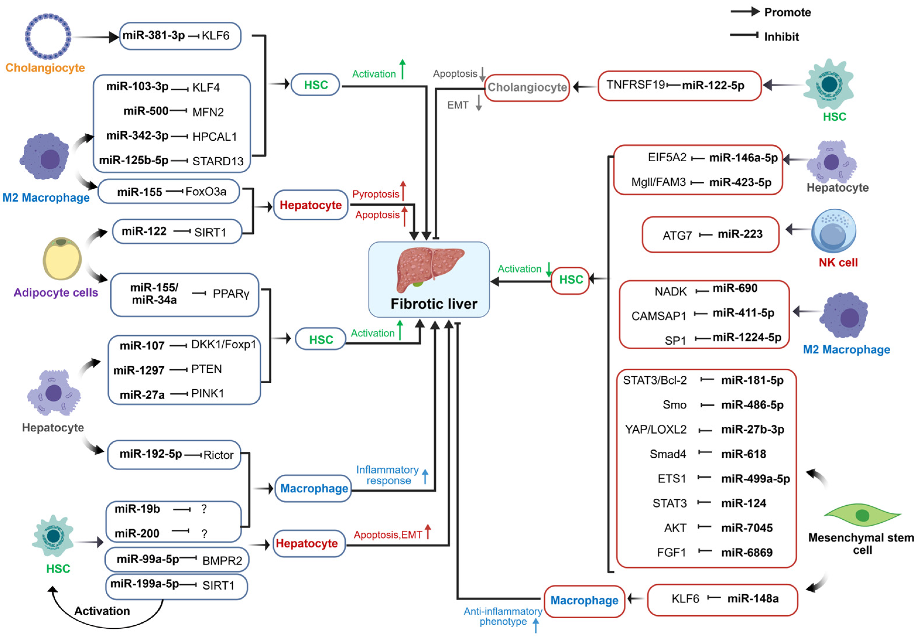

Disclaimer/Publisher’s Note: The statements, opinions and data contained in all publications are solely those of the individual author(s) and contributor(s) and not of MDPI and/or the editor(s). MDPI and/or the editor(s) disclaim responsibility for any injury to people or property resulting from any ideas, methods, instructions or products referred to in the content. |
© 2026 by the authors. Licensee MDPI, Basel, Switzerland. This article is an open access article distributed under the terms and conditions of the Creative Commons Attribution (CC BY) license.