


Abstract
Skin aging is a multifactorial process that is regulated and preceded by changes at the cellular and molecular level, including chronic low-grade inflammation, senescence and altered intercellular communication. Several therapeutic approaches, including mesotherapy and dermal fillers, have been developed to ameliorate the visible signs of skin aging; however, their effects are primarily focused on improving tissue structure and appearance rather than directly targeting the molecular mechanisms underlying the aging process. Here, we used an in vitro model of UVB-induced dermal fibroblast senescence to evaluate the effect of a polycomponent formula based on amino acids and vitamins on key hallmarks of skin aging. Our results showed the formula modulated senescence, epigenetic and repair genes in senescent dermal fibroblasts (senomorphic effect), while exerting a reduction in senescent cell number (senolytic effect). Conditioned medium from senescent fibroblasts treated with the formula stimulated extracellular matrix production and cell activation in healthy fibroblasts. Moreover, the formula upregulated growth factor gene expression in adipose-derived stem cells. Thus, our research shows a potent biostimulatory and regenerative response of the proposed formula in aged skin and adipose stem cells, indicating the potential use of these compounds in dermal fillers and other injectable products aimed at regenerating and rejuvenating skin and deep adipose tissue.
1. Introduction
Skin aging is a complex biological phenomenon characterized by the interplay between intrinsic and extrinsic environmental factors. At the molecular level, cells undergo a progressive decline in their functions and accumulation of damage that results in epigenetic alterations, genomic damage, mitochondrial dysfunction and cellular senescence, among other hallmarks [1]. These changes favor a disrupted tissue microenvironment through chronic, low-grade inflammatory state known as “Inflammaging”. At the tissue level, this translates into extracellular matrix alterations, based on decreased collagen and elastin production, impaired TGF-β signaling and increased matrix metalloproteinase activity [2,3]. Consequently, these molecular, cellular and tissue changes are responsible for the visual changes observed in aged skin, including loss of firmness and elasticity and increased sagging, dryness, wrinkles and laxity. Interestingly, these changes are not only dependent on dermal extracellular matrix alterations, but also influenced by deeper tissue compartments, such as adipose tissue. Aged adipose tissue is characterized by loss of fat deposits in differentiated adipocytes and decreased adipose-derived stem cell activity, this last cell type being a key source of growth factors and cytokines that stimulate neighboring cells both in adipose and near tissues, such as tendons, bones and other connective tissues [4]. In fact, these fat compartments are usually another target of injectable biomaterials to induce physical restoration of volume and redensification to correct visual skin-aging signs.
As current available treatments for skin aging, mesotherapy and dermal filler injections are widely used by physicians to correct wrinkles and loss of firmness. The biomaterials included in these injectables work either by physically filling the skin compartments that cause skin aging signs (such as hyaluronic acid) or act through a controlled inflammatory reaction that frequently derives in undesired adverse effects caused by foreign body reactions to the biomaterial (such as calcium hydroxyapatite) [5,6]. Thus, it is essential to develop new treatments that include biostimulatory molecules that target the root cause of skin aging signs, which are the aforementioned molecular hallmarks of aging. Previous research has indicated the use of different compounds inside mesotherapy products to improve skin brightness or hydration [7,8], but whether these compounds regulate key pathways such as senescence or epigenetic modifications remains unknown. Interestingly, compounds regulating senescence proinflammatory phenotype (called senomorphics) or that specifically cause apoptosis in senescent cells (called senolytics) have shown promise in other in vitro and animal studies regarding tissue rejuvenation and aged cellular function restoration [9,10,11,12].
Hence, our aim was to elucidate the effect of a combination of amino acids and vitamins on key hallmarks of aging in aged dermal fibroblasts and adipose-derived stem cells (ADSCs). Our findings indicated that the proposed formula exerted beneficial effects on epigenetic, repair and senescence biomarkers in senescent fibroblasts, while restoring altered intercellular communication by boosting healthy fibroblasts activation and extracellular matrix synthesis following treatment with conditioned medium from senescent fibroblasts. Moreover, the tested formula upregulated growth factor, adipogenic and epigenetic repair gene expression in ADSCs, further indicating a potential benefit in the paracrine regenerative potential of stem cells located in deep fat tissues.
2. Materials and Methods
2.1. Cell Culture and Treatment
Human dermal fibroblasts (HDFs) (Promocell, Heidelberg, Germany) were cultured in DMEM (Capricorn Scientific GmbH, Ebsdorfergrund, Germany) supplemented with 10% fetal bovine serum (Fisher Scientific, Hampton, NH, USA) and Zellshield (Minerva Biolabs GmbH, Berlin, Germany). ADSCs (Promocell, Heidelberg, Germany) were cultured in Mesenchymal Stem Cell Growth Medium 2 (Promocell, Heidelberg, Germany). Cells were incubated at 37 °C in 5% CO2 and 90% humidity.
A formula containing ascorbic acid, cyanocobalamin, thiamine, retinol, pyridoxine, niacinamide, tocopherol, biotin, glycine, arginine, glutathione, lysine and proline was kindly provided by mesoestetic Pharma Group S.L, containing 1 mM, 2 µM, 100 µM, 1 µM, 200 µM, 5 mM, 50 µM, 100 µM, 5 mM, 5 mM, 5 mM, 500 µM and 500 µM as final concentration, respectively. The concentrations were selected based on viable doses according to previous studies [13,14,15,16,17,18,19,20,21,22,23,24,25]. The compounds were diluted in culture medium and used for in vitro evaluation.
Cell viability was assessed using the sulforhodamine B (SRB) assay, as previously described, to determine whether the selected combination of compounds exerted any cytotoxic effects [26]. Briefly, cells were treated with the combination of compounds for 24 h and consequently incubated with 10% trichloroacetic acid at 4 °C for 1 h. The SRB solution was then added and cells were incubated for 30 min at RT. The SRB solution was removed, cells were washed 4 times with 1% acetic acid, and they were allowed to dry at RT. The dye was dissolved with 10 mM Tris base solution (pH 10) and the absorbance was measured at 540 nm using a microplate reader (multiskan FC, Thermo Fisher Scientific, Waltham, MA, USA).
2.2. UVB-Induced Senescence in Human Dermal Fibroblasts
HDFs were plated at a density of 6 × 10⁴ cells/plate in 6-well plates. After 24 h, cell culture medium was removed, and cells were irradiated at 25 mJ/cm2 of UVB light (312 nm) in a Bio LINK Crosslinker BLX-312/365 (Witec ag) in 1 mL of PBS. PBS was then removed and fresh cell culture medium was added to each well. After 3 days, cells were irradiated again under the same conditions. After 2 days, cells were processed according to the gene expression (qPCR) or β-Gal staining protocols described in the next subsections. For non-irradiated control conditions, HDF were plated at a density of 6 × 10⁴ cells/plate in 6-well plates. After 2 days, cells were trypsinized and reseeded at 6 × 10⁴ cells/plate in fresh cell culture medium in 6-well plates. After 3 days, cells were trypsinized again and reseeded at 8 × 10⁴ cells/plate in fresh cell culture medium containing the compound in 6-well plates. After 2 days, cells were processed as irradiated cells conditions. These trypsinization and cell passaging steps are performed to avoid excessive cell confluence for non-irradiated cells at the end of the experiment, considering that non-irradiated cells keep on proliferating, but UVB-irradiated cells show significantly reduced proliferation during the culture and irradiation protocol.
2.3. Gene Expression Quantification by qPCR
RNA was extracted from cell pellets using the Total RNA Purification Kit (Norgen, Thorold, ON, Canada) according to the manufacturer’s protocol. Extracted RNA was quantified using a NanoDrop™ ND-1000 UV–Vis spectrophotometer (Thermo Fisher Scientific, Waltham, MA, USA).
For gene expression quantification, 300 ng of RNA was retrotranscribed to cDNA using the PrimeScript™ RT reagent kit (Takara Bio Inc., Kusatsu City, Japan). cDNA was diluted 1/10 in ultrapure water, and the qPCR mix was prepared by adding cDNA (12 ng), SYBR Green Supermix (Bio-Rad, Hercules, CA, USA), and forward and reverse primers (0.2 μM).
The primers and their respective sequences are listed in Table 1. The qPCR protocol included one step at 95 °C for 30 s, 40 cycles at 95 °C for 10 s, and 60 °C for 1 min. The results were analyzed with Bio-Rad™ CFX Manager software (version 3.1). ACTB was used as a housekeeping gene to normalize gene expression.
Table 1.
List of primers used for qPCR assay.
2.4. β-Galactosidase Staining
After UVB irradiation protocol, cells were washed in PBS and fixed with 1 mL of 10% formalin (Sigma Aldrich, St Louis, MO, USA). Detection of senescence-associated beta-galactosidase (SA-β-Gal) activity was performed as previously described [27]. Bright field pictures were taken for every condition using a CKX41 phase contrast microscope (Olympus, Tokyo, Japan) and the number of cells was manually counted using ImageJ (1.51k). Cells stained in blue were counted as senescent while non-stained cells were counted as non-senescent. The percentage of senescent cells was then calculated by dividing the number of counted senescent cells by the total number of cells in each condition.
2.5. Characterization of Cell Communication Effect Through Conditioned Medium
After the treatment of senescent fibroblasts with the formula and before cell harvesting, the media of senescent cells treated for 48 h with the formula (conditioned medium) was harvested and transferred to another plate of non-senescent human dermal fibroblasts. Cells were incubated for 24 h in conditioned medium and later harvested by trypsinization and processed for qPCR assay as described in the previous section.
2.6. Statistical Analysis
The experiments were performed in triplicate. Statistical significance was determined through Student’s t-test (* p < 0.05, ** p < 0.01, *** p < 0.001) using Microsoft Excel™ version 2408 (Microsoft Corporation, Redmond, WA, USA). This parametric approach was selected to compare the means of two groups based on independent biological replicates, with each biological replicate considered as an individual experimental unit to avoid pseudo replication. The use of Student’s t-test was considered appropriate for the analysis of continuous quantitative variables, including percentages derived from cell counts, provided that the assumptions of approximate normality and homogeneity of variances were reasonably satisfied. Although the limited number of biological replicates (n = 3 per condition) restricts the ability to reliably assess normality, the results were interpreted considering this limitation.
3. Results
3.1. Effect of Polycomponent Formula on Cell Viability
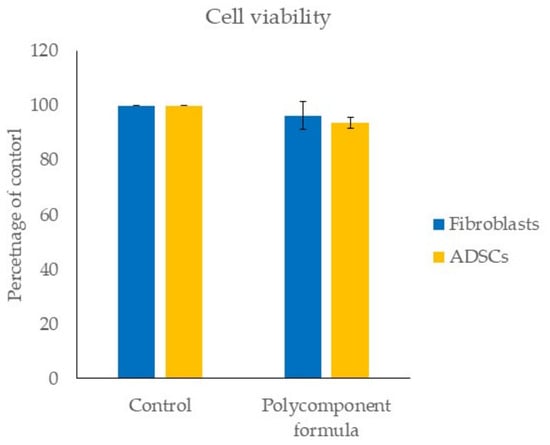
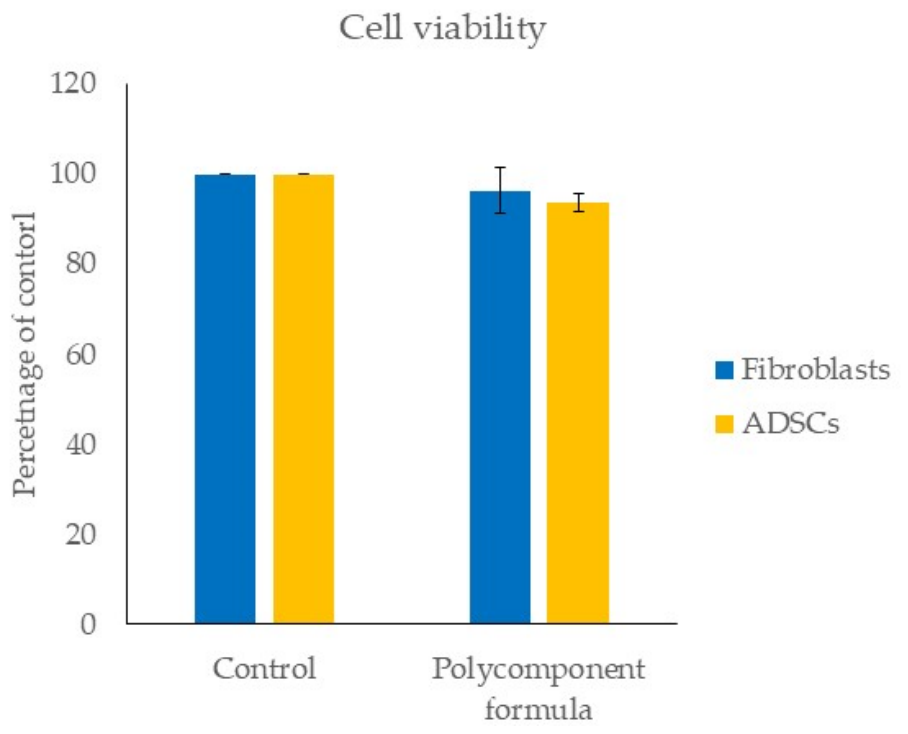
Cell viability using SRB assay was performed to confirm that the concentration of compounds used in the subsequent assays was not inducing a cytotoxic effect. As shown in Figure 1, the selected concentration of the combination of ingredients did not alter cell viability in either dermal fibroblasts or ADSCs. Thus, the specified concentration of compounds was used for the subsequent experiments in both cell types.
Figure 1.
Cell viability assay using SRB reagent in fibroblasts and ADSCs treated for 24 h with the polycomponent formula. Bar graphs indicate mean ± SD (n = 3). Statistical analysis was performed using Student’s t-test. No significant changes between control and treated cells were observed according to this statistical analysis method.
3.2. Effect of Polycomponent Formula on Senescent Dermal Fibroblasts
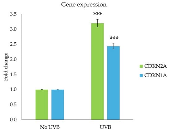
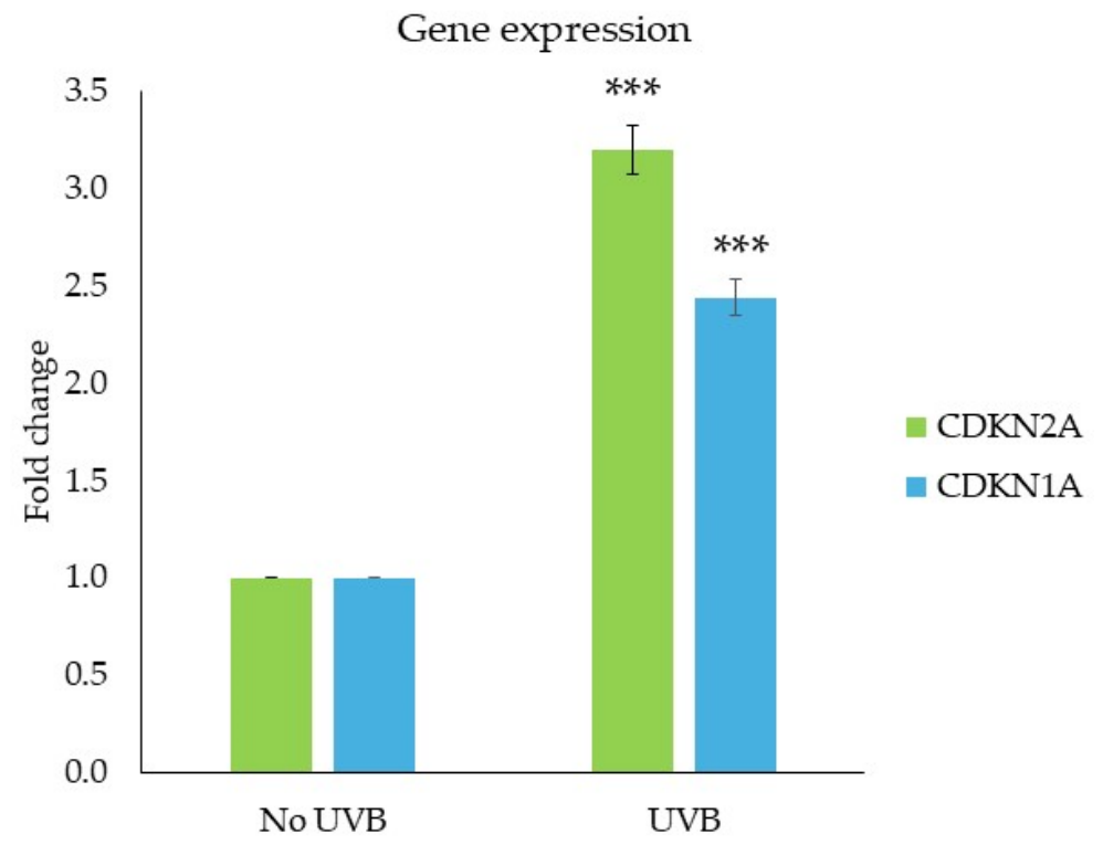
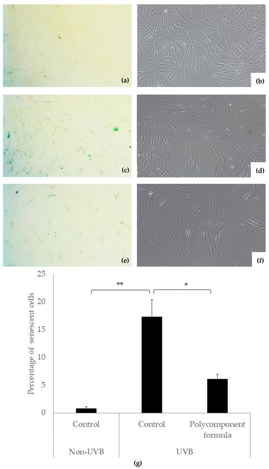
Human dermal fibroblasts senescence was successfully induced using several UVB irradiations. Typical senescent features that confirm senescent state include upregulation of canonical senescence-associated cell cycle inhibitors p16INK4a and p21CIP1 (Figure 2), decreased LMNB1 expression (Table 2) and increased SA-β-Gal staining (Figure 3). To evaluate the effect on senescent cell phenotype/activity (senomorphic effect), senescent dermal fibroblasts were treated for 48 h with the polycomponent formula. After the treatment, qPCR assay revealed that treated cells showed upregulation of genes involved in senescence regulation (DNMT1, LMNB1), epigenetic modulation (BMI1, EZH2, SIRT1, SIRT3, SIRT6) and cell and tissue repair processes (EGF, GDF11, HBEGF, KU70, NAMPT) (Table 2), suggesting a positive senomorphic effect on senescent fibroblasts.
Figure 2.
Gene expression levels of canonical regulators of senescence-associated cell cycle arrest (CDKN2A/p16INK4a and CDKN1A/p21CIP1) in non-UVB irradiated fibroblasts and UVB-irradiated fibroblasts. Bar graphs indicate mean ± SD (n = 3). Statistical analysis was performed using Student’s t-test, where *** indicates p < 0.001.
Table 2.
Gene expression levels of senescence, epigenetic and repair markers in control fibroblasts, UVB-induced senescent fibroblasts and UVB-induced senescent fibroblasts treated with the polycomponent formula for 48 h. Standard deviation (SD, n = 3) for each quantification is provided in brackets. Statistical analysis was performed using Student’s t-test, where “a” indicates p < 0.05 comparing no UVB control vs. UVB-induced senescent control; “b” indicates p < 0.01 comparing no UVB control vs. UVB-induced senescent control; “c” indicates p < 0.001 comparing no UVB control vs. UVB-induced senescent control; “d” indicates p < 0.05 comparing UVB-induced senescent control vs. UVB-induced senescent treated with the formula; “e” indicates p < 0.01 comparing UVB-induced senescent control vs. UVB-induced senescent treated with the formula.
Figure 3.
Senescence-associated beta-galactosidase (SA-β-Gal) staining (40× magnification) of non-UVB control fibroblasts (a,b), UVB-induced senescent control fibroblasts (c,d) and UVB-induced senescent fibroblasts treated with the formula (e,f). Representative pictures of triplicates are shown, where (a,c,e) correspond to bright field and (b,d,f) correspond to phase-contrast. (g) Percentage of senescent cells was calculated by counting the number of stained cells in blue (senescent) and the total number of cells in each condition. Bar graphs indicate mean ± SD (n = 3). Statistical analysis was performed using Student’s t-test, where * indicates p < 0.05 and ** indicates p < 0.01.
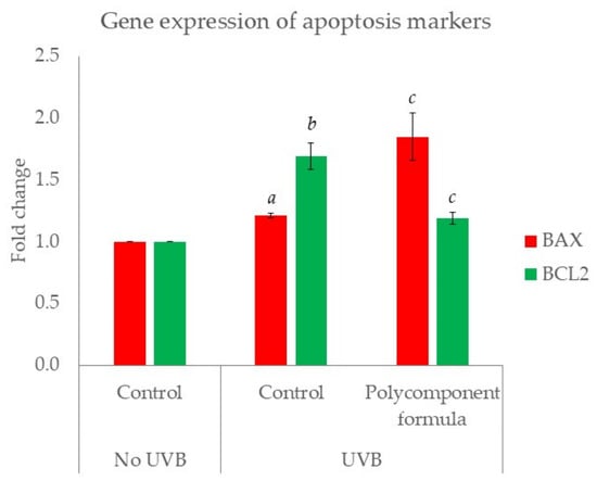
Senescent cell death (senolytic effect) was evaluated by SA-β-Gal staining and apoptosis-related gene expression by qPCR. As shown in Figure 3 and Figure 4, treatment with the formula for 48 h significantly reduced the percentage of SA-β-Gal-stained cells, upregulated pro-apoptotic gene BAX and downregulated anti-apoptotic gene BCL2. Taken together, the reduction in SA-β-gal-positive cells, the modulation of apoptosis-related markers, and the absence of cytotoxicity in normal fibroblasts as demonstrated by the SRB viability assay (Figure 1) support a senolytic effect of the formula by promoting the elimination of senescent fibroblasts while preserving the viability of non-senescent cells.
Figure 4.
Gene expression levels of apoptosis regulators (BAX, BCL2) in control fibroblasts, UVB-induced senescent fibroblasts and UVB-induced senescent fibroblasts treated with the polycomponent formula for 48 h. Bar graphs indicate mean ± SD (n = 3). Statistical analysis was performed using Student’s t-test, where “a” indicates p < 0.01 comparing no UVB control vs. UVB-induced senescent control; “b” indicates p < 0.001 comparing no UVB control vs. UVB-induced senescent control; “c” indicates p < 0.05 comparing UVB-induced senescent control vs. UVB-induced senescent cells treated with formula.
3.3. Effect of Conditioned Medium from Senescent Fibroblasts on Healthy Fibroblasts
To evaluate the effect of the treatment on altered intercellular communication, conditioned medium from senescent fibroblasts treated for 48 h with the formula was transferred to healthy fibroblasts and gene expression was quantified by qPCR. As shown in Table 3, two genes involved in fibroblast activity and extracellular matrix (DPT and ACTA2) were downregulated by conditioned medium from non-treated cells, indicating that senescent phenotype can alter cell function of neighboring cells. When senescent cells were treated with the polycomponent formula, healthy fibroblasts incubated with conditioned medium from senescent cells showed upregulated levels of fibroblast activity and extracellular matrix production, suggesting that the treatment restores altered cell communication between senescent and healthy fibroblasts.
Table 3.
Gene expression levels of fibroblasts activity and extracellular matrix in fibroblasts incubated in conditioned medium (CM) from healthy cells, fibroblasts incubated in CM from senescent cells and fibroblasts incubated in CM from senescent cells and treated with the polycomponent formula for 24 h. Standard deviation (SD, n = 3) for each quantification is provided in brackets, and statistical analysis was performed using Student’s t-test, where “a” indicates p < 0.01 comparing CM from healthy cells and CM from senescent cells; “b” indicates p < 0.05 comparing CM from senescent cells and CM from senescent cells treated with formula; “c” indicates p < 0.01 comparing CM from senescent cells and CM from senescent cells treated with formula.
3.4. Effect of Polycomponent Formula on Adipose-Derived Stem Cells (ADSCs)
ADSCs were cultured for 24 h in the presence of the formula to assess the effect on genes involved in tissue regeneration and repair of deep tissues. As shown in Table 4, the formula upregulated adipogenic (ADIPOQ, PPARG), growth factors (FGFB, GDF11, HGF, PDGF, VEGF), proliferation (CD44) and cellular function/resistance to stress (SIRT1, SIRT3, SIRT6). Overall, these changes indicate an early transcriptional response of ADSCs to the formula, characterized by the modulation of adipogenesis-related genes together with increased expression of factors associated with tissue repair and cellular function.
Table 4.
Gene expression levels of proliferation, adipogenic phenotype, growth factors, cell function and resistance to stress in control ADSCs and ADSCs treated with the polycomponent formula for 24 h. Standard deviation (SD, n = 3) for each quantification is provided in brackets, and statistical analysis was performed using Student’s t-test, where * indicates p < 0.05, ** indicates p < 0.01 and *** indicates p < 0.001.
4. Discussion
Cellular senescence is increasingly recognized as a central hallmark of aging and a major driver of skin aging. Rather than acting independently, senescence is closely interconnected with the other hallmarks of aging, amplifying tissue dysfunction through reciprocal feedback mechanisms [28]. The accumulation of senescent cells promotes the secretion of the senescence-associated secretory phenotype (SASP), which establishes a chronic pro-inflammatory microenvironment and contributes to inflammaging. This persistent low-grade inflammation disrupts tissue homeostasis, impairs extracellular matrix integrity, and reduces the regenerative capacity of the skin [1,2]. Collectively, these processes underlie the progressive loss of firmness, elasticity, and volume that characterize aged skin. Therefore, targeting senescence and its downstream effects has become a key objective in the development of interventions aimed at preventing or reversing the visible signs of skin aging.
Within this framework, regenerative medicine focuses on restoring cellular functions that progressively decline with age by promoting endogenous repair mechanisms and tissue regeneration [29,30]. Unlike approaches that rely primarily on inflammatory-driven remodeling, an ideal regenerative strategy should provide controlled biostimulation while minimizing excessive or prolonged inflammation, which may compromise tissue homeostasis and long-term outcomes. Consequently, biomaterials capable of promoting tissue regeneration through physiological activation of resident cells, without eliciting an exacerbated inflammatory response, represent a promising strategy to achieve more effective and durable skin rejuvenation.
In this context, our research highlights a combination of vitamins and amino acids with biologically relevant effects on key aging pathways in both aged dermal fibroblasts and ADSCs. Previous studies using similar compounds for mesotherapy purposes have shown benefits on collagen synthesis or antioxidant effect [8,31,32,33], rather than evaluating the effect on key hallmarks of aging such as senescence or altered intercellular communication.
First, we observed a combined senolytic and senomorphic effect of the tested formula in a model of photoaging-induced senescence (Table 2, Figure 3 and Figure 4), by reducing the population of senescent cells while stimulating repair, epigenetic modulation and regeneration markers in the surviving cells. This dual effect represents a distinctive approach in the field of skin rejuvenation, as previous studies have largely focused on either promoting the elimination of senescent cells or attenuating the senescent phenotype, whereas the present findings suggest that both effects may be achieved through the same intervention [9,34]. Of note, the tested formula upregulated GDF11 among other biologically relevant genes in the skin. Previous research has proven the key role of this growth factor in preserving youthful phenotypes in various types of skin cells [35]. However, to the best of our knowledge, this is the first study to report the upregulation of this gene in response to an injectable treatment or compound.
Second, the beneficial effect of the formula on senescent fibroblasts was validated by testing the effect of the SASP on neighboring cells, which accounts for another of the key hallmarks of aging—altered intercellular communication [36]. For this, conditioned medium from senescent fibroblasts was transferred to healthy fibroblasts and gene expression was evaluated by qPCR. The results showed that the conditioned medium from senescent cells treated with the formula upregulated genes involved in extracellular matrix (DPT, DCN) and fibroblasts activation (GDF11, ACTA2) in healthy fibroblasts. Thus, the plausible explanation for this effect is the positive modulation of the secreted factors by senescent cells with a consequent beneficial activation of healthy fibroblasts. Nevertheless, these findings should be interpreted with caution. Since the conditioned medium obtained from formula-treated senescent fibroblasts may contain residual amounts of the tested compounds, a direct effect of these compounds on the healthy recipient fibroblasts cannot be completely excluded. Therefore, although the observed modulation of DPT and ACTA2 expression is consistent with an alteration of the paracrine effects exerted by senescent fibroblasts, the present experimental design does not allow this effect to be unequivocally attributed to restored paracrine signaling. Future experiments including a control in which healthy fibroblasts are exposed to fresh medium containing an equivalent residual concentration of the formula would be necessary to distinguish between direct effects of the compounds and effects mediated by changes in the senescent cell secretome.
Finally, the formula was also tested on ADSCs, as these cells are increasingly becoming of interest in regenerative medicine. Particularly, these cells can differentiate to a wide variety of cells including adipocytes, chondrocytes or osteoblasts among others, while also producing growth factors and cytokines that boost regenerative and rejuvenating processes in deep tissues such as fat compartments [4,37]. Based on these abilities, these cells are being used in autologous fat grafting for regenerative purposes. Interestingly, previous studies have focused on using injectable products to boost the activation of resident ADSCs as an alternative to autologous procedures, which are more technically difficult and can derive in risks and adverse effects in the patients [38]. Specifically, hyaluronic acid (HA) complexes can induce adipogenic gene upregulation after 7 days. In our experiment, the formula upregulated adipogenic genes, but to a lesser extent than HA. The difference might be explained by our reduced time of treatment (24 h vs. 7 days). Interestingly, our research shows that the formula can upregulate growth factors gene expression and CD44, the receptor that mediates HA-induced cell proliferation. Thus, the combination of HA and this polycomponent formula might have a synergistic effect on the early activation of ADSCs. The increased expression of PPARG and ADIPOQ observed after 24 h of treatment indicates an early modulation of genes associated with adipogenic regulation and adipose tissue function. Given the short treatment period and the absence of a specific adipogenic induction protocol, these transcriptional changes should not be interpreted as evidence of terminal adipogenic differentiation. Rather, they suggest that the formula induces an early transcriptional response in ADSCs involving pathways related to their adipogenic potential. Importantly, the aim of this experiment was to assess the acute responsiveness of ADSCs to the formula in the context of tissue biostimulation, rather than to promote their differentiation into mature adipocytes. Further studies involving prolonged adipogenic induction and functional endpoints, such as lipid accumulation, would be required to establish whether these early molecular changes translate into enhanced adipogenic differentiation.
Despite these promising findings, several limitations of the present study should be acknowledged. First, the cytokines and growth factors released by senescent cells, present in the conditioned medium and responsible for the beneficial effects on healthy fibroblasts remain unknown. Second, the experiment on ADSCs lacks a functional demonstration of the positive effect of the formula, as the observed changes correspond to gene expression changes, but not protein or pathway alterations. These mRNA changes may not necessarily translate into corresponding changes at the protein level. Therefore, future studies assessing protein abundance and/or secretion should validate these transcriptional responses and confirm their functional relevance, particularly regarding fibroblast activation and the production of growth factors by ADSCs. Finally, the presented results are observed in in vitro models of skin aging, and thus these effects should be confirmed in ex vivo and clinical studies to finally assure the observed regenerative and rejuvenating potential of the formula. Therefore, future research could be oriented to solve these limitations.
5. Conclusions
Our findings demonstrate that the proposed combination of active ingredients positively modulates key pathways involved in regeneration, senescence, and tissue repair in aged dermal fibroblasts and adipose-derived stem cells (ADSCs). These results highlight its potential as a bioactive formulation for mesotherapy products and dermal fillers designed to promote tissue biostimulation and rejuvenation. By targeting both superficial skin layers and deeper anatomical structures relevant to regenerative and aesthetic medicine, including the deep fat compartments, this approach represents a promising strategy for enhancing tissue regeneration and addressing age-related changes through a comprehensive regenerative mechanism.
Author Contributions
Conceptualization, A.M.-G.; methodology, A.M.-G. and H.C.; software, H.C. and L.G.-R.; validation, A.M.-G. and L.S.; formal analysis, A.M.-G. and L.G.-R.; investigation, A.M.-G., H.C. and L.G.-R.; resources, A.M.-G. and L.S.; data curation, A.M.-G. and L.G.-R.; writing—original draft preparation, A.M.-G.; writing—review and editing, L.S.; visualization, A.M.-G.; supervision, A.M.-G. and L.S.; project administration, A.M.-G. and L.S.; funding acquisition, L.S. All authors have read and agreed to the published version of the manuscript.
Funding
The authors declare that this study received funding from Mesoestetic Pharma Group s.l.
Institutional Review Board Statement
Not applicable.
Informed Consent Statement
Not applicable.
Data Availability Statement
The original contributions presented in this study are included in the article. Further inquiries can be directed to the corresponding author.
Conflicts of Interest
All authors were employed by the company Mesoestetic Pharma Group s.l. The funder was not involved in the study design, collection, analysis, interpretation of data, the writing of this article or the decision to submit it for publication. There is no any commercial conflicts of interest in this article.
References
- Jin, S.; Li, K.; Zong, X.; Eun, S.; Morimoto, N.; Guo, S. Hallmarks of Skin Aging: Update. Aging Dis. 2023, 14, 2167–2176. [Google Scholar] [CrossRef] [Scilit] [PubMed]
- Ganguly, B.; Hota, M.; Pradhan, J. Skin Aging: Implications of UV Radiation, Reactive Oxygen Species and Natural Antioxidants. In Reactive Oxygen Species; IntechOpen: London, UK, 2021. [Google Scholar]
- He, R.; Sun, M.; Liu, T.; Geng, L.; Xing, C.; Xu, P.; Peng, Y.; Fang, Y. Hallmarks of the Aging Skin Microenvironment: Components and Mechanisms. J. Cell. Physiol. 2025, 240, e70111. [Google Scholar] [CrossRef] [Scilit] [PubMed]
- Zhang, Y.; Jiang, Y.; Yang, X.; Huang, Y.; Pan, A.; Liao, Y. Adipose tissue senescence: Biological changes, hallmarks and therapeutic approaches. Mech. Ageing Dev. 2024, 222, 111988. [Google Scholar] [CrossRef] [Scilit] [PubMed]
- Lee, J.C.; Daniels, M.A.; Roth, M.Z. Mesotherapy, Microneedling, and Chemical Peels. Clin. Plast. Surg. 2016, 43, 583–595. [Google Scholar] [CrossRef] [Scilit] [PubMed]
- Fallacara, A.; Manfredini, S.; Durini, E.; Vertuani, S. Hyaluronic Acid Fillers in Soft Tissue Regeneration. Facial Plast. Surg. 2017, 33, 87–96, Erratum in Facial Plast. Surg. 2017, 33, 244. https://doi.org/10.1055/s-0037-1601851. [Google Scholar] [CrossRef] [Scilit] [PubMed]
- Siquier-Dameto, G.; Boadas-Vaello, P.; Verdú, E. Intradermal Treatment with a Hyaluronic Acid Complex Supplemented with Amino Acids and Antioxidant Vitamins Improves Cutaneous Hydration and Viscoelasticity in Healthy Subjects. Antioxidants 2024, 13, 770. [Google Scholar] [CrossRef] [Scilit] [PubMed]
- Scarano, A.; Qorri, E.; Sbarbati, A.; Gehrke, S.A.; Marchetti, M.; Desiderio, V.; Amuso, D.; Tari, S. Mesotherapy with hyaluronic acid solutions enriched by amino acids in the neck area: Open-label uncontrolled, monocentric study. J. Cosmet. Laser Ther. 2025, 27, 121–126. [Google Scholar] [CrossRef] [Scilit] [PubMed]
- Zhang, L.; Pitcher, L.E.; Prahalad, V.; Niedernhofer, L.J.; Robbins, P.D. Targeting cellular senescence with senotherapeutics: Senolytics and senomorphics. FEBS J. 2023, 290, 1362–1383. [Google Scholar] [CrossRef] [Scilit] [PubMed]
- De Luca, F.; Gola, F.; Azzalin, A.; Casali, C.; Gaiaschi, L.; Milanesi, G.; Vicini, R.; Rossi, P.; Bottone, M.G. A Lombard Variety of Sweet Pepper Regulating Senescence and Proliferation: The Voghera Pepper. Nutrients 2024, 16, 1681. [Google Scholar] [CrossRef] [Scilit] [PubMed]
- Gola, F.; Gaiaschi, L.; Roda, E.; De Luca, F.; Ferulli, F.; Vicini, R.; Rossi, P.; Bottone, M.G. Voghera Sweet Pepper: A Potential Ally against Oxidative Stress and Aging. Int. J. Mol. Sci. 2023, 24, 3782. [Google Scholar] [CrossRef] [Scilit] [PubMed]
- Gola, F.; Casali, C.; Gaiaschi, L.; Roda, E.; Milanesi, G.; De Luca, F.; Bottone, M.G. Voghera Sweet Pepper Regulates Cell Death Pathways in an Aging In Vitro Model. Nutrients 2025, 17, 2147. [Google Scholar] [CrossRef] [Scilit] [PubMed]
- Mohammed, B.M.; Fisher, B.J.; Kraskauskas, D.; Ward, S.; Wayne, J.S.; Brophy, D.F.; Fowler, A.; Yager, D.R.; Natarajan, R. Vitamin C promotes wound healing through novel pleiotropic mechanisms. Int. Wound J. 2016, 13, 572–584. [Google Scholar] [CrossRef] [Scilit] [PubMed]
- Krugluger, W.; Stiefsohn, K.; Laciak, K.; Moser, K.; Moser, C. Vitamin B12 Activates the Wnt-Pathway in Human Hair Follicle Cells by Induction of beta-Catenin and Inhibition of Glycogensynthase Kinase-3 Transcription. J. Cosmet. Dermatol. Sci. Appl. 2011, 1, 25–29. [Google Scholar] [CrossRef]
- Huang, H.M.; Chen, H.L.; Gibson, G.E. Thiamine and oxidants interact to modify cellular calcium stores. Neurochem Res. 2010, 35, 2107–2116. [Google Scholar] [CrossRef] [Scilit] [PubMed]
- Gimeno, A.; Zaragozá, R.; Vivó-Sesé, I.; Viña, J.R.; Miralles, V.J. Retinol, at concentrations greater than the physiological limit, induces oxidative stress and apoptosis in human dermal fibroblasts. Exp. Dermatol. 2004, 13, 45–54. [Google Scholar] [CrossRef] [Scilit] [PubMed]
- Fujishiro, M.; Yahagi, S.; Takemi, S.; Nakahara, M.; Sakai, T.; Sakata, I. Pyridoxine stimulates filaggrin production in human epidermal keratinocytes. Mol. Biol. Rep. 2021, 48, 5513–5518. [Google Scholar] [CrossRef] [Scilit] [PubMed]
- Kang, H.T.; Lee, H.I.; Hwang, E.S. Nicotinamide extends replicative lifespan of human cells. Aging Cell 2006, 5, 423–436. [Google Scholar] [CrossRef] [Scilit] [PubMed]
- Villalón-García, I.; Álvarez-Córdoba, M.; Povea-Cabello, S.; Talaverón-Rey, M.; Villanueva-Paz, M.; Luzón-Hidalgo, R.; Suárez-Rivero, J.M.; Suárez-Carrillo, A.; Munuera-Cabeza, M.; Salas, J.J.; et al. Vitamin E prevents lipid peroxidation and iron accumulation in PLA2G6-Associated Neurodegeneration. Neurobiol. Dis. 2022, 165, 105649. [Google Scholar] [CrossRef] [Scilit] [PubMed]
- Duchi, S.; Torregrosa, P.R.; Hajuj, A.; Molho, D.; Shkoor, R.; Saada, N.A.; Fernández, D.G.; Goldstein, D.; Pérez-Fernández, A. The formulation and in vitro evaluation of WS Biotin, a novel encapsulated form of D-Biotin with improved water solubility for hair and skin treatment applications. Int. J. Cosmet. Sci. 2024, 46, 119–129. [Google Scholar] [CrossRef] [Scilit] [PubMed]
- Tripathi, R.; Yang, X.; Liu, D.; Lu, W.; Wang, B. Biotin-mediated drug delivery: Does the biotin transporter mediate uptake of biotin conjugates? J. Enzym. Inhib. Med. Chem. 2025, 40, 2554896. [Google Scholar] [CrossRef] [Scilit] [PubMed]
- Kocic, H.; Arsic, I.; Stankovic, M.; Tiodorovic, D.; Ciric, V.; Kocic, G. Proliferative, anti-apoptotic and immune-enhancing effects of L-arginine in culture of skin fibroblasts. J. Biol. Regul. Homeost. Agents 2017, 31, 667–672. [Google Scholar] [PubMed]
- Montero, D.; Tachibana, C.; Winther, J.R.; Appenzeller-Herzog, C. Intracellular glutathione pools are heterogeneously concentrated. Redox Biol. 2013, 1, 508–513. [Google Scholar] [CrossRef] [Scilit] [PubMed]
- Lin, X.; Li, S.; Zou, Y.; Zhao, F.Q.; Liu, J.; Liu, H. Lysine Stimulates Protein Synthesis by Promoting the Expression of ATB0,+ and Activating the mTOR Pathway in Bovine Mammary Epithelial Cells. J. Nutr. 2018, 148, 1426–1433. [Google Scholar] [CrossRef] [Scilit] [PubMed]
- Krishnan, N.; Dickman, M.B.; Becker, D.F. Proline modulates the intracellular redox environment and protects mammalian cells against oxidative stress. Free Radic. Biol. Med. 2008, 44, 671–681. [Google Scholar] [CrossRef] [Scilit] [PubMed]
- Vichai, V.; Kirtikara, K. Sulforhodamine B colorimetric assay for cytotoxicity screening. Nat. Protoc. 2006, 1, 1112–1116. [Google Scholar] [CrossRef] [Scilit] [PubMed]
- Debacq-Chainiaux, F.; Erusalimsky, J.D.; Campisi, J.; Toussaint, O. Protocols to detect senescence-associated beta-galactosidase (SA-betagal) activity, a biomarker of senescent cells in culture and in vivo. Nat. Protoc. 2009, 4, 1798–1806. [Google Scholar] [CrossRef] [Scilit] [PubMed]
- Chin, T.; Lee, X.E.; Ng, P.Y.; Lee, Y.; Dreesen, O. The role of cellular senescence in skin aging and age-related skin pathologies. Front. Physiol. 2023, 14, 1297637. [Google Scholar] [CrossRef] [Scilit] [PubMed]
- Dutra Alves, N.S.; Reigado, G.R.; Santos, M.; Caldeira, I.S.D.; Hernandes, H.d.S.; Freitas-Marchi, B.L.; Zhivov, E.; Chambergo, F.S. Advances in regenerative medicine-based approaches for skin regeneration and rejuvenation. Front. Bioeng. Biotechnol. 2025, 13, 1527854. [Google Scholar] [CrossRef] [Scilit] [PubMed]
- Berardesca, E.; Cameli, N. Perspectives of Regenerative Medicine in Dermatology and Cosmetology. Cosmetics 2024, 11, 188. [Google Scholar] [CrossRef] [Scilit]
- Deglesne, P.A.; Arroyo, R.; Ranneva, E.; Deprez, P. In vitro study of RRS HA injectable mesotherapy/biorevitalization product on human skin fibroblasts and its clinical utilization. Clin. Cosmet. Investig. Dermatol. 2016, 9, 41–53. [Google Scholar] [CrossRef] [Scilit] [PubMed]
- Jäger, C.; Brenner, C.; Habicht, J.; Wallich, R. Bioactive reagents used in mesotherapy for skin rejuvenation in vivo induce diverse physiological processes in human skin fibroblasts in vitro- a pilot study. Exp. Dermatol. 2012, 21, 72–75. [Google Scholar] [CrossRef] [Scilit] [PubMed]
- Prikhnenko, S.; Dalens, M. Polycomponent mesotherapy formulations for the treatment of skin aging and improvement of skin quality. Clin. Cosmet. Investig. Dermatol. 2015, 8, 151–157. [Google Scholar] [CrossRef] [Scilit] [PubMed]
- Martel, J.; Ojcius, D.M.; Wu, C.; Peng, H.; Voisin, L.; Perfettini, J.; Ko, Y.; Young, J.D. Emerging use of senolytics and senomorphics against aging and chronic diseases. Med. Res. Rev. 2020, 40, 2114–2131. [Google Scholar] [CrossRef] [Scilit] [PubMed]
- Rochette, L.; Mazini, L.; Meloux, A.; Zeller, M.; Cottin, Y.; Vergely, C.; Malka, G. Anti-Aging Effects of GDF11 on Skin. Int. J. Mol. Sci. 2020, 21, 2598. [Google Scholar] [CrossRef] [Scilit] [PubMed]
- Sun, H.; Xia, T.; Ma, S.; Lv, T.; Li, Y. Intercellular communication is crucial in the regulation of healthy aging via exosomes. Pharmacol. Res. 2025, 212, 107591. [Google Scholar] [CrossRef] [Scilit] [PubMed]
- Ou, M.Y.; Zhang, H.; Tan, P.C.; Zhou, S.B.; Li, Q.F. Adipose tissue aging: Mechanisms and therapeutic implications. Cell Death Dis. 2022, 13, 300. [Google Scholar] [CrossRef] [Scilit] [PubMed]
- Stellavato, A.; La Noce, M.; Corsuto, L.; Pirozzi, A.V.A.; De Rosa, M.; Papaccio, G.; Schiraldi, C.; Tirino, V. Hybrid Complexes of High and Low Molecular Weight Hyaluronans Highly Enhance HASCs Differentiation: Implication for Facial Bioremodelling. Cell. Physiol. Biochem. 2017, 44, 1078–1092. [Google Scholar] [CrossRef] [Scilit] [PubMed]


Disclaimer/Publisher’s Note: The statements, opinions and data contained in all publications are solely those of the individual author(s) and contributor(s) and not of MDPI and/or the editor(s). MDPI and/or the editor(s) disclaim responsibility for any injury to people or property resulting from any ideas, methods, instructions or products referred to in the content. |
© 2026 by the authors. Licensee MDPI, Basel, Switzerland. This article is an open access article distributed under the terms and conditions of the Creative Commons Attribution (CC BY) license.