


Abstract
Antibody-based therapeutics have transformed the management of several vision-threatening ocular diseases by enabling targeted modulation of pathological angiogenesis, inflammation, and disease-associated molecular pathways. However, their clinical effectiveness remains constrained by anatomical and physiological barriers that limit ocular tissue penetration, therapeutic durability, and treatment accessibility. This review provides a comprehensive overview of antibody and related biologic therapeutics across six major ocular disease domains: age-related macular degeneration (AMD), diabetic retinopathy (DR) and diabetic macular edema (DME), retinal vein occlusion (RVO), uveitis, glaucoma, and thyroid eye disease (TED). We first discuss the molecular mechanisms underlying their therapeutic actions, including inhibition of angiogenic signaling, modulation of inflammatory and immune pathways, complement inhibition, and regulation of apoptosis and fibrosis. We then summarize the clinical applications of established and emerging biologic therapies, highlighting their molecular targets, therapeutic outcomes, and limitations. Particular emphasis is placed on advances in antibody engineering and ocular drug delivery, including bispecific antibodies, Fc engineering, antibody–drug conjugates, sustained-release systems, and targeted delivery platforms. Despite substantial progress, repeated administration, variable therapeutic responses, and limited treatment durability remain important challenges. Integrating next-generation biologic platforms with innovative ocular delivery technologies may support more effective, durable, and individualized therapeutic strategies for ocular diseases.
1. Introduction
The eye, with its intricate anatomy and immune-privileged environment, presents unique barriers to effective drug delivery, particularly for chronic and vision-threatening diseases that demand sustained, localized treatment [1,2]. Traditional therapeutic options have often fallen short due to limited tissue penetration, short intraocular half-life, or significant systemic side effects [3]. Over the past two decades, antibody-based therapies have revolutionized the field of ophthalmology, providing unprecedented precision in targeting disease-specific molecular pathways with improved efficacy and tolerability [4,5,6,7,8].
Antibody and related biologic therapeutics have become important treatment options for several ocular disorders, including age-related macular degeneration (AMD) [8], diabetic retinopathy (DR) and diabetic macular edema (DME) [4], retinal vein occlusion (RVO) [5], uveitis [6], and thyroid eye disease (TED) [7], while antibody-based strategies for glaucoma remain largely investigational [9] (Figure 1). These conditions involve distinct pathological mechanisms, ranging from retinal neovascularization and vascular leakage to immune dysregulation, orbital tissue remodeling, and progressive optic neuropathy. Although some molecular pathways overlap across ocular diseases, their therapeutic relevance and clinical validation vary considerably between indications.
Figure 1.
Major ocular disease domains and their anatomical sites of pathology. Age-related macular degeneration (AMD), diabetic retinopathy (DR) and diabetic macular edema (DME), retinal vein occlusion (RVO), uveitis, thyroid eye disease (TED), and glaucoma affect distinct ocular structures and can cause vision impairment. AMD involves drusen accumulation, choroidal neovascularization, or geographic atrophy in the macula. DR is characterized by retinal microvascular damage and pathological neovascularization; DME involves retinal thickening from vascular leakage. RVO causes retinal venous obstruction and macular oedema. Uveitis causes inflammation of the uveal tract and may disrupt the blood–retinal barrier. TED induces orbital inflammation and tissue remodeling, resulting in proptosis, and in severe cases, optic neuropathy. Glaucoma involves progressive optic nerve damage, often associated with elevated intraocular pressure. Shared features vary by disease and include inflammation, vascular dysfunction, and neurodegeneration.
Among the most significant advances has been the development of therapeutics targeting vascular endothelial growth factor (VEGF). Agents such as ranibizumab and aflibercept have become first-line treatments for neovascular AMD and diabetic retinal diseases by effectively halting pathological angiogenesis and reducing vascular leakage [10]. Similarly, biologic agents targeting inflammatory pathways, particularly tumor necrosis factor-α (TNF-α), have expanded treatment options for selected patients with noninfectious uveitis who respond inadequately to conventional immunosuppressive therapies [11], especially when conventional steroids or immunosuppressants fail.
Despite these advances, ocular diseases continue to contribute substantially to the global burden of visual impairment. The World Health Organization estimates that at least 2.2 billion people worldwide experience near or distance vision impairment, with major consequences for quality of life, independence, and socioeconomic well-being [12]. Population aging and the increasing prevalence of diabetes are expected to further increase the demand for effective and durable ocular therapies. Moreover, repeated intraocular administration, variable treatment responses, limited therapeutic durability, and restricted drug distribution remain important challenges in the long-term management of several ocular conditions.
The molecular specificity and structural versatility of antibody therapeutics provide opportunities for targeting disease-associated pathways while enabling further optimization of their pharmacological properties [13]. Advances in biologic engineering have introduced bispecific antibodies, Fc-modified constructs, and antibody–drug conjugates (ADCs), whereas innovations in ocular drug delivery are being explored to improve therapeutic exposure, tissue distribution, and treatment durability. These developments offer opportunities to address the limitations of conventional biologic therapies, although their clinical maturity and applicability differ substantially among ocular indications.
Relevant literature was identified through a focused search of PubMed/MEDLINE, supplemented by official regulatory documents and prescribing information, including sources from the U.S. Food and Drug Administration and DailyMed. The literature discussed spans publications from 1994 to 2026, with particular emphasis on recent clinical and translational advances. Priority was given to original clinical trials, clinical studies, and primary mechanistic investigations relevant to the molecular targets, therapeutic agents, indications, and delivery strategies discussed. Review articles were consulted for background information and reference tracing. Evidence was interpreted according to study design, therapeutic modality, and clinical development status.
This narrative review provides an integrated overview of antibody and related biologic therapeutics across six major ocular disease domains, encompassing age-related macular degeneration, diabetic retinal diseases, retinal vein occlusion, uveitis, glaucoma, and thyroid eye disease. Particular attention is given to the molecular mechanisms underlying therapeutic activity, the clinical evidence supporting established and emerging interventions, and advances in antibody engineering and ocular drug delivery. By distinguishing antibody-based agents from related biologic modalities and evaluating their therapeutic applications in the context of disease-specific mechanisms and clinical development, this review aims to connect molecular understanding with current treatment strategies and future therapeutic opportunities. Ultimately, these perspectives may help guide the development of more targeted, durable, and clinically applicable biologic therapies for ocular diseases.
2. Disease Context, Pathobiology, and Unmet Therapeutic Needs in Major Ocular Diseases
Major ocular diseases differ substantially in their anatomical sites of pathology, dominant molecular drivers, clinical manifestations, and unmet therapeutic needs. This section summarizes the disease context and pathobiological features that provide the rationale for targeted biologic intervention. The corresponding molecular mechanisms and clinical evidence for specific therapeutic agents are discussed separately in Section 3 and Section 4.
Antibodies (Abs) are engineered proteins designed to selectively bind to antigens, such as soluble growth factors or membrane-bound receptors, thereby blocking, neutralizing, or modulating their biological activity [14]. Structurally, a conventional monoclonal antibody (mAb) comprises two heavy chains and two light chains forming a Y-shaped configuration [15]. The antigen-binding fragments (Fab regions) at the tips of the “arms” confer target specificity, while the crystallizable fragment (Fc region) at the “stem” modulates immune responses and governs pharmacokinetic behavior [16]. Beyond traditional mAbs, next-generation formats have been developed to overcome therapeutic limitations. These include bispecific antibodies (bsAbs), which bind two distinct targets to address multifactorial disease mechanisms [13], and antibody–drug conjugates (ADCs), which enable selective payload delivery to diseased tissues [17] (Figure 2). Such innovations have expanded the therapeutic arsenal in ophthalmology, offering improved durability, tissue targeting, and treatment outcomes.
Figure 2.
Structures of monoclonal, bispecific, and antibody–drug conjugates. Monoclonal antibodies (mAbs) bind single antigens via variable regions and mediate immune effector functions. Bispecific antibodies (bsAbs) engage two distinct targets, enhancing functional versatility. Antibody–drug conjugates (ADCs) combine antigen specificity with selective cytotoxic delivery. In all formats, antigen recognition is mediated by complementarity-determining regions (CDRs), enabling targeted therapeutic applications.
The following sections will explore how these antibody-based strategies have been applied across a range of major ocular diseases, highlighting disease-specific targets, clinical applications, and the therapeutic innovations that have reshaped the landscape of ophthalmic care.
2.1. Age-Related Macular Degeneration (AMD)
Age-related macular degeneration (AMD) is a leading cause of vision loss in adults over the age of 50, primarily affecting central vision due to pathological changes in the macula. The disease is broadly classified into two forms: dry (non-exudative) and wet (exudative) AMD [18].
Dry AMD is characterized by the progressive accumulation of drusen, lipid-rich extracellular deposits, between the retinal pigment epithelium (RPE) and Bruch’s membrane [19]. This is often accompanied by geographic atrophy (GA) and the degeneration of RPE cells, ultimately leading to photoreceptor damage and central vision loss [20]. In contrast, wet AMD, responsible for the majority of severe visual impairment, involves choroidal neovascularization (CNV), the abnormal proliferation of blood vessels from the choroid into the subretinal space [21]. CNV is primarily driven by upregulated vascular endothelial growth factor (VEGF) expression, which enhances vascular permeability, promotes leakage, and causes hemorrhage [21]. These events culminate in retinal edema, photoreceptor degeneration, and irreversible vision loss.
Therapeutic advances in AMD have been driven largely by improved control of pathological angiogenesis in neovascular disease, and more recently, by strategies targeting complement-mediated degeneration in geographic atrophy [8]. Nevertheless, important limitations remain, including repeated intraocular treatment, variable patient response, incomplete disease control, and limited restoration of established retinal damage. These challenges continue to motivate the development of more durable and mechanism-based therapeutic approaches.
2.2. Uveitis
Uveitis refers to inflammation of the uveal tract, comprising the iris, ciliary body, and choroid. Based on the anatomical location of inflammation, it is classified into [22,23,24] anterior, intermediate, posterior, or panuveitis [25]. Uveitis may arise from infectious etiologies, but a significant proportion of cases are associated with autoimmune or systemic inflammatory diseases such as rheumatoid arthritis, sarcoidosis, ankylosing spondylitis, and Behçet’s disease [26].
The pathogenesis of uveitis involves the infiltration of immune cells into ocular tissues and the release of pro-inflammatory cytokines, including tumor necrosis factor-alpha (TNF-α), interleukin-1 (IL-1), and interleukin-6 (IL-6) [22,23,24]. These cytokines disrupt the blood–retinal barrier, leading to retinal edema, vitreous opacities, and in advanced cases, damage to the retina or optic nerve, resulting in permanent vision loss.
Noninfectious uveitis is commonly managed with corticosteroids and conventional immunosuppressive agents, but long-term treatment may be limited by corticosteroid-related complications, incomplete disease control, or treatment intolerance. These limitations have encouraged the development of targeted biologic approaches directed against inflammatory cytokines and immune-cell pathways [27,28]. In selected patients with refractory or steroid-dependent disease, such strategies may improve inflammatory control and reduce corticosteroid exposure, although treatment must be individualized according to disease phenotype, systemic involvement, prior response, and therapy-specific safety considerations.
In summary, antibody-based therapies have expanded treatment options for noninfectious uveitis by enabling the targeted modulation of inflammatory pathways. Their use is particularly relevant in selected patients with refractory or steroid-dependent disease, but treatment should be individualized according to disease phenotype, systemic involvement, prior therapeutic response, and agent-specific safety considerations.
2.3. Diabetic Retinopathy (DR) and Diabetic Macular Edema (DME)
DR is a leading microvascular complication of diabetes mellitus and remains one of the primary causes of vision loss among working-age adults worldwide [29]. Prolonged hyperglycemia leads to retinal vascular damage, triggering a cascade of pathological events. DR typically progresses from non-proliferative diabetic retinopathy (NPDR), characterized by microaneurysms, retinal hemorrhages, and vascular leakage, to proliferative diabetic retinopathy (PDR), which involves neovascularization and increased risk of vitreous hemorrhage or retinal detachment.
DME, a major cause of visual impairment in DR patients, is marked by thickening of the macula due to the leakage of fluid from hyperpermeable retinal vessels [30]. A central mediator in both DR and DME is VEGF, which promotes pathological angiogenesis and increases vascular permeability [31].
Therapeutic advances in DR and DME have been driven largely by improved control of pathological vascular permeability and neovascularization [4]. However, important limitations remain, including repeated intravitreal treatment, incomplete or variable response, persistent edema in some patients, and progressive structural retinal damage. These challenges continue to motivate the development of more durable, mechanism-based, and combination therapeutic strategies.
In summary, DR and DME remain major causes of vision loss in diabetes despite substantial therapeutic progress. Continued efforts are needed to improve treatment durability, reduce injection burden, and address disease mechanisms not fully controlled by current vascular-targeted therapies.
2.4. Retinal Vein Occlusion (RVO)
RVO is a major retinal vascular disorder and an important cause of visual impairment, particularly in older adults [32]. It is classified as central retinal vein occlusion (CRVO) or branch retinal vein occlusion (BRVO), depending on the site of venous obstruction. Impaired venous drainage increases retinal venous pressure, promotes hemorrhage and vascular leakage, and can cause macular oedema. In ischaemic cases, retinal hypoxia is associated with increased vascular endothelial growth factor (VEGF) levels, contributing to vascular permeability and pathological neovascularization, including iris neovascularization that may lead to neovascular glaucoma [33]. Macular oedema is a major cause of visual loss in both CRVO and BRVO, making the suppression of vascular leakage a central therapeutic objective [34,35].
Despite improvements in visual outcomes, anti-VEGF treatment does not eliminate the need for continued monitoring or repeated intravitreal injections. Long-term observations and analyses of clinical-trial data demonstrate variability in anatomical and visual responses, with incomplete early resolution of oedema associated with less favorable subsequent outcomes in some patients [36,37].
2.5. Glaucoma
Glaucoma encompasses a group of progressive optic neuropathies characterized by the degeneration of retinal ganglion cells (RGCs) and optic nerve damage, often associated with elevated intraocular pressure (IOP). It remains a leading cause of irreversible blindness worldwide [9]. The most prevalent form, primary open-angle glaucoma (POAG), results from impaired aqueous humor outflow, leading to sustained elevation in IOP and subsequent retinal damage [9].
Although intraocular pressure (IOP) reduction remains the cornerstone of glaucoma management, disease progression can occur despite treatment, motivating the investigation of additional mechanisms of retinal ganglion cell injury [38]. Experimental studies have implicated complement activation, glial responses, and inflammatory signaling in glaucomatous neurodegeneration, while clinical studies have identified altered cytokine profiles in ocular fluids [39,40]. These findings support the biological relevance of inflammatory pathways but do not by themselves establish that antibody-mediated blockade of these targets provides neuroprotection in patients.
Preclinical studies have reported protective effects from the modulation of inflammatory and complement-related pathways [41], including TNF-α, IL-6, glial activation, and complement signaling [42]. However, the evidence for direct antibody-based neuroprotection in glaucoma remains limited, and no antibody therapy is currently established for preserving retinal ganglion cells or optic nerve function in clinical practice [43,44]. These approaches should therefore be regarded as investigational strategies that require further validation in well-designed preclinical and clinical studies.
In summary, antibody-based approaches in glaucoma remain experimental. Their potential value lies in targeting disease-associated inflammatory and neurodegenerative mechanisms as adjuncts to IOP-lowering therapy, but clinical efficacy has not yet been established.
2.6. Thyroid Eye Disease (TED)
TED is an autoimmune inflammatory disorder commonly associated with Graves’ disease [45]. It is characterized by immune-mediated inflammation and the remodeling of orbital tissues, including the extraocular muscles and retro-orbital fat. This leads to clinical symptoms such as proptosis, eyelid retraction, ocular discomfort, and diplopia, and in severe cases, compressive optic neuropathy [46]. The pathogenesis of TED involves the activation of orbital fibroblasts by autoantibodies, most notably those targeting the insulin-like growth factor-1 receptor (IGF-1R), which synergize with thyroid-stimulating hormone receptor (TSHR) signaling. This interaction promotes the production of glycosaminoglycans, recruitment of inflammatory cells, cytokine release, and pathological tissue expansion [47,48].
Traditional management of TED includes systemic corticosteroids, orbital radiotherapy, and surgical intervention, depending on disease activity and severity. However, variable therapeutic response, treatment-related adverse effects, persistent proptosis or diplopia, and incomplete control of orbital tissue remodeling remain important unmet needs. Increasing the understanding of receptor-mediated fibroblast activation and autoimmune signaling has therefore provided a rationale for more targeted biologic approaches. These strategies are discussed in detail in Section 3 and Section 4.
In summary, TED represents a distinct autoimmune orbital disorder in which inflammatory activity and tissue remodeling can produce persistent functional and structural consequences. Although conventional anti-inflammatory and surgical approaches remain important, incomplete response, disease recurrence, and residual proptosis or diplopia highlight the need for therapies that more directly target disease-driving molecular pathways. These unmet needs provide the rationale for the targeted biologic strategies discussed in Section 3 and Section 4.
3. Mechanism of Action for Antibody-Based Therapies
Antibody-based therapies have emerged as a transformative strategy in the treatment of ocular diseases, offering the targeted modulation of disease-specific pathways with high specificity and minimal off-target effects. Through their unique binding properties, monoclonal antibodies, bispecific antibodies, and ADCs can neutralize pathogenic molecules, block receptor–ligand interactions, and modulate immune responses contributing to ocular pathology.
In ophthalmology, these agents exert therapeutic effects through diverse mechanisms that address key pathological features such as angiogenesis, inflammation, autoimmunity, neurodegeneration, and tissue remodeling. The following subsections outline the major mechanisms by which antibody-based therapies exert their clinical benefits across a range of ocular diseases.
3.1. Inhibition of Angiogenic Growth Factors (VEGF Pathway)
In diseases such as AMD and DR, overexpression of vascular endothelial growth factor-A (VEGF-A) is a key driver of pathological angiogenesis, vascular leakage, and chronic inflammation. Hypoxia-induced VEGF release promotes endothelial cell proliferation and the formation of fragile neovessels, resulting in retinal edema and vision impairment.
VEGF-targeting biologics include the monoclonal antibody bevacizumab (Avastin), the antigen-binding fragment ranibizumab (Lucentis), and the VEGF-trap fusion protein aflibercept (Eylea®), all of which inhibit VEGF signaling through distinct molecular formats [49,50] (Figure 3). This inhibition blocks downstream angiogenic signaling and vascular permeability. Clinical trials have validated this strategy, with ranibizumab shown to significantly improve visual acuity in patients with neovascular AMD [49]. These therapies remain first-line treatment for wet AMD, DR, and diabetic macular edema (DME) (Figure 3).
Figure 3.
Therapeutic targets and mechanisms of antibody-based treatments in ocular diseases. Anti-VEGF agents and the VEGF-A/Ang-2 bispecific antibody faricimab suppress pathological angiogenesis and vascular leakage in retinal vascular diseases, including nAMD, DR/DME, and RVO. In noninfectious uveitis, selected antibodies targeting TNF-α, IL-6 receptors, or CD20 have been used or investigated. Importantly, CD20-targeting antibodies such as rituximab mediate B-cell depletion, whereas T-cell co-stimulation blockade represents a distinct immunomodulatory mechanism and should not be attributed to rituximab. Complement-targeting therapies have indication-specific applications in geographic atrophy (GA); avacincaptad pegol is a C5-targeting RNA aptamer, rather than an antibody, and is approved for GA, not uveitis. Teprotumumab inhibits IGF-1R signaling in thyroid eye disease (TED). The illustration summarizes potential molecular targets and mechanisms, not the clinical efficacy or regulatory approval of every depicted strategy. DR: diabetic retinopathy; AMD: age-related macular degeneration; VEGF-A: vascular endothelial growth factor A; Ang-2: angiopoietin-2; TED: thyroid eye disease; TNF-α: tumor necrosis factor-alpha; IL: interleukin; IGF-1R: insulin-like growth factor-1 receptor; CD20: cluster of differentiation 20.
3.2. Neutralization of Pro-Inflammatory Cytokines
The dysregulated expression of pro-inflammatory cytokines is a central driver of tissue damage and immune-mediated pathology in several ocular diseases, including non-infectious uveitis, glaucoma, and thyroid eye disease (TED). Key cytokines implicated in these processes include tumor necrosis factor-alpha (TNF-α), interleukin-6 (IL-6), interleukin-1beta (IL-1β), and interleukin-17 (IL-17) [51]. These molecules contribute to immune cell infiltration, disruption of the blood–retinal barrier, local tissue edema, and progressive neurodegeneration.
In autoimmune uveitis, elevated levels of TNF-α have been consistently observed in both the aqueous humor and serum of affected patients, correlating with disease activity [51]. Infliximab (Remicade) and adalimumab (Humira), monoclonal antibodies that neutralize TNF-α, have demonstrated significant clinical benefit in reducing ocular inflammation, prolonging remission, and enabling corticosteroid tapering in refractory cases [52]. These agents are now central to immunosuppressive protocols for non-infectious posterior uveitis and uveitis associated with systemic autoimmune diseases.
Other cytokine pathways are also being investigated. IL-6 can contribute to inflammatory signaling, and in appropriate cytokine contexts, support the differentiation of naïve CD4+ T cells towards a Th17 phenotype; TGF-β and IL-6 contribute to differentiation, whereas IL-23 is particularly important for pathogenic stabilization, maintenance, and the expansion of Th17 responses. IL-1β signals through the IL-1 receptor, recruiting MyD88 and IRAK family kinases and activating downstream NF-κB and MAPK pathways that regulate inflammatory gene expression [53]. Tocilizumab, an anti-IL-6 receptor antibody, has been investigated for refractory uveitis. Cytokine blockade for glaucoma remains experimental and is not yet established as neuroprotective care.
In TED, IL-6 and IL-17 have been investigated in relation to orbital fibroblast activation and autoimmune inflammation [54,55]. Secukinumab (anti-IL-17A) and ustekinumab (anti-IL-12/23) have not established efficacy as ocular therapies; evidence from trials in other diseases must not be interpreted as proof of benefit in uveitis or TED. Overall, cytokine-targeted biologics have indication-specific evidence and should not be described as a uniformly validated strategy across inflammatory eye diseases (Figure 3).
3.3. Inhibition of Immune Cell Recruitment and Modulation of Lymphocyte Function
Non-infectious uveitis is largely driven by dysregulated activation and the recruitment of immune cells, particularly T lymphocytes, macrophages, and B cells. These cells infiltrate ocular tissues, release inflammatory mediators, and drive sustained inflammation that leads to tissue damage and visual impairment [56].
TNF-α plays a central role in orchestrating immune cell recruitment to sites of inflammation. Its overexpression in the ocular environment enhances the expression of adhesion molecules and chemokines, promoting the infiltration of activated T cells and macrophages [57]. Infliximab, a chimeric monoclonal antibody against TNF-α, blocks this recruitment cascade, resulting in reduced inflammation and protection against tissue damage [58]. Clinical studies have confirmed the efficacy of infliximab and other anti-TNF-α agents in managing refractory non-infectious uveitis, especially in cases linked to systemic autoimmune diseases such as Behçet’s disease and sarcoidosis.
Beyond cytokine blockade, more targeted immunomodulatory strategies have emerged. Rituximab, a monoclonal antibody against CD20, depletes B cells and indirectly dampens T-cell activation and autoantibody production [56]. It has shown promise in refractory uveitis, particularly in patients with underlying systemic autoimmune conditions or those unresponsive to TNF-α inhibitors [28]. Moreover, modulation of T-cell co-stimulation, through the blockade of pathways such as CD28-CD80/86, is an area of active research. This approach aims to suppress autoreactive T-cell proliferation without inducing global immunosuppression.
Together, the inhibition of immune cell recruitment and direct modulation of lymphocyte function represent critical strategies in the treatment of non-infectious uveitis, offering opportunities for disease-specific immune suppression with reduced systemic toxicity.
3.4. Complement System Inhibition
The complement system is an essential component of innate immunity, and its dysregulation contributes to the pathogenesis of age-related macular degeneration (AMD), particularly geographic atrophy (GA). Activation of the classical, lectin, and alternative complement pathways converges on C3 and subsequently C5, promoting inflammatory signaling and membrane attack complex (MAC) formation. Cleavage of C5 generates C5a, a potent inflammatory mediator, and C5b, which initiates MAC assembly through interactions with C6–C9. Excessive complement activation may contribute to retinal pigment epithelium (RPE) injury [59], photoreceptor degeneration, and progression of GA [60] (Figure 4).
Figure 4.
Complement activation pathways and therapeutic intervention in geographic atrophy. The classical, lectin, and alternative complement pathways converge on C3 and C5 activation, leading to inflammatory signaling and membrane attack complex (MAC) formation. Eculizumab, a monoclonal antibody targeting C5, is illustrated as an example of the molecular inhibition of terminal complement activation. Importantly, this mechanism does not imply demonstrated clinical efficacy in geographic atrophy, as the COMPLETE trial failed to show a significant reduction in lesion progression. In contrast, pegcetacoplan (C3 inhibitor) and avacincaptad pegol (C5-targeting RNA aptamer) have demonstrated modest reductions in geographic atrophy lesion growth and received U.S. FDA approval. Lampalizumab, an anti-factor D antibody fragment, failed to demonstrate significant efficacy in the CHROMA and SPECTRI trials. The illustration represents complement pathway mechanisms rather than the relative clinical efficacy or regulatory status of individual agents [61,62,63,64].
Despite this mechanistic rationale, clinical outcomes of complement-targeting therapies differ substantially among agents. Eculizumab, a humanized monoclonal antibody targeting C5, inhibits terminal complement activation but failed to significantly reduce GA enlargement in the randomized COMPLETE trial [61]. Similarly, lampalizumab, an antibody fragment targeting complement factor D, failed to meet its primary efficacy endpoints in the phase 3 CHROMA and SPECTRI trials. In contrast, the C3 inhibitor pegcetacoplan and the C5-targeting RNA aptamer avacincaptad pegol demonstrated modest reductions in GA lesion growth in the OAKS/DERBY and GATHER2 trials, respectively, leading to their approval by the U.S. Food and Drug Administration [62,63]. Importantly, neither approved agent is an antibody-based therapeutic, and neither has been demonstrated to restore lost retinal tissue [61,62,63,64]. Figure 4 illustrates the molecular mechanisms of complement inhibition rather than establishing the clinical efficacy of individual agents.
Collectively, these findings establish complement inhibition as a clinically relevant but agent-dependent therapeutic approach for GA. The contrasting outcomes of successful and unsuccessful trials underscore the importance of distinguishing molecular target engagement from demonstrated clinical efficacy. Although approved complement inhibitors can slow GA lesion enlargement, their benefits must be balanced against treatment burden and potential adverse events, including neovascular AMD and injection-related complications.
3.5. IGF-1R Inhibition and Fibroblast Modulation
TED is characterized by autoimmune-driven inflammation and fibroblast-mediated tissue remodeling within the orbit. Central to this process is the pathological activation of orbital fibroblasts, driven in part by signaling through the insulin-like growth factor-1 receptor (IGF-1R), which interacts with thyroid-stimulating hormone receptor (TSHR) in orbital fibroblasts to amplify pathogenic signaling [65].
The pivotal randomized trial demonstrated improvements in proptosis and other measures of active TED in selected adults [7]. However, treatment response and durability vary among patients, and clinically important adverse effects, including hearing impairment and hyperglycemia, should be considered when evaluating its use.
Teprotumumab is an established targeted option for selected patients with TED, but response durability, patient selection, adverse effects, and access remain important clinical considerations. Detailed trial outcomes and dosing are summarized in Section 4.
3.6. Targeting Pathological Apoptosis in Glaucoma
Glaucoma is a progressive optic neuropathy characterized by irreversible loss of retinal ganglion cells (RGCs), in which apoptosis is a major mode of cell death [66]. Both intrinsic mitochondrial and extrinsic death-receptor pathways contribute to RGC degeneration, with caspase-9 and caspase-8 acting as key initiator caspases, respectively.
The intrinsic pathway can be activated by elevated intraocular pressure (IOP), oxidative stress, and mitochondrial dysfunction. These insults promote mitochondrial outer membrane permeabilization (MOMP), resulting in the release of cytochrome c into the cytosol. Cytochrome c associates with Apaf-1 to form the apoptosome, leading to the activation of caspase-9 and downstream effector caspases such as caspase-3 [67,68,69,70] (Figure 5). Experimental studies indicate that the modulation of mitochondrial apoptotic signaling can reduce RGC loss, supporting this pathway as a potential neuroprotective target.
Figure 5.
Molecular mechanisms of retinal ganglion cell apoptosis in glaucoma. Retinal ganglion cell apoptosis in glaucoma involves both intrinsic and extrinsic apoptotic pathways. In the intrinsic pathway, pro-apoptotic BCL-2 family proteins BAX and BAK promote mitochondrial outer membrane permeabilization (MOMP), regulated by the balance of pro- and anti-apoptotic BCL-2 proteins. MOMP results in the release of cytochrome c and inhibitors of apoptosis protein (IAP) antagonists (IAPi) into the cytosol. Cytochrome c binds apoptotic protease-activating factor 1 (APAF1), recruiting caspase-9 to form the apoptosome and activate caspase-3. IAPi blocks the caspase-inhibitory effects of XIAP, enabling apoptosis. The extrinsic pathway is initiated by death receptor ligation, leading to the recruitment of FADD and caspase-8 to form the death-inducing signaling complex (DISC). Activation of caspase-8 results in the cleavage of caspase-3, or cleavage of BID to truncated BID (tBID), which induces MOMP and amplifies the intrinsic pathway. TNFR1 activation also recruits TRADD, RIPK1, and FADD, leading to caspase-8 activation. Caspase-8 activity is modulated by cFLIP, a pro-survival factor that inhibits full caspase-8 activation. Together, these pathways converge on mitochondrial dysfunction and caspase activation, culminating in retinal ganglion cell apoptosis. Fas: Fas cell-surface death receptor; FasL: Fas ligand; TNF-α: tumor necrosis factor-alpha; TNF-R: tumor necrosis factor-receptor; FADD: Fas-associated protein with death domain; TRADD: TNF receptor-associated protein with death domain; TRAIL-R: TNF-related apoptosis-inducing ligand receptor; cFLIP: cellular FLICE-inhibitory protein; FLICE: FADD-like ICE (interleukin-1β-converting enzyme)-like enzyme; tBID: truncated BH3(BCL-2 homology 3)-interacting domain; BCL-2: B-cell lymphoma-2; Bax: BCL-2-associated X protein; Bak: BCL-2 antagonist/killer; BCL-xL: B-cell lymphoma-extra-large (long isoform); Smac: second mitochondria-derived activator of caspases; XIAP: X-linked inhibitor of apoptosis protein; NLRP3: NOD-like receptor pyrin domain-containing protein 3; NOD: nucleotide-binding oligomerization domain.
The extrinsic pathway is initiated by ligand–receptor interactions, including Fas ligand (FasL) binding to the Fas receptor, followed by formation of the death-inducing signaling complex (DISC) and activation of caspase-8 [71,72] (Figure 5). Activated caspase-8 can directly stimulate downstream effector caspases or amplify mitochondrial apoptosis through the cleavage of BID to truncated BID (tBID). Fas signaling has therefore attracted interest as a therapeutic target in glaucoma. Notably, ONL1204, which has shown neuroprotective activity in experimental glaucoma, is a small-peptide Fas receptor antagonist rather than an anti-FasL monoclonal antibody [73].
These findings support apoptosis-related pathways as potential therapeutic targets in glaucoma, but direct evidence for antibody-based inhibition of these pathways remains limited. In particular, intracellular targets such as caspases present major challenges for antibody delivery and target engagement. Thus, although advances in antibody engineering and intracellular delivery may eventually enable new approaches to apoptotic signaling, antibody-based neuroprotection in glaucoma remains investigational. The current evidence supporting apoptosis-related therapeutic targets in glaucoma, together with the corresponding molecular entities and evidence levels, is summarized in Table 1.
Table 1.
Apoptosis-related therapeutic targets in glaucoma: molecular entities, proposed mechanisms, and evidence levels.
3.7. Anti-Fibrotic and Extracellular Matrix-Related Mechanisms
Fibrotic and extracellular matrix (ECM) remodeling contributes particularly to advanced diabetic retinopathy, where fibrovascular membrane formation and tissue contraction can promote tractional retinal damage and visual loss. Chronic hyperglycemia, vascular leakage, inflammation, and ischemia can alter retinal cell–matrix interactions and ECM composition, thereby contributing to progressive structural remodeling of the diabetic retina [75,76].
Integrins are cell-surface adhesion receptors that regulate interactions between cells and the extracellular matrix and participate in angiogenesis, inflammation, and tissue remodeling. Among these receptors, integrin αvβ3 has been implicated in pathological retinal neovascularization and vascular responses associated with diabetic retinal disease [77]. Experimental inhibition of αvβ3 signaling has reduced retinal neovascularization and related vascular abnormalities in preclinical models, supporting this pathway as a potential therapeutic target [77,78].
However, much of the available evidence for αvβ3 inhibition derives from peptide, polypeptide, or small-molecule antagonists rather than therapeutic antibodies. Direct evidence that anti-αvβ3 antibodies suppress retinal fibrosis or provide clinically meaningful benefit in DR or DME remains limited. Accordingly, integrin-targeted antibody strategies should currently be regarded as investigational rather than established anti-fibrotic therapies.
In summary, the modulation of integrin- and ECM-associated signaling represents a potential strategy for targeting vascular and fibrotic remodeling in diabetic retinal disease. Further studies are required to determine whether antibody-based integrin inhibition can provide therapeutic benefits beyond established anti-VEGF approaches.
3.8. Bispecific Antibodies and Dual-Targeting Strategies
Bispecific antibodies (bsAbs) represent an emerging class of biologics capable of simultaneously binding two distinct targets [79], offering enhanced therapeutic precision in complex ocular diseases. This dual-targeting capability is particularly advantageous in retinal pathologies like AMD and DME, where multiple signaling pathways contribute to disease progression.
A leading example is faricimab, a bispecific antibody that simultaneously targets VEGF-A and angiopoietin-2 (Ang-2) [79]. VEGF-A drives angiogenesis and vascular permeability, while Ang-2 destabilizes endothelial junctions and promotes vascular inflammation. By co-inhibiting both ligands, faricimab enhances vascular stability more effectively than anti-VEGF monotherapies. Clinical trials have demonstrated that faricimab not only reduces macular edema, but also enables extended treatment intervals without compromising visual outcomes, addressing the burden of frequent intravitreal injections in chronic disease management [79].
Beyond faricimab, the bispecific antibody platform is being adapted to target other combinatorial axes, such as VEGF plus inflammatory cytokines e.g., VEGF/TNF-α or VEGF/IL-6) or VEGF plus integrins [80], aiming to synergistically suppress angiogenesis, inflammation, and fibrosis in a single therapeutic agent.
In summary, bispecific antibodies provide a promising strategy for enhancing therapeutic efficacy, reducing injection frequency, and potentially addressing limitations of single-pathway therapy in retinal diseases. As molecular targets in ocular pathology become increasingly defined, dual-targeting approaches are expected to play an expanding role in precision ophthalmology.
4. Clinical Evidence for Antibody Therapeutics and Related Biologics Across Ocular Diseases
This section reviews the clinical applications, trial outcomes, and regulatory status of antibody therapeutics across major ocular diseases, with selected non-antibody biologics included as clinically relevant comparators. Each agent is identified by its molecular class to distinguish monoclonal antibodies from other biologic modalities, including the VEGF-receptor–Fc fusion protein aflibercept, the pegylated cyclic peptide pegcetacoplan, and the RNA aptamer avacincaptad pegol. Regulatory indications and dosing are presented according to current U.S. prescribing information. Although anti-VEGF agents are central to the management of neovascularization and vascular leakage, their clinical benefits are indication-specific and should not be generalized as disease-modifying effects across ocular disorders. The agents discussed and their clinical roles are summarized in Table 2.
Table 2.
Selected antibody therapeutics and related biologics used or investigated in ocular disease.
4.1. Age-Related Macular Degeneration (AMD)
For neovascular AMD (nAMD), intravitreal VEGF inhibition is established care. The CATT randomized trial found similar average visual-acuity outcomes with ranibizumab and off-label bevacizumab at one year, although dosing schedule and systemic adverse-event profiles differed [49]. VIEW 1 and VIEW 2 established the efficacy of intravitreal aflibercept regimens compared with ranibizumab, while HAWK and HARRIER demonstrated non-inferior visual outcomes with brolucizumab versus aflibercept in nAMD [50,84]. These are distinct molecules and formulations; aflibercept is a receptor–Fc fusion protein, whereas ranibizumab is an antibody Fab fragment, bevacizumab is a full-length monoclonal antibody, and brolucizumab is a single-chain antibody fragment. Bevacizumab remains off-label for intraocular use in the United States and is commonly administered as a compounded intravitreal preparation.
The therapeutic objective in nAMD is to suppress active neovascularization and fluid, preserve or improve vision, and tailor injection intervals to disease activity. Treatment burden, incomplete response, recurrence, and the need for ongoing monitoring remain clinically important. Brolucizumab can permit extended intervals in some patients, but its use must be weighed against the recognized risk of intraocular inflammation, retinal vasculitis and retinal vascular occlusion; a claim of superior overall safety would not be justified by the efficacy trials alone.
Geographic atrophy (GA), an advanced form of non-neovascular AMD, requires a separate evidence statement. Systemic eculizumab, an anti-C5 monoclonal antibody, did not significantly reduce GA lesion growth in the COMPLETE randomized trial [61]. Lampalizumab, an antibody fragment targeting complement factor D, failed to meet the primary efficacy endpoint in the phase 3 CHROMA and SPECTRI trials [64]. These results do not support a general claim that complement inhibition as a class has proven efficacy. In contrast, the OAKS and DERBY phase 3 trials found that pegcetacoplan reduced GA lesion growth relative to the sham, with effects varying by dosing schedule and follow-up; it did not restore lost retinal tissue [62]. GATHER2 found a modest reduction in GA growth with avacincaptad pegol compared with the sham at 12 months [63]. Pegcetacoplan and avacincaptad pegol are U.S.-approved for slowing GA lesion growth, but neither is an antibody. These findings indicate the slowing of anatomical enlargement rather than the reversal of established atrophy or guaranteeing the preservation of visual function.
4.2. Diabetic Retinopathy (DR) and Diabetic Macular Edema (DME)
For centre-involving DME with visual impairment, intravitreal anti-VEGF agents establish first-line options for many patients. In Protocol T, aflibercept, bevacizumab and ranibizumab all improved vision; the relative benefit depended on baseline visual acuity, with aflibercept producing greater average gains in eyes with worse starting acuity [4]. The RISE and RIDE trials independently demonstrated visual and anatomical benefit with ranibizumab for DME [90]. Bevacizumab is widely used off-label, whereas ranibizumab and aflibercept have U.S. indications for DME. These results support agent selection based on baseline vision, response, safety, access and treatment burden not a universal ranking independent of clinical context.
In DR without centre-involving DME, anti-VEGF treatment can reduce progression to vision-threatening disease and may improve retinopathy severity, but it does not remove the need for follow-up [105,106]. In proliferative DR, pan retinal photocoagulation remains an established treatment; injection-based strategies require reliable, repeated attendance because disease activity can recur after treatment is interrupted [107]. Therefore, anti-VEGF therapy should be described as controlling disease manifestations and reducing complications, rather than curing the underlying diabetic retinal disease.
The ranibizumab port-delivery system (Susvimo) is distinguished from standard intravitreal ranibizumab injections because it uses a surgically implanted, refillable ocular device for continuous drug delivery. Susvimo received U.S. FDA approval for neovascular age-related macular degeneration (nAMD) in October 2021; the indication was expanded to diabetic macular oedema (DME) in February 2025 and DR in May 2025 [108]. The implant has a distinct procedural and safety profile, including a boxed warning for endophthalmitis [108]. Under the current U.S. prescribing information, the implant continuously delivers ranibizumab 2 mg, with refill-exchanges every 24 weeks for nAMD and DME and every 36 weeks for DR. Each indication requires a previous response to at least two intravitreal VEGF-inhibitor injections [108]. These implant-specific refill intervals differ from the dosing schedules used for standard intravitreal ranibizumab. Dosing schedules for ranibizumab injections, aflibercept 2 mg or 8 mg, and brolucizumab likewise vary according to product, formulation, and indication.
Faricimab (Vabysmo®) is an FDA-approved bispecific antibody administered by intravitreal injection for nAMD, DME, and macular oedema following RVO. In the phase III TENAYA and LUCERNE trials, faricimab achieved visual and anatomical outcomes non-inferior to aflibercept in nAMD, with disease-activity-guided dosing intervals of up to 16 weeks in eligible patients [109]. The YOSEMITE and RHINE trials similarly demonstrated non-inferior visual-acuity outcomes compared with aflibercept in DME and supported personalized dosing intervals extending to 16 weeks [13]. These studies indicate that faricimab can maintain treatment efficacy while reducing injection frequency in some patients; however, the treatment interval remains dependent on indication, disease activity, treatment response, and the applicable prescribing information.
4.3. Retinal Vein Occlusion (RVO)
Macular oedema secondary to branch or central RVO is treated with intravitreal VEGF inhibitors. The BRAVO and CRUISE phase 3 trials demonstrated that ranibizumab improved visual acuity and reduced macular oedema in branch and central RVO, respectively [34,35]. The HORIZON extension showed that many patients, particularly those with central RVO, continued to require monitoring and retreatment beyond the initial trial period [110]. The VIBRANT trial also demonstrated benefit with aflibercept for macular oedema due to branch RVO [81]. These studies support anti-VEGF treatment of RVO-associated oedema, not a claim that the underlying venous occlusion or retinal ischemia is reversed.
VEGF inhibition is a principal pharmacological treatment for RVO-associated macular oedema. Randomized clinical trials have demonstrated improved visual acuity and reduced retinal thickening with intravitreal ranibizumab in BRVO and CRVO [34,35], as well as with aflibercept in BRVO and CRVO [111,112]. The SCORE2 trial further showed that intravitreal bevacizumab was non-inferior to aflibercept for visual acuity at six months in patients with macular oedema due to CRVO or hemiretinal RVO, although secondary anatomical outcomes differed between treatment groups [32]. Intravitreal corticosteroids, including the dexamethasone implant, provide an alternative for selected patients, particularly when anti-VEGF treatment is unsuitable or response is inadequate; their use requires the consideration of adverse effects, including increased intraocular pressure and cataract [113].
Treatment decisions therefore require individualized assessment of visual function, optical coherence tomography findings and retinal perfusion, together with surveillance for neovascular complications. Future research should prioritize strategies that sustain treatment response, reduce injection burden and identify patients most likely to benefit from specific anti-VEGF agents or alternative therapies. Current clinical evidence supports VEGF-targeting antibody fragments and fusion proteins for RVO-associated macular oedema, while further investigation is needed to establish the role of additional antibody-based approaches in this indication.
4.4. Uveitis
For non-infectious intermediate, posterior, and panuveitis, adalimumab has the strongest pivotal randomized-trial evidence among the biologics discussed here and is U.S.-approved for selected adult patients. VISUAL I evaluated active disease, whereas VISUAL II assessed the prevention of flare in corticosteroid-controlled inactive disease; both trials demonstrated a longer time to treatment failure with adalimumab than with the placebo under their respective study protocols [6,114]. The approved dosing regimen is defined by the current Humira prescribing information or corresponding biosimilar label and is indication-specific. Accordingly, adalimumab should be considered an established therapy for selected forms of non-infectious uveitis rather than as a universally approved treatment across all uveitis subtypes or associated systemic conditions.
Infliximab, a chimeric anti-TNF monoclonal antibody, is used off-label for selected cases of refractory non-infectious uveitis, particularly Behçet-associated uveoretinitis. Evidence of benefit comes primarily from prospective multicenter observational studies and case series rather than large placebo-controlled phase III trials [115,116]. Intravenous infusion is the established systemic route, with treatment schedules individualized according to ocular disease activity, response and associated systemic disease [115,116]. Pediatric observational studies also support its use in refractory non-infectious uveitis, although dosing should be based on uveitis-specific evidence and specialist assessment rather than extrapolation from pediatric inflammatory bowel disease regimens [117]. Intravitreal infliximab remains experimental: although early pilot studies reported short-term improvements, subsequent research documented severe intraocular inflammatory reactions and frequent treatment failure [118,119]. It should therefore not be presented as a routine alternative to systemic therapy.
Tocilizumab, an IL-6 receptor antibody, has been evaluated in the small randomized, open-label STOP-Uveitis study, which assessed intravenous treatment in non-infectious intermediate, posterior or panuveitis [27]. This supports consideration in selected refractory cases, not a broad approved indication. Rituximab has been reported in small case series for selected refractory non-infectious uveitis phenotypes, including chronic recurrent Vogt–Koyanagi–Harada (VKH)-associated uveitis. Retrospective studies have reported disease remission, improved visual outcomes and reduced corticosteroid requirements in some patients; however, the evidence remains limited by small sample sizes, uncontrolled designs and heterogeneous treatment protocols [28,94,120,121]. Rituximab is not FDA-approved for uveitis. Dosing schedules established for lymphoma, vasculitis or other systemic indications should not be presented as standard ocular regimens unless they are explicitly supported by the cited uveitis-specific study.
Secukinumab did not meet the primary efficacy endpoints in the SHIELD, INSURE and ENDURE uveitis trials [96,97]. It should therefore be described as unsuccessful in its tested uveitis programmes, not as an established treatment based on mechanistic plausibility or responses in other diseases. Similarly, studies of ustekinumab in inflammatory bowel disease do not establish its efficacy for intraocular inflammation. Evidence for uveitis remains limited to small clinical reports, including two cases of non-infectious uveitis associated with Crohn’s disease, and is insufficient to establish a standard ocular regimen [104]. Across this subsection, treatment should be individualized according to anatomical subtype and disease severity, after excluding infectious causes, and should account for associated systemic disease, previous treatment response and corticosteroid-related risks [122,123].
4.5. Glaucoma
This subsection focuses on strategies intended to preserve retinal ganglion cells beyond intraocular pressure (IOP) reduction. No antibody-based neuroprotective treatment is currently established for glaucoma, and the evidence identified in this review does not demonstrate clinical benefit from an antibody candidate in preserving retinal ganglion cells or visual-field function. Current management therefore remains centred on IOP reduction and the monitoring of structural and functional progression.
Apoptotic and inflammatory pathways, including Fas signaling, caspase activation and tumor necrosis factor-α (TNF-α), represent potential mechanistic targets for investigation; however, evidence implicating these pathways in retinal injury does not itself establish therapeutic efficacy. For example, ONL1204, a small-peptide Fas receptor antagonist, reduced retinal ganglion-cell loss, axonal degeneration and neuroinflammatory responses in an inducible mouse model of glaucoma [73]. Separate experimental studies implicate the Fas ligand in glaucoma-associated retinal ganglion-cell death [71]. These findings support further investigation of Fas-pathway inhibition but do not establish the efficacy of an anti-FasL monoclonal antibody. Accordingly, this review distinguishes antibody-specific experimental evidence from indirect mechanistic findings and preclinical therapeutic strategies. Table 1 identifies the molecular entity and evidence category for each proposed intervention and distinguishes direct antibody evidence from mechanistic or conceptual evidence.
4.6. Thyroid Eye Disease (TED)
Teprotumumab is a fully human monoclonal antibody targeting insulin-like growth factor-1 receptor (IGF-1R) and is the principal antibody-based targeted therapy discussed for thyroid eye disease (TED). In the phase 3 OPTIC trial, eight intravenous infusions 10 mg/kg initially, followed by 20 mg/kg every three weeks for seven additional infusions improved proptosis, diplopia, disease activity and disease-specific quality of life compared with the placebo in adults with active, moderate-to-severe TED [7,98]. The earlier phase 2 trial reported concordant clinical benefit [98]. These clinical findings should be distinguished from the mechanistic rationale discussed in Section 3.
Teprotumumab received initial U.S. FDA approval in 2020. Current U.S. prescribing information indicates the treatment of thyroid eye disease regardless of disease activity or duration [99]. TEPEZZA was developed by Horizon Therapeutics, subsequently acquired by Amgen. Evidence in chronic or low-activity TED is supported by a later placebo-controlled trial, although response magnitude and benefit–risk should be assessed in the context of the population studied [124]. Safety considerations include hyperglycemia, hearing impairment, exacerbation of inflammatory bowel disease, infusion reactions, and postmarketing reports of encephalopathy [99]. Responses are not universal, and recurrence or incomplete response may require further treatment or other management strategies, as illustrated by the OPTIC-X extension study [125].
5. Barriers and Innovations in the Ocular Delivery of Antibody Therapeutics
Effective delivery of monoclonal antibodies and related biologics to ocular tissues remains a substantial pharmaceutical challenge because anatomical barriers and physiological clearance limit drug penetration and retention. The blood–retinal barrier restricts the movement of systemically administered molecules into the retina, while corneal and other ocular tissue barriers, tear drainage and vascular clearance constrain topical and periocular delivery to the posterior segment [126]. Experimental studies have also demonstrated limited permeability of large biologics, including bevacizumab, across the retinal pigment epithelium [127]. Intravitreal injection therefore provides a direct route to the vitreous and posterior segment, although ocular distribution and elimination remain dependent on molecular properties and tissue physiology [128,129] (Figure 6).
Figure 6.
Routes of antibody delivery for eye disease treatment. Antibody-based therapies target a range of eye diseases, with delivery routes optimized by disease location. Intravitreal injection remains standard for posterior segment diseases such as neovascular age-related macular degeneration, and diabetic retinopathy. Anterior segment conditions like dry eye disease may benefit from topical administration, though limited penetration remains a challenge. Systemic and periocular routes are used for inflammatory diseases including uveitis. Advances in nanoparticles, hydrogels and sustained-release implants aim to improve eye bioavailability, reduce injection frequency and enhance tissue targeting, offering promising strategies to overcome physiological barriers and improve therapeutic outcomes in chronic eye diseases. TED: thyroid eye disease; AMD: age-related macular degeneration; DR: diabetic retinopathy.
Intravitreal administration provides direct access to the vitreous and posterior segment and is an established route for delivering biologics to the retina and choroid. It is widely used to treat retinal vascular diseases, including nAMD, DR and DME. Randomized clinical trials have demonstrated substantial clinical benefit with intravitreal ranibizumab and aflibercept in nAMD and DME [4,8,50,130]. However, treatment may require repeated injections, and procedural risks include endophthalmitis, retinal tear or detachment, and transient intraocular pressure elevation [131,132,133]. Table 3 distinguishes treatment-related adverse events and serious procedural risks from symptoms and complications attributable to the underlying disease. The inclusion of glaucoma-related outcomes does not imply that antibody therapies are approved for glaucoma neuroprotection.
Table 3.
Indication-specific clinical benefits and treatment-related adverse effects of antibody therapeutics and selected related biologics in ocular diseases.
In response, sustained-release delivery systems are being investigated to reduce treatment burden and prolong intraocular drug exposure. Clinically established examples include biodegradable intravitreal implants such as the dexamethasone-containing Ozurdex®, whereas experimental approaches include injectable or thermoresponsive hydrogels designed for the prolonged delivery of antibody fragments and monoclonal antibodies [140]. Such platforms aim to maintain therapeutic concentrations in ocular tissues while minimizing injection frequency, thereby enhancing patient compliance and clinical outcomes.
Among the ocular delivery strategies, subretinal administration (Figure 6) provides direct access to the retinal pigment epithelium (RPE) and photoreceptors and can concentrate on therapeutic material near these target cells [141]. Its clinical application is established primarily in retinal gene therapy, with investigational approaches also explored for selected retinal disorders, including neovascular age-related macular degeneration (nAMD). However, subretinal delivery requires specialized vitreoretinal surgery and creates a local retinal detachment, introducing procedural risks and limiting its suitability for the routine administration of therapeutics that require repeated dosing [142].
In contrast, suprachoroidal administration (Figure 6), facilitated by microneedle-based platforms, offers a minimally invasive approach for directing therapeutics towards the choroid and retina while limiting exposure to anterior ocular tissues. Preclinical studies have demonstrated targeted posterior-segment delivery of bevacizumab [143]. However, evidence for this route remains molecule-specific, and the clinical findings for corticosteroids should not be extrapolated directly to monoclonal antibodies.
Topical delivery (Figure 6) is non-invasive and convenient, but conventional formulations generally achieve limited posterior-segment exposure because of tear clearance and ocular tissue barriers. Experimental studies have investigated cell-penetrating peptides to enhance the topical delivery of ranibizumab and bevacizumab, although these approaches remain investigational and may require frequent administration [144]. Topical therapies remain established for accessible anterior-segment conditions, including glaucoma, whereas TED requires disease-specific management and should not be presented as a routine indication for topical antibody treatment.
Systemic administration, by intravenous or subcutaneous routes, is used for selected severe or refractory inflammatory eye diseases and orbital disorders. Ocular pharmacokinetic studies show that exposure depends on molecular size and antibody format, while clinical trials support systemic adalimumab for selected forms of non-infectious uveitis and intravenous teprotumumab for TED [3,6,7,145]. Systemic treatment may nevertheless cause off-target effects and requires agent-specific risk assessment.
Periocular routes, including subconjunctival injection (Figure 6), offer an alternative means of regional delivery. Experimental studies demonstrate that bevacizumab can reach ocular tissues following subconjunctival administration, although posterior-segment concentrations are lower than those achieved with intravitreal injection [145]. Sustained-release depots and controlled-release devices remain promising formulation strategies, but their applicability to antibody therapeutics requires direct validation. Overall, improving ocular delivery requires balancing anatomical targeting, invasiveness, local exposure, dosing frequency and systemic safety [146].
6. Strategies to Enhance Antibody Therapeutics and Related Biologic Therapies for Ocular Diseases: Current Advances and Future Directions
Despite the transformative impact of antibody-based therapies in ophthalmology, several challenges remain, including limited durability, suboptimal tissue penetration, immune-related adverse events, and serious side-effects (Table 3). To address these limitations, ongoing research has focused on innovative strategies to enhance the pharmacological performance, targeting precision, and delivery profiles of antibody therapeutics. This section outlines emerging approaches aimed at improving the clinical efficacy and translational potential of antibody therapies for ocular diseases.
6.1. Structural Engineering of Antibodies
Recent advancements in antibody engineering, including humanization, affinity maturation, and Fc region modification have significantly optimized therapeutic efficacy for ocular applications [147]. These structural refinements enhance antigen-binding specificity and affinity, extend serum half-life, and enable the tailored modulation of immune effector functions. Notably, Fc engineering can prolong systemic antibody half-life, whereas its effects on ocular distribution and persistence are more complex and require molecule- and tissue-specific validation [148,149]. Consequently, systemic pharmacokinetic improvements should not be assumed to translate directly into prolonged intraocular durability or reduced injection frequency. Concurrently, structural modifications aimed at minimizing immunogenic epitopes and off-target interactions have improved safety profiles and long-term tolerability, which are critical considerations in the management of chronic ocular diseases.
6.2. Bispecific Antibody Technologies
Bispecific antibodies (bsAbs) represent a next-generation therapeutic modality capable of concurrently targeting two distinct molecular pathways. In ophthalmology, this dual-targeting approach has shown considerable promise, as exemplified by faricimab, which targets VEGF-A and angiopoietin-2, which achieved visual outcomes non-inferior to aflibercept in phase 3 trials and permitted extended dosing intervals in eligible patients [13,109,150]. The structural versatility of bsAbs enables a wide range of molecular formats, including dual Fab arms, tandem single-chain variable fragments (scFvs), and asymmetric IgG architectures, each engineered to optimize tissue penetration, intraocular retention, and pharmacokinetic profiles. Moreover, these constructs can be further modified to extend ocular half-life and minimize immunogenicity, facilitating sustained therapeutic efficacy with reduced treatment burden.
6.3. Long-Acting Delivery Systems
The need for frequent intravitreal injections remains a major challenge in the management of chronic ocular diseases, placing a substantial burden on both patients and healthcare systems. To address this issue, long-acting delivery systems are being developed to provide sustained intraocular exposure to antibody therapeutics. Experimental approaches include thermoresponsive hydrogel formulations designed to prolong intraocular biologic exposure. Such systems have recently been shown to support the prolonged delivery of structurally intact and biologically active Fab fragments and monoclonal antibodies [140]. In parallel, implantable port delivery systems, such as the ranibizumab port delivery system, provide sustained intraocular drug delivery through a refillable reservoir and have progressed to clinical use [93]. Together, these technologies may reduce treatment burden and improve the durability of ocular biologic therapy.
6.4. Enhanced Retinal Penetration and Bioavailability
Achieving effective drug delivery to the posterior segment of the eye remains a significant barrier in ocular therapeutics. One strategy to improve retinal and subretinal penetration involves reducing the molecular size of antibody-based agents [151]. Smaller constructs, such as antigen-binding fragments (Fabs), exemplified by ranibizumab, or single-chain variable fragments (scFvs), retain high target specificity while exhibiting enhanced tissue diffusivity and faster intraocular distribution [151,152]. Further improvements in ocular bioavailability can be achieved through advanced delivery strategies, including conjugation with cell-penetrating peptides [144] or the exploitation of receptor-mediated transcytosis pathways, such as those involving the transferrin receptor. These approaches facilitate more efficient traversal across ocular barriers and enable targeted delivery to retinal tissues, offering the potential for increased therapeutic efficacy with lower dosing requirements.
6.5. Intracellular and Subcellular Targeting
Conventional antibodies primarily target extracellular or cell-surface antigens, limiting their direct application to intracellular pathways implicated in retinal degeneration. Oxidative stress, lysosomal dysfunction and NLRP3 inflammasome activation have been investigated in retinal pigment epithelial cells and experimental models of age-related macular degeneration [153,154]. These findings provide a rationale for exploring approaches that extend antibody activity to intracellular targets. Experimental strategies include nanocarrier-mediated delivery, antibody engineering and conjugation with cell-penetrating peptides or protein-transduction domains, including TAT-derived sequences [155]. Studies in non-ocular cell systems have demonstrated the cytosolic delivery of intact antibodies and modulation of intracellular proteins, although efficient cellular entry and endosomal escape remain important challenges [156]. The therapeutic application of these technologies to retinal neurons, retinal pigment epithelial cells and microglia remains investigational, requiring direct evidence of cell-specific delivery, intracellular target engagement, biological activity and ocular safety.
6.6. Personalized Medicine and Biomarker-Guided Therapies
The heterogeneity of ocular diseases and variability in treatment response provide a rationale for more individualized therapeutic strategies. Biomarker-guided approaches aim to identify patient subgroups with distinct disease characteristics or treatment requirements, potentially improving treatment selection and reducing unnecessary exposure [157]. In neovascular age-related macular degeneration and diabetic macular oedema, studies of aqueous-humour proteins have identified candidate biomarkers associated with anti-VEGF treatment response, while pharmacodynamic analyses have characterized intraocular VEGF-A and angiopoietin-2 dynamics during faricimab treatment [158]. These findings support the further investigation of intraocular molecular profiles, although their use for selecting specific antibody therapies remains unvalidated. In parallel, machine-learning models incorporating optical coherence tomography features and clinical data have shown potential for predicting anatomical response, visual outcomes and treatment burden [159,160,161]. Integration of proteomic, transcriptomic, metabolomic and imaging data may ultimately improve patient stratification; however, prospective validation and demonstration of clinical utility are required before these approaches can reliably guide individual treatment decisions.
7. Conclusions
Antibody therapeutics have transformed the management of ocular diseases by enabling targeted modulation of angiogenesis, inflammation, immune dysregulation, complement activation, fibrosis, and neurodegeneration. Approved antibody and related biologic therapies targeting VEGF, inflammatory pathways, complement signaling, and IGF-1R have established the clinical value of targeted biologic intervention across AMD, DR/DME, RVO, uveitis, and TED. At the same time, next-generation platforms, including bispecific antibodies, Fc-engineered antibodies, antibody–drug conjugates, and long-acting delivery systems, are expanding the scope and durability of antibody-based intervention. However, frequent administration, variable therapeutic responses, resistance, delivery barriers, and long-term safety remain important limitations. Future progress will depend on integrating antibody engineering with sustained-release technologies, improved ocular delivery strategies, and biomarker-guided therapeutic decision-making. Together, these advances may support more durable, precise, and clinically feasible antibody-based treatments for preserving vision in patients with ocular diseases.
Author Contributions
P.F.M.: Writing–original draft, Visualization. T.-L.Y.: Writing—original draft, Visualization, Funding acquisition. G.H.: Visualization, Investigation, Data curation. J.-S.J.: Visualization, Data curation. A.G.D.: Investigation, Data curation. C.-H.Y.: Writing—review, Supervision, Funding acquisition. Y.-H.C.: Writing—review & editing, Supervision, Funding acquisition. Y.-W.C.: Writing—review & editing, Supervision. All authors have read and agreed to the published version of the manuscript.
Funding
This work was supported by grants from the National Science and Technology Council, Taiwan (NSTC 112-2320-B-038-MY3) and Ditmanson Medical Foundation, Chia-Yi Christian Hospital (R114-072).
Institutional Review Board Statement
Not applicable.
Informed Consent Statement
Not applicable.
Data Availability Statement
No new data were created or analyzed in this study. Data sharing is not applicable to this article.
Acknowledgments
During the preparation of this work, the authors used ChatGPT (GPT 5.5, OpenAI, San Francisco, CA, USA) to assist in editing the manuscript for clarity and grammar in English, and its image-generation tool to assist in producing preliminary schematic illustrations. The resulting images were subsequently redrawn, edited, relabeled, and reorganized by the authors. All scientific content, figure composition, annotations, and interpretation were independently reviewed and verified by the authors, who take full responsibility for the content of the published article and for the accuracy and integrity of the final figures.
Conflicts of Interest
The authors declare no conflict of interest.
References
- Beck, R.W.; Maguire, M.G.; Bressler, N.M.; Glassman, A.R.; Lindblad, A.S.; Ferris, F.L. Visual acuity as an outcome measure in clinical trials of retinal diseases. Ophthalmology 2007, 114, 1804–1809. [Google Scholar] [CrossRef] [Scilit] [PubMed]
- Hutton-Smith, L.A.; Gaffney, E.A.; Byrne, H.M.; Maini, P.K.; Gadkar, K.; Mazer, N.A. Ocular Pharmacokinetics of Therapeutic Antibodies Given by Intravitreal Injection: Estimation of Retinal Permeabilities Using a 3-Compartment Semi-Mechanistic Model. Mol. Pharm. 2017, 14, 2690–2696. [Google Scholar] [CrossRef] [Scilit] [PubMed]
- Bussing, D.; Li, Y.; Guo, L.; Verma, A.; Sullivan, J.M.; Shah, D.K. Pharmacokinetics of Monoclonal Antibody and Antibody Fragments in The Mouse Eye Following Intravitreal Administration. J. Pharm. Sci. 2023, 112, 2276–2284. [Google Scholar] [CrossRef] [Scilit] [PubMed]
- Wells, J.A.; Glassman, A.R.; Ayala, A.R.; Jampol, L.M.; Aiello, L.P.; Antoszyk, A.N.; Arnold-Bush, B.; Baker, C.W.; Bressler, N.M.; Browning, D.J.; et al. Aflibercept, bevacizumab, or ranibizumab for diabetic macular edema. N. Engl. J. Med. 2015, 372, 1193–1203. [Google Scholar] [CrossRef] [Scilit] [PubMed]
- Tadayoni, R.; Paris, L.P.; Danzig, C.J.; Abreu, F.; Khanani, A.M.; Brittain, C.; Lai, T.Y.Y.; Haskova, Z.; Sakamoto, T.; Kotecha, A.; et al. Efficacy and Safety of Faricimab for Macular Edema due to Retinal Vein Occlusion: 24-Week Results from the BALATON and COMINO Trials. Ophthalmology 2024, 131, 950–960. [Google Scholar] [CrossRef] [Scilit] [PubMed]
- Jaffe, G.J.; Dick, A.D.; Brézin, A.P.; Nguyen, Q.D.; Thorne, J.E.; Kestelyn, P.; Barisani-Asenbauer, T.; Franco, P.; Heiligenhaus, A.; Scales, D.; et al. Adalimumab in Patients with Active Noninfectious Uveitis. N. Engl. J. Med. 2016, 375, 932–943. [Google Scholar] [CrossRef] [Scilit] [PubMed]
- Douglas, R.S.; Kahaly, G.J.; Patel, A.; Sile, S.; Thompson, E.H.Z.; Perdok, R.; Fleming, J.C.; Fowler, B.T.; Marcocci, C.; Marinò, M.; et al. Teprotumumab for the Treatment of Active Thyroid Eye Disease. N. Engl. J. Med. 2020, 382, 341–352. [Google Scholar] [CrossRef] [Scilit] [PubMed]
- Rosenfeld, P.J.; Brown, D.M.; Heier, J.S.; Boyer, D.S.; Kaiser, P.K.; Chung, C.Y.; Kim, R.Y. Ranibizumab for Neovascular Age-Related Macular Degeneration. N. Engl. J. Med. 2006, 355, 1419–1431. [Google Scholar] [CrossRef] [Scilit] [PubMed]
- Macanian, J.; Sharma, S.C. Pathogenesis of Glaucoma. Encyclopedia 2022, 2, 1803–1810. [Google Scholar] [CrossRef] [Scilit]
- Pham, B.; Thomas, S.M.; Lillie, E.; Lee, T.; Hamid, J.; Richter, T.; Janoudi, G.; Agarwal, A.; Sharpe, J.P.; Scott, A.; et al. Anti-vascular endothelial growth factor treatment for retinal conditions: A systematic review and meta-analysis. BMJ Open 2019, 9, e022031. [Google Scholar] [CrossRef] [Scilit] [PubMed]
- Saadoun, D.; Ghembaza, A.; Touhami, S.; Girszyn, N.; Bielefeld, P.; Seve, P.; Pugnet, G.; Andre, M.; Leclercq, M.; Rogier, T.; et al. Adalimumab, Anakinra, and Tocilizumab in Patients With Noninfectious Uveitis: A Multicenter Randomized Controlled Trial. Am. J. Ophthalmol. 2026, 285, 202–212. [Google Scholar] [CrossRef] [Scilit] [PubMed]
- Raviglione, M.C.; Tediosi, F.; Villa, S.; Casamitjana, N.; Plasència, A. Global Health Essentials; Springer: Berlin/Heidelberg, Germany, 2023. [Google Scholar]
- Wykoff, C.C.; Abreu, F.; Adamis, A.P.; Basu, K.; Eichenbaum, D.A.; Haskova, Z.; Lin, H.; Loewenstein, A.; Mohan, S.; Pearce, I.A.; et al. Efficacy, durability, and safety of intravitreal faricimab with extended dosing up to every 16 weeks in patients with diabetic macular oedema (YOSEMITE and RHINE): Two randomised, double-masked, phase 3 trials. Lancet 2022, 399, 741–755. [Google Scholar] [CrossRef] [Scilit] [PubMed]
- Castelli, M.S.; McGonigle, P.; Hornby, P.J. The pharmacology and therapeutic applications of monoclonal antibodies. Pharmacol. Res. Perspect. 2019, 7, e00535. [Google Scholar] [CrossRef] [Scilit] [PubMed]
- Yan, X.; Chen, L.; Shen, Y.; Miao, F.; Zhang, Y.; Zhang, J. Novel format single-domain antibodies using human VL domain as scaffold. Int. J. Biol. Macromol. 2026, 361, 151940. [Google Scholar] [CrossRef] [Scilit] [PubMed]
- Bussel, J.B.; Cines, D.B.; Blumberg, R.S. Neonatal Fc Receptor—Biology and Therapeutics. N. Engl. J. Med. 2025, 392, 1621–1635. [Google Scholar] [CrossRef] [Scilit] [PubMed]
- Lee, S.J.; Kim, S.; Jo, D.H.; Cho, C.S.; Kim, S.R.; Kang, D.; Chae, J.; Yoo, D.K.; Ha, S.; Chung, J.; et al. Specific ablation of PDGFRβ-overexpressing pericytes with antibody-drug conjugate potently inhibits pathologic ocular neovascularization in mouse models. Commun. Med. 2021, 1, 58. [Google Scholar] [CrossRef] [Scilit] [PubMed]
- Tak, N.; Reddy, A.J.; Martel, J.; Martel, J.B. Clinical Wide-Field Retinal Image Deep Learning Classification of Exudative and Non-Exudative Age-Related Macular Degeneration. Cureus 2021, 13, e17579. [Google Scholar] [CrossRef] [Scilit] [PubMed]
- Bressler, N.M.; Silva, J.C.; Bressler, S.B.; Fine, S.L.; Green, W.R. Clinicopathologic correlation of drusen and retinal pigment epithelial abnormalities in age-related macular degeneration. Retina 1994, 14, 130–142. [Google Scholar] [CrossRef] [Scilit]
- Bird, A.C.; Phillips, R.L.; Hageman, G.S. Geographic atrophy: A histopathological assessment. JAMA Ophthalmol. 2014, 132, 338–345. [Google Scholar] [CrossRef] [Scilit] [PubMed]
- Spilsbury, K.; Garrett, K.L.; Shen, W.Y.; Constable, I.J.; Rakoczy, P.E. Overexpression of vascular endothelial growth factor (VEGF) in the retinal pigment epithelium leads to the development of choroidal neovascularization. Am. J. Pathol. 2000, 157, 135–144. [Google Scholar] [CrossRef] [Scilit] [PubMed]
- Valentincic, N.V.; de Groot-Mijnes, J.D.; Kraut, A.; Korosec, P.; Hawlina, M.; Rothova, A. Intraocular and serum cytokine profiles in patients with intermediate uveitis. Mol. Vis. 2011, 17, 2003–2010. [Google Scholar] [PubMed]
- Barros Ferreira, L.; Ashander, L.M.; Ma, Y.; Appukuttan, B.; Williams, K.A.; Best, G.; Smith, J.R. Effects of tumor necrosis factor-α and interleukin-1β on human retinal endothelial cells. Cytokine 2024, 173, 156407. [Google Scholar] [CrossRef] [Scilit] [PubMed]
- Luna, J.D.; Chan, C.C.; Derevjanik, N.L.; Mahlow, J.; Chiu, C.; Peng, B.; Tobe, T.; Campochiaro, P.A.; Vinores, S.A. Blood-retinal barrier (BRB) breakdown in experimental autoimmune uveoretinitis: Comparison with vascular endothelial growth factor, tumor necrosis factor alpha, and interleukin-1beta-mediated breakdown. J. Neurosci. Res. 1997, 49, 268–280. [Google Scholar] [CrossRef] [Scilit]
- Jabs, D.A.; Nussenblatt, R.B.; Rosenbaum, J.T. Standardization of uveitis nomenclature for reporting clinical data. Results of the First International Workshop. Am. J. Ophthalmol. 2005, 140, 509–516. [Google Scholar] [CrossRef] [Scilit] [PubMed]
- Jakob, E.; Reuland, M.S.; Mackensen, F.; Harsch, N.; Fleckenstein, M.; Lorenz, H.M.; Max, R.; Becker, M.D. Uveitis subtypes in a german interdisciplinary uveitis center--analysis of 1916 patients. J. Rheumatol. 2009, 36, 127–136. [Google Scholar] [CrossRef] [Scilit] [PubMed]
- Sepah, Y.J.; Sadiq, M.A.; Chu, D.S.; Dacey, M.; Gallemore, R.; Dayani, P.; Hanout, M.; Hassan, M.; Afridi, R.; Agarwal, A.; et al. Primary (Month-6) Outcomes of the STOP-Uveitis Study: Evaluating the Safety, Tolerability, and Efficacy of Tocilizumab in Patients With Noninfectious Uveitis. Am. J. Ophthalmol. 2017, 183, 71–80. [Google Scholar] [CrossRef] [Scilit] [PubMed]
- Lasave, A.F.; You, C.; Ma, L.; Abusamra, K.; Lamba, N.; Valdes Navarro, M.; Meese, H.; Foster, C.S. Long-term outcomes of rituximab therapy in patients with noninfectious posterior uveitis refractory to conventional immunosuppressive therapy. Retina 2018, 38, 395–402. [Google Scholar] [CrossRef] [Scilit] [PubMed]
- Kropp, M.; Golubnitschaja, O.; Mazurakova, A.; Koklesova, L.; Sargheini, N.; Vo, T.K.S.; de Clerck, E.; Polivka, J., Jr.; Potuznik, P.; Polivka, J.; et al. Diabetic retinopathy as the leading cause of blindness and early predictor of cascading complications-risks and mitigation. Epma J. 2023, 14, 21–42. [Google Scholar] [CrossRef] [Scilit] [PubMed]
- Zheng, C.; Cao, D.; Ma, R.; Hao, X.; Jin, L.; Hu, Y. Current status of multi-target therapeutics for diabetic macular oedema: Mechanisms and outcomes from clinical trials. Diabetes Obes. Metab. 2026, 28, 1649–1660. [Google Scholar] [CrossRef] [Scilit] [PubMed]
- Arrigo, A.; Aragona, E.; Bandello, F. VEGF-targeting drugs for the treatment of retinal neovascularization in diabetic retinopathy. Ann. Med. 2022, 54, 1089–1111. [Google Scholar] [CrossRef] [Scilit] [PubMed]
- Scott, I.U.; VanVeldhuisen, P.C.; Ip, M.S.; Blodi, B.A.; Oden, N.L.; Awh, C.C.; Kunimoto, D.Y.; Marcus, D.M.; Wroblewski, J.J.; King, J.; et al. Effect of Bevacizumab vs Aflibercept on Visual Acuity Among Patients with Macular Edema Due to Central Retinal Vein Occlusion: The SCORE2 Randomized Clinical Trial. JAMA 2017, 317, 2072–2087. [Google Scholar] [CrossRef] [Scilit] [PubMed]
- Boyd, S.R.; Zachary, I.; Chakravarthy, U.; Allen, G.J.; Wisdom, G.B.; Cree, I.A.; Martin, J.F.; Hykin, P.G. Correlation of increased vascular endothelial growth factor with neovascularization and permeability in ischemic central vein occlusion. Arch. Ophthalmol. 2002, 120, 1644–1650. [Google Scholar] [CrossRef] [Scilit] [PubMed]
- Campochiaro, P.A.; Heier, J.S.; Feiner, L.; Gray, S.; Saroj, N.; Rundle, A.C.; Murahashi, W.Y.; Rubio, R.G. Ranibizumab for macular edema following branch retinal vein occlusion: Six-month primary end point results of a phase III study. Ophthalmology 2010, 117, 1102–1112.e1101. [Google Scholar] [CrossRef] [Scilit] [PubMed]
- Brown, D.M.; Campochiaro, P.A.; Singh, R.P.; Li, Z.; Gray, S.; Saroj, N.; Rundle, A.C.; Rubio, R.G.; Murahashi, W.Y. Ranibizumab for macular edema following central retinal vein occlusion: Six-month primary end point results of a phase III study. Ophthalmology 2010, 117, 1124–1133.e1121. [Google Scholar] [CrossRef] [Scilit] [PubMed]
- Bhisitkul, R.B.; Campochiaro, P.A.; Shapiro, H.; Rubio, R.G. Predictive value in retinal vein occlusions of early versus late or incomplete ranibizumab response defined by optical coherence tomography. Ophthalmology 2013, 120, 1057–1063. [Google Scholar] [CrossRef] [Scilit] [PubMed]
- Campochiaro, P.A.; Hafiz, G.; Channa, R.; Shah, S.M.; Nguyen, Q.D.; Ying, H.; Do, D.V.; Zimmer-Galler, I.; Solomon, S.D.; Sung, J.U.; et al. Antagonism of vascular endothelial growth factor for macular edema caused by retinal vein occlusions: Two-year outcomes. Ophthalmology 2010, 117, 2387–2394.e2381–2385. [Google Scholar] [CrossRef] [Scilit] [PubMed]
- Leske, M.C.; Heijl, A.; Hussein, M.; Bengtsson, B.; Hyman, L.; Komaroff, E. Factors for glaucoma progression and the effect of treatment: The early manifest glaucoma trial. Arch. Ophthalmol. 2003, 121, 48–56. [Google Scholar] [CrossRef] [Scilit] [PubMed]
- Kuehn, M.H.; Kim, C.Y.; Ostojic, J.; Bellin, M.; Alward, W.L.; Stone, E.M.; Sakaguchi, D.S.; Grozdanic, S.D.; Kwon, Y.H. Retinal synthesis and deposition of complement components induced by ocular hypertension. Exp. Eye Res. 2006, 83, 620–628. [Google Scholar] [CrossRef] [Scilit] [PubMed]
- Chua, J.; Vania, M.; Cheung, C.M.; Ang, M.; Chee, S.P.; Yang, H.; Li, J.; Wong, T.T. Expression profile of inflammatory cytokines in aqueous from glaucomatous eyes. Mol. Vis. 2012, 18, 431–438. [Google Scholar] [PubMed]
- Rolle, T.; Ponzetto, A.; Malinverni, L. The Role of Neuroinflammation in Glaucoma: An Update on Molecular Mechanisms and New Therapeutic Options. Front. Neurol. 2020, 11, 612422. [Google Scholar] [CrossRef] [Scilit] [PubMed]
- He, S.; Stankowska, D.L.; Ellis, D.Z.; Krishnamoorthy, R.R.; Yorio, T. Targets of Neuroprotection in Glaucoma. J. Ocul. Pharmacol. Ther. 2018, 34, 85–106. [Google Scholar] [CrossRef] [Scilit] [PubMed]
- Guan, C.; Zhang, L.; Fomo, K.N.; Yang, J.; Pfeiffer, N.; Grus, F.H. Targeting glial fibrillary acidic protein in glaucoma: A monoclonal antibody approach to modulate glial reactivity and neuroinflammation for neuroprotection. J. Neuroinflammation 2025, 22, 159. [Google Scholar] [CrossRef] [Scilit] [PubMed]
- Reinehr, S.; Gomes, S.C.; Gassel, C.J.; Asaad, M.A.; Stute, G.; Schargus, M.; Dick, H.B.; Joachim, S.C. Intravitreal Therapy Against the Complement Factor C5 Prevents Retinal Degeneration in an Experimental Autoimmune Glaucoma Model. Front. Pharmacol. 2019, 10, 1381. [Google Scholar] [CrossRef] [Scilit] [PubMed]
- Rashad, R.; Pinto, R.; Li, E.; Sohrab, M.; Distefano, A.G. Thyroid Eye Disease. Life 2022, 12, 2084. [Google Scholar] [CrossRef] [Scilit] [PubMed]
- Bartley, G.B.; Fatourechi, V.; Kadrmas, E.F.; Jacobsen, S.J.; Ilstrup, D.M.; Garrity, J.A.; Gorman, C.A. Clinical features of Graves’ ophthalmopathy in an incidence cohort. Am. J. Ophthalmol. 1996, 121, 284–290. [Google Scholar] [CrossRef] [Scilit] [PubMed]
- Krieger, C.C.; Place, R.F.; Bevilacqua, C.; Marcus-Samuels, B.; Abel, B.S.; Skarulis, M.C.; Kahaly, G.J.; Neumann, S.; Gershengorn, M.C. TSH/IGF-1 Receptor Cross Talk in Graves’ Ophthalmopathy Pathogenesis. J. Clin. Endocrinol. Metab. 2016, 101, 2340–2347. [Google Scholar] [CrossRef] [Scilit] [PubMed]
- Latif, R.; Mezei, M.; Davies, T.F. Mechanisms in Thyroid Eye Disease: The TSH Receptor Interacts Directly With the IGF-1 Receptor. Endocrinology 2025, 166, bqaf009. [Google Scholar] [CrossRef] [Scilit] [PubMed]
- Martin, D.F.; Maguire, M.G.; Ying, G.S.; Grunwald, J.E.; Fine, S.L.; Jaffe, G.J. Ranibizumab and bevacizumab for neovascular age-related macular degeneration. N. Engl. J. Med. 2011, 364, 1897–1908. [Google Scholar] [CrossRef] [Scilit] [PubMed]
- Heier, J.S.; Brown, D.M.; Chong, V.; Korobelnik, J.F.; Kaiser, P.K.; Nguyen, Q.D.; Kirchhof, B.; Ho, A.; Ogura, Y.; Yancopoulos, G.D.; et al. Intravitreal aflibercept (VEGF trap-eye) in wet age-related macular degeneration. Ophthalmology 2012, 119, 2537–2548, Erratum in Ophthalmology 2013, 120, 209–210. [Google Scholar] [CrossRef] [Scilit] [PubMed]
- Bonacini, M.; Soriano, A.; Cimino, L.; De Simone, L.; Bolletta, E.; Gozzi, F.; Muratore, F.; Nicastro, M.; Belloni, L.; Zerbini, A.; et al. Cytokine Profiling in Aqueous Humor Samples From Patients With Non-Infectious Uveitis Associated With Systemic Inflammatory Diseases. Front. Immunol. 2020, 11, 358. [Google Scholar] [CrossRef] [Scilit]
- Fabiani, C.; Vitale, A.; Rigante, D.; Emmi, G.; Lopalco, G.; Sota, J.; Vannozzi, L.; Guerriero, S.; Bitossi, A.; Orlando, I.; et al. Efficacy of anti-tumour necrosis factor-α monoclonal antibodies in patients with non-infectious anterior uveitis. Clin. Exp. Rheumatol. 2019, 37, 301–305. [Google Scholar] [PubMed]
- Pereira, M.; Gazzinelli, R.T. Regulation of innate immune signaling by IRAK proteins. Front. Immunol. 2023, 14, 1133354. [Google Scholar] [CrossRef] [Scilit] [PubMed]
- Sun, X.; Jiang, Y.; Liu, Z.; Zhao, L. Association of IL-6 and IL-17 with thyroid eye disease. Front. Endocrinol. 2026, 17, 1798278. [Google Scholar] [CrossRef] [Scilit]
- Akhter, S.; Tasnim, F.M.; Islam, M.N.; Rauf, A.; Mitra, S.; Emran, T.B.; Alhumaydhi, F.A.; Khalil, A.A.; Aljohani, A.S.M.; Al Abdulmonem, W.; et al. Role of Th17 and IL-17 Cytokines on Inflammatory and Auto-immune Diseases. Curr. Pharm. Des. 2023, 29, 2078–2090. [Google Scholar] [CrossRef] [Scilit]
- Meng, T.; Nie, L.; Wang, Y. Role of CD4(+) T cell-derived cytokines in the pathogenesis of uveitis. Clin. Exp. Med. 2025, 25, 49. [Google Scholar] [CrossRef] [Scilit]
- Sijssens, K.M.; Rijkers, G.T.; Rothova, A.; Stilma, J.S.; Schellekens, P.A.W.J.F.; de Boer, J.H. Cytokines, chemokines and soluble adhesion molecules in aqueous humor of children with uveitis. Exp. Eye Res. 2007, 85, 443–449. [Google Scholar] [CrossRef] [Scilit]
- Xiong, A.; Liu, D.; Chen, H.; Yang, G.; Xiong, C.; Shuai, Y.; He, L.; Guo, Z.; Zhang, L.; Yang, Y.; et al. The Efficacy and Safety of Infliximab in Refractory Noninfectious Uveitis: A Meta-Analysis of Observational Studies. Front. Pharmacol. 2021, 12, 620340. [Google Scholar] [CrossRef] [Scilit]
- Katschke, K.J.; Xi, H.; Cox, C.; Truong, T.; Malato, Y.; Lee, W.P.; McKenzie, B.; Arceo, R.; Tao, J.; Rangell, L.; et al. Classical and alternative complement activation on photoreceptor outer segments drives monocyte-dependent retinal atrophy. Sci. Rep. 2018, 8, 7348, Correction in Sci. Rep. 2018, 8, 13055. [Google Scholar] [CrossRef] [Scilit]
- Kassa, E.; Ciulla, T.A.; Hussain, R.M.; Dugel, P.U. Complement inhibition as a therapeutic strategy in retinal disorders. Expert. Opin. Biol. Ther. 2019, 19, 335–342. [Google Scholar] [CrossRef] [Scilit]
- Yehoshua, Z.; de Amorim Garcia Filho, C.A.; Nunes, R.P.; Gregori, G.; Penha, F.M.; Moshfeghi, A.A.; Zhang, K.; Sadda, S.; Feuer, W.; Rosenfeld, P.J. Systemic complement inhibition with eculizumab for geographic atrophy in age-related macular degeneration: The COMPLETE study. Ophthalmology 2014, 121, 693–701. [Google Scholar] [CrossRef] [Scilit]
- Heier, J.S.; Lad, E.M.; Holz, F.G.; Rosenfeld, P.J.; Guymer, R.H.; Boyer, D.; Grossi, F.; Baumal, C.R.; Korobelnik, J.F.; Slakter, J.S.; et al. Pegcetacoplan for the treatment of geographic atrophy secondary to age-related macular degeneration (OAKS and DERBY): Two multicentre, randomised, double-masked, sham-controlled, phase 3 trials. Lancet 2023, 402, 1434–1448. [Google Scholar] [CrossRef] [Scilit]
- Khanani, A.M.; Patel, S.S.; Staurenghi, G.; Tadayoni, R.; Danzig, C.J.; Eichenbaum, D.A.; Hsu, J.; Wykoff, C.C.; Heier, J.S.; Lally, D.R.; et al. Efficacy and safety of avacincaptad pegol in patients with geographic atrophy (GATHER2): 12-month results from a randomised, double-masked, phase 3 trial. Lancet 2023, 402, 1449–1458. [Google Scholar] [CrossRef] [Scilit]
- Holz, F.G.; Sadda, S.R.; Busbee, B.; Chew, E.Y.; Mitchell, P.; Tufail, A.; Brittain, C.; Ferrara, D.; Gray, S.; Honigberg, L.; et al. Efficacy and Safety of Lampalizumab for Geographic Atrophy Due to Age-Related Macular Degeneration: Chroma and Spectri Phase 3 Randomized Clinical Trials. JAMA Ophthalmol. 2018, 136, 666–677. [Google Scholar] [CrossRef] [Scilit]
- Slentz, D.H.; Nelson, C.C.; Smith, T.J. Teprotumumab: A novel therapeutic monoclonal antibody for thyroid-associated ophthalmopathy. Expert. Opin. Investig. Drugs 2020, 29, 645–649. [Google Scholar] [CrossRef] [Scilit]
- Zhao, W.J.; Fan, C.L.; Hu, X.M.; Ban, X.X.; Wan, H.; He, Y.; Zhang, Q.; Xiong, K. Regulated Cell Death of Retinal Ganglion Cells in Glaucoma: Molecular Insights and Therapeutic Potentials. Cell Mol. Neurobiol. 2023, 43, 3161–3178. [Google Scholar] [CrossRef] [Scilit] [PubMed]
- Ju, W.K.; Kim, K.Y.; Lindsey, J.D.; Angert, M.; Patel, A.; Scott, R.T.; Liu, Q.; Crowston, J.G.; Ellisman, M.H.; Perkins, G.A.; et al. Elevated hydrostatic pressure triggers release of OPA1 and cytochrome C, and induces apoptotic cell death in differentiated RGC-5 cells. Mol. Vis. 2009, 15, 120–134. [Google Scholar]
- Huang, W.; Dobberfuhl, A.; Filippopoulos, T.; Ingelsson, M.; Fileta, J.B.; Poulin, N.R.; Grosskreutz, C.L. Transcriptional Up-Regulation and Activation of Initiating Caspases in Experimental Glaucoma. Am. J. Pathol. 2005, 167, 673–681. [Google Scholar] [CrossRef] [Scilit][Green Version]
- Hänninen, V.A.; Pantcheva, M.B.; Freeman, E.E.; Poulin, N.R.; Grosskreutz, C.L. Activation of caspase 9 in a rat model of experimental glaucoma. Curr. Eye Res. 2002, 25, 389–395. [Google Scholar] [CrossRef] [Scilit]
- Huang, W.; Fileta, J.B.; Dobberfuhl, A.; Filippopolous, T.; Guo, Y.; Kwon, G.; Grosskreutz, C.L. Calcineurin cleavage is triggered by elevated intraocular pressure, and calcineurin inhibition blocks retinal ganglion cell death in experimental glaucoma. Proc. Natl. Acad. Sci. USA 2005, 102, 12242–12247. [Google Scholar] [CrossRef] [Scilit]
- Gregory, M.S.; Hackett, C.G.; Abernathy, E.F.; Lee, K.S.; Saff, R.R.; Hohlbaum, A.M.; Moody, K.S.; Hobson, M.W.; Jones, A.; Kolovou, P.; et al. Opposing roles for membrane bound and soluble Fas ligand in glaucoma-associated retinal ganglion cell death. PLoS ONE 2011, 6, e17659. [Google Scholar] [CrossRef] [Scilit]
- Medema, J.P.; Scaffidi, C.; Kischkel, F.C.; Shevchenko, A.; Mann, M.; Krammer, P.H.; Peter, M.E. FLICE is activated by association with the CD95 death-inducing signaling complex (DISC). Embo J. 1997, 16, 2794–2804. [Google Scholar] [CrossRef] [Scilit]
- Krishnan, A.; Kocab, A.J.; Zacks, D.N.; Marshak-Rothstein, A.; Gregory-Ksander, M. A small peptide antagonist of the Fas receptor inhibits neuroinflammation and prevents axon degeneration and retinal ganglion cell death in an inducible mouse model of glaucoma. J. Neuroinflammation 2019, 16, 184. [Google Scholar] [CrossRef] [Scilit]
- Rajakrishna, N.; Lim, S.T.; Wang, X.; Wong, T.T. Caspase-mediated pathways in retinal ganglion cell injury: A novel therapeutic target for glaucoma. Front. Cell Dev. Biol. 2025, 13, 1586240. [Google Scholar] [CrossRef] [Scilit]
- Resnikoff, H.A.; Miller, C.G.; Schwarzbauer, J.E. Implications of fibrotic extracellular matrix in diabetic retinopathy. Exp. Biol. Med. 2022, 247, 1093–1102. [Google Scholar] [CrossRef] [Scilit]
- Roy, S.; Bae, E.; Amin, S.; Kim, D. Extracellular matrix, gap junctions, and retinal vascular homeostasis in diabetic retinopathy. Exp. Eye Res. 2015, 133, 58–68. [Google Scholar] [CrossRef] [Scilit]
- Friedlander, M.; Theesfeld, C.L.; Sugita, M.; Fruttiger, M.; Thomas, M.A.; Chang, S.; Cheresh, D.A. Involvement of integrins alpha v beta 3 and alpha v beta 5 in ocular neovascular diseases. Proc. Natl. Acad. Sci. USA 1996, 93, 9764–9769. [Google Scholar] [CrossRef] [Scilit]
- Xie, Z.; Wu, X.J.; Cheng, R.W.; Cui, J.H.; Yuan, S.T.; Zhou, J.W.; Liu, Q.H. JP1, a polypeptide specifically targeting integrin alphaVbeta3, ameliorates choroidal neovascularization and diabetic retinopathy in mice. Acta Pharmacol. Sin. 2023, 44, 897–912. [Google Scholar] [CrossRef] [Scilit]
- Panos, G.D.; Lakshmanan, A.; Dadoukis, P.; Ripa, M.; Motta, L.; Amoaku, W.M. Faricimab: Transforming the Future of Macular Diseases Treatment—A Comprehensive Review of Clinical Studies. Drug Des. Dev. Ther. 2023, 17, 2861–2873. [Google Scholar] [CrossRef] [Scilit]
- Choi, S.M.; Lee, J.H.; Ko, S.; Hong, S.S.; Jin, H.E. Mechanism of Action and Pharmacokinetics of Approved Bispecific Antibodies. Biomol. Ther. 2024, 32, 708–722, Erratum in Biomol. Ther. 2025, 33, 1085. [Google Scholar] [CrossRef] [Scilit]
- Clark, W.L.; Boyer, D.S.; Heier, J.S.; Brown, D.M.; Haller, J.A.; Vitti, R.; Kazmi, H.; Berliner, A.J.; Erickson, K.; Chu, K.W.; et al. Intravitreal Aflibercept for Macular Edema Following Branch Retinal Vein Occlusion: 52-Week Results of the VIBRANT Study. Ophthalmology 2016, 123, 330–336. [Google Scholar] [CrossRef] [Scilit]
- Korobelnik, J.F.; Lanzetta, P.; Leal, S.; Holz, F.G.; Clark, W.L.; Eichenbaum, D.; Iida, T.; Sun, X.; Berliner, A.J.; Schulze, A.; et al. Intravitreal Aflibercept 8 mg in Neovascular Age-Related Macular Degeneration: Ninety-Six-Week Results from the Randomized Phase 3 PULSAR Trial. Ophthalmology 2026, 133, 39–50. [Google Scholar] [CrossRef] [Scilit]
- Martin, D.F.; Maguire, M.G.; Fine, S.L.; Ying, G.S.; Jaffe, G.J.; Grunwald, J.E.; Toth, C.; Redford, M.; Ferris, F.L., 3rd. Ranibizumab and Bevacizumab for Treatment of Neovascular Age-related Macular Degeneration: Two-Year Results. Ophthalmology 2020, 127, S135–S145. [Google Scholar] [CrossRef] [Scilit]
- Dugel, P.U.; Koh, A.; Ogura, Y.; Jaffe, G.J.; Schmidt-Erfurth, U.; Brown, D.M.; Gomes, A.V.; Warburton, J.; Weichselberger, A.; Holz, F.G. HAWK and HARRIER: Phase 3, Multicenter, Randomized, Double-Masked Trials of Brolucizumab for Neovascular Age-Related Macular Degeneration. Ophthalmology 2020, 127, 72–84. [Google Scholar] [CrossRef] [Scilit]
- Cheung, C.M.G.; Lim, J.I.; Priglinger, S.; Querques, G.; Margaron, P.; Patel, S.; Souverain, A.; Willis, J.R.; Yang, M.; Guymer, R. Anatomic Outcomes with Faricimab vs Aflibercept in Head-to-Head Dosing Phase of the TENAYA/LUCERNE Trials in Neovascular Age-related Macular Degeneration. Ophthalmology 2025, 132, 519–526. [Google Scholar] [CrossRef] [Scilit]
- Hattenbach, L.O.; Abreu, F.; Arrisi, P.; Basu, K.; Danzig, C.J.; Guymer, R.; Haskova, Z.; Heier, J.S.; Kotecha, A.; Liu, Y.; et al. BALATON and COMINO: Phase III Randomized Clinical Trials of Faricimab for Retinal Vein Occlusion: Study Design and Rationale. Ophthalmol. Sci. 2023, 3, 100302. [Google Scholar] [CrossRef] [Scilit]
- Horiguchi, N.; Kamoi, K.; Horie, S.; Iwasaki, Y.; Kurozumi-Karube, H.; Takase, H.; Ohno-Matsui, K. A 10-year follow-up of infliximab monotherapy for refractory uveitis in Behçet’s syndrome. Sci. Rep. 2020, 10, 22227. [Google Scholar] [CrossRef] [Scilit]
- Sharma, S.M.; Damato, E.; Hinchcliffe, A.E.; Andrews, C.D.; Myint, K.; Lee, R.; Dick, A.D. Long-term efficacy and tolerability of TNFα inhibitors in the treatment of non-infectious ocular inflammation: An 8-year prospective surveillance study. Br. J. Ophthalmol. 2021, 105, 1256–1262. [Google Scholar] [CrossRef] [Scilit]
- Narayanan, R.; Kuppermann, B.D.; Jones, C.; Kirkpatrick, P. Ranibizumab. Nat. Rev. Drug Discov. 2006, 5, 815–816. [Google Scholar] [CrossRef] [Scilit]
- Nguyen, Q.D.; Brown, D.M.; Marcus, D.M.; Boyer, D.S.; Patel, S.; Feiner, L.; Gibson, A.; Sy, J.; Rundle, A.C.; Hopkins, J.J.; et al. Ranibizumab for diabetic macular edema: Results from 2 phase III randomized trials: RISE and RIDE. Ophthalmology 2012, 119, 789–801. [Google Scholar] [CrossRef] [Scilit]
- Pieramici, D.J.; Awh, C.C.; Chang, M.; Emanuelli, A.; Holekamp, N.M.; Hu, A.Y.; Suñer, I.J.; Wykoff, C.C.; Brittain, C.; Howard, D.; et al. Port Delivery System With Ranibizumab vs Monitoring in Nonproliferative Diabetic Retinopathy Without Macular Edema: The Pavilion Randomized Clinical Trial. JAMA Ophthalmol. 2025, 143, 317–325. [Google Scholar] [CrossRef] [Scilit]
- Khanani, A.M.; Campochiaro, P.A.; Graff, J.M.; Marcus, D.M.; Miller, D.; Mittra, R.A.; Regillo, C.; Sheth, V.S.; Bobbala, A.; Gune, S.; et al. Continuous Ranibizumab via Port Delivery System vs Monthly Ranibizumab for Treatment of Diabetic Macular Edema: The Pagoda Randomized Clinical Trial. JAMA Ophthalmol. 2025, 143, 326–335. [Google Scholar] [CrossRef] [Scilit]
- Holekamp, N.M.; Campochiaro, P.A.; Chang, M.A.; Miller, D.; Pieramici, D.; Adamis, A.P.; Brittain, C.; Evans, E.; Kaufman, D.; Maass, K.F.; et al. Archway Randomized Phase 3 Trial of the Port Delivery System with Ranibizumab for Neovascular Age-Related Macular Degeneration. Ophthalmology 2022, 129, 295–307. [Google Scholar] [CrossRef] [Scilit]
- Abu El-Asrar, A.M.; Dheyab, A.; Khatib, D.; Struyf, S.; Van Damme, J.; Opdenakker, G. Efficacy of B Cell Depletion Therapy with Rituximab in Refractory Chronic Recurrent Uveitis Associated with Vogt-Koyanagi-Harada Disease. Ocul. Immunol. Inflamm. 2022, 30, 750–757. [Google Scholar] [CrossRef] [Scilit]
- Umran, R.M.R.; Shukur, Z.Y.H. Rituximab for sight-threatening refractory pediatric Vogt-Koyanagi-Harada disease. Mod. Rheumatol. 2018, 28, 197–199. [Google Scholar] [CrossRef] [Scilit]
- Dick, A.D.; Tugal-Tutkun, I.; Foster, S.; Zierhut, M.; Melissa Liew, S.H.; Bezlyak, V.; Androudi, S. Secukinumab in the treatment of noninfectious uveitis: Results of three randomized, controlled clinical trials. Ophthalmology 2013, 120, 777–787. [Google Scholar] [CrossRef] [Scilit]
- Letko, E.; Yeh, S.; Foster, C.S.; Pleyer, U.; Brigell, M.; Grosskreutz, C.L. Efficacy and safety of intravenous secukinumab in noninfectious uveitis requiring steroid-sparing immunosuppressive therapy. Ophthalmology 2015, 122, 939–948. [Google Scholar] [CrossRef] [Scilit]
- Smith, T.J.; Kahaly, G.J.; Ezra, D.G.; Fleming, J.C.; Dailey, R.A.; Tang, R.A.; Harris, G.J.; Antonelli, A.; Salvi, M.; Goldberg, R.A.; et al. Teprotumumab for Thyroid-Associated Ophthalmopathy. N. Engl. J. Med. 2017, 376, 1748–1761. [Google Scholar] [CrossRef] [Scilit]
- Horizon Therapeutics USA, Inc. TEPEZZA® (Teprotumumab-Trbw) for Injection, for Intravenous Use: US Prescribing Information. DailyMed, U.S. National Library of Medicine. Available online: https://dailymed.nlm.nih.gov/dailymed/drugInfo.cfm?setid=3e6c54a1-cefd-4a5b-a855-ab9f268b6cce (accessed on 21 October 2026).
- Vegas-Revenga, N.; Calvo-Río, V.; Mesquida, M.; Adán, A.; Hernández, M.V.; Beltrán, E.; Valls Pascual, E.; Díaz-Valle, D.; Díaz-Cordovés, G.; Hernandez-Garfella, M.; et al. Anti-IL6-Receptor Tocilizumab in Refractory and Noninfectious Uveitic Cystoid Macular Edema: Multicenter Study of 25 Patients. Am. J. Ophthalmol. 2019, 200, 85–94. [Google Scholar] [CrossRef] [Scilit]
- Leclercq, M.; Jacquot, R.; Charbonnier, C.; Desbois, A.C.; Maalouf, G.; Touhami, S.; Ghembaza, A.; Domont, F.; Leroux, G.; Sales de Gauzy, T.; et al. Comparative Effectiveness of Intravenous versus Subcutaneous Tocilizumab for Refractory Uveitis: A Retrospective Analysis. Ophthalmology 2025, 132, 1134–1141. [Google Scholar] [CrossRef] [Scilit]
- Perez-Moreiras, J.V.; Gomez-Reino, J.J.; Maneiro, J.R.; Perez-Pampin, E.; Romo Lopez, A.; Rodríguez Alvarez, F.M.; Castillo Laguarta, J.M.; Del Estad Cabello, A.; Gessa Sorroche, M.; España Gregori, E.; et al. Efficacy of Tocilizumab in Patients With Moderate-to-Severe Corticosteroid-Resistant Graves Orbitopathy: A Randomized Clinical Trial. Am. J. Ophthalmol. 2018, 195, 181–190. [Google Scholar] [CrossRef] [Scilit]
- Mugheddu, C.; Atzori, L.; Del Piano, M.; Lappi, A.; Pau, M.; Murgia, S.; Zucca, I.; Rongioletti, F. Successful ustekinumab treatment of noninfectious uveitis and concomitant severe psoriatic arthritis and plaque psoriasis. Dermatol. Ther. 2017, 30, e12527. [Google Scholar] [CrossRef] [Scilit]
- Chateau, T.; Angioi, K.; Peyrin-Biroulet, L. Two Cases of Successful Ustekinumab Treatment for Non-infectious Uveitis Associated With Crohn’s Disease. J. Crohns Colitis 2020, 14, 571. [Google Scholar] [CrossRef] [Scilit]
- Maturi, R.K.; Glassman, A.R.; Josic, K.; Antoszyk, A.N.; Blodi, B.A.; Jampol, L.M.; Marcus, D.M.; Martin, D.F.; Melia, M.; Salehi-Had, H.; et al. Effect of Intravitreous Anti-Vascular Endothelial Growth Factor vs Sham Treatment for Prevention of Vision-Threatening Complications of Diabetic Retinopathy: The Protocol W Randomized Clinical Trial. JAMA Ophthalmol. 2021, 139, 701–712. [Google Scholar] [CrossRef] [Scilit]
- Brown, D.M.; Wykoff, C.C.; Boyer, D.; Heier, J.S.; Clark, W.L.; Emanuelli, A.; Higgins, P.M.; Singer, M.; Weinreich, D.M.; Yancopoulos, G.D.; et al. Evaluation of Intravitreal Aflibercept for the Treatment of Severe Nonproliferative Diabetic Retinopathy: Results From the PANORAMA Randomized Clinical Trial. JAMA Ophthalmol. 2021, 139, 946–955. [Google Scholar] [CrossRef] [Scilit]
- Sivaprasad, S.; Prevost, A.T.; Vasconcelos, J.C.; Riddell, A.; Murphy, C.; Kelly, J.; Bainbridge, J.; Tudor-Edwards, R.; Hopkins, D.; Hykin, P. Clinical efficacy of intravitreal aflibercept versus panretinal photocoagulation for best corrected visual acuity in patients with proliferative diabetic retinopathy at 52 weeks (CLARITY): A multicentre, single-blinded, randomised, controlled, phase 2b, non-inferiority trial. Lancet 2017, 389, 2193–2203. [Google Scholar] [CrossRef] [Scilit] [PubMed]
- Genentech, Inc. SUSVIMO® (Ranibizumab Injection) for Intravitreal Use via SUSVIMO Ocular Implant: US Prescribing Information. DailyMed, U.S. National Library of Medicine. Available online: https://dailymed.nlm.nih.gov/dailymed/drugInfo.cfm?setid=9c6d9e2a-9a77-4d11-b692-de87cfde3444 (accessed on 21 October 2026).
- Heier, J.S.; Khanani, A.M.; Quezada Ruiz, C.; Basu, K.; Ferrone, P.J.; Brittain, C.; Figueroa, M.S.; Lin, H.; Holz, F.G.; Patel, V.; et al. Efficacy, durability, and safety of intravitreal faricimab up to every 16 weeks for neovascular age-related macular degeneration (TENAYA and LUCERNE): Two randomised, double-masked, phase 3, non-inferiority trials. Lancet 2022, 399, 729–740. [Google Scholar] [CrossRef] [Scilit]
- Heier, J.S.; Campochiaro, P.A.; Yau, L.; Li, Z.; Saroj, N.; Rubio, R.G.; Lai, P. Ranibizumab for macular edema due to retinal vein occlusions: Long-term follow-up in the HORIZON trial. Ophthalmology 2012, 119, 802–809. [Google Scholar] [CrossRef] [Scilit]
- Campochiaro, P.A.; Clark, W.L.; Boyer, D.S.; Heier, J.S.; Brown, D.M.; Vitti, R.; Kazmi, H.; Berliner, A.J.; Erickson, K.; Chu, K.W.; et al. Intravitreal aflibercept for macular edema following branch retinal vein occlusion: The 24-week results of the VIBRANT study. Ophthalmology 2015, 122, 538–544. [Google Scholar] [CrossRef] [Scilit]
- Brown, D.M.; Heier, J.S.; Clark, W.L.; Boyer, D.S.; Vitti, R.; Berliner, A.J.; Zeitz, O.; Sandbrink, R.; Zhu, X.; Haller, J.A. Intravitreal aflibercept injection for macular edema secondary to central retinal vein occlusion: 1-year results from the phase 3 COPERNICUS study. Am. J. Ophthalmol. 2013, 155, 429–437.e427. [Google Scholar] [CrossRef] [Scilit]
- Haller, J.A.; Bandello, F.; Belfort, R., Jr.; Blumenkranz, M.S.; Gillies, M.; Heier, J.; Loewenstein, A.; Yoon, Y.H.; Jacques, M.L.; Jiao, J.; et al. Randomized, sham-controlled trial of dexamethasone intravitreal implant in patients with macular edema due to retinal vein occlusion. Ophthalmology 2010, 117, 1134–1146.e1133. [Google Scholar] [CrossRef] [Scilit]
- Nguyen, Q.D.; Merrill, P.T.; Jaffe, G.J.; Dick, A.D.; Kurup, S.K.; Sheppard, J.; Schlaen, A.; Pavesio, C.; Cimino, L.; Van Calster, J.; et al. Adalimumab for prevention of uveitic flare in patients with inactive non-infectious uveitis controlled by corticosteroids (VISUAL II): A multicentre, double-masked, randomised, placebo-controlled phase 3 trial. Lancet 2016, 388, 1183–1192, Erratum in Lancet 2016, 388, 1160. [Google Scholar] [CrossRef] [Scilit] [PubMed]
- Okada, A.A.; Goto, H.; Ohno, S.; Mochizuki, M. Multicenter study of infliximab for refractory uveoretinitis in Behçet disease. Arch. Ophthalmol. 2012, 130, 592–598. [Google Scholar] [CrossRef] [Scilit]
- Joseph, A.; Raj, D.; Dua, H.S.; Powell, P.T.; Lanyon, P.C.; Powell, R.J. Infliximab in the treatment of refractory posterior uveitis. Ophthalmology 2003, 110, 1449–1453. [Google Scholar] [CrossRef] [Scilit]
- Kreps, E.O.; Epps, S.J.; Consejo, A.; Dick, A.D.; Guly, C.M.; Ramanan, A.V. Infliximab in chronic non-infectious paediatric uveitis refractory to previous biologic therapy. Eye 2024, 38, 871–876. [Google Scholar] [CrossRef] [Scilit]
- Markomichelakis, N.; Delicha, E.; Masselos, S.; Sfikakis, P.P. Intravitreal infliximab for sight-threatening relapsing uveitis in Behçet disease: A pilot study in 15 patients. Am. J. Ophthalmol. 2012, 154, 534–541.e531. [Google Scholar] [CrossRef] [Scilit]
- Refaat, M.; Abdullatif, A.M.; Hamza, M.M.; Macky, T.A.; El-Agha, M.H.; Ragab, G.; Soliman, M.M. Monthly Intravitreal Infliximab in Behçet’s Disease Active Posterior Uveitis: A Long-Term Safety Study. Retina 2021, 41, 1739–1747. [Google Scholar] [CrossRef] [Scilit]
- Bolletta, E.; Gozzi, F.; Mastrofilippo, V.; Pipitone, N.; De Simone, L.; Croci, S.; Invernizzi, A.; Adani, C.; Iannetta, D.; Coassin, M.; et al. Efficacy of Rituximab Treatment in Vogt-Koyanagi-Harada Disease Poorly Controlled by Traditional Immunosuppressive Treatment. Ocul. Immunol. Inflamm. 2022, 30, 1303–1308. [Google Scholar] [CrossRef] [Scilit]
- Dolz-Marco, R.; Gallego-Pinazo, R.; Díaz-Llopis, M. Rituximab in refractory Vogt-Koyanagi-Harada disease. J. Ophthalmic Inflamm. Infect. 2011, 1, 177–180. [Google Scholar] [CrossRef] [Scilit]
- Dick, A.D.; Rosenbaum, J.T.; Al-Dhibi, H.A.; Belfort, R., Jr.; Brézin, A.P.; Chee, S.P.; Davis, J.L.; Ramanan, A.V.; Sonoda, K.H.; Carreño, E.; et al. Guidance on Noncorticosteroid Systemic Immunomodulatory Therapy in Noninfectious Uveitis: Fundamentals Of Care for UveitiS (FOCUS) Initiative. Ophthalmology 2018, 125, 757–773. [Google Scholar] [CrossRef] [Scilit]
- Jabs, D.A.; Busingye, J. Approach to the diagnosis of the uveitides. Am. J. Ophthalmol. 2013, 156, 228–236. [Google Scholar] [CrossRef] [Scilit]
- Douglas, R.S.; Couch, S.; Wester, S.T.; Fowler, B.T.; Liu, C.Y.; Subramanian, P.S.; Tang, R.; Nguyen, Q.T.; Maamari, R.N.; Ugradar, S.; et al. Efficacy and Safety of Teprotumumab in Patients With Thyroid Eye Disease of Long Duration and Low Disease Activity. J. Clin. Endocrinol. Metab. 2024, 109, 25–35. [Google Scholar] [CrossRef] [Scilit]
- Douglas, R.S.; Kahaly, G.J.; Ugradar, S.; Elflein, H.; Ponto, K.A.; Fowler, B.T.; Dailey, R.; Harris, G.J.; Schiffman, J.; Tang, R.; et al. Teprotumumab Efficacy, Safety, and Durability in Longer-Duration Thyroid Eye Disease and Re-treatment: OPTIC-X Study. Ophthalmology 2022, 129, 438–449. [Google Scholar] [CrossRef] [Scilit]
- Khan, S.; Do, C.W.; Ho, E.A. Recent updates on drug delivery approaches for improved ocular delivery with an insight into nanostructured drug delivery carriers for anterior and posterior segment disorders. Drug Deliv. Transl. Res. 2025, 15, 1828–1876. [Google Scholar] [CrossRef] [Scilit]
- Ramsay, E.; Hagström, M.; Vellonen, K.S.; Boman, S.; Toropainen, E.; Del Amo, E.M.; Kidron, H.; Urtti, A.; Ruponen, M. Role of retinal pigment epithelium permeability in drug transfer between posterior eye segment and systemic blood circulation. Eur. J. Pharm. Biopharm. 2019, 143, 18–23. [Google Scholar] [CrossRef] [Scilit]
- Bakri, S.J.; Snyder, M.R.; Reid, J.M.; Pulido, J.S.; Singh, R.J. Pharmacokinetics of intravitreal bevacizumab (Avastin). Ophthalmology 2007, 114, 855–859. [Google Scholar] [CrossRef] [Scilit]
- Sinapis, C.I.; Routsias, J.G.; Sinapis, A.I.; Sinapis, D.I.; Agrogiannis, G.D.; Pantopoulou, A.; Theocharis, S.E.; Baltatzis, S.; Patsouris, E.; Perrea, D. Pharmacokinetics of intravitreal bevacizumab (Avastin®) in rabbits. Clin. Ophthalmol. 2011, 5, 697–704. [Google Scholar] [CrossRef] [Scilit]
- Brown, D.M.; Kaiser, P.K.; Michels, M.; Soubrane, G.; Heier, J.S.; Kim, R.Y.; Sy, J.P.; Schneider, S. Ranibizumab versus verteporfin for neovascular age-related macular degeneration. N. Engl. J. Med. 2006, 355, 1432–1444. [Google Scholar] [CrossRef] [Scilit]
- Thinggaard, B.S.; Pedersen, F.; Kawasaki, R.; Wied, J.; Subhi, Y.; Grauslund, J.; Stokholm, L. Risk of post-injection endophthalmitis peaks within the first three injections of anti-vascular endothelial growth factor therapy: A nationwide registry-based study. Acta Ophthalmol. 2024, 102, 953–962. [Google Scholar] [CrossRef] [Scilit]
- Gabrielle, P.H.; Nguyen, V.; Arnould, L.; Viola, F.; Zarranz-Ventura, J.; Barthelmes, D.; Creuzot-Garcher, C.; Gillies, M. Incidence, Risk Factors, and Outcomes of Rhegmatogenous Retinal Detachment after Intravitreal Injections of Anti-VEGF for Retinal Diseases: Data from the Fight Retinal Blindness! Registry. Ophthalmol. Retin. 2022, 6, 1044–1053. [Google Scholar] [CrossRef] [Scilit]
- Kim, J.E.; Mantravadi, A.V.; Hur, E.Y.; Covert, D.J. Short-term intraocular pressure changes immediately after intravitreal injections of anti-vascular endothelial growth factor agents. Am. J. Ophthalmol. 2008, 146, 930–934.e931. [Google Scholar] [CrossRef] [Scilit]
- Monés, J.; Srivastava, S.K.; Jaffe, G.J.; Tadayoni, R.; Albini, T.A.; Kaiser, P.K.; Holz, F.G.; Korobelnik, J.F.; Kim, I.K.; Pruente, C.; et al. Risk of Inflammation, Retinal Vasculitis, and Retinal Occlusion-Related Events with Brolucizumab: Post Hoc Review of HAWK and HARRIER. Ophthalmology 2021, 128, 1050–1059. [Google Scholar] [CrossRef] [Scilit]
- Ngo Ntjam, N.; Thulliez, M.; Paintaud, G.; Salvo, F.; Angoulvant, D.; Pisella, P.J.; Bejan-Angoulvant, T. Cardiovascular Adverse Events With Intravitreal Anti-Vascular Endothelial Growth Factor Drugs: A Systematic Review and Meta-analysis of Randomized Clinical Trials. JAMA Ophthalmol. 2021, 139, 610–619. [Google Scholar] [CrossRef] [Scilit]
- Zafar, S.; Walder, A.; Virani, S.; Biggerstaff, K.; Orengo-Nania, S.; Chang, J.; Channa, R. Systemic Adverse Events Among Patients With Diabetes Treated With Intravitreal Anti-Vascular Endothelial Growth Factor Injections. JAMA Ophthalmol. 2023, 141, 658–666. [Google Scholar] [CrossRef] [Scilit]
- Jabs, D.A.; Sugar, E.A.; Burke, A.E.; Altaweel, M.; Thorne, J.E.; Baksh, S.; Lim, L.; Thomas, A.; Yeh, S.; Holbrook, J.T. Adalimumab versus Conventional Immunosuppression for Uveitis (ADVISE) Trial. Ophthalmology 2026, 133, 310–325. [Google Scholar] [CrossRef] [Scilit]
- Suhler, E.B.; Jaffe, G.J.; Fortin, E.; Lim, L.L.; Merrill, P.T.; Dick, A.D.; Brezin, A.P.; Nguyen, Q.D.; Thorne, J.E.; Van Calster, J.; et al. Long-Term Safety and Efficacy of Adalimumab in Patients with Noninfectious Intermediate Uveitis, Posterior Uveitis, or Panuveitis. Ophthalmology 2021, 128, 899–909. [Google Scholar] [CrossRef] [Scilit]
- Ohno, S.; Umebayashi, I.; Matsukawa, M.; Goto, T.; Yano, T. Safety and efficacy of infliximab in the treatment of refractory uveoretinitis in Behçet’s disease: A large-scale, long-term postmarketing surveillance in Japan. Arthritis Res. Ther. 2019, 21, 2. [Google Scholar] [CrossRef] [Scilit]
- Pilipenko, I.; Stenberg, K.; Puranen, J.; Toropainen, E.; Määttä, I.; Jeltsch, M.; Jauhiainen, S.; Nieminen, T.; Laakkonen, J.P.; Ylä-Herttuala, S.; et al. Thermoresponsive hydrogel for long-acting delivery of structurally intact and biologically active Fab fragment and monoclonal antibody. Acta Biomater. 2026, 219, 167–179. [Google Scholar] [CrossRef] [Scilit]
- Nork, T.M.; Murphy, C.J.; Kim, C.B.; Ver Hoeve, J.N.; Rasmussen, C.A.; Miller, P.E.; Wabers, H.D.; Neider, M.W.; Dubielzig, R.R.; McCulloh, R.J.; et al. Functional and anatomic consequences of subretinal dosing in the cynomolgus macaque. Arch. Ophthalmol. 2012, 130, 65–75. [Google Scholar] [CrossRef] [Scilit]
- Yao, T.T.; Yang, Y.; Jin, X.L.; Wang, Y.X.; Zhou, Y.L.; Xu, A.J.; He, F.L.; Wang, Z.Y. Intraocular pharmacokinetics of anti-vascular endothelial growth factor agents by intraoperative subretinal versus intravitreal injection in silicone oil-filled eyes of proliferative diabetic retinopathy: A randomized controlled pilot study. Acta Ophthalmol. 2020, 98, e795–e800. [Google Scholar] [CrossRef] [Scilit]
- Patel, S.R.; Berezovsky, D.E.; McCarey, B.E.; Zarnitsyn, V.; Edelhauser, H.F.; Prausnitz, M.R. Targeted administration into the suprachoroidal space using a microneedle for drug delivery to the posterior segment of the eye. Investig. Ophthalmol. Vis. Sci. 2012, 53, 4433–4441. [Google Scholar] [CrossRef] [Scilit]
- de Cogan, F.; Hill, L.J.; Lynch, A.; Morgan-Warren, P.J.; Lechner, J.; Berwick, M.R.; Peacock, A.F.A.; Chen, M.; Scott, R.A.H.; Xu, H.; et al. Topical Delivery of Anti-VEGF Drugs to the Ocular Posterior Segment Using Cell-Penetrating Peptides. Investig. Ophthalmol. Vis. Sci. 2017, 58, 2578–2590. [Google Scholar] [CrossRef] [Scilit]
- Nomoto, H.; Shiraga, F.; Kuno, N.; Kimura, E.; Fujii, S.; Shinomiya, K.; Nugent, A.K.; Hirooka, K.; Baba, T. Pharmacokinetics of bevacizumab after topical, subconjunctival, and intravitreal administration in rabbits. Investig. Ophthalmol. Vis. Sci. 2009, 50, 4807–4813. [Google Scholar] [CrossRef] [Scilit]
- Kim, S.H.; Galbán, C.J.; Lutz, R.J.; Dedrick, R.L.; Csaky, K.G.; Lizak, M.J.; Wang, N.S.; Tansey, G.; Robinson, M.R. Assessment of subconjunctival and intrascleral drug delivery to the posterior segment using dynamic contrast-enhanced magnetic resonance imaging. Investig. Ophthalmol. Vis. Sci. 2007, 48, 808–814. [Google Scholar] [CrossRef] [Scilit][Green Version]
- Lazar, G.A.; Dang, W.; Karki, S.; Vafa, O.; Peng, J.S.; Hyun, L.; Chan, C.; Chung, H.S.; Eivazi, A.; Yoder, S.C.; et al. Engineered antibody Fc variants with enhanced effector function. Proc. Natl. Acad. Sci. USA 2006, 103, 4005–4010. [Google Scholar] [CrossRef] [Scilit] [PubMed]
- Mackness, B.C.; Jaworski, J.A.; Boudanova, E.; Park, A.; Valente, D.; Mauriac, C.; Pasquier, O.; Schmidt, T.; Kabiri, M.; Kandira, A.; et al. Antibody Fc engineering for enhanced neonatal Fc receptor binding and prolonged circulation half-life. MAbs 2019, 11, 1276–1288. [Google Scholar] [CrossRef] [Scilit]
- Naware, S.; Kulkarni, S.; Salvi, S.; Patel, D.; Shah, D.K. The Effect of FcRn Binding on Ocular Disposition of Monoclonal Antibodies. Antibodies 2026, 15, 27. [Google Scholar] [CrossRef] [Scilit]
- Khanani, A.M.; Guymer, R.H.; Basu, K.; Boston, H.; Heier, J.S.; Korobelnik, J.F.; Kotecha, A.; Lin, H.; Silverman, D.; Swaminathan, B.; et al. TENAYA and LUCERNE: Rationale and Design for the Phase 3 Clinical Trials of Faricimab for Neovascular Age-Related Macular Degeneration. Ophthalmol. Sci. 2021, 1, 100076. [Google Scholar] [CrossRef] [Scilit]
- Hutton-Smith, L.A.; Gaffney, E.A.; Byrne, H.M.; Maini, P.K.; Schwab, D.; Mazer, N.A. A Mechanistic Model of the Intravitreal Pharmacokinetics of Large Molecules and the Pharmacodynamic Suppression of Ocular Vascular Endothelial Growth Factor Levels by Ranibizumab in Patients with Neovascular Age-Related Macular Degeneration. Mol. Pharm. 2016, 13, 2941–2950. [Google Scholar] [CrossRef] [Scilit]
- Bussing, D.; Li, Z.; Li, Y.; Chang, H.P.; Chang, H.Y.; Guo, L.; Verma, A.; Shah, D.K. Pharmacokinetics of Monoclonal Antibody and Antibody Fragments in the Mouse Eye Following Systemic Administration. Aaps J. 2021, 23, 116. [Google Scholar] [CrossRef] [Scilit]
- Wang, Y.; Hanus, J.W.; Abu-Asab, M.S.; Shen, D.; Ogilvy, A.; Ou, J.; Chu, X.K.; Shi, G.; Li, W.; Wang, S.; et al. NLRP3 Upregulation in Retinal Pigment Epithelium in Age-Related Macular Degeneration. Int. J. Mol. Sci. 2016, 17, 73. [Google Scholar] [CrossRef] [Scilit]
- Liao, Y.; Zhang, H.; He, D.; Wang, Y.; Cai, B.; Chen, J.; Ma, J.; Liu, Z.; Wu, Y. Retinal Pigment Epithelium Cell Death Is Associated With NLRP3 Inflammasome Activation by All-trans Retinal. Investig. Ophthalmol. Vis. Sci. 2019, 60, 3034–3045. [Google Scholar] [CrossRef] [Scilit]
- Choi, D.K.; Bae, J.; Shin, S.M.; Shin, J.Y.; Kim, S.; Kim, Y.S. A general strategy for generating intact, full-length IgG antibodies that penetrate into the cytosol of living cells. MAbs 2014, 6, 1402–1414. [Google Scholar] [CrossRef] [Scilit]
- Wang, H.H.; Tsourkas, A. Cytosolic delivery of inhibitory antibodies with cationic lipids. Proc. Natl. Acad. Sci. USA 2019, 116, 22132–22139. [Google Scholar] [CrossRef] [Scilit]
- Cao, X.; Sanchez, J.C.; Dinabandhu, A.; Guo, C.; Patel, T.P.; Yang, Z.; Hu, M.W.; Chen, L.; Wang, Y.; Malik, D.; et al. Aqueous proteins help predict the response of patients with neovascular age-related macular degeneration to anti-VEGF therapy. J. Clin. Investig. 2022, 132, e144469. [Google Scholar] [CrossRef] [Scilit]
- Diack, C.; Avery, R.L.; Cheung, C.M.G.; Csaky, K.G.; Gibiansky, L.; Jaminion, F.; Gibiansky, E.; Sickert, D.; Stoilov, I.; Cosson, V.; et al. Ocular Pharmacodynamics of Intravitreal Faricimab in Patients With Neovascular Age-Related Macular Degeneration or Diabetic Macular Edema. Transl. Vis. Sci. Technol. 2024, 13, 13. [Google Scholar] [CrossRef] [Scilit]
- Cao, J.; You, K.; Jin, K.; Lou, L.; Wang, Y.; Chen, M.; Pan, X.; Shao, J.; Su, Z.; Wu, J.; et al. Prediction of response to anti-vascular endothelial growth factor treatment in diabetic macular oedema using an optical coherence tomography-based machine learning method. Acta Ophthalmol. 2021, 99, e19–e27. [Google Scholar] [CrossRef] [Scilit]
- Gallardo, M.; Munk, M.R.; Kurmann, T.; De Zanet, S.; Mosinska, A.; Karagoz, I.K.; Zinkernagel, M.S.; Wolf, S.; Sznitman, R. Machine Learning Can Predict Anti-VEGF Treatment Demand in a Treat-and-Extend Regimen for Patients with Neovascular AMD, DME, and RVO Associated Macular Edema. Ophthalmol. Retin. 2021, 5, 604–624. [Google Scholar] [CrossRef] [Scilit]
- Chandra, R.S.; Ying, G.S. Predicting Visual Acuity Responses to Anti-VEGF Treatment in the Comparison of Age-related Macular Degeneration Treatments Trials Using Machine Learning. Ophthalmol. Retin. 2024, 8, 419–430. [Google Scholar] [CrossRef] [Scilit]
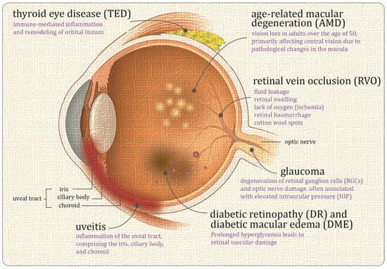
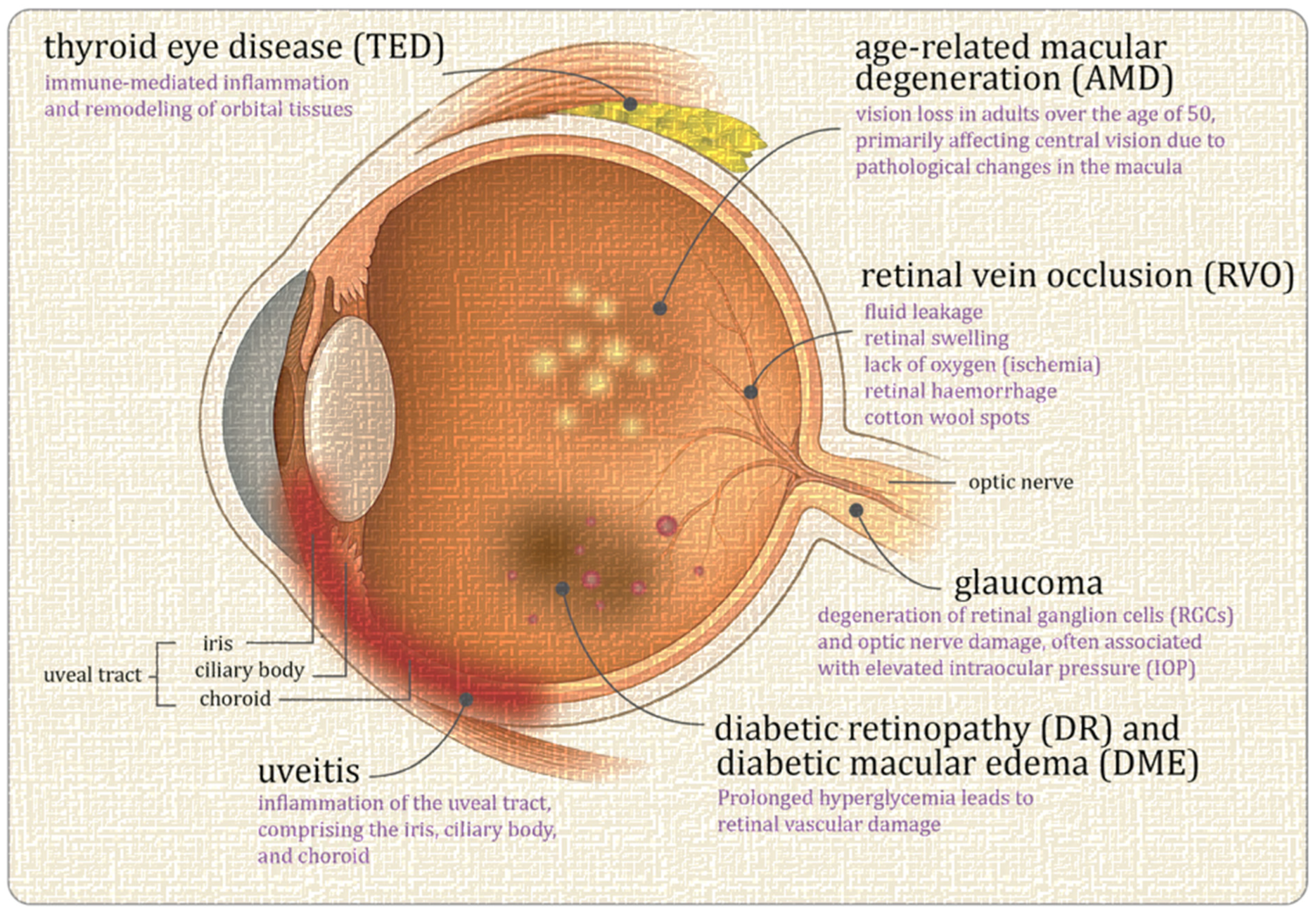
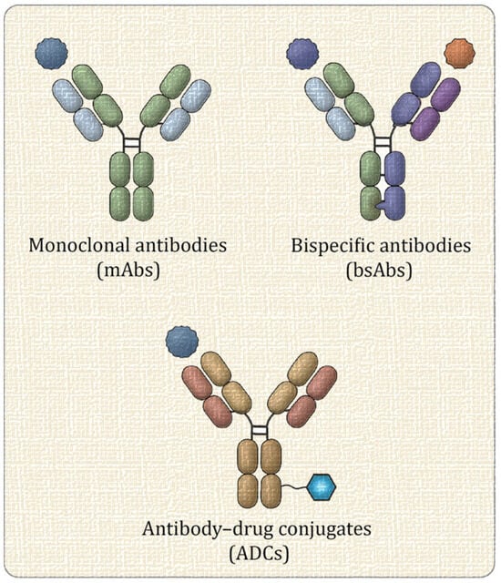
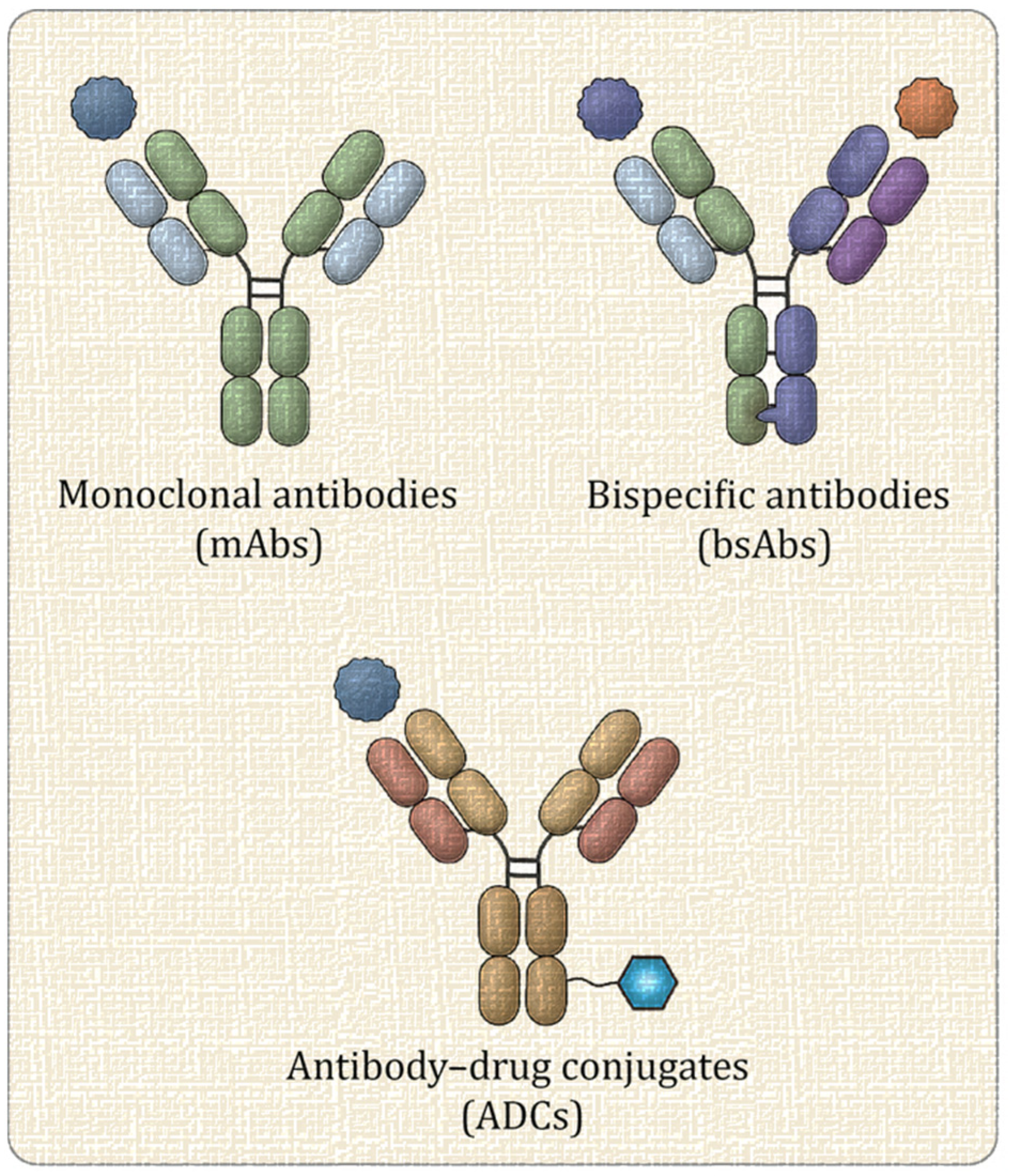
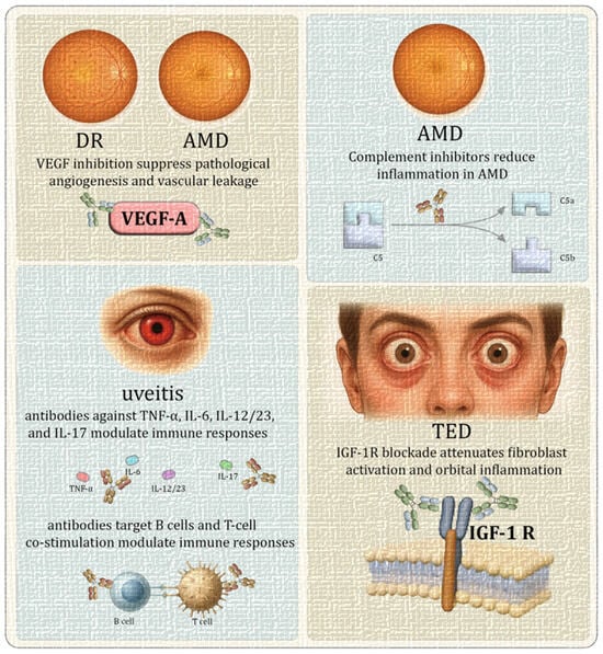
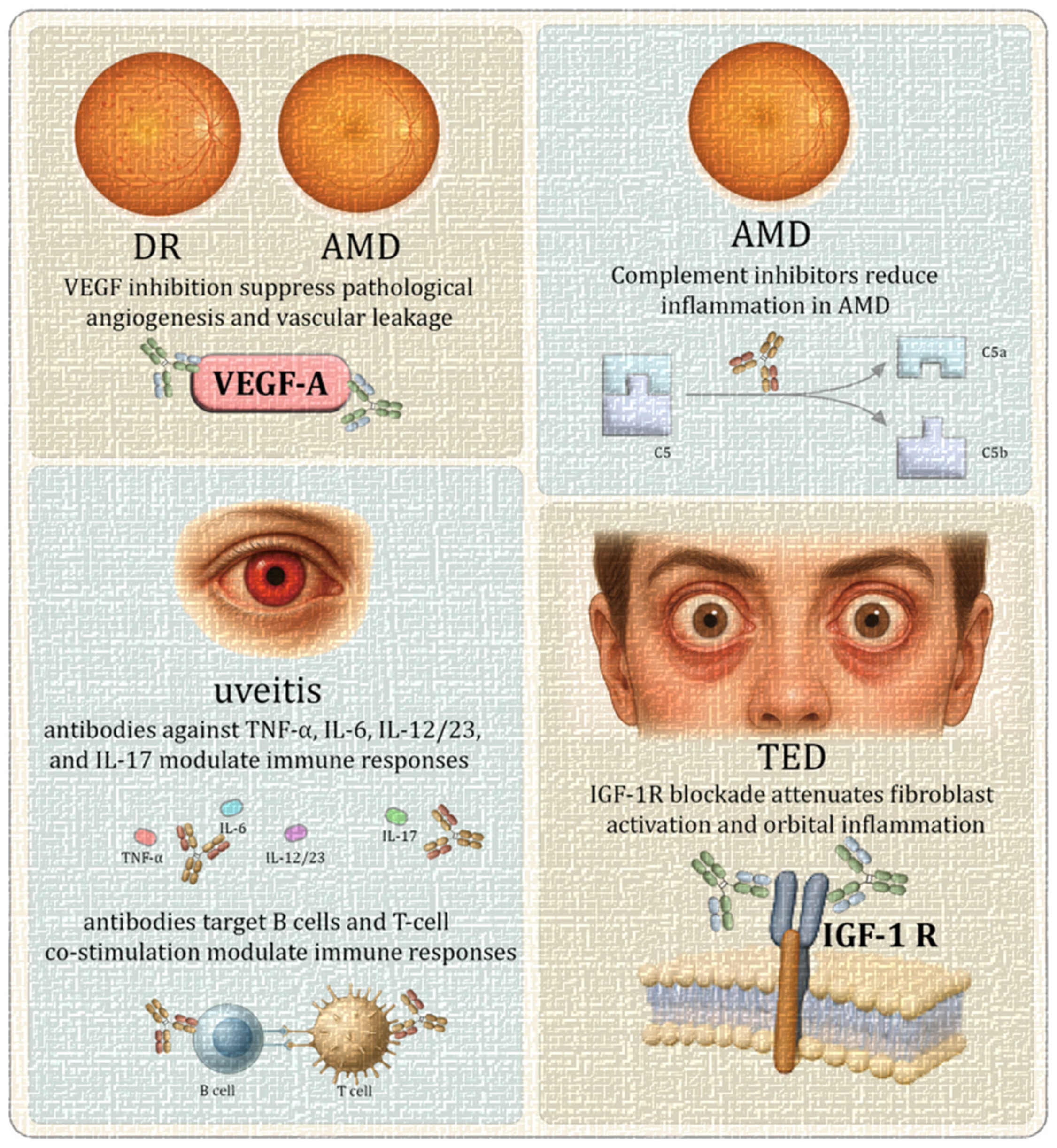
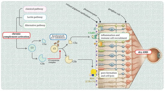
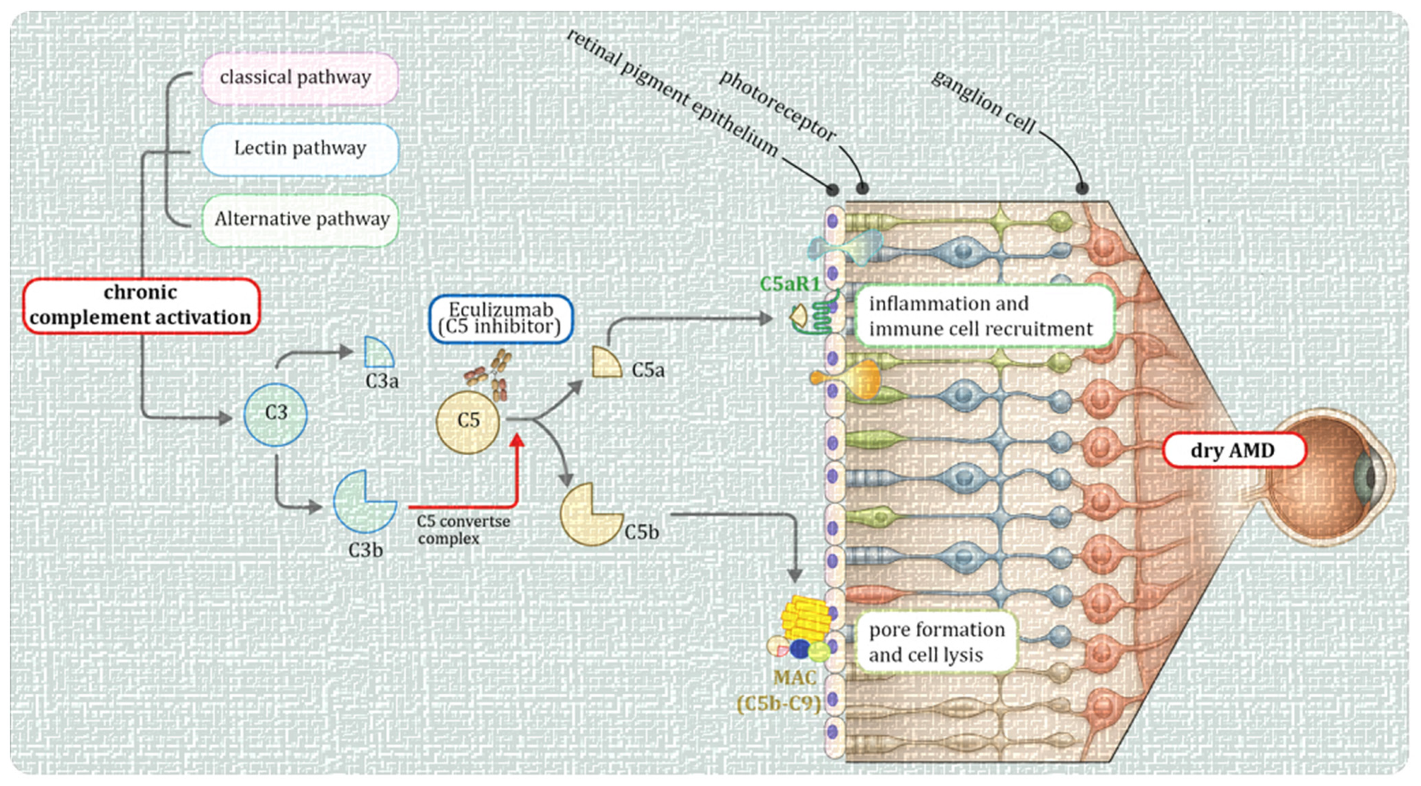
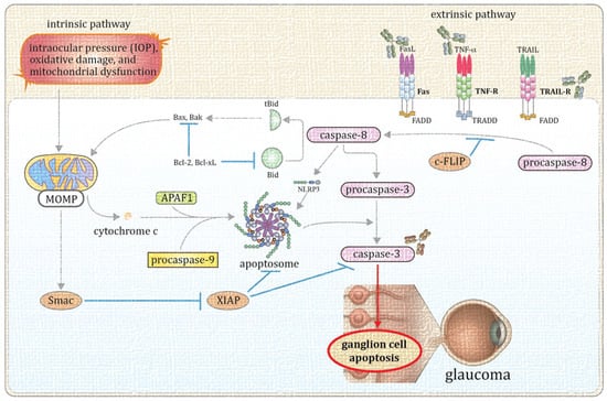
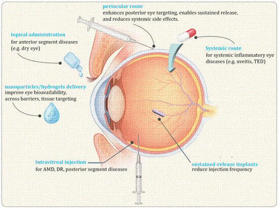
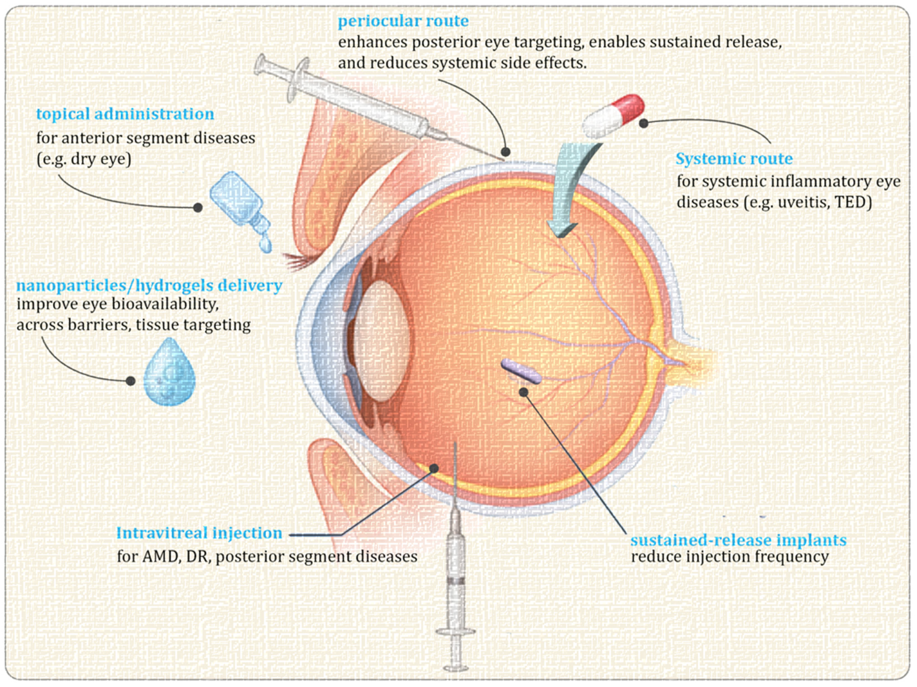

Disclaimer/Publisher’s Note: The statements, opinions and data contained in all publications are solely those of the individual author(s) and contributor(s) and not of MDPI and/or the editor(s). MDPI and/or the editor(s) disclaim responsibility for any injury to people or property resulting from any ideas, methods, instructions or products referred to in the content. |
© 2026 by the authors. Licensee MDPI, Basel, Switzerland. This article is an open access article distributed under the terms and conditions of the Creative Commons Attribution (CC BY) license.