Next Issue
Volume 44, April
Previous Issue
Volume 44, February
 
 
cimb-logo

Journal Browser

Journal Browser

Curr. Issues Mol. Biol., Volume 44, Issue 3 (March 2022) – 29 articles

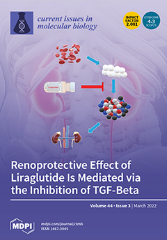
Cover Story (view full-size image): The aim of this research was to assess the effects of Liraglutide in the cell culture model of diabetic nephropathy on cell viability, antioxidant (GSH) and transforming growth factor-beta 1 (TGF-β1) levels, and extracellular matrix (ECM) expression. Metabolic activity was assessed by measuring Akt, pAkt, GSK3β, pGSK3β, pSTAT3, SOCS3, iNOS, and NOX4 protein expression with Western blot. The results attained in this study support a possible protective role of Liraglutide in this model probably mediated via inhibition of TGF-β1; however, this effect is dependent on the extent of cellular damage and type of toxic environment. Based on WB analysis, we have revealed signaling pathways involved in cytoprotective and cytotoxic effects of the drug itself. View this paper
  • Issues are regarded as officially published after their release is announced to the table of contents alert mailing list.
  • You may sign up for e-mail alerts to receive table of contents of newly released issues.
  • PDF is the official format for papers published in both, html and pdf forms. To view the papers in pdf format, click on the "PDF Full-text" link, and use the free Adobe Reader to open them.
Order results
Result details
Section
Select all
Export citation of selected articles as:
10 pages, 1552 KiB  
Article
Anti-Inflammatory Effect of Three Isolated Compounds of Physalis alkekengi var. franchetii (PAF) in Lipopolysaccharide-Activated RAW 264.7 Cells
by Hyun-Jung Park, Hyun Soo Shim, Ah-Reum Han, Eun-Kyoung Seo, Kyu-Ri Kim, Bong Hee Han and Insop Shim
Curr. Issues Mol. Biol. 2022, 44(3), 1407-1416; https://doi.org/10.3390/cimb44030094 - 21 Mar 2022
Cited by 2 | Viewed by 2361
Abstract
(1) Background: Three isolated compounds from Physalis alkekengi var. franchetii (PAF) have been investigated to possess a variety of biological activities. Their structures were elucidated by spectroscopic analysis (Ultraviolet (UV), High-resolution electrospray mass spectrometry (HR-ESI-Ms), and their anti-inflammatory effects were evaluated in vitro; [...] Read more.
(1) Background: Three isolated compounds from Physalis alkekengi var. franchetii (PAF) have been investigated to possess a variety of biological activities. Their structures were elucidated by spectroscopic analysis (Ultraviolet (UV), High-resolution electrospray mass spectrometry (HR-ESI-Ms), and their anti-inflammatory effects were evaluated in vitro; (2) Methods: To investigate the mechanisms of action of PAF extracts and their isolated compounds, their anti-inflammatory effects were assessed in RAW 264.7 macrophages stimulated by lipopolysaccharide (LPS). RAW 264.7 cells were treated with different concentrations of Physalis alkekengi var. franchetii three isolated compounds of PAF for 30 min prior to stimulation with or without LPS for the indicated times. The inflammatory cytokines, interleukin (IL)-1β and tumor necrosis factor (TNF)-α were determined using reverse transcription-polymerase chain (RT-PCR); (3) Results Treatment of RAW 264.7 cells with LPS alone resulted in significant increases in inflammatory cytokine production as compared to the control group (p < 0.001). However, with the treatment of isophysalin B 100 μg/mL, there was a significant decrease in the mRNA expression levels of TNF-α in LPS-stimulated raw 264.7 cells (p < 0.001). With treatment of physalin 1–100 μg/mL, there was a markedly decrease in the mRNA expression levels of TNF-α in LPS stimulated raw 264.7 (p < 0.05). Moreover, TNF-α mRNA (p < 0.05) and IL-1β mRNA (p < 0.001) mRNA levels were significantly suppressed after treatment with 3′,7-dimethylquercetin in LPS stimulated Raw 264.7 cells; (4) Conclusions: These findings suggest that three isolated compounds from can suppress inflammatory responses in LPS stimulated macrophage. Full article
Show Figures

Figure 1

12 pages, 1926 KiB  
Article
Cyclin D1 Serves as a Poor Prognostic Biomarker in Stage I Gastric Cancer
by Se-Il Go, Gyung Hyuck Ko, Won Sup Lee, Jeong-Hee Lee, Sang-Ho Jeong, Young-Joon Lee, Soon Chan Hong and Woo Song Ha
Curr. Issues Mol. Biol. 2022, 44(3), 1395-1406; https://doi.org/10.3390/cimb44030093 - 20 Mar 2022
Cited by 3 | Viewed by 1994
Abstract
TNM stage still serves as the best prognostic marker in gastric cancer (GC). The next step is to find prognostic biomarkers that detect subgroups with different prognoses in the same TNM stage. In this study, the expression levels of epidermal growth factor receptor [...] Read more.
TNM stage still serves as the best prognostic marker in gastric cancer (GC). The next step is to find prognostic biomarkers that detect subgroups with different prognoses in the same TNM stage. In this study, the expression levels of epidermal growth factor receptor (EGFR) and cyclin D1 were assessed in 96 tissue samples, including non-tumorous tissue, adenoma, and carcinoma. Then, the prognostic impact of EGFR and cyclin D1 was retrospectively investigated in 316 patients who underwent R0 resection for GC. EGFR positivity increased as gastric tissue became malignant, and cyclin D1 positivity was increased in all the tumorous tissues. However, there was no survival difference caused by the EGFR positivity, while the cyclin D1-postive group had worse overall survival (OS) than the cyclin D1-negative group in stage I GC (10-year survival rate (10-YSR): 62.8% vs. 86.5%, p = 0.010). In subgroup analyses for the propensity score-matched (PSM) cohort, there were also significant differences in the OS according to the cyclin D1 positivity in stage I GC but not in stage II and III GC. Upon multivariate analysis, cyclin D1 positivity was an independent prognostic factor in stage I GC. In conclusion, cyclin D1 may be a useful biomarker for predicting prognosis in stage I GC. Full article
(This article belongs to the Special Issue Molecules at Play in Cancer)
Show Figures

Figure 1

19 pages, 367 KiB  
Review
Features of the Metabolisms of Cardiac Troponin Molecules—Part 1: The Main Stages of Metabolism, Release Stage
by Aleksey Michailovich Chaulin
Curr. Issues Mol. Biol. 2022, 44(3), 1376-1394; https://doi.org/10.3390/cimb44030092 - 20 Mar 2022
Cited by 3 | Viewed by 2358
Abstract
Cardiac troponins (cTns) have long been the most valuable and specific biomarkers for detecting ischemic myocardial cells (MCs) injury, which is one of the key signs of myocardial infarction (MI). Modern methods (highly sensitive and ultra-sensitive immunoassays (hs-cTns)) of detection are an important [...] Read more.
Cardiac troponins (cTns) have long been the most valuable and specific biomarkers for detecting ischemic myocardial cells (MCs) injury, which is one of the key signs of myocardial infarction (MI). Modern methods (highly sensitive and ultra-sensitive immunoassays (hs-cTns)) of detection are an important and indispensable tool for the early diagnosis of MI and the choice of patient management protocols. Timely diagnosis of MI can significantly improve the prognosis of patients. However, in real clinical practice, doctors often face a significant problem when using cTns—the difficulty of differential diagnosis due to frequent and unexplained increases in the concentration of cTns in blood serum. In addition, there is conflicting information that may potentially affect the diagnostic capabilities and value of cTns: the influence of certain biological factors (diurnal rhythm, gender and age) on serum cTns levels; extra-cardiac expression of cTns; the possibilities of non-invasive diagnosis of MI; and other pathological conditions that cause non-ischemic injury to MCs. To solve these problems, it is necessary to concentrate on studying the metabolism of cTns. The review of our current knowledge about cTns metabolism consists of two parts. In this (first) part of the manuscript, the main stages of cTns metabolism are briefly described and the mechanisms of cTns release from MCs are considered in detail. Full article
(This article belongs to the Collection Feature Papers in Current Issues in Molecular Biology)
23 pages, 9556 KiB  
Review
Putative Mechanisms Underlying the Beneficial Effects of Polyphenols in Murine Models of Metabolic Disorders in Relation to Gut Microbiota
by Yoshimi Niwano, Hidetsugu Kohzaki, Midori Shirato, Shunichi Shishido and Keisuke Nakamura
Curr. Issues Mol. Biol. 2022, 44(3), 1353-1375; https://doi.org/10.3390/cimb44030091 - 18 Mar 2022
Cited by 5 | Viewed by 2686
Abstract
The beneficial effects of polyphenols on metabolic disorders have been extensively reported. The interaction of these compounds with the gut microbiota has been the focus of recent studies. In this review, we explored the fundamental mechanisms underlying the beneficial effects of polyphenols in [...] Read more.
The beneficial effects of polyphenols on metabolic disorders have been extensively reported. The interaction of these compounds with the gut microbiota has been the focus of recent studies. In this review, we explored the fundamental mechanisms underlying the beneficial effects of polyphenols in relation to the gut microbiota in murine models of metabolic disorders. We analyzed the effects of polyphenols on three murine models of metabolic disorders, namely, models of a high-fat diet (HFD)-induced metabolic disorder, dextran sulfate sodium (DSS)-induced colitis, and a metabolic disorder not associated with HFD or DSS. Regardless of the model, polyphenols ameliorated the effects of metabolic disorders by alleviating intestinal oxidative stress, improving inflammatory status, and improving intestinal barrier function, as well as by modulating gut microbiota, for example, by increasing the abundance of short-chain fatty acid-producing bacteria. Consequently, polyphenols reduce circulating lipopolysaccharide levels, thereby improving inflammatory status and alleviating oxidative imbalance at the lesion sites. In conclusion, polyphenols likely act by regulating intestinal functions, including the gut microbiota, and may be a safe and suitable therapeutic agent for various metabolic disorders. Full article
(This article belongs to the Special Issue Polyphenols as Cellular Metabolic Regulators)
Show Figures

Figure 1

21 pages, 2507 KiB  
Article
Multigene Panel Sequencing Reveals Cancer-Specific and Common Somatic Mutations in Colorectal Cancer Patients: An Egyptian Experience
by Amira Salah El-Din Youssef, Mohamed A. Abdel-Fattah, Mai M. Lotfy, Auhood Nassar, Mohamed Abouelhoda, Ahmed O. Touny, Zeinab K. Hassan, Mohammed Mohey Eldin, Abeer A. Bahnassy, Hussein Khaled and Abdel Rahman N. Zekri
Curr. Issues Mol. Biol. 2022, 44(3), 1332-1352; https://doi.org/10.3390/cimb44030090 - 18 Mar 2022
Cited by 4 | Viewed by 4178
Abstract
This study aims at identifying common pathogenic somatic mutations at different stages of colorectal carcinogenesis in Egyptian patients. Our cohort included colonoscopic biopsies collected from 120 patients: 20 biopsies from patients with inflammatory bowel disease, 38 from colonic polyp patients, and 62 from [...] Read more.
This study aims at identifying common pathogenic somatic mutations at different stages of colorectal carcinogenesis in Egyptian patients. Our cohort included colonoscopic biopsies collected from 120 patients: 20 biopsies from patients with inflammatory bowel disease, 38 from colonic polyp patients, and 62 from patients with colorectal cancer. On top of this, the cohort included 20 biopsies from patients with non-specific mild to moderated colitis. Targeted DNA sequencing using a customized gene panel of 96 colorectal related genes running on the Ion Torrent NGS technology was used to process the samples. Our results revealed that 69% of all cases harbored at least one somatic mutation. Fifty-seven genes were found to carry 232 somatic non-synonymous variants. The most frequently pathogenic somatic mutations were localized in TP53, APC, KRAS, and PIK3CA. In total, 16 somatic mutations were detected in the CRC group and in either the IBD or CP group. In addition, our data showed that 51% of total somatic variants were CRC-specific variants. The average number of CRC-specific variants per sample is 2.4. The top genes carrying CRC-specific mutations are APC, TP53, PIK3CA, FBXW7, ATM, and SMAD4. It seems obvious that TP53 and APC genes were the most affected genes with somatic mutations in all groups. Of interest, 85% and 28% of the APC and TP53 deleterious somatic mutations were located in Exon 14 and Exon 3, respectively. Besides, 37% and 28% of the total somatic mutations identified in APC and TP53 were CRC-specific variants, respectively. Moreover, we identified that, in 29 somatic mutations in 21 genes, their association with CRC patients was unprecedented. Ten detected variants were likely to be novel: six in PIK3CA and four variants in FBXW7. The detected P53, Wnt/βcatenin, Angiogenesis, EGFR, TGF-β and Interleukin signaling pathways were the most altered pathways in 22%, 16%, 12%, 10%, 9% and 9% of the CRC patients, respectively. These results would contribute to a better understanding of the colorectal cancer and in introducing personalized therapies for Egyptian CRC patients. Full article
(This article belongs to the Special Issue Linking Genomic Changes with Cancer in the NGS Era)
Show Figures

Figure 1

6 pages, 1349 KiB  
Article
hERG1 Potassium Channel Expression in Colorectal Adenomas: Comparison with Other Preneoplastic Lesions of the Gastrointestinal Tract
by Elena Lastraioli, Jessica Iorio, Federica Petrelli, Anna Tomezzoli, Serena Battista, Maria Raffaella Ambrosio, Mariella Chiudinelli, Federica De Salvatore, Luca Messerini, Vincenzo Villanacci, Luca Saragoni and Annarosa Arcangeli
Curr. Issues Mol. Biol. 2022, 44(3), 1326-1331; https://doi.org/10.3390/cimb44030089 - 17 Mar 2022
Cited by 2 | Viewed by 1735
Abstract
Preneoplastic lesions represent a useful target for early diagnosis and follow-up of gastrointestinal malignancies. hERG1 channel expression was tested by immunohistochemistry (IHC) in a cohort of colorectal adenoma samples belonging to Italian subjects. Overall, hERG1 was expressed in 56.5% of cases with both [...] Read more.
Preneoplastic lesions represent a useful target for early diagnosis and follow-up of gastrointestinal malignancies. hERG1 channel expression was tested by immunohistochemistry (IHC) in a cohort of colorectal adenoma samples belonging to Italian subjects. Overall, hERG1 was expressed in 56.5% of cases with both high staining intensity and a high percentage of positive cells. Moreover, hERG1 was expressed in a higher percentage of dysplastic adenomas with respect to hyperplastic lesions, and the proportion of positive samples further increased in patients with high-grade dysplasia. Comparing hERG1 expression in other preneoplastic lesions of the GI tract (gastric dysplasia and Barrett’s esophagus), it emerged that in all the conditions, hERG1 was expressed with a diffused pattern, throughout the cell, with variable staining intensity within the samples. The highest expression was detected in gastric dysplasia samples and the lowest in Barrett’s esophagus at similar levels observed in colorectal adenomas. Our results show that hERG1 is aberrantly expressed in human preneoplastic lesions of the gastrointestinal tract and has a different pattern of expression and role in the different sites. Overall, the detection of hERG1 expression in preneoplastic lesions could represent a novel diagnostic or prognostic marker of progression in the gastrointestinal tract. Full article
(This article belongs to the Special Issue Molecular-Based Approaches in Therapy for Gastrointestinal Cancers)
Show Figures

Figure 1

10 pages, 3857 KiB  
Article
Rotating Magnetic Field-Assisted Reactor Enhances Mechanisms of Phage Adsorption on Bacterial Cell Surface
by Bartłomiej Grygorcewicz, Rafał Rakoczy, Marta Roszak, Maciej Konopacki, Marian Kordas, Agnieszka Piegat, Natalia Serwin, Elżbieta Cecerska-Heryć, Miroslawa El Fray and Barbara Dołęgowska
Curr. Issues Mol. Biol. 2022, 44(3), 1316-1325; https://doi.org/10.3390/cimb44030088 - 17 Mar 2022
Cited by 7 | Viewed by 2295
Abstract
Growing interest in bacteriophage research and use, especially as an alternative treatment option for multidrug-resistant bacterial infection, requires rapid development of production methods and strengthening of bacteriophage activities. Bacteriophage adsorption to host cells initiates the process of infection. The rotating magnetic field (RMF) [...] Read more.
Growing interest in bacteriophage research and use, especially as an alternative treatment option for multidrug-resistant bacterial infection, requires rapid development of production methods and strengthening of bacteriophage activities. Bacteriophage adsorption to host cells initiates the process of infection. The rotating magnetic field (RMF) is a promising biotechnological method for process intensification, especially for the intensification of micromixing and mass transfer. This study evaluates the use of RMF to enhance the infection process by influencing bacteriophage adsorption rate. The RMF exposition decreased the t50 and t75 of bacteriophages T4 on Escherichia coli cells and vb_SauM_A phages on Staphylococcus aureus cells. The T4 phage adsorption rate increased from 3.13 × 10⁻⁹ mL × min−1 to 1.64 × 10⁻⁸ mL × min−1. The adsorption rate of vb_SauM_A phages exposed to RMF increased from 4.94 × 10⁻⁹ mL × min−1 to 7.34 × 10⁻⁹ mL × min−1. Additionally, the phage T4 zeta potential changed under RMF from −11.1 ± 0.49 mV to −7.66 ± 0.29 for unexposed and RMF-exposed bacteriophages, respectively. Full article
(This article belongs to the Collection Feature Papers in Current Issues in Molecular Biology)
Show Figures

Figure 1

22 pages, 1479 KiB  
Review
Molecular, Viral and Clinical Features of Alcohol- and Non-Alcohol-Induced Liver Injury
by Manuela G. Neuman, Helmut K. Seitz, Rolf Teschke, Stephen Malnick, Kamisha L. Johnson-Davis, Lawrence B. Cohen, Anit German, Nicolas Hohmann, Bernhardo Moreira, George Moussa and Mihai Opris
Curr. Issues Mol. Biol. 2022, 44(3), 1294-1315; https://doi.org/10.3390/cimb44030087 - 16 Mar 2022
Cited by 5 | Viewed by 2957
Abstract
Hepatic cells are sensitive to internal and external signals. Ethanol is one of the oldest and most widely used drugs in the world. The focus on the mechanistic engine of the alcohol-induced injury has been in the liver, which is responsible for the [...] Read more.
Hepatic cells are sensitive to internal and external signals. Ethanol is one of the oldest and most widely used drugs in the world. The focus on the mechanistic engine of the alcohol-induced injury has been in the liver, which is responsible for the pathways of alcohol metabolism. Ethanol undergoes a phase I type of reaction, mainly catalyzed by the cytoplasmic enzyme, alcohol dehydrogenase (ADH), and by the microsomal ethanol-oxidizing system (MEOS). Reactive oxygen species (ROS) generated by cytochrome (CYP) 2E1 activity and MEOS contribute to ethanol-induced toxicity. We aimed to: (1) Describe the cellular, pathophysiological and clinical effects of alcohol misuse on the liver; (2) Select the biomarkers and analytical methods utilized by the clinical laboratory to assess alcohol exposure; (3) Provide therapeutic ideas to prevent/reduce alcohol-induced liver injury; (4) Provide up-to-date knowledge regarding the Corona virus and its affect on the liver; (5) Link rare diseases with alcohol consumption. The current review contributes to risk identification of patients with alcoholic, as well as non-alcoholic, liver disease and metabolic syndrome. Additional prevalence of ethnic, genetic, and viral vulnerabilities are presented. Full article
Show Figures

Figure 1

10 pages, 1355 KiB  
Article
Bicarbonate-Independent Sodium Conductance of Na/HCO3 Cotransporter NBCn1 Decreases NMDA Receptor Function
by Inyeong Choi, Hansoo Yang, Eunjin Kim and Soojung Lee
Curr. Issues Mol. Biol. 2022, 44(3), 1284-1293; https://doi.org/10.3390/cimb44030086 - 13 Mar 2022
Cited by 1 | Viewed by 2108
Abstract
The sodium bicarbonate cotransporter NBCn1 is an electroneutral transporter with a channel activity that conducts Na+ in a HCO3-independent manner. This channel activity was suggested to functionally affect other membrane proteins which permeate Na+ influx. We previously reported [...] Read more.
The sodium bicarbonate cotransporter NBCn1 is an electroneutral transporter with a channel activity that conducts Na+ in a HCO3-independent manner. This channel activity was suggested to functionally affect other membrane proteins which permeate Na+ influx. We previously reported that NBCn1 is associated with the NMDA receptors (NMDARs) at the molecular and physiological levels. In this study, we examined whether NBCn1 channel activity affects NMDAR currents and whether this effect involves the interaction between the two proteins. NBCn1 and the NMDAR subunits GluN1A/GluN2A were expressed in Xenopus oocytes, and glutamate currents produced by the receptors were measured using two-electrode voltage clamp. In the absence of CO2/HCO3, NBCn1 channel activity decreased glutamate currents mediated by GluN1A/GluN2A. NBCn1 also decreased the slope of the current–voltage relationships for the glutamate current. Similar effects on the glutamate current were observed with and without PSD95, which can cluster NBCn1 and NMDARs. The channel activity was also observed in the presence of CO2/HCO3. We conclude that NBCn1 channel activity decreases NMDAR function. Given that NBCn1 knockout mice develop a downregulation of NMDARs, our results are unexpected and suggest that NBCn1 has dual effects on NMDARs. It stabilizes NMDAR expression but decreases receptor function by its Na+ channel activity. The dual effects may play an important role in fine-tuning the regulation of NMDARs in the brain. Full article
(This article belongs to the Collection Feature Papers in Current Issues in Molecular Biology)
Show Figures

Figure 1

11 pages, 1620 KiB  
Brief Report
Repeated Home-Applied Dual-Light Antibacterial Photodynamic Therapy Can Reduce Plaque Burden, Inflammation, and aMMP-8 in Peri-Implant Disease—A Pilot Study
by Hanna Lähteenmäki, Tommi Pätilä, Ismo T. Räisänen, Esko Kankuri, Taina Tervahartiala and Timo Sorsa
Curr. Issues Mol. Biol. 2022, 44(3), 1273-1283; https://doi.org/10.3390/cimb44030085 - 08 Mar 2022
Cited by 6 | Viewed by 4874
Abstract
Until now, in clinical dentistry, antibacterial photodynamic therapy (aPDT) has been restricted to in-office treatments, which hampers repeated applications. This pilot study tested the benefit of a commercially available Lumoral® device designed for regular periodontal dual-light aPDT treatment at home. Seven patients [...] Read more.
Until now, in clinical dentistry, antibacterial photodynamic therapy (aPDT) has been restricted to in-office treatments, which hampers repeated applications. This pilot study tested the benefit of a commercially available Lumoral® device designed for regular periodontal dual-light aPDT treatment at home. Seven patients with peri-implant disease applied dual-light aPDT daily in addition to their normal dental hygiene for four weeks. A single Lumoral® treatment includes an indocyanine green mouth rinse followed by 40 J/cm2 radiant exposure to a combination of 810 nm and 405 nm light. A point-of-care analysis of active-matrix metalloproteinase (aMMP-8), visible plaque index (VPI), bleeding on probing (BOP), and peri-implant pocket depth (PPD) measurements was performed on day 0, day 15, and day 30. Reductions in aMMP-8 (p = 0.047), VPI (p = 0.03), and BOP (p = 0.03) were observed, and PPD was measured as being 1 mm lower in the implant (p = ns). These results suggest a benefit of regular application of dual-light aPDT in peri-implantitis. Frequently repeated application can be a promising approach to diminishing the microbial burden and to lowering the tissue destructive proteolytic and inflammatory load around dental implants. Further studies in larger populations are warranted to show the long-term benefits. Full article
(This article belongs to the Special Issue Advances in Research on Molecular Oral Microorganisms)
Show Figures

Figure 1

16 pages, 4880 KiB  
Article
Short-Term Functional and Morphological Changes in the Primary Cultures of Trigeminal Ganglion Cells
by Carla Pires Veríssimo, Lionete Gall Acosta Filha, Fábio Jorge Moreira da Silva, Harrison Westgarth, Juliana De Mattos Coelho Aguiar, Bruno Pontes, Vivaldo Moura-Neto, Parisa Gazerani and Marcos F. DosSantos
Curr. Issues Mol. Biol. 2022, 44(3), 1257-1272; https://doi.org/10.3390/cimb44030084 - 08 Mar 2022
Cited by 6 | Viewed by 3344
Abstract
Several studies have proved that glial cells, as well as neurons, play a role in pain pathophysiology. Most of these studies have focused on the contribution of central glial cells (e.g., microglia and astrocytes) to neuropathic pain. Likewise, some works have suggested that [...] Read more.
Several studies have proved that glial cells, as well as neurons, play a role in pain pathophysiology. Most of these studies have focused on the contribution of central glial cells (e.g., microglia and astrocytes) to neuropathic pain. Likewise, some works have suggested that peripheral glial cells, particularly satellite glial cells (SGCs), and the crosstalk between these cells and the sensory neurons located in the peripheral ganglia, play a role in the phenomenon that leads to pain. Nonetheless, the study of SGCs may be challenging, as the validity of studying those cells in vitro is still controversial. In this study, a research protocol was developed to examine the potential use of primary mixed neuronal–glia cell cultures obtained from the trigeminal ganglion cells (TGCs) of neonate mice (P10–P12). Primary cultures were established and analyzed at 4 h, 24 h, and 48 h. To this purpose, phase contrast microscopy, immunocytochemistry with antibodies against anti-βIII-tubulin and Sk3, scanning electron microscopy, and time-lapse photography were used. The results indicated the presence of morphological changes in the cultured SGCs obtained from the TGCs. The SGCs exhibited a close relationship with neurons. They presented a round shape in the first 4 h, and a more fusiform shape at 24 h and 48 h of culture. On the other hand, neurons changed from a round shape to a more ramified shape from 4 h to 48 h. Intriguingly, the expression of SK3, a marker of the SGCs, was high in all samples at 4 h, with some cells double-staining for SK3 and βIII-tubulin. The expression of SK3 decreased at 24 h and increased again at 48 h in vitro. These results confirm the high plasticity that the SGCs may acquire in vitro. In this scenario, the authors hypothesize that, at 4 h, a group of the analyzed cells remained undifferentiated and, therefore, were double-stained for SK3 and βIII-tubulin. After 24 h, these cells started to differentiate into SCGs, which was clearer at 48 h in the culture. Mixed neuronal–glial TGC cultures might be implemented as a platform to study the plasticity and crosstalk between primary sensory neurons and SGCs, as well as its implications in the development of chronic orofacial pain. Full article
(This article belongs to the Section Biochemistry, Molecular and Cellular Biology)
Show Figures

Figure 1

10 pages, 901 KiB  
Article
The Selective Agonist for Sphingosine-1-Phosphate Receptors Siponimod Increases the Expression Level of NR4A Genes in Microglia Cell Line
by Francesca Montarolo, Serena Martire, Fabiana Marnetto, Paola Valentino, Sabdi Valverde, Marco Alfonso Capobianco and Antonio Bertolotto
Curr. Issues Mol. Biol. 2022, 44(3), 1247-1256; https://doi.org/10.3390/cimb44030083 - 07 Mar 2022
Cited by 4 | Viewed by 2633
Abstract
Fingolimod (FTY720) and siponimod (BAF312) are selective agonists for sphingosine-1-phosphate (S1P) receptors approved for the treatment of relapsing–remitting (RR) and secondary progressive (SP) multiple sclerosis (MS), respectively. BAF312 exerts pro-myelination and neuro-protective functions on CNS resident cells, although the underlying molecular mechanism is [...] Read more.
Fingolimod (FTY720) and siponimod (BAF312) are selective agonists for sphingosine-1-phosphate (S1P) receptors approved for the treatment of relapsing–remitting (RR) and secondary progressive (SP) multiple sclerosis (MS), respectively. BAF312 exerts pro-myelination and neuro-protective functions on CNS resident cells, although the underlying molecular mechanism is not yet fully understood. NR4A2 is an anti-inflammatory gene, belonging to the NR4A family, whose expression is reduced in blood from treatment-naïve patients with RRMS, but is restored in patients treated with FTY720 for more than two years. We performed an in vitro study to investigate the potential involvement of the NR4A genes in the protective and restorative effects of BAF312. We showed that BAF312 enhances the expression of NR4A1 and NR4A2 in the N9 microglial cell line, but has no effect in the peripheral blood mononuclear cells and oligodendrocytes. This study suggests a novel molecular mechanism of action for the selective agonists for S1P receptors within the CNS. Full article
(This article belongs to the Special Issue Signaling Pathways, Development, and Biomarkers in Neuropathy)
Show Figures

Figure 1

12 pages, 16599 KiB  
Article
LED Light-Induced ROS Differentially Regulates Focal Adhesion Kinase Activity in HaCaT Cell Viability
by Jun-Sub Kim and Ssang-Taek Steve Lim
Curr. Issues Mol. Biol. 2022, 44(3), 1235-1246; https://doi.org/10.3390/cimb44030082 - 04 Mar 2022
Cited by 5 | Viewed by 2753
Abstract
In this study, changes in cell signaling mechanisms in skin cells induced by various wavelengths and intensities of light-emitting diodes (LED) were investigated, focusing on the activity of focal adhesion kinase (FAK) in particular. We examined the effect of LED irradiation on cell [...] Read more.
In this study, changes in cell signaling mechanisms in skin cells induced by various wavelengths and intensities of light-emitting diodes (LED) were investigated, focusing on the activity of focal adhesion kinase (FAK) in particular. We examined the effect of LED irradiation on cell survival, the generation of intracellular reactive oxygen species (ROS), and the activity of various cell-signaling proteins. Red LED light increased cell viability at all intensities, whereas strong green and blue LED light reduced cell viability, and this effect was reversed by NAC or DPI treatment. Red LED light caused an increase in ROS formation according to the increase in the intensity of the LED light, and green and blue LED lights led to sharp increases in ROS formation. In the initial reaction to LEDs, red LED light only increased the phosphorylation of FAK and extracellular-signal regulated protein kinase (ERK), whereas green and blue LED lights increased the phosphorylation of inhibitory-κB Kinase α (IKKα), c-jun N-terminal kinase (JNK), and p38. The phosphorylation of these intracellular proteins was reduced via FAK inhibitor, NAC, and DPI treatments. Even after 24 h of LED irradiation, the activity of FAK and ERK appeared in cells treated with red LED light but did not appear in cells treated with green and blue LED lights. Furthermore, the activity of caspase-3 was confirmed along with cell detachment. Therefore, our results suggest that red LED light induced mitogenic effects via low levels of ROS–FAK–ERK, while green and blue LED lights induced cytotoxic effects via cellular stress and apoptosis signaling resulting from high levels of ROS. Full article
(This article belongs to the Collection Feature Papers in Current Issues in Molecular Biology)
Show Figures

Figure 1

11 pages, 1694 KiB  
Article
Tremella fuciformis Crude Polysaccharides Attenuates Steatosis and Suppresses Inflammation in Diet-Induced NAFLD Mice
by Tariq Jamal Khan, Xiaofei Xu, Xiaoling Xie, Ximing Dai, Pingnan Sun, Qingdong Xie and Xiaoling Zhou
Curr. Issues Mol. Biol. 2022, 44(3), 1224-1234; https://doi.org/10.3390/cimb44030081 - 03 Mar 2022
Cited by 9 | Viewed by 3216
Abstract
Nonalcoholic fatty liver disease (NAFLD) is a chronic liver disorder characterized by an enhanced accumulation of lipids, which affects around 40% of the world’s population. The T. fuciformis fungus possesses immunomodulatory activity and other beneficial properties that may alleviate steatosis through a different [...] Read more.
Nonalcoholic fatty liver disease (NAFLD) is a chronic liver disorder characterized by an enhanced accumulation of lipids, which affects around 40% of the world’s population. The T. fuciformis fungus possesses immunomodulatory activity and other beneficial properties that may alleviate steatosis through a different mechanism. The present study was designed to evaluate the effect T. fuciformis crude polysaccharides (TFCP) on inflammatory and lipid metabolism gene expression, oxidative stress, and lipid profile. Mice were divided into groups receiving (a) a normal chow diet (NCD), (b) a methionine–choline-deficient (MCD) diet, and (c) a MCD diet with TFCP. Liver histopathology was performed, and the hepatic gene expression levels were estimated using qRT-PCR. The lipid profiles, ALT, AST, and efficient oxidative enzymes were analyzed using ELISA. The TFCP administration in the MCD-fed mice suppressed hepatic lipid accumulation, lipid metabolism-associated genes (HMGCR, FABP, SREBP, ACC, and FAS), and inflammation-associated genes (IL-1β, TLR4, TNF-α, and IL-6) whilst enhancing the expression of HNF4α genes. TFCP mitigated against oxidative stress and normalized healthy lipid profiles. These results highlighted that TFCP prevents NAFLD through the inhibition of oxidative stress and inflammation, suggesting TFCP would potentially be an effective therapeutic agent against NAFLD progression. Full article
(This article belongs to the Section Bioorganic Chemistry and Medicinal Chemistry)
Show Figures

Figure 1

9 pages, 2870 KiB  
Article
Evaluation of Ectopic Mitochondrial DNA in HeLa Cells
by Mohammad T. Hussan, Noriko Matsui and Hideaki Matsui
Curr. Issues Mol. Biol. 2022, 44(3), 1215-1223; https://doi.org/10.3390/cimb44030080 - 02 Mar 2022
Viewed by 3054
Abstract
The presence of ectopic DNA in the cytoplasm induces inflammation and cell death. It has been widely reported that leakage of nuclear DNA into the cytoplasm can mainly be sensed by cyclic GMP-AMP synthase (cGAS). We recently reported that mitochondria-derived cytoplasmic double-stranded DNA [...] Read more.
The presence of ectopic DNA in the cytoplasm induces inflammation and cell death. It has been widely reported that leakage of nuclear DNA into the cytoplasm can mainly be sensed by cyclic GMP-AMP synthase (cGAS). We recently reported that mitochondria-derived cytoplasmic double-stranded DNA (dsDNA) that has escaped lysosomal degradation induces significant cytotoxicity in cultured cells and in vivo. Cytoplasmic mitochondrial DNA is assumed to be involved in various diseases and disorders, and more and more papers have been published confirming this. On the other hand, the current method for evaluating mitochondrial DNA in the cytoplasm may not be quantitative. Here, we introduce in detail a method to evaluate ectopic mitochondrial DNA in cells. This method is useful in basic research as well as in the study of aging, Parkinson’s disease, Alzheimer’s disease, heart failure, autoimmune diseases, cancer, and other conditions. Full article
Show Figures

Figure 1

12 pages, 2074 KiB  
Article
Zinc Finger E-Box Binding Homeobox 2 as a Prognostic Biomarker in Various Cancers and Its Correlation with Infiltrating Immune Cells in Ovarian Cancer
by Hye-Ran Kim, Choong Won Seo, Sang Jun Han, Jae-Ho Lee and Jongwan Kim
Curr. Issues Mol. Biol. 2022, 44(3), 1203-1214; https://doi.org/10.3390/cimb44030079 - 01 Mar 2022
Cited by 2 | Viewed by 2845
Abstract
This study investigated the expression of zinc finger E-box binding homeobox 2 (ZEB2), its prognostic significance in various cancers, and the correlation between ZEB2 and infiltrating immune cells and ZEB2-related proteins in ovarian cancer (OV). The Gene Expression Profiling Interactive Analysis tool was [...] Read more.
This study investigated the expression of zinc finger E-box binding homeobox 2 (ZEB2), its prognostic significance in various cancers, and the correlation between ZEB2 and infiltrating immune cells and ZEB2-related proteins in ovarian cancer (OV). The Gene Expression Profiling Interactive Analysis tool was used to analyze RNA sequencing data and cancer survival rates, based on normal and tumor tissue data available in The Cancer Genome Atlas (TCGA) database. The Kaplan–Meier plotter and PrognoScan databases were used to analyze the prognostic value of ZEB2 in OV (n = 1144). The Tumor Immune Estimation Resource was used to investigate the correlation between ZEB2 and infiltrating immune cells in various cancers, including OV. High ZEB2 expression was associated with a poorer prognosis in OV. In OV, ZEB2 is positively correlated with CD8+T cells, neutrophils, macrophages, and dendritic cell invasion; and ZEB2 is negatively correlated with tumor-infiltrating B cells. The STRING database was used to investigate the correlations with ZEB2-related proteins. The results reveal that ZEB2 was positively correlated with SMAD1 and SMAD2 in OV. Our findings may serve as a potential prognostic biomarker, and provide novel insights into the tumor immunology in OV. Thus, ZEB2 may be a potential diagnostic and therapeutic target in OV. Full article
(This article belongs to the Special Issue Molecules at Play in Cancer)
Show Figures

Figure 1

12 pages, 846 KiB  
Review
Alternate Causes for Pathogenesis of Exfoliation Glaucoma, a Multifactorial Elastotic Disorder: A Literature Review
by Munmun Chakraborty and Aparna Rao
Curr. Issues Mol. Biol. 2022, 44(3), 1191-1202; https://doi.org/10.3390/cimb44030078 - 01 Mar 2022
Cited by 10 | Viewed by 2750
Abstract
Exfoliation glaucoma (XFG) is the most recognizable form of secondary open-angle glaucoma associated with a high risk of blindness. This disease is characterized by white flaky granular deposits in the anterior chamber that leads to the elevation of intraocular pressure (IOP) and subsequent [...] Read more.
Exfoliation glaucoma (XFG) is the most recognizable form of secondary open-angle glaucoma associated with a high risk of blindness. This disease is characterized by white flaky granular deposits in the anterior chamber that leads to the elevation of intraocular pressure (IOP) and subsequent glaucomatous optic nerve damage. Conventionally, XFG is known to respond poorly to medical therapy, and surgical intervention is the only management option in most cases. Various genetic and nongenetic factors are known to be linked to the development of XFG. Despite decades of research on the genetic factors in exfoliation syndrome (XFS) by study groups and global consortia involving different ethnic populations, the pathogenesis of XFS and the mechanism of onset of glaucoma still remains an unsolved mystery. The key lies in understanding how the function of a gene (or set of genes) is altered by environmental triggers, along with other molecular events that underlie the key disease attributes, namely, oxidative stress and the disruption of the blood–aqueous barrier (BAB). It remains a challenge to evolve a theory encompassing all factions of molecular events occurring independently or parallelly that determine the disease manifestation (phenotype) or the stage of the disease in the eye (or in any tissue) in exfoliation. Our enhanced understanding of the underlying molecular pathophysiology of XFG, beyond the known genes or polymorphisms involved in the disease, will lead to improved diagnosis and management and the ability to recognize how the environment influences these key events that lead to the disease phenotype or disease progression. This review summarizes the recent observations and discoveries of four key factors that may hold the answers to the non-lysyl oxidase-like 1 (LOXL1) mechanisms behind XFG pathogenesis, namely, the epigenetic factor miRNA, disordered autophagy along with the potential involvement of mitochondrial mutations, and a compromised aqueous–blood barrier. Full article
(This article belongs to the Section Molecular Medicine)
Show Figures

Figure 1

9 pages, 5817 KiB  
Article
MicroRNA-1270 Inhibits Cell Proliferation, Migration, and Invasion via Targeting IRF8 in Osteoblast-like Cell Lines
by Eric Gustavo Ramírez-Salazar, Erika Victoria Almeraya, Tania Valentina López-Perez, Zacarías Jiménez-Salas, Nelly Patiño and Rafael Velázquez-Cruz
Curr. Issues Mol. Biol. 2022, 44(3), 1182-1190; https://doi.org/10.3390/cimb44030077 - 01 Mar 2022
Cited by 3 | Viewed by 2810
Abstract
Osteoporosis (OP) is the most common bone disease affecting elderly individuals. The diagnosis of this pathology is most commonly made on the basis of bone fractures. Several microRNAs (miRNAs/miRs) have been identified as possible biomarkers for the diagnosis and treatment of OP. miRNAs [...] Read more.
Osteoporosis (OP) is the most common bone disease affecting elderly individuals. The diagnosis of this pathology is most commonly made on the basis of bone fractures. Several microRNAs (miRNAs/miRs) have been identified as possible biomarkers for the diagnosis and treatment of OP. miRNAs can regulate gene expression, and determining their functions can provide potential pharmacological targets for treating OP. A previous study showed that miR-1270 was upregulated in monocytes derived from postmenopausal women with OP. Therefore, the present study aimed to uncover the role of miR-1270 in regulating bone metabolism. To reveal the mechanism underlying the regulatory effect of miR-1270 on interferon regulatory factor 8 (IRF8) expression, luciferase assay, reverse transcription-quantitative PCR, and Western blot analysis were performed. The results suggest that miR-1270 could regulate the mRNA and protein expression levels of IRF8 by directly binding to its 3′-untranslated region. The effects of miR-1270 overexpression and IRF8 silencing on cell proliferation, migration, and invasion were also evaluated. To the best of our knowledge, the current study was the first to support the crucial role of miR-1270 in bone metabolism via modulation of IRF8 expression. In addition, miR-1270 overexpression could attenuate human osteoblast-like cells’ proliferation and migration ability. Full article
(This article belongs to the Section Biochemistry, Molecular and Cellular Biology)
Show Figures

Figure 1

13 pages, 2366 KiB  
Article
Antimicrobial Properties of a Peptide Derived from the Male Fertility Factor kl2 Protein of Drosophila melanogaster
by Bernadetta Bilska, Urszula Godlewska, Milena Damulewicz, Krzysztof Murzyn, Mateusz Kwitniewski, Joanna Cichy and Elżbieta Pyza
Curr. Issues Mol. Biol. 2022, 44(3), 1169-1181; https://doi.org/10.3390/cimb44030076 - 28 Feb 2022
Viewed by 2396
Abstract
Antimicrobial peptides (AMPs) are important components of innate immunity. Here, we report the antimicrobial properties of a peptide derived from the Male fertility factor kl2 (MFF-kl2) protein of Drosophila melanogaster, which was identified as a functional analog of the mammalian antibacterial chemerin-p4 [...] Read more.
Antimicrobial peptides (AMPs) are important components of innate immunity. Here, we report the antimicrobial properties of a peptide derived from the Male fertility factor kl2 (MFF-kl2) protein of Drosophila melanogaster, which was identified as a functional analog of the mammalian antibacterial chemerin-p4 peptide. The antimicrobial activity of multifunctional chemerin is mainly associated with a domain localized in the middle of the chemerin sequence, Val66-Pro85 peptide (chemerin-p4). Using bioinformatic tools, we found homologs of the chemerin-p4 peptide in the proteome of D. melanogaster. One of them is MFF-p1, which is a part of the MFF kl2 protein, encoded by the gene male fertility factor kl2 (kl-2) located on the long arm of the Y chromosome. The second detected peptide (Z-p1) is a part of the Zizimin protein belonging to DOCK family, which is involved in cellular signaling processes. After testing the antimicrobial properties of both peptides, we found that only MFF-p1 possesses these properties. Here, we demonstrate its antimicrobial potential both in vitro and in vivo after infecting D. melanogaster with bacteria. MFF-p1 strongly inhibits the viable counts of E. coli and B. subtilis after 2 h of treatment and disrupts bacterial cells. The expression of kl-2 is regulated by exposure to bacteria and by the circadian clock. Full article
(This article belongs to the Section Bioinformatics and Systems Biology)
Show Figures

Figure 1

20 pages, 6689 KiB  
Article
DNA Methylation Patterns According to Fatty Liver Index and Longitudinal Changes from the Korean Genome and Epidemiology Study (KoGES)
by Young Kyung Ko, Hayeon Kim, Yoonseok Lee, Young-Sun Lee and Jeong-An Gim
Curr. Issues Mol. Biol. 2022, 44(3), 1149-1168; https://doi.org/10.3390/cimb44030075 - 27 Feb 2022
Cited by 3 | Viewed by 2795
Abstract
The role of differentially methylated regions (DMRs) in nonalcoholic fatty liver disease (NAFLD) is unclear. This study aimed to identify the role of DMR in NAFLD development and progression using the Korean Genome and Epidemiology Study (KoGES) cohort. We used laboratory evaluations and [...] Read more.
The role of differentially methylated regions (DMRs) in nonalcoholic fatty liver disease (NAFLD) is unclear. This study aimed to identify the role of DMR in NAFLD development and progression using the Korean Genome and Epidemiology Study (KoGES) cohort. We used laboratory evaluations and Illumina Methylation 450 k DNA methylation microarray data from KoGES. The correlation between fatty liver index (FLI) and genomic CpG sites was analyzed in 322 subjects. Longitudinal changes over 8 years were confirmed in 33 subjects. To identify CpG sites and genes related to FLI, we obtained enrichment terms for 6765 genes. DMRs were identified for both high (n = 128) and low (n = 194) groups on the basis of FLI 30 in 142 men and 180 women. To confirm longitudinal changes in 33 subjects, the ratio of follow-up and baseline investigation values was obtained. Correlations and group comparisons were performed for the 8 year change values. PITPNM3, RXFP3, and THRB were hypermethylated in the increased FLI groups, whereas SLC9A2 and FOXI3 were hypermethylated in the decreased FLI groups. DMRs describing NAFLD were determined, and functions related to inflammation were identified. Factors related to longitudinal changes are suggested, and blood circulation-related functions appear to be important in the management of NAFLD. Full article
Show Figures

Figure 1

22 pages, 770 KiB  
Review
Use of Next-Generation Sequencing for Identifying Mitochondrial Disorders
by Shafi Mahmud, Suvro Biswas, Shamima Afrose, Mohasana Akter Mita, Md. Robiul Hasan, Mst. Sharmin Sultana Shimu, Gobindo Kumar Paul, Sanghyun Chung, Md. Abu Saleh, Sultan Alshehri, Momammed M. Ghoneim, Maha Alruwaily and Bonglee Kim
Curr. Issues Mol. Biol. 2022, 44(3), 1127-1148; https://doi.org/10.3390/cimb44030074 - 27 Feb 2022
Cited by 7 | Viewed by 6645
Abstract
Mitochondria are major contributors to ATP synthesis, generating more than 90% of the total cellular energy production through oxidative phosphorylation (OXPHOS): metabolite oxidation, such as the β-oxidation of fatty acids, and the Krebs’s cycle. OXPHOS inadequacy due to large genetic lesions in mitochondrial [...] Read more.
Mitochondria are major contributors to ATP synthesis, generating more than 90% of the total cellular energy production through oxidative phosphorylation (OXPHOS): metabolite oxidation, such as the β-oxidation of fatty acids, and the Krebs’s cycle. OXPHOS inadequacy due to large genetic lesions in mitochondrial as well as nuclear genes and homo- or heteroplasmic point mutations in mitochondrially encoded genes is a characteristic of heterogeneous, maternally inherited genetic disorders known as mitochondrial disorders that affect multisystemic tissues and organs with high energy requirements, resulting in various signs and symptoms. Several traditional diagnostic approaches, including magnetic resonance imaging of the brain, cardiac testing, biochemical screening, variable heteroplasmy genetic testing, identifying clinical features, and skeletal muscle biopsies, are associated with increased risks, high costs, a high degree of false-positive or false-negative results, or a lack of precision, which limits their diagnostic abilities for mitochondrial disorders. Variable heteroplasmy levels, mtDNA depletion, and the identification of pathogenic variants can be detected through genetic sequencing, including the gold standard Sanger sequencing. However, sequencing can be time consuming, and Sanger sequencing can result in the missed recognition of larger structural variations such as CNVs or copy-number variations. Although each sequencing method has its own limitations, genetic sequencing can be an alternative to traditional diagnostic methods. The ever-growing roster of possible mutations has led to the development of next-generation sequencing (NGS). The enhancement of NGS methods can offer a precise diagnosis of the mitochondrial disorder within a short period at a reasonable expense for both research and clinical applications. Full article
(This article belongs to the Special Issue Mitochondrial Genome 2024)
Show Figures

Figure 1

12 pages, 2431 KiB  
Article
Intracellular Reverse Transcription of Pfizer BioNTech COVID-19 mRNA Vaccine BNT162b2 In Vitro in Human Liver Cell Line
by Markus Aldén, Francisko Olofsson Falla, Daowei Yang, Mohammad Barghouth, Cheng Luan, Magnus Rasmussen and Yang De Marinis
Curr. Issues Mol. Biol. 2022, 44(3), 1115-1126; https://doi.org/10.3390/cimb44030073 - 25 Feb 2022
Cited by 58 | Viewed by 2167292
Abstract
Preclinical studies of COVID-19 mRNA vaccine BNT162b2, developed by Pfizer and BioNTech, showed reversible hepatic effects in animals that received the BNT162b2 injection. Furthermore, a recent study showed that SARS-CoV-2 RNA can be reverse-transcribed and integrated into the genome of human cells. In [...] Read more.
Preclinical studies of COVID-19 mRNA vaccine BNT162b2, developed by Pfizer and BioNTech, showed reversible hepatic effects in animals that received the BNT162b2 injection. Furthermore, a recent study showed that SARS-CoV-2 RNA can be reverse-transcribed and integrated into the genome of human cells. In this study, we investigated the effect of BNT162b2 on the human liver cell line Huh7 in vitro. Huh7 cells were exposed to BNT162b2, and quantitative PCR was performed on RNA extracted from the cells. We detected high levels of BNT162b2 in Huh7 cells and changes in gene expression of long interspersed nuclear element-1 (LINE-1), which is an endogenous reverse transcriptase. Immunohistochemistry using antibody binding to LINE-1 open reading frame-1 RNA-binding protein (ORFp1) on Huh7 cells treated with BNT162b2 indicated increased nucleus distribution of LINE-1. PCR on genomic DNA of Huh7 cells exposed to BNT162b2 amplified the DNA sequence unique to BNT162b2. Our results indicate a fast up-take of BNT162b2 into human liver cell line Huh7, leading to changes in LINE-1 expression and distribution. We also show that BNT162b2 mRNA is reverse transcribed intracellularly into DNA in as fast as 6 h upon BNT162b2 exposure. Full article
Show Figures

Figure 1

28 pages, 9558 KiB  
Article
Renoprotective Effect of Liraglutide Is Mediated via the Inhibition of TGF-Beta 1 in an LLC-PK1 Cell Model of Diabetic Nephropathy
by Vjera Ninčević, Milorad Zjalić, Tea Omanović Kolarić, Martina Smolić, Tomislav Kizivat, Lucija Kuna, Aleksandar Včev, Ashraf Tabll and Ines Bilić Ćurčić
Curr. Issues Mol. Biol. 2022, 44(3), 1087-1114; https://doi.org/10.3390/cimb44030072 - 25 Feb 2022
Cited by 4 | Viewed by 3668
Abstract
Background: Recently published research demonstrated direct renoprotective effects of the glucagon-like peptide-1 receptor agonist GLP 1 RA, but the relevant molecular mechanisms are still not clear. The aim of this research was to assess the effects of Liraglutide in a cell culture model [...] Read more.
Background: Recently published research demonstrated direct renoprotective effects of the glucagon-like peptide-1 receptor agonist GLP 1 RA, but the relevant molecular mechanisms are still not clear. The aim of this research was to assess the effects of Liraglutide in a cell culture model of diabetic nephropathy on cell viability, antioxidant (GSH) and transforming growth factor beta 1 (TGF- β1) levels and extracellular matrix (ECM) expression. The metabolic activity in hyperglycemic conditions and the effect of Liraglutide treatment were assessed by measuring Akt, pAkt, GSK3β, pGSK3β, pSTAT3, SOCS3, iNOS and NOX4 protein expression with Western blot. F actin distribution was used to assess the structural changes of the cells upon treatment. Materials and methods: The cells were exposed to high glucose (HG30 mM) followed by 0.5 mM H2O2 and a combination of glucose and H2O2 during 24 h. Subsequently, the cells were treated with different combinations of HG30, H2O2 and Liraglutide. Cell viability was determined by an MTT colorimetric test, and the GSH, TGF-β1 concentration and ECM expression were measured using a spectrophotometric/microplate reader assay and an ELISA kit, respectively. Western blotting was used to detect the protein level of Akt, pAkt, GSK3β, pGSK3β, pSTAT3, SOCS3, iNOS and NOX4. The F-actin cytoskeleton was visualized with Phalloidin stain and subsequently quantified. Results: Cell viability was decreased as well as GSH levels in cells treated with a combination of HG30/H2O2, and HG30 alone (p < 0.001). The addition of Liraglutide improved the viability in cells treated with HG30, but it did not affect the cell viability in the cell treated with the addition of H2O2. GSH increased with the addition of Liraglutide in HG30/H2O2 (p < 0.001) treated cells, with no effect in cells treated only with HG30. TGF-β1 levels (p < 0.001) were significantly increased in HG30 and HG30/H2O2. The addition of Liraglutide significantly decreased the TGF-β1 levels (p < 0.01; p < 0.05) in all treated cells. The synthesis of collagen was significantly increased in HG30/H2O2 (p < 0.001), while the addition of Liraglutide in HG30/H2O2 significantly decreased collagen (p < 0.001). Akt signaling was not significantly affected by treatment. The GSK3b and NOX4 levels were significantly reduced (p < 0.01) after the peroxide and glucose treatment, with the observable restoration upon the addition of Liraglutide suggesting an important role of Liraglutide in oxidative status regulation and mitochondrial activity. The treatment with Liraglutide significantly upregulated STAT3 (p < 0.01) activity, with no change in SOCS3 indicating a selective regulation of the STAT 3 signaling pathway in glucose and the oxidative overloaded environment. A significant reduction in the distribution of F-actin was observed in cells treated with HG30/H2O2 (p < 0.01). The addition of Liraglutide to HG30-treated cells led to a significant decrease of distribution of F-actin (p < 0.001). Conclusion: The protective effect of Liraglutide is mediated through the inhibition of TGF beta, but this effect is dependent on the extent of cellular damage and the type of toxic environment. Based on the WB analysis we have revealed the signaling pathways involved in cytoprotective and cytotoxic effects of the drug itself, and further molecular studies in vitro and vivo are required to elucidate the complexity of the pathophysiological mechanisms of Liraglutide under conditions of hyperglycemia and oxidative stress. Full article
Show Figures

Figure 1

12 pages, 851 KiB  
Article
Effects of Essential Amino Acid Deficiency on General Control Nonderepressible 2/Eukaryotic Initiation Factor 2 Signaling and Proteomic Changes in Primary Bovine Mammary Epithelial Cells
by Zulma Tatiana Ruiz-Cortés, Peter Yoder and Mark D. Hanigan
Curr. Issues Mol. Biol. 2022, 44(3), 1075-1086; https://doi.org/10.3390/cimb44030071 - 25 Feb 2022
Cited by 2 | Viewed by 2381
Abstract
We hypothesized that the general control nonderepressible 2 (GCN2)/eukaryotic initiation factor 2 (eIF2) signaling pathway and intracellular protein synthesis (PS) are regulated to maintain milk PS in primary bovine mammary epithelial cells (MECs) under essential amino acid (EAA) starvation conditions. We cultured MECs [...] Read more.
We hypothesized that the general control nonderepressible 2 (GCN2)/eukaryotic initiation factor 2 (eIF2) signaling pathway and intracellular protein synthesis (PS) are regulated to maintain milk PS in primary bovine mammary epithelial cells (MECs) under essential amino acid (EAA) starvation conditions. We cultured MECs with 0%, 2% (depletion), and 100% (control) EAA for two exposure times (8 and 24 h), followed by three refeeding (RF) times with 100% EAA (0, 8, and 24 h). Subsequently, we measured cell viability, total protein concentration, and proliferation. Western blotting was used to quantify the levels of casein and the expression of total GCN2 and eIF2, as well as phosphorylated GCN2 (GCN2P) and eIF2 (eIF2P). The ISOQuant method was used to assess MEC proteomes, and the resultant data were analyzed using the Kruskal–Wallis test, nonpaired Wilcoxon rank post-hoc test, and ANOVA–Tukey test, as well as principal component analyses and multiple regressions models. Differences in cell viability were observed between the control versus the depleted and repleted MECs, respectively, where 97.2–99.8% viability indicated low cell death rates. Proliferation (range, 1.02–1.55 arbitrary units (AU)) was affected by starvation for 12 and 24 h and repletion for 24 h, but it was not increased compared with the control. Total protein expression was unaffected by both depletion and repletion treatments (median 3158 µg/mL). eIF2P expression was significantly increased (p < 0.05) after treatment with 2% EAA for 8 and 24 h compared with 2% EAA with 8 h + 24 h RF and 2% EAA with 24 h + 8 h RF. GCN2P also showed significantly increased expression (p < 0.05) after treatment with 2% EAA for 24 h compared with the control and 2% EAA with 24 h + 8 h RF. Intracellular casein/α-tubulin expression was unaffected by 2% EAA compared with control (0.073 ± 0.01 AU versus 0.086 ± 0.02 AU, respectively). We studied 30 of the detected 1180 proteins, 16 of which were differentially expressed in starved and refed MECs. Cells faced with EAA deficiency activated the GCN2P/eIF2P pathway, and the lack of change in the levels of casein and other milk proteins suggested that the EAA deficit was mitigated by metabolic flexibility to maintain homeostasis. Full article
Show Figures

Figure 1

13 pages, 37866 KiB  
Article
Nordentatin Inhibits Neuroblastoma Cell Proliferation and Migration through Regulation of GSK-3 Pathway
by Chantana Boonyarat, Panatchakorn Boonput, Nantakorn Tongloh, Rawiwun Kaewamatawong, Suchada Chaiwiwatrakul, Chavi Yenjai and Pornthip Waiwut
Curr. Issues Mol. Biol. 2022, 44(3), 1062-1074; https://doi.org/10.3390/cimb44030070 - 24 Feb 2022
Cited by 5 | Viewed by 2487
Abstract
Cancer is caused by abnormal cell changes leading to uncontrolled cell growth. The specific characteristics of cancer cells, including the loss of apoptotic control and the ability to migrate into and invade the surrounding tissue, result in cancer cell metastasis to other parts [...] Read more.
Cancer is caused by abnormal cell changes leading to uncontrolled cell growth. The specific characteristics of cancer cells, including the loss of apoptotic control and the ability to migrate into and invade the surrounding tissue, result in cancer cell metastasis to other parts of the body. Therefore, the inhibition of the proliferation, migration, and invasion of cancer cells are the principal goals in the treatment of cancer. This study aimed to investigate the inhibitory activity of nordentatin, a coumarin derivative isolated from Clausena harmandiana, regarding the proliferation and migration of human neuroblastoma cells (SH-SY5Y). Nordentatin at a concentration of 100 µM showed cell cytotoxicity toward SH-SY5Y that was significantly different from that of the control group (p < 0.01) at 24, 48, and 72 h. Moreover, nordentatin inhibited SH-SY5Y proliferation by inhibiting the antiapoptotic protein Mcl-1, leading to the cleavage of caspase-3 and resulting in the inhibition of a migratory protein, MMP-9, through the GSK-3 pathway (compared with cells treated with a GSK inhibitor). These results suggest that nordentatin inhibited the proliferation and migration of neuroblastoma cells through the GSK-3 pathway. Full article
(This article belongs to the Topic Cancer Biology and Therapy)
Show Figures

Graphical abstract

16 pages, 3697 KiB  
Hypothesis
Drug Investigation to Dampen the Comorbidity of Rheumatoid Arthritis and Osteoporosis via Molecular Docking Test
by Ki-Kwang Oh, Md. Adnan and Dong-Ha Cho
Curr. Issues Mol. Biol. 2022, 44(3), 1046-1061; https://doi.org/10.3390/cimb44030069 - 23 Feb 2022
Cited by 5 | Viewed by 3914
Abstract
At present, most rheumatoid arthritis (RA) patients are at risk of osteoporosis (OP), which is increased by 1.5 times compared to non-RA individuals. Hence, we investigated overlapping targets related directly to the occurrence and development of RA and OP through public databases (DisGeNET, [...] Read more.
At present, most rheumatoid arthritis (RA) patients are at risk of osteoporosis (OP), which is increased by 1.5 times compared to non-RA individuals. Hence, we investigated overlapping targets related directly to the occurrence and development of RA and OP through public databases (DisGeNET, and OMIM) and literature. A total of 678 overlapping targets were considered as comorbid factors, and 604 out of 678 were correlated with one another. Interleukin 6 (IL-6), with the highest degree of value in terms of protein–protein interaction (PPI), was considered to be a core target against comorbidity. We identified 31 existing small molecules (< 1000 g/mol) as IL-6 inhibitors, and 19 ligands were selected by the 3 primary criteria (Lipinski’s rule, TPSA, and binding energy). We postulated that MD2-TLR4-IN-1 (PubChem ID: 138454798), as confirmed by the three criteria, was the key ligand to alleviate comorbidity between RA and OP. In conclusion, we described a promising active ligand (MD2-TLR4-IN-1), and a potential target (IL-6) against comorbidity of RA and OP, providing scientific evidence for a further clinical trial. Full article
Show Figures

Figure 1

19 pages, 7672 KiB  
Article
An Insight into the Structural Requirements and Pharmacophore Identification of Carbonic Anhydrase Inhibitors to Combat Oxidative Stress at High Altitudes: An In-Silico Approach
by Amena Ali, Abuzer Ali, Musarrat Husain Warsi, Mohammad Akhlaquer Rahman, Mohamed Jawed Ahsan and Faizul Azam
Curr. Issues Mol. Biol. 2022, 44(3), 1027-1045; https://doi.org/10.3390/cimb44030068 - 23 Feb 2022
Cited by 3 | Viewed by 2426
Abstract
Carbonic anhydrases (CA) inhibitory action could be linked to the treatment of a number of ailments, including cancer, osteoporosis, glaucoma, and several neurological problems. For the development of effective CA inhibitors, a variety of heterocyclic rings have been investigated. Furthermore, at high altitudes, [...] Read more.
Carbonic anhydrases (CA) inhibitory action could be linked to the treatment of a number of ailments, including cancer, osteoporosis, glaucoma, and several neurological problems. For the development of effective CA inhibitors, a variety of heterocyclic rings have been investigated. Furthermore, at high altitudes, oxygen pressure drops, resulting in the formation of reactive oxygen and nitrogen species, and CA inhibitors having role in combating this oxidative stress. Acetazolamide contains thiadiazole ring, which has aroused researchers’ interest because of its CA inhibitory action. In the present study, we used a number of drug design tools, such as pharmacophore modeling, 3D QSAR, docking, and virtual screening on twenty-seven 1,3,4-thiadiazole derivatives that have been described as potential CA inhibitors in the literature. An atom-based 3D-QSAR analysis was carried out to determine the contribution of individual atoms to model generation, while a pharmacophore mapping investigation was carried out to find the common unique pharmacophoric properties required for biological activity. The coefficient of determination for both the training and test sets were statistically significant in the generated model. The best QSAR model was chosen based on the values of R2 (0.8757) and Q2 (0.7888). A molecular docking study was also conducted against the most potent analogue 4m, which has the highest SP docking score (−5.217) (PDB ID: 6g3v). The virtual screening revealed a number of promising compounds. The screened compound ZINC77699643 interacted with the amino acid residues, Pro201 and Thr199, in the virtual screening study (PDB ID: 6g3v). These interactions demonstrated the significance of the CA inhibitory activity of the compound. Furthermore, ADME study revealed useful information regarding compound’s drug-like properties. Therefore, the findings of the present investigation could aid in the development of more potent CA inhibitors, which could benefit the treatment of oxidative stress at high altitudes. Full article
Show Figures

Figure 1

15 pages, 3368 KiB  
Article
Anticancer and Apoptotic Activity in Cervical Adenocarcinoma HeLa Using Crude Extract of Ganoderma applanatum
by Anley Teferra Kiddane, Min-Jae Kang, Truc Cong Ho, Adane Tilahun Getachew, Maheshkumar Prakash Patil, Byung-Soo Chun and Gun-Do Kim
Curr. Issues Mol. Biol. 2022, 44(3), 1012-1026; https://doi.org/10.3390/cimb44030067 - 22 Feb 2022
Cited by 6 | Viewed by 3482
Abstract
Cancer is currently one of the foremost health challenges and a leading cause of death worldwide. Cervical cancer is caused by cofactors, including oral contraceptive use, smoking, multiparity, and HIV infection. One of the major and considerable etiologies is the persistent infection of [...] Read more.
Cancer is currently one of the foremost health challenges and a leading cause of death worldwide. Cervical cancer is caused by cofactors, including oral contraceptive use, smoking, multiparity, and HIV infection. One of the major and considerable etiologies is the persistent infection of the oncogenic human papilloma virus. G. applanatum is a valuable medicinal mushroom that has been widely used as a folk medicine for the treatment and prevention of various diseases. In this study, we obtained crude extract from G. applanatum mushroom with a subcritical water extraction method; cell viability assay was carried out and the crude extract showed an antiproliferative effect in HeLa cells with IC50 of 1.55 ± 0.01 mg/mL; however, it did not show any sign of toxicity in HaCaT. Protein expression was detected by Western blot, stability of IκBα and downregulation of NFκB, IKKα, IKKβ, p-NFκB-65(Ser 536) and p-IKKα/β(Ser 176/180), suggesting loss of survival in a dose-dependent manner. RT-qPCR revealed RNA/mRNA expression; fold changes of gene expression in Apaf-1, caspase-3, cytochrome-c, caspase-9, Bax and Bak were increased, which implies apoptosis, and NFκB was decreased in a dose-dependent manner. DNA fragmentation was seen in the treatment groups as compared to the control group using gel electrophoresis. Identification and quantification of compounds were carried out by GC–MS and HPLC, respectively; 2(5H)furanone with IC50 of 1.99 ± 0.01 μg/mL could be the responsible anticancer compound. In conclusion, these findings suggest the potential use of the crude extract of G. applanatum as a natural source with anticancer activity against cervical cancer. Full article
Show Figures

Figure 1

14 pages, 1426 KiB  
Article
Ceramide-1-Phosphate as a Potential Regulator of the Second Sodium Pump from Kidney Proximal Tubules by Triggering Distinct Protein Kinase Pathways in a Hierarchic Way
by Lindsey M. P. Cabral, Fernando G. Almeida, Gloria M. R. S. Grelle, Adalberto Vieyra, Celso Caruso-Neves and Marcelo Einicker-Lamas
Curr. Issues Mol. Biol. 2022, 44(3), 998-1011; https://doi.org/10.3390/cimb44030066 - 22 Feb 2022
Viewed by 2028
Abstract
Kidney proximal tubules are a key segment in the reabsorption of solutes and water from the glomerular ultrafiltrate, an essential process for maintaining homeostasis in body fluid compartments. The abundant content of Na+ in the extracellular fluid determines its importance in the [...] Read more.
Kidney proximal tubules are a key segment in the reabsorption of solutes and water from the glomerular ultrafiltrate, an essential process for maintaining homeostasis in body fluid compartments. The abundant content of Na+ in the extracellular fluid determines its importance in the regulation of extracellular fluid volume, which is particularly important for different physiological processes including blood pressure control. Basolateral membranes of proximal tubule cells have the classic Na+ + K+-ATPase and the ouabain-insensitive, K+-insensitive, and furosemide-sensitive Na+-ATPase, which participate in the active Na+ reabsorption. Here, we show that nanomolar concentrations of ceramide-1 phosphate (C1P), a bioactive sphingolipid derived in biological membranes from different metabolic pathways, promotes a strong inhibitory effect on the Na+-ATPase activity (C1P50 ≈ 10 nM), leading to a 72% inhibition of the second sodium pump in the basolateral membranes. Ceramide-1-phosphate directly modulates protein kinase A and protein kinase C, which are known to be involved in the modulation of ion transporters including the renal Na+-ATPase. Conversely, we did not observe any effect on the Na+ + K+-ATPase even at a broad C1P concentration range. The significant effect of ceramide-1-phosphate revealed a new potent physiological and pathophysiological modulator for the Na+-ATPase, participating in the regulatory network involving glycero- and sphingolipids present in the basolateral membranes of kidney tubule cells. Full article
(This article belongs to the Section Molecular Medicine)
Show Figures

Figure 1

Previous Issue
Next Issue
Back to TopTop