The Precision, Inter-Rater Reliability, and Accuracy of a Handheld Scanner Equipped with a Light Detection and Ranging Sensor in Measuring Parts of the Body—A Preliminary Validation Study

 , , , ,
  and
and
Abstract
1. Introduction
2. Materials and Methods
2.1. Study Design
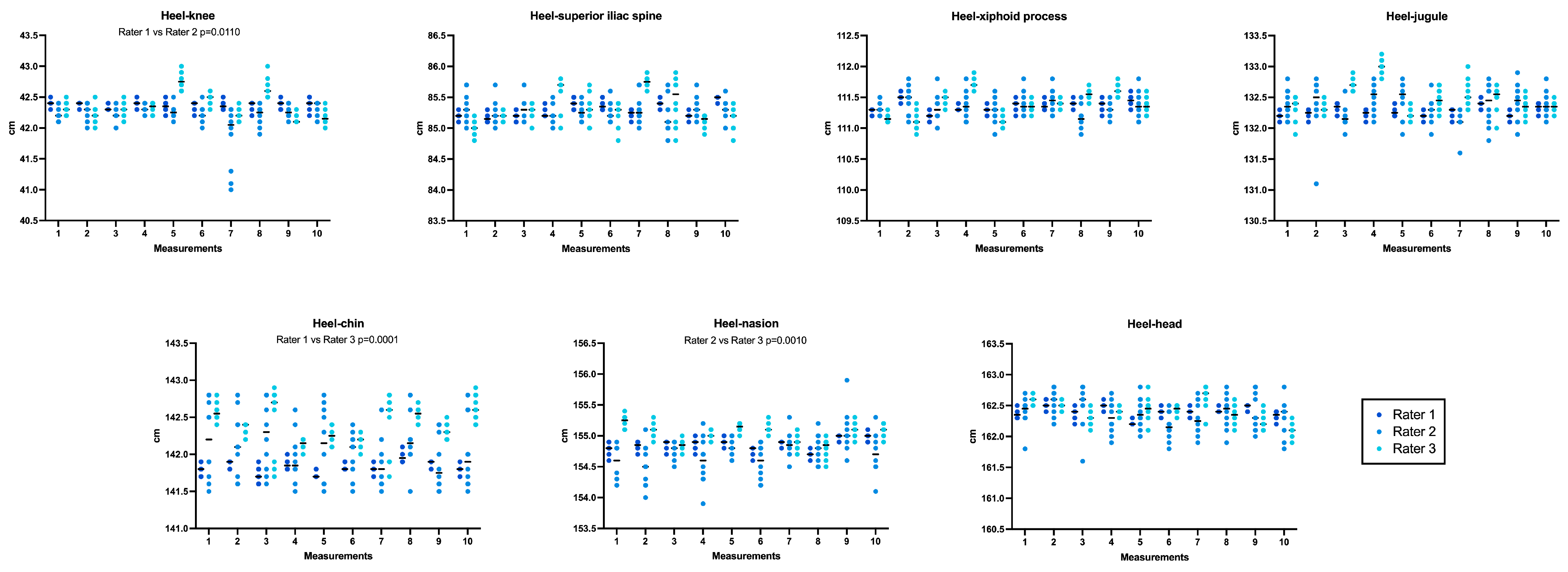
- The precision or intra-rater variability, defined as the degree to which repeated measures performed in different moments produce similar results, was tested by comparing, for each anthropometric measure, the measures performed across the ten days;
- The inter-rater reliability, defined as the degree of concordance among different raters who analyze the same parameter independently of each other, was tested by comparing, for each anthropometric measure, the measures performed across the ten days by the 3 involved raters and by comparing the median values obtained by each rater for all the anthropometric measures using the intraclass correlation coefficient (ICC);
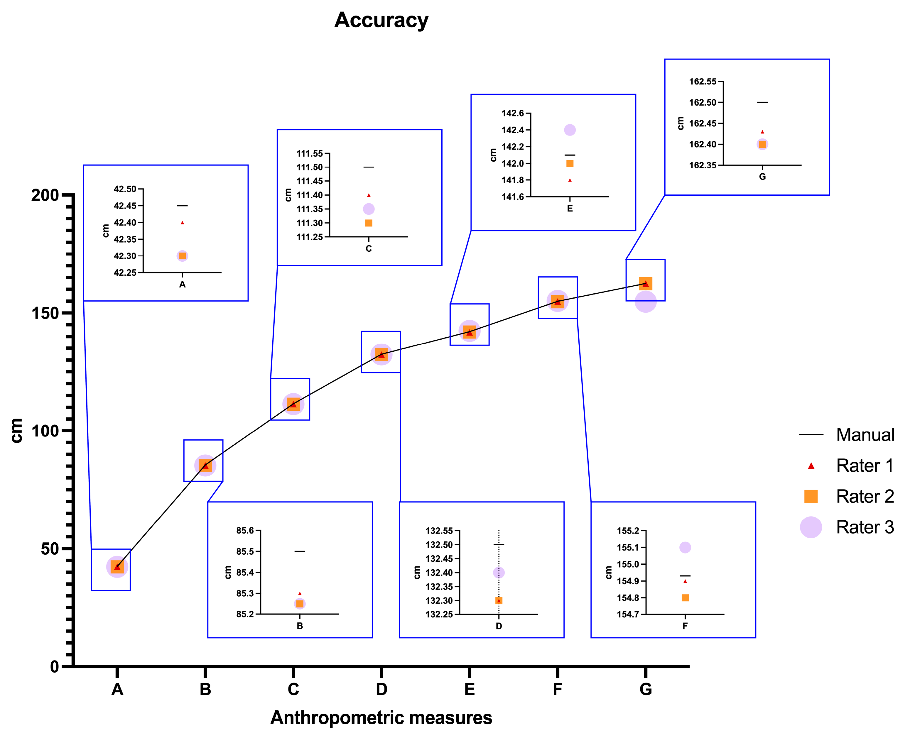
- Accuracy of the handheld scanner, indicating the closeness between the true value and the value measured with the investigated technique, was evaluated by determining the percentage error between the real (manual) measurement and the median of each digital distance measured by the three raters involved (resulting in 100 measurements for each anthropometric measure).
2.2. Manual Measurements
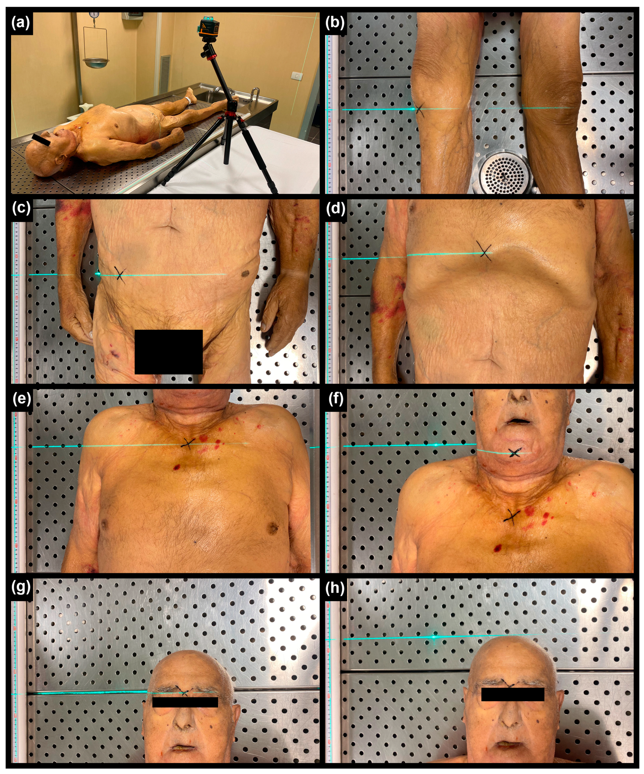
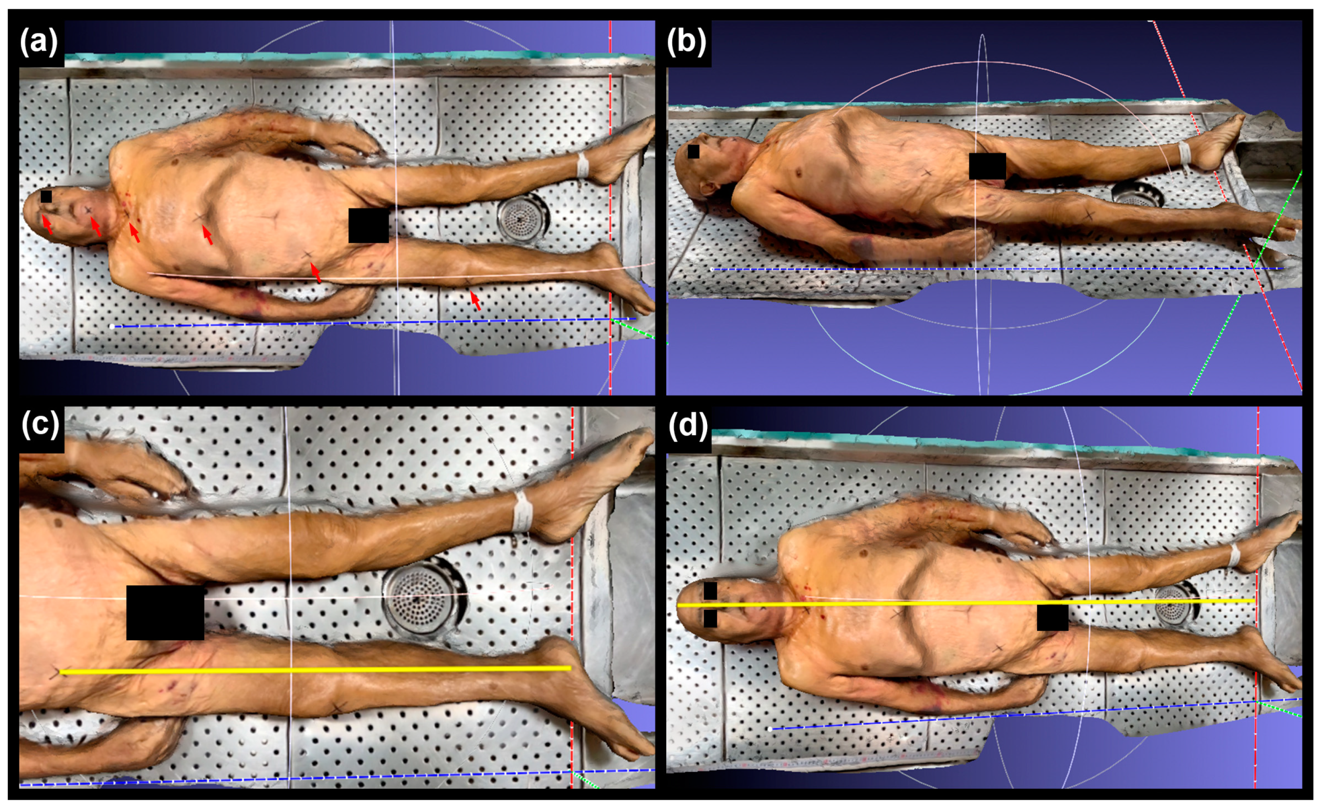
- Identified and marked 6 anthropometric landmarks on the body using an “X” (Figure 1);
- Measured 7 longitudinal distances using a 2-meter rigid meter placed beside the corpse, aligned parallel to its main axis. Additionally, a laser level (Laser Level mode CM-701, Cigman, Essen, Germany) was used to facilitate the orthogonal transposition of the specifically marked landmarks. Each longitudinal distance was measured ten times.
2.3. Body Scan
2.4. Digital Measurements
2.5. Statistical Analyses
3. Results
4. Discussion
Author Contributions
Funding
Institutional Review Board Statement
Informed Consent Statement
Data Availability Statement
Conflicts of Interest
References
- Cette Recommandation a été adoptée par le Comité des Ministres le 2 février 1999, lors de la 658e réunion des Délégués des Ministres. Recommendation No. R (99) 3 of the Committee of Ministers to Member States on the Harmonization of Medico-Legal Autopsy Rules. Forensic Sci. Int. 2000, 111, 31–58. [Google Scholar] [CrossRef] [Scilit]
- Ubelaker, D.H. A History of Forensic Anthropology. Am. J. Phys. Anthropol. 2018, 165, 915–923. [Google Scholar] [CrossRef] [Scilit] [PubMed]
- Blau, S.; Ubelaker, D.H. (Eds.) Handbook of Forensic Anthropology and Archaeology; Routledge: London, UK, 2016. [Google Scholar]
- Blau, S.; Briggs, C.A. The Role of Forensic Anthropology in Disaster Victim Identification (DVI). Forensic Sci. Int. 2011, 205, 29–35. [Google Scholar] [CrossRef] [Scilit] [PubMed]
- Cattaneo, C. Forensic Anthropology: Developments of a Classical Discipline in the New Millennium. Forensic Sci. Int. 2007, 165, 185–193. [Google Scholar] [CrossRef] [Scilit] [PubMed]
- DiMaio, V.J. Gunshot Wounds Practical Aspects of Firearms, Ballistics, and Forensic Techniques, 2nd ed.; CRC Press: Boca Raton, FL, USA, 1999; ISBN 0-8493-8163-0. [Google Scholar]
- Kneubuehl, B.P.; Coupland, R.M.; Rothschild, M.A.; Thali, M.J. Wound Ballistics, Basics and Applications; Springer: Berlin/Heidelberg, Germany, 2011; ISBN 978-3-642-20355-8. [Google Scholar]
- Haag, L.C. Shooting Incident Reconstruction; Elsevier: Amsterdam, The Netherlands, 2005. [Google Scholar]
- Burke, M.P. Forensic Medical Investigation of Motor Vehicle Incidents; CRC Press: Boca Raton, FL, USA, 2006. [Google Scholar]
- Buck, U.; Buße, K.; Campana, L.; Gummel, F.; Schyma, C.; Jackowski, C. What happened before the run over? Morphometric 3D reconstruction. Forensic Sci. Int. 2020, 306, 110059. [Google Scholar] [CrossRef] [Scilit] [PubMed]
- Han, I.; Park, C.S. Characteristic analysis and reconstruction method of falls from windows. Forensic Sci. Int. 2022, 330, 111134. [Google Scholar] [CrossRef] [Scilit] [PubMed]
- Madea, B. (Ed.) Handbook of Forensic Medicine; Wiley Blackwell: Chichester, UK, 2014. [Google Scholar]
- Brinkmann, B. Harmonisation of Medico-Legal Autopsy Rules. Int. J. Legal Med. 1999, 113, 1–14. [Google Scholar] [CrossRef] [Scilit]
- Cecchetto, G.; Bajanowski, T.; Cecchi, R.; Favretto, D.; Grabherr, S.; Ishikawa, T.; Kondo, T.; Montisci, M.; Pfeiffer, H.; Bonati, M.R.; et al. Back to the Future—Part 1. The Medico-Legal Autopsy from Ancient Civilization to the Post-Genomic Era. Int. J. Legal Med. 2017, 131, 1069–1083. [Google Scholar] [CrossRef] [Scilit]
- Cecchi, R.; Cusack, D.; Ludes, B.; Madea, B.; Vieira, D.N.; Keller, E.; Payne-James, J.; Sajantila, A.; Vali, M.; Zoia, R.; et al. European Council of Legal Medicine (ECLM) on-Site Inspection Forms for Forensic Pathology, Anthropology, Odontology, Genetics, Entomology and Toxicology for Forensic and Medico-Legal Scene and Corpse Investigation: The Parma Form. Int. J. Legal Med. 2022, 136, 1037–1049. [Google Scholar] [CrossRef] [Scilit]
- Saukko, P.; Knight, B. Knight’s Forensic Pathology; CRC Press: Boca Raton, FL, USA, 2015. [Google Scholar]
- INTERPOL. Disaster Victim Identification Guide; INTERPOL: Lyon, France, 2023. [Google Scholar]
- Fourie, Z.; Damstra, J.; Gerrits, P.O.; Ren, Y. Evaluation of Anthropometric Accuracy and Reliability Using Different Three-Dimensional Scanning Systems. Forensic Sci. Int. 2011, 207, 127–134. [Google Scholar] [CrossRef] [Scilit]
- Shamata, A.; Thompson, T. Documentation and Analysis of Traumatic Injuries in Clinical Forensic Medicine Involving Structured Light Three-Dimensional Surface Scanning versus Photography. J. Forensic Leg. Med. 2018, 58, 93–100. [Google Scholar] [CrossRef] [Scilit]
- Näther, S.; Buck, U.; Thali, M.J. Photogrammetry-Based Optical Surface Scanning. In Brogdon’s Forensic Radiology; CRC Press: Boca Raton, FL, USA, 2010. [Google Scholar]
- Bennett, J.P.; Liu, Y.E.; Quon, B.K.; Kelly, N.N.; Wong, M.C.; Kennedy, S.F.; Chow, D.C.; Garber, A.K.; Weiss, E.J.; Heymsfield, S.B.; et al. Assessment of Clinical Measures of Total and Regional Body Composition from a Commercial 3-Dimensional Optical Body Scanner. Clin. Nutr. 2022, 41, 211–218. [Google Scholar] [CrossRef] [Scilit] [PubMed]
- Cascos, R.; Ortiz Del Amo, L.; Álvarez-Guzmán, F.; Antonaya-Martín, J.L.; Celemín-Viñuela, A.; Gómez-Costa, D.; Zafra-Vallejo, M.; Agustín-Panadero, R.; Gómez-Polo, M. Accuracy between 2D Photography and Dual-Structured Light 3D Facial Scanner for Facial Anthropometry: A Clinical Study. J. Clin. Med. 2023, 12, 3090. [Google Scholar] [CrossRef] [Scilit] [PubMed]
- Peschel, O.; Szeimies, U.; Vollmar, C.; Kirchhoff, S. Postmortem 3-D Reconstruction of Skull Gunshot Injuries. Forensic Sci. Int. 2013, 233, 45–50. [Google Scholar] [CrossRef] [Scilit]
- Ditkofsky, N.G.; Maresky, H.; Mathur, S. Imaging Ballistic Injuries. Can. Assoc. Radiol. J. 2020, 71, 335–343. [Google Scholar] [CrossRef] [Scilit]
- Ferrara, S.D.; Cecchetto, G.; Cecchi, R.; Favretto, D.; Grabherr, S.; Ishikawa, T.; Kondo, T.; Montisci, M.; Pfeiffer, H.; Bonati, M.R.; et al. Back to the Future—Part 2. Post-Mortem Assessment and Evolutionary Role of the Bio-Medicolegal Sciences. Int. J. Legal Med. 2017, 131, 1085–1101. [Google Scholar] [CrossRef] [Scilit] [PubMed]
- Van Kan, R.A.T.; Haest, I.I.H.; Lobbes, M.B.I.; Kroll, J.; Ernst, S.R.; Kubat, B.; Hofman, P.A.M. Post-Mortem Computed Tomography in Forensic Investigations of Lethal Gunshot Incidents: Is There an Added Value? Int. J. Legal Med. 2019, 133, 1889–1894. [Google Scholar] [CrossRef] [Scilit] [PubMed]
- Giorgetti, A.; Giraudo, C.; Viero, A.; Bisceglia, M.; Lupi, A.; Fais, P.; Quaia, E.; Montisci, M.; Cecchetto, G.; Viel, G. Radiological Investigation of Gunshot Wounds: A Systematic Review of Published Evidence. Int. J. Legal Med. 2019, 133, 1149–1158. [Google Scholar] [CrossRef] [Scilit]
- Zyoud, T.Y.T.; Abdul Rashid, S.N.; Suppiah, S.; Mahmud, R.; Kabeer, A.; Abd Manaf, R.; Abdul Rahim, E. Estimation of Body Height from Spinal Length Measurements Using Post-Mortem Computed Tomographic Images. Malays. J. Pathol. 2020, 42, 423–431. [Google Scholar]
- Web-site American Geosciences Institute. Available online: https://www.americangeosciences.org/critical-issues/faq/what-lidar-and-what-it-used (accessed on 11 December 2023).
- Maiese, A.; Manetti, A.C.; Ciallella, C.; Fineschi, V. The Introduction of a New Diagnostic Tool in Forensic Pathology: LiDAR Sensor for 3D Autopsy Documentation. Biosensors 2022, 12, 132. [Google Scholar] [CrossRef] [Scilit]
- Pelletti, G.; Cecchetto, G.; Viero, A.; Fais, P.; Weber, M.; Miotto, D.; Montisci, M.; Viel, G.; Giraudo, C. Accuracy, Precision and Inter-Rater Reliability of Micro-CT Analysis of False Starts on Bones. A Preliminary Validation Study. Leg. Med. 2017, 29, 38–43. [Google Scholar] [CrossRef] [Scilit] [PubMed]
- Koo, T.K.; Li, M.Y. A Guideline of Selecting and Reporting Intraclass Correlation Coefficients for Reliability Research. J. Chiropr. Med. 2016, 15, 155–163. [Google Scholar] [CrossRef] [Scilit] [PubMed]
- Langley, N.R.; Meadows Jantz, L.; McNulty, S.; Maijanen, H.; Ousley, S.D.; Jantz, R.L. Error Quantification of Osteometric Data in Forensic Anthropology. Forensic Sci. Int. 2018, 287, 183–189. [Google Scholar] [CrossRef] [Scilit] [PubMed]
- Carew, R.M.; French, J.; Morgan, R.M. 3D Forensic Science: A New Field Integrating 3D Imaging and 3D Printing in Crime Reconstruction. Forensic Sci. Int. Synergy 2021, 3, 100205. [Google Scholar] [CrossRef] [Scilit]



| Anthropometric Measurement | Median Manual Measurement (IQ) (cm) | Digital Measurement | ||||
|---|---|---|---|---|---|---|
| Intra-Rater Reliability | ICC | |||||
| Median (IQ) (cm) Significant Differences | Median (IQ) (cm) Significant Differences | Median (IQ) (cm) Significant Differences | Single Measures | Average Measures | ||
| A. Heel–knee | 42.45 (0.05) | 42.40 (0.05) p = 0.5330 | 42.20 (0.07) p = 0.8822 | 42.30 (0.33) p = 0.4384 | 1.000 | 1.000 |
| B. Heel–superior iliac spine | 85.50 (0.05) | 85.50 (0.20) p = 0.1022 | 85.20 (0.09) p = 0.8532 | 85.39 (0.44) p = 0.2025 | ||
| C. Heel–xiphoid process | 111.50 (0.01) | 111.38 (0.11) p = 0.6143 | 111.32 (0.11) p = 0.9565 | 111.38 (0.40) p = 0.2854 | ||
| D. Heel–jugule | 132.50 (0.05) | 132.26 (0.12) p = 0.6417 | 132.39 (0.16) p = 0.9320 | 132.44 (0.22) p = 0.0980 | ||
| E. Heel–chin | 142.10 (0.10) | 141.80 (0.13) p = 0.1393 | 142.10 (0.27) p = 0.1919 | 142.47 (0.36) p = 0.0136 * | ||
| F. Heel–nasion | 154.93 (0.10) | 154.90 (0.13) p = 0.1045 | 154.67 (0.31) p = 0.6108 | 155.10 (0.22) p = 0.2814 | ||
| G. Heel–head | 162.50 (0.06) | 162.4 (0.14) p = 0.8864 | 162.39 (0.15) p = 0.5401 | 162.41 (0.25) p = 0.5456 | ||
| Anthropometric Measurement | Accuracy Max Percent Error (Median) | |||
|---|---|---|---|---|
| Rater 1 | Rater 2 | Rater 3 | Median among Raters | |
| A. Heel–knee | <0.6% (0.1) Excellent | <0.8% (0.4) Excellent | <1.3% (0.4) Excellent | <0.6% (0.4) Excellent |
| B. Heel–superior iliac spine | <0.5% (0.2) Excellent | <0.6% (0.3) Excellent | <0.8% (0.3) Excellent | <0.4% (0.2) Excellent |
| C. Heel–xiphoid process | <0.4% (0.1) Excellent | <0.5% (0.2) Excellent | <0.5% (0.1) Excellent | <0.2% (0.1) Excellent |
| D. Heel–jugule | <0.3% (0.2) Excellent | <0.5% (0.2) Excellent | <0.5% (0.1) Excellent | <0.2% (0.2) Excellent |
| E. Heel–chin | <0.4% (0.2) Excellent | <0.5% (0.1) Excellent | <0.6% (0.2) Excellent | <0.1% (0.1) Excellent |
| F. Heel–nasion | <0.2% (0.0) Excellent | <0.5% (0.1) Excellent | <0.3% (0.1) Excellent | <0.1% (0.0) Excellent |
| G. Heel–head | <0.2% (0.0) Excellent | <0.5% (0.1) Excellent | <0.4% (0.1) Excellent | <0.1% (0.1) Excellent |
Disclaimer/Publisher’s Note: The statements, opinions and data contained in all publications are solely those of the individual author(s) and contributor(s) and not of MDPI and/or the editor(s). MDPI and/or the editor(s) disclaim responsibility for any injury to people or property resulting from any ideas, methods, instructions or products referred to in the content. |
© 2024 by the authors. Licensee MDPI, Basel, Switzerland. This article is an open access article distributed under the terms and conditions of the Creative Commons Attribution (CC BY) license (https://creativecommons.org/licenses/by/4.0/).
Share and Cite
Callegari, E.; Agnolucci, J.; Angiola, F.; Fais, P.; Giorgetti, A.; Giraudo, C.; Viel, G.; Cecchetto, G. The Precision, Inter-Rater Reliability, and Accuracy of a Handheld Scanner Equipped with a Light Detection and Ranging Sensor in Measuring Parts of the Body—A Preliminary Validation Study. Sensors 2024, 24, 500. https://doi.org/10.3390/s24020500
Callegari E, Agnolucci J, Angiola F, Fais P, Giorgetti A, Giraudo C, Viel G, Cecchetto G. The Precision, Inter-Rater Reliability, and Accuracy of a Handheld Scanner Equipped with a Light Detection and Ranging Sensor in Measuring Parts of the Body—A Preliminary Validation Study. Sensors. 2024; 24(2):500. https://doi.org/10.3390/s24020500
Chicago/Turabian StyleCallegari, Enrica, Jacopo Agnolucci, Francesco Angiola, Paolo Fais, Arianna Giorgetti, Chiara Giraudo, Guido Viel, and Giovanni Cecchetto. 2024. "The Precision, Inter-Rater Reliability, and Accuracy of a Handheld Scanner Equipped with a Light Detection and Ranging Sensor in Measuring Parts of the Body—A Preliminary Validation Study" Sensors 24, no. 2: 500. https://doi.org/10.3390/s24020500
APA StyleCallegari, E., Agnolucci, J., Angiola, F., Fais, P., Giorgetti, A., Giraudo, C., Viel, G., & Cecchetto, G. (2024). The Precision, Inter-Rater Reliability, and Accuracy of a Handheld Scanner Equipped with a Light Detection and Ranging Sensor in Measuring Parts of the Body—A Preliminary Validation Study. Sensors, 24(2), 500. https://doi.org/10.3390/s24020500