Article

Brain MR Image Enhancement for Tumor Segmentation Using 3D U-Net

by
Faizad Ullah
1,2,
Shahab U. Ansari
2,
Muhammad Hanif
2,
Mohamed Arselene Ayari
3,4,
Muhammad Enamul Hoque Chowdhury
5,
Amith Abdullah Khandakar
5 and
Muhammad Salman Khan
1,6,*
1
Artificial Intelligence in Healthcare, Intelligent Information Processing Lab, National Center of Artificial Intelligence, University of Engineering and Technology, Peshawar 25120, Pakistan
2
Faculty of Computer Science and Engineering, Ghulam Ishaq Khan Institute of Engineering Sciences and Technology, Topi 23640, Pakistan
3
Technology Innovation and Engineering Education, College of Engineering, Qatar University, Doha 2713, Qatar
4
Department of Civil and Architectural Engineering, College of Engineering, Qatar University, Doha 2713, Qatar
5
Department of Electrical Engineering, College of Engineering, Qatar University, Doha 2713, Qatar
6
Department of Electrical Engineering (JC), University of Engineering and Technology, Peshawar 24241, Pakistan
*
Author to whom correspondence should be addressed.
Sensors 2021, 21(22), 7528; https://doi.org/10.3390/s21227528
Submission received: 5 October 2021 / Revised: 7 November 2021 / Accepted: 9 November 2021 / Published: 12 November 2021
(This article belongs to the Section Fault Diagnosis & Sensors)

Abstract

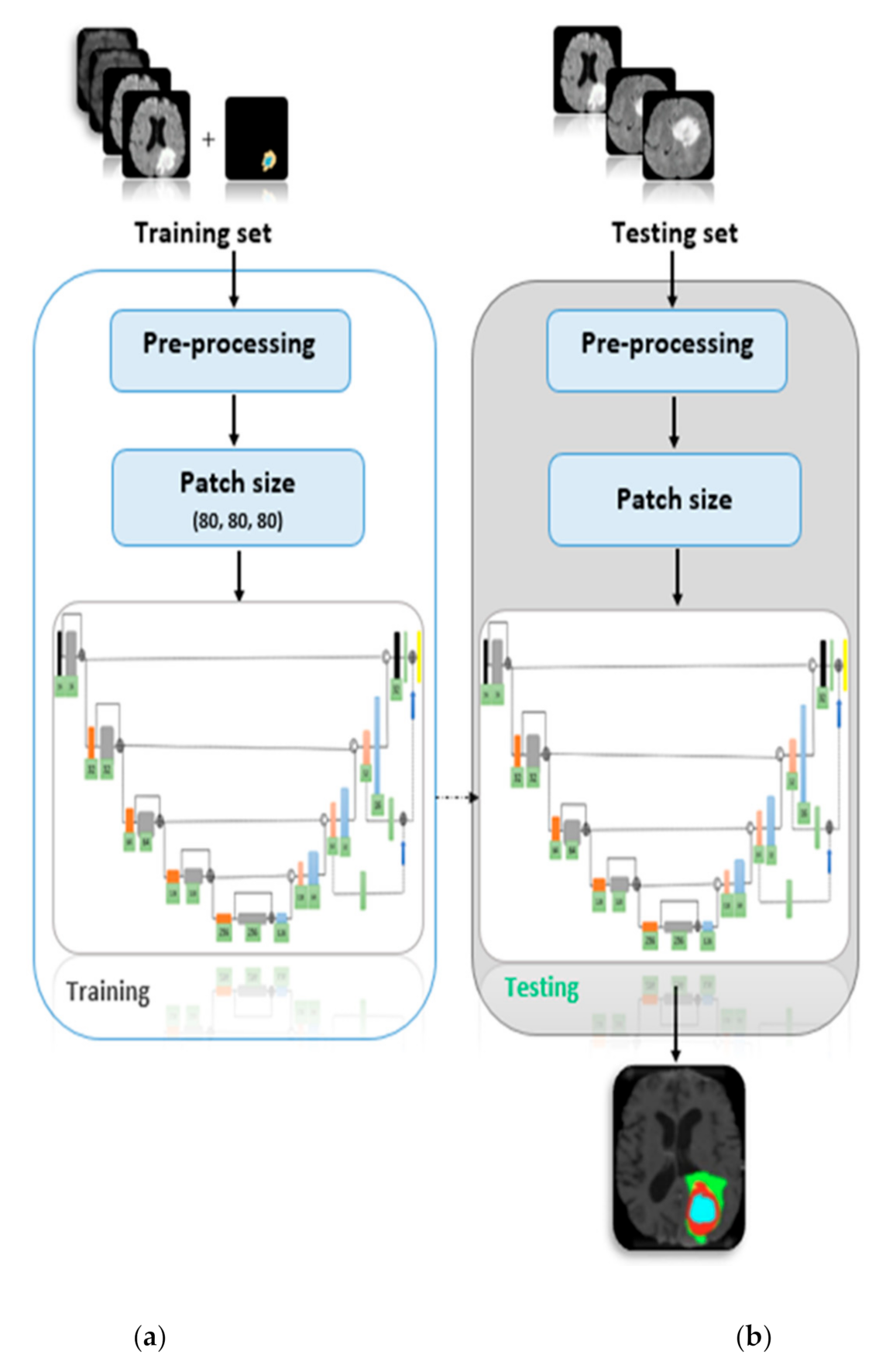
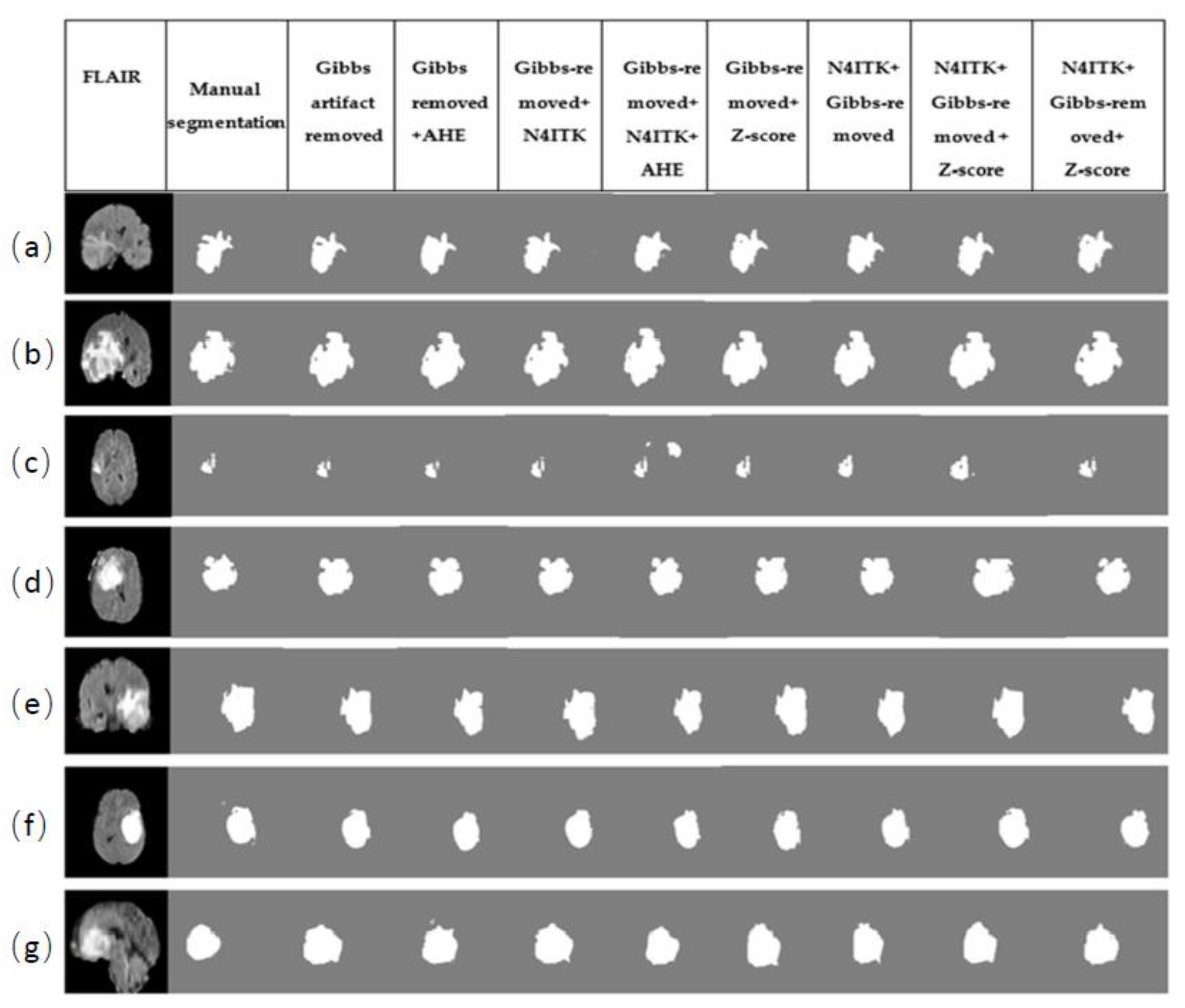
MRI images are visually inspected by domain experts for the analysis and quantification of the tumorous tissues. Due to the large volumetric data, manual reporting on the images is subjective, cumbersome, and error prone. To address these problems, automatic image analysis tools are employed for tumor segmentation and other subsequent statistical analysis. However, prior to the tumor analysis and quantification, an important challenge lies in the pre-processing. In the present study, permutations of different pre-processing methods are comprehensively investigated. In particular, the study focused on Gibbs ringing artifact removal, bias field correction, intensity normalization, and adaptive histogram equalization (AHE). The pre-processed MRI data is then passed onto 3D U-Net for automatic segmentation of brain tumors. The segmentation results demonstrated the best performance with the combination of two techniques, i.e., Gibbs ringing artifact removal and bias-field correction. The proposed technique achieved mean dice score metrics of 0.91, 0.86, and 0.70 for the whole tumor, tumor core, and enhancing tumor, respectively. The testing mean dice scores achieved by the system are 0.90, 0.83, and 0.71 for the whole tumor, core tumor, and enhancing tumor, respectively. The novelty of this work concerns a robust pre-processing sequence for improving the segmentation accuracy of MR images. The proposed method overcame the testing dice scores of the state-of-the-art methods. The results are benchmarked with the existing techniques used in the Brain Tumor Segmentation Challenge (BraTS) 2018 challenge.
Keywords: brain tumor segmentation; deep learning; Gibbs ringing artifact; image enhancement; medical image processing brain tumor segmentation; deep learning; Gibbs ringing artifact; image enhancement; medical image processing