


Abstract
This review aims to describe and discuss the different functions of the endolysosomal system, from homeostasis to its vital role during viral infections. We will initially describe endolysosomal system’s main functions, presenting recent data on how its compartments are essential for host defense to explore later how SARS-CoV-2 (Severe Acute Respiratory Syndrome Coronavirus 2) and other coronaviruses subvert these organelles for their benefit. It is clear that to succeed, pathogens’ evolution favored the establishment of ways to avoid, escape, or manipulate lysosomal function. The unavoidable coexistence with such an unfriendly milieu imposed on viruses the establishment of a vast array of strategies to make the most out of the invaded cell’s machinery to produce new viruses and maneuvers to escape the host’s defense system.
Keywords:
lysosome; endosome; acidification; coronaviruses; endocytosis; autophagy; SARS-CoV-2; virus cycle; immune evasion; immunity 1. The Endolysosomal System
Let us begin by briefly describing the organelles and processes this review will focus on.
1.1. Lysosomes
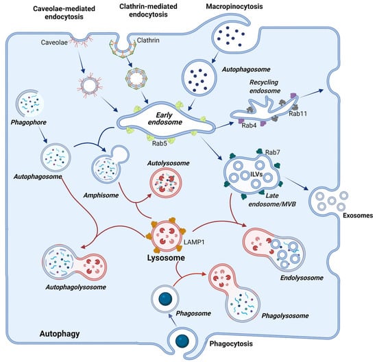
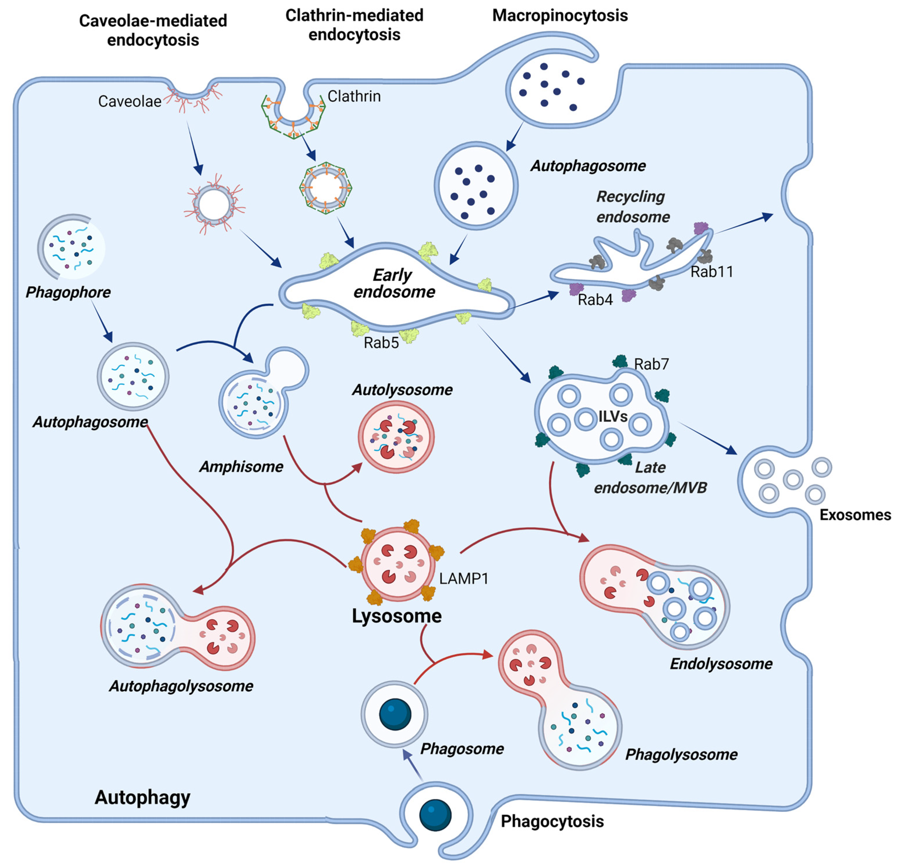
The lysosome is the cellular terminal station where the degradation of internalized material occurs. It performs a crucial role in keeping homeostasis, and defective function can lead to disease. These organelles store a vast repertoire of inactive acid hydrolases, activated upon fusion with the acidic late endosomes. This merge forms the endolysosomes, where degradation takes place [1]. It is important to emphasize that the lysosome term is commonly used to define the degradative compartment of the cell, but this definition is not accurate. The degradative compartments are the vesicles that originate from the fusion with lysosomes, such as endolysosomes, autolysosomes, and phagolysosomes (Figure 1). These coexist with terminal storage lysosomes that are not acidic and serve to keep acidic hydrolases that will be activated when needed upon fusion with cargo-containing acidic vesicles [1]. We will respect the nomenclature adopted in each cited article to construct this review. Still, the reader must be aware of the broad meaning the term lysosome may have in some articles, where it may be used as a synonym for endolysosomes.
Figure 1.
The endolysosomal system. Illustration showing entry pathways into cells and compartments mentioned throughout this review that constitute the endolysosomal system. These vesicles traffic throughout the cell and exchange their contents through ‘’kiss-and-run” or “full-fusion” events. Particles from the extracellular milieu can access cells by caveolae-mediated endocytosis, clathrin-mediated endocytosis, macropinocytosis, and phagocytosis. Particles entering through endocytosis and micropinocytosis are delivered to early endosomes. Vesicles generated during autophagy are depicted on the left side of the figure. Please refer to the main text for a detailed description of the fusion events and specific molecular markers of each of these compartments. Image created using BioRender.
Lysosomes and the other vesicles of the endolysosomal system have their specificities, including cargo, resident proteins, membrane markers, morphology, redox environment, and pH. Vesicles resulting from the fusion of distinct compartments–such as amphisomes, autolysosomes, endolysosomes, phagolysosomes-exhibit hybrid characteristics from the parental compartments (Figure 1). Together, these features allow these organelles to accomplish their functions. The luminal acidic pH of degradative compartments can be as low as 4.5–5.0, and this is maintained by the V-type ATPase, and ion transporters and channels keep the ionic balance in the lumen. To shield the luminal lysosome membrane against the destructive potential of acid hydrolases, LAMPs (lysosome-associated membrane proteins) are highly glycosylated in their luminal N-portion [2]. Also, as mentioned before, these hydrolases are only fully activated when lysosomes and late endosomes fuse. After fusion, lysosomes are reformed by a maturation process that condensates its contents and recaptures endosomal-specific proteins [3].
1.2. Endocytosis and the Endolysosomal System
The endosomal system comprises vesicle compartments in the cell that regulate the traffic and fate of material from the cell itself or acquired after internalization.
Endocytosis is the cellular process that allows the intake of fluid, solutes, macromolecules, and particles through membrane invagination. It may be constitutive or receptor-dependent, and in each case, the plasma membrane forms invaginations through distinct mechanisms, dependent on specific molecules and structures, such as clathrin, caveolae, Cdc42 (Cell Division Cycle 42), Rho/Rac, endophilin A2 toxins, and FEME. The variety of these mechanisms is not the scope of this review, and readers should refer to excellent reviews in the area, such as one of Sandvig and collaborators [4].
Following membrane invaginations, the newly formed vesicles fuse with early endosomes, where the mildly acidic pH (ranging from 5.9 to 6.8) provokes the dissociation of many ligands from their receptors. In endosomal compartments, ingested material to be destroyed (large particles such as viruses) and molecules to be recycled are sorted out. The early endosome is responsible for sorting and delivering distinct molecules to their destination compartments. This is achieved by the highly organized feature of these compartments where vacuolar sorting domains and tubular recycling domains are found. The first contains the material to be degraded and mature to late endosomes, while the later domains have a high surface membrane/lumen ratio to recycle molecules to other compartment membranes such as plasma membrane, TGN, and recycling endosomes, without losing nutrients. To achieve correct sorting, membrane subdomains of the endosome are distinctly enriched in Rab5, Rab4, Rab11, Arf1/COPI (ADP-Ribosylation Factor 1/Coat Protein I), retromer, and caveolae [5,6,7].
The Rab proteins-small GTPases (guanosine triphosphatases) of the Ras superfamily-are crucial for vesicle trafficking and fusion and participate in several steps of this process. For instance, Rab5, a component of early endosomes, regulates vesicle fusion and sorting of molecules and is lost in the maturing process into late endosomes, while Rab7 is acquired in this maturation. Rab7 has crucial roles in endolysosomes, regulating processes, such as lysosomal enzymes trafficking, fusion with lysosomes, vesicle acidification, and degradation [8]. In addition, Rab4 and Rab11 handle the recycling of membrane receptors to the plasma membrane via recycling endosomes [5,9,10].
Membrane fusion involves the coordinated interaction of Rab GTPases, SNAREs (soluble N-ethylmaleimide-sensitive factor attachment protein receptor), the ESCRT (endosomal sorting complexes required for transport) machinery, and tethering complexes-CORVET (class C core vacuole/endosome tethering) and HOPS (homotypic fusion and vacuole protein sorting). The SNARE family of proteins comprises 38 members, classified according to the presence of conserved glutamine (Q) or arginine (R) residues in R-SNARES or Q-SNARES (further distinguished in Qa-, Qb- and Qc-SNARE). The process of fusion depends on the interaction between SNARES of opposing membranes. SNARES assemble in a quadruple helix forming a zipper that pulls the membranes so close together that they fuse [11]. ESCRT is a multimolecular complex that drives membrane remodeling. It consists of four main complexes (ESCRT-0, -I, -II, and -III) and associated proteins. ESCRT-0 interacts with ubiquitylated proteins, recruiting ESCRT-I, -II -III in a step-wise manner. Tethering complexes CORVET and HOPS share a core comprising the Vps (Vacuolar Protein Sorting) proteins Vps11, Vps13, Vps18, and Vps33 associated with Vps3 and Vps8 in CORVET or Vps39 and Vps41 in HOPS. Whereas CORVET participates in fusing early endosomes to other vesicles, HOPS is a tethering complex involved in fusion between the lysosome and other vesicles [12,13,14].
In summary, as early endosomes mature to late endosomes, there is an exchange of GTPases, tethering complexes, and also SNAREs in a highly controlled mechanism that assures identity and proper functioning of each compartment. We will refer to specific members of these families of proteins along this review to describe events important for the coronaviruses cycle.
Early endosomes also communicate with the Golgi, receiving macromolecules, such as acid hydrolases from the trans-Golgi network. These hydrolases may also be secreted by the cell through the secretory pathway and endocytosed back after they bind to mannose-6-phosphatase receptors (M6PR) present in the plasma membrane [15]. Thus, communication with other compartments is essential for the maturation of early endosomes into late endosomes, allowing the acquisition of distinct molecules and the delivery of others. Late endosomes pH ranges between 6.0–4.8, and the maturation of early into late endosomes is also characterized by the formation of inward budding of endosomal membrane creating intraluminal vesicles (ILVs) [6]. As the maturation process continues, ILVs accumulate, and in late endosomes, they are abundant and characterize multi-vesicular bodies (MVBs). MVBs can fuse to lysosomes, where ILVs are degraded, or fuse to the membrane liberating ILVs, constituting the exosomes [6].
MVBs are formed by two distinct mechanisms, one dependent and the other independent of ESCRT. The Vps4 provides the energy for dissociating the inward budding vesicles from the endosomal membrane. Fusion of MVBs with lysosomes heads the ubiquitin-tagged proteins to degradation [16,17]. An alternative mechanism relies on the activity of SMase (sphingomyelinases), enzymes exhibiting different optimal pHs (alkaline, neutral, or acid), that hydrolyses SM (sphingomyelin) of endosomal lipid rafts to ceramide. Plasma and endosomal membranes have an asymmetrical lipid composition, with SM enriched in the non-cytosolic leaflet. Whereas sphingomyelin has a cylinder topology and a high affinity for membrane cholesterol that contributes to the compact and rigid characteristic of lipid rafts, ceramide, with its conical topology, favors vesicle budding by influencing cholesterol efflux, membrane fluidity, and curving [18]. ESCRT-dependent and -independent pathways can be inhibited by different drugs, such as manumycin, an inhibitor of Ras farnesylation, and GW4869, an inhibitor of the nSMase (neutral SMase), respectively [17,19].
1.3. Lysosomes as Signaling Hubs: From Lysosomal Exocytosis to Autophagy Control
ILVS release from the cell during lysosomal exocytosis constitutes an essential way of cell communication [20]. Lysosome exocytosis is also involved in membrane repair, degradation, and remodeling of the extracellular matrix and release of non-degraded material. Thus, lysosomes not only act inside the cell but function beyond the limits of the plasma membrane. To accomplish all these tasks, lysosomes’ location needs to be highly dynamic, which is made possible by interaction with extensive microtubule network going either outwards using kinesin or inwards using dynein.
The anterograde movement of lysosomes depends on the complex of proteins in the lysosomal membrane composed by BORC, the small GTPase Arl8, and the kinesin-binding protein PLEKHM2/SKIP (Pleckstrin homology domain-containing family M member 2/Salmonella-induced filaments A and kinesin-interacting protein) that connects lysosomes to kinesin [21,22,23,24]. Distinct GTPases are also implicated depending on the cell type, for example, Rab27a and Rab27b in HeLa cells, Rab11 in K562 cells, Rab35 in Oli-neu cells [17,25,26,27].
Lysosomes’ interaction with the actin cytoskeleton permits its appropriate positioning close to the membrane, which facilitates fusion between these two compartments. This event also requires mobilization of specific SNARES of both organelles, such as VAMP7 and SNAP23, which dock the lysosome to the internal face of the plasma membrane, respectively. Besides, fusion is also dependent on Ca2+ release from the lysosomal lumen through channels such as TMPRL1. The presence of Ca2+ triggers lysosomal synaptotagmin VII interaction with the Qb,c-SNARE SNAP23, and the Qa-SNARE Syntaxin 4, facilitating the formation of the SNARE complex with the R-SNARE VAMP7 [20,28].
The location of lysosomes is also associated with their function as a metabolic hub that integrates cell signals triggering or inhibiting anabolic/catabolic activity [29].
A key player in this setting is mTORC1 (mechanistic target of rapamycin complex 1)-a complex composed by mTOR (mechanistic target of rapamycin), and the adaptor proteins Raptor and MLST8 (Mammalian lethal with SEC13 protein 8/Target of rapamycin complex subunit LST8)-that in its active form localizes to the cytosolic membrane of late endosomes and lysosomes. Active mTORC1 senses hormonal cues and the metabolic status of the cell, promoting ribosomes biosynthesis, protein translation, lipogenesis while inhibiting autophagy. mTORC1 is activated by growth-factor-activated Rheb (Ras homolog enriched in brain) GTPase and by amino acids-activated Rag GTPases [30,31,32].
The protein synthesis promoting activity of Rheb is inhibited by the tuberous sclerosis complex (TSC). Growth factors liberate Rheb from TSC inhibition by activating PI3K and PKB/Akt1 and subsequent phosphorylation of TSC, which becomes sequestered in the cytoplasm, away from lysosomes. Amino acids activate Rag by stimulating its interaction with Raptor, bringing mTORC1 to late endosomes and lysosomal membranes, where mTORC1 interacts with Rheb. Interestingly, activation of mTORC1 by amino acids requires the activity of v-ATPase. The active mTORC1 phosphorylates substrates, such as S6 kinase (S6K1), eukaryotic translation initiation factor 4E-binding protein 1 (4E-BP1), Unc-51 like autophagy activating kinase (ULK1), and transcription factor EB (TFEB). Overall mTORC1 actions stimulate anabolic activity and suppress activators of autophagy. Lysosome positioning close to the plasma membrane can help enhance mTORC1 activation because activators such as Akt1 are close by [30,31,32].
During starvation, anabolic activities are inhibited, and autophagy of self-organelles and macromolecules is activated to provide nutrients for the cell. To this end, the biogenesis of lysosomes and autophagosomes is stimulated, as well as the fusion of these organelles. Autophagy begins with mobilization of Atg (autophagy-related gene) proteins to the phagophore assembly site, and nucleation of a cup-shaped membrane termed IM (isolation membrane). This structure forms the phagophore, which expands surrounding cargoes until it confines them in double-membrane vesicles, which characterize the autophagosome. The outer membrane of autophagosomes fuses with the membrane of lysosomes delivering its cargo for degradation in the newly formed autophagolysosome [33]. This process involves the coordinated recruitment of distinct complexes of proteins that we will describe briefly for understanding the mechanisms that coronaviruses manipulate for their own benefit, which will be depicted later in this review.
In nutrient-rich conditions, mTORC1 interacts with the ULK1 (Unc-51 Like Autophagy Activating Kinase 1) complex, formed by ULK1 itself, Atg13, FIP200, and Atg101 (family kinase-interacting protein of 200 kD) and phosphorylates ULK1 at specific residues (serines 637 and 757 in mouse and serines 638 and 758 in human; the first is phosphorylated by AMPK and MTOR and the second solely by mTOR) [34,35,36,37], rendering ULK1 inactive. Starvation induces inactivation and moving of mTORC1 to the cytoplasm. The phosphorylation of ULK1 is then relieved by phosphatases, and ULK1 is autophosphorylated (at serine 1042 and threonines 180 and 1046) [38]. In this form, ULK1 becomes active, phosphorylates FIP200 (at serine 943, 986, and 1323) and Atg13 (at serines 203 and 318) [39,40], moves to the endoplasmic reticulum recruiting a second complex, termed the PI3KC3 (phosphatidylinositol 3-kinase catalytic subunit type 3) complex II. This complex consists of Vps34-a class III phosphatidylinositol 3-kinase, Beclin-1 (Bcl-2-interacting protein 1), Vps15, and Atg14L and generates the omegasomes-PI(3)P-enriched subdomains of the ER, where autophagosome formation is initiated [37,41].
PI3KC3 I complex I is composed of Vps34, Beclin-1, Vps15, and UVRAG, instead of Atg14L [42,43]. Whereas Atg14L collects mainly in the autophagosome, UVRAG localizes to early and late endosomes [42,44]. The PI3K complex I also participates in autophagosome formation and depends on the interaction of UVRAG with Beclin1 [45]. Interestingly, UVRAG also engages Vps34 to the autophagosome and stimulates Rab7 promoting the fusion of autophagosomes with late endosomes/lysosomes. The activity of UVRAG depends on the v-ATPase [44].
Proteins, such as WIPI2B (WD repeat domain phosphoinositide interacting 2b) and DFCP1 (Double FYVE Containing Protein) that contain PI3P-binding domains, are recruited and promote the expansion of the isolation membranes IMs [46,47]. WIPI2 binds Atg16L1 (Atg 16 like 1) and recruit another complex composed of Atg12, Atg5, and Atg16L1 that stimulates the conjugation of Atg8 family proteins-which comprises LC3 (microtubule- associated protein light chain 3) proteins and GABARAPs (γ-aminobutyric acid receptor-associated proteins) to membrane PE (phosphatidylethanolamine). This event gives rise to LC3II-the lipidated form of LC3, a signature of the autophagic process. Atg8s interacts with members of the autophagic machinery containing the LC3-interacting region (LIR), including LIR-containing cargo receptors, and ultimately regulates the elongation and closure of the autophagosomes carrying the material to be degraded. The membranes required for the elongation of autophasomes are recycled from various cell organelles, such as mitochondria, Golgi, and recycling endosomes [33,48]. A deterioration of the energy level also triggers autophagy, which is sensed by AMPK through the ratio of ATP/AMP. AMPK activates TSC2, which in turn inhibits mTOR [49]. Beyond starvation and decline of cell energy, autophagy can be induced during development and by stressors, such as hypoxia and infections, allowing the disposal of unwanted material, such as aggregated proteins, damaged organelles, and invading pathogens.
Meanwhile, the dephosphorylated form of TFEB translocates to the nucleus and activates the transcription of autophagy-related genes and genes of the CLEAR (Coordinated Lysosomal Expression and Regulation) network that ultimately control the lysosomal biogenesis [50,51]. In this situation, the levels of Arl8 and motor protein KIF2 (Kinesin-related protein 2) diminish, and both lysosomes and autophagosomes move towards the nucleus where they fuse, leading to degradation of autophagosome contents to alleviate starvation, maintaining the cell alive [29]. Arl8 also promotes the fusion of lysosomes with autophagosomes and endosomes [52]. Fusion of autophagosomes/amphisomes with late endosomes/lysosomes depends on a protein complex formed by the Qa SNARE STX17 (sintaxin 17), the Qbc SNARE SNAP29 (Synaptosome Associated Protein 29), and the R-SNARE VAMP8 (Vesicle-Associated Membrane Protein 8) to lysosomes [53]. HOPS interact with SYX17 mediating the assembly of SNAREs [54,55], and this interaction of HOPS and STX17 is essential for HOPS to tether autophagosome to lysosome ensuring their fusion.
An autophagosome fuses to numerous lysosomes so that autophagy provokes a depletion of the latter. To prevent this, besides controlling the biogenesis of new lysosomes, autophagy also triggers an alternative mechanism of lysosomal biogenesis, named the autophagic lysosomal reformation mechanism that consists of restoring them from tubular structures derived from autophagosomes [56].
In conclusion, lysosomes sense and respond to many stimuli that reflect regulated degradation requirements, such as starvation, accumulation of defective proteins, membrane repair, pathogen infection, etc. These kinds of stimuli may overwhelm the endolysosomal system depending on their intensity, and lysosome functioning can be drastically altered. It was recently proposed that lysosomes display a set of signs that characterize a lysosomal stress response, which would be (1) increased intralysosomal pH, (2) increased lysosome size, (3) membrane permeabilization, (4) cationic eflux, (5) repositioning intracellularly, (6) misfolded protein aggregation, (7) LDL cholesterol accumulation, (8) redox catastrophe, and (9) bioenergetic crisis [57].
Concerning increased intralysosomal pH, although it was proposed that perinuclear lysosomes are more acidic than peripheral ones (where a higher permeability to protons and a less active V-type ATPase would lead to an increased pH) [58], a recent study using a genetically encoded fluorescence-based probe demonstrated that peripheral lysosomes are as acidic as perinuclear ones [59]. So some issues still need to be resolved. Concerning cellular location, the lysosomal position is dynamic and also associated with its different functions [2]; for instance, lysosome positioning close to the plasma membrane can help enhance mTORC1 activation because activators such as Akt1 are close by.
3. Genome-Wide Studies Reveal Components of the Endolysosomal System as Potential Pharmacological Targets against COVID-19
The advancement in high-throughput technologies allowed the recognition of many genes and cellular signaling networks that are potential targets for therapy against SARS-CoV-2 and COVID-19.
A study of genome-wide CRISP knockout screening to identify host factors that confer resistance to SARS-CoV-2 and the common cold coronaviruses 229E and OC43, followed by a gene ontology enrichment analysis identified genes essential for this resistance and organized them in biological networks, such as macroautophagy and phospholipid metabolic process, lysosome to endosome transport and membrane fusion, Golgi vesicle transport, Rab protein signaling, phagosome maturation and cholesterol metabolic process. The cells used in this study were Huh7.5.1 for OC43 and 229E infections and Huh7.5.1 cells overexpressing ACE2 and TMPRSS2 to optimize SARS-CoV-2 infection [124].
Host genes related to the virus entry were detected, such as the ones coding for ACE2 and ANPEP, the respective receptors for SARS-CoV-2 and 229E, and multiple genes involved in heparan sulfate biosynthesis, involved in infection by OC43 that uses sialic acid or glycosaminoglycans for cell entry. Genes of the cholesterol metabolism pathway were identified in all three coronaviruses screens (SCAP, MBTPS1, MBTPS2, LDLR, and NPC1). The common cold viruses screens exhibit genes related to the phosphatidylinositol metabolism, such as PIK3C3, UVRAG, BECN1, and PIK3R4), involved in endosome sorting, endomembrane homeostasis, and autophagy; some of their products were already mentioned in this review. Specific to these viruses were also the genes involved in the endosome and phagosome maturation (RAB7A, members of the HOPS complex-VPS11, VPS16, VPS18 and VPS33A, the Ccz1-Mon1 guanosine exchange factor complex -CCZ1, CC1B, and C18orf8, the WDR81-WDR91 complex) and others associated to lysosome and autophagosome function (SPNS1, TOLLIP, TMEM41B, AMBRA1). Other genes related to lysosomal enzymes and their traffic from the Golgi to the lysosomes include CTSL1, M6PR, and GNPTAB.
In the SARS-CoV-2 screen, the top hit was the lysosomal transmembrane protein TMEM106B and VAC14, a member of the PIKfyve complex. This study highlights the central role of the endosomal system for coronaviruses infection.
Another CRISPR loss-of-function screen of human alveolar basal epithelial carcinoma cells infected with SARS-CoV-2 confirmed the central role cholesterol plays during SARS-CoV-2 infection [151]. These results agree with the cholesterol function during coronaviruses cell entry, as depicted earlier in this review (Teixeira et al., submitted) [118,122,125]. Furthermore, cholesterol may also play an essential part in many other steps of the viral cycle, such as the production of lipid droplets-which fuels infection [152,153], formation of double-membrane vesicles required for the replication organelles, production of membranes for the autophagic process, and virus particle production.
Interactomes are very informative in searching for host factors on which SARS-CoV-2 depends to succeed. Gordon and colleagues expressed 26 out of the 29 SARS-CoV-2 proteins in HEK293/17 cells and concluded that 40% of host interacting proteins relate to vesicles or their traffic [154]. The uncovered interactions (where the name of the viral protein is followed by the bracketed name of the host protein or cellular process) revealed that viral infection may remodel the trafficking of ER and Golgi, modulate ER stress response and modify the vesicle compartments to favor replication or to mediate escape from immune responses: NSP8 (SRP-signal recognition particle), ORF8 (protein quality control in the ER), M (morphology of the ER), NSP13 (organization of the centrosome and Golgi), NSP2 (WASH), NSP6 and M (vacuolar ATPase), NSP7 (Rab proteins), NSP10 (AP2), E (AP3) and ORF3a (HOPS), NSP6 and ORF9c (Sigma receptors implicated in lipid remodelling). These are valuable results since many of these proteins are known drug targets then tested. The authors identified two classes of effective drugs against SARS-CoV-2 infection–protein biogenesis inhibitors (zotatifin, ternatin-4, and PS3061) and ligands of the sigma-1 and sigma-2 receptors (haloperidol, PB28, PD-144418, hydroxychloroquine, clemastine, cloperastine, progesterone and siramesin) [154].
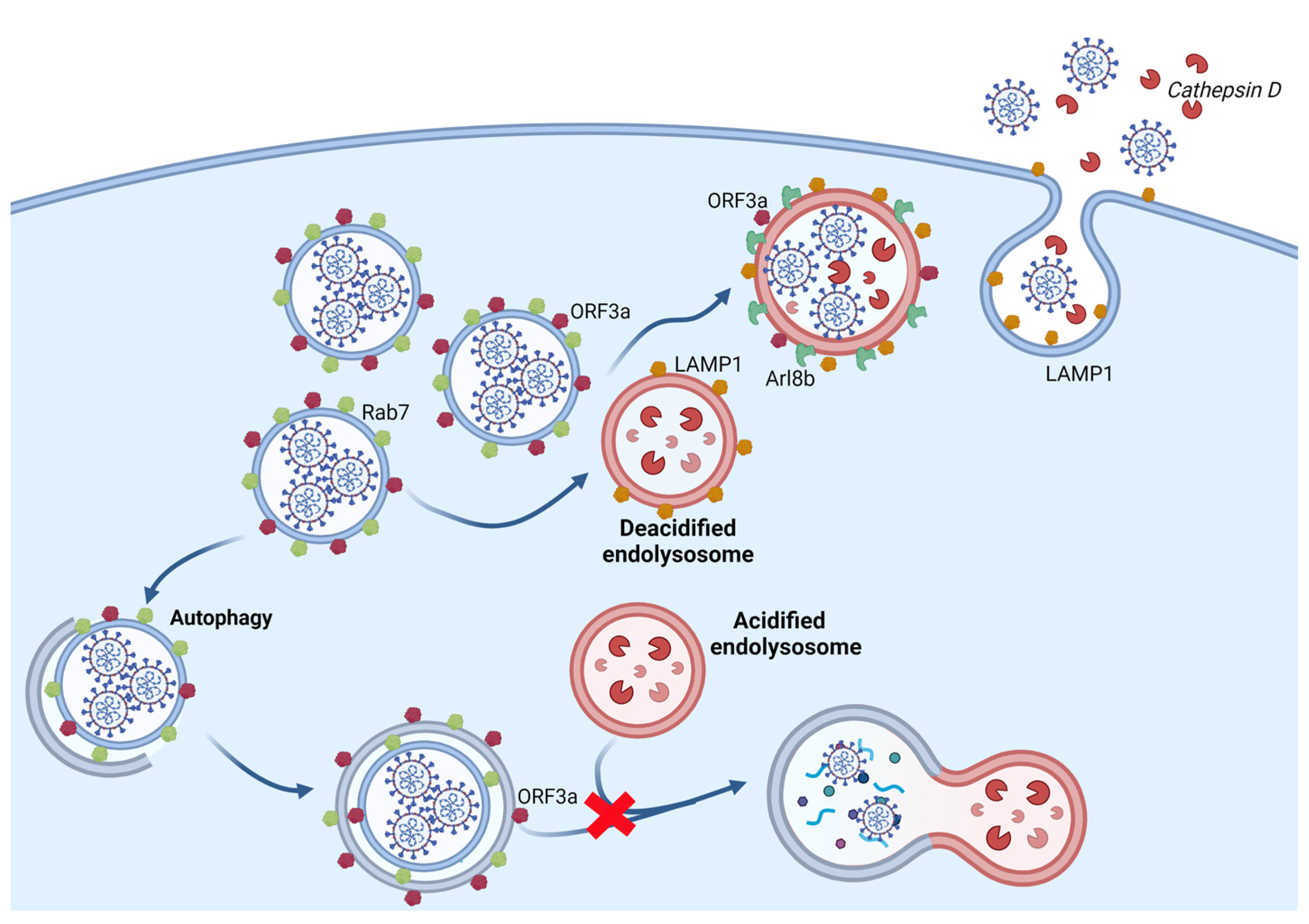
While the studies described above used HEK293/17, Stukalov and coworkers used A549 (lung carcinoma epithelial cells) to express each of SARS-CoV and SARS-CoV2 proteins and performed the interactome of viral and host proteins, as well as the alterations caused in the transcriptome, proteome, phosphoproteome, ubiquitinome of cells expressing the viral proteins [155]. With this approach, the authors recognized shared and specific mechanisms used by these two viruses highlighting the role of ORF3a and ORF8 of SARS-CoV2 in manipulating the autophagy and the TGFβ signaling pathway, respectively. The central role of ORF3a in managing autophagy confirms once more the results reported elsewhere, described in this review [139,140,141,142,148]. ORF3 blocks the autophagic flux, causing the accumulation of autophagy-related proteins (SQSTM1, GABARAPL2, NBR1, CALCOCO2, MAP1LC3A, MAP1LC3B, and TAX1BP1). Based on the interactome results, the authors propose that the ability of ORF3 to abrogate autophagy may be due to the interaction of the ORF3 protein with the HOPS complex (Vps11, Vps16, Vps18, Vps39 and Vps41). The disruptive role of ORF3 and Vps39 was later confirmed by Chen and colleagues’ study [139]. They also suggest the differential phosphorylation of regulatory sites on TSC2, mTORC1 complex, ULK1, RPS6, and SQSTM1, and ubiquitination of critical components, such as MAP1LC3A, GABARAPL2, Vps33A, and VAMP8, may explain the ability of ORF3 to inhibit autophagy [155].
In the same study, but now using Vero cells infected with an MOI of 0.01 of SARS-CoV-2, authors test drug efficacy against the virus. In this case, rapamycin suppressed SARS-CoV-2 replication, but the highest antiviral activities were obtained with the following drugs: gilteritinib-a designated inhibitor of FLT3 and AXL; ipatasertib-an AKT inhibitor; and prinomastat and marimastat-matrix metalloprotease (MMP) inhibitors. Importantly, these drugs exhibited minor effects on cell growth [155].
4. Conclusions
In this review, we gathered information about the central role of lysosomes in controlling cellular signaling, sensing cellular stress and the metabolic/energy status, degrading intra- and extracellular materials including cell invaders, communicating with the external environment, and participating in the innate immune response. To perform these varied tasks, lysosomes use many protein complexes, which are mobilized in an orchestrated way, allowing interaction with other vesicles of the endolysosomal system and the plasma membrane. Here, our intention was not to cover in depth these molecular pathways but to present a general view of lysosome biology to help the reader understand these processes and how coronaviruses highjack the endolysosomal system and its functions to their benefit.
Viruses provoke a radical shift in cell metabolism and manipulate the cellular machinery to replicate and disseminate. Endomembranes are remodeled to allow virus internalization, replication, assembly, and egress. Coronaviruses handle the lysosomal apparatus to avoid cellular defense mechanisms such as autophagy, luminal degradation, antigen presentation and ultimately explore these organelles as a route of escape. SARS-CoV-2 exhibits traits that make it especially skillful in exploiting the endosomal system, which is reflected in its high effectiveness to infect and spread. The concentrated efforts of the scientific community to comprehend SARS-CoV-2 unveiled many mechanisms involved in infection at an unprecedented speed. This translates into developing vaccines and promising drugs in under two years. Although this review was certainly born already outdated, it highlights the idea that the best way to win is to know our enemy and its tactics well. All the cited work, and those we fail to mention, help us do that. All the gathered information so far indicates that a promising course of action to control the new coronavirus could be the use of pharmaceuticals that interfere with components of the endolysosomal system. The drugs being tested at present -molnupiravir, which introduces mutations into the viral genome or Paxlovid that inhibits viral protein (Nature 599, 358–359 (2021)) processing act directly against the virus, and we can envisage the emergence of resistant strains. The advantage of developing drugs that would act on components of the endolysosomal system is that one would interfere with virus uptake or egress without selecting the virus.
Summed to that, the use of nanotechnology to optimize drug delivery and efficacy represents a powerful tool against COVID-19 (for a review, see [156]. Niclosamide, an approved anti-helminthic drug that stimulates autophagy, has broad antiviral activity but low oral bioavailability [157,158]. Importantly, it has antiviral activity against Alpha (B.1.1.7), Beta (B.1.351), and Delta (B.1617.2) SARS-CoV-2 variants in human primary lung epithelial cells, as well as in the cell lineages Vero and Caco-2 [158]. Nanotechnology has been used to improve the delivery of niclosamide to treat conditions, such as cancer and now COVID-19 [159,160]. With this purpose, the study of Brunaugh and coworkers report the development of an inhalable formulation of niclosamide associated with lysozyme, which helps the solubilization and delivery to the primary sites of infection of coronaviruses at high dose concentrations and improves the antiviral potency of niclosamide in vitro and in vivo against MERS-CoV and SARS-CoV-2 infection using a murine model [160]. Inhaled and intranasal application of niclosamide was tested in a Phase I trial that revealed that its use is safe, well-tolerated, and presents mild adverse effects, such as transient irritation of the upper airways [161].
While SARS-CoV-2 keeps surprising us with new variants, scientists try to learn and understand its biology better to control its spread around the globe. We hope the knowledge of viral strategies of infection associated with the development of vaccines and the advance of new pharmacological approaches will grant us the end this pandemic soon.
Author Contributions
Conceptualization, C.J.G.d.A.; software, F.S.P.-D.; writing—original draft preparation, C.J.G.d.A.; writing—critical review and editing, A.L.M.G., literature search, D.C.-S., preparation of figures D.C.-S. and F.S.P.-D.; funding acquisition, C.J.G.d.A. All authors have read and agreed to the published version of the manuscript.
Funding
This research was funded by Oswaldo Cruz Foundation/FIOCRUZ Inova Program, by Fundação De Amparo À Pesquisa Do Estado Do Rio De Janeiro (FAPERJ) and by Conselho Nacional De Desenvolvimento Científico E Tecnológico (CNPq). The funders had no role in study design, data collection and analysis, decision to publish, or preparation of the manuscript.
Institutional Review Board Statement
Not applicable.
Informed Consent Statement
Not applicable.
Conflicts of Interest
The authors declare no conflict of interest.
References
- Bright, N.A.; Davis, L.J.; Luzio, J.P. Endolysosomes Are the Principal Intracellular Sites of Acid Hydrolase Activity. Curr. Biol. 2016, 26, 2233–2245. [Google Scholar] [CrossRef] [Scilit]
- Pu, J.; Guardia, C.M.; Keren-Kaplan, T.; Bonifacino, J.S. Mechanisms and Functions of Lysosome Positioning. J. Cell Sci. 2016, 129, 4329–4339. [Google Scholar] [CrossRef] [Scilit]
- Luzio, J.P.; Hackmann, Y.; Dieckmann, N.M.G.; Griffiths, G.M. The Biogenesis of Lysosomes and Lysosome-Related Organelles. Cold Spring Harb. Perspect. Biol. 2014, 6, 1–17. [Google Scholar] [CrossRef] [Scilit]
- Sandvig, K.; Kavaliauskiene, S.; Skotland, T. Clathrin-Independent Endocytosis: An Increasing Degree of Complexity. Histochem. Cell Biol. 2018, 150, 107–118. [Google Scholar] [CrossRef] [Scilit]
- Ward, E.S.; Martinez, C.; Vaccaro, C.; Zhou, J.; Tang, Q.; Ober, R.J. From Sorting Endosomes to Exocytosis: Association of Rab4 and Rab11 GTPases with the Fc Receptor, FcRn, during Recycling. Mol. Biol. Cell 2005, 16, 2028–2038. [Google Scholar] [CrossRef] [Scilit]
- Huotari, J.; Helenius, A. Endosome Maturation. EMBO J. 2011, 30, 3481–3500. [Google Scholar] [CrossRef] [Scilit] [PubMed]
- Yuan, W.; Song, C. The Emerging Role of Rab5 in Membrane Receptor Trafficking and Signaling Pathways. Biochem. Res. Int. 2020, 2020, 1–10. [Google Scholar] [CrossRef] [Scilit]
- Wandinger-Ness, A.; Zerial, M. Rab Proteins and the Compartmentalization of the Endosomal System. Cold Spring Harb. Perspect. Biol. 2014, 6, a022616. [Google Scholar] [CrossRef] [Scilit]
- Li, H.; Li, H.-F.; Felder, R.A.; Periasamy, A.; Jose, P.A. Rab4 and Rab11 Coordinately Regulate the Recycling of Angiotensin II Type I Receptor as Demonstrated by Fluorescence Resonance Energy Transfer Microscopy. J. Biomed. Opt. 2008, 13, 031206. [Google Scholar] [CrossRef] [Scilit]
- Saftig, P.; Klumperman, J. Lysosome Biogenesis and Lysosomal Membrane Proteins: Trafficking Meets Function. Nat. Rev. Mol. Cell Biol. 2009, 10, 623–635. [Google Scholar] [CrossRef] [Scilit]
- Jahn, R.; Scheller, R.H. SNAREs—Engines for Membrane Fusion. Nat. Rev. Mol. Cell Biol. 2006, 7, 631–643. [Google Scholar] [CrossRef] [Scilit]
- Balderhaar, H.J.K.; Ungermann, C. CORVET and HOPS Tethering Complexes—Coordinators of Endosome and Lysosome Fusion. J. Cell Sci. 2013, 126, 1307–1316. [Google Scholar] [CrossRef] [Scilit] [PubMed]
- Solinger, J.A.; Spang, A. Tethering Complexes in the Endocytic Pathway: CORVET and HOPS. FEBS J. 2013, 280, 2743–2757. [Google Scholar] [CrossRef] [Scilit] [PubMed]
- van der Kant, R.; Jonker, C.T.H.; Wijdeven, R.H.; Bakker, J.; Janssen, L.; Klumperman, J.; Neefjes, J. Characterization of the Mammalian CORVET and HOPS Complexes and Their Modular Restructuring for Endosome Specificity. J. Biol. Chem. 2015, 290, 30280–30290. [Google Scholar] [CrossRef] [Scilit] [PubMed]
- Klumperman, J.; Hille, A.; Veenendaal, T.; Oorschot, V.; Stoorvogel, W.; von Figura, K.; Geuze, H.J. Differences in the Endosomal Distributions of the Two Mannose 6-Phosphate Receptors. J. Cell Biol. 1993, 121, 997–1010. [Google Scholar] [CrossRef] [Scilit] [PubMed]
- Hurley, J.H. ESCRTs Are Everywhere. EMBO J 2015, 34, 2398–2407. [Google Scholar] [CrossRef] [Scilit]
- Catalano, M.; O’Driscoll, L. Inhibiting Extracellular Vesicles Formation and Release: A Review of EV Inhibitors. J. Extracell. Vesicles 2020, 9, 1703244. [Google Scholar] [CrossRef] [Scilit]
- Trajkovic, K.; Hsu, C.; Chiantia, S.; Rajendran, L.; Wenzel, D.; Wieland, F.; Schwille, P.; Brügger, B.; Simons, M. Ceramide Triggers Budding of Exosome Vesicles into Multivesicular Endosomes. Science 2008, 319, 1244–1247. [Google Scholar] [CrossRef] [Scilit]
- Datta, A.; Kim, H.; Lai, M.; McGee, L.; Johnson, A.; Moustafa, A.A.; Jones, J.C.; Mondal, D.; Ferrer, M.; Abdel-Mageed, A.B. Manumycin A Suppresses Exosome Biogenesis and Secretion via Targeted Inhibition of Ras/Raf/ERK1/2 Signaling and HnRNP H1 in Castration-Resistant Prostate Cancer Cells. Cancer Lett. 2017, 408, 73–81. [Google Scholar] [CrossRef] [Scilit]
- Tancini, B.; Buratta, S.; Delo, F.; Sagini, K.; Chiaradia, E.; Pellegrino, R.M.; Emiliani, C.; Urbanelli, L. Lysosomal Exocytosis: The Extracellular Role of an Intracellular Organelle. Membranes 2020, 10, 406. [Google Scholar] [CrossRef] [Scilit]
- Hofmann, I.; Munro, S. An N-Terminally Acetylated Arf-like GTPase Is Localised to Lysosomes and Affects Their Motility. J. Cell Sci. 2006, 119, 1494–1503. [Google Scholar] [CrossRef] [Scilit] [PubMed]
- Rosa-Ferreira, C.; Munro, S. Arl8 and SKIP Act Together to Link Lysosomes to Kinesin-1. Dev. Cell 2011, 21, 1171–1178. [Google Scholar] [CrossRef] [Scilit] [PubMed]
- Khatter, D.; Sindhwani, A.; Sharma, M. Arf-like GTPase Arl8: Moving from the Periphery to the Center of Lysosomal Biology. Cell. Logist. 2015, 5, e1086501. [Google Scholar] [CrossRef] [Scilit] [PubMed]
- Pu, J.; Schindler, C.; Jia, R.; Jarnik, M.; Backlund, P.; Bonifacino, J.S. BORC, a Multisubunit Complex That Regulates Lysosome Positioning. Dev. Cell 2015, 33, 176–188. [Google Scholar] [CrossRef] [Scilit]
- Savina, A.; Vidal, M.; Colombo, M.I. The Exosome Pathway in K562 Cells Is Regulated by Rab11. J. Cell Sci. 2002, 115, 2505–2515. [Google Scholar] [CrossRef] [Scilit] [PubMed]
- Ostrowski, M.; Carmo, N.B.; Krumeich, S.; Fanget, I.; Raposo, G.; Savina, A.; Moita, C.F.; Schauer, K.; Hume, A.N.; Freitas, R.P.; et al. Rab27a and Rab27b Control Different Steps of the Exosome Secretion Pathway. Nat. Cell Biol. 2010, 12, 19–30. [Google Scholar] [CrossRef] [Scilit]
- Hsu, C.; Morohashi, Y.; Yoshimura, S.I.; Manrique-Hoyos, N.; Jung, S.Y.; Lauterbach, M.A.; Bakhti, M.; Grønborg, M.; Möbius, W.; Rhee, J.S.; et al. Regulation of Exosome Secretion by Rab35 and Its GTPase-Activating Proteins TBC1D10A-C. J. Cell Biol. 2010, 189, 223–232. [Google Scholar] [CrossRef] [Scilit]
- Rao, S.K.; Huynh, C.; Proux-Gillardeaux, V.; Galli, T.; Andrews, N.W. Identification of SNAREs Involved in Synaptotagmin VII-Regulated Lysosomal Exocytosis. J. Biol. Chem. 2004, 279, 20471–20479. [Google Scholar] [CrossRef] [Scilit]
- Korolchuk, V.I.; Saiki, S.; Lichtenberg, M.; Siddiqi, F.H.; Roberts, E.A.; Imarisio, S.; Jahreiss, L.; Sarkar, S.; Futter, M.; Menzies, F.M.; et al. Lysosomal Positioning Coordinates Cellular Nutrient Responses. Nat. Cell Biol. 2011, 13, 453–462. [Google Scholar] [CrossRef] [Scilit]
- Puertollano, R. MTOR and Lysosome Regulation. F1000Prime Rep. 2014, 6, 1–7. [Google Scholar] [CrossRef] [Scilit]
- Saxton, R.A.; Sabatini, D.M. MTOR Signaling in Growth, Metabolism, and Disease. Cell 2017, 168, 960–976. [Google Scholar] [CrossRef] [Scilit] [PubMed]
- Dossou, A.S.; Bosu, A. The Emerging Roles of MTORC1 in Macromanaging Autophagy. Cancers 2019, 10, 1422. [Google Scholar] [CrossRef] [Scilit] [PubMed]
- Dikic, I.; Elazar, Z. Mechanism and Medical Implications of Mammalian Autophagy. Nat. Rev. Mol. Cell Biol. 2018, 19, 349–364. [Google Scholar] [CrossRef] [Scilit]
- Ganley, I.G.; Lam, D.H.; Wang, J.; Ding, X.; Chen, S.; Jiang, X. ULK1·ATG13·FIP200 Complex Mediates MTOR Signaling and Is Essential for Autophagy. J. Biol. Chem. 2009, 284, 12297–12305. [Google Scholar] [CrossRef] [Scilit] [PubMed]
- Hosokawa, N.; Sasaki, T.; Iemura, S.I.; Natsume, T.; Hara, T.; Mizushima, N. Atg101, a Novel Mammalian Autophagy Protein Interacting with Atg13. Autophagy 2009, 5, 973–979. [Google Scholar] [CrossRef] [Scilit]
- Shang, L.; Wang, X. AMPK and MTOR Coordinate the Regulation of Ulk1 and Mammalian Autophagy Initiation. Autophagy 2011, 7, 924–926. [Google Scholar] [CrossRef] [Scilit]
- Jung, C.H.; Jun, C.B.; Ro, S.-H.; Kim, Y.-M.; Otto, N.M.; Cao, J.; Kundu, M.; Kim, D.-H. ULK-Atg13-FIP200 Complexes Mediate MTOR Signaling to the Autophagy Machinery. Mol. Biol. Cell 2009, 20, 1992–2003. [Google Scholar] [CrossRef] [Scilit]
- Bach, M.; Larance, M.; James, D.E.; Ramm, G. The Serine/Threonine Kinase ULK1 Is a Target of Multiple Phosphorylation Events. Biochem. J. 2011, 440, 283–291. [Google Scholar] [CrossRef] [Scilit]
- Egan, D.F.; Chun, M.G.H.; Vamos, M.; Zou, H.; Rong, J.; Miller, C.J.; Lou, H.J.; Raveendra-Panickar, D.; Yang, C.C.; Sheffler, D.J.; et al. Small Molecule Inhibition of the Autophagy Kinase ULK1 and Identification of ULK1 Substrates. Mol. Cell 2015, 59, 285–297. [Google Scholar] [CrossRef] [Scilit]
- Lin, M.G.; Hurley, J.H. Structure and Function of the ULK1 Complex in Autophagy. Curr. Opin. Cell Biol. 2016, 39, 61–68. [Google Scholar] [CrossRef] [Scilit]
- Russell, R.C.; Tian, Y.; Yuan, H.; Park, H.W.; Chang, Y.Y.; Kim, J.; Kim, H.; Neufeld, T.P.; Dillin, A.; Guan, K.L. ULK1 Induces Autophagy by Phosphorylating Beclin-1 and Activating VPS34 Lipid Kinase. Nat. Cell Biol. 2013, 15, 741–750. [Google Scholar] [CrossRef] [Scilit]
- Itakura, E.; Mizushima, N. Atg14 and UVRAG: Mutually Exclusive Subunits of Mammalian Beclin 1-PI3K Complexes. Autophagy 2009, 5, 534–536. [Google Scholar] [CrossRef] [Scilit] [PubMed]
- Itakura, E.; Mizushima, N. Characterization of Autophagosome Formation Site by a Hierarchical Analysis of Mammalian Atg Proteins. Autophagy 2010, 6, 764–776. [Google Scholar] [CrossRef] [Scilit]
- Liang, C.; Lee, J.S.; Inn, K.S.; Gack, M.U.; Li, Q.; Roberts, E.A.; Vergne, I.; Deretic, V.; Feng, P.; Akazawa, C.; et al. Beclin1-Binding UVRAG Targets the Class C Vps Complex to Coordinate Autophagosome Maturation and Endocytic Trafficking. Nat. Cell Biol. 2008, 10, 776–787. [Google Scholar] [CrossRef] [Scilit] [PubMed]
- Liang, C.; Feng, P.; Ku, B.; Dotan, I.; Canaani, D.; Oh, B.H.; Jung, J.U. Autophagic and Tumour Suppressor Activity of a Novel Beclin1-Binding Protein UVRAG. Nat. Cell Biol. 2006, 8, 688–698. [Google Scholar] [CrossRef] [Scilit] [PubMed]
- Mercer, T.J.; Gubas, A.; Tooze, S.A. A Molecular Perspective of Mammalian Autophagosome Biogenesis. J. Biol. Chem. 2018, 293, 5386–5395. [Google Scholar] [CrossRef] [Scilit] [PubMed]
- Zhao, Y.G.; Zhang, H. Autophagosome Maturation: An Epic Journey from the ER to Lysosomes. J. Cell Biol. 2019, 218, 757–770. [Google Scholar] [CrossRef] [Scilit] [PubMed]
- Grasso, D.; Renna, F.J.; Vaccaro, M.I. Initial Steps in Mammalian Autophagosome Biogenesis. Front. Cell Dev. Biol. 2018, 6, 1–10. [Google Scholar] [CrossRef] [Scilit]
- Tripathi, D.N.; Chowdhury, R.; Trudel, L.J.; Tee, A.R.; Slack, R.S.; Walker, C.L.; Wogan, G.N. Reactive Nitrogen Species Regulate Autophagy through ATM-AMPK-TSC2-Mediated Suppression of MTORC1. Proc. Natl. Acad. Sci. USA 2013, 110. [Google Scholar] [CrossRef] [Scilit]
- Sardiello, M.; Ballabio, A. Lysosomal Enhancement: A CLEAR Answer to Cellular Degradative Needs. Cell Cycle 2009, 8, 4021–4022. [Google Scholar] [CrossRef] [Scilit]
- Settembre, C.; di Malta, C.; Polito, V.A.; Arencibia, M.G.; Vetrini, F.; Erdin, S.; Erdin, S.U.; Huynh, T.; Medina, D.; Colella, P.; et al. TFEB Links Autophagy to Lysosomal Biogenesis. Science 2011, 332, 1429–1433. [Google Scholar] [CrossRef] [Scilit] [PubMed]
- Boda, A.; Lőrincz, P.; Takáts, S.; Csizmadia, T.; Tóth, S.; Kovács, A.L.; Juhász, G. Drosophila Arl8 Is a General Positive Regulator of Lysosomal Fusion Events. Biochim. Biophys. Acta Mol. Cell Res. 2019, 1866, 533–544. [Google Scholar] [CrossRef] [Scilit] [PubMed]
- Itakura, E.; Kishi-Itakura, C.; Mizushima, N. The Hairpin-Type Tail-Anchored SNARE Syntaxin 17 Targets to Autophagosomes for Fusion with Endosomes/Lysosomes. Cell 2012, 151, 1256–1269. [Google Scholar] [CrossRef] [Scilit] [PubMed]
- Jiang, P.; Nishimura, T.; Sakamaki, Y.; Itakura, E.; Hatta, T.; Natsume, T.; Mizushima, N. The HOPS Complex Mediates Autophagosome-Lysosome Fusion through Interaction with Syntaxin 17. Mol. Biol. Cell 2014, 25, 1327–1337. [Google Scholar] [CrossRef] [Scilit]
- Takáts, S.; Pircs, K.; Nagy, P.; Varga, Á.; Kárpáti, M.; Hegedus, K.; Kramer, H.; Kovács, A.L.; Sass, M.; Juhász, G. Interaction of the HOPS Complex with Syntaxin 17 Mediates Autophagosome Clearance in Drosophila. Mol. Biol. Cell 2014, 25, 1338–1354. [Google Scholar] [CrossRef] [Scilit]
- Chen, Y.; Yu, L. Autophagic Lysosome Reformation. Exp. Cell Res. 2013, 319, 142–146. [Google Scholar] [CrossRef] [Scilit] [PubMed]
- Lakpa, K.L.; Khan, N.; Afghah, Z.; Chen, X.; Geiger, J.D. Lysosomal Stress Response (LSR): Physiological Importance and Pathological Relevance. J. Neuroimmune Pharmacol. 2021, 16, 219–237. [Google Scholar] [CrossRef] [Scilit]
- Johnson, D.E.; Ostrowski, P.; Jaumouillé, V.; Grinstein, S. The Position of Lysosomes within the Cell Determines Their Luminal PH. J. Cell Biol. 2016, 212, 677–692. [Google Scholar] [CrossRef] [Scilit]
- Ponsford, A.H.; Ryan, T.A.; Raimondi, A.; Cocucci, E.; Wycislo, S.A.; Fröhlich, F.; Swan, L.E.; Stagi, M. Live Imaging of Intra-Lysosome PH in Cell Lines and Primary Neuronal Culture Using a Novel Genetically Encoded Biosensor. Autophagy 2021, 17, 1500–1518. [Google Scholar] [CrossRef] [Scilit]
- Yamauchi, Y.; Helenius, A. Virus Entry at a Glance. J. Cell Sci. 2013, 126, 1289–1295. [Google Scholar] [CrossRef] [Scilit]
- Carty, M.; Guy, C.; Bowie, A.G. Detection of Viral Infections by Innate Immunity. Biochem. Pharmacol. 2021, 183, 114316. [Google Scholar] [CrossRef] [Scilit] [PubMed]
- Ripa, I.; Andreu, S.; López-Guerrero, J.A.; Bello-Morales, R. Membrane Rafts: Portals for Viral Entry. Front. Microbiol. 2021, 12, 120. [Google Scholar] [CrossRef] [Scilit]
- Mayor, S.; Parton, R.G.; Donaldson, J.G. Clathrin-Independent Pathways of Endocytosis. Cold Spring Harb. Perspect. Biol. 2014, 6, a016758. [Google Scholar] [CrossRef] [Scilit] [PubMed]
- Pelkmans, L.; Kartenbeck, J.; Helenius, A. Caveolar Endocytosis of Simian Virus 40 Reveals a New Two-Step Vesicular-Transport Pathway to the ER. Nat. Cell Biol. 2001, 3, 473–483. [Google Scholar] [CrossRef] [Scilit] [PubMed]
- Damm, E.M.; Pelkmans, L.; Kartenbeck, J.; Mezzacasa, A.; Kurzchalia, T.; Helenius, A. Clathrin- and Caveolin-1-Independent Endocytosis: Entry of Simian Virus 40 into Cells Devoid of Caveolae. J. Cell Biol. 2005, 168, 477–488. [Google Scholar] [CrossRef] [Scilit] [PubMed]
- Ewers, H.; Römer, W.; Smith, A.E.; Bacia, K.; Dmitrieff, S.; Chai, W.; Mancini, R.; Kartenbeck, J.; Chambon, V.; Berland, L.; et al. GM1 Structure Determines SV40-Induced Membrane Invagination and Infection. Nat. Cell Biol. 2010, 12, 11–18. [Google Scholar] [CrossRef] [Scilit]
- Cheng, J.P.X.; Nichols, B.J. Caveolae: One Function or Many? Trends Cell Biol. 2016, 26, 177–189. [Google Scholar] [CrossRef] [Scilit]
- Parton, R.G. Caveolae: Structure, Function, and Relationship to Disease. Annu. Rev. Cell Dev. Biol. 2018, 34, 111–136. [Google Scholar] [CrossRef] [Scilit]
- Thomsen, P.; Roepstorff, K.; Stahlhut, M.; van Deurs, B. Caveolae Are Highly Immobile Plasma Membrane Microdomains, Which Are Not Involved in Constitutive Endocytic Trafficking. Mol. Biol. Cell 2002, 13, 238–250. [Google Scholar] [CrossRef] [Scilit]
- Le, P.U.; Guay, G.; Altschuler, Y.; Nabi, I.R. Caveolin-1 Is a Negative Regulator of Caveolae-Mediated Endocytosis to the Endoplasmic Reticulum. J. Biol. Chem. 2002, 277, 3371–3379. [Google Scholar] [CrossRef] [Scilit]
- Hoffmann, C.; Berking, A.; Agerer, F.; Buntru, A.; Neske, F.; Chhatwal, G.S.; Ohlsen, K.; Hauck, C.R. Caveolin Limits Membrane Microdomain Mobility and Integrin-Mediated Uptake of Fibronectin-Binding Pathogens. J. Cell Sci. 2010, 123, 4280–4291. [Google Scholar] [CrossRef] [Scilit] [PubMed]
- Lajoie, P.; Kojic, L.D.; Nim, S.; Li, L.; Dennis, J.W.; Nabi, I.R. Caveolin-1 Regulation of Dynamin-Dependent, Raft-Mediated Endocytosis of Cholera Toxin-B Sub-Unit Occurs Independently of Caveolae. J. Cell. Mol. Med. 2009, 13, 3218–3225. [Google Scholar] [CrossRef] [Scilit] [PubMed]
- Pelkmans, L.; Zerial, M. Kinase-Regulated Quantal Assemblies and Kiss-and-Run Recycling of Caveolae. Nature 2005, 436, 128–133. [Google Scholar] [CrossRef] [Scilit] [PubMed]
- Lajoie, P.; Nabi, I.R. Lipid Rafts, Caveolae, and Their Endocytosis, 1st ed.; Elsevier Inc.: Amsterdam, The Netherlands, 2010; Volume 282. [Google Scholar]
- Parton, R.G.; Joggerst, B.; Simons, K. Regulated Internalization of Caveolae. J. Cell Biol. 1994, 127, 1199–1215. [Google Scholar] [CrossRef] [Scilit] [PubMed]
- Pol, A.; Lu, A.; Pons, M.; Peiró, S.; Enrich, C. Epidermal Growth Factor-Mediated Caveolin Recruitment to Early Endosomes and MAPK Activation. Role of Cholesterol and Actin Cytoskeleton. J. Biol. Chem. 2000, 275, 30566–30572. [Google Scholar] [CrossRef] [Scilit]
- Tran, D.; Carpentier, J.L.; Sawano, F.; Gorden, P.; Orci, L. Ligands Internalized through Coated or Noncoated Invaginations Follow a Common Intracellular Pathway. Proc. Natl. Acad. Sci. USA 1987, 84, 7957–7961. [Google Scholar] [CrossRef] [Scilit]
- Pelkmans, L.; Bürli, T.; Zerial, M.; Helenius, A. Caveolin-Stabilized Membrane Domains as Multifunctional Transport and Sorting Devices in Endocytic Membrane Traffic. Cell 2004, 118, 767–780. [Google Scholar] [CrossRef] [Scilit]
- Gagescu, R.; Demaurex, N.; Parton, R.G.; Hunziker, W.; Huber, L.A.; Gruenberg, J. The Recycling Endosome of Madin-Darby Canine Kidney Cells Is a Mildly Acidic Compartment Rich in Raft Components. Mol. Biol. Cell 2000, 11, 2775–2791. [Google Scholar] [CrossRef] [Scilit]
- Liebl, D.; Difato, F.; Horníková, L.; Mannová, P.; Štokrová, J.; Forstová, J. Mouse Polyomavirus Enters Early Endosomes, Requires Their Acidic PH for Productive Infection, and Meets Transferrin Cargo in Rab11-Positive Endosomes. J. Virol. 2006, 80, 4610–4622. [Google Scholar] [CrossRef] [Scilit]
- Hill, M.M.; Bastiani, M.; Luetterforst, R.; Kirkham, M.; Kirkham, A.; Nixon, S.J.; Walser, P.; Abankwa, D.; Oorschot, V.M.J.; Martin, S.; et al. PTRF-Cavin, a Conserved Cytoplasmic Protein Required for Caveola Formation and Function. Cell 2008, 132, 113–124. [Google Scholar] [CrossRef] [Scilit]
- Hayer, A.; Stoeber, M.; Ritz, D.; Engel, S.; Meyer, H.H.; Helenius, A. Caveolin-1 Is Ubiquitinated and Targeted to Intralumenal Vesicles in Endolysosomes for Degradation. J. Cell Biol. 2010, 191, 615–629. [Google Scholar] [CrossRef] [Scilit] [PubMed]
- Parton, R.G.; Howes, M.T. Revisiting Caveolin Trafficking: The End of the Caveosome. J. Cell Biol. 2010, 191, 439–441. [Google Scholar] [CrossRef] [Scilit] [PubMed]
- Mercer, J.; Schelhaas, M.; Helenius, A. Virus Entry by Endocytosis. Annu. Rev. Biochem. 2010, 79, 803–833. [Google Scholar] [CrossRef] [Scilit] [PubMed]
- Chaudhary, N.; Gomez, G.A.; Howes, M.T.; Lo, H.P.; McMahon, K.A.; Rae, J.A.; Schieber, N.L.; Hill, M.M.; Gaus, K.; Yap, A.S.; et al. Endocytic Crosstalk: Cavins, Caveolins, and Caveolae Regulate Clathrin-Independent Endocytosis. PLoS Biol. 2014, 12, e1001832. [Google Scholar] [CrossRef] [Scilit] [PubMed]
- Meier, O.; Boucke, K.; Hammer, S.V.; Keller, S.; Stidwill, R.P.; Hemmi, S.; Greber, U.F. Adenovirus Triggers Macropinocytosis and Endosomal Leakage Together with Its Clathrin-Mediated Uptake. J. Cell Biol. 2002, 158, 1119–1131. [Google Scholar] [CrossRef] [Scilit] [PubMed]
- Zhou, P.; Yang, X.L.; Wang, X.G.; Hu, B.; Zhang, L.; Zhang, W.; Si, H.R.; Zhu, Y.; Li, B.; Huang, C.L.; et al. A Pneumonia Outbreak Associated with a New Coronavirus of Probable Bat Origin. Nature 2020, 579, 270–273. [Google Scholar] [CrossRef] [Scilit] [PubMed]
- Letko, M.; Marzi, A.; Munster, V. Functional Assessment of Cell Entry and Receptor Usage for SARS-CoV-2 and Other Lineage B Betacoronaviruses. Nat. Microbiol. 2020, 5, 562–569. [Google Scholar] [CrossRef] [Scilit]
- Daly, J.L.; Simonetti, B.; Klein, K.; Chen, K.E.; Williamson, M.K.; Antón-Plágaro, C.; Shoemark, D.K.; Simón-Gracia, L.; Bauer, M.; Hollandi, R.; et al. Neuropilin-1 Is a Host Factor for SARS-CoV-2 Infection. Science 2020, 370, 861–865. [Google Scholar] [CrossRef] [Scilit]
- Wang, K.; Chen, W.; Zhou, Y.S.; Lian, J.Q.; Zhang, Z.; Du, P.; Gong, L.; Zhang, Y.; Cui, H.Y.; Geng, J.J.; et al. SARS-CoV-2 Invades Host Cells via a Novel Route: CD147-Spike Protein. bioRxiv 2020. [Google Scholar] [CrossRef] [Scilit]
- Liu, L.; Chopra, P.; Li, X.; Bouwman, K.M.; Tompkins, S.M.; Wolfert, M.A.; de Vries, R.P.; Boons, G.J. Heparan Sulfate Proteoglycans as Attachment Factor for SARS-CoV-2. ACS Cent. Sci. 2021, 7, 1009–1018. [Google Scholar] [CrossRef] [Scilit]
- Clausen, T.M.; Sandoval, D.R.; Spliid, C.B.; Pihl, J.; Perrett, H.R.; Painter, C.D.; Narayanan, A.; Majowicz, S.A.; Kwong, E.M.; McVicar, R.N.; et al. SARS-CoV-2 Infection Depends on Cellular Heparan Sulfate and ACE2. Cell 2020, 183, 1043–1057.e15. [Google Scholar] [CrossRef] [Scilit] [PubMed]
- Amraei, R.; Yin, W.; Napoleon, M.A.; Suder, E.L.; Berrigan, J.; Zhao, Q.; Olejnik, J.; Chandler, K.B.; Xia, C.; Feldman, J.; et al. CD209L/L-SIGN and CD209/DC-SIGN Act as Receptors for SARS-CoV-2. ACS Cent. Sci. 2021, 7, 1156–1165. [Google Scholar] [CrossRef] [Scilit] [PubMed]
- Carlos, A.J.; Ha, D.P.; Yeh, D.W.; van Krieken, R.; Tseng, C.C.; Zhang, P.; Gill, P.; Machida, K.; Lee, A.S. The Chaperone GRP78 Is a Host Auxiliary Factor for SARS-CoV- 2 and GRP78 Depleting Antibody Blocks Viral Entry and Infection. J. Biol. Chem. 2021, 296, 100759. [Google Scholar] [CrossRef] [Scilit] [PubMed]
- Wang, S.; Qiu, Z.; Hou, Y.; Deng, X.; Xu, W.; Zheng, T.; Wu, P.; Xie, S.; Bian, W.; Zhang, C.; et al. AXL Is a Candidate Receptor for SARS-CoV-2 That Promotes Infection of Pulmonary and Bronchial Epithelial Cells. Cell Res. 2021, 31, 126–140. [Google Scholar] [CrossRef] [Scilit]
- Ichimura, T.; Mori, Y.; Aschauer, P.; Padmanabha Das, K.M.; Padera, R.F.; Weins, A.; Nasr, M.L.; Bonventre, J. v KIM-1/TIM-1 Is a Receptor for SARS-CoV-2 in Lung and Kidney. Medrxiv Prepr. Serv. Health Sci. 2020. [Google Scholar] [CrossRef] [Scilit]
- Hoffmann, M.; Kleine-Weber, H.; Pöhlmann, S. A Multibasic Cleavage Site in the Spike Protein of SARS-CoV-2 Is Essential for Infection of Human Lung Cells. Mol. Cell 2020, 78, 779–784.e5. [Google Scholar] [CrossRef] [Scilit]
- Hoffmann, M.; Kleine-Weber, H.; Schroeder, S.; Krüger, N.; Herrler, T.; Erichsen, S.; Schiergens, T.S.; Herrler, G.; Wu, N.H.; Nitsche, A.; et al. SARS-CoV-2 Cell Entry Depends on ACE2 and TMPRSS2 and Is Blocked by a Clinically Proven Protease Inhibitor. Cell 2020, 181, 271–280.e8. [Google Scholar] [CrossRef] [Scilit]
- Zhu, Y.; Feng, F.; Hu, G.; Wang, Y.; Yu, Y.; Zhu, Y.; Xu, W.; Cai, X.; Sun, Z.; Han, W.; et al. A Genome-wide CRISPR Screen Identifies Host Factors That Regulate SARS-CoV-2 Entry. Nat. Commun. 2021, 12, 961. [Google Scholar] [CrossRef] [Scilit] [PubMed]
- Papa, G.; Mallery, D.L.; Albecka, A.; Welch, L.G.; Cattin-Ortolá, J.; Luptak, J.; Paul, D.; McMahon, H.T.; Goodfellow, I.G.; Carter, A.; et al. Furin Cleavage of SARS-CoV-2 Spike Promotes but Is Not Essential for Infection and Cell-Cell Fusion. PLoS Pathog. 2021, 17, e1009246. [Google Scholar] [CrossRef] [Scilit]
- Shang, J.; Ye, G.; Shi, K.; Wan, Y.; Luo, C.; Aihara, H.; Geng, Q.; Auerbach, A.; Li, F. Structural Basis of Receptor Recognition by SARS-CoV-2. Nature 2020, 581, 221–224. [Google Scholar] [CrossRef] [Scilit]
- Walls, A.C.; Park, Y.J.; Tortorici, M.A.; Wall, A.; McGuire, A.T.; Veesler, D. Structure, Function, and Antigenicity of the SARS-CoV-2 Spike Glycoprotein. Cell 2020, 181, 281–292.e6. [Google Scholar] [CrossRef] [Scilit] [PubMed]
- Wrapp, D.; Wang, N.; Corbett, K.S.; Goldsmith, J.A.; Hsieh, C.L.; Abiona, O.; Graham, B.S.; McLellan, J.S. Cryo-EM Structure of the 2019-NCoV Spike in the Prefusion Conformation. Science 2020, 367, 1260–1263. [Google Scholar] [CrossRef] [Scilit] [PubMed]
- Koch, J.; Uckeley, Z.M.; Doldan, P.; Stanifer, M.; Boulant, S.; Lozach, P. TMPRSS2 Expression Dictates the Entry Route Used by SARS-CoV-2 to Infect Host Cells. EMBO J. 2021, 40, 1–20. [Google Scholar] [CrossRef] [Scilit] [PubMed]
- Kawase, M.; Shirato, K.; van der Hoek, L.; Taguchi, F.; Matsuyama, S. Simultaneous Treatment of Human Bronchial Epithelial Cells with Serine and Cysteine Protease Inhibitors Prevents Severe Acute Respiratory Syndrome Coronavirus Entry. J. Virol. 2012, 86, 6537–6545. [Google Scholar] [CrossRef] [Scilit]
- Shirato, K.; Kawase, M.; Matsuyama, S. Wild-Type Human Coronaviruses Prefer Cell-Surface TMPRSS2 to Endosomal Cathepsins for Cell Entry. Virology 2018, 517, 9–15. [Google Scholar] [CrossRef] [Scilit] [PubMed]
- Ou, X.; Liu, Y.; Lei, X.; Li, P.; Mi, D.; Ren, L.; Guo, L.; Guo, R.; Chen, T.; Hu, J.; et al. Characterization of Spike Glycoprotein of SARS-CoV-2 on Virus Entry and Its Immune Cross-Reactivity with SARS-CoV. Nat. Commun. 2020, 11, 1620. [Google Scholar] [CrossRef] [Scilit]
- Bollavaram, K.; Leeman, T.H.; Lee, M.W.; Kulkarni, A.; Upshaw, S.G.; Yang, J.; Song, H.; Platt, M.O. Multiple Sites on SARS-CoV-2 Spike Protein Are Susceptible to Proteolysis by Cathepsins B, K, L, S, and V. Protein Sci. 2021, 30, 1131–1143. [Google Scholar] [CrossRef] [Scilit]
- Mille, J.K.; Whittaker, G.R. Host Cell Entry of Middle East Respiratory Syndrome Coronavirus after Two-Step, Furin-Mediated Activation of the Spike Protein. Proc. Natl. Acad. Sci. USA 2014, 111, 15214–15219. [Google Scholar] [CrossRef] [Scilit]
- Park, J.E.; Li, K.; Barlan, A.; Fehr, A.R.; Perlman, S.; McCray, P.B.; Gallagher, T. Proteolytic Processing of Middle East Respiratory Syndrome Coronavirus Spikes Expands Virus Tropism. Proc. Natl. Acad. Sci. USA 2016, 113, 12262–12267. [Google Scholar] [CrossRef] [Scilit]
- Zhou, Y.; Vedantham, P.; Lu, K.; Agudelo, J.; Carrion, R.; Nunneley, J.W.; Barnard, D.; Pöhlmann, S.; McKerrow, J.H.; Renslo, A.R.; et al. Protease Inhibitors Targeting Coronavirus and Filovirus Entry. Antivir. Res. 2015, 116, 76–84. [Google Scholar] [CrossRef] [Scilit]
- Iwata-Yoshikawa, N.; Okamura, T.; Shimizu, Y.; Hasegawa, H.; Takeda, M.; Nagata, N. TMPRSS2 Contributes to Virus Spread and Immunopathology in the Airways of Murine Models after Coronavirus Infection. J. Virol. 2019, 93, e01815-18. [Google Scholar] [CrossRef] [Scilit] [PubMed]
- Mingo, R.M.; Simmons, J.A.; Shoemaker, C.J.; Nelson, E.A.; Schornberg, K.L.; D’Souza, R.S.; Casanova, J.E.; White, J.M. Ebola Virus and Severe Acute Respiratory Syndrome Coronavirus Display Late Cell Entry Kinetics: Evidence That Transport to NPC1 + Endolysosomes Is a Rate-Defining Step. J. Virol. 2015, 89, 2931–2943. [Google Scholar] [CrossRef] [Scilit] [PubMed]
- Earnest, J.T.; Hantak, M.P.; Li, K.; McCray, P.B.; Perlman, S.; Gallagher, T. The Tetraspanin CD9 Facilitates MERS-Coronavirus Entry by Scaffolding Host Cell Receptors and Proteases. PLoS Pathog. 2017, 13, 1–22. [Google Scholar] [CrossRef] [Scilit] [PubMed]
- Chambers, J.P.; Yu, J.; Valdes, J.J.; Arulanandam, B.P. SARS-CoV-2, Early Entry Events. J. Pathog. 2020, 2020, 1–11. [Google Scholar] [CrossRef] [Scilit] [PubMed]
- Sherman, E.J.; Emmer, B.T. ACE2 Protein Expression within Isogenic Cell Lines Is Heterogeneous and Associated with Distinct Transcriptomes. Sci. Rep. 2021, 11, 1–13. [Google Scholar] [CrossRef] [Scilit] [PubMed]
- Bayati, A.; Kumar, R.; Francis, V.; McPherson, P.S. SARS-CoV-2 Infects Cells after Viral Entry via Clathrin-Mediated Endocytosis. J. Biol. Chem. 2021, 296, 100306. [Google Scholar] [CrossRef] [Scilit]
- Li, X.; Zhu, W.; Fan, M.; Zhang, J.; Peng, Y.; Huang, F.; Wang, N.; He, L.; Zhang, L.; Holmdahl, R.; et al. Dependence of SARS-CoV-2 Infection on Cholesterol-Rich Lipid Raft and Endosomal Acidification. Comput. Struct. Biotechnol. J. 2021, 19, 1933–1943. [Google Scholar] [CrossRef] [Scilit]
- Li, G.M.; Li, Y.G.; Yamate, M.; Li, S.M.; Ikuta, K. Lipid Rafts Play an Important Role in the Early Stage of Severe Acute Respiratory Syndrome-Coronavirus Life Cycle. Microbes Infect. 2007, 9, 96–102. [Google Scholar] [CrossRef] [Scilit]
- Wang, S.; Li, W.; Hui, H.; Tiwari, S.K.; Zhang, Q.; Croker, B.A.; Rawlings, S.; Smith, D.; Carlin, A.F.; Rana, T.M. Cholesterol 25-Hydroxylase Inhibits SARS -CoV-2 and Other Coronaviruses by Depleting Membrane Cholesterol. EMBO J. 2020, 39, 1–13. [Google Scholar] [CrossRef] [Scilit]
- Zang, R.; Case, J.B.; Yutuc, E.; Ma, X.; Shen, S.; Castro, M.F.G.; Liu, Z.; Zeng, Q.; Zhao, H.; Son, J.; et al. Cholesterol 25-Hydroxylase Suppresses SARS-CoV-2 Replication by Blocking Membrane Fusion. Proc. Natl. Acad. Sci. USA 2020, 117, 32105–32113. [Google Scholar] [CrossRef] [Scilit]
- Sanders, D.W.; Jumper, C.C.; Ackerman, P.J.; Bracha, D.; Donlic, A.; Kim, H.; Kenney, D.; Castello-Serrano, I.; Suzuki, S.; Tamura, T.; et al. SARS-CoV-2 Requires Cholesterol for Viral Entry and Pathological Syncytia Formation. eLife 2021, 10, 1–47. [Google Scholar] [CrossRef] [Scilit] [PubMed]
- Kang, Y.L.; Chou, Y.Y.; Rothlauf, P.W.; Liu, Z.; Soh, T.K.; Cureton, D.; Case, J.B.; Chen, R.E.; Diamond, M.S.; Whelan, S.P.J.; et al. Inhibition of PIKfyve Kinase Prevents Infection by Zaire Ebolavirus and SARS-CoV-2. Proc. Natl. Acad. Sci. USA 2020, 117, 20803–20813. [Google Scholar] [CrossRef] [Scilit] [PubMed]
- Wang, R.; Simoneau, C.R.; Kulsuptrakul, J.; Bouhaddou, M.; Travisano, K.A.; Hayashi, J.M.; Carlson-Stevermer, J.; Zengel, J.R.; Richards, C.M.; Fozouni, P.; et al. Genetic Screens Identify Host Factors for SARS-CoV-2 and Common Cold Coronaviruses. Cell 2021, 184, 106–119.e14. [Google Scholar] [CrossRef] [Scilit] [PubMed]
- Wang, H.; Yang, P.; Liu, K.; Guo, F.; Zhang, Y.; Zhang, G.; Jiang, C. SARS Coronavirus Entry into Host Cells through a Novel Clathrin- and Caveolae-Independent Endocytic Pathway. Cell Res. 2008, 18, 290–301. [Google Scholar] [CrossRef] [Scilit]
- Inoue, Y.; Tanaka, N.; Tanaka, Y.; Inoue, S.; Morita, K.; Zhuang, M.; Hattori, T.; Sugamura, K. Clathrin-Dependent Entry of Severe Acute Respiratory Syndrome Coronavirus into Target Cells Expressing ACE2 with the Cytoplasmic Tail Deleted. J. Virol. 2007, 81, 8722–8729. [Google Scholar] [CrossRef] [Scilit]
- Buchrieser, J.; Dufloo, J.; Hubert, M.; Monel, B.; Planas, D.; Rajah, M.M.; Planchais, C.; Porrot, F.; Guivel-Benhassine, F.; van der Werf, S.; et al. Syncytia Formation by SARS-CoV-2-infected Cells. EMBO J. 2020, 39, 1–12. [Google Scholar] [CrossRef] [Scilit]
- Jackson, C.B.; Farzan, M.; Chen, B.; Choe, H. Mechanisms of SARS-CoV-2 Entry into Cells. Nat. Rev. Mol. Cell Biol. 2022, 23, 3–20. [Google Scholar] [CrossRef] [Scilit]
- Snijder, E.J.; Decroly, E.; Ziebuhr, J. The Nonstructural Proteins Directing Coronavirus RNA Synthesis and Processing. Adv. Virus Res. 2016, 96, 59–126. [Google Scholar] [CrossRef] [Scilit]
- Wolff, G.; Limpens, R.W.A.L.; Zevenhoven-Dobbe, J.C.; Laugks, U.; Zheng, S.; de Jong, A.W.M.; Koning, R.I.; Agard, D.A.; Grünewald, K.; Koster, A.J.; et al. A Molecular Pore Spans the Double Membrane of the Coronavirus Replication Organelle. Science 2020, 369, 1395–1398. [Google Scholar] [CrossRef] [Scilit]
- Tooze, J.; Tooze, S.A.; Fuller, S.D. Sorting of Progeny Coronavirus from Condensed Secretory Protein at the Exit from the Trans-Golgi Network at AtT20 Cells. J. Cell Biol. 1987, 105, 1215–1226. [Google Scholar] [CrossRef] [Scilit]
- Cohen, J.R.; Lin, L.D.; Machamer, C.E. Identification of a Golgi Complex-Targeting Signal in the Cytoplasmic Tail of the Severe Acute Respiratory Syndrome Coronavirus Envelope Protein. J. Virol. 2011, 85, 5794–5803. [Google Scholar] [CrossRef] [Scilit] [PubMed]
- Perrier, A.; Bonnin, A.; Desmarets, L.; Danneels, A.; Goffard, A.; Rouillé, Y.; Dubuisson, J.; Belouzard, S. The C-Terminal Domain of the MERS CoronavirusMprotein Contains a Trans-Golgi Network Localization Signal. J. Biol. Chem. 2019, 294, 14406–14421. [Google Scholar] [CrossRef] [Scilit] [PubMed]
- Ghosh, S.; Dellibovi-Ragheb, T.A.; Kerviel, A.; Pak, E.; Qiu, Q.; Fisher, M.; Takvorian, P.M.; Bleck, C.; Hsu, V.W.; Fehr, A.R.; et al. β-Coronaviruses Use Lysosomes for Egress Instead of the Biosynthetic Secretory Pathway. Cell 2020, 183, 1520–1535.e14. [Google Scholar] [CrossRef] [Scilit] [PubMed]
- V’kovski, P.; Kratzel, A.; Steiner, S.; Stalder, H.; Thiel, V. Coronavirus Biology and Replication: Implications for SARS-CoV-2. Nat. Rev. Microbiol. 2021, 19, 155–170. [Google Scholar] [CrossRef] [Scilit] [PubMed]
- Bracquemond, D.; Muriaux, D. Betacoronavirus Assembly: Clues and Perspectives for Elucidating SARS-CoV-2 Particle Formation and Egress. mBio 2021. [Google Scholar] [CrossRef] [Scilit] [PubMed]
- Ducatelle, R.; Hoorens, J. Archives of Virology Significance of Lysosomes in the Morphogenesis of Coronaviruses. Arch. ViroIogy 1984, 79, 1–12. [Google Scholar] [CrossRef] [Scilit]
- Yue, Y.; Nabar, N.R.; Shi, C.S.; Kamenyeva, O.; Xiao, X.; Hwang, I.Y.; Wang, M.; Kehrl, J.H. SARS-Coronavirus Open Reading Frame-3a Drives Multimodal Necrotic Cell Death. Cell Death Dis. 2018, 9, 904. [Google Scholar] [CrossRef] [Scilit]
- Chen, D.; Zheng, Q.; Sun, L.; Ji, M.; Li, Y.; Deng, H.; Zhang, H. ORF3a of SARS-CoV-2 Promotes Lysosomal Exocytosis-Mediated Viral Egress. Dev. Cell 2021, 56, 3250–3263. [Google Scholar] [CrossRef] [Scilit]
- Miao, G.; Zhao, H.; Li, Y.; Ji, M.; Chen, Y.; Shi, Y.; Bi, Y.; Wang, P.; Zhang, H. ORF3a of the COVID-19 Virus SARS-CoV-2 Blocks HOPS Complex-Mediated Assembly of the SNARE Complex Required for Autolysosome Formation. Dev. Cell 2021, 56, 427–442.e5. [Google Scholar] [CrossRef] [Scilit]
- Qu, Y.; Wang, X.; Zhu, Y.; Wang, W.; Wang, Y.; Hu, G.; Liu, C.; Li, J.; Ren, S.; Xiao, M.Z.X.; et al. ORF3a-Mediated Incomplete Autophagy Facilitates Severe Acute Respiratory Syndrome Coronavirus-2 Replication. Front. Cell Dev. Biol. 2021, 9, 1–11. [Google Scholar] [CrossRef] [Scilit]
- Zhang, Y.; Sun, H.; Pei, R.; Mao, B.; Zhao, Z.; Li, H.; Lin, Y.; Lu, K. The SARS-CoV-2 Protein ORF3a Inhibits Fusion of Autophagosomes with Lysosomes. Cell Discov. 2021, 7, 31. [Google Scholar] [CrossRef] [Scilit] [PubMed]
- Cottam, E.M.; Whelband, M.C.; Wileman, T. Coronavirus NSP6 Restricts Autophagosome Expansion. Autophagy 2014, 10, 1426–1441. [Google Scholar] [CrossRef] [Scilit] [PubMed]
- Tan, Y.J.; Lim, S.G.; Hong, W. Understanding the Accessory Viral Proteins Unique to the Severe Acute Respiratory Syndrome (SARS) Coronavirus. Antivir. Res. 2006, 72, 78–88. [Google Scholar] [CrossRef] [Scilit] [PubMed]
- Siu, K.L.; Yuen, K.S.; Castano-Rodriguez, C.; Ye, Z.W.; Yeung, M.L.; Fung, S.Y.; Yuan, S.; Chan, C.P.; Yuen, K.Y.; Enjuanes, L.; et al. Severe Acute Respiratory Syndrome Coronavirus ORF3a Protein Activates the NLRP3 Inflammasome by Promoting TRAF3-Dependent Ubiquitination of ASC. FASEB J. 2019, 33, 8865–8877. [Google Scholar] [CrossRef] [Scilit]
- Kanzawa, N.; Nishigaki, K.; Hayashi, T.; Ishii, Y.; Furukawa, S.; Niiro, A.; Yasui, F.; Kohara, M.; Morita, K.; Matsushima, K.; et al. Augmentation of Chemokine Production by Severe Acute Respiratory Syndrome Coronavirus 3a/X1 and 7a/X4 Proteins through NF-ΚB Activation. FEBS Lett. 2006, 580, 6807–6812. [Google Scholar] [CrossRef] [Scilit]
- Castaño-rodriguez, C.; Honrubia, J.M.; Gutiérrez-álvarez, J.; Dediego, M.L.; Nieto-torres, J.L.; Jimenez-guardeño, J.M.; Regla-nava, J.A.; Fernandez-delgado, R.; Verdia-báguena, C.; Queralt-martín, M.; et al. Role of Severe Acute Respiratory Syndrome Coronavirus Viroporins E, 3a, and 8a in Replication and Pathogenesis. MBio 2018, 9, 1–23. [Google Scholar] [CrossRef] [Scilit]
- Gassen, N.C.; Papies, J.; Bajaj, T.; Emanuel, J.; Dethloff, F.; Chua, R.L.; Trimpert, J.; Heinemann, N.; Niemeyer, C.; Weege, F.; et al. SARS-CoV-2-Mediated Dysregulation of Metabolism and Autophagy Uncovers Host-Targeting Antivirals. Nat. Commun. 2021, 12, 1–15. [Google Scholar] [CrossRef] [Scilit]
- Liu, Y.; Lear, T.; Larsen, M.; Lin, B.; Cao, Q.; Alfaras, I.; Kennerdell, J.; Salminen, L.; Camarco, D.; Lockwood, K.; et al. Modulation of Lysosomal Function as a Therapeutic Approach for Coronaviral Infections. Res. Sq. 2021, 1–18. [Google Scholar] [CrossRef] [Scilit]
- Datta, G.; Miller, N.M.; Halcrow, P.W.; Khan, N.; Colwell, T.; Geiger, J.D.; Chen, X. SARS-CoV-2 S1 Protein Induces Endolysosome Dysfunction and Neuritic Dystrophy. Front. Cell. Neurosci. 2021, 15, 1–9. [Google Scholar] [CrossRef] [Scilit]
- Daniloski, Z.; Jordan, T.X.; Wessels, H.H.; Hoagland, D.A.; Kasela, S.; Legut, M.; Maniatis, S.; Mimitou, E.P.; Lu, L.; Geller, E.; et al. Identification of Required Host Factors for SARS-CoV-2 Infection in Human Cells. Cell 2021, 184, 92–105.e16. [Google Scholar] [CrossRef] [Scilit]
- da Silva Gomes Dias, S.; Soares, V.C.; Ferreira, A.C.; Sacramento, C.Q.; Fintelman-Rodrigues, N.; Temerozo, J.R.; Teixeira, L.; da Silva, M.A.N.; Barreto, E.; Mattos, M.; et al. Lipid Droplets Fuel SARS-CoV-2 Replication and Production of Inflammatory Mediators. PLoS Pathog. 2020, 16, 1–19. [Google Scholar] [CrossRef] [Scilit]
- Yuan, S.; Yan, B.; Cao, J.; Ye, Z.W.; Liang, R.; Tang, K.; Luo, C.; Cai, J.; Chu, H.; Chung, T.W.H.; et al. SARS-CoV-2 Exploits Host DGAT and ADRP for Efficient Replication. Cell Discov. 2021, 7, 100. [Google Scholar] [CrossRef] [Scilit] [PubMed]
- Gordon, D.E.; Jang, G.M.; Bouhaddou, M.; Xu, J.; Obernier, K.; White, K.M.; O’Meara, M.J.; Rezelj, V.V.; Guo, J.Z.; Swaney, D.L.; et al. A SARS-CoV-2 Protein Interaction Map Reveals Targets for Drug Repurposing. Nature 2020, 583, 459–468. [Google Scholar] [CrossRef] [Scilit] [PubMed]
- Stukalov, A.; Girault, V.; Grass, V.; Karayel, O.; Bergant, V.; Urban, C.; Haas, D.A.; Huang, Y.; Oubraham, L.; Wang, A.; et al. Multilevel proteomics reveals host perturbations by SARS-CoV-2 and SARS-CoV. Nature 2021, 594, 246–252. [Google Scholar] [CrossRef] [Scilit]
- Seyfoori, A.; Barough, M.S.; Mokarram, P.; Ahmadi, M.; Mehrbod, P.; Sheidary, A.; Madrakian, T.; Kiumarsi, M.; Walsh, T.; McAlinden, K.D.; et al. Emerging Advances of Nanotechnology in Drug and Vaccine Delivery against Viral Associated Respiratory Infectious Diseases (VARID). Int. J. Mol. Sci. 2021, 22, 6937. [Google Scholar] [CrossRef] [Scilit]
- Jeon, S.; Ko, M.; Lee, J.; Choi, I.; Byun, S.Y.; Park, S.; Shum, D.; Kim, S. Identification of Antiviral Drug Candidates against SARS-CoV-2 from FDA-Approved Drugs. Antimicrob. Agents Chemother. 2020, 64, 1–9. [Google Scholar] [CrossRef] [Scilit]
- Weiss, A.; Touret, F.; Baronti, C.; Gilles, M.; Hoen, B.; Nougairède, A.; de Lamballerie, X.; Sommer, M.O.A. Niclosamide Shows Strong Antiviral Activity in a Human Airway Model of SARS-CoV-2 Infection and a Conserved Potency against the Alpha (B.1.1.7), Beta (B.1.351) and Delta Variant (B.1.617.2). PLoS ONE 2021, 16, e0260958. [Google Scholar] [CrossRef] [Scilit]
- Schweizer, M.T.; Haugk, K.; McKiernan, J.S.; Gulati, R.; Cheng, H.H.; Maes, J.L.; Dumpit, R.F.; Nelson, P.S.; Montgomery, B.; McCune, J.S.; et al. A Phase I Study of Niclosamide in Combination with Enzalutamide in Men with Castration-Resistant Prostate Cancer. PLoS ONE 2018, 13, e0198389. [Google Scholar] [CrossRef] [Scilit]
- Brunaugh, A.D.; Seo, H.; Warnken, Z.; Ding, L.; Seo, S.H.; Smyth, H.D.C. Development and Evaluation of Inhalable Composite Niclosamide-Lysozyme Particles: A Broad-Spectrum, Patient-Adaptable Treatment for Coronavirus Infections and Sequalae. PLoS ONE 2021, 16, e0246803. [Google Scholar] [CrossRef] [Scilit]
- Backer, V.; Sjöbring, U.; Sonne, J.; Weiss, A.; Hostrup, M.; Johansen, H.K.; Becker, V.; Sonne, D.P.; Balchen, T.; Jellingsø, M.; et al. A Randomized, Double-Blind, Placebo-Controlled Phase 1 Trial of Inhaled and Intranasal Niclosamide: A Broad Spectrum Antiviral Candidate for Treatment of COVID-19. Lancet Reg. Health Eur. 2021, 4, 100084. [Google Scholar] [CrossRef] [Scilit]
Publisher’s Note: MDPI stays neutral with regard to jurisdictional claims in published maps and institutional affiliations. |
© 2022 by the authors. Licensee MDPI, Basel, Switzerland. This article is an open access article distributed under the terms and conditions of the Creative Commons Attribution (CC BY) license (https://creativecommons.org/licenses/by/4.0/).