


Abstract
In recent years, remarkable progress in nanomedicine has been achieved, leading to the development of several nanocarriers which aim to enhance the therapeutic efficacy in cancer treatment. Owing to their high versatility and highly tunable physicochemical properties, alumina (Al2O3) and silica (SiO2) substrates represent promising and innovative nanoplatforms that are widely used in biomedical applications, such as drug-delivery, diagnosis, and biosensing in cancer. In particular, such platforms possess multiple advantageous properties, including mechanical stability, high loading capacity, tunable porosity, excellent biocompatibility, and in vitro and in vivo low toxicity. In this review article, we discuss their emerging role as biosensing platforms and drug delivery systems in oncology. As such, we describe how these substrates enable the incorporation of antibodies against various cancer biomarkers [e.g., cancer antigen 15-3 (CA15-3), serum amyloid A1 (SAA1), epithelial cell adhesion molecule (EpCAM), or human epidermal growth factor receptor 2 (HER2)] for the detection of multiple malignancies. Furthermore, we highlight the development of highly promising alumina- and silica-based platforms for drug delivery (e.g., chemotherapeutics, photosensitizers, or gene delivery agents) in cancer. Ultimately, by providing a comprehensive overview alongside a critical analysis, we demonstrate that such nanostructures represent promising platforms for potential clinical translation in cancer medicine, helping to mitigate the limitations of conventional cancer therapies.
Keywords:
alumina; silica; cancer; biosensors; drug-delivery; theranostics; substrates; nanoplatforms 1. Introduction
Biomedical applications are traditionally divided into diagnostic approaches, focusing on disease detection, and therapeutic approaches, which aim to treat the disease [1]. However a new approach, termed theranostics, has gained considerable attention [2,3]. This term was originally used by Funkhouser to describe a system that integrates both diagnostic imaging agents and therapeutic compounds, allowing simultaneous diagnosis and treatment [2,3]. This approach can also offer real-time feedback and allow treatment strategies to be tailored to individual patients, a property of great significance, especially in oncology given the heterogeneity of tumor evolution [2,3].
Cancer remains a predominant cause of morbidity and mortality worldwide [4]. Cancer cells exhibit a range of distinct biological hallmarks, including sustained proliferative signaling, resistance to programmed cell death, and the ability to invade surrounding tissues and metastasize [5]. In this context, biosensors have emerged as the means to measure a wide spectrum of tumor analytes and as a fast and accurate tool for cancer detection and monitoring [6]. Therefore, their use in early and accurate diagnosis allows for improved prognosis and application of effective treatment strategies, hence, reducing cancer mortality rate [6]. The current frontline of therapeutic approaches includes, among others, surgery, chemotherapy, radiotherapy, hormonal therapy, and the evolving field of immunotherapy [7]. Specifically, surgery and radiotherapy primarily eradicate localized tumors, whereas chemotherapy and hormone-based therapies are employed to control systemic disease by disrupting cell-division processes or growth signaling pathways [7].
However, in many cases, conventional antitumor strategies are often insufficient for effectively combating cancer [8]. Over the last two decades, the field of cancer nanomedicine has evolved significantly, driven by the immense need to overcome crucial challenges of cancer diagnostics and [8]. Nanotechnology seems to overcome, to some extent, several limitations of chemotherapeutics, including low tumor specificity, and therefore, unwanted and severe off-target effects that compromise the continuation and efficacy of the treatment [8]. The increased interest in improving and using nanomaterials for cancer is based on their capacity to deliver anticancer drugs and monitor disease progression and treatment response in real-time, while it minimizes the multiple life-threatening side effects [8,9].
Despite the fact that several previously fatal cancer types have been transformed into chronic and manageable diseases, core hindrances such as systemic toxicity, drug resistance, and tumor heterogeneity remain persistent challenges for modern medicine [7]. These limitations have led to the demand for the development of advanced, targeted and controllable drug delivery and diagnostic platforms, such as the nanomaterial-based systems [7,8,9]. Intriguingly, inorganic nanostructured materials, such as alumina (Al2O3) and silica (SiO2) substrates, have gained prominence as promising therapeutic platforms [10,11]. Highly investigated and tested systems, such as polymeric nanoparticles and organic nanocarriers, are currently facing a plethora of challenges, including low biological compatibility and stability, and increased in vivo toxicity [12,13]. Other nanocarrier platforms, such as metal–organic frameworks (MOFs) and calcium carbonate-based (CaCO3) carriers, encounter similar difficulties related to their stability in physiological conditions and potential toxicity, caused by the release of metal ions or organic linkers [14,15]. In contrast to the aforementioned substrates, alumina- and silica-based nanocarriers possess multiple advantageous properties, making them among the most promising candidate carriers for clinical theranostic purposes [10,11]. This is mainly attributed to the fact that they exhibit superior mechanical stability, tunable size, porosity, surface chemistry, increased surface-to-volume ratio, high biocompatibility, and resistance to uncontrolled drug release and degradation [10,11]. By exploiting these properties in combination with activating stimuli present in the tumor microenvironment, such as acidic pH, reactive oxygen species (ROS)-response mechanisms and overexpressed enzymes, on site dissolution of the nanomaterial-drug complex and subsequent localized drug release can be achieved [6]. An additional way to achieve on demand activation of drug administration is possible when applying light or ultrasounds [10,11]. Hence, these two substrates serve as ideal scaffolds for biosensing applications and chemotherapeutic or other anticancer drugs loading, providing a promising bridge between traditional cancer care and next-generation nanomedicine [10,11].
In this review, we present a comprehensive overview of alumina- and silica-based substrates, detailing their synthesis strategies, their unique traits and physicochemical properties, as well as their restricted limitations, both in functionality and efficacy. We further highlight their emerging role as potential biosensing platforms for cancer diagnosis and as drug delivery systems, engineered to mitigate the limitations of conventional cancer therapies. Lastly, we conclude that toward future clinical translation, it is crucial to consider the immunological interactions and biodistribution of alumina- and silica-based substrates.
2. Insights into Alumina and Silica Substrates
2.1. Synthesis and Design of Alumina Substrates
Alumina substrates, composed of aluminum oxide (Al2O3), have emerged in the field of nanomedicine due to their strength, heat conductivity, and excellent electrical insulation. In their standard configuration, alumina substrates acquire the form of flat ceramic plates that serve as foundational platforms for electronic circuits and microdevices. However, the anodization of aluminum promotes the transformation of its smooth surface into another form. Particularly, nanoporous anodic alumina (NAA) features a highly ordered honeycomb arrangement of cylindrical pores, vertically aligned and tightly packed. NAA exhibits high surface area, tunable surface chemistry, and optical responsiveness qualities, which have attracted significant attention in biomedical applications, particularly in biosensing and cancer theranostics [16,17].
NAA is typically fabricated by electrochemical anodization of pure aluminum, using sulfuric, oxalic, or phosphoric acid electrolytes. Its pore pattern is formed under constant voltage, which promotes the oxide formation and dissolution, creating a self-organized porous layer [16]. Masuda and Fukuda introduced a method, known as two-step anodization, which improves the regularity of the pores (Figure 1). Particularly, in this process, the initial oxide is formed, removed, and used as a mold for the aluminum to maximize the uniform pore during the second anodization [18]. Of note, there are several anodization processes that are characterized by distinct variations. While mild anodization promotes precise control over oxide layer formation with slow growth kinetics, hard anodization facilitates the oxide development and results in the formation of deeper, well-defined pores [16]. Alternative processing methods such as voltage pulsing, annealing, barrier-layer thinning, funnel-shaped pores, and hierarchical structures endorse tune pore depth, geometry, and optical characteristics.
Figure 1.
Two-step anodization. Schematic illustration of the two-step anodization process, consisting of two sequential electrochemical steps that yield highly ordered and functional anodic oxide layers. During the first step (upper panel), the aluminum film oxidizes and forms a poorly ordered porous alumina layer. Through chemical etching, the underlying surface forms a more ordered pore structure. Finally, the second anodization step (lower panel) proceeds, enabling controlled pore widening.
2.2. Synthesis and Design of Silica Substrates
Silica substrates are materials composed of silicon dioxide (SiO2), which may exist in either crystalline or amorphous forms and serve as versatile platforms across numerous biomedical and technological applications. A wide range of synthetic techniques are employed to generate mesoporous silica substrates, including sol–gel methods, microwave-assisted synthesis, and chemical etching. Among these, the sol–gel method remains the most valuable one, due to its low-temperature processing, tunable parameters, and facile functionalization [19]. In the aforementioned method, silica precursors such as silicon alkoxides undergo hydrolysis to form silanol groups (Si-OH), which subsequently create siloxane (Si–O–Si) networks. To obtain ordered mesoporous silica, structure-directing agents (SDAs), such as surfactants or block copolymers, are required to template periodic pore architectures, revealed upon template removal by calcination or solvent extraction. Furthermore, thin films can be fabricated through sol–gel-derived approaches, including evaporation-induced self-assembly (EISA), electro-assisted self-assembly (EASA), or Stöber solution growth method. While EISA typically produces horizontal pores and Stöber requires smooth and reactive substrates, the more recent EASA method enables vertically aligned channels on a variety of conductive surfaces. The latter is a key feature for enhancing mass transport in applications including biosensing and drug delivery [20].
2.3. Functionalization of Alumina Substrates
The surface functionalization of alumina substrates is an essential step for their application in the field of cancer theranostics. One of the most-used functionalization methods of these substrates is metal deposition (Figure 2), which is achieved using techniques such as sputtering, evaporation, or chemical vapor deposition (CVD), effectively improving its conductivity, catalytic activity, or antimicrobial behavior. Another effective functionalization strategy involves coating alumina substrates with diverse thin films. In cancer theranostics, coatings based on metals such as gold, silver, platinum, palladium, titanium, nickel, and cobalt have been widely explored. In fact, silver-modified alumina substrates have been shown to rejuvenate fibroblast cells, underscoring the potential application of the aforementioned pads in the field of tissue regeneration [16,21]. An additional functionalization method is related to the formation of polymers like polypyrrole (PPy), that can be electropolymerized directly inside the pores, creating nanowires which change surface topography and cell adhesion behavior [22]. A distinct polymer that is used in the procedures mentioned above is polymethylmethacrylate (PMMA). Particularly, alumina coatings of PMMA, fabricated through the electrophoretic deposition (EPD) procedure, have shown increased alkaline phosphatase (ALP) enzymatic activity and strong osteoconductive potential. The latter represents a key characteristic that contributes to the translational relevance of findings in bone cancer and implant-related research [22]. Other chemical functionalization methods like silanization with (3-Aminopropyl) triethoxysilane (APTES), phosphonic acid grafting, atomic layer deposition (ALD) coatings, or sol–gel modification enable alumina surfaces to selectively bind to drugs, peptides, DNA strands, nanoparticles, or antibodies (Table 1). These methods are capable of turning these platforms into drug reservoirs, biosensors, or selective microfluidic interfaces [16]. Ultimately, alumina can be combined with polymers through EPD, modified with sodium cholate NaCh, forming hybrid films on steel or alumina surfaces [23].
Figure 2.
Metal deposition on Nanoporous Anodic Alumina. The nanoplatforms acquire new traits after functionalization via metal deposition, based on the coating material. Furthermore, the combination of these metals, either as coating or as payloads, gives the nanoplatforms higher specificity and application versatility.
Table 1.
Different functionalization methods of alumina and silica substrates.
2.4. Functionalization of Silica Substrates
Functionalization of silica substrates involves enriching the mesoporous surface with organic or inorganic groups, biomolecules, or ions in order to enhance hydrophilicity, stability, and selectivity [20]. This can be achieved via co-condensation during sol–gel synthesis (Table 1, Figure 3), as mentioned before, which distributes functional groups throughout the material. Alternatively, the post-grafting process is utilized to attach the groups following pore formation [24]. Representative functional groups, frequently employed in silica surface modification, include amino groups (e.g., APTES), which enable peptide conjugation and increase hydrophobicity; thiol groups (e.g., mercaptopropyl) for disulfide-based biofunctionalization; as well as carboxyl and hydroxyl groups for the adsorption of biomolecules [20]. Lastly, functionalization with PEG or other stabilizing groups mitigates silica hydrolysis in biological environments, thereby enhancing controlled delivery [20]. These groups also improve biocompatibility, since the release of solubilized silica and encapsulated drugs during dissolution may pose cytotoxicity risks [25].
Figure 3.
Sol–gel synthesis. Schematic illustration of a wet-chemical method leading to the formation of silica powders and thin films. Initially, TEOS, other alkoxysilanes, and acidic aqueous solution undergo hydrolysis (upper panel) and condensation reactions, forming a colloidal “sol” (middle panel). Upon addition of complexing and cross-linking agents, and by fine-tuning of pH, the sol evolves into a gel network with tunable particle size, porosity, and structural organization (lower panel).
2.5. Properties of Alumina Substrates
The biomedical appeal of alumina stems not solely from its exceptional structural robustness but also from its remarkable physicochemical adaptability, which enables diverse functionalization and broad applicability across medical contexts. In particular, alumina exhibits strong resistance to thermal and chemical degradation, coupled with increased mechanical durability. Notably, its chemical structure is often described as “onion-like” due to its multiple layers. Although there is ongoing debate regarding the number of distinct layers of the structure (two, three, or four), there is consensus that the outermost regions are enriched with electrolyte-derived species, while the inner domains consist predominantly of pure Al2O3. Lastly, the abundance of hydroxyl groups on its surface is particularly useful for further modification [16].
In the field of cancer research, alumina has been explored in three major directions: studying cancer cell behavior, building scaffold-like platforms for tumor microenvironments, and sensing biomarkers for diagnostics. Nanoporous alumina, especially when combined with PPy nanowires, enhances cell spreading, cytoskeletal extension, and motility in MCF and HeLa cells, which makes it useful for studies on metastasis or mechanical signaling [22]. Regarding bone-related cancers, polymethyl methacrylate (PMMA) alumina composite coatings have demonstrated enhanced metabolism in Saos-2 osteosarcoma cells and increased ALP activity, showing promising results in tumor-supporting scaffolds and bone–implant interfaces [23].
Perhaps the most active research area is biomarker detection using NAA-based optical biosensors. Structures such as Fabry–Pérot interferometers, rugate filters, or Bragg reflectors exploit optical interference effects, enhanced by pore geometry, to detect microRNAs, circulating tumor DNA, and protein biomarkers with increased sensitivity, rendering them suitable for liquid biopsy applications and early cancer diagnostics [16].
Altogether, alumina substrates ranging from dense ceramic plates to finely engineered nanoporous structures offer a highly adaptable material platform. Their ability to combine mechanical durability, surface tunability, optical properties, and biological compatibility position them as ideal candidates for integration into next-generation cancer-focused diagnostic and therapeutic systems.
To rationalize how alumina substrates interact with biological systems, it is important to consider the mechanistic role of pore architecture and surface chemistry. In the first case, in vitro and in vivo studies have demonstrated that nanoporous alumina membranes with 200 nm pores induce a stronger inflammatory response, characterized by increased macrophage activation and secretion of proinflammatory cytokines, compared to 20 nm pores [26]. Many additional studies support the differential behavior of cells grown on alumina substrates with deep versus shallow alumina pores, highlighting pore depth as a key mechanistic factor in regulating biological responses [27]. This represents a crucial design parameter for alumina-based biosensing and drug-delivery systems. Additionally, biological responses are strongly governed by surface chemistry in alumina-based substrates, including adsorption of biomolecules at the alumina surface. The physicochemical and mechanical properties of alumina substrates, such as hydroxylation and charge, significantly influence protein interactions and cellular behavior, modulating cell adhesion and morphology [28].
2.6. Properties of Silica Substrates
Mesoporous silica materials, defined by their interconnected 2–50 nm pore networks, exhibit high surface area (700–1000 m2/g) and large pore volume (>0.9 cm3/g) along with tunable pore size. The latter renders them appropriate substrates for efficient molecule loading, diffusion, as well as controlled drug release. Of note, their surface silanol groups improve their biocompatibility, thereby facilitating their chemical functionalization, resulting in enhanced material targeting and stability [19]. Particularly, ordered mesoporous thin films improve diffusion kinetics compared to disordered structures. Thus, they demonstrate increased suitability for biosensing applications [20].
Similar to alumina nanoparticles, the surface chemistry and pore architecture of silica-based substrates are important determinants governing biological responses. Surface chemistry critically influences biological responses, as variations in surface silanol density, charge, and chemical modification directly regulate interactions with cell membranes and intracellular processing. In parallel, mesoporosity and pore architecture modulate the effective contact area between particles and cells, as well as internalization processes [29]. Collectively, these parameters enable the rational design of biocompatible silica-based platforms, supporting their effective utilization in biosensing and drug-delivery applications [29]. For instance, in a recent study, Hasany et al. designed silica nanoparticles and demonstrated that, even in the absence of therapeutic or sensing moieties, surface chemistry alone can significantly influence cellular responses. Particularly, they developed silica nanoparticles bearing various surface functionalities, inducing differential cell viability in both 2D and 3D cellular systems [30]. Additional studies in RAW 264.7 murine macrophages demonstrate that the highly positive charged silica nanoparticles exhibit the greatest cell-surface adsorption and trigger higher cytotoxicity compared to other types. More broadly, nanoparticle surface chemistry has been shown to influence protein corona composition and inflammatory responses, a topic discussed later in this review [31]. In this context, protein corona formation relies significantly on the surface charge and physicochemical properties such as particle size, hydrophobicity, and chemical structure [31].
3. Theranostic Applications of Alumina and Silica Substrates
3.1. Beneficial Traits of Alumina Substrates for Biosensing Applications
As mentioned above, NAAs, due to their unique properties, are applied in a plethora of scientific fields, including biosensing. More specifically, NAAs have ideal characteristics for in vivo settings as they evade immune clearance, they are bioresorbable, and demonstrate high biocompatibility [32]. In line with this notion, alumina substrates, especially NAAs, present robust advantages for biosensing due to their high surface area, their tunable pore structures, enhanced chemical/physical stability, cost-effective fabrication, and high compatibility with various detection methods [17], as mentioned above. All the functionalization methods take advantage of the high density of hydroxyl groups reported on the aluminium oxide because of its preparation process, which can be further increased by boiling samples in oxygen peroxide. Alterations in the physical properties of the NAAs surface, such as optical response, electrode potential, and transmembrane potential, form the basis for the development of highly sensitive systems to the formation of the desired immunocomplex. Notably, the oligonucleotide-based sensors present high selectivity, specificity and affinity, even higher than those of antibodies [17]. All in all, these characteristics can be effectively used to modify the sensitivity of biosensors based on nanochannel blockage by designing nanochannels with suitable geometrical features considering the size of the biomolecules involved (Figure 4, Table 2).
Figure 4.
Advantages and disadvantages of alumina and silica substrates in cancer theranostics. A schematic overview of alumina and silica, comparing their key material properties, functional capabilities, and biomedical performance. The (left panel) highlights significant advantages such as tunable pore size and geometrical characteristics, surface functionalisation flexibility, high drug-loading capacity, increased tumor permeability, biocompatibility, stability, cost-effectiveness, low toxicity, and large-scale production feasibility. The (right panel) summarises the main limitations, which include haemolysis, immune recognition and clearance, enzyme encapsulation challenges, restricted circulation movement, and preferred medication routes. The Venn-diagram layout emphasises overlapping and complementary features across alumina and silica substrates.
Table 2.
Unique properties of alumina and silica substrates for cancer theranostics.
Of note, there are also limitations in the use of alumina substrates as biosensing platforms. As for the immunosensors, the principal challenges of non-labelled sensors regard their requirements in performing sufficiently high selectivity and physical properties changes. On the other hand, oligonucleotide-based sensors seem to overcome the limitations of immunosensors since they can be reproduced with increased accuracy, modified easily, while at the same time, large-scale production is possible, and their diversity is very high in comparison to antibodies. Other NAA biosensors, like the ones that incorporate enzymes, exhibit serious disadvantages, since enzymes’ structure is extremely sensitive, which complicates their use as sensors with high sensitivity and stability [17]. More specifically, the main challenges for enzymes in inorganic membranes refer to preventing denaturation function, from harsh surface interactions, ensuring efficient activity, encapsulation difficulties, managing enzyme orientation, and alleviating issues such as enzyme leaching or aggregation, often requiring specific immobilization techniquesto balance cost, stability, and activity for applications like biosensors or biocatalysis. More specifically, inorganic surfaces can provoke the unfolding of enzymes or stretch, altering the active site shape and reducing activity. The process of immobilizing an enzyme to a solid inorganic surface, particularly through strong covalent bonds, can inadvertently pull the protein into structural changes, leading to reduced catalytic activity or complete denaturation. All the aforementioned are further influenced by the dimensional characteristics of the platform. Moreover, enzymes can be encapsulated within solid structures, either by incorporating them in the manufacturing process or by filtering an enzyme solution through a membrane so that the enzymes will be trapped in the pores. Moreover, with the layer-by-layer method, enzymes can be trapped in a network of polyelectrolytes of alternating charges. Although entrapment and encapsulation offer a shielding microenvironment within the solid substrates and are characterized by the minimal effect on the enzyme structure, the catalytic efficiency can be hampered by mass transport limitations. Finally, as long as it concerns the stimulus complexity in enzyme based biosensors, the pH levels in TME can denature enzymes before they are even fully immobilized [33].
3.2. Biosensing Applications of Alumina Substrates for Cancer Detection
Biosensors are widely used analytical tools in healthcare, enabling early diagnosis as well as real-time monitoring of diseases and treatments. These capabilities stem from their high specificity and sensitivity, combined with their cost-effective nature and scalability [34,35,36]. Biosensors consist of a bio-receptor, which recognizes the target molecule, and a transducer, which converts this interaction into a measurable signal [34,35,36]. Their classification is commonly based on: (i) the type of molecule that binds to the bio-receptor and (ii) the type of signal transduction employed [34,35,36].
Alumina substrates, due to their unique properties, are broadly applied in a plethora of scientific fields, including biosensing (Figure 5). NAA-based biosensor designs have been developed for the diagnosis of various diseases [37]. In this section, we focus on the NAA-based sensors used for cancer diagnosis and disease progression monitoring that have been developed over the past 15 years.
Figure 5.
Alumina- and silica-based agents for biosensing and drug delivery. Schematic representation showing the surface modifications of alumina and silica substrates for biosensing applications (left panel), along with the various drug-loading carriers that can be incorporated into these materials, that renders them appropriate for drug-delivery systems (right panel). A wide range of functional agents, including antibodies, enzymes, oligonucleotides, photosensitizers, siRNAs, miRNAs and chemotherapeutics, can be immobilized or encapsulated within the nanoplatform (middle panel). The scaffolds that are designed (right panel) enable selective detection of various cancer biomarkers, targeted therapy and multifunctional theranostic applications.
An important study in 2011 introduced a method designed for the detection of cancer antigen 15-3 (CA15-3), a biomarker associated with breast cancer progression. Porous anodic alumina was used as the substrate, and antibodies specific to CA15-3 were immobilized on the surface of its pores. Following the binding of the biomarker, gold nanoparticles (AuNPs) were added as blocking agents. This resulted in partial blocking of the passage of electroactive elements, preventing them from reaching the electrode located at the bottom of the pores. The reduction led to decreased oxidation, thus resulting in a decrease in the measured signal, meaning the detection of CA15-3 [38]. Another recent development involves a system for detecting circulating tumor cells (CTCs), which serve a key role in metastasis. Particularly, biotinylated epithelial cell adhesion molecule antibodies (anti-EpCAM) were fixed on gold-coated nanoporous alumina. Upon formation of antibody-cell complexes, a shift occurred in the interferometric reflectance spectrum, enabling detection of the captured CTCs [39]. Furthermore, Jae-Sung Lee et al. developed a biosensor for the detection of lung cancer, taking advantage of the lung cancer-specific biomarker serum amyloid A1 (SAA1). In greater detail, they coated an NAA substrate with Ni/Au and restrained SAA1-specific antibodies. Using both localize surface plasmon resonance (LRSP) and interferometry, the researchers achieved a limit of detection (LOD) of 100 ag/mL, highlighting the increased sensitivity of that method [40]. Another application using an immuno-based biosensor was reported in a recent study, in which anti-CD3 and anti-EpCAM antibodies were fixed on the surface of porous alumina to efficiently capture exosomes, as cancer patients typically exhibit increased exosome levels compared to healthy individuals. The captured exosomes blocked the entrance of the pores and reduced ion flow (measured using a picoammeter) [41]. Moreover, a recent study reported the construction of an enzyme-based sensor for the detection of lung, colorectal and parotic cancer biomarkers using cytochrome C. Particularly, they immobilized trypsin to an NAA platform in order to cleave the heme groups of cytochrome C and produce peptides that can oxidaze 2,2-azino-bis(3-ethylbenzothiazoline-6-sulfonic acid) (ABTS) to create ABTS-anions. In the presence of hydrogen peroxide (H2O2), the solution becomes green, indicating high risk for cancer progression [42].
A distinct category of biosensors includes oligonucleotide-based sensors, which have been developed for the detection of breast, colorectal and cervical cancer [43,44,45]. In the first case, researchers aimed to detect miR-99a-5p, which is an early indicator of breast cancer. The NAA pores were filled with rhodamine B and capped with a complementary oligonucleotide connected to (3-Aminopropyl) triethoxysilane (APTES). When the miRNA binds to the oligonucleotide, the cap is removed, the fluorogen is released and the absorption is measured [45]. In line with this notion, for the detection of changes in the levels of the oncomarker 8-oxo-7,8-dihydro-2-deoxyguanosine (8-oxo-dG), a sequence of DNA with a tertiary structure that can bind a molecular target, known as an aptamer, was used as a cap [44]. A supplementary strategy for rhodamine implementation B system has been published by Hernández-Montoto et al., who synthesized oligonucleotide probes in order to detect hr-HPV types which increase the risk of cervical cancer [43]. Additionally, a research team recently reported the synthesis of an alumina nanoporous substrate coated with Au that employs an aptamer in order to bind cancer biomarker Mucin-1, thus constricting the passage of ions through the pores and creating a detectable signal [46]. Similarly, AuNPs-engineered alumina nanoporous membranes have been developed, aiming to achieve sensitive electrochemical detection of exosomes, while a nanochannel based on dendrimer-gold network and aluminum oxide arrays enabled the detection of miRNA-122, highly associated with liver cancer [47,48].
3.3. Beneficial Traits of Silica Substrates for Biosensing Applications
Silica-based systems and mesoporous silica nanoparticles (MSNs) provide several unique traits that bring robust benefits for developing biosensors. One of their most useful characteristics refers to the stable functionalization of their surface. Thus, various functional groups such as thiol, amine, hydroxyl, halogen, and others are attached to the silica surface in a one-step reaction with different alkoxysilanes. This feature enables the use of versatile functionalization strategies, facilitating the desired final functionalization on the NP surface. The tunable porosity of MSNs is another distinctive property that renders their use suitable for fabricating biosensors. Their pores can be filled with signaling molecules such as dyes or electroactive species for developing optical or electrochemical biosensors, respectively. Furthermore, the distribution of their molecules can be modified by surface-functionalization and pore-blocking species. Targeted release of cargo molecules can be attained by attaching pore-blocking species to the MSNs via stimuli-cleavable linkers. Therefore, the structure of the employed linkers encompasses functional groups that can be cleaved upon reaction with specific reagents (e.g., disulfide groups for cleavage by reduction agents, glutathione), change in pH (e.g., hydrazone or acetal linkages for cleavage by acidification), or after the interaction with other external factors such as magnetic field or light irradiation. Another unique trait of MSNs refers to the fact that they are not optically active and conductive without the loaded signaling molecules, which results in the enhancement of the signal/noise ratio [49].
The fabrication of biosensors for cancer biomarkers based on MSNs is also promising since their large-scale production is affordable. However, when expensive recognition elements such as antibodies or aptamers are used, the cost of the final products is elevated. Nevertheless, heterogeneous post-modification may enable the redeployment of residual recognition elements following centrifugation of the MSNs, thereby reducing the overall cost of biosensor production [49].
It is obvious that the silica-based systems offer unique advantages in drug delivery and biosensing. Despite their robust potential in the aforementioned diagnostic applications for cancer, a vital challenge lies in their transport (diffusion, convection) through different media (circulation, membranes, porous materials) to ensure that the biosensor reaches the desired surface or reaction site with high specificity and affinity [50]. For example, the mass transport of molecularly imprinted polymers (MIP) films incorporating silica is impeded from slow diffusion to cavities with reduced accessibility, thus restricting the selectivity of MIP-assisted biosensing applications. As long as it concerns the enzyme-based biosensors, a significant limitation is related to the enzyme encapsulation capacity of the porous materials.
Although numerous efforts have been made to develop MSNs for biosensing applications, their performance is still impeded by several significant limitations. Specifically, a key challenge is assessing and mitigating their biocompatibility and toxicity in vivo, since surface silanol groups may interact with red blood cells and induce hemolysis or immune reaction [51,52]. These traits depend on their properties and specifically on their particle size, dose, surface chemistry, and the presence of any drug payload. Surface functionalization of MSNs, particularly through the application of biocompatible polymer coatings decrease cytotoxicity by decreasing particle aggregation while improving cellular uptake efficiency and reducing recognition by the immune system. Furthermore, another way to diminish the toxicity of the carriers refers to the coating of MSNs with biocompatible polymers such as 3-APTES or PEG, which decrease interactions with cell membranes and thus mitigate their potential to induce toxicity. Optimizing the size of NPs is also significant as the NPs that are too small are possible to accumulate in organs such as the kidneys, whereas oversized particles risk causing embolism in blood vessels or triggering immune responses. Moreover, as long as it concerns the specificity and sensitivity of MSNs, the surface chemistry and the particle size are two critical parameters to balance, which are correlated by the structural properties of the materials. For instance, expanding pore size promotes the selective accommodation of larger-sized analytes, while reducing surface area and deteriorating detection sensitivity. Thus, design compromise is required to attain enhanced sensitivity and specificity in detection, especially for complex clinical use [53] (Table 2).
3.4. Biosensing Applications of Silica Substrates for Cancer Detection
Determining the presence and measuring the concentration of cancer-related biomarkers in body fluids enables both the detection of cancer across various stages and the tracking of treatment effectiveness [54]. Nanobiosensors, which involve mesoporous silica nanoparticles (MSNs) and mesoporous silica films (MSFs), are useful tools for the incorporation of sensing elements and the subsequent detection of those biomarkers in the diagnosis of cancer [49,55] (Figure 5). Several MSN-based biosensors have been developed to target biomarkers of different cancer types [49].
In breast cancer, electrochemiluminescence (ECL)-based cyto-immunosensing strategies are widely used for the detection of various biomarkers. Particularly, a recently described method utilizes a vertically oriented silica-based mesoporous material (SBMM), which is integrated with an electrode and combined with a chitosan–luminol composite. Chitosan serves as an adhesive matrix to enhance stability and facilitate antibody immbilization for the selective detection of metastatic breast cancer cells [56]. Furthermore, another ECL immunosensor for breast cancer has been reported for the quantification of the biomarker CA15-3. In this system, Ru(dcbpy)32+, PEI, and AuNPs are immobilized on dendritic mesoporous silica nanoparticles (DMSNs) to form an efficient ECL sensing platform. The specific antigen–antibody recognition of CA15-3 introduces Cu2O@poly(dopamine) nanoparticles as dual quenchers, resulting in a concentration-dependent decrease in the ECL signal [57]. Of note, a recent scientific work described the synthesis of a peroxidase-mimicking mesoporous silica–gold nanocluster hybrid platform (MSN–AuNC–anti-HER2), where human epidermal growth factor receptor 2 antibodies (anti-HER2) are immobilized on the MSN surface, and gold nanoclusters are confined within the mesopores. This platform catalyzes H2O2-mediated oxidation of TMB for colorimetric detection of HER2-positive breast cancer cells [58]. In line with these findings, Yu Chen et al. fabricated a mesoporous silica-based ECL system modified with phenyl-boronic acid, loaded with Ru(dcbpy)32+, and capped with polyhydroxy-functionalized AuNPs, allowing breast cancer cell detection through signal modulation. Specifically, this system exploits endogenous H2O2, produced by MCF-7 cells, which oxidizes the arylboronic ester linker, triggering the release of Ru(phen)32+ and subsequent enhancement of the ECL signal [59].
Numerous studies have employed silica-based nanoplatforms as carriers and signal amplification scaffolds for the detection of lung cancer biomarkers. For instance, Yuan Zhang et al. fabricated a core-shell nanozyme (CPT/DM-FA) based on dendritic mesoporous silica nanoparticles with a MnO2 shell, which enables triple-mode glutathione (GSH) sensing through MnO2-mediated signal transduction (fluorescence, UV–vis, and colorimetric) and specific cancer cell detection (through folic acid surface modification) [60]. Another scientific work refers to the fabrication of a biosensor for cytokeratin fragment 19 (CYFRA 21-1) detection. The latter utilizes a pH stimulus-responsive release strategy wherein polyethylenimine-modified silica (SiO2-PEI) serves as a carrier, glucose functions as the inducer for controlled release, and the ECL signal is generated through the specific interaction between antibody and antigen [61]. Of note, an electrochemical immunosensor for CYFRA 21-1 detection has been recently developed. This employs a silicon nitride–molybdenum disulfide composite on multi-walled carbon nanotubes as the sensor platform, with core-shell magnetic mesoporous silica nanoparticles@gold nanoparticles acting as signal amplification elements. The antibodies that target CYFRA21-1 are immobilized on the platform via electrostatic/ionic interactions, enabling sandwich-type voltametric immunosensing for lung cancer biomarker detection [62]. In line with these findings, a scientific team recently described an immunosensor for the detection of CYFRA 21-1 in lung cancer based on a personal glucose meter. This immunosensor is fabricated by utilizing polyethylenimine-modified mesoporous silica nanoparticles (MSN-PEI) loaded with glucose and CYFRA 21-1 antibody-labeled gold nanoparticles. The antibody–antigen connection triggers glucose release, and its subsequent measurement indirectly reflects the presence of CYFRA 21-1 [63].
In prostate cancer, there are different nanoprobes used for biosensing. Particularly, a scientific team created fluorescent nanoprobes composed of N-acetyl-l-cysteine capped-copper nanoclusters (NAC-CuNCs) incorporated into three-dimensional mesoporous silica particles (MSiO2), combined with MnO2 nanosheets. The latter refers to a turn-off/turn-on fluorescent detection platform for prostate cancer biomarker acid phosphatase (ACP), where the fluorescence signal stems from MnO2 reduction. As a result, ACP-triggered hydrolysis of l-ascorbic acid-2-phosphate reduces MnO2 to restore fluorescence [64]. In another recent work, a state potentiometric sensor for sarcosine, a prostate cancer biomarker, was designed. This was fabricated by utilizing a molecularly imprinted polymer (MIP) polymerized over silica nanoparticles to achieve high selectivity in phosphate-buffered solution and simulated body fluid for prostate cancer detection [65]. Furthermore, a recent study is related to the synthesis of pyrroloquinoline quinone (PQQ)-decorated mesoporous silica nanoparticles (MSNs) functionalized with anti-prostate specific antigen (anti-PSA) antibody. This antibody act as a catalytic and recognition element in a sandwich-type colorimetric immunoassay, where PSA bridges magnetic capture beads and the nanoparticles to enable signal amplification through catalytic reduction of Fe(III)-ferrozine to Fe(II)-ferrozine [66].
In the context of pancreatic cancer diagnostics, a controlled-release electrochemical immunosensor targeting cancer antigen 19 (CA 19-9) has been developed, employing glucose-loaded mesoporous silica nanoparticles (MSNs) capped with ZnS and functionalized with anti-CA19-9 antibody. The system achieves release through dithiothreitol (DTT)-mediated disulfide bond cleavage, thereby liberating glucose for subsequent oxidation on a three-dimensional cactus-like nickel–cobalt layered double hydroxide/copper selenide nanosheet-modified electrode, where the electrochemical signal is generated [67]. In line with these findings, a multichannel light-addressable photoelectrochemical (PEC) sensor has been engineered to enable multiplexed detection of pancreatic cancer biomarkers, including glypican-1 (GPC1), carcinoembryonic antigen (CEA), and glutathione (GSH). This platform integrates gold nanocluster/graphene oxide-based PEC immunosensors for GPC1 and CEA with carbon dot@mesoporous silica bead-based PEC sensors for GSH, thereby facilitating simultaneous and highly sensitive biomarker analysis [68].
As for cervical cancer, MSNs are widely employed as platforms for the immobilization of sensing elements. Particularly, a scientific team developed MSNs loaded with methylene blue (MB) and capped with chitosan, which act as a redox probe nano-depot, functionalized with anti-E6 antibody 2 for HPV16 E6 oncoprotein detection. A glassy carbon electrode modified with dendritic palladium–boron–phosphorus nanospheres (PdBP-NSs) and anti-E6 antibody 1 enables sandwich-type electrochemical sensing, where MSNs modulate MB electropolymerization and amplify the signal response [69]. In line with these results, a recent work reported the synthesis of a sandwich-type electrochemical immunosensor for cervical cancer biomarker SCCA, which utilizes highly branched PtCo nanocrystals (PtCo BNCs) as electrode substrates for enhanced conductivity and antibody loading, combined with dendritic mesoporous SiO2@AuPt nanoparticles (DM-SiO2@AuPt NPs) loaded with thionine as an electroactive signal label for amplified electrochemical detection [70]. A label-free electrochemical cytosensor for HeLa cells employs folic acid-functionalized vertically ordered mesoporous silica films (VMSFs) on an indium tin oxide (ITO) electrode to prevent direct cell contact while maintaining electrode activity. The specific adhesion of HeLa cells to the surface impedes the transport of the Fe (CN)63− redox probe through the nanochannels, resulting in a decreased electrochemical response that is used as the analytical signal for quantitative determination [71].
In ovarian cancer, biphenyl-loaded mesoporous silica nanoparticles (BDMSNs) combined with multiplex lateral flow immunoassay (MLFIA) enable simultaneous detection of ovarian cancer biomarkers CA125 and Human Epididymis Protein 4 (HE4), leveraging the aggregation-induced emission property and high biotin–streptavidin affinity of BDMSNs as fluorescent signal reporters with robust antibody enrichment [72]. A sandwich-type magneto-immunosensor for simultaneous detection of three ovarian cancer biomarkers (HE4, Alpha-fetoprotein-AFP, and CA 125) employs screen-printed electrodes combined with electroactive nanomaterials, including gold nanoparticles, CdTe, and PbS quantum dots conjugated with specific antibodies, and MSNs, serving as signal amplification elements for enhanced electrochemical signal readout [73].
As regards other cancer types, an electrochemical cytosensor for colorectal cancer stem cell (HT-29 CSC) detection utilizes a nanocomposite of mesoporous silica nanoparticles (MSNs) and platinum nanoparticles (PtNPs) on a glassy carbon electrode with biotinylated monoclonal antibodies targeting the Cancer Stem Cell (CSC) marker CD133, enabling sensitive cell detection through enhanced mass and charge transfer [74]. A dual-signal-amplified sandwich-type electrochemical immunoassay for CEA detection employs dual-labeled mesoporous silica nanospheres (amine-functionalized SBA-15 with entrapped Au nanorods and covalently conjugated HRP and anti-CEA antibody) combined with NiO@Au- and anti-CEA antibody-decorated graphene as a conductive layer for remarkable sensitivity enhancement [75].
3.5. Beneficial Traits of Alumina-Based Drug Delivery Systems
Given that a wide range of nanomaterials have been developed to achieve targeted drug release, inorganic nanoporous structures such as NAA and mesoporous silica substrates have attracted increased interest from researchers due to several factors. More specifically, NAA demonstrates a series of advantages for the development of drug delivery systems due to its excellent physicochemical and biocompatible properties. In line with this notion, NAA not only present excellent chemical inertness, enhanced mechanical strength, tunable chemistry, and controlled pore dimensions, volumes for loading and releasing drugs in a controlled manner, but also a cost-effective fabrication process [76]. The geometrical characteristics of these materials, such as pore dimensions, size, shape, interpore distance, and length, can be adjusted by varying anodization voltage, type, electrolyte concentration, and temperature. Thus, the tunable features of NAA make it an appropriate material for the loading of different anti-cancer drugs with high affinity and increased drug loading efficiency. Regarding drug release from NAA platforms, it is primarily based on molecular diffusion from the pores and is mainly governed by the pore dimensions. Therefore, the adjustment of pore diameter and pore depth has been considered a common strategy to control drug release performance [77]. Conventionally anodized aluminum is capable of forming a self-organized nanochannel array, characterized by a precisely regulated pore size distribution, which secures the high specificity in delivering drugs in targeted cells [78]. All in all, the physicochemical properties, biocompatibility, stability, and customizable surface functionalization of NAA render them as a highly effective platform for both high-affinity drug loading and precise, targeted drug delivery (Figure 4, Table 2).
On the contrary, regarding the limitations of NAA for drug delivery platforms, these mainly arise from their restricted loading capacity, which refers to their inability to encapsulate large quantities of their payload or extended chemical compounds (unable to load large drugs). Furthermore, the inadequate cellular uptake of some agents reduces the therapeutic efficiency of the antitumor agent, which may result in their aggregation in healthy organs. This point reflects the limitations of certain drug delivery NAA-based systems in clinical use [79]. However, the aforementioned limitation can be easily reversed by taking advantage of stimuli-responsiveness of alumina. Leveraging internal or external stimuli, such as pH or temperature, enables site-specific drug release, thus preventing any interaction with the healthy tissues. Finally, alumina-based materials lack inherent stimulus-responsive activation, thus limiting their spatiotemporal control in complicated biological settings. However, hybrid designs, especially rare-earth-doped alumina systems, reduce their functional restrictions by including redox activity, antimicrobial effects, and age-resistant properties. The aforementioned techniques allow the conversion of alumina from a structural material to a multipurpose bioactive platform, rather than a trigger-dependent therapeutic system [80].
3.6. Drug Delivery Systems Based on Alumina Substrates for Cancer Therapy
Alumina substrates have significantly emerged within the field of nanomedicine as scaffolds for the development of highly promising platforms, particularly for drug delivery applications in cancer [76,79,81] (Figure 5). Their superiority over alternative materials arises from their unique combination of physical and chemical properties. Alumina surfaces can additionally be coated with biodegradable, chemical, or pH-responsive agents to regulate and trigger drug release [81]. Given that these substrates are characterized by increased biocompatibility, decreased toxicity, significant mechanical strength, and chemical resistance under both oxidizing and reducing conditions, the aforementioned features position them as an exceptional, targeted drug delivery system, extremely necessary to be exploited for therapeutic purposes in oncology [76,79,81].
Porta-i-Batalla et al. developed NAA structures with varying pore dimensions, aiming to investigate the relevance of pore architectures with drug release rates. In this research, doxorubicin (DOX), a chemotherapeutic agent highly efficient in treating various types of cancer, was used as a model drug [81,82]. Interestingly, they demonstrated that these fabricated NAA structures prevent the undesired high initial drug release burst, thus providing a more stable and sustained drug-release profile. This controlled behavior reduces the risk of high-dose drug exposure and enhances local therapeutic availability [81,83,84]. Overall, these findings underscore the potential of using DOX-loaded alumina structures to achieve targeted elimination of tumor cells combined with reduced systemic toxicity. Another scientific work supporting targeted delivery approaches based on nanomaterials was reported by Gao et al., describing the synthesis of hyaluronic acid-coated mesoporous hollow alumina nanoparticles (HMHAs), applied for the treatment of liver cancer [85]. Particularly, HMHAs were loaded with paclitaxel—another widely used chemotherapeutic drug—and then investigated for their efficacy in drug delivery. A comparative analysis that was held between the free drug and drug-loaded HMHAs, proved that the latter exhibited higher cellular uptake, therefore, increased delivery of paclitaxel in the liver tumor tissues, and significantly elevated apoptosis induction in cancer cells of the liver [85,86].
Of note, Ajalli et al. developed a more advanced drug transport system, the Chi-tosan/Gamma-Alumina/Fe3O4@5-FU nanocarrier, which induced cytotoxicity in breast cancer cells (MCF-7 cell line) [87]. Tested with various loading assays, these new nanocomposites achieved controlled, stable and pH-sensitive release of 5-FU, also known as the pyrimidine analog fluorouracil, at the acidic environment of the tumors. The results extracted from flow cytometry analysis and (3-(4,5-Dimethylthiazol-2-yl)-2,5-Diphenyltetrazolium Bromide) (MTT assay), delineated that these nanocarriers induce increased levels of DNA damage and finally, trigger apoptosis in breast cancer masses [87,88]. Following a similar design strategy, Nafaji et al. and Nematollahi et al. reported two distinct nanocarrier composites with similarly high performance. In the first case, curcumin—a natural polyphenolic phytoalexin used in cancer therapy—was delivered in lung cancer cells and L929 cell line (used to test the biocompatibility of the transport system), via the Sodium Alginate/Zein/γ-Alumina nanocarriers, providing a promising, highly biocompatible and tumor-targeting drug delivery platform [89,90]. More recently, the anticancer effect of curcumin was also demonstrated using SA/PVP/γ-Al2O3 nanocarriers and carboxymethyl starch-modified gamma alumina nanoparticles for targeted delivery in colon and breast cancer therapy, respectively [91,92]. In the second case, the drug model of the study was quercetin (QC), a natural flavonoid used as an anticancer agent, which was loaded in Chitosan (CS)/Polyvinylpyrrolidone (PVP)/γ-Alumina nanocomposites and tested on MCF-7 breast cancer cells [93,94]. The QC-loaded CS/PVP/γ-Alumina nanocarriers showed significant superiority in achieving targeted eradication of cancer cells compared to treatment using only free QC [93].
More recently, Jia and colleagues moved the field of nanomedicine a step further by developing a system that enhanced the efficacy of metalloimmunotherapy by combining it with conventional anti-cancer therapy [95]. Under this scope, they created a magnesium-containing and methotrexate/5′-C-phosphate-G-3′ (CpG)-loaded nano-aluminum carrier, called NanoAlum [95]. Peritumoral injection of NanoAlum effectively converted “cold” tumors into immunologically active or “hot” tumors, by enhancing intratumoral T cell infiltration and activity. More explicitly, these nanocarriers enhanced the antigen-specific, anti-tumor immunity mediated by T-cells, leading to the apoptosis of cancer cells [95,96]. Furthermore, a recent study from Sichuan University reported the fabrication of nanocarriers, used to combine immunotherapy and photo-thermal therapy (PTT) [97,98]. Hence, they proceeded in coating alumina (Al2O3) nanocarriers with polydopamine, a material that exhibits high photothermal efficiency [98]. The main purpose of the study was to eliminate cancer cells by triggering downstream immune responses that concurrently contribute to avoiding tumor relapses [98]. After testing the nanocarriers in vitro, in terms of cellular uptake and cytotoxicity, they investigated their efficacy in vivo by injecting them into B16F10 melanoma allografts in mice. In the next steps, the mice underwent irradiation, which shrinks the tumor masses, resulting in the release of tumor-associated antigens. Thus, they described a mechanism by which the tumor antigens are being presented on the surface of mature dendritic cells, leading to immune responses activation [98]. The combinatorial administration of Al2O3 nanocarriers together with the immune adjuvant CpG effectively reduced melanoma tumor volume and the impending risk of antigen-mediated metastasis or tumor recurrence [98,99].
3.7. Beneficial Traits of Silica-Based Drug Delivery Systems
Silica and porous silica materials can be used as versatile platforms for drug delivery, overcoming challenges such as poor drug solubility, limited biodistribution, and lack of selectivity. Their high drug-loading capacity, which arises from to their ability to encapsulate large amounts and release drugs in a controlled manner, has driven extensive research in this area. Their unique characteristics of high surface area, controllable pore structure, and increased biocompatibility render them as a highly efficient tool for drug delivery. More specifically, MSNs exhibit exceptionally high surface areas, reaching approximately 1000 m2/g and pore volumes in the range of 0.6–1.3 cm3/g, features that markedly improve their drug-loading capacity and overall delivery efficiency. The surface of MSNs can be easily modified with various functional groups, securing the specificity of drug delivery and the controlled release of cargo. Their size and shape can be tuned, ranging from 20 to 600 nm in diameter, and from spherical to rod-like morphologies. Furthermore, these nanoparticles can passively enter tumors through the enhanced permeability and retention effect, and they can also be actively targeted by various ligands, while its PEGylation prolongs circulation time and availability. Additionally, MSNs not only exhibit low toxicity in vivo, but also have been utilized for co-delivery of anticancer drugs and siRNA, offering a potential strategy to overcome multidrug resistance in chemotherapy. Overall, MSN represent a robust and useful tool for novel and reliable cancer therapies, with potential for combinatorial therapies and gene delivery in oncology (Figure 4, Table 2) [25].
Despite the numerous advantages of silica substrates as drug delivery systems, they also exhibit some key limitations. Firstly, silica-based nanoparticles are preferably injected intravenously, and then, they circulate in the blood stream until they ascertain their target. One fundamental obstacle of this medication route refers to the prevention of premature clearance from the systemic circulation and the reassurance of the safe and long-term application of nano-DDSs. The latter is also described as hemocompatibility which is the interaction of foreign material with the blood system. Another challenge for silica-based systems is related to the avoidance of hemolysis. The underlying mechanism for hemolysis of amorphous SNPs is not fully understood but there are some hypotheses with the most prevalent supporting that the amount of surface silanol groups are responsible for the hemolysis in both SNPs and MSNs. More specifically, the degree of hydrophilicity (related to surface silanols -SiOH) and hydrophobicity (related to surface siloxanes -Si-O-Si-) of silica particles is a suggested determinant of the aforementioned toxicity. There are distinct proposed mechanisms to reduce the toxicity of silica-based systems provoked by hemolysis. For instance, thermal treatment of silica dust alleviates the structure, decreases the population of surface silanols, and accomplishes a reduction of their hemolytic activity. Other chemical treatments severely affecting the surface and consequently decreasing the hemolytic action of silica based systems have been also described to the involvement of silanols such as the adsorption of polymers (e.g., poly(2-vinylpyridine-N-oxide) (PVPNO), proteins, and lipids like albumin, lecithin, serum, plasma and corona, chloroquine, and aluminum compounds (e.g., AlCl3, aluminosilicate compounds, and aluminum lactate). A reduction in hemolytic activity has also demonstrated following the substitution of surface hydroxyl groups with trimethylsilyl groups and through hydrofluoric-acid etching of silica particles. Moreover, particle size appears to influence hemolytic behavior in both micro- and nanoscale silicas since among micrometric particles, smaller quartz species exhibit greater membranolytic potential than their larger counterparts. Finally, the hemolytic response of silica particles is also modulated by environmental factors, including temperature, pH, and the physicochemical characteristics of the surrounding medium [100].
As long as it concerns the immune responses, one major factor that must be considered, in these treatments is the activation of the immune and complement system. The activation of the complement system is influenced not only by particle size, surface charge, and functional group density, but also by surface curvature, and especially, the presence of surface defects (Figure 4, Table 2) [101].
Additionally, pH-based triggers are limited in deep-seated cancers because intratumoral acidity is low, spatially varied, and not specific to cancer, resulting in reduced treatment consistency or off-target activation. Although light-based triggers provide temporal control, they present poor tissue penetration and require oxygen for photodynamic treatment (PDT). They also exhibit difficulty in restricting thermal or photochemical effects at depth without causing collateral damage. Hybrid systems that combine tumor-microenvironment cues, such as pH- or enzyme-enhanced uptake, with/or externally applied energies such as NIR light, ultrasound, magnetic fields, or ionizing radiation to improve both selectivity and depth reach, are vital for their development. Such multimodal designs transform weak or unreliable single triggers into robust, spatially and temporally controlled activation mechanisms suitable for deep solid tumors [102].
3.8. Drug Delivery Systems Based on Silica Substrates for Cancer Therapy
Silica substrates and mesoporous silica nanoparticles (MSNs) have emerged as efficient carriers for therapeutic agents against cancer cells [52]. This ability is attributed to a wide range of internal (pH, redox potential and enzyme activity) and external (light, temperature and magnetic field) stimuli that mediate the intracellular release of drugs targeting neoplastic cells [103]. In this section, we present a concise overview of silica-based materials employed as drug-delivery vehicles, which are capable of overcoming various limitations frequently encountered in conventional cancer therapies, such as poor distribution, multidrug resistance (MDR) and non-specific targeting, commonly observed in traditional cancer therapy [104] (Figure 5).
MSNs have been extensively investigated in cancer drug-delivery research, compared to silica substrates, which typically promote localized rather than systemic drug release [105]. Notably, Lebold et al. developed mesoporous thin silica films featuring nanometer-sized pores, which were utilized as drug carriers for DOX [106]. Of note, drug diffusion and dynamics inside the nanopores are regulated by pore size and surface modifications [106].
Additional studies demonstrate that DOX-loaded MSNs show encouraging outcomes, including in vivo inhibition of glioma growth, antitumor efficacy in breast and pancreatic cancer, and cytotoxicity in HeLa cells [37,107,108,109]. Moreover, a recent study reported the development of an MSN system co-loaded with DOX and β-elemene, a natural chemical compound, resulting in improved synergistic chemotherapy outcomes in esophageal cancer [110]. Accordingly, MSNs-DOX nanoconjugates exhibit increased bioavailability and therapeutic efficacy, paving the way for targeted cancer treatments and the development of personalized medicine [110]. The therapeutic index of cervical cancer treatment using MSNs has been further enhanced through the co-delivery of cisplatin and aluminum chloride phthalocyanine (AlClPc) within the same MSN platform [111]. Interestingly, multifunctional MSNs delivering paclitaxel together with tetrandrine in breast cancer cells also showed promising results, effectively overcoming tumor MDR [112]. In an effort to treat advanced or metastatic breast cancers, El-Shahawy et al. took advantage of the unique properties of MSNs and designed a theranostic nanoplatform comprised of magnetite nanoparticles with mesoporous silica, coated with chitosan. The aforementioned nanostructured system was further loaded with Abemaciclib, an inhibitor of cyclin-dependent kinases 4 and 6 [113]. Moreover, in a recent study, functionalized hollow MSNs were synthesized and utilized as nanocarriers for the antineoplastic agent fludarabine [114]. Specifically, fludarabine-loaded, alkylammonium-functionalized hollow MSNs significantly reduced MCF-7 cell viability in a pH-dependent manner, compared to the free drug [114]. Ultimately, in the context of breast cancer, the synergistic therapeutic effects of curcumin and silymarin incorporated in MSN have been outlined, while MSNs loaded with resveratrol have also been used for targeted cancer therapy, offering additional therapeutic options in this type of cancer [115,116].
Another promising approach for cancer treatment is photodynamic therapy (PDT) [97]. Silica-based nanoparticles have been widely explored as carriers in order to deliver the photosensitizers (PSs) directly to neoplastic cells and mitigate their limitations in clinical applications [117]. Depending on the type of PS used, various platforms have been developed, including porphyrin-loaded MSNs, phthalocyanine-loaded MSNs, chlorin e6-loaded MSNs, indocyanine green-loaded MSNs, and others [117]. Examples of such advances include the design of silica-PS conjugates for PDT against pancreatic cancer cells [118]; the development of 3D dendritic mesoporous silica nanoparticles for the delivery of novel hydrophobic photosensitizers [119]; and the engineering of dendritic mesoporous organosilica nanoparticles (MONs) capable of encapsulating indocyanine green (ICG) photosensitizer on macromolecular catalase (CAT) in order to overcome hypoxia of tumor cells and enhance PTD therapy [120]. In line with these strategies, Makhadmeh et al. encapsulated temoporfin (porphyrin derivative) in silica nanoparticles, enabling its controlled release and reducing off-target toxicity, thereby providing insights into efficient breast cancer therapy [121]. Intriguingly, silica nanoparticles have also been explored as potential platforms for dual chemo-photodynamic treatment, exhibiting promising results in human breast (MCF-7) and lung (A549) cancer lines [122].
Gene delivery has gained considerable attention in cancer therapy, as it enables the transfer of therapeutic genes into a cancer cell or tissue [123]. The incorporation of positively charged groups (amines, quaternary ammonium and guanidine groups) on the surface of MSNs favors the binding of negatively charged DNA or small interfering RNA (siRNA) molecules [124]. Leveraging these features, multiple studies have designed surface-functionalized MSNs for gene delivery in cancer [124]. For instance, MSN23 designed by Kim et al., is characterized by very large pores (>15 nm) and efficiently delivers siRNAs to the target and silences vascular endothelial growth factor (VEGF) in in vivo MDA-MB-231 xenografts [125]. Further research indicates that MSN platforms can simultaneously deliver DOX and Bcl-2 siRNAs, targeting Bcl-2 mRNAs, thereby triggering apoptosis and improving chemotherapy outcomes [126]. Tumor growth inhibition can also be achieved by delivering tumor suppressor genes, such as TP53, or siRNAs that silence oncogenes, suppressing cancer cell proliferation in lung and breast cancer [124]. Regarding breast cancer, human epidermal growth factor receptor 2 (HER2), an established oncogene responsible for this type of malignancy, has also been recognized as a therapeutic target. Thus, MSNs carrying siRNAs that knock down HER2 have been developed to enhance the existing therapeutic strategies against breast cancer [127]. In addition, P-glycoprotein (Pgp), which is overexpressed in drug-resistant breast cancer cells, drove Meng et al. to develop MSNs for the protected delivery of stably bound DOX and Pgp siRNA to the tumor site [128]. Given that oncogenic minibrain-related kinase (Mirk) and polo-like kinase 1 (PLK1) represent validated therapeutic targets, polylysine-functionalized large-pore mesoporous silica nanoparticles (MSNs) have been demonstrated to effectively deliver siRNAs directed against these kinases in osteosarcoma cells [129]. Ultimately, additional types of nucleic acids can be delivered through MSN complexes. In particular, tumor-suppressive miR-34a and anti-sense oligonucleotides targeting the oncogenic miR-21 can selectively be de-livered resulting in a decrease of neuroblastoma growth tumors and breast cancer cells, respectively [130,131].
In the past decade, immunotherapy, which is used to boost natural defenses to eliminate malignant cells, has emerged as a powerful strategy for treating numerous forms of cancer [132]. Notably, considering their unique properties, MSNs can significantly enhance cancer immunotherapy [133]. In brief, several research groups have combined MSN-based cancer vaccines with conventional chemotherapeutics such as DOX [134]. Furthermore, such platforms could be co-delivered with checkpoint inhibitors to block immune checkpoint proteins, the “brakes” of the immune system, thereby enabling more effective targeting and elimination of malignant cells [134]. Lastly, MSNs not only act as adjuvants, inducing both humoral and cell-mediated immune responses, but also serve as antigen carriers and immunomodulatory scaffolds [133].
3.9. Superiority of Alumina and Silica Substrates over Similar Types of Nanoparticles and Traditional Methods
Over the years, several different nanoplatforms have been constructed and studied regarding their potential applications in biosensing as well as drug delivery. Some well-known platforms are nanogels and lipidic nanocarriers. Polymeric nanocarriers offer great control over specific target detection and site-selective drug release [135]. Nanogels’ highly tunable properties offer substantial flexibility in terms of applications in theranostics [136]. They display key characteristics that allow for situational properties’ modification, such as size, charge, architecture and softness, based on the needs [137]. Lipid-based nanoplatforms, such as micelles, due to their hydrophobic properties and their ability to mimic organic structures, can carry a variety of payloads into cells. A great application of said carriers has been the mRNA vaccines, especially after COVID-19 [138]. However, their structural integrity is not very stable. These platforms possess incomparable attributes; however, alumina- and silica-based nanoplatforms offer improved characteristics with minimal trade-offs, mainly nano-bio unwanted immune reactions. Silica nanoplatforms offer high tunability of pore size and surface area, like nanogels, but with the advantage of more robust architectural chemistry, allowing for a greater variety of possible attachments and structures, offering them high versatility and enhanced uptake potential by the target cells/tissues [139,140] along with low toxicity. Alumina platforms also offer high versatility since they combine structural rigidity and a large modifiable surface. They can be constructed via the use of different techniques that result in different properties (pore size, architecture, etc.). Apart from their use in tissue repair, such as bone reformation, their cytotoxic properties against cancer cells and their low toxicity in normal tissues, along with their aforementioned characteristics, make alumina nanoplatforms ideal for pathology detection and cancer treatment.
Similarly, several techniques have been developed over the years to cover the needs of cancer diagnosis. Methods such as enzyme-linked immunosorbent assay (ELISA) and polymerase chain reaction (PCR) offer detection of cancer markers and genes in a variety of biological samples, have the benefit of detecting cancer-related material in low concentrations and are also easy to perform. Nevertheless, alumina- and silica-based nanoplatforms offer all the advantages of the aforementioned techniques while omitting several of their limitations. Characteristically, their porous structure makes them the perfect candidate for pathological marker detection, since they can encapsulate a variety of detection-mediating molecules. Furthermore, their characteristics allow them to detect markers with higher efficacy than already established, commercially available kits, down to the pico- and femto-per-ml scale and even detect nucleic acids without the need of copy amplification. Apart from greater detection range, these nanoplatforms offer extremely fast run-times from a few minutes down to a couple of seconds. Along with these perks that make them highly adaptable, alumina and silica platforms also offer immense structural robustness (some being stable for over 150 days) retaining their sensitivity, combined with low toxicity and potential use for in vivo imaging [49,141,142,143].
4. Understanding the Immune Interactions and Biodistribution of Alumina and Silica Substrates Toward Clinical Translation
Toward future clinical translation, it is crucial to understand the immunological interactions of alumina- and silica-based substrates. Once these nanoparticles enter the circulation, they rapidly engage with components of the immune system, and their biological fate depends on several factors such as protein corona formation, opsonization and subsequent macrophage uptake (Figure 6).
Figure 6.
Immune interactions, biodistribution, and clinical translation of alumina- and silica-based nanoplatforms. Once alumina- and silica-based nanoplatforms enter the circulation, they interact with immune components through protein corona formation and opsonization (A), which govern their in vivo biodistribution and clearance, with primary accumulation and clearance involving the liver, spleen, kidneys, brain, and bladder (B). Ultimately, these processes critically influence the translational and clinical potential of cancer theranostic platforms (C). “Mouse and human icons were provided by Servier Medical Art (https://smart.servier.com/), licensed under CC BY 4.0 (https://creativecommons.org/licenses/by/4.0/)”.
In this context, alumina nanoplatforms interact strongly with the immune guardians through protein adsorption and immune recognition. These particles rapidly ac-quire a protein-rich corona, a dynamic layer of proteins that adsorb around nanoparticles from biological fluids, and their surface functionalization influences the composition of the adsorbed proteins upon exposure to biological fluids [144]. Additionally, opsonization (the deposition of complement proteins on the surface of nanoparticles) of alumina-based platforms enhances their recognition and uptake by macrophages, thus modulating immune responses and amplifying their adjuvant effects [145]. In greater detail, this implies that immune activation can be achieved from the inorganic alumina lattice itself rather than from the therapeutic payload. Mechanistically, such alum-mediated adjuvanticity arises from particle internalization and the subsequent phagolysosomal destabilization and lysosomal rupture [102,146]. The latter leads to cathepsin release, and consequently to the activation of the NLRP3 inflammasome, leading to the caspase-1-dependent secretion of the pro-inflammatory cytokines IL-1β and IL-18, independently of the loaded cargo [147].
Similarly, multiple studies highlight the significant influence of protein corona formation in shaping the functionality of silica nanoparticles, thus modulating cellular interactions and immunological responses. Clemments et al., synthesized mesoporous silica particles with a range of diameters, showing that the composition of protein corona depends strongly on particle porosity and characteristics [147]. Additional studies in macrophages demonstrate that protein corona formation mitigates toxicity for all silica nanoparticles regardless of size [148]. More recently, an independent group reported that plasma protein adsorption onto MSNs regulates immune signaling pathways, highlighting the importance of corona formation and composition in regulating host and silica-based nanomaterials interactions [149]. Furthermore, protein corona formation is a crucial factor controlling the differential uptake of silica nanoparticles by human M1 macrophages and M2 polarized macrophages [150]. In parallel, silica substrates can also intrinsically activate immune signaling pathways. In particular, multiple studies that support that silica-induced cellular stress promotes the release of host-derived self-DNA, including mitochondrial DNA, into the cytoplasm, thereby activating the cGAS-STING pathway and downstream type I interferon signaling [151]. Additionally, silica-based nanomaterials have been reported to indirectly modulate toll-like receptor (TLR)-associated signaling. For example, a recent study in macro-phages pre-exposured to silica nanoparticles supports alterations in TLR4 localization, thereby influencing endolysosomal trafficking and NF-κB activation following cellular uptake [152].
The biodistribution of alumina and silica substrates is strongly influenced by the patient-specific nature of protein corona formation. To enable effective translation into clinical studies, it is therefore crucial to understand the dynamic evolution of the protein corona formation across different patient backgrounds. Key biological parameters, including biological sex, genetic ancestry, disease state, environment and biological age, significantly affect the pharmacokinetics and pharmacodynamics of these nanocarriers, thereby challenging the traditional “one-size-fits-all” strategy [153]. A commonly adopted strategy to mitigate immune clearance is the reduction of opsonization by making alumina and silica nanoparticles less visible to the immune system, thereby increasing their circulation half-life and potentially improving therapeutic efficacy [154]. In greater depth, methods targeting the attachment of polymer, protein, or bio-mimetic coatings have been developed and used as stealth agents on silica and alumina substrates. For example, polyethene glycol (PEG) is the most commonly used stealth agent, as it is water-soluble and enables extension of nanocarriers’ circulation half-life [155]. Beyond PEGylation, zwitterionic polymers (sulfobetaines, phosphorylcholine, and peptides) have also emerged as potential stealth agents, due to their similar behavior to PEG, enabling increased circulation in in vivo tumor-bearing nude mice [156]. However, in recent years, research has increasingly focused on biologically derived coatings, where the incorporation of hydroxyethyl starch (HES) onto the surface of alumina- and silica-based substrates can efficiently reduce their clearance [156]. Furthermore, an alternative strategy for the construction of stealth nanoparticles involves a priori design of protein coatings (dysopsonins, albumin and apolipoproteins) and bio-mimetic coronas (blood cell proteins and patient-derived proteins) to deliberately tune or exploit the protein corona for enhanced targeted delivery and sneak past the immune system’s defenses [157,158,159]. In contrast to the above stealth agents, several studies report that coating silica nanoparticles with poly(2-methyl-2-oxazoline) (PMOXA) does not prevent immune recognition, as it triggers C1q-mediated complement activation and C3 opsonization, leading to phagocytic clearance [160].
The immune-mediated interactions directly influence the biodistribution and clearance of alumina- and silica-based nanoplatforms, which are strongly size- and design-dependent (Figure 6) [11]. Alumina nanoparticles (AlNPs) exhibit efficient systemic absorption and predominantly accumulate in reticuloendothelial system (RES) organs, most notably the liver and spleen, with additional retention in the kidneys and, in certain models, the brain [161]. In vivo toxicological assessments of anodic alumina nanotubes further indicate a route-dependent biodistribution profile: while intravenous administration leads to accumulation in the liver and spleen due to RES sequestration, subcutaneous implantation facilitates local clearance through active immune cell infiltration [162]. Despite the growing body of data describing the short-term biodistribution of alumina-based nanoplatforms, systematic long-term, multi-month studies remain an ongoing challenge. Consequently, as the majority of available in vivo investigations are limited to short-term observations, the ultimate fate of alumina nanostructures beyond the therapeutic window remains insufficiently understood. Although persistent accumulation in RES has been reported, as discussed above, it remains unclear whether this reflects permanent sequestration or gradual, slow clearance.
In parallel, silica nanoparticles display biodistribution profiles that are strongly dependent on particle size and surface properties [163]. Following systemic administration, ultrasmall silica nanoparticles (<5–6 nm) are efficiently cleared via renal excretion in human imaging studies, whereas larger particles predominantly accumulate in RES organs, exhibiting prolonged hepatic and splenic retention [164]. Notably, the biodistribution and biodegradation of silica nanostructures vary with physicochemical properties, including particle geometry and shape, raising organ-specific clearance and safety considerations [163]. Regarding long-term biodistribution, in vivo studies in mice have reported up to 8 weeks persistence of silica nanoparticles in RES organs, raising concerns about their bioaccumulation over time [165]. Furthermore, the kinetics of in vivo degradation and clearance of silica nanomaterials are strongly influenced by design parameters. However, comparative long-term clearance profiles as a function of pore geometry are currently lacking. As noted above, existing literature predominantly interrogates short-term biodistribution for both alumina and silica substrates regarding geometry-dependent clearance and persistence. Collectively, these findings highlight the need for long-term biodistribution and clearance studies spanning several months, coupled with pharmacokinetic and histopathological analyses on alumina- and silica-based substrates.
Taken together, advancing alumina- and silica-based platforms toward clinical translation requires a thorough understanding of their safety, biodistribution and toxicological profiles, including ethical considerations related to long-term safety and patient risk (Figure 6). In the case of silica-based systems, limited but important progress has been made toward clinical studies. Interestingly, Philips et al. reported a first-in-human clinical trial employing renally excreted silica substrates radiolabeled with 124I for positron emission tomography (PET) imaging. This nanoplatform enables the detection of lesions, cancer staging and treatment options in patients with melanoma and brain cancer [166]. In addition, clinical studies have investigated gold shell-silica nanoparticles capable of generating heat for photothermal tumor ablation in head, neck and prostate cancers [167,168]. However, despite extensive preclinical data, the translation of silica-based nanoplatforms into further human clinical trials remains an open challenge due to various biological barriers. These include discrepancies between animal models and human physiology, which contributes to the existing gap between pre-clinical toxicological studies and patient safety outcomes [169]. Additional key biological hurdles that must be addressed for silica nanoparticles’ clinical translation include establishing safety following chronic exposure and comprehensive long-term toxicological profiles across different routes of administration [167]. Regarding alumina-based nanoparticles, despite the extensive and promising preclinical data, they have not yet advanced into clinical trials. Although alumina nanoparticles have shown antitumor efficacy in in vivo models, comprehensive pharmacokinetic and toxicity studies are lacking. Importantly, existing non-oncological in vivo studies provide valuable insights into their biodistribution and safety profile yet are insufficient to support clinical translation in cancer settings due to the bioaccumulation of inorganic materials and the potential immunogenicity and toxicity [170,171,172,173].
Beyond biological challenges, significant technological barriers should be addressed before the clinical translation of alumina- and silica-based platforms. Despite the availability of sophisticated fabrication strategies (EASA, ALD and hierarchical pore engineering), which enable the precise control of particle size, pore structure and functionality, the progression beyond laboratory-scale systems remains challenging. In particular, achieving reproducible large-scale fabrication for clinical grade manufacturing still needs optimization in order to maintain the same pore size, surface area and particle morphology [174,175]. Furthermore, multiple studies highlight that the production of defect-free nanoplatforms, with tightly controlled physicochemical characteristics, still remains an open challenge, limiting their progression beyond proof-of-concept studies toward regulatory approval and broader clinical trials [176,177].
5. Conclusions and Future Directions
Since the emergence of nanomedicine in the 1990s, a great expansion of research in the field of cancer treatment has been achieved. In this review article, the biomedical applications of alumina and silica substrates in cancer have been discussed. The key features of these materials, including mechanical stability, tunable porosity, and high biocompatibility, highlight their potential as highly promising candidates in cancer theranostics. In particular, both alumina and silica have been used in order to develop highly sensitive platforms for the detection of numerous cancer biomarkers, including CA15-3, CD3, SAA1, HER2, and EpCAM. NAA platforms, owing to their highly organised pore design and strong optical interference effects, often achieve extremely low detection limits, ranging from femtogram (10−15 g/mL) to attogram (10−18 g/mL) range [178], thus making early-stage and label-free biomarker identification possible. In parallel, mesoporous silica-based systems offer enhanced chemical flexibility and support a wider variety of signal amplification techniques, enabling the recognition of numerous cancer biomarkers with great sensitivity. Importantly, material selection for biosensing platforms should not be guided by sensitivity alone; translational context, target analyte characteristics, and the intended sensing modality must also be carefully considered.
Taking all the aforementioned points into consideration, it is evident that both alumina- and silica-based systems constitute very innovative and promising platforms in the biomedicine field of cancer theragnostic. As is presented in Table 2, all the geometrical characteristics of alumina- and silica-based systems are tunable (size, shape, length, and pores). The latter serves as a cornerstone for their increased sensitivity and specificity, both in targeted drug loading and release. Furthermore, these traits, in conjunction with their easy modification of functionalization of their surface, are the basis for the creation of very sensitive biosensors. In line with this notion, alumina and silica systems demonstrate increased stability and biocompatibility while their cytotoxicity is very low, both in vivo and in vitro. Of note, there are some features that differentiate between the two systems, such as drug-loading capacity. Particularly, silica-based systems can carry larger amounts of the drug than alumina, and they are characterized by enhanced tumor permeability. Regarding their respective limitations, each system appears to offset the shortcomings of the other. For example, silica-based systems are injected intravenously, which may influence their movement through the circulation. Moreover, their silanol groups promote hemolysis, both drawbacks that alumina-based systems fully overcome. Lastly, the enzyme-based biosensing systems for both of them present difficulties in enzyme encapsulation, which renders them very sensitive. All in all, both alumina- and silica-based systems exert multiple and unique advantages in comparison to traditional biomarkers and other nanoparticles, while their disadvantages are very limited and possible to overcome in the future.
As long as it concerns the restricted loading capacity of NAA, their pore properties can be conveniently tailored by changing the parameters of anodization. The latter have been exploited for creating NAAs for a plethora of applications in order to load large molecules. More specifically, a significant progress toward NAA fabrication with wider pore ordering was accomplished by Masuda and Fukuda, which changed this material growth to a substantial degree. Their simple electrochemical method uses a two-step anodization protocol in order to achieve the fabrication of highly ordered nanostructures that rely on the self-ordering phenomenon. All in all, the geometrical characteristics such as pore dimensions, size, shape, interpore distance, and length can be controlled by modifying anodization voltage, type, electrolyte concentration, and temperature [76,179].
Apart from the enormous potential for next-generation bioanalysis, multifunctional alumina- and silica-based nanostructures have been developed, addressing challenges of conventional cancer treatments, including drug toxicity and resistance, as well as tumor heterogeneity. For instance, the incorporation of chemotherapeutics that are used in everyday clinical practice (e.g., doxorubicin), combined with other compounds, has shown improved bioavailability and therapeutic efficacy. Furthermore, a plethora of recent advances in nanoporous anodic alumina refer to the creation of sensing platforms based on the interaction of oligonucleotides and antibodies with bioelements that can help in disease detection and monitoring in current oncology. More specifically, nucleic acids can bind to distinct elements such as small organic molecules, proteins, or cells with increased selectivity, specificity, and affinity, even higher than those of antibodies [17]. The fabrication of biosensors for cancerous mutation detection has gained increased interest. DNA sensors have been manufactured with NAA oligonucleotide modified substrates using several detection techniques: voltammetry, impedimetric sensing, RIfS, fluorescence and LSPR. The aforementioned traits prove the crucial role of alumina-based platforms in the nucleic-acid-based therapeutics that dominate current oncology pipelines. Another promising application of alumina and silica substrates is associated with the detection and elimination of senescent cells, which are increasingly recognized as contributors to cancer progression and therapy resistance [180,181,182]. In this context, the combinatorial administration of chemotherapeutics and senolytics using these platforms may prevent tumor relapses, expanding patients’ health span [183].
Cancer treatment requires the integration of multiple therapeutic modalities to effectively address tumor complexity and overcome resistant mechanisms. Therefore, the combination of alumina- and silica-based nanoplatforms with other types of treatments such as radiotherapy [184], hyperthermia [185], pulsed electromagnetic field stimulation (PEMFs) [186] and targeted molecular therapies [187] could be proven to be highly advantageous. Based on their unique properties, both alumina and silica materials have also shown encouraging outcomes in infectious diseases, Alzheimer’s disease and various metabolic pathologies (e.g., diabetes, osteoporosis, etc.) [188]. To conclude, these materials are poised to contribute substantially to the cure of cancer and other complex diseases, ultimately supporting the development of more effective therapies and improving patient survival and quality of life.
Despite their potential in controlled drug delivery and nanomedicine, a significant translation gap exists between lab-scale fabrication techniques and industrial-scale manufacturing. Maintaining precise batch-to-batch repeatability of nanopore dimensions, pore ordering, and surface chemistry is challenging, but extremely necessary as every technical detail can have an immense impact on drug loading efficiency, release kinetics, and biological interactions. Furthermore, when compared to clinically approved and industrially used platforms such as liposomal formulations, alumina and silica-based systems frequently face economic and practical constraints, such as high production costs, more complex quality control requirements, and, most importantly, less established regulatory pathways [189,190]. These factors, taken together, restrict large-scale industrial manufacturing, emphasizing the importance of scalable fabrication processes and rigorous techno-economic evaluations to support clinical translation.
Future research on alumina- and silica-based platforms should increasingly focus on bridging the gap between material design and specific oncological applications, moving beyond descriptive demonstrations toward rational, function-oriented engineering. In line with the synthesis and functionalization strategies discussed, key research hotspots are expected to involve the development of stimuli-responsive and multifunctional theranostic systems, in which pore architecture, surface chemistry, and material composition are deliberately optimized to match the requirements of biosensing, drug delivery, or combined applications [191]. Specifically, the development of highly ordered optical and electrochemical biosensors, implantable diagnostic devices, and mechanically defined substrates for studying tumor cells behavior and microenvironmental interactions can be significantly supported by nanoporous anodic alumina platforms. At the same time, silica-based systems, particularly mesoporous and dendritic architectures, have the potential to accelerate future advancements by designing large-pore, surface-functionalized carriers that can successfully load and release chemotherapeutics, photosensitizers, and nucleic acid-based therapeutics, thus enabling increasingly complex combinatorial therapies [192]. However, despite the aforementioned advantages, the presence of some limitations, presented in Table 2, is still under investigation for the improvement of alumina- and silica-based nanoplatforms translated into clinically useful tools for cancer theranostics. Long-term research on biocompatibility, biodegradation, clearance, and immunological interactions is also ongoing, especially for hybrid and multifunctional platforms.
The current oncology theranostic landscape is characterized by multi-marker approaches for cancer detection and treatment. Liquid biopsies serve as a cornerstone in multi-marker cancer diagnostics since they offer a sensitive, specific, cost-effective and non-invasive alternative to the conventional surgical tumor biopsy. To this day, the majority of alumina- and silica-based biosensors, utilized for liquid sample analysis, are designed for single-analyte detection, such as well-established tumor biomarkers commonly used in cancer diagnostics (e.g. CA15-3, SAA1, EpCAM, HER2) [49]. However, there is growing recognition that multiomic biosensing approaches capable of simultaneously interrogating DNA, RNA, and protein biomarkers are essential to better capture tumor heterogeneity from liquid biopsies. Tumor evolution is characterized by constant genetic, transcriptomic, proteomic and metabolomic alterations that can be identified through multiomic analysis, leading to personalized early diagnosis and effective treatment. Due to their tunability in size, capacity and number of pores, alumina and silica substrates constitute ideal scaffolds for the immobilization and spatial organization of multiple biosensing systems. Additionally, as it has already been highlighted, alumina and silica nanomaterials exhibit high biocompatibility and low toxicity, thereby offering a further critical advantage for their integration into biosensing platforms intended for biological fluids and clinical diagnostic applications. Although multiomic biosensors conjugated on alumina or silica substrates remain at an early stage of development, recent advances in microfluidics, surface patterning, and signal multiplexing suggest that such platforms are technically feasible and represent a compelling future direction for next-generation cancer diagnostics.
Nanoporous anodic alumina has mostly been studied in the field of materials science and biosensing [193]. Although these studies offer valuable insights, they typically examine silica and alumina substrates independently, while they solely focus on their therapeutic and diagnostic applications [194]. In contrast, this review integrates silica- and alumina-based systems within a unified theranostic framework, providing a comprehensive overview alongside a critical analysis of synthesis strategies, surface functionalization methods, advantages and limitations, and their corresponding biological applications on biosensing and drug delivery.
Author Contributions
Conceptualization, N.L.; methodology, D.-F.T., P.P., G.T., and N.L.; formal analysis, D.-F.T., P.P., G.T., S.V., C.K., and N.L.; investigation, D.-F.T., P.P., G.T., S.V., C.K., G.P., M.M., M.P., N.P. (Nikolaos Papanikolaou), E.-M.T., M.-A.G., and N.L.; resources, E.A.P., N.P. (Natassa Pippa), V.G.G., and N.L.; writing—original draft preparation, D.-F.T., P.P., G.T., S.V., C.K.; G.P., M.M., M.P., N.P. (Nikolaos Papanikolaou), E.-M.T., M.-A.G.; writing—review and editing, D.-F.T., P.P., G.T., S.V., C.K.; G.P., M.M., M.P., N.P. (Nikolaos Papanikolaou), E.-M.T., M.-A.G., E.A.P., N.P. (Natassa Pippa), V.G.G., and N.L.; visualization, G.T., D.-F.T., and S.V.; supervision, N.L. All authors have read and agreed to the published version of the manuscript.
Funding
This research received no external funding.
Institutional Review Board Statement
Not applicable.
Informed Consent Statement
Not applicable.
Data Availability Statement
No new data were created or analyzed in this study. Data sharing is not applicable to this article.
Acknowledgments
The authors would like to thank NETPORE COST Action 20126 and particularly Lluis F. Marsal for the inspiration to write this review, through the continuous scientific interaction during several meetings, summer schools, short-term scientific missions, and conferences that are supported from this COST action.
Conflicts of Interest
The authors declare no conflicts of interest.
Abbreviations
The following abbreviations are used in this manuscript:
| 8-oxo-dG | 8-oxo-7,8-dihydro-2′-deoxyguanosine |
| ABTS | 2,2′-azino-bis(3-ethylbenzothiazoline-6-sulfonic acid) |
| ACP | acid phosphatase |
| Al2O3 | aluminum oxide |
| Al2O3NPs | alumina nanoparticles |
| AlClPc | aluminum chloride phthalocyanine |
| ALD | atomic layer deposition |
| ALP | alkaline phosphatase |
| anti-EpCAM | epithelial cell adhesion molecule antibodies |
| APTES | (3-Aminopropyl)triethoxysilane |
| AuNPs | gold nanoparticles |
| BDMSNs | biphenyl-decorated mesoporous silica nanoparticles |
| CA15-3 | cancer antigen 15-3 |
| CA 19-9 | cancer antigen 19-9 |
| CAT | catalase |
| CEA | carcinoembryonic antigen |
| CpG | 5′-C-phosphate-G-3′ |
| CS | chitosan |
| CTCs | circulating tumor cells |
| CVD | chemical vapor deposition |
| CYFRA 21-1 | cytokeratin fragment 19 |
| DMSNs | dendritic mesoporous silica nanoparticles |
| DOX | doxorubicin |
| DTT | dithiothreitol |
| EASA | electro-assisted self-assembly |
| ECL | electrochemiluminescence |
| EISA | evaporation-induced self-assembly |
| ELISA | enzyme-linked immunosorbent assay |
| EPD | electrophoretic deposition |
| GPC1 | glypican-1 |
| GSH | glutathione |
| H2O2 | hydrogen peroxide |
| HER2 | human epidermal growth factor receptor 2 |
| HMHAs | hyaluronic acid-coated mesoporous hollow alumina nanoparticles |
| ICG | indocyanine green |
| ITO | indium tin oxide |
| LOD | limit of detection |
| LRSP | localize surface plasmon resonance |
| MB | methylene blue |
| MDR | multidrug resistance |
| MIP | molecularly imprinted polymer |
| Mirk | minibrain-related kinase |
| MLFIA | multiplex lateral flow immunoassay |
| MONs | mesoporous organosilica nanoparticles |
| MSiO2 | mesoporous silica particles |
| MSNs | mesoporous silica nanoparticles |
| MSF | mesoporous silica films |
| MTT | (3-(4,5-Dimethylthiazol-2-yl)-2,5-Diphenyltetrazolium Bromide) |
| NAA | nanoporous anodic alumina |
| PCR | polymerase chain reaction |
| PDT | photodynamic therapy |
| PEC | photoelectrochemical |
| Pgp | P-glycoprotein |
| PLK1 | polo-like kinase 1 |
| PMMA | polymethylmethacrylate |
| PPy | polypyrrole |
| PQQ | pyrroloquinoline quinone |
| PSA | prostate specific antigen |
| PSs | photosensitizers |
| PtNPs | platinum nanoparticles |
| PTT | photothermal therapy |
| PVP | polyvinylpyrrolidone |
| QC | quercetin |
| ROS | reactive oxygen species |
| SAA1 | serum amyloid A1 |
| SBMM | silica-based mesoporous material |
| SDAs | structure-directing agents |
| Si–O–Si | siloxane |
| Si-OH | silanol groups |
| SiO2-PEI | polyethylenimine-modified silica |
| siRNA | small interfering RNA |
| VEGF | vascular endothelial growth factor |
| VMSFs | vertically ordered mesoporous silica films |
References
- Boulaiz, H.; Alvarez, P.J.; Ramirez, A.; Marchal, J.A.; Prados, J.; Rodríguez-Serrano, F.; Perán, M.; Melguizo, C.; Aranega, A.; Boulaiz, H.; et al. Nanomedicine: Application Areas and Development Prospects. Int. J. Mol. Sci. 2011, 12, 3303–3321. [Google Scholar] [CrossRef] [Scilit] [PubMed]
- Pires, I.C.B.; Filho, I.A.S.; Nunes, C.T.T.; Junior, S.A.; Mathis, J.M. Mesoporous Silica Nanoparticles for Breast Cancer Theranostics: Advances in Imaging and Therapy. J. Nanopart. Res. 2025, 27, 181. [Google Scholar] [CrossRef] [Scilit]
- Kelkar, S.S.; Reineke, T.M. Theranostics: Combining Imaging and Therapy. Bioconjugate Chem. 2011, 22, 1879–1903. [Google Scholar] [CrossRef] [Scilit] [PubMed]
- Bray, F.; Laversanne, M.; Sung, H.; Ferlay, J.; Siegel, R.L.; Soerjomataram, I.; Jemal, A. Global Cancer Statistics 2022: GLOBOCAN Estimates of Incidence and Mortality Worldwide for 36 Cancers in 185 Countries. CA Cancer J. Clin. 2024, 74, 229–263. [Google Scholar] [CrossRef] [Scilit]
- Hanahan, D.; Weinberg, R.A. Hallmarks of Cancer: The next Generation. Cell 2011, 144, 646–674. [Google Scholar] [CrossRef] [Scilit] [PubMed]
- Bohunicky, B.; Mousa, S.A. Biosensors: The New Wave in Cancer Diagnosis. Nanotechnol. Sci. Appl. 2010, 4, 1–10. [Google Scholar] [CrossRef] [Scilit]
- Liu, B.; Zhou, H.; Tan, L.; Siu, K.T.H.; Guan, X.-Y. Exploring Treatment Options in Cancer: Tumor Treatment Strategies. Signal Transduct. Target. Ther. 2024, 9, 175. [Google Scholar] [CrossRef] [Scilit] [PubMed]
- Giri, P.M.; Banerjee, A.; Layek, B. A Recent Review on Cancer Nanomedicine. Cancers 2023, 15, 2256. [Google Scholar] [CrossRef] [Scilit]
- Sabit, H.; Abdel-Hakeem, M.; Shoala, T.; Abdel-Ghany, S.; Abdel-Latif, M.M.; Almulhim, J.; Mansy, M. Nanocarriers: A Reliable Tool for the Delivery of Anticancer Drugs. Pharmaceutics 2022, 14, 1566. [Google Scholar] [CrossRef] [Scilit] [PubMed]
- Sun, Z.; Wang, W.; Wang, R.; Duan, J.; Hu, Y.; Ma, J.; Zhou, J.; Xie, S.; Lu, X.; Zhu, Z.; et al. Aluminum Nanoparticles Enhance Anticancer Immune Response Induced by Tumor Cell Vaccine. Cancer Nanotechnol. 2010, 1, 63–69. [Google Scholar] [CrossRef] [Scilit] [PubMed]
- Mosseri, A.; Sanchez-Uriel, L.; Garcia-Peiro, J.I.; Hornos, F.; Hueso, J.L. A Review of Silica-Based Nanoplatforms for Anticancer Cargo Delivery. Int. J. Mol. Sci. 2025, 26, 5850. [Google Scholar] [CrossRef] [Scilit]
- Xiao, X.; Teng, F.; Shi, C.; Chen, J.; Wu, S.; Wang, B.; Meng, X.; Essiet Imeh, A.; Li, W. Polymeric Nanoparticles—Promising Carriers for Cancer Therapy. Front. Bioeng. Biotechnol. 2022, 10. [Google Scholar] [CrossRef] [Scilit]
- Janjua, T.I.; Cao, Y.; Kleitz, F.; Linden, M.; Yu, C.; Popat, A. Silica Nanoparticles: A Review of Their Safety and Current Strategies to Overcome Biological Barriers. Adv. Drug Deliv. Rev. 2023, 203, 115115. [Google Scholar] [CrossRef] [Scilit]
- Meng, X.; Shen, Y.; Zhao, H.; Lu, X.; Wang, Z.; Zhao, Y. Redox-Manipulating Nanocarriers for Anticancer Drug Delivery: A Systematic Review. J. Nanobiotechnology 2024, 22, 587. [Google Scholar] [CrossRef] [Scilit] [PubMed]
- Lin, C.; Akhtar, M.; Li, Y.; Ji, M.; Huang, R.; Lin, C.; Akhtar, M.; Li, Y.; Ji, M.; Huang, R. Recent Developments in CaCO3 Nano-Drug Delivery Systems: Advancing Biomedicine in Tumor Diagnosis and Treatment. Pharmaceutics 2024, 16, 275. [Google Scholar] [CrossRef] [Scilit] [PubMed]
- Kumeria, T.; Santos, A.; Losic, D. Nanoporous Anodic Alumina Platforms: Engineered Surface Chemistry and Structure for Optical Sensing Applications. Sensors 2014, 14, 11878–11918. [Google Scholar] [CrossRef] [Scilit] [PubMed]
- Xifre-Perez, E.; Ferre-Borrull, J.; Marsal, L.F. Oligonucleotic Probes and Immunosensors Based on Nanoporous Anodic Alumina for Screening of Diseases. Adv. Mater. Technol. 2022, 7, 2101591. [Google Scholar] [CrossRef] [Scilit]
- Masuda, H.; Fukuda, K. Ordered Metal Nanohole Arrays Made by a Two-Step Replication of Honeycomb Structures of Anodic Alumina. Science 1995, 268, 1466–1468. [Google Scholar] [CrossRef] [Scilit] [PubMed]
- Emrie, D.B. Sol–Gel Synthesis of Nanostructured Mesoporous Silica Powder and Thin Films. J. Nanomater. 2024, 2024, 6109770. [Google Scholar] [CrossRef] [Scilit]
- Sartori, B.; Amenitsch, H.; Marmiroli, B. Functionalized Mesoporous Thin Films for Biotechnology. Micromachines 2021, 12, 740. [Google Scholar] [CrossRef] [Scilit] [PubMed]
- Kiradzhiyska, D.; Milcheva, N.; Mancheva, R.; Batsalova, T.; Dzhambazov, B.; Zahariev, N. Preparation and Preliminary Evaluation of Silver-Modified Anodic Alumina for Biomedical Applications. Metals 2022, 12, 51. [Google Scholar] [CrossRef] [Scilit]
- Ahmed El-Said, W.; Yea, C.-H.; Jung, M.; Kim, H.; Choi, J.-W. Analysis of Effect of Nanoporous Alumina Substrate Coated with Polypyrrole Nanowire on Cell Morphology Based on AFM Topography. Ultramicroscopy 2010, 110, 676–681. [Google Scholar] [CrossRef] [Scilit] [PubMed]
- D’Elia, A.; Deering, J.; Clifford, A.; Lee, B.E.J.; Grandfield, K.; Zhitomirsky, I. Electrophoretic Deposition of Polymethylmethacrylate and Composites for Biomedical Applications. Colloids Surf. B Biointerfaces 2020, 188, 110763. [Google Scholar] [CrossRef] [Scilit] [PubMed]
- Maleki, A.; Kettiger, H.; Schoubben, A.; Rosenholm, J.M.; Ambrogi, V.; Hamidi, M. Mesoporous Silica Materials: From Physico-Chemical Properties to Enhanced Dissolution of Poorly Water-Soluble Drugs. J. Control. Release 2017, 262, 329–347. [Google Scholar] [CrossRef] [Scilit]
- Khoz, R.; Yazdian, F.; Pourmadadi, M.; Rahdar, A.; Fathi-karkan, S.; Pandey, S. Current Trends in Silica Based Drug Delivery Systems. Eur. J. Med. Chem. Rep. 2024, 12, 100206. [Google Scholar] [CrossRef] [Scilit]
- Pujari, S.; Hoess, A.; Shen, J.; Thormann, A.; Heilmann, A.; Tang, L.; Karlsson-Ott, M. Effects of Nanoporous Alumina on Inflammatory Cell Response. J. Biomed. Mater. Res. Part A 2014, 102, 3773–3780. [Google Scholar] [CrossRef] [Scilit]
- Thakur, S.; Massou, S.; Benoliel, A.M.; Bongrand, P.; Hanbucken, M.; Sengupta, K. Depth Matters: Cells Grown on Nano-Porous Anodic Alumina Respond to Pore Depth. Nanotechnology 2012, 23, 255101. [Google Scholar] [CrossRef] [Scilit] [PubMed]
- Alasvand, N.; Banijamali, S.; Milan, P.B.; Mozafari, M. Cellular Response to Alumina. In Handbook of Biomaterials Biocompatibility; Woodhead Publishing: Sawston, UK, 2020; pp. 335–352. [Google Scholar]
- Tarn, D.; Ashley, C.E.; Xue, M.; Carnes, E.C.; Zink, J.I.; Brinker, C.J. Mesoporous Silica Nanoparticle Nanocarriers: Biofunctionality and Biocompatibility. Acc. Chem. Res. 2013, 46, 792–801. [Google Scholar] [CrossRef] [Scilit] [PubMed]
- Hasany, M.; Taebnia, N.; Yaghmaei, S.; Shahbazi, M.-A.; Mehrali, M.; Dolatshahi-Pirouz, A.; Arpanaei, A. Silica Nanoparticle Surface Chemistry: An Important Trait Affecting Cellular Biocompatibility in Two and Three Dimensional Culture Systems. Colloids Surf. B Biointerfaces 2019, 182, 110353. [Google Scholar] [CrossRef] [Scilit] [PubMed]
- González-García, L.E.; MacGregor, M.N.; Visalakshan, R.M.; Lazarian, A.; Cavallaro, A.A.; Morsbach, S.; Mierczynska-Vasilev, A.; Mailänder, V.; Landfester, K.; Vasilev, K. Nanoparticles Surface Chemistry Influence on Protein Corona Composition and Inflammatory Responses. Nanomaterials 2022, 12, 682. [Google Scholar] [CrossRef] [Scilit]
- Al-Moameri, H.; Nahi, Z.; Raheem, D.; Al-sharify, N. A Review on The Biomedical Applications of Alumina. J. Eng. Sustain. Dev. 2020, 24. [Google Scholar] [CrossRef] [Scilit]
- Sigurdardóttir, S.B.; Lehmann, J.; Ovtar, S.; Grivel, J.-C.; Negra, M.D.; Kaiser, A.; Pinelo, M. Enzyme Immobilization on Inorganic Surfaces for Membrane Reactor Applications: Mass Transfer Challenges, Enzyme Leakage and Reuse of Materials. Adv. Synth. Catal. 2018, 360, 2578–2607. [Google Scholar] [CrossRef] [Scilit]
- Naresh, V.; Lee, N. A Review on Biosensors and Recent Development of Nanostructured Materials-Enabled Biosensors. Sensors 2021, 21, 1109. [Google Scholar] [CrossRef] [Scilit]
- Fracchiolla, N.S.; Artuso, S.; Cortelezzi, A. Biosensors in Clinical Practice: Focus on Oncohematology. Sensors 2013, 13, 6423–6447. [Google Scholar] [CrossRef] [Scilit]
- Bhatia, D.; Paul, S.; Acharjee, T.; Ramachairy, S.S. Biosensors and Their Widespread Impact on Human Health. Sens. Int. 2024, 5, 100257. [Google Scholar] [CrossRef] [Scilit]
- Domagalski, J.T.; Xifre-Perez, E.; Marsal, L.F. Recent Advances in Nanoporous Anodic Alumina: Principles, Engineering, and Applications. Nanomaterials 2021, 11, 430. [Google Scholar] [CrossRef] [Scilit] [PubMed]
- de la Escosura-Muñiz, A.; Merkoçi, A. A Nanochannel/Nanoparticle-Based Filtering and Sensing Platform for Direct Detection of a Cancer Biomarker in Blood. Small 2011, 7, 675–682. [Google Scholar] [CrossRef] [Scilit]
- Kumeria, T.; Kurkuri, M.D.; Diener, K.R.; Parkinson, L.; Losic, D. Label-Free Reflectometric Interference Microchip Biosensor Based on Nanoporous Alumina for Detection of Circulating Tumour Cells. Biosens. Bioelectron. 2012, 35, 167–173. [Google Scholar] [CrossRef] [Scilit]
- Lee, J.-S.; Kim, S.-W.; Jang, E.-Y.; Kang, B.-H.; Lee, S.-W.; Sai-Anand, G.; Lee, S.-H.; Kwon, D.-H.; Kang, S.-W. Rapid and Sensitive Detection of Lung Cancer Biomarker Using Nanoporous Biosensor Based on Localized Surface Plasmon Resonance Coupled with Interferometry. J. Nanomater. 2015, 2015, 183438. [Google Scholar] [CrossRef] [Scilit]
- Wang, C.; Jin, D.; Yu, Y.; Tang, L.; Sun, Y.; Sun, Z.; Zhang, G.-J. A Dual Antibody-Modified Nanochannel Biosensor for Capture and Identification of Exosomes. Sens. Actuators B Chem. 2020, 314, 128056. [Google Scholar] [CrossRef] [Scilit]
- Amouzadeh Tabrizi, M.; Ferré-Borrull, J.; Marsal, L.F. Highly Sensitive IRS Based Biosensor for the Determination of Cytochrome c as a Cancer Marker by Using Nanoporous Anodic Alumina Modified with Trypsin. Biosens. Bioelectron. 2020, 149, 111828. [Google Scholar] [CrossRef] [Scilit]
- Hernández-Montoto, A.; Aranda, M.N.; Caballos, I.; López-Palacios, A.; Tormo-Mas, M.Á.; Pemán, J.; Rodríguez, M.P.; Picornell, C.; Aznar, E.; Martínez-Máñez, R. Human Papilloma Virus DNA Detection in Clinical Samples Using Fluorogenic Probes Based on Oligonucleotide Gated Nanoporous Anodic Alumina Films. Adv. Healthc. Mater. 2023, 12, 2203326. [Google Scholar] [CrossRef] [Scilit]
- Pla, L.; Sancenón, F.; Martínez-Bisbal, M.C.; Bañuls, C.; Estañ, N.; Botello-Marabotto, M.; Aznar, E.; Sáez, G.; Santiago-Felipe, S.; Martínez-Máñez, R. A New 8-Oxo-7,8-2′deoxyguanosine Nanoporous Anodic Alumina Aptasensor for Colorectal Cancer Diagnosis in Blood and Urine. Nanoscale 2021, 13, 8648–8657. [Google Scholar] [CrossRef] [Scilit]
- Garrido-Cano, I.; Pla, L.; Santiago-Felipe, S.; Simón, S.; Ortega, B.; Bermejo, B.; Lluch, A.; Cejalvo, J.M.; Eroles, P.; Martínez-Máñez, R. Nanoporous Anodic Alumina-Based Sensor for miR-99a-5p Detection as an Effective Early Breast Cancer Diagnostic Tool. ACS Sens. 2021, 6, 1022–1029. [Google Scholar] [CrossRef] [Scilit] [PubMed]
- Pan, M.; Cai, J.; Li, S.; Xu, L.; Ma, W.; Xu, C.; Kuang, H. Aptamer-Gated Ion Channel for Ultrasensitive Mucin 1 Detection. Anal. Chem. 2021, 93, 4825–4831. [Google Scholar] [CrossRef] [Scilit] [PubMed]
- Wang, Y.; Zhang, H. Gold Nanoparticles Engineered Nanoporous Membrane with Sandwich Structure for Sensitive Electrochemical Quantification of Exosome. Sens. Actuators B Chem. 2023, 393, 134294. [Google Scholar] [CrossRef] [Scilit]
- Ma, W.; Liu, L.; Jiang, Z.; Zhao, C.; Lv, Z.; Yang, Y.; Xu, Y.; Wang, L.; Chen, L.; Li, S. A Bioinspired Heterostructured Nanochannel Based on Dendrimer-Gold Network and Aluminum Oxide Arrays for Sensitive Detection of miRNA. Anal. Chim. Acta 2024, 1307, 342630. [Google Scholar] [CrossRef] [Scilit]
- Mladenović, M.; Jarić, S.; Mundžić, M.; Pavlović, A.; Bobrinetskiy, I.; Knežević, N.Ž. Biosensors for Cancer Biomarkers Based on Mesoporous Silica Nanoparticles. Biosensors 2024, 14, 326. [Google Scholar] [CrossRef] [Scilit] [PubMed]
- Awawdeh, K.; Buttkewitz, M.A.; Bahnemann, J.; Segal, E. Enhancing the Performance of Porous Silicon Biosensors: The Interplay of Nanostructure Design and Microfluidic Integration. Microsyst. Nanoeng. 2024, 10, 100. [Google Scholar] [CrossRef] [Scilit] [PubMed]
- Jafari, S.; Derakhshankhah, H.; Alaei, L.; Fattahi, A.; Varnamkhasti, B.S.; Saboury, A.A. Mesoporous Silica Nanoparticles for Therapeutic/Diagnostic Applications. Biomed. Pharmacother. 2019, 109, 1100–1111. [Google Scholar] [CrossRef] [Scilit]
- Dutta Gupta, Y.; Mackeyev, Y.; Krishnan, S.; Bhandary, S. Mesoporous Silica Nanotechnology: Promising Advances in Augmenting Cancer Theranostics. Cancer Nano 2024, 15, 9. [Google Scholar] [CrossRef] [Scilit]
- Liu, X.; Song, N.; Qian, D.; Gu, S.; Pu, J.; Huang, L.; Liu, J.; Qian, K. Porous Inorganic Materials for Bioanalysis and Diagnostic Applications. ACS Biomater. Sci. Eng. 2022, 8, 4092–4109. [Google Scholar] [CrossRef] [Scilit] [PubMed]
- Barhoum, A.; Altintas, Z.; Devi, K.S.S.; Forster, R.J. Electrochemiluminescence Biosensors for Detection of Cancer Biomarkers in Biofluids: Principles, Opportunities, and Challenges. Nano Today 2023, 50, 101874. [Google Scholar] [CrossRef] [Scilit]
- Erkmen, C.; Sanko, V.; Ozturk, B.O.; Quinchía, J.; Orozco, J.; Kuralay, F. Mesoporous Silica-Based Electrochemical Biosensors in the Determination of Cancer Biomarkers: Current Progress on Analytical Performance and Future Trends. TrAC Trends Anal. Chem. 2024, 179, 117876. [Google Scholar] [CrossRef] [Scilit]
- Nasrollahpour, H.; Mahdipour, M.; Isildak, I.; Rashidi, M.-R.; Naseri, A.; Khalilzadeh, B. A Highly Sensitive Electrochemiluminescence Cytosensor for Detection of SKBR-3 Cells as Metastatic Breast Cancer Cell Line: A Constructive Phase in Early and Precise Diagnosis. Biosens. Bioelectron. 2021, 178, 113023. [Google Scholar] [CrossRef] [Scilit] [PubMed]
- Wang, W.; Feng, D.; Wang, Y.; Kan, X. Ruthenium Poly(Ethylenimine)/Gold Nanoparticles Immobilized on Dendritic Mesoporous Silica Nanoparticles for a CA15-3 Electrochemiluminescence Immunosensor via Cu2O@PDA Dual Quenching. ACS Appl. Nano Mater. 2023, 6, 19271–19278. [Google Scholar] [CrossRef] [Scilit]
- Li, M.; Lao, Y.-H.; Mintz, R.L.; Chen, Z.; Shao, D.; Hu, H.; Wang, H.-X.; Tao, Y.; Leong, K.W. A Multifunctional Mesoporous Silica–Gold Nanocluster Hybrid Platform for Selective Breast Cancer Cell Detection Using a Catalytic Amplification-Based Colorimetric Assay. Nanoscale 2019, 11, 2631–2636. [Google Scholar] [CrossRef] [Scilit]
- Chen, Y.; Chen, Z.; Fang, L.; Weng, A.; Luo, F.; Guo, L.; Qiu, B.; Lin, Z. Electrochemiluminescence Sensor for Cancer Cell Detection Based on H2O2-Triggered Stimulus Response System. J. Anal. Test. 2020, 4, 128–135. [Google Scholar] [CrossRef] [Scilit]
- Zhang, Y.; Meng, W.; Li, X.; Wang, D.; Shuang, S.; Dong, C. Dendritic Mesoporous Silica Nanoparticle-Tuned High-Affinity MnO2 Nanozyme for Multisignal GSH Sensing and Target Cancer Cell Detection. ACS Sustain. Chem. Eng. 2022, 10, 5911–5921. [Google Scholar] [CrossRef] [Scilit]
- Huang, Z.; Zhao, L.; Li, Y.; Wang, H.; Ma, H.; Wei, Q.; Wu, D. Glucose Oxidation Induced pH Stimuli Response Controlled Release Electrochemiluminescence Biosensor for Ultrasensitive Detection of CYFRA 21-1. Talanta 2024, 266, 124955. [Google Scholar] [CrossRef] [Scilit]
- Yola, M.L.; Atar, N.; Özcan, N. A Novel Electrochemical Lung Cancer Biomarker Cytokeratin 19 Fragment Antigen 21-1 Immunosensor Based on Si3N4/MoS2 Incorporated MWCNTs and Core–Shell Type Magnetic Nanoparticles. Nanoscale 2021, 13, 4660–4669. [Google Scholar] [CrossRef] [Scilit] [PubMed]
- Lv, F.; Wang, M.; Ma, H.; Hu, L.; Wei, Q.; Wu, D. Sensitive Detection of CYFRA21-1 by a Controlled Release Sensor Based on Personal Glucose Meter. Sens. Actuators B Chem. 2022, 371, 132543. [Google Scholar] [CrossRef] [Scilit]
- Chen, S.; Li, Z.; Xue, R.; Huang, Z.; Jia, Q. Confining Copper Nanoclusters in Three Dimensional Mesoporous Silica Particles: Fabrication of an Enhanced Emission Platform for “Turn off-on” Detection of Acid Phosphatase Activity. Anal. Chim. Acta 2022, 1192, 339387. [Google Scholar] [CrossRef] [Scilit]
- Fernández-Puig, S.; Lazo-Fraga, A.R.; Korgel, B.A.; Oza, G.; Dutt, A.; Vallejo-Becerra, V.; Valdés-González, A.C.; Chávez-Ramírez, A.U. Molecularly Imprinted Polymer-Silica Nanocomposite Based Potentiometric Sensor for Early Prostate Cancer Detection. Mater. Lett. 2022, 309, 131324. [Google Scholar] [CrossRef] [Scilit]
- Xia, N.; Deng, D.; Mu, X.; Liu, A.; Xie, J.; Zhou, D.; Yang, P.; Xing, Y.; Liu, L. Colorimetric Immunoassays Based on Pyrroloquinoline Quinone-Catalyzed Generation of Fe(II)-Ferrozine with Tris(2-Carboxyethyl)Phosphine as the Reducing Reagent. Sens. Actuators B Chem. 2020, 306, 127571. [Google Scholar] [CrossRef] [Scilit]
- Li, B.; Li, Y.; Li, C.; Yang, J.; Liu, D.; Wang, H.; Xu, R.; Zhang, Y.; Wei, Q. An Ultrasensitive Split-Type Electrochemical Immunosensor Based on Controlled-Release Strategy for Detection of CA19-9. Biosens. Bioelectron. 2023, 227, 115180. [Google Scholar] [CrossRef] [Scilit] [PubMed]
- Xiao, G.; Ge, H.; Yang, Q.; Zhang, Z.; Cheng, L.; Cao, S.; Ji, J.; Zhang, J.; Yue, Z. Light-Addressable Photoelectrochemical Sensors for Multichannel Detections of GPC1, CEA and GSH and Its Applications in Early Diagnosis of Pancreatic Cancer. Sens. Actuators B Chem. 2022, 372, 132663. [Google Scholar] [CrossRef] [Scilit]
- Wen, T.; Xia, C.; Yu, Q.; Yu, Y.; Li, S.; Zhou, C.; Sun, K.; Yue, S. A Dual-Signal Electrochemical Immunosensor for the Detection of HPV16 E6 Oncoprotein Based on PdBP Dendritic Ternary Nanospheres and MBSi-Chi Nanocomposites. Analyst 2022, 147, 2272–2279. [Google Scholar] [CrossRef] [Scilit]
- Zhang, J.-X.; Lv, C.-L.; Tang, C.; Jiang, L.-Y.; Wang, A.-J.; Feng, J.-J. Ultrasensitive Sandwich-Typed Electrochemical Immunoassay for Detection of Squamous Cell Carcinoma Antigen Based on Highly Branched PtCo Nanocrystals and Dendritic Mesoporous SiO2@AuPt Nanoparticles. Microchim. Acta 2022, 189, 416. [Google Scholar] [CrossRef] [Scilit] [PubMed]
- Zeng, Z.; Zhao, Y.; Yang, L.; Xi, F.; Su, D. Vertically Ordered Mesoporous Silica Film-Assisted Electrochemical Cytosensor for the Sensitive Detection of HeLa Cells. Front. Chem. 2023, 11. [Google Scholar] [CrossRef] [Scilit]
- Liu, H.; Cao, J.; Ding, S.-N. Simultaneous Detection of Two Ovarian Cancer Biomarkers in Human Serums with Biotin-Enriched Dendritic Mesoporous Silica Nanoparticles-Labeled Multiplex Lateral Flow Immunoassay. Sens. Actuators B Chem. 2022, 371, 132597. [Google Scholar] [CrossRef] [Scilit]
- Kovarova, A.; Kastrati, G.; Pekarkova, J.; Metelka, R.; Drbohlavova, J.; Bilkova, Z.; Selesovska, R.; Korecka, L. Biosensor with Electrochemically Active Nanocomposites for Signal Amplification and Simultaneous Detection of Three Ovarian Cancer Biomarkers. Electrochim. Acta 2023, 469, 143213. [Google Scholar] [CrossRef] [Scilit]
- Sadi, S.; Khalilzadeh, B.; Mahdipour, M.; Sokouti Nasimi, F.; Isildak, I.; Davaran, S.; Rashidi, M.-R.; Bani, F. Early Stage Evaluation of Cancer Stem Cells Using Platinum Nanoparticles/CD133+ Enhanced Nanobiocomposite. Cancer Nano 2023, 14, 55. [Google Scholar] [CrossRef] [Scilit]
- Krishnan, S.; He, X.; Zhao, F.; Zhang, Y.; Liu, S.; Xing, R. Dual Labeled Mesoporous Silica Nanospheres Based Electrochemical Immunosensor for Ultrasensitive Detection of Carcinoembryonic Antigen. Anal. Chim. Acta 2020, 1133, 119–127. [Google Scholar] [CrossRef] [Scilit] [PubMed]
- Kapruwan, P.; Ferré-Borrull, J.; Marsal, L.F. Nanoporous Anodic Alumina Platforms for Drug Delivery Applications: Recent Advances and Perspective. Adv. Mater. Interfaces 2020, 7, 2001133. [Google Scholar] [CrossRef] [Scilit]
- Porta-i-Batalla, M.; Eckstein, C.; Xifré-Pérez, E.; Formentín, P.; Ferré-Borrull, J.; Marsal, L.F. Sustained, Controlled and Stimuli-Responsive Drug Release Systems Based on Nanoporous Anodic Alumina with Layer-by-Layer Polyelectrolyte. Nanoscale Res. Lett. 2016, 11, 372. [Google Scholar] [CrossRef] [Scilit] [PubMed]
- Esmat, N.; Mabrouk, M.; Beherei, H.H.; El-Sayed, E.-S.M.; Omar, A. Harnessing Computational and Experimental Strategies to Enhance Nanoporous Alumina Membranes for Neural Drug Delivery. J. Drug Deliv. Sci. Technol. 2025, 112, 107267. [Google Scholar] [CrossRef] [Scilit]
- Pourmadadi, M.; Farokh, A.; Rahmani, E.; Shamsabadipour, A.; Eshaghi, M.M.; Rahdar, A.; Ferreira, L.F.R. Porous Alumina as Potential Nanostructures for Drug Delivery Applications, Synthesis and Characteristics. J. Drug Deliv. Sci. Technol. 2022, 77, 103877. [Google Scholar] [CrossRef] [Scilit]
- Dong, Z.; Lu, X.; Zhang, W.; Tang, S.; Liu, H.; Zhang, R.; Liang, Y.; Liu, X.; Zhang, Y. Current Applications and Future Perspectives on Rare-Earth-Based Materials in Stomatology. iScience 2025, 28, 113220. [Google Scholar] [CrossRef] [Scilit]
- Porta-i-Batalla, M.; Xifré-Pérez, E.; Eckstein, C.; Ferré-Borrull, J.; Marsal, L.F. 3D Nanoporous Anodic Alumina Structures for Sustained Drug Release. Nanomaterials 2017, 7, 227. [Google Scholar] [CrossRef] [Scilit] [PubMed]
- Rivankar, S. An Overview of Doxorubicin Formulations in Cancer Therapy. J. Cancer Res. Ther. 2014, 10, 853. [Google Scholar] [CrossRef] [Scilit]
- Yoo, J.; Won, Y.-Y. Phenomenology of the Initial Burst Release of Drugs from PLGA Microparticles. ACS Biomater. Sci. Eng. 2020, 6, 6053–6062. [Google Scholar] [CrossRef] [Scilit] [PubMed]
- Allison, S.D. Analysis of Initial Burst in PLGA Microparticles. Expert Opin. Drug Deliv. 2008, 5, 615–628. [Google Scholar] [CrossRef] [Scilit] [PubMed]
- Gao, Y.; Hu, L.; Liu, Y.; Xu, X.; Wu, C. Targeted Delivery of Paclitaxel in Liver Cancer Using Hyaluronic Acid Functionalized Mesoporous Hollow Alumina Nanoparticles. BioMed Res. Int. 2019, 2019, 2928507. [Google Scholar] [CrossRef] [Scilit]
- Markman, M.; Mekhail, T.M. Paclitaxel in Cancer Therapy. Expert Opin. Pharmacother. 2002, 3, 755–766. [Google Scholar] [CrossRef] [Scilit] [PubMed]
- Ajalli, N.; Pourmadadi, M.; Yazdian, F.; Rashedi, H.; Navaei-Nigjeh, M.; Díez-Pascual, A.M. Chitosan/Gamma-Alumina/Fe3O4@5-FU Nanostructures as Promising Nanocarriers: Physiochemical Characterization and Toxicity Activity. Molecules 2022, 27, 5369. [Google Scholar] [CrossRef] [Scilit] [PubMed]
- Mafi, A.; Rezaee, M.; Hedayati, N.; Hogan, S.D.; Reiter, R.J.; Aarabi, M.-H.; Asemi, Z. Melatonin and 5-Fluorouracil Combination Chemotherapy: Opportunities and Efficacy in Cancer Therapy. Cell Commun. Signal 2023, 21, 33. [Google Scholar] [CrossRef] [Scilit]
- Wang, W.; Li, M.; Wang, L.; Chen, L.; Goh, B.-C. Curcumin in Cancer Therapy: Exploring Molecular Mechanisms and Overcoming Clinical Challenges. Cancer Lett. 2023, 570, 216332. [Google Scholar] [CrossRef] [Scilit]
- Najafi, M.; Faradonbeh, D.R.; Hajebi, S.; Khoddam, Z.; Chamanara, M.; Ramezanpour, S.; Shakerimoghaddam, A.; Fasihi, H.; Basanti, M.; Omidi, M.H.; et al. Synthesis and Characterization of pH-Responsive Sodium Alginate/Zein/γ- Alumina Nanocarriers for Targeted Curcumin Delivery in Lung Cancer. Nano Life 2024, 16, 2550004. [Google Scholar] [CrossRef] [Scilit]
- Pourmadadi, M.; Hafezi, A.R.; Mafirad, S.; Nouri, M.; Shirazi, M.; Younesi, M.; Ajalli, N.; Rahdar, A.; Fathi-karkan, S.; Del Bakhshayesh, A.R.; et al. Development and Evaluation of SA/PVP/γ-Al2O3 Nanocarriers for Targeted Delivery of Curcumin in Colon Cancer Therapy. J. Drug Deliv. Sci. Technol. 2025, 105, 106632. [Google Scholar] [CrossRef] [Scilit]
- Adeli, F.; Pourmadadi, M.; Abdouss, M.; Rahdar, A.; Fathi-karkan, S.; Samandari, S.S.; Díez-Pascual, A.M. Controlled Curcumin Delivery via Carboxymethyl Starch-Modified Gamma Alumina Nanoparticles in a Polyethylene Glycol-Based Hydrogel. J. Appl. Polym. Sci. 2024, 141, e55946. [Google Scholar] [CrossRef] [Scilit]
- Nematollahi, E.; Pourmadadi, M.; Yazdian, F.; Fatoorehchi, H.; Rashedi, H.; Nigjeh, M.N. Synthesis and Characterization of Chitosan/Polyvinylpyrrolidone Coated Nanoporous γ-Alumina as a pH-Sensitive Carrier for Controlled Release of Quercetin. Int. J. Biol. Macromol. 2021, 183, 600–613. [Google Scholar] [CrossRef] [Scilit] [PubMed]
- Tang, S.-M.; Deng, X.-T.; Zhou, J.; Li, Q.-P.; Ge, X.-X.; Miao, L. Pharmacological Basis and New Insights of Quercetin Action in Respect to Its Anti-Cancer Effects. Biomed. Pharmacother. 2020, 121, 109604. [Google Scholar] [CrossRef] [Scilit]
- Jia, Y.; Hu, J.; Zhu, C.; Li, Z.; Yang, X.; Liu, R.; Zeng, L.; Zhang, L. Engineered NanoAlum from Aluminum Turns Cold Tumor Hot for Potentiating Cancer Metalloimmunotherapy. J. Control. Release 2023, 354, 770–783. [Google Scholar] [CrossRef] [Scilit]
- Khosravi, G.-R.; Mostafavi, S.; Bastan, S.; Ebrahimi, N.; Gharibvand, R.S.; Eskandari, N. Immunologic Tumor Microenvironment Modulators for Turning Cold Tumors Hot. Cancer Commun. 2024, 44, 521–553. [Google Scholar] [CrossRef] [Scilit]
- Cai, Y.; Chai, T.; Nguyen, W.; Liu, J.; Xiao, E.; Ran, X.; Ran, Y.; Du, D.; Chen, W.; Chen, X. Phototherapy in Cancer Treatment: Strategies and Challenges. Signal Transduct. Target. Ther. 2025, 10, 115. [Google Scholar] [CrossRef] [Scilit] [PubMed]
- Chen, W.; Qin, M.; Chen, X.; Wang, Q.; Zhang, Z.; Sun, X. Combining Photothermal Therapy and Immunotherapy against Melanoma by Polydopamine-Coated Al2O3 Nanoparticles. Theranostics 2018, 8, 2229–2241. [Google Scholar] [CrossRef] [Scilit]
- Lou, Y.; Liu, C.; Lizée, G.; Peng, W.; Xu, C.; Ye, Y.; Rabinovich, B.A.; Hailemichael, Y.; Gelbard, A.; Zhou, D.; et al. Antitumor Activity Mediated by CpG: The Route of Administration Is Critical. J. Immunother. 2011, 34, 279. [Google Scholar] [CrossRef] [Scilit] [PubMed]
- Pavan, C.; Tomatis, M.; Ghiazza, M.; Rabolli, V.; Bolis, V.; Lison, D.; Fubini, B. In Search of the Chemical Basis of the Hemolytic Potential of Silicas. Chem. Res. Toxicol. 2013, 26, 1188–1198. [Google Scholar] [CrossRef] [Scilit]
- Karaman, D.S.; Kettiger, H. Silica-Based Nanoparticles as Drug Delivery Systems: Chances and Challenges. In Inorganic Frameworks as Smart Nanomedicines; William Andrew Publishing: Norwich, NY, USA, 2018; pp. 1–40. [Google Scholar]
- Palanikumar, L.; Kalmouni, M.; Houhou, T.; Abdullah, O.; Ali, L.; Pasricha, R.; Straubinger, R.; Thomas, S.; Afzal, A.J.; Barrera, F.N.; et al. pH-Responsive Upconversion Mesoporous Silica Nanospheres for Combined Multimodal Diagnostic Imaging and Targeted Photodynamic and Photothermal Cancer Therapy. ACS Nano 2023, 17, 18979–18999. [Google Scholar] [CrossRef] [Scilit] [PubMed]
- Ahmed, H.; Gomte, S.S.; Prathyusha, E.; A, P.; Agrawal, M.; Alexander, A. Biomedical Applications of Mesoporous Silica Nanoparticles as a Drug Delivery Carrier. J. Drug Deliv. Sci. Technol. 2022, 76, 103729. [Google Scholar] [CrossRef] [Scilit]
- Koohi Moftakhari Esfahani, M.; Alavi, S.E.; Cabot, P.J.; Islam, N.; Izake, E.L. Application of Mesoporous Silica Nanoparticles in Cancer Therapy and Delivery of Repurposed Anthelmintics for Cancer Therapy. Pharmaceutics 2022, 14, 1579. [Google Scholar] [CrossRef] [Scilit] [PubMed]
- Hesse, D.; Ehlert, N.; Lüenhop, T.; Smoczek, A.; Glage, S.; Behrens, P.; Müller, P.P.; Esser, K.-H.; Lenarz, T.; Stieve, M.; et al. Nanoporous Silica Coatings as a Drug Delivery System for Ciprofloxacin: Outcome of Variable Release Rates in the Infected Middle Ear of Rabbits. Otol. Neurotol. 2013, 34, 1138–1145. [Google Scholar] [CrossRef] [Scilit] [PubMed]
- Lebold, T.; Jung, C.; Michaelis, J.; Bräuchle, C. Nanostructured Silica Materials as Drug-Delivery Systems for Doxorubicin: Single Molecule and Cellular Studies. Nano Lett. 2009, 9, 2877–2883. [Google Scholar] [CrossRef] [Scilit] [PubMed]
- Chen, Z.-A.; Wu, C.-H.; Wu, S.-H.; Huang, C.-Y.; Mou, C.-Y.; Wei, K.-C.; Yen, Y.; Chien, I.-T.; Runa, S.; Chen, Y.-P.; et al. Receptor Ligand-Free Mesoporous Silica Nanoparticles: A Streamlined Strategy for Targeted Drug Delivery across the Blood-Brain Barrier. ACS Nano 2024, 18, 12716–12736. [Google Scholar] [CrossRef] [Scilit] [PubMed]
- Racles, C.; Zaltariov, M.-F.; Peptanariu, D.; Vasiliu, T.; Cazacu, M. Functionalized Mesoporous Silica as Doxorubicin Carriers and Cytotoxicity Boosters. Nanomaterials 2022, 12, 1823. [Google Scholar] [CrossRef] [Scilit]
- Qi, G.; Shi, G.; Wang, S.; Hu, H.; Zhang, Z.; Yin, Q.; Li, Z.; Hao, L. A Novel pH-Responsive Iron Oxide Core-Shell Magnetic Mesoporous Silica Nanoparticle (M-MSN) System Encapsulating Doxorubicin (DOX) and Glucose Oxidase (Gox) for Pancreatic Cancer Treatment. Int. J. Nanomed. 2023, 18, 7133–7147. [Google Scholar] [CrossRef] [Scilit]
- Zhan, W.; Li, H.; Guo, Y.; Du, G.; Wu, Y.; Zhang, D. Construction of Biocompatible Dual-Drug Loaded Complicated Nanoparticles for in Vivo Improvement of Synergistic Chemotherapy in Esophageal Cancer. Front. Oncol. 2020, 10, 622. [Google Scholar] [CrossRef] [Scilit]
- Vivero-Escoto, J.L.; Elnagheeb, M. Mesoporous Silica Nanoparticles Loaded with Cisplatin and Phthalocyanine for Combination Chemotherapy and Photodynamic Therapy in Vitro. Nanomaterials 2015, 5, 2302–2316. [Google Scholar] [CrossRef] [Scilit] [PubMed]
- Jia, L.; Li, Z.; Shen, J.; Zheng, D.; Tian, X.; Guo, H.; Chang, P. Multifunctional Mesoporous Silica Nanoparticles Mediated Co-Delivery of Paclitaxel and Tetrandrine for Overcoming Multidrug Resistance. Int. J. Pharm. 2015, 489, 318–330. [Google Scholar] [CrossRef] [Scilit]
- El-Shahawy, A.A.G.; Zohery, M.; El-Dek, S.I. Theranostics Platform of Abemaciclib Using Magnetite@silica@chitosan Nanocomposite. Int. J. Biol. Macromol. 2022, 221, 634–643. [Google Scholar] [CrossRef] [Scilit] [PubMed]
- Park, S.S.; Ha, C.-S. pH-Responsive Hollow Mesoporous Silica Nanoparticles with Fludarabine for Cancer Therapy. Nanomaterials 2025, 15, 1308. [Google Scholar] [CrossRef] [Scilit]
- Sadegha, S.; Varshochian, R.; Dadras, P.; Hosseinzadeh, H.; Sakhtianchi, R.; Mirzaie, Z.H.; Shafiee, A.; Atyabi, F.; Dinarvand, R. Mesoporous Silica Coated SPIONs Containing Curcumin and Silymarin Intended for Breast Cancer Therapy. Daru 2022, 30, 331–341. [Google Scholar] [CrossRef] [Scilit]
- Gu, Y.; Fei, Z. Mesoporous Silica Nanoparticles Loaded with Resveratrol Are Used for Targeted Breast Cancer Therapy. J. Oncol. 2022, 2022, 8471331. [Google Scholar] [CrossRef] [Scilit]
- Nady, D.S.; Hassan, A.; Amin, M.U.; Bakowsky, U.; Fahmy, S.A. Recent Innovations of Mesoporous Silica Nanoparticles Combined with Photodynamic Therapy for Improving Cancer Treatment. Pharmaceutics 2023, 16, 14. [Google Scholar] [CrossRef] [Scilit]
- Er, O.; Tuncel, A.; Ocakoglu, K.; Ince, M.; Kolatan, E.H.; Yilmaz, O.; Aktaş, S.; Yurt, F. Radiolabeling, In Vitro Cell Uptake, and In Vivo Photodynamic Therapy Potential of Targeted Mesoporous Silica Nanoparticles Containing Zinc Phthalocyanine. Mol. Pharm. 2020, 17, 2648–2659. [Google Scholar] [CrossRef] [Scilit] [PubMed]
- Cai, X.; Luo, Y.; Song, Y.; Liu, D.; Yan, H.; Li, H.; Du, D.; Zhu, C.; Lin, Y. Integrating in Situ Formation of Nanozymes with Three-Dimensional Dendritic Mesoporous Silica Nanospheres for Hypoxia-Overcoming Photodynamic Therapy. Nanoscale 2018, 10, 22937–22945. [Google Scholar] [CrossRef] [Scilit] [PubMed]
- Huang, Y.; Shen, K.; Si, Y.; Shan, C.; Guo, H.; Chen, M.; Wu, L. Dendritic Organosilica Nanospheres with Large Mesopores as Multi-Guests Vehicle for Photoacoustic/Ultrasound Imaging-Guided Photodynamic Therapy. J. Colloid. Interface Sci. 2021, 583, 166–177. [Google Scholar] [CrossRef] [Scilit] [PubMed]
- Makhadmeh, G.N.; AlZoubi, T.; Aljarrah, A.M.; Abu Mhareb, M.H.; Alami, J.H.A.; Zyoud, S.H. Enhancing Photodynamic Therapy Efficacy through Silica Nanoparticle-Mediated Delivery of Temoporfin for Targeted in Vitro Breast Cancer Treatment. Photodiagnosis Photodyn. Ther. 2024, 46, 104034. [Google Scholar] [CrossRef] [Scilit]
- Shi, C.-E.; You, C.-Q.; Pan, L. Facile Formulation of Near-Infrared Light-Triggered Hollow Mesoporous Silica Nanoparticles Based on Mitochondria Targeting for on-Demand Chemo/Photothermal/Photodynamic Therapy. Nanotechnology 2019, 30, 325102. [Google Scholar] [CrossRef] [Scilit] [PubMed]
- Huayamares, S.G.; Loughrey, D.; Kim, H.; Dahlman, J.E.; Sorscher, E.J. Nucleic Acid-Based Drugs for Patients with Solid Tumours. Nat. Rev. Clin. Oncol. 2024, 21, 407–427. [Google Scholar] [CrossRef] [Scilit] [PubMed]
- Khaliq, N.U.; Lee, J.; Kim, J.; Kim, Y.; Yu, S.; Kim, J.; Kim, S.; Sung, D.; Kim, H. Mesoporous Silica Nanoparticles as a Gene Delivery Platform for Cancer Therapy. Pharmaceutics 2023, 15, 1432. [Google Scholar] [CrossRef] [Scilit] [PubMed]
- Na, H.-K.; Kim, M.-H.; Park, K.; Ryoo, S.-R.; Lee, K.E.; Jeon, H.; Ryoo, R.; Hyeon, C.; Min, D.-H. Efficient Functional Delivery of siRNA Using Mesoporous Silica Nanoparticles with Ultralarge Pores. Small 2012, 8, 1752–1761. [Google Scholar] [CrossRef] [Scilit]
- Chen, A.M.; Zhang, M.; Wei, D.; Stueber, D.; Taratula, O.; Minko, T.; He, H. Co-Delivery of Doxorubicin and Bcl-2 siRNA by Mesoporous Silica Nanoparticles Enhances the Efficacy of Chemotherapy in Multidrug-Resistant Cancer Cells. Small 2009, 5, 2673–2677. [Google Scholar] [CrossRef] [Scilit] [PubMed]
- Ngamcherdtrakul, W.; Morry, J.; Gu, S.; Castro, D.J.; Goodyear, S.M.; Sangvanich, T.; Reda, M.M.; Lee, R.; Mihelic, S.A.; Beckman, B.L.; et al. Cationic Polymer Modified Mesoporous Silica Nanoparticles for Targeted SiRNA Delivery to HER2+ Breast Cancer. Adv. Funct. Mater. 2015, 25, 2646–2659. [Google Scholar] [CrossRef] [Scilit]
- Meng, H.; Liong, M.; Xia, T.; Li, Z.; Ji, Z.; Zink, J.I.; Nel, A.E. Engineered Design of Mesoporous Silica Nanoparticles to Deliver Doxorubicin and P-Glycoprotein siRNA to Overcome Drug Resistance in a Cancer Cell Line. ACS Nano 2010, 4, 4539–4550. [Google Scholar] [CrossRef] [Scilit]
- Hartono, S.B.; Gu, W.; Kleitz, F.; Liu, J.; He, L.; Middelberg, A.P.J.; Yu, C.; Lu, G.Q.M.; Qiao, S.Z. Poly-L-Lysine Functionalized Large Pore Cubic Mesostructured Silica Nanoparticles as Biocompatible Carriers for Gene Delivery. ACS Nano 2012, 6, 2104–2117. [Google Scholar] [CrossRef] [Scilit] [PubMed]
- Li, H.; Mu, Y.; Lu, J.; Wei, W.; Wan, Y.; Liu, S. Target-Cell-Specific Fluorescence Silica Nanoprobes for Imaging and Theranostics of Cancer Cells. Anal. Chem. 2014, 86, 3602–3609. [Google Scholar] [CrossRef] [Scilit] [PubMed]
- Cha, W.; Fan, R.; Miao, Y.; Zhou, Y.; Qin, C.; Shan, X.; Wan, X.; Li, J. Mesoporous Silica Nanoparticles as Carriers for Intracellular Delivery of Nucleic Acids and Subsequent Therapeutic Applications. Molecules 2017, 22, 782. [Google Scholar] [CrossRef] [Scilit]
- Zhang, Y.; Zhang, Z. The History and Advances in Cancer Immunotherapy: Understanding the Characteristics of Tumor-Infiltrating Immune Cells and Their Therapeutic Implications. Cell Mol. Immunol. 2020, 17, 807–821. [Google Scholar] [CrossRef] [Scilit] [PubMed]
- Escriche-Navarro, B.; Escudero, A.; Lucena-Sánchez, E.; Sancenón, F.; García-Fernández, A.; Martínez-Máñez, R. Mesoporous Silica Materials as an Emerging Tool for Cancer Immunotherapy. Adv. Sci. 2022, 9, e2200756. [Google Scholar] [CrossRef] [Scilit] [PubMed]
- Zheng, D.-W.; Chen, J.-L.; Zhu, J.-Y.; Rong, L.; Li, B.; Lei, Q.; Fan, J.-X.; Zou, M.-Z.; Li, C.; Cheng, S.-X.; et al. Highly Integrated Nano-Platform for Breaking the Barrier between Chemotherapy and Immunotherapy. Nano Lett. 2016, 16, 4341–4347. [Google Scholar] [CrossRef] [Scilit]
- De, R.; Mahata, M.K.; Kim, K.-T. Structure-Based Varieties of Polymeric Nanocarriers and Influences of Their Physicochemical Properties on Drug Delivery Profiles. Adv. Sci. 2022, 9, 2105373. [Google Scholar] [CrossRef] [Scilit]
- Rajabi, M.; Feyzbakhsh, H. Polymeric Nanocarriers in Drug Delivery, Bioimaging, and Biosensing: Advances and Perspectives on Nanomicelles, Nanogels, and Dendrimers. RSC Adv. 2025, 15, 40400–40438. [Google Scholar] [CrossRef] [Scilit]
- Beach, M.A.; Nayanathara, U.; Gao, Y.; Zhang, C.; Xiong, Y.; Wang, Y.; Such, G.K. Polymeric Nanoparticles for Drug Delivery. Chem. Rev. 2024, 124, 5505–5616. [Google Scholar] [CrossRef] [Scilit] [PubMed]
- Hou, X.; Zaks, T.; Langer, R.; Dong, Y. Lipid Nanoparticles for mRNA Delivery. Nat. Rev. Mater. 2021, 6, 1078–1094. [Google Scholar] [CrossRef] [Scilit] [PubMed]
- Douroumis, D.; Onyesom, I.; Maniruzzaman, M.; Mitchell, J. Mesoporous Silica Nanoparticles in Nanotechnology. Crit. Rev. Biotechnol. 2013, 33, 229–245. [Google Scholar] [CrossRef] [Scilit] [PubMed]
- Zhou, J.; Liu, C.; Zhong, Y.; Luo, Z.; Wu, L.; Zhou, J.; Liu, C.; Zhong, Y.; Luo, Z.; Wu, L. A Review of Current Developments in Functionalized Mesoporous Silica Nanoparticles: From Synthesis to Biosensing Applications. Biosensors 2024, 14, 575. [Google Scholar] [CrossRef] [Scilit]
- Tabrizi, M.A.; Ferre-Borrull, J.; Marsal, L.F.; Tabrizi, M.A.; Ferre-Borrull, J.; Marsal, L.F. Advances in Optical Biosensors and Sensors Using Nanoporous Anodic Alumina. Sensors 2020, 20, 5068. [Google Scholar] [CrossRef] [Scilit]
- Deng, J.; Toh, C.-S.; Deng, J.; Toh, C.-S. Impedimetric DNA Biosensor Based on a Nanoporous Alumina Membrane for the Detection of the Specific Oligonucleotide Sequence of Dengue Virus. Sensors 2013, 13, 7774–7785. [Google Scholar] [CrossRef] [Scilit]
- Arap, W.; Pasqualini, R.; Montalti, M.; Petrizza, L.; Prodi, L.; Rampazzo, E.; Zaccheroni, N.; Marchiò, S. Luminescent Silica Nanoparticles for Cancer Diagnosis. Curr. Med. Chem. 2013, 20, 2195–2211. [Google Scholar] [CrossRef] [Scilit]
- Meder, F.; Daberkow, T.; Treccani, L.; Wilhelm, M.; Schowalter, M.; Rosenauer, A.; Mädler, L.; Rezwan, K. Protein Adsorption on Colloidal Alumina Particles Functionalized with Amino, Carboxyl, Sulfonate and Phosphate Groups. Acta Biomater. 2012, 8, 1221–1229. [Google Scholar] [CrossRef] [Scilit]
- Gibbons, J.P.; Monopoli, M.P.; Lundqvist, M.; Montes-Burgos, I.; McGrath, E.E.; Elia, G.; Lynch, I.; Dawson, K.A.; Stanton, K.T. Human Plasma Protein Adsorption onto Alumina Nanoparticles Relevant to Orthopedic Wear. J. Appl. Biomater. Funct. Mater. 2015, 13, e145-155. [Google Scholar] [CrossRef] [Scilit]
- Kang, S.-J.; Locksley, R.M. The Inflammasome and Alum-Mediated Adjuvanticity. F1000 Biol. Rep. 2009, 1, 15. [Google Scholar] [CrossRef] [Scilit] [PubMed]
- Clemments, A.M.; Botella, P.; Landry, C.C. Protein Adsorption From Biofluids on Silica Nanoparticles: Corona Analysis as a Function of Particle Diameter and Porosity. ACS Appl. Mater. Interfaces 2015, 7, 21682–21689. [Google Scholar] [CrossRef] [Scilit] [PubMed]
- Marichal, L.; Klein, G.; Armengaud, J.; Boulard, Y.; Chédin, S.; Labarre, J.; Pin, S.; Renault, J.-P.; Aude, J.-C.; Marichal, L.; et al. Protein Corona Composition of Silica Nanoparticles in Complex Media: Nanoparticle Size Does Not Matter. Nanomaterials 2020, 10, 240. [Google Scholar] [CrossRef] [Scilit] [PubMed]
- Saikia, J.; Yazdimamaghani, M.; Hadipour Moghaddam, S.P.; Ghandehari, H. Differential Protein Adsorption and Cellular Uptake of Silica Nanoparticles Based on Size and Porosity. ACS Appl. Mater. Interfaces 2016, 8, 34820–34832. [Google Scholar] [CrossRef] [Scilit] [PubMed]
- Hoppstädter, J.; Seif, M.; Dembek, A.; Cavelius, C.; Huwer, H.; Kraegeloh, A.; Kiemer, A.K. M2 Polarization Enhances Silica Nanoparticle Uptake by Macrophages. Front. Pharmacol. 2015, 6, 55. [Google Scholar] [CrossRef] [Scilit] [PubMed]
- Benmerzoug, S.; Rose, S.; Bounab, B.; Gosset, D.; Duneau, L.; Chenuet, P.; Mollet, L.; Le Bert, M.; Lambers, C.; Geleff, S.; et al. STING-Dependent Sensing of Self-DNA Drives Silica-Induced Lung Inflammation. Nat. Commun. 2018, 9, 5226. [Google Scholar] [CrossRef] [Scilit]
- Bianchi, M.G.; Chiu, M.; Taurino, G.; Bergamaschi, E.; Cubadda, F.; Macaluso, G.M.; Bussolati, O. The TLR4/NFκB-Dependent Inflammatory Response Activated by LPS Is Inhibited in Human Macrophages Pre-Exposed to Amorphous Silica Nanoparticles. Nanomaterials 2022, 12, 2307. [Google Scholar] [CrossRef] [Scilit]
- Cisneros, E.P.; Morse, B.A.; Savk, A.; Malik, K.; Peppas, N.A.; Lanier, O.L. The Role of Patient-Specific Variables in Protein Corona Formation and Therapeutic Efficacy in Nanomedicine. J. Nanobiotechnology 2024, 22, 714. [Google Scholar] [CrossRef] [Scilit]
- Suk, J.S.; Xu, Q.; Kim, N.; Hanes, J.; Ensign, L.M. PEGylation as a Strategy for Improving Nanoparticle-Based Drug and Gene Delivery. Adv. Drug Deliv. Rev. 2016, 99, 28–51. [Google Scholar] [CrossRef] [Scilit] [PubMed]
- Pinals, R.L.; Chio, L.; Ledesma, F.; Landry, M.P. Engineering at the Nano-Bio Interface: Harnessing the Protein Corona Towards Nanoparticle Design and Function. Analyst 2020, 145, 5090–5112. [Google Scholar] [CrossRef] [Scilit]
- Zhao, J.; Qin, Z.; Wu, J.; Li, L.; Jin, Q.; Ji, J. Zwitterionic Stealth Peptide-Protected Gold Nanoparticles Enable Long Circulation without the Accelerated Blood Clearance Phenomenon. Biomater. Sci. 2017, 6, 200–206. [Google Scholar] [CrossRef] [Scilit] [PubMed]
- Meng, Q.-F.; Cheng, Y.-X.; Huang, Q.; Zan, M.; Xie, W.; Sun, Y.; Li, R.; Wei, X.; Guo, S.-S.; Zhao, X.-Z.; et al. Biomimetic Immunomagnetic Nanoparticles with Minimal Nonspecific Biomolecule Adsorption for Enhanced Isolation of Circulating Tumor Cells. ACS Appl. Mater. Interfaces 2019, 11, 28732–28739. [Google Scholar] [CrossRef] [Scilit]
- Lazarovits, J.; Chen, Y.Y.; Song, F.; Ngo, W.; Tavares, A.J.; Zhang, Y.-N.; Audet, J.; Tang, B.; Lin, Q.; Tleugabulova, M.C.; et al. Synthesis of Patient-Specific Nanomaterials. Nano Lett. 2019, 19, 116–123. [Google Scholar] [CrossRef] [Scilit]
- Oh, J.Y.; Kim, H.S.; Palanikumar, L.; Go, E.M.; Jana, B.; Park, S.A.; Kim, H.Y.; Kim, K.; Seo, J.K.; Kwak, S.K.; et al. Cloaking Nanoparticles with Protein Corona Shield for Targeted Drug Delivery. Nat. Commun. 2018, 9, 4548. [Google Scholar] [CrossRef] [Scilit]
- Tavano, R.; Gabrielli, L.; Lubian, E.; Fedeli, C.; Visentin, S.; Polverino De Laureto, P.; Arrigoni, G.; Geffner-Smith, A.; Chen, F.; Simberg, D.; et al. C1q-Mediated Complement Activation and C3 Opsonization Trigger Recognition of Stealth Poly(2-Methyl-2-Oxazoline)-Coated Silica Nanoparticles by Human Phagocytes. ACS Nano 2018, 12, 5834–5847. [Google Scholar] [CrossRef] [Scilit] [PubMed]
- Krause, B.C.; Kriegel, F.L.; Rosenkranz, D.; Dreiack, N.; Tentschert, J.; Jungnickel, H.; Jalili, P.; Fessard, V.; Laux, P.; Luch, A. Aluminum and Aluminum Oxide Nanomaterials Uptake after Oral Exposure - a Comparative Study. Sci. Rep. 2020, 10, 2698. [Google Scholar] [CrossRef] [Scilit] [PubMed]
- Wang, Y.; Zinonos, I.; Zysk, A.; Panagopoulos, V.; Kaur, G.; Santos, A.; Losic, D.; Evdokiou, A. In Vivo Toxicological Assessment of Electrochemically Engineered Anodic Alumina Nanotubes: A Study of Biodistribution, Subcutaneous Implantation and Intravenous Injection. J. Mater. Chem. B 2017, 5, 2511–2523. [Google Scholar] [CrossRef] [Scilit] [PubMed]
- Yu, J.; Dan, N.; Eslami, S.M.; Lu, X. State of the Art of Silica Nanoparticles: An Overview on Biodistribution and Preclinical Toxicity Studies. AAPS J. 2024, 26, 35. [Google Scholar] [CrossRef] [Scilit]
- Peng, C.; Huang, Y.; Zheng, J. Renal Clearable Nanocarriers: Overcoming the Physiological Barriers for Precise Drug Delivery and Clearance. J. Control. Release 2020, 322, 64–80. [Google Scholar] [CrossRef] [Scilit]
- Malfatti, M.A.; Palko, H.A.; Kuhn, E.A.; Turteltaub, K.W. Determining the Pharmacokinetics and Long-Term Biodistribution of SiO2 Nanoparticles in Vivo Using Accelerator Mass Spectrometry. Nano Lett. 2012, 12, 5532–5538. [Google Scholar] [CrossRef] [Scilit]
- Phillips, E.; Penate-Medina, O.; Zanzonico, P.B.; Carvajal, R.D.; Mohan, P.; Ye, Y.; Humm, J.; Gönen, M.; Kalaigian, H.; Schöder, H.; et al. Clinical Translation of an Ultrasmall Inorganic Optical-PET Imaging Nanoparticle Probe. Sci. Transl. Med. 2014, 6, 260ra149. [Google Scholar] [CrossRef] [Scilit] [PubMed]
- Janjua, T.I.; Cao, Y.; Yu, C.; Popat, A. Clinical Translation of Silica Nanoparticles. Nat. Rev. Mater. 2021, 6, 1072–1074. [Google Scholar] [CrossRef] [Scilit]
- Rastinehad, A.R.; Anastos, H.; Wajswol, E.; Winoker, J.S.; Sfakianos, J.P.; Doppalapudi, S.K.; Carrick, M.R.; Knauer, C.J.; Taouli, B.; Lewis, S.C.; et al. Gold Nanoshell-Localized Photothermal Ablation of Prostate Tumors in a Clinical Pilot Device Study. Proc. Natl. Acad. Sci. USA 2019, 116, 18590–18596. [Google Scholar] [CrossRef] [Scilit]
- Metselaar, J.M.; Lammers, T. Challenges in Nanomedicine Clinical Translation. Drug Deliv. Transl. Res. 2020, 10, 721–725. [Google Scholar] [CrossRef] [Scilit] [PubMed]
- Li, H.; Huang, T.; Wang, Y.; Pan, B.; Zhang, L.; Zhang, Q.; Niu, Q. Toxicity of Alumina Nanoparticles in the Immune System of Mice. Nanomedicine 2020, 15, 927–946. [Google Scholar] [CrossRef] [Scilit]
- Zhang, Q.; Ding, Y.; He, K.; Li, H.; Gao, F.; Moehling, T.J.; Wu, X.; Duncan, J.; Niu, Q. Exposure to Alumina Nanoparticles in Female Mice During Pregnancy Induces Neurodevelopmental Toxicity in the Offspring. Front. Pharmacol. 2018, 9, 253. [Google Scholar] [CrossRef] [Scilit] [PubMed]
- Zhang, Q.L.; Li, M.Q.; Ji, J.W.; Gao, F.P.; Bai, R.; Chen, C.Y.; Wang, Z.W.; Zhang, C.; Niu, Q. In Vivo Toxicity of Nano-Alumina on Mice Neurobehavioral Profiles and the Potential Mechanisms. Int. J. Immunopathol. Pharmacol. 2011, 24, 23S–29S. [Google Scholar]
- Park, E.-J.; Lee, G.-H.; Yoon, C.; Jeong, U.; Kim, Y.; Cho, M.-H.; Kim, D.-W. Biodistribution and Toxicity of Spherical Aluminum Oxide Nanoparticles. J. Appl. Toxicol. 2016, 36, 424–433. [Google Scholar] [CrossRef] [Scilit]
- Castillo, R.R.; de la Torre, L.; García-Ochoa, F.; Ladero, M.; Vallet-Regí, M. Production of MCM-41 Nanoparticles with Control of Particle Size and Structural Properties: Optimizing Operational Conditions during Scale-Up. Int. J. Mol. Sci. 2020, 21, 7899. [Google Scholar] [CrossRef] [Scilit]
- Pattnaik, S.; Pathak, K. Mesoporous Silica Molecular Sieve Based Nanocarriers: Transpiring Drug Dissolution Research. Curr. Pharm. Des. 2017, 23, 467–480. [Google Scholar] [CrossRef] [Scilit]
- Lérida-Viso, A.; Estepa-Fernández, A.; García-Fernández, A.; Martí-Centelles, V.; Martínez-Máñez, R. Biosafety of Mesoporous Silica Nanoparticles; towards Clinical Translation. Adv. Drug Deliv. Rev. 2023, 201, 115049. [Google Scholar] [CrossRef] [Scilit]
- Budiman, A.; Mutmainah, L.; Anjelina, M.; Fitriawati, M.K.; Pilihanto, E.I.; Amaliah, S.; Aulifa, D.L. The Application of Mesoporous Silica Nanoparticles in Enhancing the Efficacy of Anti-Atherosclerosis Therapies: A Review. Int. J. Nanomed. 2025, 20, 9825–9856. [Google Scholar] [CrossRef] [Scilit] [PubMed]
- Yeom, S.-H.; Kim, O.-G.; Kang, B.-H.; Kim, K.-J.; Yuan, H.; Kwon, D.-H.; Kim, H.-R.; Kang, S.-W. Highly Sensitive Nano-Porous Lattice Biosensor Based on Localized Surface Plasmon Resonance and Interference. Opt. Express 2011, 19, 22882–22891. [Google Scholar] [CrossRef] [Scilit] [PubMed]
- Choudhari, K.S.; Choi, C.-H.; Chidangil, S.; George, S.D.; Choudhari, K.S.; Choi, C.-H.; Chidangil, S.; George, S.D. Recent Progress in the Fabrication and Optical Properties of Nanoporous Anodic Alumina. Nanomaterials 2022, 12, 444. [Google Scholar] [CrossRef] [Scilit] [PubMed]
- Magkouta, S.; Veroutis, D.; Pousias, A.; Papaspyropoulos, A.; Pippa, N.; Lougiakis, N.; Kambas, K.; Lagopati, N.; Polyzou, A.; Georgiou, M.; et al. A Fluorophore-Conjugated Reagent Enabling Rapid Detection, Isolation and Live Tracking of Senescent Cells. Mol. Cell 2023, 83, 3558–3573.e7. [Google Scholar] [CrossRef] [Scilit] [PubMed]
- Zampetidis, C.P.; Galanos, P.; Angelopoulou, A.; Zhu, Y.; Polyzou, A.; Karamitros, T.; Kotsinas, A.; Lagopati, N.; Mourkioti, I.; Mirzazadeh, R.; et al. A Recurrent Chromosomal Inversion Suffices for Driving Escape from Oncogene-Induced Senescence via subTAD Reorganization. Mol. Cell 2021, 81, 4907–4923.e8. [Google Scholar] [CrossRef] [Scilit] [PubMed]
- Pantelis, P.; Tremoulis, D.C.; Evangelou, K.; Bakouros, P.; Magkouta, S.; Ntintas, O.A.; Veroutis, D.; Theocharous, G.; Kostopoulos, I.V.; Thanos, D.-F.; et al. Immune Cell Senescence Drives Responsiveness to Immunotherapy in Melanoma. Mol. Cancer 2025, 24, 308. [Google Scholar] [CrossRef] [Scilit] [PubMed]
- Magkouta, S.; Veroutis, D.; Papaspyropoulos, A.; Georgiou, M.; Lougiakis, N.; Pippa, N.; Havaki, S.; Palaiologou, A.; Thanos, D.-F.; Kambas, K.; et al. Generation of a Selective Senolytic Platform Using a Micelle-Encapsulated Sudan Black B Conjugated Analog. Nat. Aging 2025, 5, 162–175. [Google Scholar] [CrossRef] [Scilit] [PubMed]
- Petroni, G.; Cantley, L.C.; Santambrogio, L.; Formenti, S.C.; Galluzzi, L. Radiotherapy as a Tool to Elicit Clinically Actionable Signalling Pathways in Cancer. Nat. Rev. Clin. Oncol. 2022, 19, 114–131. [Google Scholar] [CrossRef] [Scilit]
- Bala, V.-M.; Lampropoulou, D.I.; Grammatikaki, S.; Kouloulias, V.; Lagopati, N.; Aravantinos, G.; Gazouli, M. Nanoparticle-Mediated Hyperthermia and Cytotoxicity Mechanisms in Cancer. Int. J. Mol. Sci. 2023, 25, 296. [Google Scholar] [CrossRef] [Scilit]
- Pantelis, P.; Theocharous, G.; Veroutis, D.; Vagena, I.-A.; Polyzou, A.; Thanos, D.-F.; Kyrodimos, E.; Kotsinas, A.; Evangelou, K.; Lagopati, N.; et al. Pulsed Electromagnetic Fields (PEMFs) Trigger Cell Death and Senescence in Cancer Cells. Int. J. Mol. Sci. 2024, 25, 2473. [Google Scholar] [CrossRef] [Scilit]
- Min, H.-Y.; Lee, H.-Y. Molecular Targeted Therapy for Anticancer Treatment. Exp. Mol. Med. 2022, 54, 1670–1694. [Google Scholar] [CrossRef] [Scilit]
- Xu, B.; Li, S.; Shi, R.; Liu, H. Multifunctional Mesoporous Silica Nanoparticles for Biomedical Applications. Signal Transduct. Target. Ther. 2023, 8, 435. [Google Scholar] [CrossRef] [Scilit]
- Tang, F.; Li, L.; Chen, D. Mesoporous Silica Nanoparticles: Synthesis, Biocompatibility and Drug Delivery. Adv. Mater. 2012, 24, 1504–1534. [Google Scholar] [CrossRef] [Scilit] [PubMed]
- Bulbake, U.; Doppalapudi, S.; Kommineni, N.; Khan, W. Liposomal Formulations in Clinical Use: An Updated Review. Pharmaceutics 2017, 9, 12. [Google Scholar] [CrossRef] [Scilit] [PubMed]
- Yang, B.; Chen, Y.; Shi, J. Material Chemistry of Two-Dimensional Inorganic Nanosheets in Cancer Theranostics. Chem 2018, 4, 1284–1313. [Google Scholar] [CrossRef] [Scilit]
- Gao, Y.; Gao, D.; Shen, J.; Wang, Q. A Review of Mesoporous Silica Nanoparticle Delivery Systems in Chemo-Based Combination Cancer Therapies. Front. Chem. 2020, 8, 598722. [Google Scholar] [CrossRef] [Scilit]
- Feng, S.; Ji, W. Advanced Nanoporous Anodic Alumina-Based Optical Sensors for Biomedical Applications. Front. Nanotechnol. 2021, 3. [Google Scholar] [CrossRef] [Scilit]
- Wang, Y.; Qiu, Y.; Yin, S.; Zhang, L.; Shi, K.; Gao, H.; Zhang, Z.; He, Q. A Functional Nanocarrier That Copenetrates Extracellular Matrix and Multiple Layers of Tumor Cells for Sequential and Deep Tumor Autophagy Inhibitor and Chemotherapeutic Delivery. Autophagy 2016, 13, 359–370. [Google Scholar] [CrossRef] [Scilit] [PubMed]
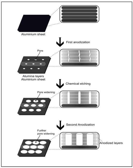
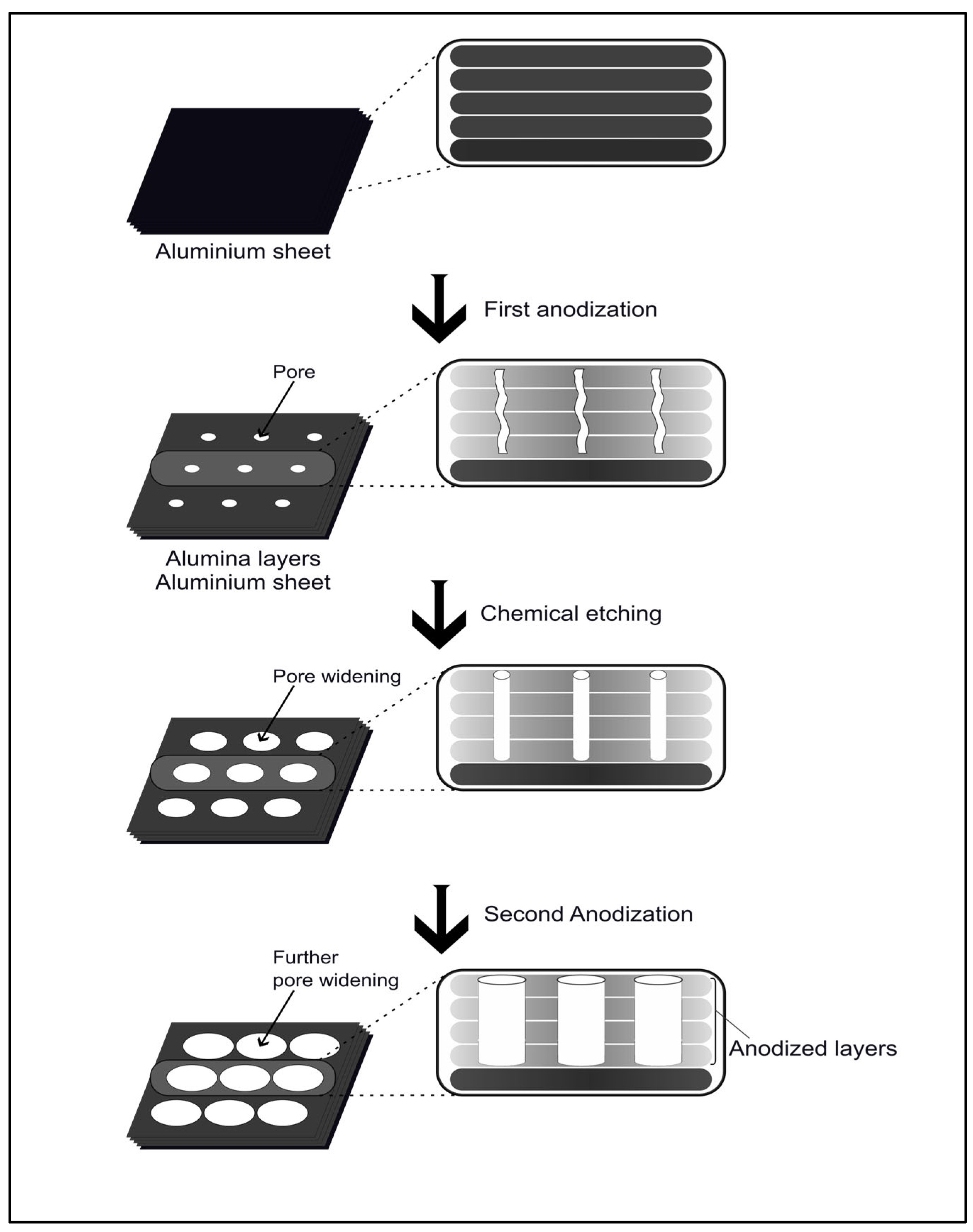
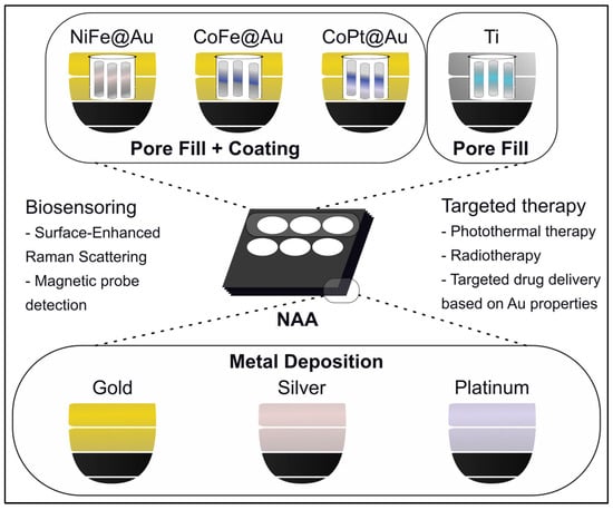
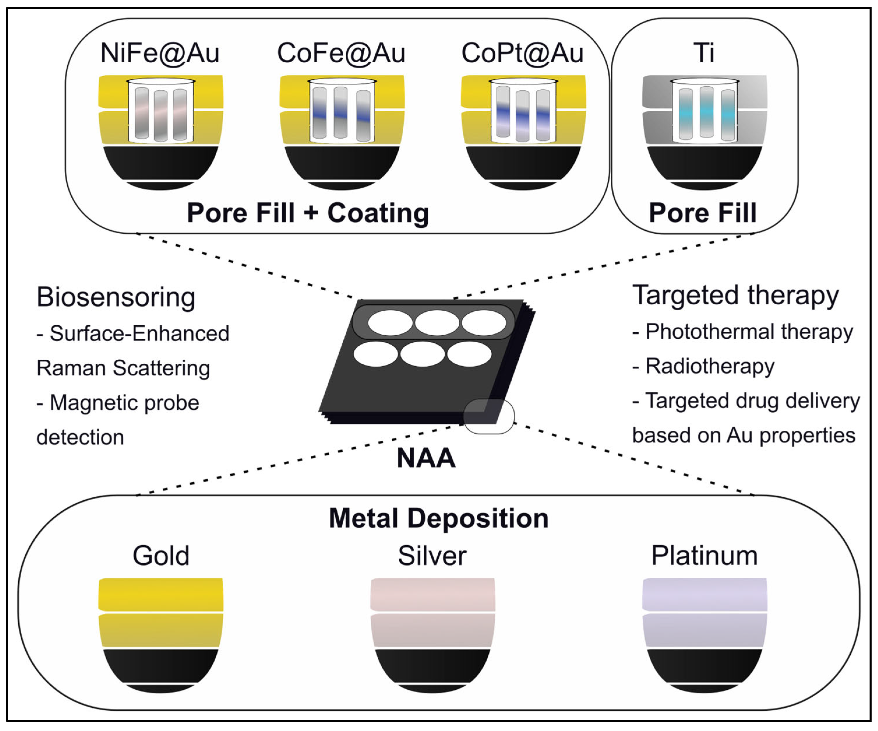
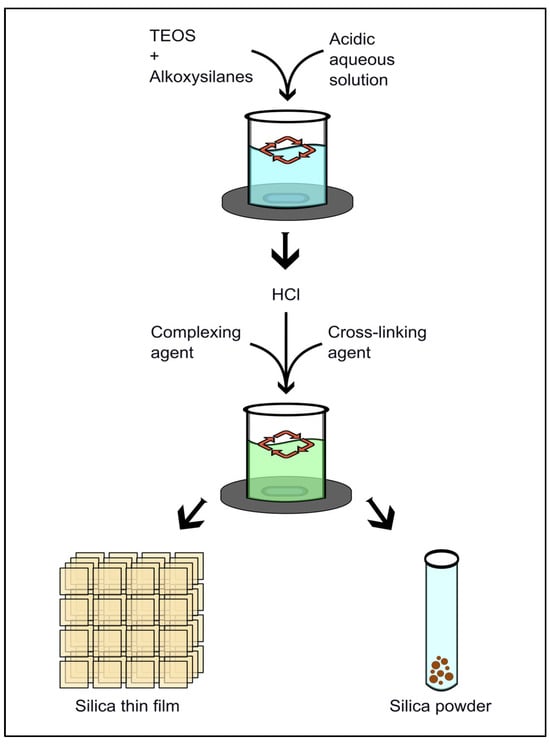
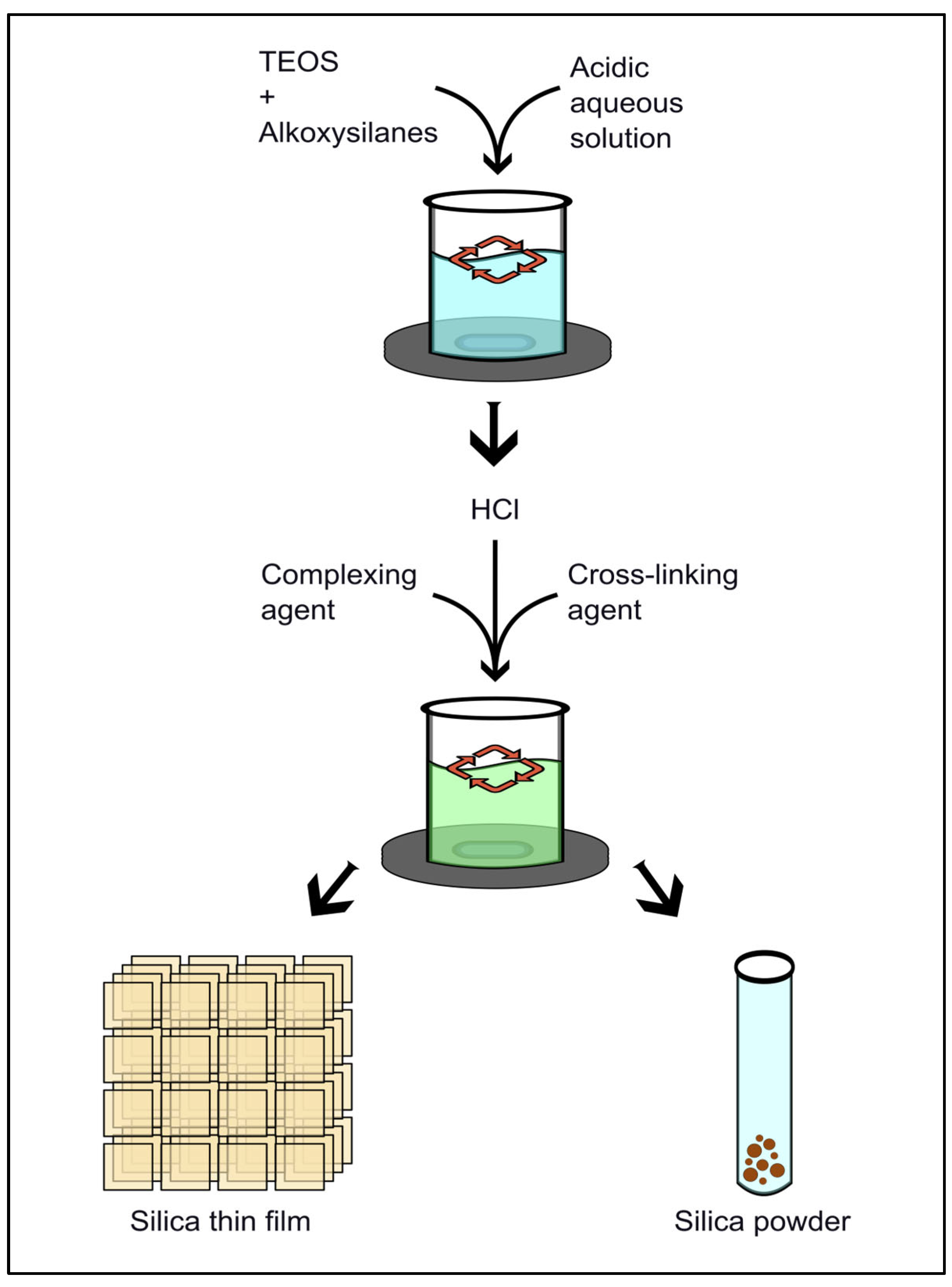
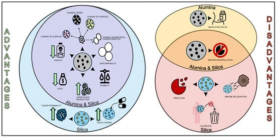
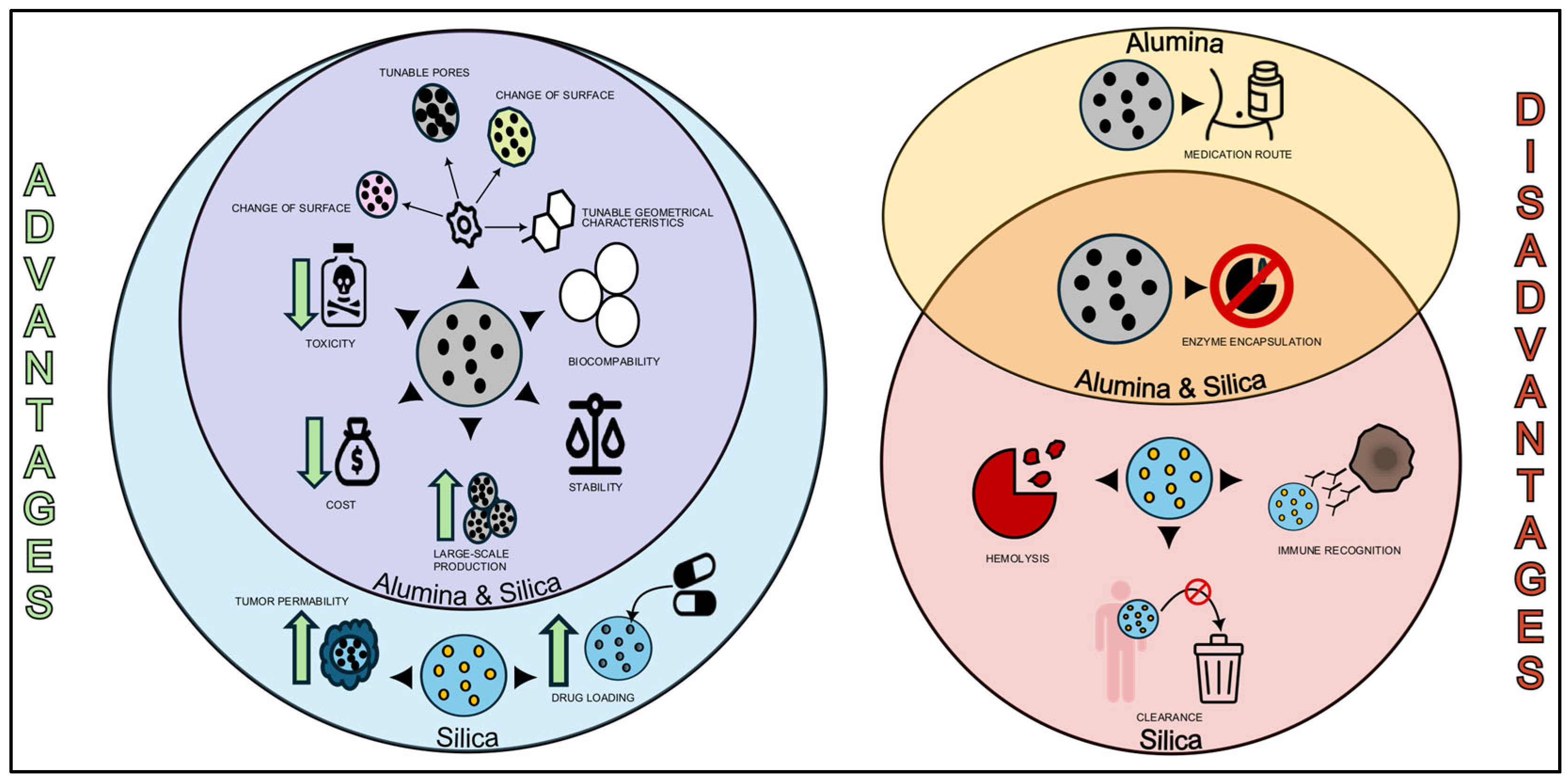
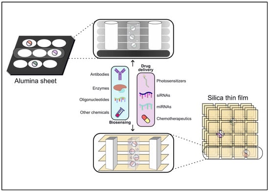
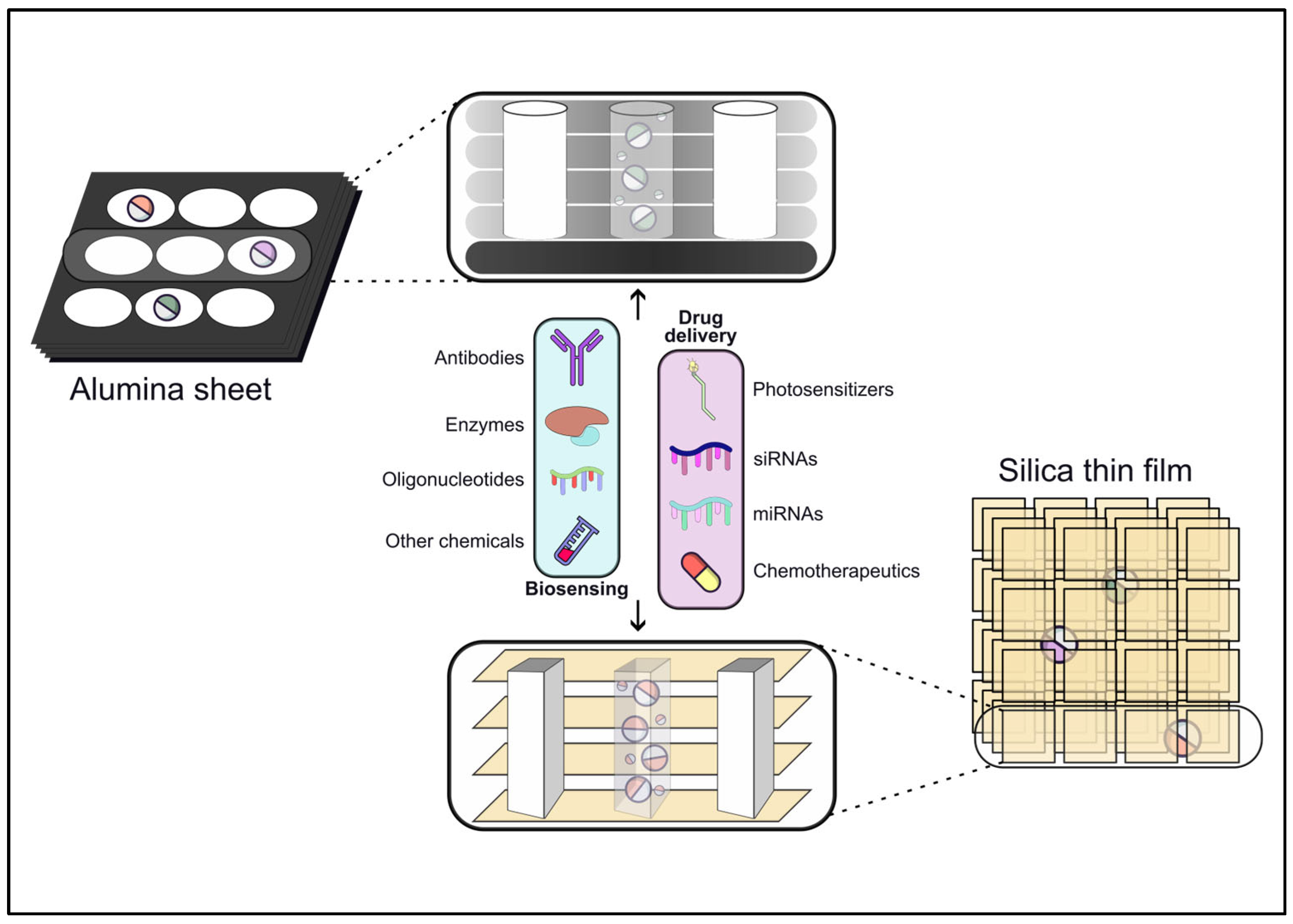
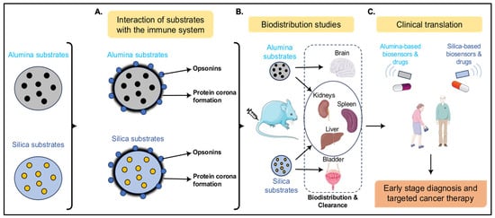
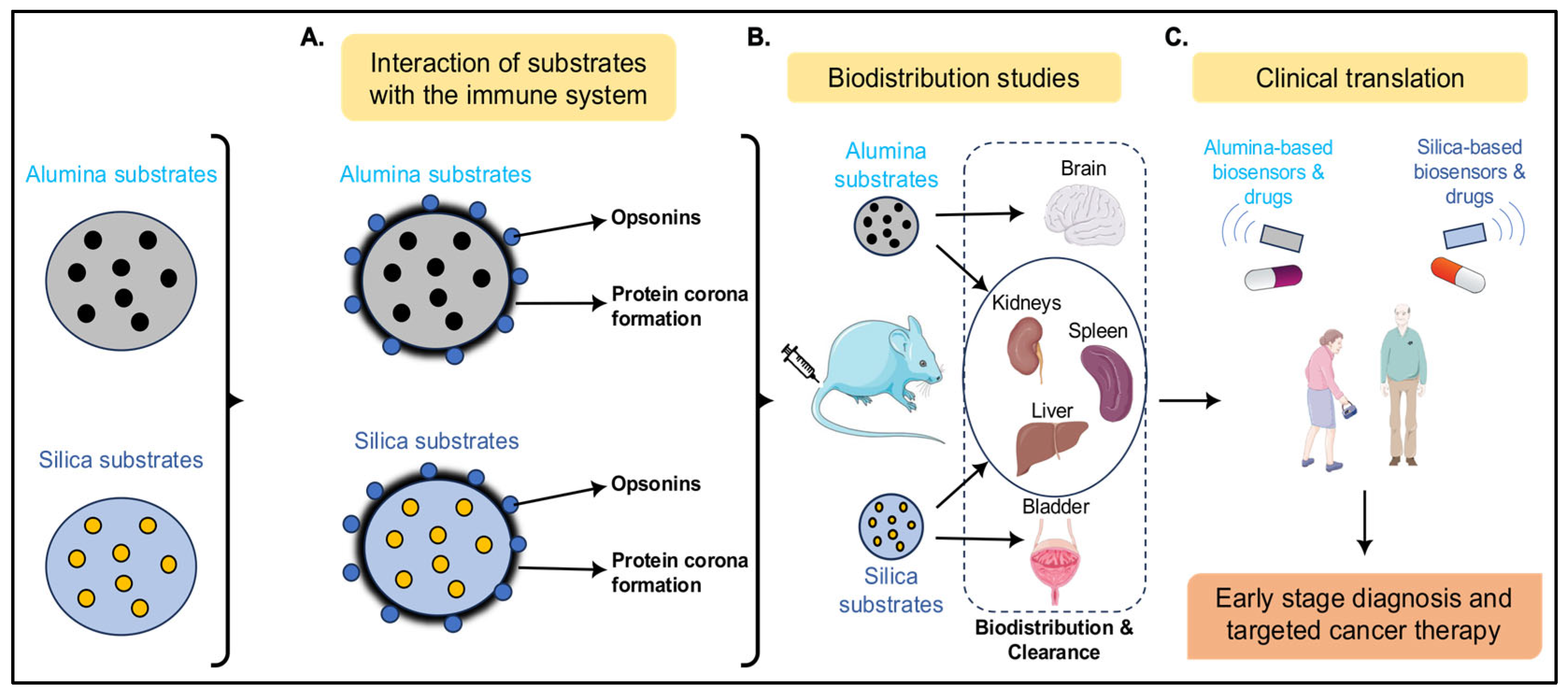



Disclaimer/Publisher’s Note: The statements, opinions and data contained in all publications are solely those of the individual author(s) and contributor(s) and not of MDPI and/or the editor(s). MDPI and/or the editor(s) disclaim responsibility for any injury to people or property resulting from any ideas, methods, instructions or products referred to in the content. |
© 2026 by the authors. Licensee MDPI, Basel, Switzerland. This article is an open access article distributed under the terms and conditions of the Creative Commons Attribution (CC BY) license.