


Abstract
This review provides a comprehensive overview of recent progress in chitin-based nanomaterials and their composite engineering. Particular focus is placed on techniques for constructing self-assembled chitin nanofibers (ChNFs) with tightly bundled fibrillar structures, as well as strategies for fabricating composites in which the ChNFs serve as reinforcing components, combined with natural polymeric matrices. In addition, high-crystalline scaled-down (SD-)ChNFs were fabricated through partial deacetylation of the ChNFs, followed by electrostatic repulsive disassembly of the abovementioned bundled fibrils in aqueous acetic acid, which were further used to reinforce composites comprising the other polysaccharides. Mixing the SD-ChNFs with low-crystalline chitin substrates further enabled the fabrication of all-chitin composites (AChCs) that exploit crystallinity contrast to achieve enhanced tensile strength. Moreover, the AChC films exhibited high cell-adhesive properties and promoted the formation of three-dimensional cell-networks, highlighting their potential for biomedical applications.
1. Introduction
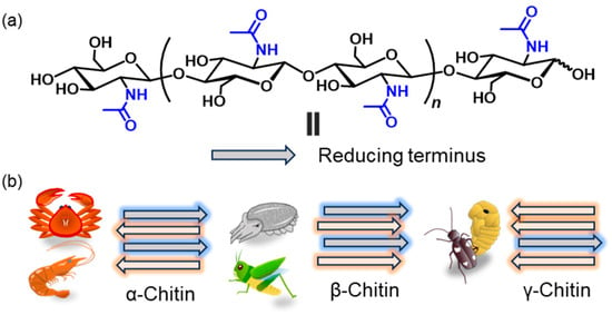
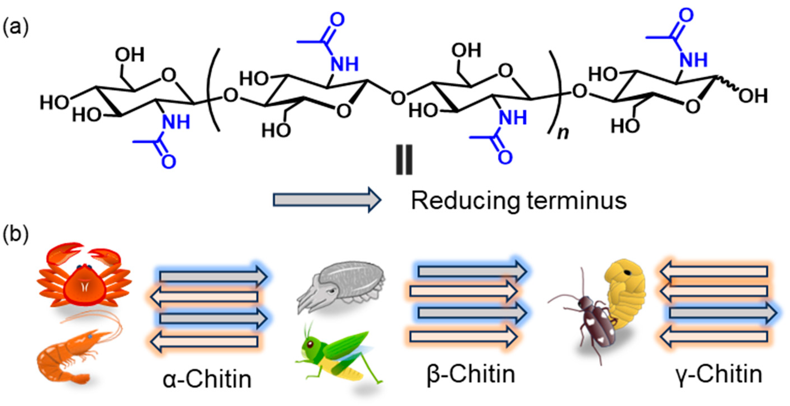
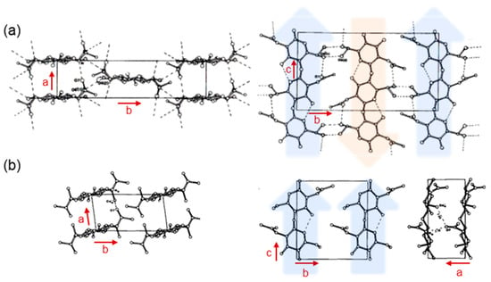
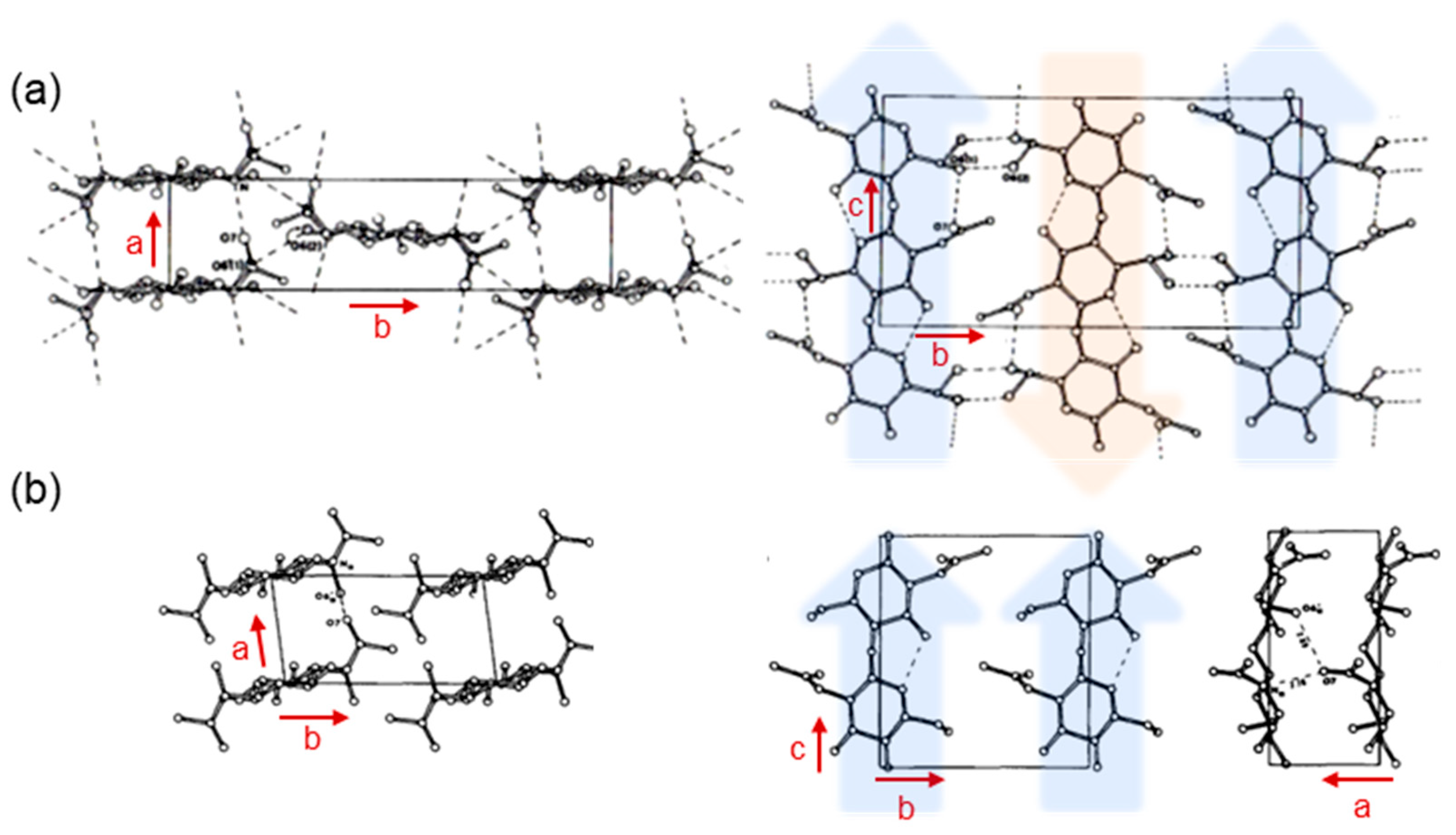
Chitin is one of the most abundant structural polysaccharides in nature and is mainly biosynthesized in the exoskeletons of crustaceans and insects such as shrimps, crabs, and crickets [1,2,3,4,5,6,7,8,9]. It is a linear polysaccharide composed of N-acetyl-D-glucosamine (GlcNAc) units linked through β(1→4) glycosidic bonds (Figure 1a) and is known to exhibit a broad molecular weight on the order of ~104 to ~105 [10,11]. In addition, acetamido groups at C-2 position in the GlcNAc units form strong intermolecular hydrogen bonds, which contribute to the characteristically high crystallinity and stiffness of chitin [12]. The crystalline polymorphs of chitin are classified into three forms (α, β, and γ) according to the chain arrangement (Figure 1b). The most abundant form is α-chitin, which is found in the shells of crabs and shrimps and features antiparallel chain alignment. β-Chitin occurs in the squid pen and cuttlebone of common cuttlefish, as well as in certain insect-derived materials. γ-Chitin, a relatively rare polymorph exhibiting a mixed arrangement of α- and β-type packing, has been reported in the cocoon fibers of Ptinus beetles [13]. As a more sophisticated structural representation, the ab and bc projections of α-chitin and β-Chitin are illustrated in Figure 2a,b [14,15]. The α-chitin lattice exhibits exceptionally tight molecular packing because an extensive alternating array of interchain hydrogen bonds provides a highly stabilized crystalline framework. In contrast, the β-chitin polymorph forms fewer hydrogen bonds, and their geometrical arrangement is less favorable for cohesive packing. As a result, the intermolecular interactions in β-chitin are substantially weaker. Molecular dynamics (MD) simulations, conducted in aqueous environments, have demonstrated that, unlike the robust and structurally persistent α-chitin, the crystalline order of β-chitin undergoes rapid hydration-induced destabilization and readily collapses [16].
Figure 1.
(a) Chemical structure of chitin and (b) schematic images for representative crystalline structure, α-, β-, and γ-chitins.
Figure 2.
Projection of the proposed model for (a) α-chitin (adapted with permission from Ref. [15]. Copyright 1978, Elsevier) and (b) β-chitin (adapted with permission from Ref. [14]. Copyright 1975, Wiley).
Chitin have garnered considerable attention across various research fields owing to their ability to form nanoscale materials such as nanofibers [17,18]. Because of their outstanding characteristics, including light weight, high tensile strength, and low thermal expansion coefficients, nano-fibrillated derivatives such as nanofibers, nanocrystals, and nanowhiskers have been extensively explored as versatile, stable, and functional forms of chitin [19,20,21,22,23,24,25]. Two principal strategies have been reported for the construction of chitin nanofibers, i.e., top-down and bottom-up approaches. The top-down approach involves the disintegration of native chitin microfibrils through mechanical or chemical treatments [18,26,27,28,29,30,31]. Native chitin has a hierarchical structure composed of bundles assembled from nanofibers (microfibrils), and accordingly, size reduction can be achieved by disintegration of the assemblies [32]. This approach generally yields chitin nanofibers with widths on the order of 10 nm and lengths of several micrometers or longer, while some variability in fiber morphology is commonly observed. In contrast, the bottom-up approach relies on dissolving/swelling chitin to temporarily disrupt its crystalline structure, followed by a regeneration process using poor solvents or electrospinning, leading to reconstructing fibrous morphologies. The bottom-up approach enables the facile formation of nanostructures with a wide range of sizes, morphologies, and fiber lengths, spanning from fine particles to fibrous nanofibers. As discussed later, it allows the size of small particles with diameters ranging from several nanometers to several hundred nanometers to be tuned [33,34,35,36].
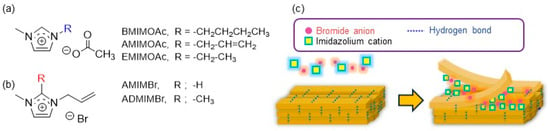
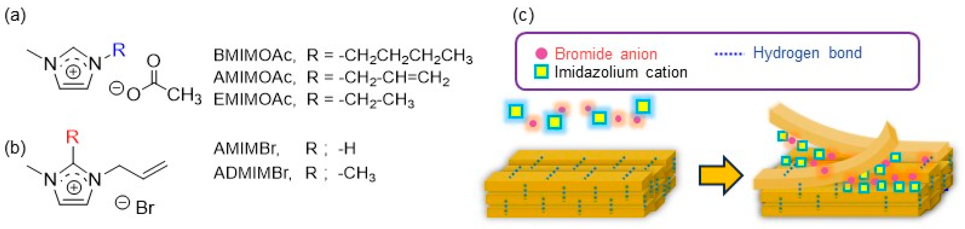
Ionic liquids (ILs) comprise a family of salts that exhibit liquid-phase behavior near ambient temperatures, where a number of such solvent systems share similar structural and physicochemical features. Rogers et al. demonstrated in 2002 that an IL, such as 1-butyl-3-methylimidazolium chloride (BMIMCl), efficiently dissolved cellulose, which subsequently positioned ILs as highly effective solvents for polysaccharides [37,38,39,40,41]. ILs have also been utilized as effective media for preparing chitin nanofibers (ChNFs) via a regenerative self-assembling process upon bottom-up approach, and thereafter, with the rapid expansion of research in this field, they have further been recognized as one of solvent systems capable of dissolving chitin [42,43,44,45,46]. 1-Butyl-3-methylimidazolium acetate (BMIMOAc) enabled dissolution of chitin from crab shells up to approximately 6 wt%, and the resulting solutions could be subsequently reformed into chitin-sponge architectures [47]. Since then, systematic screening of structurally distinct IL families has led to the reporting of ionic liquids such as 1-allyl-3-methylimidazolium acetate (AMIMOAc) [42] and 1-ethyl-3-methylimidazolium acetate (EMIMOAc) (Figure 3a) [48]. Overall, the above dissolution studies of chitin in ILs suggest that ILs with carboxylate anions generally act as good solvents for chitin.
Figure 3.
(a,b) Representative ionic liquids (ILs) capable of dissolving chitin. (c) Plausible dissolution process of chitin in AMIMBr predicted by molecular dynamics (MD) simulations.
Another type of IL, reported in our previous work, 1-allyl-3-methylimidazolium bromide (AMIMBr) was found to dissolve chitin up to 4.8 wt% [49,50]. Furthermore, when higher amounts of chitin (6.5–10.7 wt%) were mixed with AMIMBr, ion gels were formed after standing at room temperature, followed by heating at 100 °C and cooling [51] (Figure 3b and Figure 4a). To understand the dissolution mechanisms in the ILs, particularly in AMIMBr, MD simulations have provided mechanistic insight into how ILs influence the disintegration of chitin crystallites [52]. The simulations have indicated that bromide ions in AMIMBr efficiently disrupt hydrogen bonds associated with the acetamido groups, thereby promoting progressive chain liberation from the outermost crystalline layers. Once the chains are released, AMIM+ act to suppress re-aggregation into an ordered phase (Figure 3c). Recently, it has also been demonstrated that 1-allyl-2,3-dimethylimidazolium bromide (ADMIMBr), a structural analog of imidazolium-based ILs, serves as an efficient chitin solvent [53].
Based on the demonstrated dissolution of chitin in AMIMBr, our laboratory has been developing a fabrication strategy for ChNFs and exploring their translation into advanced functional nanomaterials. The regeneration from chitin/AMIMBr ion gels using methanol with ultrasonication yielded a dispersion of self-assembled ChNFs upon bottom-up process (Figure 4b) [49,54,55,56]. The resulting dispersions were subjected to filtration to produce ChNF films with highly entangled nanofibrous morphologies (Figure 4d). Transmission electron microscopy (TEM) revealed that the ChNFs were composed of bundles of thinner fibrils averaging 20–60 nm in width and several hundred nanometers in length [57] (Figure 4c).
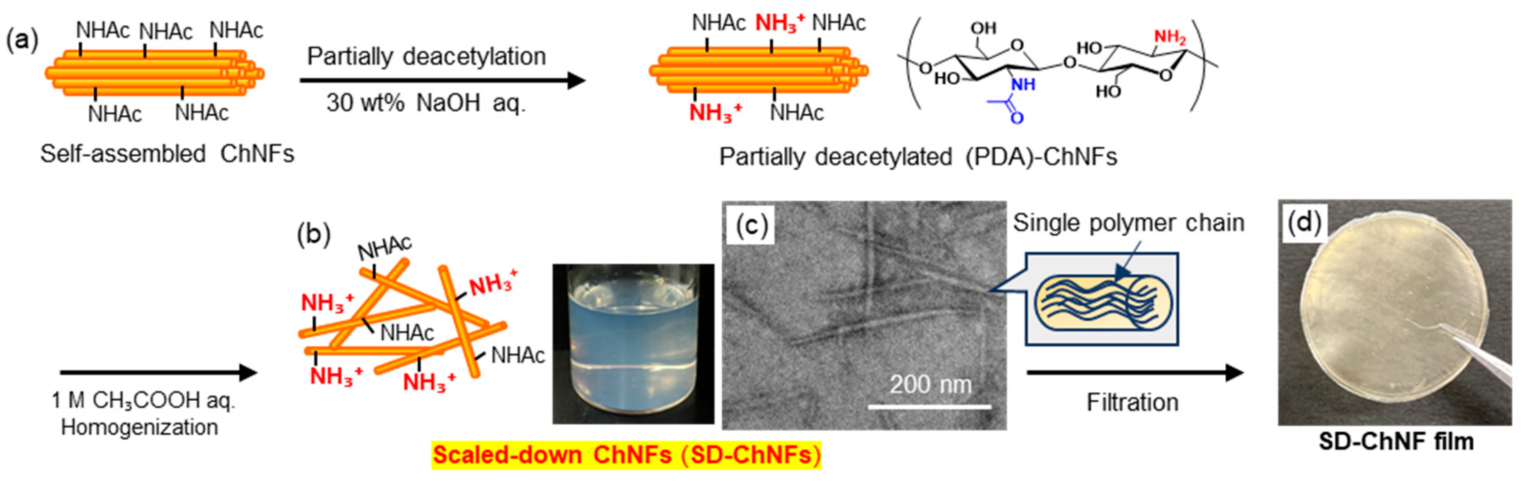
Partial deacetylation (PDA) of the acetamido groups on ChNFs using 30 wt% aqueous NaOH introduced amino functionalities along the nanofiber surfaces (Figure 5a). Subsequent dispersion of the PDA-ChNFs in 1 M aqueous acetic acid, assisted by homogenization, promoted the formation of individualized, scaled-down (SD-)ChNFs through electrostatic repulsion among the protonated amino (ammonium) groups (Figure 5b). As a result, thinner nanofiber structure was observed in the TEM image (Figure 5c), which was completely different from the bundled morphology observed for the original ChNFs (Figure 4c). The resulting SD-ChNFs were then collected by suction filtration to produce flexible films with densely entangled morphologies (Figure 5d).
The obtained self-assembled and SD-ChNFs are solely composed of chitin and considered to possess intrinsic biocompatibility, biodegradability, and mechanical robustness. Owing to these properties, they are expected to serve as sustainable bio-nanomaterials for biomedical scaffolds, film substrates, and environmentally friendly composite systems, where nanoscale organization plays a key role in determining their material performance [18,25,58,59,60,61,62,63,64,65,66,67,68,69,70,71,72,73,74,75,76,77,78,79,80,81,82,83,84,85,86,87,88].
Figure 4.
Schematic representation of (a) chitin/1-allyl-3-methylimidazolium bromide (AMIMBr) ion gel formation and (b) subsequent self-assembly to form chitin nanofibers (ChNFs), (c) a representative transmission electron microscopy (TEM) image of the ChNFs, and (d) the resulting continuous film (adapted with permission from Ref. [55]. Copyright 2021, Elsevier).
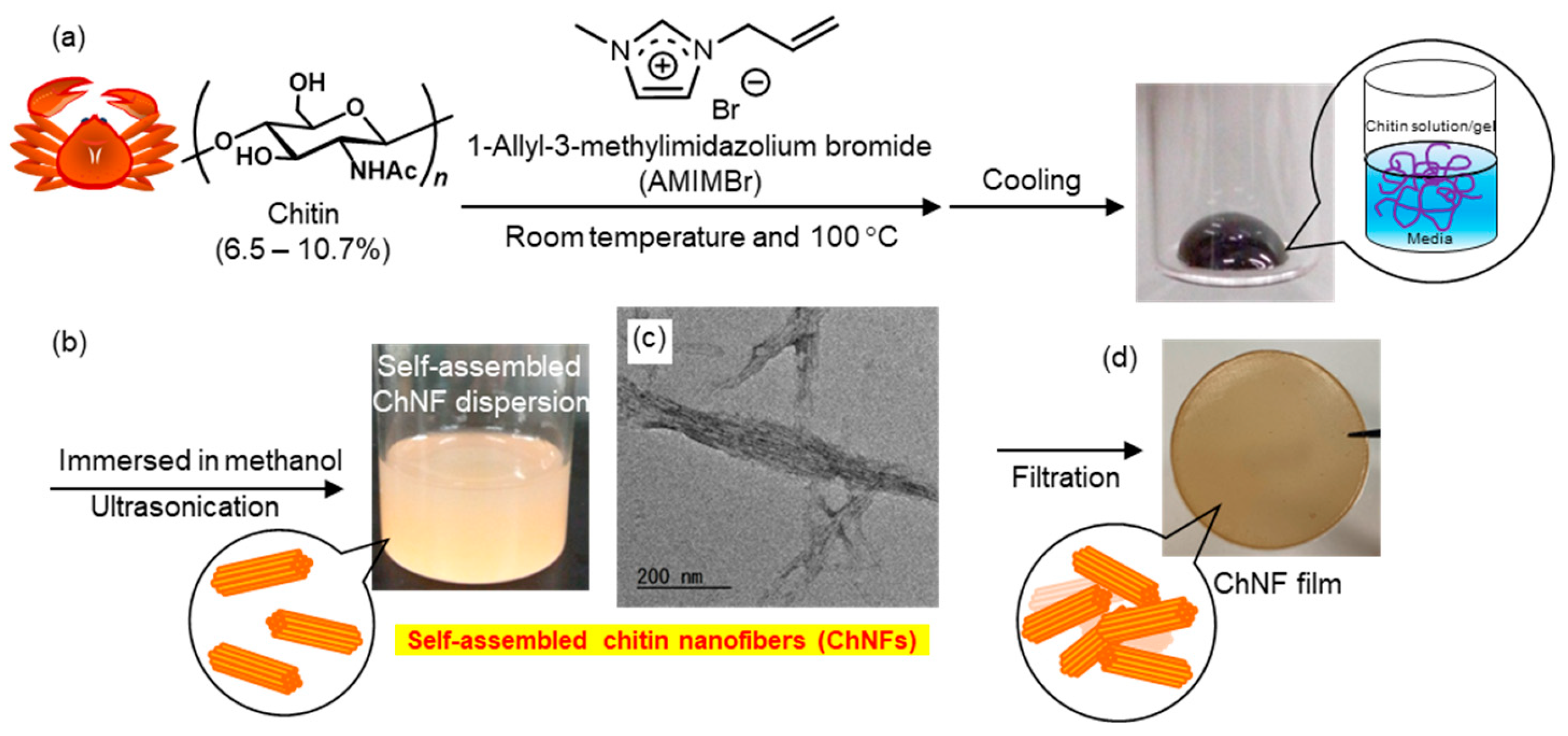
Figure 5.
Procedures for the preparation of (a) partially deacetylated (PDA)-ChNFs and (b) subsequent scaled-down ChNFs (SD-ChNFs), (c) a representative TEM image of the SD-ChNFs, and (d) the resulting continuous SD-ChNFs film (adapted with permission from Ref. [55]. Copyright 2021, Elsevier).
2. Fabrication of Polymer Composites Based on Chitin Nanofibers
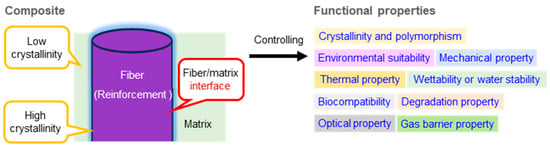
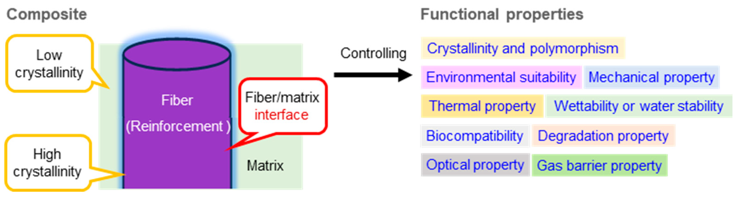
Composite structures can be constructed from a single component or by combinations of multiple components. In such systems, the interface between the reinforcement phase and the matrix phase should be precisely controlled, which is recognized as a key factor for governing the overall mechanical property of the composite (Figure 6) [79,88,89,90,91,92,93,94,95,96,97,98,99,100,101,102,103,104,105,106,107]. Variations in crystallinity between the fiber and matrix components critically influence interactions and load distribution and consequently modulate a broad spectrum of functional characteristics. These include mechanical performance, thermal and environmental stabilities, wettability and water resistance, optical and gas barrier properties, biocompatibility, and degradation behavior. Thus, the rational design of composite materials necessitates deliberate tuning of component crystallinity together with quantitative engineering of the fiber–matrix interface to achieve targeted structural and functional outcomes. In our research, we have focused on the ChNFs as a reinforcing component, and accordingly, have examined their composition with various polymeric substrates as matrix candidates [108,109,110]. In recent years, we have also investigated systems using natural polymers as the matrixes.
Figure 6.
Schematic illustration of a polymer composite structure and its functions.
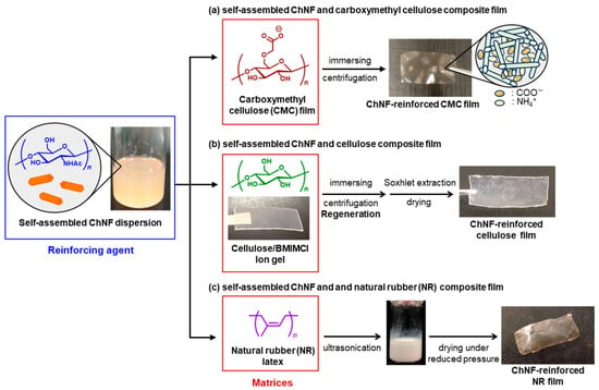
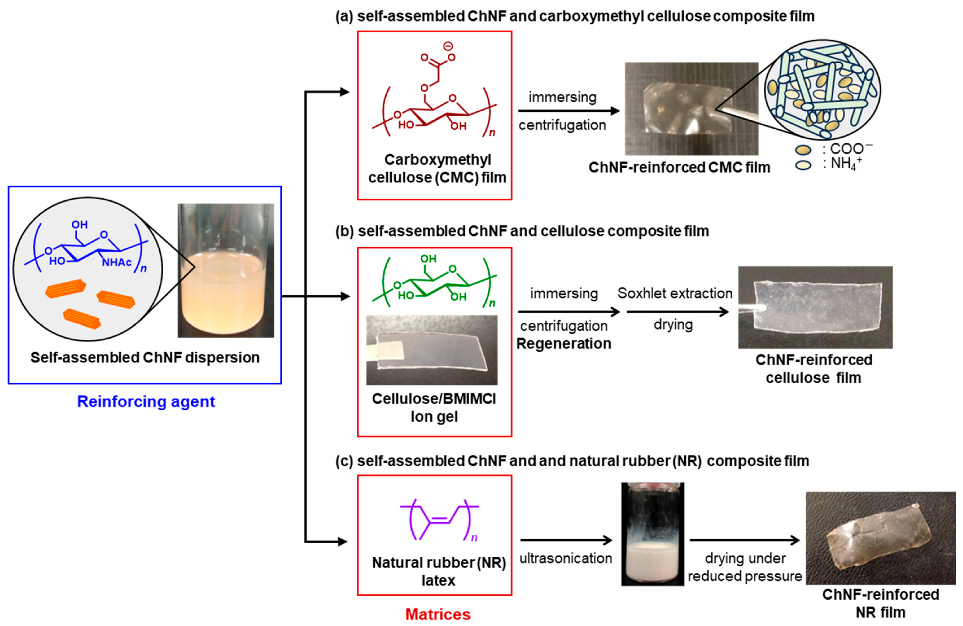
As an example, we established a simple method to fabricate ChNF-reinforced carboxymethyl cellulose (CMC) films through electrostatic interaction between ammonium and carboxylate groups (Figure 7a) [111]. CMC films were immersed in self-assembled ChNF dispersions prepared by regeneration from the chitin/AMIMBr ion gels. The absorbed ChNF amount in the composites increased with ChNF content in the dispersions, as confirmed by SEM and IR analyses. Tensile testing revealed that the ChNFs effectively reinforced the CMC films to enhance mechanical strength. The results indicate that the self-assembled ChNFs serve as promising components for compatibilizing with acidic polysaccharides, offering a versatile route to develop practical bio-based composite materials.
Figure 7.
Schematic illustration of preparations of (a) self-assembled ChNF/carboxymethyl cellulose (CMC) composite, (b) self-assembled ChNF/cellulose composite, and (c) self-assembled ChNF/natural rubber (NR) composite.
In another approach, the self-assembled ChNF-reinforced cellulose films were prepared via stepwise regenerations from the two ion gels (Figure 7b) [112]. The ChNF dispersion was obtained by the abovementioned regeneration process from the chitin/AMIMBr ion gel using methanol. In parallel, a cellulose/BMIMCl ion gel was prepared and immersed in the ChNF dispersion, allowing simultaneous regeneration of cellulose nanofibers and formation of a ChNF/cellulose composite film. IR and SEM analyses confirmed distribution of the ChNFs throughout the film. The X-ray diffraction (XRD) result revealed the amorphous structure of the regenerated cellulose component in the composite film. Tensile testing showed that the ChNFs significantly enhanced the mechanical strength. The regeneration-based approach offers a versatile route for fabricating well compatible, ChNF-reinforced polysaccharide materials applicable to various natural biopolymers.
Furthermore, the self-assembled ChNF film was found to be dispersible in ammonia aqueous solution. In addition, natural rubber (NR) is known to form an anionically charged colloidal latex with ammonia aqueous solution. Accordingly, ChNF/NR latex dispersions were obtained by mixing the two dispersions in different ratios under ultrasonication (Figure 7c) [113]. The resulting dispersions were evaporated in a drying tray under reduced pressure, yielding ChNF/NR composite films, in which the ChNFs acted as a reinforcing component, as an increase in the ChNF content strengthened the composite films. On the other hand, heating the ChNF/NR mixture for removal of ammonia, followed by lyophilization, yielded porous composite materials. The formation of the porous structures was attributed to aggregation of the ChNFs and NR upon loss of ammonia and the subsequent generation of void spaces between aggregated domains during the lyophilization process.
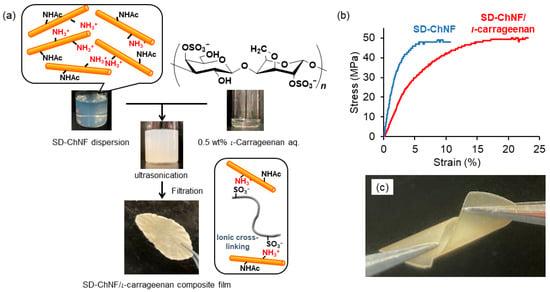
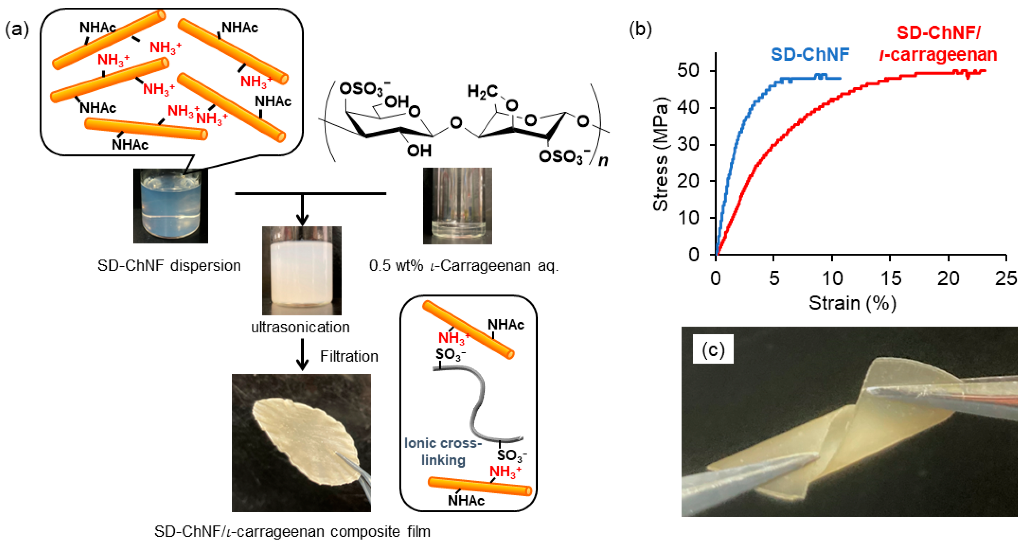
To further impart functional properties to the SD-ChNF film, we investigated the fabrication of a composite with a ι-carrageenan film by electrostatic interaction as exploited for CMC (Figure 8a) [55]. ι-Carrageenan is a water-soluble phycocolloid derived from red algae, composed of alternating 1,3-linked α-D-galactopyranose and 1,4-linked β-3,6-anhydro-D-galactopyranose residues bearing sulfate groups at the C-4 and C-2 positions [114]. A dispersion of the ChNFs in aqueous acetic acid was added dropwise onto a viscous 0.5 wt% ι-carrageenan aqueous solution (NH3+:SO3− feed ratio = 1:1.2). The resulting mixture was ultrasonicated for 10 min and then filtered to yield the composite film. Based on the film weight, the NH3+:SO3− ratio was calculated to be 1:0.76, indicating that the composite was efficiently formed from two components bearing positive and negative charges. The stress–strain curve of the SD-ChNF/ι-carrageenan composite film under tensile mode exhibited a markedly improved elongation at break while maintaining a tensile strength comparable to that of the neat SD-ChNF film (Figure 8b). The twisting test further revealed the flexible yet brittle nature of the composite film (Figure 7c). These results suggest that combining the SD-ChNFs with anionic polysaccharides facilitates the formation of efficient ion-pair interaction, which in turn contributes to the overall mechanical strength of the composite film.
Figure 8.
Schematic illustration of (a) preparation of SD-ChNF/ι-carrageenan composite film with dispersion solutions, (b) stress–strain curves of SD-ChNF and composite films, and (c) photograph for twisting test of composite film.
3. Concept and Design of All-Chitin Composites Based on Single-Polymer Composite Principle
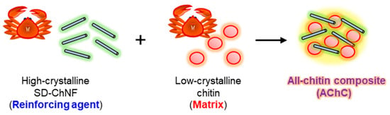
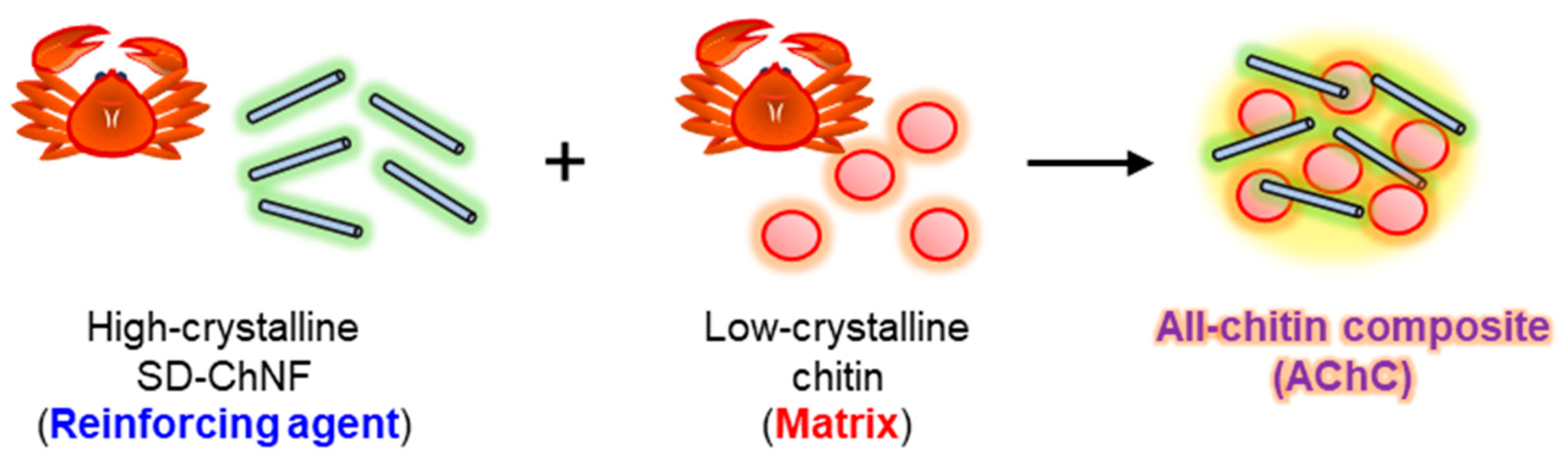
Single-polymer composites (SPCs) are defined as composed entirely of a single polymer [115,116]. Their properties are governed by intermolecular interactions, the balance of amorphous and crystalline regions, and hydrogen bonding networks, which distinguish them from conventional multi-component composites. Moreover, their thermal stability and degradation products can be readily predicted [18,117]. To translate the SPC concept into a chitin-based system, we have hypothesized that all-chitin composites (AChCs) are constructed by deliberately exploiting crystallinity contrast within a single polymer. In this design concept, highly crystalline SD-ChNFs would serve as the reinforcing phase, whereas low-crystalline chitin would serve as the matrix. We further postulated that integrating these structurally distinct yet chemically identical components would create a self-reinforced composite framework, in which mechanical reinforcement, stress transfer, and functional tunability arise solely from the controlled hierarchical organization of chitin itself (Figure 9).
Figure 9.
Schematic illustration for concept of all-chitin composite (AChC).
3.1. Fabrication of AChCs Using Low-Crystalline SD-ChNFs Prepared via Trifluoroacetic Acid Treatment
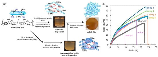
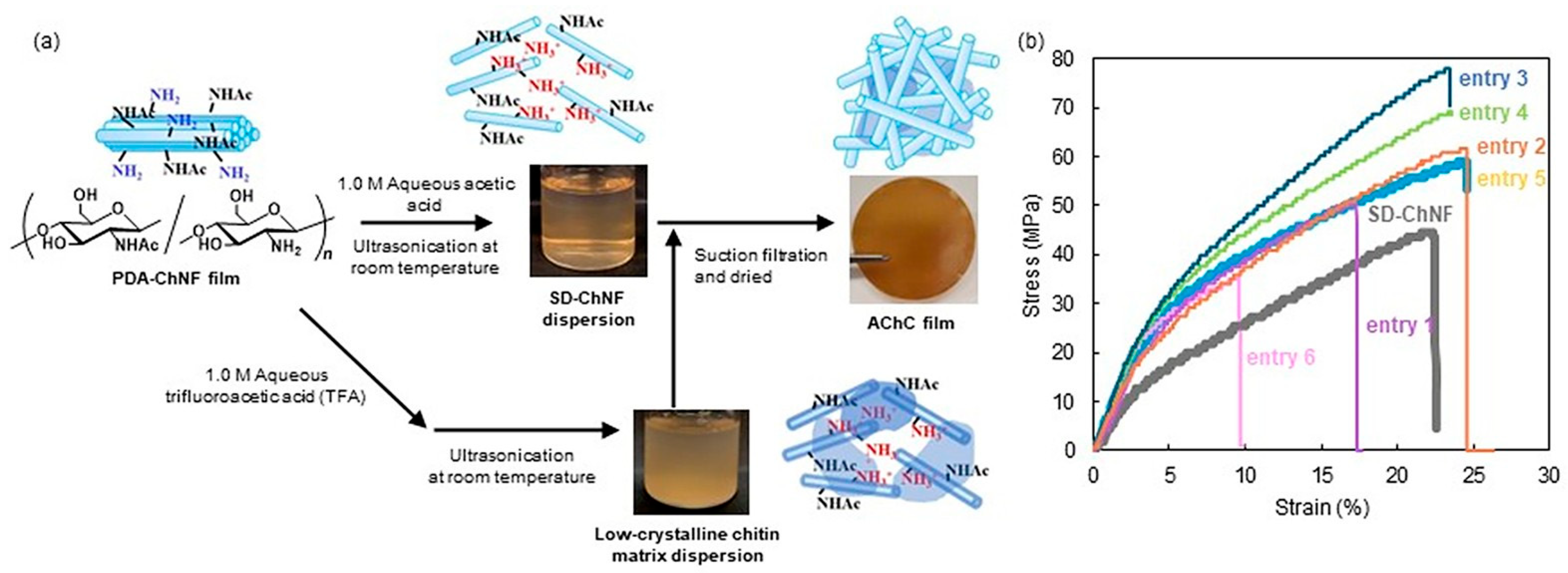
We first attempted to fabricate AChCs by blending highly crystalline SD-ChNFs with a low-crystalline chitin substrate derived from the PDA-ChNFs, based on a double-crystalline concept in which two chitin components with distinct crystallinities are combined (Figure 10a) [110,118,119]. To obtain a low-crystalline chitin matrix, the PDA-ChNFs were ultrasonicated in several 1M aqueous acid solutions. The XRD and IR analyses showed that acetic and formic acids preserved nanofiber morphology and maintained high crystallinity, whereas mild heating and ultrasonication in trifluoroacetic acid (TFA) effectively reduced crystallinity to a crystalline index (CI) of approximately 82%, without inducing chemical degradation. In contrast, methanesulfonic acid caused significant decomposition and was therefore excluded from further investigation. These low-crystalline chitin dispersions by TFA treatment (1M aqueous solution) were subsequently mixed with the SD-ChNF/1M aqueous acetic acid dispersions at various weight ratios, followed by filtration and drying, to prepare AChC films, as shown in Figure 10a. Increasing the proportion of the low-crystalline matrix gradually decreased the overall crystallinity of the resulting films. Tensile testing revealed a clear dependence of mechanical properties on composite composition. In this series, entries 1 through 6 correspond to the AChC films prepared with progressively increasing proportions of the low-crystalline chitin matrix, allowing systematic assessment of how matrix content influences mechanical reinforcement (entries 1, 2, and 3) (Figure 10b). Young’s modulus and tensile strength increased with the addition of the low-crystalline matrix up to an optimal SD-ChNF to matrix ratio of 1 to 0.026 (entry 3), where the composite achieved a Young’s modulus of 0.763 GPa and a tensile strength of 78 MPa, while maintaining an elongation at break similar to that of the pure SD-ChNF film. Further increases in the matrix content (entries 4 and 5) reduced reinforcement efficiency, presumably because of insufficient stress transfer at the fiber to matrix interface.
Figure 10.
Schematic illustration of (a) preparation of AChC with SD-ChNF dispersion with 1.0 M in aqueous acetic acid and low-crystalline chitin matrix dispersion with 1.0 M aqueous trifluoroacetic acid (TFA), (b) stress–strain curves of the SD-ChNF film and AChC films with varying low-crystallinity chitin/SD-ChNF weight ratios: entry 1 (0.006/1), entry 2 (0.013/1), entry 3 (0.026/1), entry 4 (0.067/1), entry 5 (0.091/1), and entry 6 (0.143/1) under tensile mode (adapted with permission from Ref. [119], Copyright 2025, Elsevier).
Overall, these results demonstrate that precise control of crystalline among chemically identical chitin-based components provides a useful strategy for introducing complementary mechanical functions into all-chitin composites without the need for external polymers.
3.2. Construction of Low-Crystalline Chitin Nanoparticles and Their Application as Matrix Components in AChCs
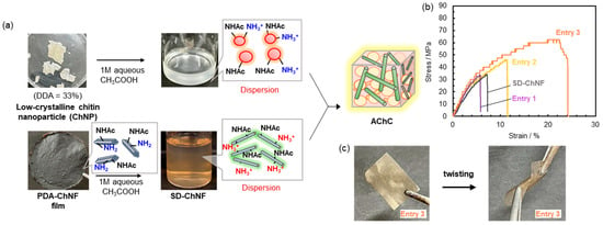
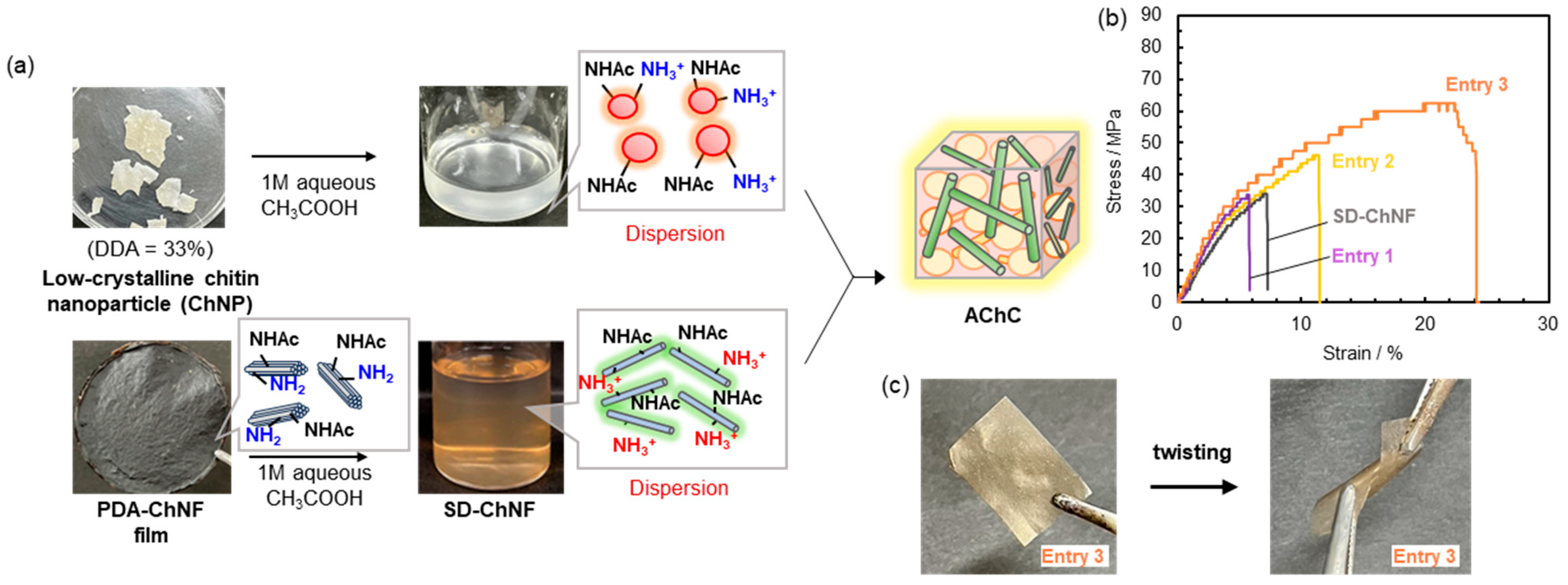
TFA has been employed in the abovementioned study because it is widely used in bio-related research as an effective solvent for weakening hydrogen-bonding interactions. However, its inherent toxicity poses limitations based on the viewpoint of practical application. In addition, the crystallinity of chitin obtained through the TFA treatment was not sufficiently reduced as low-crystalline chitin components. Therefore, in the following study, we aimed to construct low-crystalline chitin nanoparticles via an alkaline treatment in order to prepare a chitin matrix with lower crystallinity than that obtained through the previous studies based on the TFA treatment (Figure 11a). Chitin with relatively reduced crystallinity is known to be obtainable by partial deacetylation through NaOH treatment in aqueous media [110]. Additional deacetylation of the resulting low-crystalline chitin raised the degree of deacetylation (DDA) to 33%. Accordingly, the powdered chitin substrate with nanoparticle (ChNP) morphology was obtained, which dispersed well in aqueous acetic acid [120]. The SEM images revealed the following clear morphological differences; the SD-ChNFs formed nano fibrous structure, whereas the ChNPs existed as ca. 10 nm spherical particles. The XRD analysis confirmed their difference in crystallinity, where the SD-ChNF film exhibited CI over 90.5%, while the ChNPs showed CI of 57.4%, which was significantly lower than that of the TFA-treated chitin substrate. The AChC films were prepared by mixing the SD-ChNF and ChNP/1M aqueous acetic acid dispersions at defined ChNF/ChNP weight ratios (entry 1, 1/0.028; entry 2, 1/1; entry 3, 1/6.6), followed by filtration (Figure 11a). The CI values of the resulting AChC films were found to be tunable, as 65.4–88.5%, by adjusting the ChNF/ChNP weight ratios [110,121]. Increase of ChNP contents in the AChC films gradually obscured and lowered crystallinity, consistent with integration of amorphous domains. Moreover, laser microscopy showed that the SD-ChNF film displayed rough surfaces, while the AChC films became smoother with increasing ChNP contents although root-mean square roughness values did not vary significantly (0.97–2.10 µm).
Figure 11.
(a) Schematic illustration for preparation of self-reinforced AChCs from low-crystalline chitin nanoparticle (ChNP) and SD-ChNF dispersions, (b) stress–strain curves of AChC films with varying SD-ChNF/ChNP weight ratios (w/w); entry 1 (1/0.028), entry 2 (1/1), and entry 3 (1/6.6), and (c) photographs for twisting test of AChC film (entry 3) (adapted with permission from Ref. [110]. Copyright 2025, Elsevier).
Tensile testing of the AChC films revealed a clear composition-dependent trend, in which increasing the ChNP content enhanced tensile strength and Young’s modulus (Figure 11b). At the optimal composition (SD-ChNF to ChNP ratio of 1 to 6.6, entry 3), the composite exhibited markedly improved mechanical performance, reaching 64.0 MPa in tensile strength and 0.924 GPa in Young’s modulus. In contrast, films with insufficient ChNP content displayed limited reinforcement. Entry 1 showed values of 33.8 MPa (strength) and 0.742 GPa (modulus), while entry 2 exhibited 47.5 MPa and 0.769 GPa. These values were comparable to or only marginally higher than those of the reference SD-ChNF film (34.0 MPa and 0.608 GPa). The inferior performance at low-ChNP contents underscores the brittleness associated with ChNF rich compositions (entries 1 and 2) and highlights the necessity of establishing an appropriate balance between the high-crystalline SD-ChNFs and low-crystalline ChNPs. The IR analysis supported strong interfacial hydrogen bonding between acetamido and hydroxy groups, which facilitated stress transfer within the composite.
Collectively, these findings demonstrate that controlled blending of chitin components with distinct crystallinities offers an effective means to tune the mechanical response of the AChCs, and notably, that the optimally composed film in entry 3 achieves a level of structural integrity sufficient to withstand twisting deformation without fracture, thereby highlighting the robustness of the resulting AChC architecture (Figure 11c).
3.3. Hydration and Cell Adhesion Properties on AChC Films
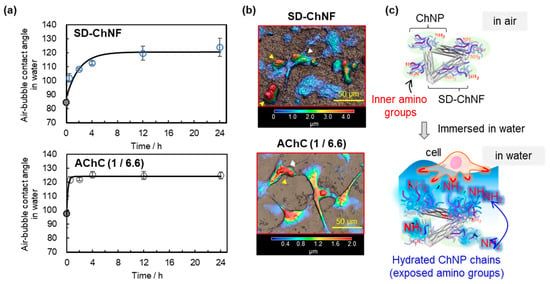
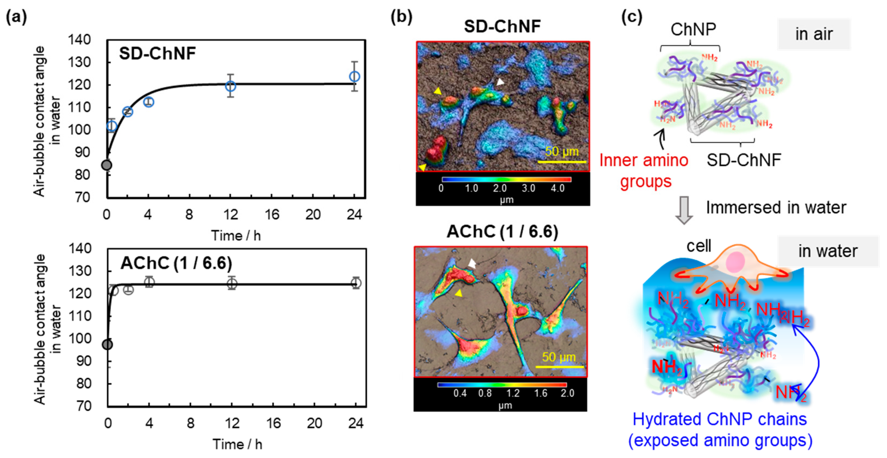
We have investigated how the hydration behavior of the AChC film composed of the highly crystalline SD-ChNFs and low-crystalline ChNPs at an affixed mixing ratio of 1 to 6.6 (wt/wt) [110,121]. Hydrophilicity was evaluated by measuring air bubble contact angles in aqueous media. In the dry state, the AChC film showed a slightly higher theoretical contact angle than the pure SD-ChNF film, reflecting the increased ChNP content and its contribution to hydrophilicity. Upon water immersion, the AChC film underwent a more rapid transition toward a hydrated, hydrophilic surface compared to the SD-ChNF film, consistent with the faster reorientation of hydrophilic amino rich ChNP domains at the interface. After 24 h of immersion, however, both films converged to similar values of approximately 120–125° (Figure 12a).
Figure 12.
(a) Air bubble contact angles against SD-ChNF and AChC (SD-ChNF/ChNP 1/6.6 wt/wt) films, (b) laser microscope images of HeLa cell adhesion on SD-ChNF and AChC films, and (c) hypothesis illustrations of AChC film surface in air to water environments (adapted with permission from Ref. [121]. Copyright 2025, Elsevier).
Cell adhesion studies using HeLa cells revealed composition-dependent responses. The SD-ChNF film supported very limited adhesion, with most HeLa cells remaining as small spherical aggregates. In contrast, the AChC film (SD ChNF/ChNP = 1/6.6 wt/wt) exhibited markedly enhanced cell adhesion, indicating that the optimized combination of highly crystalline nanofibers and low-crystalline chitin nanoparticles provides a surface environment considerably more favorable for cell adhesion (Figure 12b). The results support that hydration-induced exposure of cationic amino groups from the ChNPs at the interface enhances electrostatic interactions with negatively charged cell membranes (Figure 12c). Strong adhesion of the AChC film with high-ChNP content thus likely stems from synergy between structural support by the SD-ChNFs and hydrophilic amino-rich ChNP domains exposed in aqueous environments.
3.4. High-Density Cell Adhesion Behavior for Three-Dimensional Scaffold
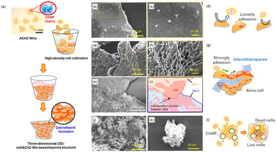
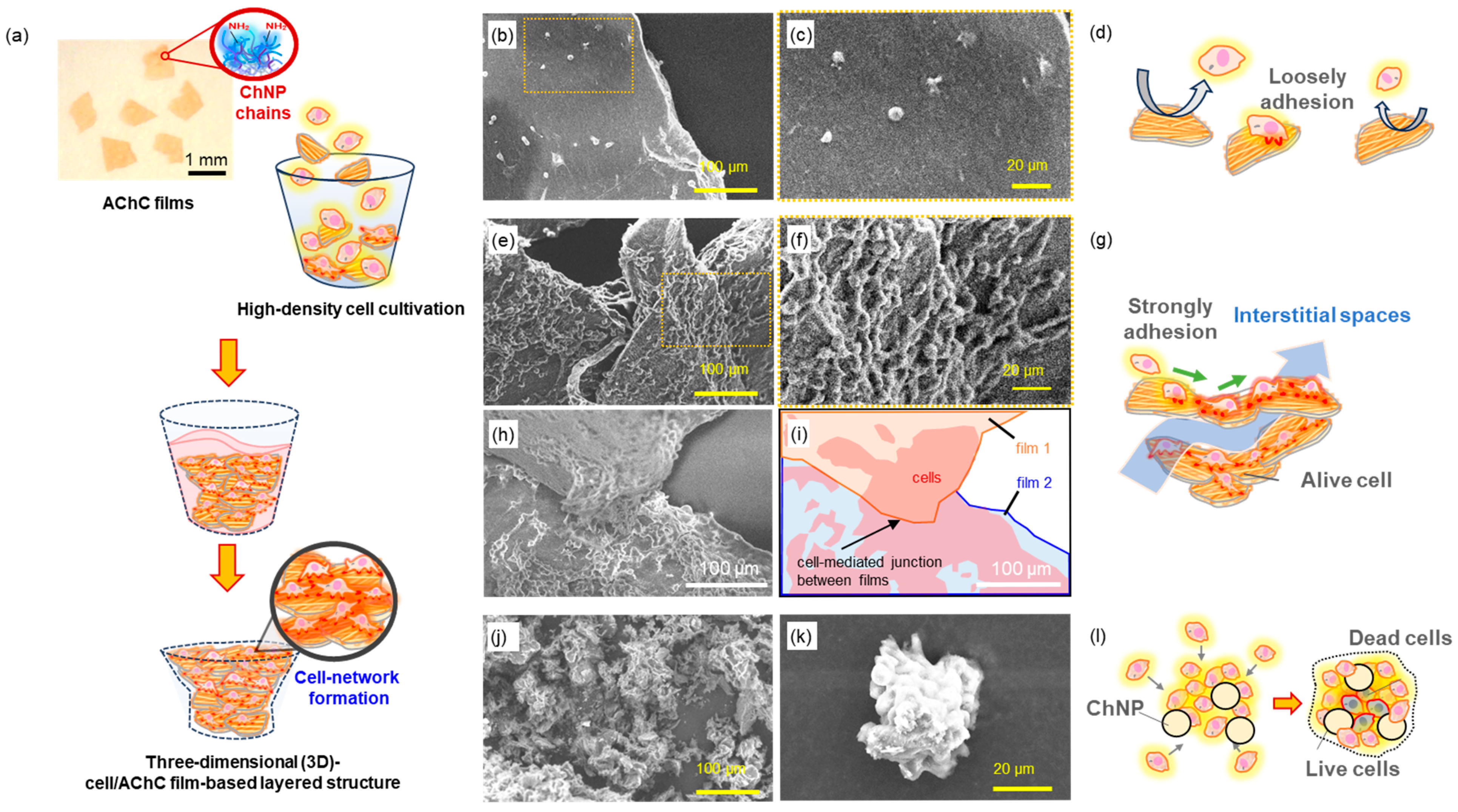
Based on the above experimental results, we hypothesized that the highly cell-adhesive AChC films could be applied to tissue-like three-dimensional (3D-)cell culture systems [122,123,124,125,126,127,128,129], such high-density culture on the AChC films would induce network-like cell adhesion that connects multiple films into a layered 3D-cell/film structure (Figure 13a) [130]. The AChC films were added to a HeLa suspension (3 × 10⁶ cells mL−1), and the SD-ChNF films and ChNP powders were also tested as references. After seeding, inversion mixing was performed every hour for 6 h, and after 4 days of static culture, cells were fixed. Sterilization tolerance indicated that the SD-ChNF and AChC films maintained flat surfaces after autoclaving at 121 °C for 20 min, whereas the ChNPs remained as 50 nm aggregates without redispersion. Thus, both film types showed significant aqueous stability owing to strong intermolecular hydrogen bonding. The SEM measurement revealed that the SD-ChNF films supported only sparse, weakly adherent spherical cells (Figure 13b–d). In contrast, the AChC film promoted pronounced cellular elongation and the formation of multicellular networks that bridged adjacent film fragments, thereby generating 3D connectivity. The overlapping arrangement of the AChC film fragments also created interstitial spaces, which likely served as pathways for fresh culture medium to permeate the structure and support sustained cellular viability (Figure 13e–i)). In contrast, the ChNP powders failed to sustain robust adhesion and instead induced only small, compact spheroids (Figure 13i,k). Such tightly aggregated cell spheroid raises concerns that, during prolonged culture, cells located in the central region may not receive sufficient nutrient and oxygen supply, potentially leading to progressive cell death within the core (Figure 13l).
Figure 13.
(a) Schematic illustrations of three-dimensional (3D-)cell/AChC film-based layered structure with high-density cell cultivation, SEM images and schematic illustration of adherent HeLa cells on (b–d) SD-ChNF films, (e–i) AChC films, and (j–l) ChNP powders.
Therefore, the AChC films outperform both the SD-ChNF films and ChNP powders as scaffolds for 3D cell culture. The optimized combination of the SD-ChNFs and ChNPs yields the film with mechanical integrity and high-cell-interaction ability. This suggests a strategy of stacking the AChC film fragments to construct 3D architectures with sufficient interstitial space for nutrient and oxygen diffusion, unlike spheroid-based approaches that often suffer from diffusion limitations.
4. Conclusions
This review outlines approaches for constructing composite materials by combining the self-assembled ChNFs with various natural polymers. It also demonstrates that the SD-ChNFs, which retain high crystallinity and are finely dispersed, can be incorporated into these systems and blended with natural polymers. Furthermore, combining the SD-ChNFs with low-crystalline chitin substrates yielded the AChCs that embody the single-polymer composite concept. These AChCs exhibited experimentally verified improvements in mechanical performance, hydration properties, and cell adhesion behavior, confirming the effectiveness of crystallinity contrast in controlling composite functionality. Future studies will focus on detailed toxicological evaluations and systematic investigations on cellular responses to facilitate further development toward medical applications. Furthermore, these fundamental insights into material properties and biosafety provide important guidelines for future industrial implementation and indicate potential applicability to large-scale structural and transportation-related sectors requiring high material volumes. Collectively, chitin nanofibers provide a coherent and experimentally validated framework for the development of sustainable nanomaterials and advanced composites for materials science and bioengineering applications.
Author Contributions
Conceptualization, methodology, validation, data curation, writing—original draft preparation, review, and editing, and funding acquisition, M.T. and J.-i.K.; supervision, project administration, J.-i.K. All authors have read and agreed to the published version of the manuscript.
Funding
This research received no external funding.
Institutional Review Board Statement
Not applicable.
Informed Consent Statement
Not applicable.
Data Availability Statement
The data presented in this study are available on request from the corresponding author.
Conflicts of Interest
The authors declare no conflicts of interest.
References
- Austin, P.R.; Brine, C.J.; Castle, J.E.; Zikakis, J.P. Chitin: New Facets of Research. Science 1981, 212, 749–753. [Google Scholar] [CrossRef] [Scilit] [PubMed]
- Minagawa, T.; Okamura, Y.; Shigemasa, Y.; Minami, S.; Okamoto, Y. Effects of molecular weight and deacetylation degree of chitin/chitosan on wound healing. Carbohydr. Polym. 2007, 67, 640–644. [Google Scholar] [CrossRef] [Scilit]
- Martínez, J.P.; Falomir, M.P.; Gozalbo, D. Chitin: A Structural Biopolysaccharide with Multiple Applications. In Encyclopedia of Life Sciences; Wiley: Hoboken, NJ, USA, 2014. [Google Scholar] [CrossRef] [Scilit]
- Zargar, V.; Asghari, M.; Dashti, A. A Review on Chitin and Chitosan Polymers: Structure, Chemistry, Solubility, Derivatives, and Applications. ChemBioEng Rev. 2015, 2, 204–226. [Google Scholar] [CrossRef] [Scilit]
- El Knidri, H.; Belaabed, R.; Addaou, A.; Laajeb, A.; Lahsini, A. Extraction, chemical modification and characterization of chitin and chitosan. Int. J. Biol. Macromol. 2018, 120, 1181–1189. [Google Scholar] [CrossRef] [Scilit]
- Torres, F.G.; Troncoso, O.P.; Pisani, A.; Gatto, F.; Bardi, G. Natural Polysaccharide Nanomaterials: An Overview of Their Immunological Properties. Int. J. Mol. Sci. 2019, 20, 5092. [Google Scholar] [CrossRef] [Scilit]
- Bai, L.; Liu, L.; Esquivel, M.; Tardy, B.L.; Huan, S.; Niu, X.; Liu, S.; Yang, G.; Fan, Y.; Rojas, O.J. Nanochitin: Chemistry, Structure, Assembly, and Applications. Chem. Rev. 2022, 122, 11604–11674. [Google Scholar] [CrossRef] [Scilit]
- Lv, J.; Lv, X.; Ma, M.; Oh, D.-H.; Jiang, Z.; Fu, X. Chitin and chitin-based biomaterials: A review of advances in processing and food applications. Carbohydr. Polym. 2023, 299, 120142. [Google Scholar] [CrossRef] [Scilit]
- Shinnerl, H.E.; Banks, I.J.; Dilger, R.N. A comparative review of chitin occurrence and quantification methodologies with applications to insect sourced materials. Carbohydr. Polym. 2026, 371, 124496. [Google Scholar] [CrossRef] [Scilit]
- Rinaudo, M. Chitin and chitosan: Properties and applications. Prog. Polym. Sci. 2006, 31, 603–632. [Google Scholar] [CrossRef] [Scilit]
- Izadi, H.; Asadi, H.; Bemani, M. Chitin: A comparison between its main sources. Front. Mater. 2025, 12, 1537067. [Google Scholar] [CrossRef] [Scilit]
- Tsurkan, M.V.; Voronkina, A.; Khrunyk, Y.; Wysokowski, M.; Petrenko, I.; Ehrlich, H. Progress in chitin analytics. Carbohydr. Polym. 2021, 252, 117204. [Google Scholar] [CrossRef] [Scilit] [PubMed]
- Kaya, M.; Mujtaba, M.; Ehrlich, H.; Salaberria, A.M.; Baran, T.; Amemiya, C.T.; Galli, R.; Akyuz, L.; Sargin, I.; Labidi, J. On chemistry of γ-chitin. Carbohydr. Polym. 2017, 176, 177–186. [Google Scholar] [CrossRef] [Scilit] [PubMed]
- Gardner, K.H.; Blackwell, J. Refinement of the structure of β-chitin. Biopolymers 1975, 14, 1581–1595. [Google Scholar] [CrossRef] [Scilit] [PubMed]
- Minke, R.; Blackwell, J. The structure of α-chitin. J. Mol. Biol. 1978, 120, 167–181. [Google Scholar] [CrossRef] [Scilit]
- Faria, R.R.; Guerra, R.F.; de Sousa Neto, L.R.; Motta, L.F.; Franca, E.d.F. Computational study of polymorphic structures of α- and β- chitin and chitosan in aqueous solution. J. Mol. Graph. Model. 2016, 63, 78–84. [Google Scholar] [CrossRef] [Scilit]
- Noda, T.; Hatakeyama, M.; Kitaoka, T. Combination of Polysaccharide Nanofibers Derived from Cellulose and Chitin Promotes the Adhesion, Migration and Proliferation of Mouse Fibroblast Cells. Nanomaterials 2022, 12, 402. [Google Scholar] [CrossRef] [Scilit]
- Islam, M.A.; Hasan, M.N.; Evan, M.S.H.; Uddin, M.J.; Tulin, W.S.; Islam, M.S.; Khandaker, M.U.; Rahman, I.M.M.; Chowdhury, F.I. Chitin nanofibers: Recent advances in preparation and applications in biomedical and beyond. RSC Adv. 2025, 15, 14655–14690. [Google Scholar] [CrossRef] [Scilit]
- Ifuku, S.; Nogi, M.; Abe, K.; Yoshioka, M.; Morimoto, M.; Saimoto, H.; Yano, H. Preparation of Chitin Nanofibers with a Uniform Width as α-Chitin from Crab Shells. Biomacromolecules 2009, 10, 1584–1588. [Google Scholar] [CrossRef] [Scilit]
- Zeng, J.-B.; He, Y.-S.; Li, S.-L.; Wang, Y.-Z. Chitin whiskers: An overview. Biomacromolecules 2012, 13, 1–11. [Google Scholar] [CrossRef] [Scilit]
- Zhang, X.; Rolandi, M. Engineering strategies for chitin nanofibers. J. Mater.Chem. B 2017, 5, 2547–2559. [Google Scholar] [CrossRef] [Scilit]
- Yang, X.; Liu, J.; Pei, Y.; Zheng, X.; Tang, K. Recent Progress in Preparation and Application of Nano-Chitin Materials. Energy Environ. Mater. 2020, 3, 492–515. [Google Scholar] [CrossRef] [Scilit]
- Kumar, S.; Foroozesh, J. Chitin nanocrystals based complex fluids: A green nanotechnology. Carbohydr. Polym. 2021, 257, 117619. [Google Scholar] [CrossRef] [Scilit] [PubMed]
- Anusiya, G.; Jaiganesh, R. A review on fabrication methods of nanofibers and a special focus on application of cellulose nanofibers. Carbohydr. Polym. Technol. Appl. 2022, 4, 100262. [Google Scholar] [CrossRef] [Scilit]
- Liao, J.; Zhou, Y.; Hou, B.; Zhang, J.; Huang, H. Nano-chitin: Preparation strategies and food biopolymer film reinforcement and applications. Carbohydr. Polym. 2023, 305, 120553. [Google Scholar] [CrossRef] [Scilit] [PubMed]
- Ifuku, S.; Saimoto, H. Chitin nanofibers: Preparations, modifications, and applications. Nanoscale 2012, 4, 3308–3318. [Google Scholar] [CrossRef] [Scilit]
- Ifuku, S. Chitin and chitosan nanofibers: Preparation and chemical modifications. Molecules 2014, 19, 18367–18380. [Google Scholar] [CrossRef] [Scilit]
- Fan, Y.; Saito, T.; Isogai, A. TEMPO-mediated oxidation of β-chitin to prepare individual nanofibrils. Carbohydr. Polym. 2009, 77, 832–838. [Google Scholar] [CrossRef] [Scilit]
- Kadokawa, J. Polymer nanocomposites based on nano chitin. In Chemical Physics of Polymer Nanocomposites; Wiley: Hoboken, NJ, USA, 2024; pp. 199–227. [Google Scholar] [CrossRef] [Scilit]
- Azuma, K.; Ifuku, S.; Osaki, T.; Okamoto, Y.; Minami, S. Preparation and Biomedical Applications of Chitin and Chitosan Nanofibers. J. Biomed. Nanotechnol. 2014, 10, 2891–2920. [Google Scholar] [CrossRef] [Scilit]
- Ifuku, S.; Kaminaka, H.; Shams, M.I. Nanochitin From Crab Shells: Production, Chemical Modification, Composite Materials, and Physiological Functions. Macromol. Rapid Commun. 2025, 46, 2400765. [Google Scholar] [CrossRef] [Scilit]
- Li, C.; Wu, J.; Shi, H.; Xia, Z.; Sahoo, J.K.; Yeo, J.; Kaplan, D.L. Fiber-Based Biopolymer Processing as a Route toward Sustainability. Adv. Mater. 2022, 34, e2105196. [Google Scholar] [CrossRef] [Scilit]
- Noh, H.K.; Lee, S.W.; Kim, J.-M.; Oh, J.-E.; Kim, K.-H.; Chung, C.-P.; Choi, S.-C.; Park, W.H.; Min, B.-M. Electrospinning of chitin nanofibers: Degradation behavior and cellular response to normal human keratinocytes and fibroblasts. Biomaterials 2006, 27, 3934–3944. [Google Scholar] [CrossRef] [Scilit]
- Zhong, C.; Cooper, A.; Kapetanovic, A.; Fang, Z.; Zhang, M.; Rolandi, M. A facile bottom-up route to self-assembled biogenic chitin nanofibers. Soft Matter 2010, 6, 5298–5301. [Google Scholar] [CrossRef] [Scilit]
- Mushi, N.E.; Butchosa, N.; Salajkova, M.; Zhou, Q.; Berglund, L.A. Nanostructured membranes based on native chitin nanofibers prepared by mild process. Carbohydr. Polym. 2014, 112, 255–263. [Google Scholar] [CrossRef] [Scilit] [PubMed]
- Zhong, T.; Liu, W.; Liu, H. Green electrospinning of chitin propionate to manufacture nanofiber mats. Carbohydr. Polym. 2021, 273, 118593. [Google Scholar] [CrossRef] [Scilit] [PubMed]
- Swatloski, R.P.; Spear, S.K.; Holbrey, J.D.; Rogers, R.D. Dissolution of Cellose with Ionic Liquids. J. Am. Chem. Soc. 2002, 124, 4974–4975. [Google Scholar] [CrossRef] [Scilit]
- Rogers, R.D.; Seddon, K.R. Ionic Liquids—Solvents of the Future? Science 2003, 302, 792–793. [Google Scholar] [CrossRef] [Scilit]
- Fort, D.A.; Remsing, R.C.; Swatloski, R.P.; Moyna, P.; Moyna, G.; Rogers, R.D. Can ionic liquids dissolve wood? Processing and analysis of lignocellulosic materials with 1-n-butyl-3-methylimidazolium chloride. Green Chem. 2007, 9, 63–69. [Google Scholar] [CrossRef] [Scilit]
- Sun, N.; Rahman, M.; Qin, Y.; Maxim, M.L.; Rodríguez, H.; Rogers, R.D. Complete dissolution and partial delignification of wood in the ionic liquid 1-ethyl-3-methylimidazolium acetate. Green Chem. 2009, 11, 646–655. [Google Scholar] [CrossRef] [Scilit]
- Wang, H.; Gurau, G.; Rogers, R.D. Ionic liquid processing of cellulose. Chem. Soc. Rev. 2012, 41, 1519–1537. [Google Scholar] [CrossRef] [Scilit]
- Wang, W.-T.; Zhu, J.; Wang, X.-L.; Huang, Y.; Wang, Y.-Z. Dissolution Behavior of Chitin in Ionic Liquids. J. Macromol. Sci. Phys. 2010, 49, 528–541. [Google Scholar] [CrossRef] [Scilit]
- Kadokawa, J. Ionic liquid as useful media for dissolution, derivatization, and nanomaterial processing of chitin. Green Sustain. Chem. 2013, 3, 19–25. [Google Scholar] [CrossRef]
- Kadokawa, J. Fabrication of nanostructured and microstructured chitin materials through gelation with suitable dispersion media. RSC Adv. 2015, 5, 12736–12746. [Google Scholar] [CrossRef] [Scilit]
- Silva, S.S.; Mano, J.F.; Reis, R.L. Ionic liquids in the processing and chemical modification of chitin and chitosan for biomedical applications. Green Chem. 2017, 19, 1208–1220. [Google Scholar] [CrossRef] [Scilit]
- Shamshina, J.L. Chitin in ionic liquids: Historical insights into the polymer’s dissolution and isolation. A review. Green Chem. 2019, 21, 3974–3993. [Google Scholar] [CrossRef] [Scilit]
- Wu, Y.; Sasaki, T.; Irie, S.; Sakurai, K. A novel biomass-ionic liquid platform for the utilization of native chitin. Polymer 2008, 49, 2321–2327. [Google Scholar] [CrossRef] [Scilit]
- Qin, Y.; Lu, X.; Sun, N.; Rogers, R.D. Dissolution or extraction of crustacean shells using ionic liquids to obtain high molecular weight purified chitin and direct production of chitin films and fibers. Green Chem. 2010, 12, 968–971. [Google Scholar] [CrossRef] [Scilit]
- Tajiri, R.; Setoguchi, T.; Wakizono, S.; Yamamoto, K.; Kadokawa, J. Preparation of Self-Assembled Chitin Nanofibers by Regeneration from Ion Gels Using Calcium Halide· dihydrate/Methanol Solutions. J. Biobased. Mater. Bioenergy 2013, 7, 655–659. [Google Scholar] [CrossRef] [Scilit]
- Kadokawa, J. An overview on acylation methods of α-chitin. Int. J. Biol. Macromol. 2024, 262, 130166. [Google Scholar] [CrossRef] [Scilit]
- Kadokawa, J. Application of ionic liquids for the functional materialization of chitin. Mater. Adv. 2022, 3, 3355–3364. [Google Scholar] [CrossRef] [Scilit]
- Uto, T.; Idenoue, S.; Yamamoto, K.; Kadokawa, J. Understanding dissolution process of chitin crystal in ionic liquids: Theoretical study. Phys. Chem. Chem. Phys. 2018, 20, 20669–20677. [Google Scholar] [CrossRef] [Scilit]
- Totani, M.; Kawano, T.; Shigenobu, Y.; Kadokawa, J. Efficient benzylation of chitin in ionic liquids. New J. Chem. 2025, 49, 11584–11587. [Google Scholar] [CrossRef] [Scilit]
- Kadokawa, J.; Takegawa, A.; Mine, S.; Prasad, K. Preparation of chitin nanowhiskers using an ionic liquid and their composite materials with poly(vinyl alcohol). Carbohydr. Polym. 2011, 84, 1408–1412. [Google Scholar] [CrossRef] [Scilit]
- Hashiguchi, T.; Yamamoto, K.; Kadokawa, J. Fabrication of highly flexible nanochitin film and its composite film with anionic polysaccharide. Carbohydr. Polym. 2021, 270, 118369. [Google Scholar] [CrossRef] [Scilit] [PubMed]
- Kadokawa, J. Preparation of Composite Materials from Self-Assembled Chitin Nanofibers. Polymers 2021, 13, 3548. [Google Scholar] [CrossRef] [Scilit]
- Kadokawa, J.; Kawano, A.; Yamamoto, K. Fabrication of semi-crystalline film by hexanoylation on self-assembled chitin nanofibers. ChemistrySelect 2019, 4, 797–801. [Google Scholar] [CrossRef] [Scilit]
- Jayakumar, R.; Prabaharan, M.; Nair, S.; Tamura, H. Novel chitin and chitosan nanofibers in biomedical applications. Biotechnol. Adv. 2010, 28, 142–150. [Google Scholar] [CrossRef] [Scilit]
- Jayakumar, R.; Menon, D.; Manzoor, K.; Nair, S.V.; Tamura, H. Biomedical applications of chitin and chitosan based nanomaterials—A short review. Carbohydr. Polym. 2010, 82, 227–232. [Google Scholar] [CrossRef] [Scilit]
- Jayakumar, R.; Chennazhi, K.P.; Srinivasan, S.; Nair, S.V.; Furuike, T.; Tamura, H. Chitin scaffolds in tissue engineering. Int. J. Mol. Sci. 2011, 12, 1876–1887. [Google Scholar] [CrossRef] [Scilit]
- Wan, A.C.; Tai, B.C. Chitin—A promising biomaterial for tissue engineering and stem cell technologies. Biotechnol. Adv. 2013, 31, 1776–1785. [Google Scholar] [CrossRef] [Scilit]
- Rolandi, M.; Rolandi, R. Self-assembled chitin nanofibers and applications. Adv. Colloid Interface Sci. 2014, 207, 216–222. [Google Scholar] [CrossRef] [Scilit]
- Ding, F.; Deng, H.; Du, Y.; Shi, X.; Wang, Q. Emerging chitin and chitosan nanofibrous materials for biomedical applications. Nanoscale 2014, 6, 9477–9493. [Google Scholar] [CrossRef] [Scilit] [PubMed]
- Yudin, V.E.; Dobrovolskaya, I.P.; Neelov, I.M.; Dresvyanina, E.N.; Popryadukhin, P.V.; Ivan’kova, E.M.; Elokhovskii, V.Y.; Kasatkin, I.A.; Okrugin, B.M.; Morganti, P. Wet spinning of fibers made of chitosan and chitin nanofibrils. Carbohydr. Polym. 2014, 108, 176–182. [Google Scholar] [CrossRef] [Scilit] [PubMed]
- Ogawa, Y.; Azuma, K.; Izawa, H.; Morimoto, M.; Ochi, K.; Osaki, T.; Ito, N.; Okamoto, Y.; Saimoto, H.; Ifuku, S. Preparation and biocompatibility of a chitin nanofiber/gelatin composite film. Int. J. Biol. Macromol. 2017, 104, 1882–1889. [Google Scholar] [CrossRef] [Scilit] [PubMed]
- Islam, S.; Bhuiyan, M.R.; Islam, M. Chitin and chitosan: Structure, properties and applications in biomedical engineering. J. Polym. Environ. 2017, 25, 854–866. [Google Scholar] [CrossRef] [Scilit]
- Dobrovolskaya, I.P.; Yudin, V.E.; Popryadukhin, P.V.; Ivan’kova, E.M.; Shabunin, A.S.; Kasatkin, I.A.; Morgantie, P. Effect of chitin nanofibrils on electrospinning of chitosan-based composite nanofibers. Carbohydr. Polym. 2018, 194, 260–266. [Google Scholar] [CrossRef] [Scilit]
- Zhong, Z.; Huang, Y.; Hu, Q.; He, W.; Duan, B.; Yan, X.; Yang, Z.; Liang, W.; Liu, Z.; Peng, Z.; et al. Elucidation of molecular pathways responsible for the accelerated wound healing induced by a novel fibrous chitin dressing. Biomater. Sci. 2019, 7, 5247–5257. [Google Scholar] [CrossRef] [Scilit]
- Zhu, K.; Tu, H.; Yang, P.; Qiu, C.; Zhang, D.; Lu, A.; Luo, L.; Chen, F.; Liu, X.; Chen, L. Mechanically strong chitin fibers with nanofibril structure, biocompatibility, and biodegradability. Chem. Mater. 2019, 31, 2078–2087. [Google Scholar] [CrossRef] [Scilit]
- Smirnova, N.V.; Kolbe, K.A.; Dresvyanina, E.N.; Grebennikov, S.F.; Dobrovolskaya, I.P.; Yudin, V.E.; Luxbacher, T.; Morganti, P. Effect of Chitin Nanofibrils on Biocompatibility and Bioactivity of the Chitosan-Based Composite Film Matrix Intended for Tissue Engineering. Materials 2019, 12, 1874. [Google Scholar] [CrossRef] [Scilit]
- Satitsri, S.; Muanprasat, C. Chitin and Chitosan Derivatives as Biomaterial Resources for Biological and Biomedical Applications. Molecules 2020, 25, 5961. [Google Scholar] [CrossRef] [Scilit]
- Bose, I.; Singh, R.; Negi, P.; Singh, Y. Chitin as bio-based nanomaterial in packaging: A review. Mater. Today Proc. 2021, 46, 11254–11257. [Google Scholar] [CrossRef] [Scilit]
- Jin, T.; Liu, T.; Lam, E.; Moores, A. Chitin and chitosan on the nanoscale. Nanoscale Horiz. 2021, 6, 505–542. [Google Scholar] [CrossRef] [Scilit] [PubMed]
- Triunfo, M.; Tafi, E.; Guarnieri, A.; Scieuzo, C.; Hahn, T.; Zibek, S.; Salvia, R.; Falabella, P. Insect chitin-based nanomaterials for innovative cosmetics and cosmeceuticals. Cosmetics 2021, 8, 40. [Google Scholar] [CrossRef] [Scilit]
- Baharlouei, P.; Rahman, A. Chitin and Chitosan: Prospective Biomedical Applications in Drug Delivery, Cancer Treatment, and Wound Healing. Mar. Drugs 2022, 20, 460. [Google Scholar] [CrossRef] [Scilit] [PubMed]
- Kodolova-Chukhontseva, V.V.; Dresvyanina, E.N.; Nashchekina, Y.A.; Dobrovol’skaya, I.P.; Bystrov, S.G.; Ivan’kova, E.M.; Yudin, V.E.; Morganti, P. Application of the Composite Fibers Based on Chitosan and Chitin Nanofibrils in Cosmetology. J. Funct. Biomater. 2022, 13, 198. [Google Scholar] [CrossRef] [Scilit]
- Kozma, M.; Acharya, B.; Bissessur, R. Chitin, Chitosan, and Nanochitin: Extraction, Synthesis, and Applications. Polymers 2022, 14, 3989. [Google Scholar] [CrossRef] [Scilit]
- Liao, J.; Hou, B.; Huang, H. Preparation, properties and drug controlled release of chitin-based hydrogels: An updated review. Carbohydr. Polym. 2022, 283, 119177. [Google Scholar] [CrossRef] [Scilit]
- Muñoz-Núñez, C.; Fernández-García, M.; Muñoz-Bonilla, A. Chitin nanocrystals: Environmentally friendly materials for the development of bioactive films. Coatings 2022, 12, 144. [Google Scholar] [CrossRef] [Scilit]
- Zhang, Z.; Li, Q.; Hatakeyama, M.; Kitaoka, T. Injectable cell-laden hydrogels fabricated with cellulose and chitosan nanofibers for bioprinted liver tissues. Biomed. Mater. 2023, 18, 045018. [Google Scholar] [CrossRef] [Scilit]
- Lee, S.; Hao, L.T.; Park, J.; Oh, D.X.; Hwang, D.S. Nanochitin and nanochitosan: Chitin nanostructure engineering with multiscale properties for biomedical and environmental applications. Adv. Mater. 2023, 35, 2203325. [Google Scholar] [CrossRef] [Scilit]
- Iroegbu, A.O.C.; Ray, S.S. Chitin nanomaterials as multifunctional systems in advanced applications–progress and challenges toward sustainability. Macromol. Mater. Eng. 2023, 308, 2300053. [Google Scholar] [CrossRef] [Scilit]
- Wang, X.; Tarahomi, M.; Sheibani, R.; Xia, C.; Wang, W. Progresses in lignin, cellulose, starch, chitosan, chitin, alginate, and gum/carbon nanotube (nano) composites for environmental applications: A review. Int. J. Biol. Macromol. 2023, 241, 124472. [Google Scholar] [CrossRef] [Scilit]
- Uddin, M.N.; Jamal, M.S.I.; Ali, M.Y.; Darda, M.A.; Mahedi, S.I. Tissue engineering and the potential use of chitin. Emerg. Mater. 2023, 6, 827–839. [Google Scholar] [CrossRef] [Scilit]
- Ganesh, S.S.; Anushikaa, R.; Swetha Victoria, V.S.; Lavanya, K.; Shanmugavadivu, A.; Selvamurugan, N. Recent Advancements in Electrospun Chitin and Chitosan Nanofibers for Bone Tissue Engineering Applications. J. Funct. Biomater. 2023, 14, 288. [Google Scholar] [CrossRef] [Scilit] [PubMed]
- Dong, X.; Shi, L.; Ma, S.; Chen, X.; Cao, S.; Li, W.; Zhao, Z.; Chen, C.; Deng, H. Chitin/chitosan nanofibers toward a sustainable future: From hierarchical structural regulation to functionalization applications. Nano Lett. 2024, 24, 12014–12026. [Google Scholar] [CrossRef] [Scilit] [PubMed]
- Hatase, R.; Li, Q.; Hatakeyama, M.; Kitaoka, T. Direct activation of Toll-like receptor 2 signaling stimulated by contact with the interfacial structures of chitin nanofibers. Int. J. Biol. Macromol. 2025, 284, 138092. [Google Scholar] [CrossRef] [Scilit]
- Panackal Shibu, R.; Jafari, M.; Sagala, S.L.; Shamshina, J.L. Chitin nanowhiskers: A review of manufacturing, processing, and the influence of content on composite reinforcement and property enhancement. RSC Appl. Polym. 2025, 3, 1031–1123. [Google Scholar] [CrossRef] [Scilit]
- Visakh, P.M.; Thomas, S. Preparation of bionanomaterials and their polymer nanocomposites from waste and biomass. Waste Biomass Valorization 2010, 1, 121–134. [Google Scholar] [CrossRef] [Scilit]
- Ifuku, S.; Shervani, Z.; Saimoto, H. Preparation of Chitin Nanofibers and Their Composites. In Biopolymer Nanocomposites; Wiley: Hoboken, NJ, USA, 2013; pp. 11–31. [Google Scholar] [CrossRef] [Scilit]
- Ifuku, S.; Ikuta, A.; Izawa, H.; Morimoto, M.; Saimoto, H. Control of mechanical properties of chitin nanofiber film using glycerol without losing its characteristics. Carbohydr. Polym. 2014, 101, 714–717. [Google Scholar] [CrossRef] [Scilit]
- Deepthi, S.; Venkatesan, J.; Kim, S.-K.; Bumgardner, J.D.; Jayakumar, R. An overview of chitin or chitosan/nano ceramic composite scaffolds for bone tissue engineering. Int. J. Biol. Macromol. 2016, 93, 1338–1353. [Google Scholar] [CrossRef] [Scilit]
- Wu, S.; Duan, B.; Lu, A.; Wang, Y.; Ye, Q.; Zhang, L. Biocompatible chitin/carbon nanotubes composite hydrogels as neuronal growth substrates. Carbohydr. Polym. 2017, 174, 830–840. [Google Scholar] [CrossRef] [Scilit]
- Yu, Z.; Ji, Y.; Bourg, V.; Bilgen, M.; Meredith, J.C. Chitin- and cellulose-based sustainable barrier materials: A review. Emerg. Mater. 2020, 3, 919–936. [Google Scholar] [CrossRef] [Scilit]
- Joseph, B.; Mavelil Sam, R.; Balakrishnan, P.; Maria, H.J.; Gopi, S.; Volova, T.; Fernandes, S.C.M.; Thomas, S. Extraction of Nanochitin from Marine Resources and Fabrication of Polymer Nanocomposites: Recent Advances. Polymers 2020, 12, 1664. [Google Scholar] [CrossRef] [Scilit] [PubMed]
- Ahmad, S.; Palvasha, B.A.; Abbasi, B.b.K.; Nazir, M.S.; Akhtar, M.N.; Tahir, Z.; Abdullah, M.A. Preparation and Applications of Polysaccharide-Based Composites. In Polysaccharides; Wiley: Hoboken, NJ, USA, 2021; pp. 543–572. [Google Scholar] [CrossRef] [Scilit]
- Peter, S.; Lyczko, N.; Gopakumar, D.; Maria, H.J.; Nzihou, A.; Thomas, S. Chitin and chitosan based composites for energy and environmental applications: A review. Waste Biomass Valorization 2021, 12, 4777–4804. [Google Scholar] [CrossRef] [Scilit]
- Kostag, M.; El Seoud, O.A. Sustainable biomaterials based on cellulose, chitin and chitosan composites—A review. Carbohydr. Polym. Technol. Appl. 2021, 2, 100079. [Google Scholar] [CrossRef] [Scilit]
- Aranaz, I.; Acosta, N. Chitin- and Chitosan-Based Composite Materials. Biomimetics 2022, 7, 1. [Google Scholar] [CrossRef] [Scilit]
- Yihun, F.A. Nanochitin preparation and its application in polymer nanocomposites: A review. Emerg. Mater. 2022, 5, 2031–2060. [Google Scholar] [CrossRef] [Scilit]
- Mayilswamy, N.; Kandasubramanian, B. Green composites prepared from soy protein, polylactic acid (PLA), starch, cellulose, chitin: A review. Emerg. Mater. 2022, 5, 727–753. [Google Scholar] [CrossRef] [Scilit]
- Liang, Y.; Zou, Y.; Wu, S.; Song, D.; Xu, W.; Zhu, K. Preparation and properties of chitin/silk fibroin biocompatible composite fibers. J. Biomater. Sci. Polym. Ed. 2023, 34, 860–874. [Google Scholar] [CrossRef] [Scilit]
- Kodolova-Chukhontseva, V.V.; Rozova, E.Y.; Dresvyanina, E.N.; Nashchekina, Y.A.; Dobrovol’skaya, I.P.; Vlasova, E.N.; Bystrov, S.G.; Popova, E.N.; Maslennikova, T.P.; Yudin, V.E.; et al. New Composite Materials Based on Chitosan Films Reinforced with Chitin Nanofibrils for Cosmetic Application. Cosmetics 2023, 10, 51. [Google Scholar] [CrossRef] [Scilit]
- Tarique, J.; Sapuan, S.M.; Aqil, N.F.; Farhan, A.; Faiz, J.I.; Shahrizan, S. A Comprehensive Review Based on Chitin and Chitosan Composites. In Composites from the Aquatic Environment; Sapuan, S.M., Ahmad, I., Eds.; Springer Nature: Singapore, 2023; pp. 15–66. [Google Scholar] [CrossRef] [Scilit]
- Li, S.; Ren, Y.; Hou, Y.; Zhan, Q.; Jin, P.; Zheng, Y.; Wu, Z. Polysaccharide-Based Composite Films: Promising Biodegradable Food Packaging Materials. Foods 2024, 13, 3674. [Google Scholar] [CrossRef] [Scilit]
- Sanz de León, A.; Pulido, J.A.; Fernández-Delgado, N.; Delgado, F.J.; Molina, S.I. Chitin Nanocomposites for Fused Filament Fabrication: Flexible Materials with Enhanced Interlayer Adhesion. ACS Appl. Mater. Interfaces 2024, 16, 35554–35565. [Google Scholar] [CrossRef] [Scilit] [PubMed]
- Zao, Y.; Ma, J.; Sun, S.; Gao, J.; Liu, L.; Jin, Y.; Li, D.; Xu, Z.; Chen, C. Crab shell-enabled fabrication of multifunctional chitin composite gels with synergistic robustness and stretchability. Chem. Eng. J. 2026, 527, 171669. [Google Scholar] [CrossRef] [Scilit]
- Prasad, K.; Kaneko, Y.; Kadokawa, J. Novel Gelling Systems of κ-, ι- and λ-Carrageenans and their Composite Gels with Cellulose Using Ionic Liquid. Macromol. Biosci. 2009, 9, 376–382. [Google Scholar] [CrossRef] [Scilit] [PubMed]
- Kadokawa, J. A Mini-Review: Fabrication of Polysaccharide Composite Materials Based on Self-Assembled Chitin Nanofibers. Materials 2024, 17, 1898. [Google Scholar] [CrossRef] [Scilit]
- Totani, M.; Tanihata, Y.; Egi, Y.; Kadokawa, J. Fabrication of self-reinforced chitin composites by double crystalline blend approach. Int. J. Biol. Macromol. 2025, 286, 138441. [Google Scholar] [CrossRef] [Scilit]
- Hatanaka, D.; Yamamoto, K.; Kadokawa, J. Preparation of chitin nanofiber-reinforced carboxymethyl cellulose films. Int. J. Biol. Macromol. 2014, 69, 35–38. [Google Scholar] [CrossRef] [Scilit]
- Kadokawa, J.; Endo, R.; Hatanaka, D.; Yamamoto, K. Preparation of Chitin Nanofiber-Reinforced Cellulose Films Through Stepwise Regenerations from Individually Prepared Ion Gels. J. Polym. Environ. 2015, 23, 348–355. [Google Scholar] [CrossRef] [Scilit]
- Kawano, A.; Yamamoto, K.; Kadokawa, J. Preparation of Self-Assembled Chitin Nanofiber-Natural Rubber Composite Sheets and Porous Materials. Biomolecules 2017, 7, 47. [Google Scholar] [CrossRef] [Scilit]
- Lipinska, A.P.; Collén, J.; Krueger-Hadfield, S.A.; Mora, T.; Ficko-Blean, E. To gel or not to gel: Differential expression of carrageenan-related genes between the gametophyte and tetasporophyte life cycle stages of the red alga Chondrus crispus. Sci. Rep. 2020, 10, 11498. [Google Scholar] [CrossRef] [Scilit]
- Karger-Kocsis, J.; Bárány, T. Single-polymer composites (SPCs): Status and future trends. Compos. Sci. Technol. 2014, 92, 77–94. [Google Scholar] [CrossRef] [Scilit]
- Baghaei, B.; Skrifvars, M. All-Cellulose Composites: A Review of Recent Studies on Structure, Properties and Applications. Molecules 2020, 25, 2836. [Google Scholar] [CrossRef] [Scilit] [PubMed]
- Babu, N.B.K.; Mensah, R.A.; Shanmugam, V.; Rashedi, A.; Athimoolam, P.; Aseer, J.R.; Das, O. Self-reinforced polymer composites: An opportunity to recycle plastic wastes and their future trends. J. Appl. Polym. Sci. 2022, 139, e53143. [Google Scholar] [CrossRef] [Scilit]
- Egi, Y.; Kontani, A.; Kadokawa, J. Fabrication of all-chitin composite films. Int. J. Biol. Macromol. 2023, 253, 127512. [Google Scholar] [CrossRef] [Scilit] [PubMed]
- Egi, Y.; Kadokawa, J. Synthesis of Mixed Chitin Esters via Acylation of Chitin in Deep Eutectic Solvents. Molecules 2023, 28, 8132. [Google Scholar] [CrossRef] [Scilit]
- Sato, K.; Noguchi, T.; Kiyose, M. Deacetylation and gelation of amorphous chitin cake. Chitin Chitosan Res. 2019, 25, 136–137. [Google Scholar]
- Totani, M.; Shinchi, H.; Kadokawa, J. Cancer cell adhesion property on all-chitin composite films with reduced crystallinity. Carbohydr. Res. 2025, 549, 109373. [Google Scholar] [CrossRef] [Scilit]
- Hutmacher, D.W. Scaffold design and fabrication technologies for engineering tissues—State of the art and future perspectives. J. Biomater. Sci. Polym. Ed. 2001, 12, 107–124. [Google Scholar] [CrossRef] [Scilit]
- Matsusaki, M.; Kadowaki, K.; Nakahara, Y.; Akashi, M. Fabrication of Cellular Multilayers with Nanometer-Sized Extracellular Matrix Films. Angew. Chem. Int. Ed. 2007, 46, 4689–4692. [Google Scholar] [CrossRef] [Scilit]
- Matsunaga, Y.T.; Morimoto, Y.; Takeuchi, S. Molding Cell Beads for Rapid Construction of Macroscopic 3D Tissue Architecture. Adv. Mater. 2011, 23, H90–H94. [Google Scholar] [CrossRef] [Scilit]
- Carletti, E.; Motta, A.; Migliaresi, C. Scaffolds for Tissue Engineering and 3D Cell Culture. In 3D Cell Culture: Methods and Protocols; Haycock, J.W., Ed.; Humana Press: Totowa, NJ, USA, 2011; pp. 17–39. [Google Scholar] [CrossRef] [Scilit]
- Loh, Q.L.; Choong, C. Three-Dimensional Scaffolds for Tissue Engineering Applications: Role of Porosity and Pore Size. Tissue Eng. Part B Rev. 2013, 19, 485–502. [Google Scholar] [CrossRef] [Scilit]
- Nikolova, M.P.; Chavali, M.S. Recent advances in biomaterials for 3D scaffolds: A review. Bioact. Mater. 2019, 4, 271–292. [Google Scholar] [CrossRef] [Scilit]
- De Pieri, A.; Rochev, Y.; Zeugolis, D.I. Scaffold-free cell-based tissue engineering therapies: Advances, shortfalls and forecast. NPJ Regen. Med. 2021, 6, 18. [Google Scholar] [CrossRef] [Scilit]
- Xie, Z.-T.; Zeng, J.; Kang, D.-H.; Saito, S.; Miyagawa, S.; Sawa, Y.; Matsusaki, M. 3D Printing of Collagen Scaffold with Enhanced Resolution in a Citrate-Modulated Gellan Gum Microgel Bath. Adv. Healthc. Mater. 2023, 12, 2301090. [Google Scholar] [CrossRef] [Scilit]
- Totani, M.; Eda, M.; Shinchi, H.; Kadokawa, J. Utilization of All-Chitin Composite Films for High-Density Three-Dimensional Cell Cultivation. Molecules 2025, 30, 4243. [Google Scholar] [CrossRef] [Scilit]
Disclaimer/Publisher’s Note: The statements, opinions and data contained in all publications are solely those of the individual author(s) and contributor(s) and not of MDPI and/or the editor(s). MDPI and/or the editor(s) disclaim responsibility for any injury to people or property resulting from any ideas, methods, instructions or products referred to in the content. |
© 2026 by the authors. Licensee MDPI, Basel, Switzerland. This article is an open access article distributed under the terms and conditions of the Creative Commons Attribution (CC BY) license.