


Epigenomes 2025, 9(2), 22; https://doi.org/10.3390/epigenomes9020022 - 13 Jun 2025
Abstract
►
Show Figures
Background: Somatic symptom disorder (SSD) in children may be influenced by stress reactivity and psychosocial factors. The glucocorticoid receptor (GR), encoded by NR3C1, is a key mediator of stress responses. However, the relationship between NR3C1 methylation and SSD remains unclear. Methods: We analyzed
[...] Read more.
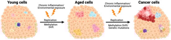
Background: Somatic symptom disorder (SSD) in children may be influenced by stress reactivity and psychosocial factors. The glucocorticoid receptor (GR), encoded by NR3C1, is a key mediator of stress responses. However, the relationship between NR3C1 methylation and SSD remains unclear. Methods: We analyzed NR3C1 exon 1F methylation in cell-free DNA from saliva in 34 children with SSD and 29 age- and sex-matched controls using bisulfite amplicon sequencing. Psychological assessments included the Beck Depression Inventory-II (BDI-II) and KINDL questionnaires to evaluate associations with methylation patterns. Results: Methylation levels showed age-related differences. In children under 13, CpG sites displayed mixed methylation, and specific sites correlated with KINDL and BDI-II scores. KINDL physical and total well-being scores negatively correlated with CpG30 and positively with CpG35; BDI-II scores negatively correlated with CpG32 and CpG35. In children aged 13 or older, CpG sites showed uniformly high methylation with no correlation to psychological measures. The SSD group showed significantly higher average methylation across the exon 1F region than controls in the older age group. These children also had more cases of orthostatic dysregulation and longer illness duration. Conclusions: This study suggests age-dependent epigenetic regulation of NR3C1 in SSD. While younger children showed CpG-specific correlations with psychological symptoms, older children demonstrated uniformly high methylation and potentially reduced gene expression, potentially reflecting cumulative stress, autonomic dysfunction, and internalizing disorders such as anxiety and depression.
Full article

Figure 1


