


Abstract
Right and bowhead whales represent key species in modern-day marine ecosystems because of their capacity to catch enormous amounts of prey items (especially calanoid copepod crustaceans). However, their evolutionary history is still not completely understood. Morphology- and molecule-based phylogenetic analyses failed to find a consensus view about the relationships of balaenids and other mysticete species and in determining the intra-family relationships of extant and fossil taxa. The fossil record may help in increasing the precision of morphology-based phylogenetic results when new balaenid species are discovered because the new fossils may add new character combinations able to change previously published hypotheses of relationships. Here, we describe a new balaenid genus and species, Quasibalaena portisantuerpiae, based on a partial skull from the Early Pliocene of Belgium. The new specimen preserves relevant portions of the cranium, periotic and tympanic bulla, and shows a character combination never observed up to now in other balaenid fossils. We compared the new taxon to all the other balaenid species known and included it in a morphology-based phylogenetic analysis. The results suggest that Balaenidae includes a stem group and two main subclades, respectively related to the extant bowhead whale (Balaena mysticetus) and to the right whales (genus Eubalaena). Quasibalaena portisantuerpiae belongs in the former subclade. Body size reconstruction allowed us to infer that the origin of the Balaena-related clade was characterized by a miniaturization process and that gigantism occurred independently in bowhead and right whales.
Keywords:
Belgium; Cetacea; fossil record; North Sea; phylogenetics; Pliocene; right whale; skull anatomy 1. Introduction
The family Balaenidae includes two extant genera: Balaena (the bowhead whale) and Eubalaena (the right whale), with four currently accepted species (Balaena mysticetus, Eubalaena glacialis, E. australis and E. japonica) [1,2]. Despite the reduced alpha diversity of living balaenids, the fossil record of this family is quite rich [3,4,5]. Several extinct balaenid taxa are now described in the scientific literature that shed light on the past diversification of this group. Balaenid diversity peaked during the Pliocene (between 5.3 and 2.6 Ma) even though a few fossils suggest that these whales could be present on our planet since the early Miocene [3,6].
Different interpretations of the morphology of extant and fossil mysticete species led to an array of phylogenetic conclusions about both origin and affinity of right and bowhead whales. Some researchers recognized that the mid-Miocene Peripolocetus vexillifer, described several decades ago from the Temblor Formation of California [7], should be recognized as the sister group of balaenids or balaenids + neobalaenids (forming the superfamily Balaenoidea and including right, bowhead and pygmy right whales) [8]. Additionally, the early Miocene Morenocetus parvus was alternatively interpreted as a basal balaenid [9] or as the sister group of Balaenoidea [3]. Total evidence and molecular analyses added further interpretations proposing that neobalaenids should be interpreted as the sister group of balaenopterids + gray whales [10,11] or as a relic of the otherwise extinct Cetotheriidae family (e.g., Ref. [12]). In any case, the monophyly of the family Balaenidae was never challenged, and we consider this family valid.
In recent years, the fossil record has made available several new specimens for comparative purposes so that more complex hypotheses of phylogenetic relationships for Balaenidae have been published (e.g., Refs. [3,13,14,15]). It is well known that new fossils add new character combinations to be considered in phylogeny inference and may have the power to change previously accepted hypotheses of relationships, supporting new and reasonably more reliable hypotheses (e.g., Refs. [16,17]). For this reason, descriptions of new fossil materials are welcome in evolutionary studies focusing on right and bowhead whales.
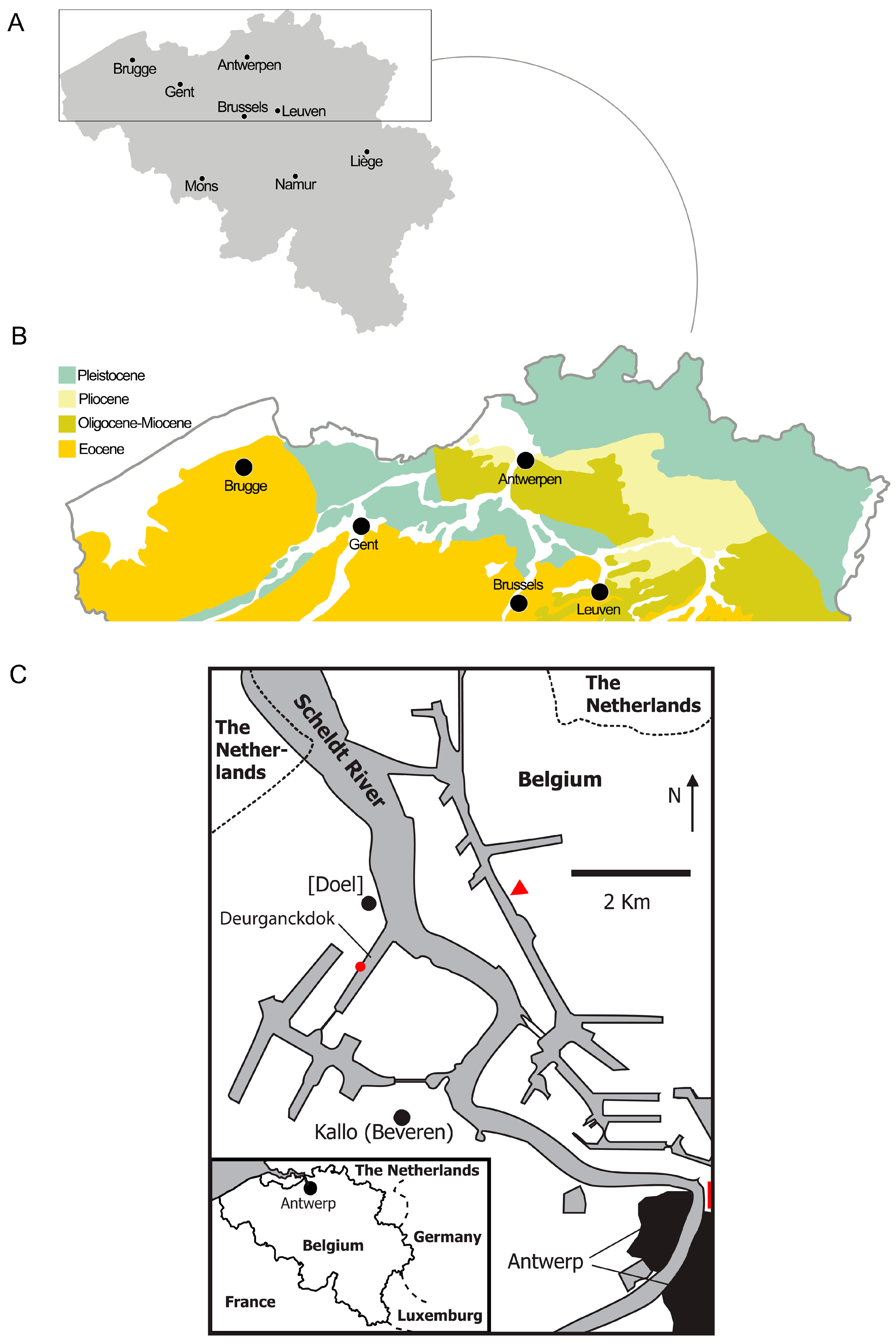
In this paper, we contribute to the increasing knowledge of balaenid evolution by describing a new specimen deriving from the Kattendijk Formation of Deurganckdok, Doel, Antwerpen (Belgium) (Figure 1). This new specimen is represented by a partial skull with a periotic still in articulation and a tympanic bulla and provides new comparative data that are used in a new phylogenetic analysis of Balaenidae.
The whale fauna from the Kattendijk Sands has been known since the 19th century and includes both balaenids, cetotheriids and balaenopterids, with some new species described in recent decades. The continuous sampling of the Kattendijk Sands at the Deurganckdok construction site, Antwerpen, resulted in the discovery of the holotype of Fragilicetus velponi [18] together with additional vertebrate and invertebrate fossils, including another large balaenopterid skeleton still under preparation. As a whole, the other finds from the Deurganckdok construction site include three fragmentary balaenid, three incomplete balaenopterid individuals, one partial cetotheriid and one odontocete endocast from the Kattendijk Sands, one fragmentary balaenid from the Luchtbal Sands, one incomplete balaenopterid from the Kruisschans Sands, two partial balaenids and one small incomplete balaenopteroid from the Oorderen Sands (M.Boss. pers. obs.). The richness of the fossil fauna of the Early Pliocene at this locality suggests that it represents a highly valuable site.
Figure 1.
Location of the discovery of the holotype of Quasibalaena portisantuerpiae gen. et sp. nov. (A) map of Belgium; the rectangle indicates the region magnified in (B). (B) geological map of northern Belgium showing the main outcrops. (C) Close-up map of Antwerp and the location of the discovery of the holotype; the red triangle indicates the tunnel–canal–dock and the red rectangle indicates the Kattendijkdok; the discovery locality (red point) is indicated in the Antwerp harbor, a hotspot of past diversity and encourages further studies, valorization and conservation efforts.
2. Geological Setting and Stratigraphic Age of the Specimen
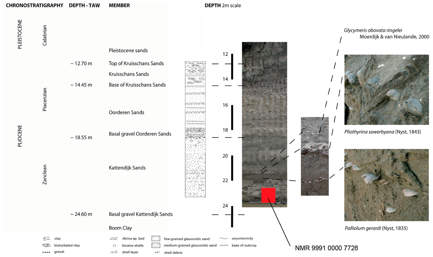
De Meuter & Laga [19] defined the Kattendijk Formation as a fine-grained to medium fine-grained, dark gray to green-gray glauconitic sand with a varying clay admixture. At the Deurganckdok construction site, mollusks and brachiopods are present, concentrated in well-separated layers (Figure 2) [20].
The Kattendijk Formation rests unconformably, directly on the Oligocene Boom clay at the type location of the specimen (pers. Obs. Mark Bosselaers; more to the north in the Deurganckdok, it rests on Miocene deposits). The Kattendijk formation consists of a single member, the Kattendijk Sands. The unit is unconformably covered by the Lillo Formation (c.q. the basal gravel of the Oorderen sands. The Kattendijk sands reach a thickness of circa 7 m at the type location of NMR 999100007728. The Deurganckdok is situated in the ‘Waasland area’, on the left bank of the Scheldt River, 13 Km WNW of Antwerp.
Figure 2.
Stratigraphic section of the locality of the discovery of the holotype of Quasibalaena portisantuerpiae. On the left side of the scheme, the chronostratigraphic subdivisions of the Pliocene are provided together with the relative heights of the different stratigraphic portions. A stratigraphic column is also shown with lithological information. The red square indicates the position of the specimen in the stratigraphic section. Details of shellbeds discussed in the text are shown on the right side of the scheme. Photographs are by Mark Bosselaers.
De Schepper et al. [21] analyzed the Kattendijk Formation for marine palynomorphs from the temporary outcrop during the construction of the tunnel–canal–dock in Antwerp. Their general age assessment showed a likely depositional range of 5.0–4.4 Ma, which supports the early Zanclean age based on other fossil groups. The Deurganckdok nannofossil (dynoflagellate cyst) assemblage reflects a neritic depositional environment in a temperate to cool climate [22]. Additional lists of microfossils are provided in Ref. [22]. The Kattendijk Formation contains several known thermophilic taxa as well as rare occurrences of the cold-tolerant species Impagidinium. Presence of the outer neritic to oceanic species Invertocysta lacrymosa and Impagidinium pallidum indicates open-water conditions [23,24], an interpretation supported by the rarity of Operculodinium israelianum, which is associated with shallow water [22].
An east–west correlation profile through the upper Neogene succession north of Antwerp, based on cone penetration tests, reveals the architecture of the lower Pliocene Kattendijk Formation. It shows a basal incision of the Kattendijk Formation down to 20 m in Miocene sands and locally even Lower Oligocene clays, cutting deeper towards the west. The incision is part of a much larger gully system in the region at the base of the Kattendijk Formation. The strongest gully incision is observed towards the west, where the type location of the specimen under investigation is situated and coincides with increases in the thickness of the Kattendijk Formation from its typical four to six meters thickness in the east towards a maximum of 15 m in the west. Correlations show that this additional thickness represents a separate sequence of the Kattendijk Formation that first filled the deepest part of the gully system (in the west, on the left bank of the Scheldt River) prior to being covered by the second sequence deposited in a larger gully system. Both sequences of the Kattendijk Formation have basal transgressive layers and are lithologically identical [25].
3. Materials and Methods
3.1. The Specimen and Its Preparation
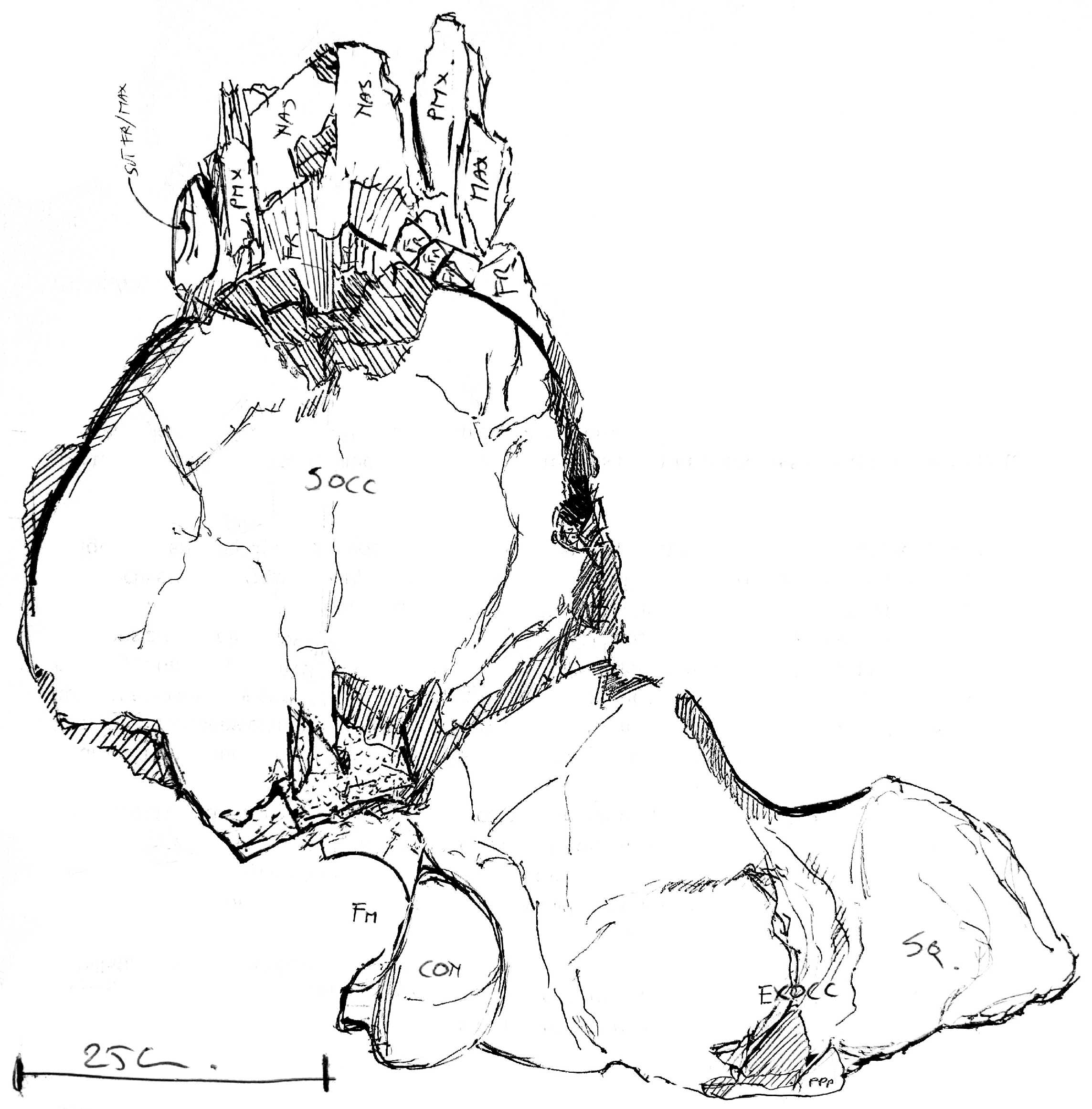
The specimen investigated in the present paper is currently part of the collection of the Natuurmuseum Rotterdam (hereinafter, NMR) identified by the accession number NMR 999100007728 (Figure 3). It was collected in June 2003 and consists of three portions: (a) part of the right posterolateral portion of the neurocranium including squamosal, exoccipital, elements of the basicranium and the right periotic; (b) a portion including the vertex (part of the anterior portion of the supraoccipital, both parietals and frontal with elements pertaining to the posteromedial elements of the rostrum); and (c) the right tympanic bulla.
Figure 3.
Original draft of the holotype skull of Quasibalaena portisantwerpiae realized by Mark Bosselaers after the end of the preparation process. Note that the scale bar equals to 25 cm. In this draft, the preliminary identification of the bones is shown.
The specimen was retrieved by construction workers and a crane operator at the Deurganckdok construction site near the hamlet of Doel; part of the village of Beveren (before Kallo), Oost-Vlaanderen province, Belgium (Figure 1). The specimen was found in a hard block of coagulated sand (sandstone) very low in the Kattendijk Sands, during leveling of the construction site floor. During recovery, the block broke and two fragments were preserved: one with the anterior part of the neurocranium and another with the right posterior part thereof. During this process, the edges of the specimen were damaged and the left posterior part of the neurocranium was lost. On the anterior side, the posterior part of the nasal bones, the premaxillae, and the right maxilla were preserved. The periotic and the tympanic bulla were also present, still in their original anatomical position.
The preparation was made using brushes and scalpels. During this process, the tympanic bulla was removed in order to prepare the ventral, medial, anterior and lateral sides of the periotic, as well as all sides of the bulla itself. After the sediment had been removed where possible, the exposed bone was treated with a 15% polyvinyl acetate solution in acetone to strengthen and plasticize the specimen, in order to prevent oxidation and/or damage.
3.2. Methodology of Taphonomic Analysis
Observations on the conservation of the specimen were made in order to get informWation about the integrity of the bones, mapping eventual fractures and breakages, and about the presence or not of signs left by scavenging activity. The sediment was visually analyzed searching for molluscs and other invertebrate and vertebrate remains useful to reconstruct the paleoecology of the site where the specimen was deposited and buried.
3.3. Anatomy, Measurements and Photography
The anatomical terminology used in the present paper is from Refs. [26,27]. The specimen was photographed using a Nikon D750 full-frame DSLR with a Tamron 24–70 mm f/2.8 lens. Additional photography was done by a Nikon D3100 DSLR with a Nikkor 18–200 mm f/3.5 lens and a Nikkor 50 mm f/1.8 lens. Measurements were taken with a Sourcingmap (300 mm) and a Tacklife D02 (150 mm) digital caliper, both with error margins to the nearest 0.01 mm.
3.4. Body Size Inference
We estimated the skeletal length of NMR 999100007728 by using the inferred occipital breadth and using the following Equation (1) [3]:
log (TL) = 1.1244 × log (OB) + 1.1869.
In Equation (1), TL is the total length of the skeleton and OB is the occipital breadth. It was demonstrated that skeletal lengths calculated with this equation and data from real specimens fall around a regression line with a r2 = 0.91, representing a high value of confidence. Based on the estimated total length of the skeleton, we estimated the body mass of this individual by using the following Equation (2) [28]:
log (BM) = 3.08 [log (TL)] − 4.84.
In Equation (2), BM is body mass and TL is total length of the skeleton. We compared the resulting values of the body length and body mass of Rotterdam with the measurements of other balaenid taxa provided by Refs. [3,15] to describe eventual patterns of body size evolution within Balaenidae. As far as the recently described Megabalaena sapporoensis is concerned, we used the total body length provided by Ref. [15], calculated the skeletal length based on Equation (6) in Ref. [3] (p. 508), and then we used the result to calculate the body mass based on Equation (2) [28].
To understand the tempo of body size change in balaenid phylogeny, we mapped a character related to body size in the phylogeny by using Mesquite 4.03 [29]. The character codes are provided in Appendix A (Appendix A.1). The character has four states corresponding to the four body size classes described in Ref. [3]: (0) small size (0–8 m), (1) medium size (9–13 m), (2) large size (14–20 m), and (3) gigantic size (>20 m).
3.5. Phylogenetic Analysis and Balaenid Diversity Through Time
A phylogenetic analysis was performed to investigate the relationships of NMR 999100007728 by using the character list and matrix provided in Appendix A (Appendix A.2 and Appendix A.3). The taxonomic sample includes 37 Operational Taxonomic Units (OTUs) with 21 balaenid, neobalaenid, stem-balaenid and stem-balaenoid OTUs. Additionally, 13 mysticete OTUs that are not related to Balaenidae are included comprising Balaenopteridae, Eschrichtiidae, Cetotheriidae, basal thalassotherian taxa, Eomysticetidae, Aetiocetidae and Mammalodontidae. Basilosauridae and Protocetidae are included as non-mysticete cetaceans. Indohyus is the outgroup. Stratigraphic ages of the OTUs were obtained from the Paleobiology Database (https://paleobiodb.org/#/; accessed on 9 June 2026) with the following three exceptions:
- Balaena montalionis. Paleobiology Database provided a stratigraphic age included between 3.6 and 2.58 Ma, but this is in contrast with the occurrence of this species in the Sabbie di Gambassi Formation, whose sedimentation was constrained in the earliest Pliocene by Ref. [30]. The latter is the assessment that we adopted here.
- Balaenula astensis. Paleobiology Database provided a stratigraphic age included between 5.3 and 3.6 Ma; this age is in contrast with the occurrence of this species in the Sabbie d’Asti Formation, whose sedimentation was constrained between 3.2 and 3.0 Ma by Ref. [31]. The latter is the chronostratigraphic assessment that we adopted here.
- Balaenula balaenopsis. The Paleobiology Database provided a stratigraphic age included between 20.45 and 1.8 Ma, which seems unlikely given the wide stratigraphic range encompassed by these data. We adopted the Belgian datations included between 5 and 4.44 Ma as the holotype material of this species (that is used in the present phylogenetic analysis) is from the Zanclean of Antwerp(Early Pliocene; see Ref. [21]).
The matrix includes 136 morphological characters. When compiling the taxon x character matrix, we assumed that general Cetacea, Chaeomysticeti, and Balaenomorpha characters (including baleen) were present in the OTUs assigned to Balaenomorpha; therefore, we coded these characters as present in the matrix independently of their preservation. Polarity of characters was decided based on the outgroup comparison criterion.
The taxon x character matrix was analyzed using TNT 1.6 [32] by its four New Technology algorithms with default parameters. Tree length, total number of nodes, Consistency and Retention indices were calculated in TNT; the Homoplasy Index was calculated by hand as it equals 1-CI. We assessed morphological support by a bootstrap analysis based on a traditional search and 1000 replicates in TNT. In the analysis, character states were considered unordered and no weight was applied.
We assessed the evolution of balaenid diversity by counting the number of described species and the number of inferred clades from each stratigraphic stage based on the results of the phylogenetic analysis performed in the present paper. We represented these observations in a dedicated plot.
4. Results
4.1. Taphonomy
Not much is known about the taphonomy of this find, as it was collected by a crane operator at the request of one of us (M.Boss.), but during his absence, in situ conditions were not recorded. It is not known whether more parts of the specimen were present at the site or not; by the time we arrived there, the place of origin had already been covered by a concrete slab.
The fact that both the periotic (petrosal) and the tympanic bulla were present in the original anatomical position (the bulla was removed during preparation to expose the periotic), indicates the skull was preserved upside down in the stratum. The broken fragment of the anterolateral process of the periotic, preserved a bit further down in the external auditory meatus, confirms this orientation. Especially, the thin bones centrally and at the lateral edges of the ventral side are heavily fragmented and crushed, which further corroborates the upside-down position.
The specimen shows signs of dorsoventral crushing that are evident because of the presence of fractures developed at right angles with respect to the longitudinal axis. These fractures are particularly evident in the area of articulation between the frontal and the posteromedial elements of the rostrum. It is possible that the crushing in this area changed the original angle between the nasals and the frontal and that now, the inclination of the nasal bones does not represent the original condition in the living animal.
Several other whale skulls found nearby at the same location and in the same stratum and comparable depth (the more basal part of the Kattendijk sands), are all, like this one, crushed and heavily broken (e.g., Fragilicetus velponi, “Balaenoptera” sibbaldina and several unpublished finds; M.Boss. pers. obs.). This indicates the skull must have been covered by a thick deposit of sediment relatively soon after the carcass had decomposed and very probably prior to fossilization.
The specimen was rather badly damaged by the crane laterally and anterodorsally during the recovery. Moreover, it broke in two, just anterior to the anterior squamosal border. Only the right half of the posterior neurocranium has been retrieved, together with the rather complete anterior neurocranium, including the posterior parts of the nasals and the posterior parts of the ascending processes of the premaxillae and maxillae.
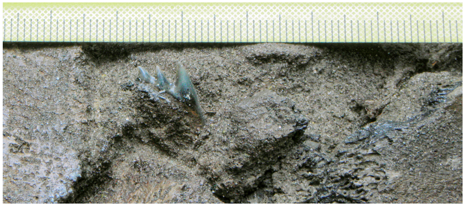
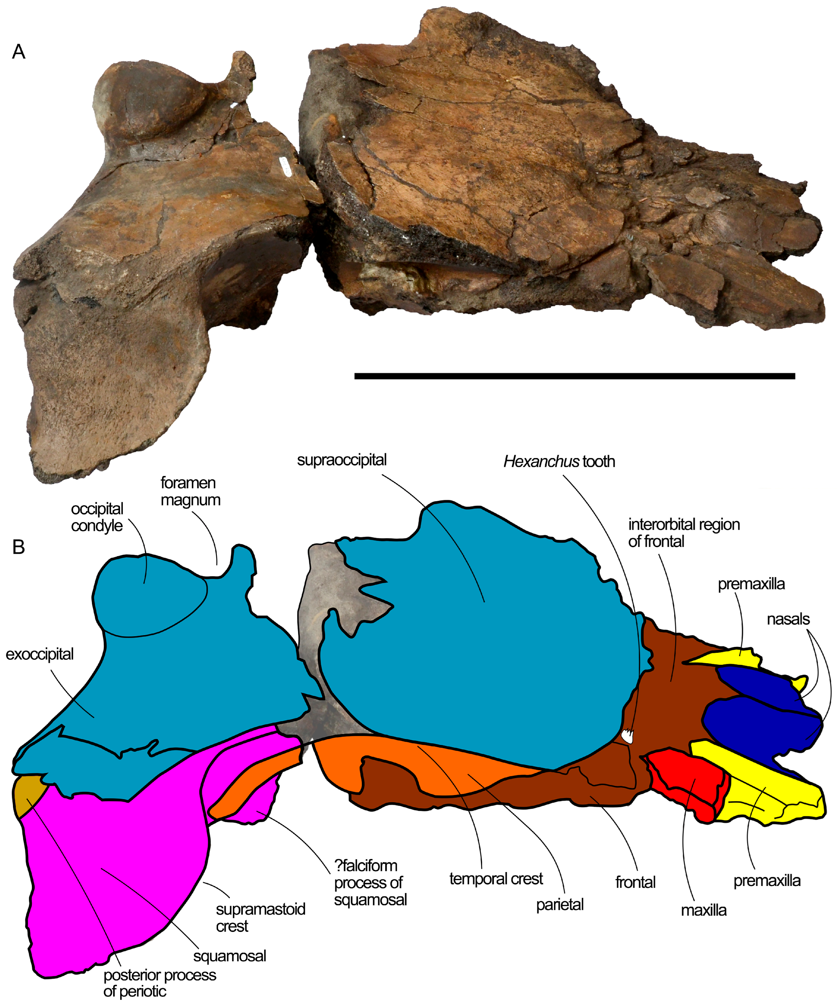
Dorsally on the anterior part of the skull, a lateral tooth of a Hexanchus gigas (Elasmobranchii; Hexanchidae; Pliocene sixgill shark) is squeezed between the right ascending posterior process of the maxilla and the right frontal (Figure 4). The tooth is oriented with the crown up and the root down, and is thus not fixed to the bone through biting. A shark possibly lost the tooth while scavenging on the carcass, as no shark bite marks could be found on any of the skull bones. The tooth has an acrocone and two distal cusps and is intact (L: 17 mm; H: 14 mm). This find is not unusual, as association between Hexanchus and whale carcasses is well known in the fossil record [33,34].
Some fairly heavily decalcified mollusc fragments were present in the surrounding sediment and were uncovered during preparation. In general, mollusc remains are scarce in this level of the Kattendijk Sands (M.Boss. pers. obs.).
4.2. Systematic Paleontology
Mammalia Linnaeus, 1758
Cetacea Brisson, 1762
Pelagiceti Uhen, 2008
Neoceti Fordyce & De Muizon, 2001
Mysticeti Flower, 1864
Chaomysticeti Mitchell, 1989
Balaenomorpha Geisler and Sanders, 2003
Balaenoidea Flower, 1865
Balaenidae Gray, 1821
Quasibalaena new genus
Figure 4.
A tooth of an Hexanchus gigas still adherent to the skull of the holotype of Quasibalaena portisantuerpiae. Scale bar in mm.
4.2.1. Etymology of Genus Name
Quasi is a Latin term for ‘almost’, ‘close to’ and is used here in reference to the close resemblance of the investigated specimen to the bowhead whale, Balaena mysticetus. Balaena is the Latin term for ‘whale’.
4.2.2. Genus Diagnosis
Quasibalaena portisantuerpiae is diagnosed by the simultaneous presence of the following characteristics: (1) wide anterior border of supraoccipital lacking a dome; (2) anterolaterally spreading of the anterior portion of the parietal dorsal to the supraorbital process of the frontal; (3) continuously convex lateral edge of the supraoccipital; (4) squared and laterally protruded exoccipital; (5) supramastoid crest of squamosal projecting laterally and posteriorly making it possible to see the posterior wall of the temporal fossa in lateral view.
This diagnosis shows that Quasibalaena portisantuerpiae shows Balaena-like characters (Nos. 5 and the lack of a supraoccipital dome as from above) and an autapomorphic character (No. 4 as from the above list, which is not observed in any other balaenid with the possible exception of Eubalaena shinshuensis; however, in the latter the preservation of this portion is not optimal and the anterior edge of the exoccipital is not completely known, limiting possible comparisons). The presence of a wide anterior edge of the supraoccipital and the continuously convex supraoccipital edge resemble more Eubalaena australis and Balaenula astensis than Balaena or Balaenella. This observation suggests that the convex supraoccipital edge and shape of the anterior border of the supraoccipital may represent archaic states maintained by Quasibalaena portisantuerpiae after its divergence from the common ancestor of the Balaena + Balaenella + Quasibalaena and the Eubalaena + Balaenula clades. The presence of a wide supraoccipital edge in the stratigraphically older Megabalaena sapporoensis and of convex lateral edges of the supraoccipital in Archeobalaena dosanko reinforce this hypothesis. See chapters 4.4 and 4.5 for further analyses of this point.
4.2.3. Type Species
Quasibalaena portisantuerpiae new species.
Quasibalalena portisantuerpiae new species.
4.2.4. Etymology of Species Name
The composed word portisantuerpiae derives from two Latin terms: portus that means ‘harbor ’, and Antuerpiae, which is from Antuerpia, the Latin name of Antwerpen. The composed word is used here to mean ‘from the Antwerpen harbor’ in reference to the location of the locality of the discovery in the Antwerpen harbor.
4.2.5. Species Diagnosis
As for genus, Quasibalaena portisantuerpiae is the only included species within the genus Quasibalaena.
4.2.6. Holotype
Partial skull with right periotic and right tympanic bulla. The holotype specimen is held by the Natuurmuseum Rotterdam with the accession number NMR 999100007728.
4.2.7. Locality and Stratigraphic Age
The holotype specimen was found in situ a few meters above the basal gravel of an outcrop of the Kattendijk Sand member of the Kattendijk Formation at the Deurganckdok construction site in Antwerpen (Belgium), 23.5 m below sea level. This is the basal-most part of the Kattandijk Sands, below the Palliolum gerardi (Nyst, 1835) level.
4.3. Description
The skull is subdivided into three portions as described in Section 3. These portions can be articulated to reconstruct most of the neurocranium. The left squamosal, right tympanic bulla, right periotic, a large part of the ventral surface of the skull, both supraorbital processes of the frontal and most of the rostrum are not preserved. The right periotic is still in articulation with the skull, but the right tympanic bulla is detached from the skull. The right tympanic bulla was originally still in contact with the right periotic, in its correct anatomical position. It is now detached from the skull in order to prepare the ventral surface of the periotic (this was done by one of us, M. Boss.). The skull is represented in Figure 5 (dorsal view), Figure 6 (left lateral view), Figure 7 (right lateral view), Figure 8 (ventral view), Figure 9 (anterior view) and Figure 10 (posterior view). In Figure 11 a reconstruction of the skull is provided in which the missing left squamosal is replaced by a mirrored copy of the right squamosal to give a more complete representation. Measurements of the skull are provided in Table 1.
Table 1.
Skull measurements (in mm) of the holotype of Quasibalaena portisantuerpiae (NMR 999100007728).
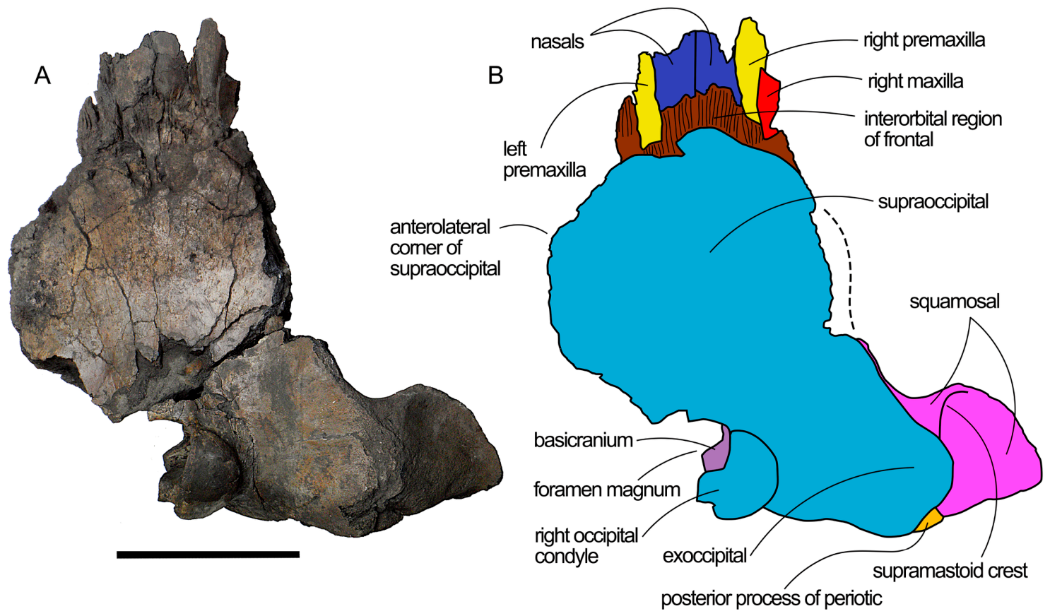
Figure 5.
Dorsal view of the holotype skull of Quasibalaena portisantuerpiae. (A), photographic representation. (B), anatomical interpretation. Scale bar equals 25 cm. The dashed line represents a reconstructed portion of the supraoccipital.
4.3.1. Nasal
The nasal bones are anteriorly incomplete. The dorsal surface is flat, and the lateral borders are parallel to the longitudinal axis of the skull. The posterior borders of the nasals are straight, with the posterolateral corner located more posteriorly than the anteromedial corner. In this way, in dorsal view, the nasofrontal suture shows a pointed shape with a pointed apex located anteriorly along the longitudinal axis of the skull. From an anterior point of view, the nasals form a sort of roof with gently descending dorsal surfaces from the midline to both sides of the narial fossa. Measurements of the nasal bones are provided in Table 1 and show that the nasals are well developed and visible in the dorsal view of this skull.
4.3.2. Premaxilla
Both ascending processes of the premaxillae are present laterally to the nasals. Their posterior ends are located close but more posteriorly to the posterolateral corner of the corresponding nasals. The transverse diameter of both ascending processes of the premaxillae diminishes approaching their posterior ends, but their posterior apices are not preserved. The measurements provided in Table 1 show that the transverse width of the ascending processes of the premaxillae is narrower than that of the nasals.
4.3.3. Maxilla
We can observe only a fragment of the ascending process of the right maxilla that is 91 mm in length and 40 mm in width, and that is developed laterally to the right ascending process of the premaxilla. The external border of the ascending process of the maxilla is concave, and the medial border is straight and projects anteriorly, paralleling the lateral edge of the ascending process of the premaxilla. The fragment of the right ascending process of the maxilla is located on the interorbital region of the frontal but is shorter than the length of this region. For this reason, anteriorly to this maxillary fragment, grooves are observed on the exposed interorbital region of the frontal for the articulation with a part of the maxilla that is now lost.
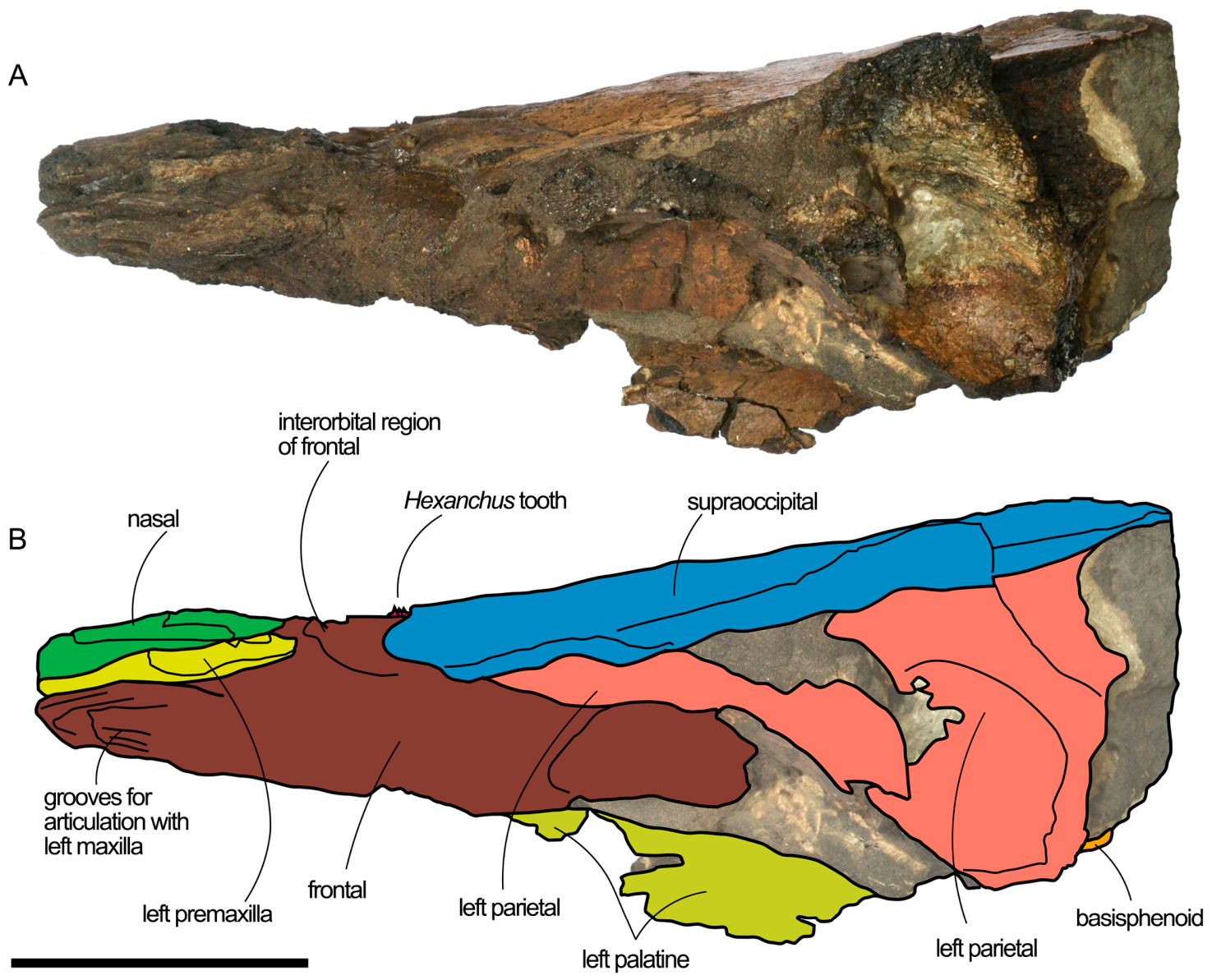
Figure 6.
Left lateral view of the holotype skull of Quasibalaena portisantuerpiae. (A) Photographic representation. (B) Anatomical interpretation. Scale bar equals 15 cm.
4.3.4. Frontal
Only the medial-most part (emergence) of the supraorbital process of the frontal is laterally preserved on both sides of the skull, including the onset of the optic canal, in addition to the interorbital region of the frontal. The latter is fully exposed, as the anterior end of the supraoccipital is missing due to breakage. The shape of the interorbital region of the frontal resembles a short bony strip developed posterolaterally. The maximum anteroposterior length of the interorbital region of the frontal is 31 mm along the midline of the skull but, more laterally, it diminishes to 28 mm. The interfrontal suture is open and is on the same line as the medial borders of the nasals. Posteriorly, the frontal is bordered by the parietal; the frontal-parietal suture is wide and still open.
Figure 7.
Right lateral view of the holotype skull of Quasibalaena portisantuerpiae. (A) Photographic representation. (B) Anatomical interpretation. Scale bar equals 50 cm.
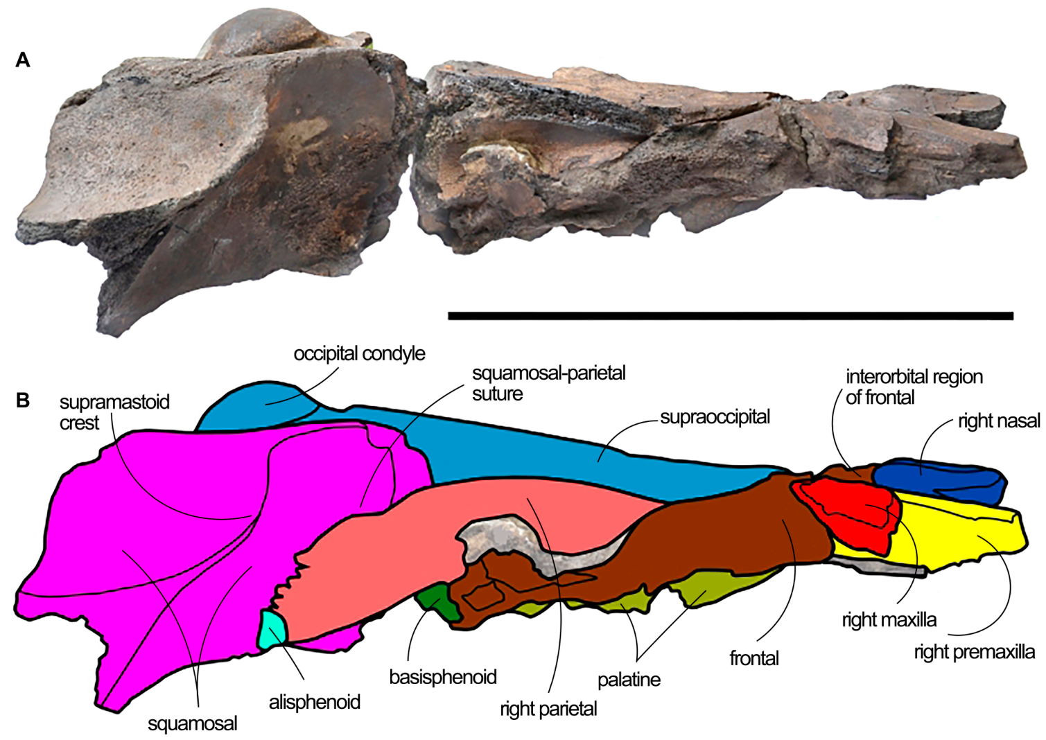
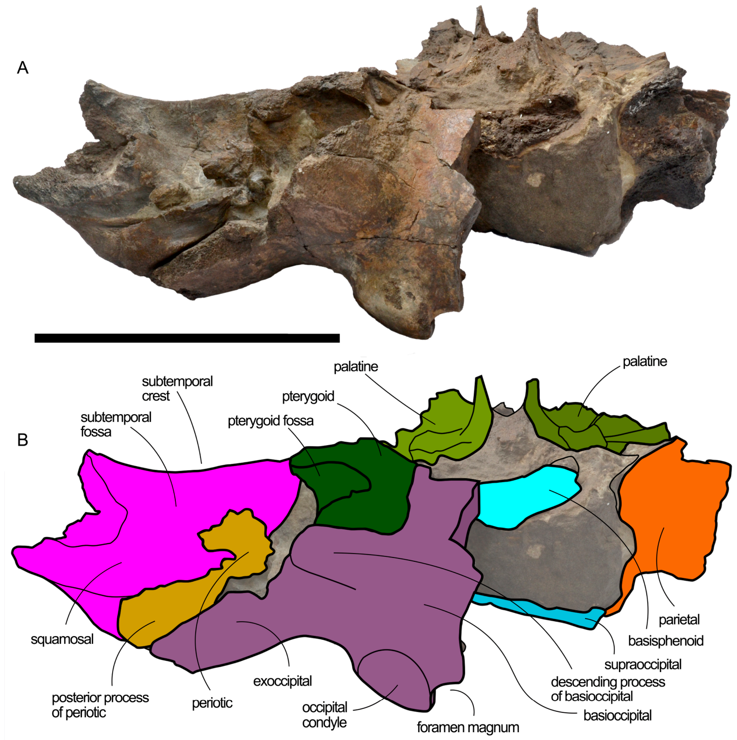
Figure 8.
Ventral view of the holotype skull of Quasibalaena portisantuerpiae. (A) Photographic representation. (B) Anatomical interpretation. Scale bar equals 50 cm. The ? Indicates that the presphenoid is tentatively identified.
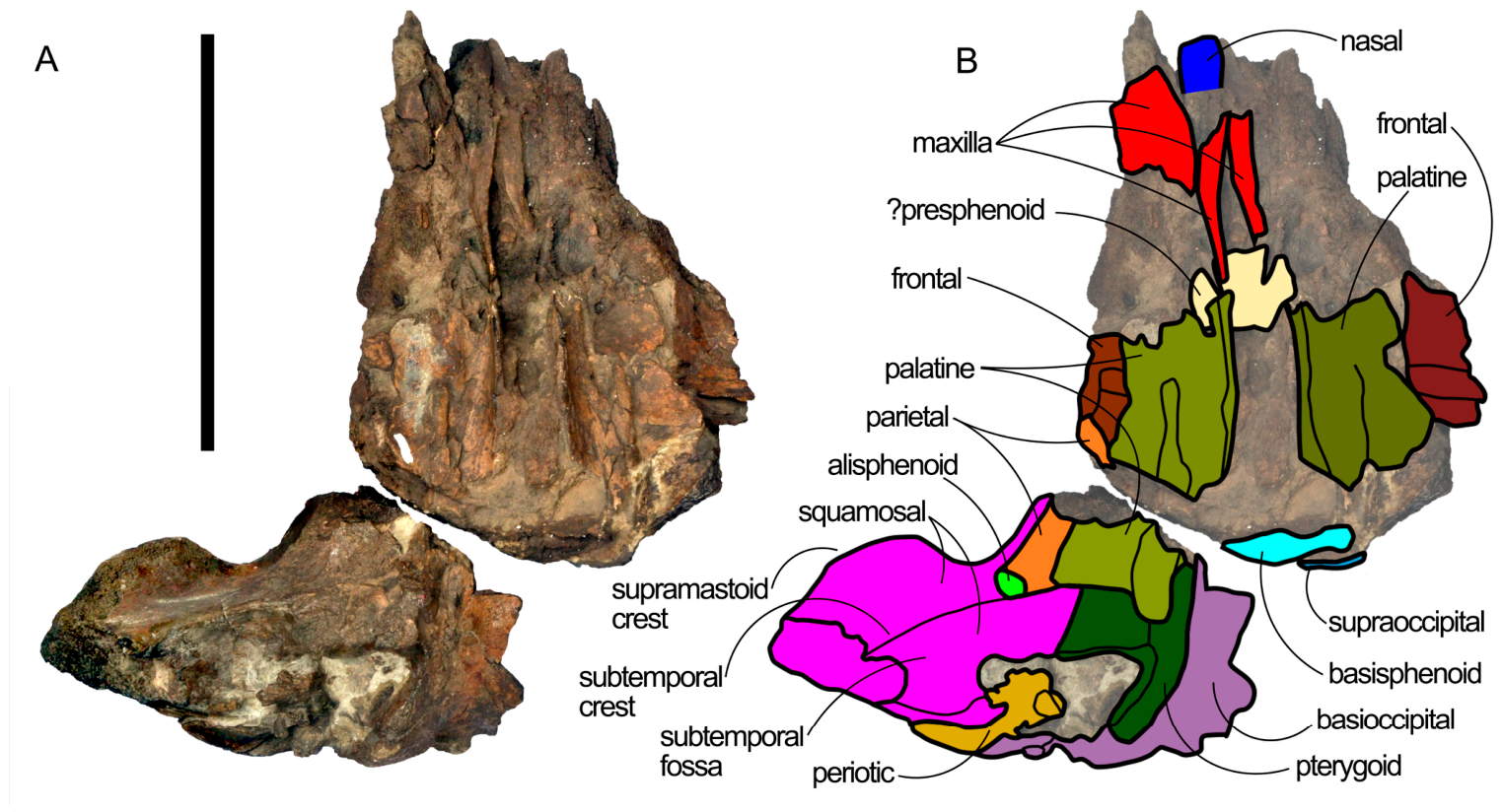
Figure 9.
Anterior and anterodorsal views of the holotype skull of Quasibalaena portisantuerpiae. (A,C) Photographic representations. (B,D) Anatomical interpretations. Scale bar equals 15 cm.
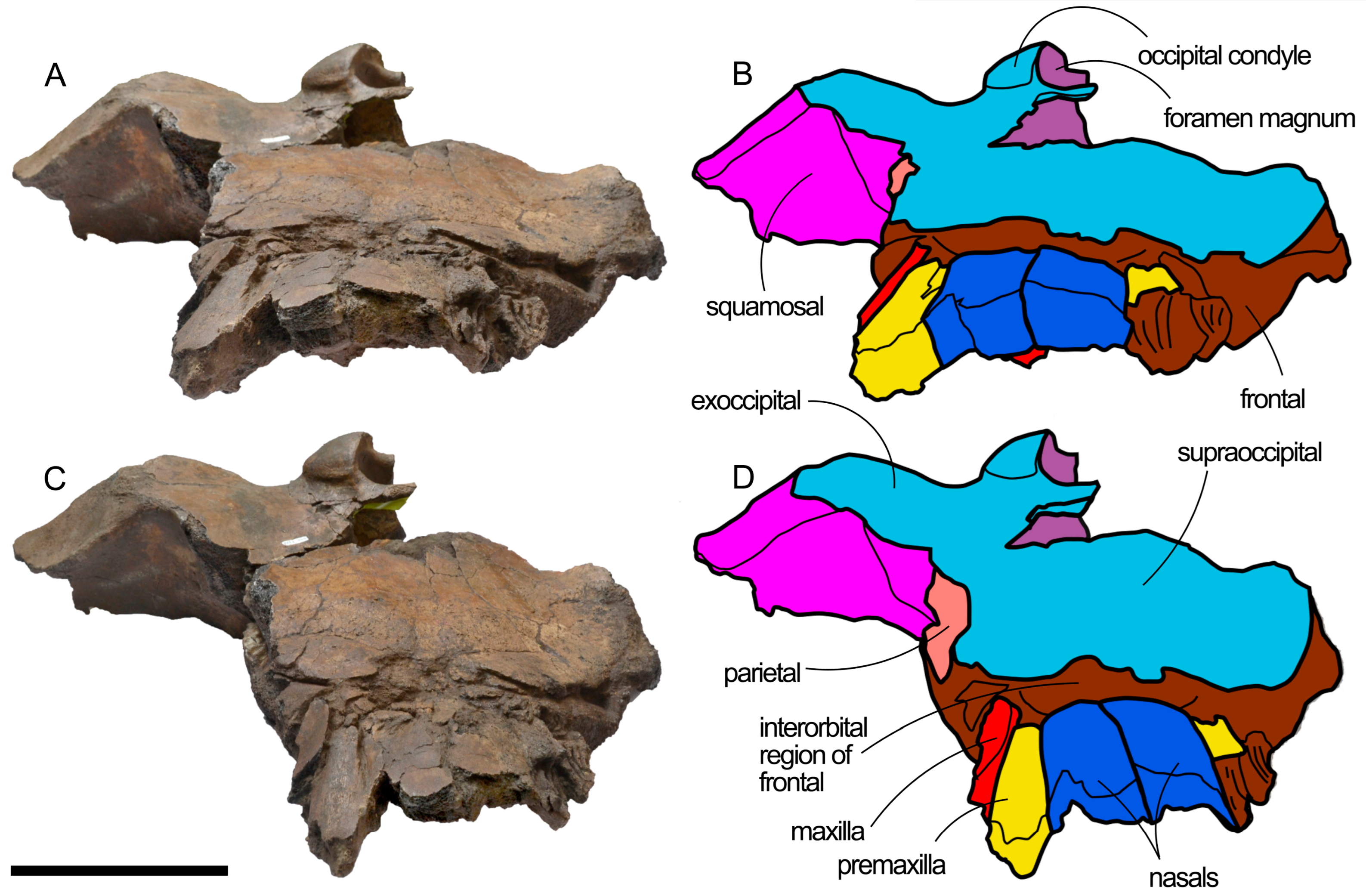
4.3.5. Parietal
Given the different degree of preservation, most of the parietal description is based on the right parietal. The fragmentary parietal is observed posteriorly to the interorbital region of the frontal; the parietal is separated from the frontal by an open frontal-parietal suture whose morphology was probably due to a dorsoventral compression of the skull rather than to a young age of the individual. We do not observe any evidence of an anteroventral corner in the frontal-parietal suture like that seen in Eubalaena and Balaenula; rather, the frontal-parietal suture projects posteroventrally in a straight manner. In lateral view, the parietal is observed along the anterolateral wall of the skull where it is overhung by the supraoccipital and shows an external concavity. The dorsal border of the parietal projects laterally and, together with the lateral border of the supraoccipital, forms the temporal crest. The temporal crest protrudes laterally and makes it impossible to observe the anterior portion of the medial wall of the temporal fossa in dorsal view. The posterior portion of the parietal squama is dorsoventrally elongated and concave. The parietal-squamosal suture is anteriorly concave; its anteroventral end is located at the border between the parietal and the posterior corner of the alisphenoid. More dorsally, the parietal-squamosal suture forms a wide curve with anterior concavity and reaches a relieved area located slightly ventrally to the temporal crest. The relieved area shows both anteroposterior and dorsoventral expansions that can be observed in the lateral view of the skull. The dorsal-most part of the parietal-squamosal suture is lost.
Figure 10.
Posterior view of the holotype skull of Quasibalaena portisantuerpiae. (A) Photographic representations. (B) Anatomical interpretations. Scale bar equals 30 cm. Note that the skull is upside down.
Only a small portion of the left parietal squama is preserved that is located posteriorly to the base of the supraorbital process of the frontal, slightly posterior to the origin of the optic channel. This fragment is concave and is 72 mm in length and 140 mm in width.
4.3.6. Occipital Region
The supraoccipital forms a wide dorsal shield that occupies almost the whole dorsal surface of the skull. The lateral border of the supraoccipital is widely concave medially and anteriorly to the nuchal crest. More anteriorly, it becomes laterally convex, forming a protruding temporal crest that prevents the observation of the medial wall of the temporal fossa from a dorsal point of view. Only a few portions of the temporal crest are preserved (Figure 12), but those portions confirm the lateral protrusion. Approaching the cranial vertex, above the base of the supraorbital process of the frontal, the anterolateral borders of the supraoccipital become rounded and converge anteromedially. Unfortunately, the anterior-most portion of the bone has broken off and is missing, and this prevents the observation of the frontal-supraoccipital interface. However, judging from the grooves on the posterior portion of the supraorbital region of the frontal, we suppose that, anteriorly, the supraoccipital formed a wide and anteriorly convex curve covering the parietal and superimposing on the posterior part of the interorbital region of the frontal.
There is no sagittal crest. There are two reliefs that are elongated paraxially and that project anterolaterally from approximately the middle of the length of the supraoccipital. These two reliefs border a central fossa.
Figure 11.
Digital restoration of the holotype skull of Quasibalaena portisantuerpiae realized through mirroring of the right squamosal on the left portion of the skull. Scale bar equals 50 cm.
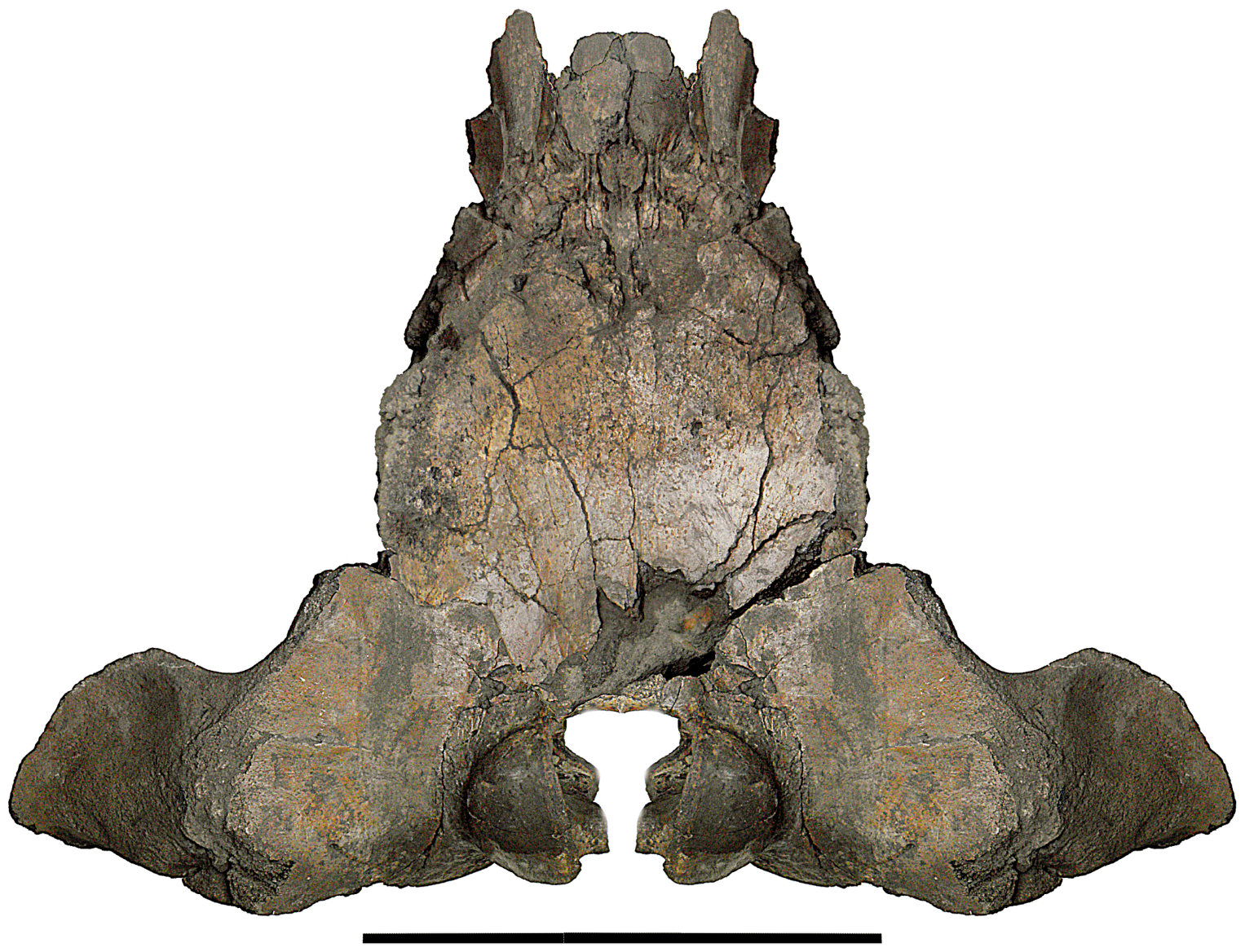
The exoccipital is rectangular and projects laterally and posteriorly from the posterolateral edge of the supraoccipital. Its surface is flat-to-slightly convex and its aspect is robust. The lateral outline of the exoccipital is externally rounded, but part of it is eroded. Unfortunately, the ventral border of the right exoccipital is eroded, and that of the left exoccipital is missing; for this reason, the paroccipital processes are not preserved. In dorsal view, the posterolateral corners of the exoccipitals are located on a transverse line that is more posterior than the occipital condyles.
Figure 12.
Dorsolateral view of the holotype skull of Quasibalaena portisantuerpiae. (A) Photographic representation. (B) Anatomical interpretation. Scale bar equals. 50 cm. The ? Indicates that the falciform process of squamosal is tentatively identified here.
The foramen magnum is transversely compressed and shows a rectangular shape. Only the right occipital condyle is preserved, which is wide and high; the medial border of the occipital condyle is straight; the lateral border is laterally convex; the articular surface is flat-to-slightly convex.
The basioccipital is preserved only on the right side of the skull. It is medially concave and elongated. More laterally, it gives rise to the long and wide descending process of the basioccipital that shows a triangular posterior face. This face is short and very wide and is developed laterally and ventrally with respect to the position of the occipital condyle, showing a crest-like shape. In lateral view, the descending process of the basioccipital is rounded, scarcely inflated. Laterally, its posterior protrusion is less than that of the occipital condyles. Anterolaterally, the descending process of the basioccipital is bordered by the exposure of the posterior process of the periotic slightly posteriorly to the squamosal. The articulation with the pterygoid is straight and proceeds along the whole anterior border of the descending process of the basioccipital.
4.3.7. Vertex
The breakage of the anterior-most border of the supraoccipital makes the reconstruction of the cranial vertex of this specimen a hard task. Observations confirmed by visual inspection include what follows: (1) the presence of a nasofrontal suture that projects anteriorly along the longitudinal axis of the skull that lacks the typical elongation observed in the nasal processes of Balaena and Balaenella; (2) the interorbital region of the frontal is anteroposteriorly reduced to a transverse strip of bone situated between the nasofrontal suture and the inferred anterior border of the supraoccipital; (3) the parietals form very narrow and triangular stripes lateral to the anterolateral borders of the supraoccipital and disappear under the supraoccipital itself. We can thus infer that the anterior border of the supraoccipital covered the whole dorsal exposure of the parietal and prevented the observation of the parietal at the cranial vertex in dorsal view. We also infer that the anterior border of the supraoccipital covered the posterior-most surface of the interorbital region of the frontal, as in all the other balaenid species known up to now.
4.3.8. Alisphenoid
There is a small exposure of the alisphenoid in the temporal fossa (Figure 8). The alisphenoid shows a triangular shape with a posterior apex. It is bordered by the squamosal dorsally, posteriorly and ventrally. Its relationships with the pterygoid are less clear, as it is not completely clear whether a small fragment of palatine was interposed between the ventral border of the alisphenoid and the pterygoid or not.
4.3.9. Squamosal
Only the right squamosal is preserved in this skull (Figure 7). The squamosal is a robust bone that projects ventrally and laterally. The body of the squamosal projects laterally and shows a robust aspect. It projects laterally and posteriorly. There is no bulging of the posterior and medial wall of the temporal fossa (in those parts that belong to the squamosal) into the temporal fossa itself. The border of the subsquamosal crest is anteriorly concave. The falciform process proceeds ventrally to the alisphenoid and makes contact with an ascending portion of the pterygoid. It is likely that the falciform process was also in contact with the palatine as in other balaenids, but this contact is not preserved in the present skull. The squamosal is in contact with the pterygoid within the pterygoid fossa and forms the anterolateral wall of the hiatus cranicus.
In posterior view, the external acoustic meatus is elongated and concave. It shows an elliptical shape and diminishes its dorsoventral diameter distally. The external acoustic meatus is also rather deep (37 mm).
In lateral view, the parietal/squamosal suture is anteriorly concave. It begins its path from the posterior corner of the alisphenoid and proceeds anterodorsally to end within a fracture. It is possible to find its dorsal-most portion in a relieved area located at the contact between the squamosal, the parietal and the supraoccipital. Posteriorly to this relieved area, the squamosal is strongly concave and is overhung by the nuchal crest. The nuchal crest is posteriorly round in dorsal view. The supramastoid crest develops from the lateral portion of the nuchal crest. The supramastoid crest is high and protrudes anterolaterally, showing an anteriorly convex border. Given the observed geometry of the skull, we suggest that the supramastoid crest projected posteroventrally and laterally, like in modern-day Balaena mysticetus and in the extinct Balaena montalionis; in this way, the medial and posterior surfaces of the temporal fossa could be visible in lateral view. The area located posteroventrally to the supramastoid crest is slightly concave.
Posteriorly, the squamosal is convex and forms an area for the attachment of the posterior process of the periotic that is slightly ventral with respect to the squamosal/exoccipital suture.
4.3.10. Ventral View of the Anterior Portion of the Skull
In ventral view, the anterior portion of the skull shows a complex pattern characterized by different fragments representing only incomplete anatomical parts (Figure 8). Anteriorly, the skull is slightly crushed along the dorsoventral axis and most of the bones are fractured and moved from their original position. Erosion is also observed in several bone fragments. Due to these post mortem processes, the reconstruction of the ventral surface of this skull is difficult.
We observed part of the vomer in an anterior fragment that is broken and crushed at several points. Anteriorly, the vomer is broken and only the posterior portion is preserved.
Two fragments are attributed to the mesethmoid; these fragments are observed laterally to the vomer at about half of the length of the block, including the anterior portion of the skull. These mesethmoid elements bear structures resembling olfactory foramens and turbinate-like crests. Laterally to the vomer, parts of the palatine bones are observed that show a flat surface and broadly rectangular shape. Laterally, the palatine is in contact with the frontal; posteriorly, it contacts the pterygoid, and posterolaterally it contacts the parietal.
On the right side of the block, the optical channel opens dorsally to the origins of the choanae. The optical channel is 42 mm in dorsoventral diameter and 37 mm in anteroposterior diameter. The left optic channel is visible but crushed near a vomer fragment.
4.3.11. Pterygoid
Only the right pterygoid is preserved in the holotype skull. The pterygoid fossa is wide and deep and shows a concave anterior wall. The roof of the pterygoid fossa is 57 mm in length and shows a posterior border projecting anterolaterally. The maximum transverse diameter of the pterygoid fossa is 70 mm. The medial part of the pterygoid is robust and thick. The pterygoid is articulated posteriorly with the descending process of the basioccipital through a straight suture.
4.3.12. Hiatus Cranicus
The hiatus cranicus is a bilateral opening observed in ventral view that is located between the pterygoid, squamosal, basioccipital, and periotic. In the present skull, it is hard to describe because some matrix still obscures some aspects of it. We can observe what follows: (1) the outline of the hiatus cranicus is irregularly shaped; the lateral border is externally convex and is formed by the squamosal; the anterior border is posteriorly concave and is formed by the squamosal in its lateral-most portion and by the roof of the pterygoid fossa in its medial-most portion; the medial border is formed by the pterygoid in its anterior-most portion and by the descending process of the basioccipital in its posterior-most portion; the posterior border is entirely formed by the exoccipital. The periotic protrudes in the hiatus cranicus at the level of its posterolateral corner; more anteriorly, the external acoustic meatus departs from the lateral border of the hiatus cranicus, slightly anteriorly to the periotic. The maximum anteroposterior diameter of the hiatus cranicus is 72 mm and its maximum transverse diameter is 90 mm.
A small bony fragment is located close to the apex of the flange on the lateral tuberosity of the periotic. It does not show characters suggesting it was part of the same flange, and we consider it an indeterminate fragment.
4.3.13. Periotic
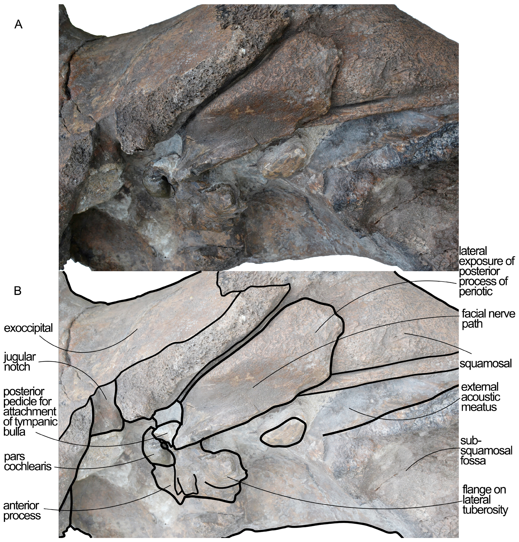
The right periotic is articulated with the skull and shows a long posterior process (Figure 13). Measurements of this periotic are provided in Table 2. The ventral surface of the posterior process of the periotic is distally flat but medially concave, showing a short and wide facial sulcus. The facial sulcus is bordered by a high relief located along the anterior border of the medial portion of the posterior process; in that relief, the posterior pedicle for the tympanic bulla is located. The posterior process of the periotic is exposed in the lateral wall of the skull, showing a triangular outline (Figure 14); as seen from the lateral view, the posterior process is high, robust and its exposure is bordered by the external acoustic meatus and the exoccipital. The posterior process of the periotic is joined to the central portion of the periotic by a wide and uniformly concave portion.
Table 2.
Measurements (in mm) of the right periotic of the holotype of Quasibalaena portisantuerpiae (NMR 999100007728).
The anterior process of the periotic is squared in ventral view and exhibits a robust aspect. The flange of the ventrolateral tuberosity is robust, projects posteriorly forming an acute angle with the posterior process of the periotic. The flange of the ventrolateral tuberosity is vaguely triangular with a broken apex. The ventral surface of both the anterior process and the flange of the ventrolateral tuberosity is rough and appears as pachyosteoschlerotic. Approaching the pars cochlearis, the anterior process increases its dorsoventral diameter and robustness. The anterior pedicle for the tympanic bulla located ventrally to the anterior process is rather large and high, suggesting that the hiatus epitympanicus was wide.
The pars cochlearis is transversely short and ventrally round. It is anteriorly bordered by an evident tensor tympani groove. The round window is wide and vaguely rounded. Observing the medial surface of the pars cochlearis is a difficult task given that the periotic is still articulated with the skull (Figure 15). What we observed includes the presence of a round internal acoustic meatus separated from the endocranial opening of the facial canal by a narrow crista transversa; the ventral border of the internal acoustic meatus is crest-like; the suprameatal area appears complex, showing an anteroposterior groove located dorsally to the internal acoustic meatus; an additional groove runs parallel to this one.
Figure 13.
Periotic of Quasibalaena portisantuerpiae in ventral view. (A) Photographic representation. (B) Anatomical interpretation. Note the shallow, wide and nearly flat path for the facial nerve on the ventral surface of the posterior process. Note also the broadly triangular ventrolateral flange of the anterior process of the periotic. Scale bar equals 5 cm.
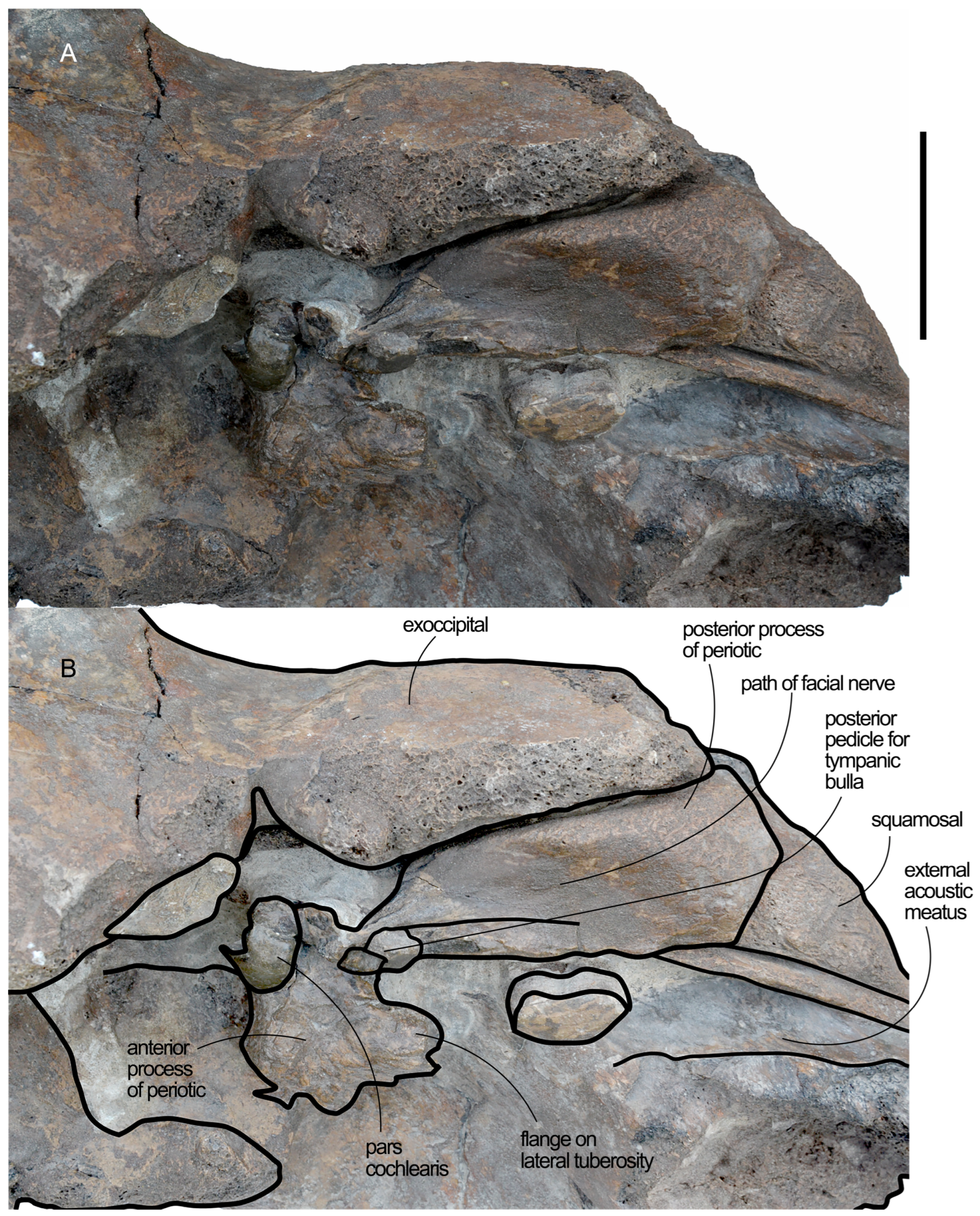
Figure 14.
Periotic of Quasibalaena portisantuerpiae in ventrolateral view. (A) Photographic representation. (B) Anatomical interpretation. Note the lateral exposure of the posterior process of the periotic between exoccipital and squamosal. Not to scale.
4.3.14. Tympanic Bulla
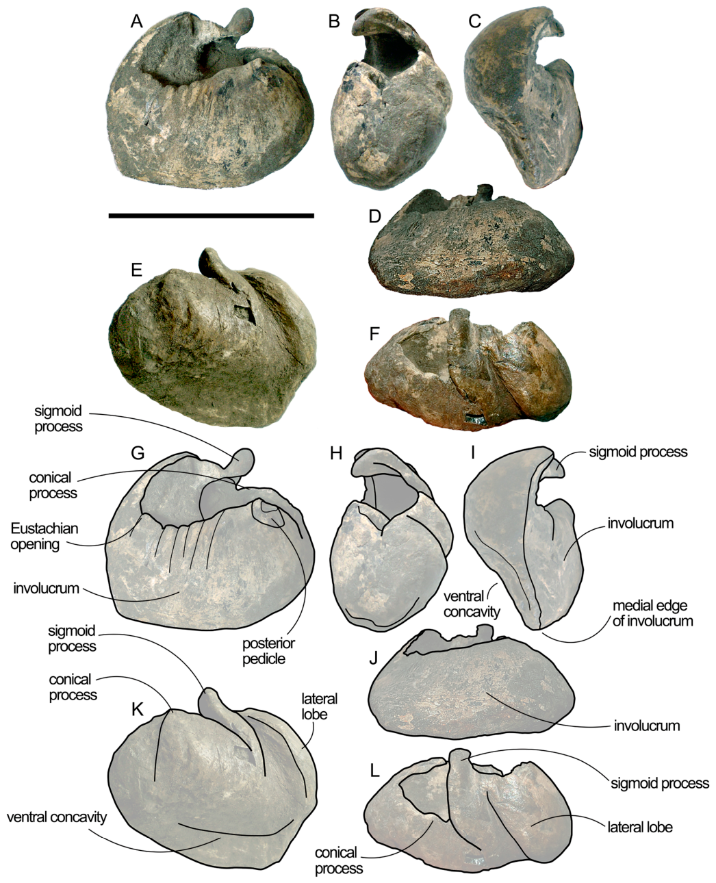
As a general observation, the bulla shows a robust aspect and is quite heavy (Table 3). The ventral surface of this bulla is concave (Figure 16). The main ridge is located along the medial border of the ventral surface. The involucrum is massive and transversely convex. The involucral ridge is very well developed and evident. The dorsomedial border of the bulla is short (45 mm in length) and shows an Eustachian opening that is wide and uniformly descending anteriorly. The posterior pedicle for the periotic is located posteriorly to the posterodorsal border of the involucrum. The posterior face of the bulla is flat and shows a laterodorsal-to-medioventral orientation. The anteromedial corner between the anterior and the medial border of the bulla is about 90°. The anterior border is uniformly convex and shows a lateral wall projecting posterodorsally.
Figure 15.
Periotic of Quasibalaena portisantuerpiae in ventromedial view. (A) Photographic representation. (B) Anatomical interpretation. Note the round pars cochlearis with remnants of endocranial foramina slightly visible. Note also the straight profile of the posterior process of the periotic marking a wide and shallow path for the facial nerve. The posterior process is exposed on the lateral surface of the skull. Scale bar equals 5 cm.
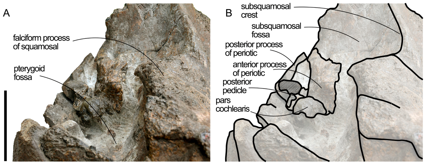
Table 3.
Measurements (in mm) of the right tympanic bulla of the holotype of Quasibalaena portisantuerpiae (NMR 999100007728).
In lateral view, the lateral lobe is present and short. The groove separating the lateral lobe from the sigmoid process is deep. The flat main surface of the sigmoid process is developed at a right angle with respect to the longitudinal axis of the bulla; its posterior surface is highly concave. The groove separating the sigmoid and the conical processes is shallow.
The conical process is not evident in lateral view and shows a flat dorsal border, as usual in Balaenidae and Neobalaenidae.
4.4. Comparisons
4.4.1. Nasals
In Balaenidae, the frontonasal suture is located at the anterior border of the interorbital region of the frontal. This suture shows different morphologies in different species. In the extant Balaena mysticetus, the frontonasal suture is anteriorly prolonged and forms two nasal processes that are long and anteriorly convex [35,36]. The nasal processes show a triangular shape in Balaenella brachyrhynus [36]; in that paper, these long processes were considered to represent true nasals. Long nasal processes are absent in Balaena montalionis [37]. In the right whales of the genus Eubalaena, there is some variation in the shape of the frontonasal suture. Capellini [38], Holder [39] and True [40] described long and anteriorly convex nasal processes in Eubalaena glacialis, with the Long Island specimen figured out by True resembling the frontonasal suture morphology of Balaenella brachyrhynus. Omura [41] described a triangular nasal process in the Kirittapu specimen of E. japonica but, later, Omura et al. [42] showed that in other specimens (61A and 61B mentioned in the article) the nasal process is short, and the frontonasal suture shows a convex edge at the base of each nasal. This same pattern is observed in the extant Eubalaena australis (Bisconti pers. obs. in specimen RGM 24757 in Naturalis Biodiversity Center, Leiden, The Netherlands). No nasal process is observed in the Pliocene Balaenula balaenopsis and Balaenula astensis [37,43], and, given the poor preservation of the relevant region, it is not possible to obtain a morphological account of the nasal process in Megabalaena sapporoensis [15].
Figure 16.
Right tympanic bulla of Quasibalaena portisantuerpiae. (A–F) Photographic representations. (G–L) Anatomical interpretations. (A,G) Dorsal view. (B,H) Posterior view. (C,I) Anterior view. (D,J) Medial view. (E,K) Ventral view. (F,L) Dorsolateral view. Scale bar equals 10 cm.
4.4.2. Parietal
The frontal-parietal suture shows the main differences among balaenid species. In the present specimen, Balaena mysticetus, Balaenella brachyrhynus, Balaena montalionis, Balaena ricei, Archaeobalaena dosanko, Eubalaena shinshuensis and Megabalaena sapporoensis, the anteroventral portion of the frontal-parietal suture is straight-to-slightly convex and projects posteriorly and ventrally (see Figure 2 of Ref. [3]). In Eubalaena glacialis, E. australis, E. japonica, E. ianitrix and Balaenula astensis, the anteroventral portion of the frontal-parietal suture forms an evident corner in lateral view that allows the parietal to increase its dorsoventral height dorsally to the supraorbital process of the frontal.
The degree of development of the eminence located along the dorsal portion of the parietal-squamosal suture is different in different balaenid species. The eminence is well developed in the present specimen, Balaena mysticetus, Balaena montalionis and Balaenella brachyrhynus, but it is scarcely developed-to-absent in Eubalaena, Balaenula and Megabalaena sapporoensis.
4.4.3. Supraoccipital
The occipital region, and especially the supraoccipital shield, shows several characters that have been used for discriminating between balaenid genera and species (Figure 17). Probably the most evident feature of the supraoccipital in Balaenidae is the dichotomous shape of this bone in the extant Balaena and Eubalaena. Bisconti [38,44] and Bisconti et al. [3] showed that in Balaena the supraoccipital shows lateral borders converging anteriorly toward a transversely narrow anterior border that may be squared or rounded; in Eubalaena, the lateral borders of the supraoccipital are mainly parallel to the longitudinal axis of the skull and the anterior border is comparatively wider than that of Balaena. Moreover, in Eubalaena the anterior surface of the supraoccipital bears a dome-like bulging that may be longitudinally crossed by a low sagittal eminence resembling a sagittal crest. This morphological pattern is absent from the supraoccipital of Balaena. The only exception to this general pattern is shown by Eubalaena australis, where the lateral borders of the supraoccipital are more converging anteriorly and the anterior edge of this bone is wide and round.
Fossil species show supraoccipital morphologies that broadly fit into this dichotomic arrangement even though differences can be demonstrated. For instance, in Balaena montalionis and Balaenella brachyrhynus, the anterior border of the supraoccipital is more squared and transversely narrow than Balaena, even though their supraoccipitals share with Balaena mysticetus anteriorly converging lateral borders of the supraoccipital and the lack of the anterior dome-like structure. Balaena ricei is probably more like the extant Balaena mysticetus in having converging lateral borders and a transversely narrow anterior edge of the supraoccipital [4]. In Balaenula astensis and Balaenula balaenopsis the lateral borders of the supraoccipital are mostly parallel to the longitudinal axis of the skull and the anterior edge is wide and round; a sort of dorsal eminence resembling the Eubalaena dome is present in Balaenula astensis. In a sense, the morphology of the supraoccipital observed in Balaenula astensis is more consistent with that of Eubalaena australis than with the other Eubalaena species.
In the fossil Eubalaena janitrix and E. shinshuensis, the supraoccipital is largely consistent with that of extant Eubalaena species [44,45]; the same observation can be made on the supraoccipital of the Late Pliocene Eubalaena from Tuscany described in Ref. [46]. Megabalaena sapporoensis is similar to extant Eubalaena glacialis and E. japonica in that it shows a wide and almost straight anterior edge of the supraoccipital and lateral borders developed parallel to the longitudinal axis of the skull [15]. In this species, however, the dorsal dome of the supraoccipital is absent, and the lateral borders are concave rather than straight as in more recent Eubalaena species. Archeobalaena dosanko and Morenocetus parvus show supraoccipitals like that of Balaenula astensis with slightly converging lateral borders and a round anterior edge [9,14].
Figure 17.
Comparative plate showing the shape of the supraoccipital and exoccipital in different extant and extinct balaenid species. Black arrowhead indicates the anterolateral corner observed in Balaena and Balaenella. White arrowhead indicates the bulge of the supraoccipital edge at the parietal-squamosal sutural interface. Not to scale. The dashed lines indicate reconstructed portions and the letter a indicates the narrow anterior edge of the supraoc-cipital in Balaena montalionis and Balaenella brachyrhynus.
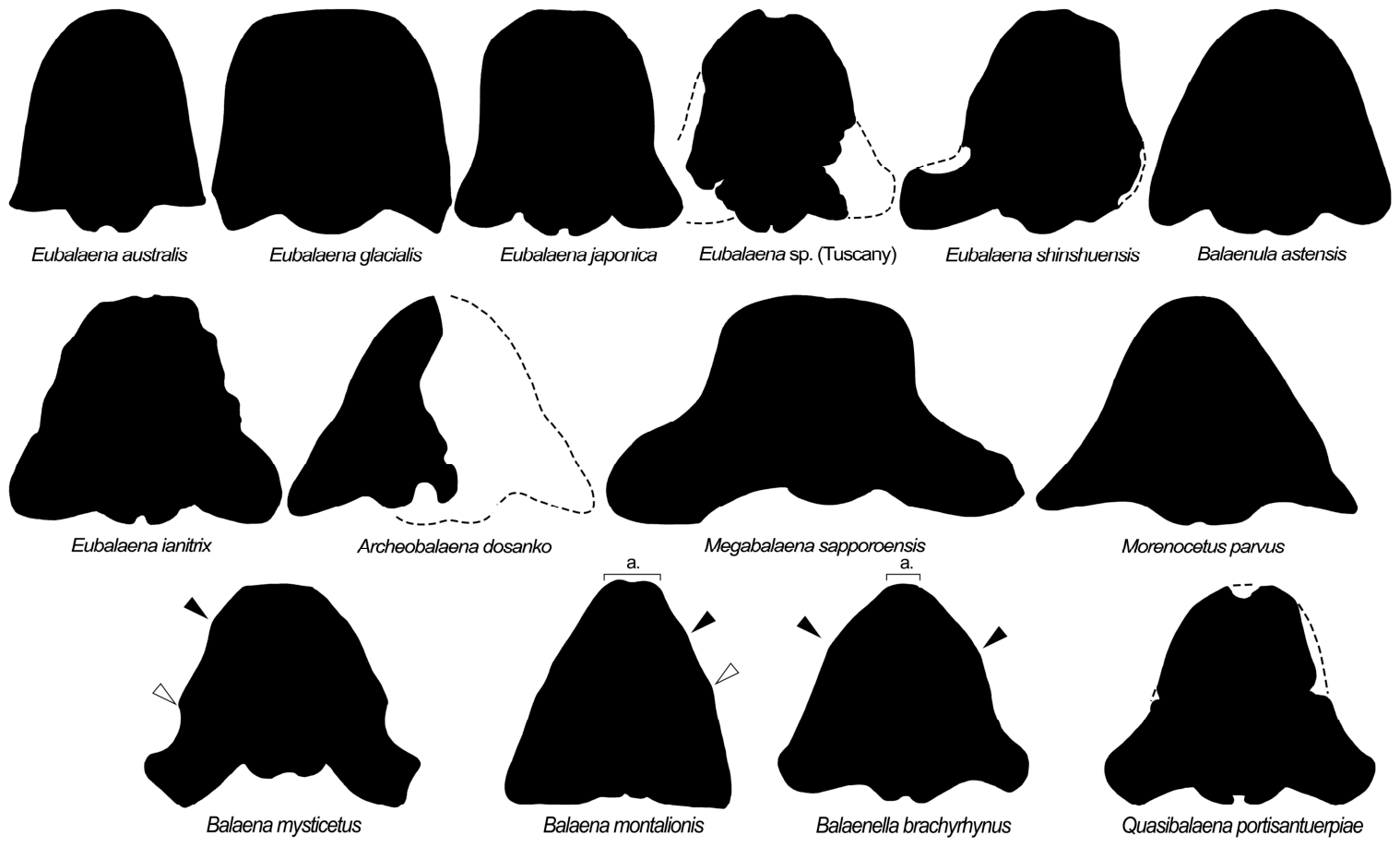
In Quasibalaena portisantuerpiae, the lateral borders of the supraoccipital are straight-to-slightly convex and are developed parallel to the longitudinal axis of the skull up to the basis of the supraorbital processes of the frontal; at that level, the external edges of the supraoccipital form a corner and converge to the midline. It is unclear whether the anterior-most edge of the supraoccipital had a shape similar to that of Balaena mysticetus or Balaena montalionis because the relevant portion is absent. A low dome-like structure is observed in the anterior surface of the supraoccipital of Quasibalaena portisantuerpiae that is crossed by a longitudinal and narrow fossa (resembling that of Eubalaena glacialis; Bisconti pers. obs. in specimen No. 264 on exposition at the Museo di Storia Naturale e del Territorio, Università di Pisa).
4.4.4. Squamosal
In Balaenidae, the lateral portion of the squamosal projects ventrally and shows a strongly reduced zygomatic process. Differences between the extant species have been listed in several papers (e.g., Refs. [3,5,37,47] and consist of different orientations of the zygomatic process of the squamosal (lateral in Balaena mysticetus and anterolateral in Eubalaena) and in different orientations of the whole lateral portion of the squamosal with respect to the inclination of the supraoccipital in lateral view. Actually, in Balaena mysticetus the supraoccipital is more vertical than in the extant Eubalaena species (see also Ref. [9]) and the lateral portion of the squamosal projects posteroventrally and forms a widely obtuse angle with respect to the dorsal supraoccipital surface; in Eubalaena the lateral portion of the squamosal projects ventrally at an almost right angle with respect to the dorsal supraoccipital surface. The same pattern is observed in Eubalaena janitrix, E. shinshuensis, in the Eubalaena sp. from Tuscany and in Megabalaena sapporoensis.
In the living Eubalaena species, E. janitrix, Eubalaena sp. from Tuscany, the zygomatic process of the squamosal projects anteriorly and prevents the view of the posterior wall of the temporal fossa from a lateral viewpoint. This pattern is not present in Balaena mysticetus, in which the zygomatic process projects laterally and allows the view of the posterior wall of the temporal fossa from a lateral viewpoint. In Balaena mysticetus and Balaenella brachyrhynus, the posterior wall of the temporal fossa can be observed in lateral view, showing a morphological pattern consistent with that seen in B. mysticetus.
Balaenula astensis and, as far as preservation allows observation, Balaenula balaenopsis, the lateral portion of the squamosal projects anteroventrally at an almost right angle with respect to the dorsal supraoccipital surface. The consequence of this skull geometry is that the craniomandibular joint is located slightly ventrally to the orbit, visibly more anteriorly than in Eubalaena, Balaena and Megabalaena.
4.4.5. Periotic
The resemblance of the periotic of Quasibalaena portisantuerpiae and of Balaena mysticetus is striking. The posterior process of the periotic in both these species is ventrally elongated with parallel borders and shows a wide and narrow facial sulcus that is scarcely defined by protruding edges of the medial portion of the posterior process itself. In Eubalaena glacialis, the posterior process of the periotic shows a complex shape characterized by pachyosteoschlerosis and irregular borders. In E. australis, the posterior process of the periotic is more inflated and the facial sulcus is more well defined by long protrusions along the medial and lateral borders of the posterior process itself. The overall shape of the posterior process of the periotic is shared with Archeobalaena dosanko and Balaenula astensis but not with Antwerpibalaena liberatlas, in which the posterior process shows a distinctive crest running along the ventromedial border and a more Eubalaena-like morphology of this portion.
The anterior process of the periotic of Quasibalaena portisantuerpiae is irregularly shaped and comparatively longer than that of Balaena mysticetus and Eubalaena. The relative length of the anterior process of the periotic of Q. portisantuerpiae is shared with Balaenella brachyrhynus. The irregular anterior border of the anterior process of Q. portisantuerpiae is not shared with any other of the accepted extant and fossil balaenid species. Even in Balaenotus insignis, a taxon in critical need of revision, the anterior process shows a linear anterior border with a well defined flange of the ventrolateral tuberosity, therefore differing from Quasibalaena portisantuerpiae.
Another obvious difference between the periotic of Q. portisantuerpiae and that of Eubalaena spp. is the presence of a transversely short pars cochlearis that is broadly rounded in ventral view. In Eubalaena, the pars cochlearis is transversely elongated and the morphology of the endocranial foramens is clearly different from that shown by all the other balaenids known up to now because this elongation process occurred during the evolution of this genus.
4.5. Phylogenetic Analysis
The phylogenetic analysis resulted in two most parsimonious cladograms whose strict consensus (Nelsen) is shown in Figure 18. The cladograms are 277 steps in length, and their CI is 0.560, RI is 0.845, and HI is 0.440. In the strict consensus tree, the monophyly of Balaenidae is supported by a bootstrap value (hereinafter, BV) of 60. Within Balaenidae, the genera Eubalaena and Balaenula are monophyletic (supported by BVs of 55 and 91, respectively). The clade including Eubalaena + Balaenula is the sister group of a clade including Balaena + Balaenella + Quasibalaena. In the latter, a monophyletic group including Balaena montalionis and Balaenella brachyrhynus was discovered that is the sister group of Balaena ricei. This large group is the sister group of a clade including Balaena mysticetus and Quasibalaena portisantuerpiae. The morphological support to these nodes was low, as BVs are lower than 50 for all the rami. The sister-group relationship of Quasibalaena portisantuerpiae and Balaena mysticetus represents an important point that will be analyzed in Section 5. Archaeobalaena dosanko and Megabalaena sapporoensis are subsequent sister groups of the Balaena + Balaenella + Quasibalaena clade. Our results confirm that Antwerpibalaena liberatlas [48] and Charadrobalaena valentinae [49] represent early diverging balaenid species not included in the clades described above.
Figure 18.
Phylogenetic analysis of Balaenidae plotted against a time scale based on the strict consensus tree. Major mysticete clades are shown in boldface. Numbers indicate bootstrap support values. Thick lines indicate the known stratigraphic ranges of operational taxonomic units. The black squares represent named clades.
In this analysis, Morenocetus parvus is the sister group of Balaenidae and can be considered a stem-balaenid taxon. This result differs from the previously published phylogenetic hypothesis published by Bisconti et al. [3], in which it was a stem-balaenoid. Presently, the horizontal and flat supraorbital processes of the frontal and the elongated interorbital region of the frontal suggest caution about inclusion of Morenocetus parvus within Balaenidae; therefore, we prefer to consider this species as a stem-balaenoid. However, our results are not in contrast with the inclusion of Morenocetus within Balaenidae.
Caperea marginata and Miocaperea pulchra are monophyletic (BV = 99) and form a clade that can be named with the traditionally recognized name Neobalaenidae. Neobalaenidae is the sister group of Balaenidae, and both form the monophyletic superfamily Balaenoidea.
Peripolocetus vexillifer is a stem-balaenoid taxon (BV = 53). The clade including Peripolocetus, Balaenoidea, their common ancestor and all the descendants of this ancestor is the sister group of Thalassotherii that comprises Balaenopteridae, Eschrichtiidae, Cetotheriidae and basal thalassotherian taxa (BV = 96). These two large clades correspond to Balaenomorpha. Horopeta umarere, Sitsqwayk cornishorum and Eomysticetidae are subsequent sister groups of Balaenomorpha and are included within Chaeomysticeti. Toothed mysticetes (comprehending Aetiocetidae and Mammalodontidae) represent the earliest-diverging rami of the Mysticeti group.
4.6. Diversity of Balaenidae Through Time
Clearly diagnosizable balaenid taxa are all from the Pliocene except for two Late Miocene species from Japan, i.e., Megabalaena sapporoensis [15] and Eubalaena shinshuensis [45]. The position of these two taxa in the strict consensus tree of Figure 18 and the phylogenetic placement of Charadrobalaena valentinae and Antwerpibalaena liberatlas implies a radiation of balaenid clades in the Late Miocene (Figure 19) that is not supported by the fossil record, suggesting that several ghost lineages have still to be discovered. Apparently, the Early Pliocene is the time at which the number of balaenid clades begins to decline, a path further observed in the Piacenzian and in the Quaternary, even though the maximum number of described balaenid species is from the Zanclean. Based on the present analysis, we expect that the fossil record analyzed up to now represents only a fraction of the true balaenid diversity of the Neogene.
Figure 19.
Diversity of Balaenidae through time. Red circles represent known (formally established) species in a given stratigraphic interval. Rectangles represent the inferred clade numbers based on the phylogenetic results of Figure 16.
4.7. Evolution of Body Size Within Balaenidae
We inferred the total length of the holotype of Quasibalaena portisantuerpiae based on the occipital breadth; the resulting total body length was 7.413 m. This information was used to infer the length of the skeleton (resulting in 5.01 m) and the body mass, which was 2.951 tons (c. 3 t). We included these data in the database used to represent the changes in body size of Balaenidae through time (Table 4) that was visualized in Figure 20.
Table 4.
Database used to visualize the changes in body size of Balaenidae species through time. The stratigraphic ages of the first column are calculated as the mean value obtained from stratigraphic intervals of occurrence provided by the Paleobiology Database, with the exceptions discussed in the text. Total lengths, skeleton lengths and body masses are from Refs. [3,15] and the present work (for Quasibalaena portisantuerpiae). Total and skeleton lengths are in meters (m) and body mass in tons (t).
Figure 20A shows the diversity of the skeletal lengths; Figure 20B illustrates the diversity of total body lengths; Figure 20C represents the diversity of body masses. Regression equations and corresponding R2 values are presented that show that logarithmic functions are those that can be more effectively used to represent the data. In any case, the R2 values are rather low (<0.6), suggesting that the data are still too sparse to allow a full understanding of the evolution of body size in Balaenidae.
Figure 20.
Changes in body size during balaenid evolution. (A) Inferred and measured (for extant species) skeletal length plotted against time. (B) Inferred total length vs. time. (C) Body mass vs. time. Regression equations are provided in the corresponding boxes. The dashed lines represent the calculated regression lines.
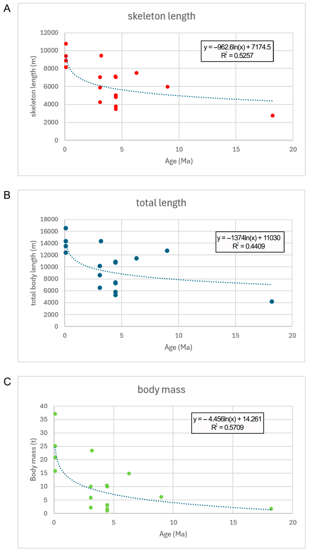
What we observe is a general increase in body size through time, with large-sized species (sensu Ref. [3]: total body length included between 14 and 20 m) occurring as early as 3+ Ma even though the upper limit of the middle-sized values is observed starting from about 9 Ma with Megabalaena sapporoensis. Interestingly, small-sized balaenid species are present throughout the Early Pliocene but disappear more recently, probably starting from the Pleistocene or the Late Pliocene when only large-to-gigantic species are observed. The reconstruction of body sizes at ancestral nodes resulted in the plot shown in Figure 21. The maximum likelihood algorithm inferred an ancestral medium size for the sister group to Morenocetus (which includes Antwerpibalaena, Eubalaena, their common ancestor, and all the descendants of this ancestor). The medium size was the more likely ancestral body size for most of the balaenid clades, suggesting that Balaenula was the result of a miniaturization process. However, the body size of Balaenula must be taken with caution because all the specimens used in the present analyses were juvenile and the final body size could be bigger in this species. Balaena mysticetus is the only large-to-gigantic species of the Balaena-related clade; in this clade, all the species are small-sized with the exception of Balaena ricei. The peculiar distribution of the small sizes among balaenids suggests that the ancestor of the Balaena-related clade derived from a miniaturization process independent of that led to the origin of Balaenula, and that the independent origins of large and gigantic sizes observed in Balaena ricei and Balaena mysticetus represented the outputs of independent size increase processes. The small size observed in the Pliocene Balaenula, Balaenella, Balaena montalionis and Quasibalaena portisantuerpiae suggests a possible link with the establishment of a warm climatic regime in the earliest Pliocene. We will discuss this point in Section 5.
Figure 21.
Ancestral state reconstruction of body size throughout balaenid phylogeny. Circles represent maximum likelihood probability distributions.
5. Discussion
5.1. Paleoecology and Diversity of Right and Bowhead Whales
As usual, each new fossil brings new information about past life and may determine large-scale changes in the way we interpret the evolutionary processes [50]. The case of the right and bowhead whales is not different from this common view, as in the last c. 30 years several new balaenid species have been described and old species have been re-interpreted, forcing the scientific community to re-assess previously accepted phylogenetic relationships and even high-rank systematics. Hot debates have developed around alternative phylogenetic hypotheses that fueled new research and re-analysis of the morphology and genetics of these endangered and iconic cetaceans.
Our work adds another piece to the puzzle of the evolutionary history of right and bowhead whales thanks to the discovery of a partial balaenid skull at the Deurganckdock construction site in Antwerpen, Belgium. This site has proven to be highly valuable in terms of paleontological richness, as the holotype of Fragilicetus velponi was found there [18] together with a large balaenopterid skeleton, still under preparation, and a vast amount of microfossils, molluscs, brachiopods and shark teeth documenting a highly diverse marine life in the earliest Pliocene of the North Sea basin (e.g., Ref. [51]).
The Miocene and Pliocene whale fauna of Belgium is known since the 19th century thanks to the richly illustrated works published by several workers (e.g., Refs. [52,53,54,55,56,57]). Parts of these fossils were revised in modern times, as the diagnoses made in the 19th century are often insufficient to warrant unambiguous identifications of the species (see Refs. [46,57,58,59,60]). In the early works, several balaenid fossils were described that contributed to unveiling the past morphological diversity of right and bowhead whales. Among them, Balaenula balaenopsis is of particular interest because, at the time of its discovery, it made it evident that small-sized balaenid species were present in the Pliocene of the North Sea. The subsequent discovery of Balaenula astensis in the upper Zanclean of the Piedmont, Northwestern Italy [4,38,61], confirmed that these small-sized balaenids were contemporaneously distributed in the western portion of the North Atlantic and the Mediterranean.
After about two centuries since the discovery of Balaenula balaenopsis, we know that large- and small-sized balaenid species were part of the communities of the late Neogene marine ecosystems worldwide. The discovery of a large-sized species like the Eubalaena sp. from the Pliocene of Tuscany [45], Eubalaena shinshuensis and Megabalaena sapporoensis respectively from the Messinian and the Tortonian (Late Miocene) of Japan [15,45] demonstrated that a tendency toward gigantism was present in Balaenidae since the early appearance of this family in the fossil record. In our work, we observe that large-sized and small-sized balaenid taxa were common in the Pliocene, and this suggests that a niche partitioning process was active in that period that was able to drive the evolution of body size in this family. During the Pliocene, the presence of a 14 m long Eubalaena sp. in the Mediterranean together with an 11 m long Charadrobalaena valentinae and a 6 m long Balaenula astensis suggests that the ecosystem dynamics were different from today and that different mechanisms allowed the co-existence of these widely different species in the same sea with respect to the oceans of today. Bisconti et al. [46] analyzed the different ecological contexts in which Pliocene and modern-day balaenids inhabited marine realms and used these observations to suggest ancestor-descendant relationships for Eubalaena ianitrix and Eubalaena glacialis. A similar point could be made for the sister group relationships of Quasibalaena portisantuerpiae and Balaena mysticetus but with an important difference: while Eubalaena ianitrix and E. glacialis were separated by about 2.5 million years, Quasibalaena portisantuerpiae and Balaena mysticetus are separated by about 5 million years, making a direct ancestorship unlikely (but not impossible).
5.2. Phylogenetics and Its Bearing on the Evolution of Balaenid Diversity
In any case, our phylogenetic results support the view that Quasibalaena portisantuerpiae is more closely related to Balaena mysticetus than all the other balaenids, posing a systematic problem that is far from easy to resolve. We found that Q. portisantuerpiae and B. mysticetus are monophyletic with another clade in which Balaena ricei, B. montalionis and Balaenella brachyrhynus are included. As shown in Figure 17, the supraoccipital morphology supports a close relationship between Balaena montalionis and Balaenella brachyrhynus to the exclusion of all the other species included in this group. Based on this result, the inclusion of Balaena montalionis within the genus Balaenella should be considered. However, we abstain from making this taxonomic operation because the morphological support for this lumping is very scarce. This raises the possibility that Balaena ricei could belong to a different genus from Balaena. We abstain from making this large-scale taxonomic revision of the balaenid species more closely related to the modern bowhead whale because we suggest that additional tests of our hypothesis are necessary based on additional balaenid fossils. These fossils should include Idiocetus guicciardinii [62,63] that is currently under description, the Balaenotus complex, and others. As there are so many additional balaenid fossils to be analyzed, we feel it is too early to make a large-scale taxonomic revision of the family.
Our analysis includes a limited number of characters, but the present morphological sample was sufficient to determine the branching order of several mysticete radiations, suggesting that it is adequate to represent the main morphological subdivisions of Mysticeti. While the position of Caperea and Miocaperea may be different from some other morphology-, molecule- and total evidence-based phylogenetic analyses, we must remark that our study is based on morphological characters that encompass all the skeletal districts without preconceived evolutionary models in mind. Ecomorphological characters were coded in our matrix as we consider these characters not necessarily deriving from homoplasy; rather, following the “ecomorphological taboo” approach published by Bisconti et al. [64], we consider these characters as historically established and conserved across the mysticete feeding guilds that correspond to different balaenomorph family-rank taxa.
As far as the intra-family relationships of Balaenidae are concerned, our dataset was adequate to provide high resolution by individuating two sub-clades in this family, even though the morphological support to nodes is reduced to a few synapomorphies. One clade is represented by the taxa closely related to Balaena and the other clade is represented by the taxa closely related to Eubalaena. The overall shape of the supraoccipital, the geometry of the supraorbital process of the frontal with respect to the rostrum, and the shape of the frontal-parietal suture suggest a close relationship between Eubalaena and Balaenula to the exclusion of the Balaena-related taxa. This result was confirmed by some other studies based on different morphological datasets [13,37,38,46]. Other works provided wildly different results demonstrating how unstable the relationships among fossil balaenid species are. The low bootstrap support values found in the present work together with those published in Refs. [47,49], and the low support found for several balaenid sub-clades in Ref. [15] show that a high amount of homoplasy is present in the balaenid lineage. This is a common problem in mysticete phylogenetics and is simply a normal issue in morphology-based phylogenetic research (see, e.g., Refs. [49,64] for reviews).
Our work is the only one in which a link between a phylogenetic hypothesis for Balaenidae and the evolution of diversity in this family is proposed. We counted both the accepted species described up to now and the implied clade number as derived from the phylogeny, and we found that there is a discrepancy between these two counts. While the number of described species peaked during the Zanclean, we observed that the implied clade number reached the maximum during the Tortonian and the Messinian that represent geological stages with a scarce balaenid fossil record.
The observed discrepancy led us to expect that a high number of ghost lineages is still to be discovered and that some of the phylogenetic relationships of balaenids may change in future studies. The recent discoveries of Antwerpibalaena liberatlas, Charadrobalaena valentinae and Megabalaena sapporoensis showed previously unrecognized balaenid branches that allow for a better understanding of morphological transformations in this group, including separate origins of the fused cervical vertebrae and the evolution of an anteriorly narrowed supraoccipital from a plesiomorphic anteriorly wide one. Surely, additional and more complete balaenid fossils will help in deciphering the relationships of this marine mammal group so fundamental for the modern-day oceanic ecology [65].
5.3. Balaenid Body Size and the Neogene Environmental Changes
As shown before in the present paper, the problem of the evolution of body size in Balaenidae is a complex one, as a simple relationship is not found between body size change and time. We observed that Morenocetus parvus was a small-sized species living in the Early Miocene and that middle-sized species are already present in the Late Miocene fossil record of the family. While this observation could support a linear interpretation of body size evolution suggesting a tendency to an increase, the Pliocene fossil record shows that small-sized balaenid species were contemporaneous, living with middle- and large-sized ones in the same basins. Our phylogenetic results and the mapping of the body size characters on the phylogenetic branching of Balaenidae suggest that two miniaturization processes occurred during the evolution of this family. One led to the origin of the Balaena-related clade including Balaenella brachyrhynus, Balaena montalionis and Quasibalaena portisantuerpiae, and the other led to the origin of Balaenula. The origin of the gigantism of Balaena mysticetus was due to a new evolutionary process that occurred recently, probably in the Pleistocene. The middle- and large-sized Eubalaena species maintained a character inherited by their common ancestor. The incompleteness of the balaenid fossil record represents a problem in this scenario, of course; therefore, descriptions of new balaenid fossils are crucial to better understand the evolution of diversity of these highly impactful species in the marine ecosystems.
The proliferation of small-sized balaenid species in the upper Zanclean could be related to the peculiar paleoclimatic conditions of that period. Bosselaers and Munsterman [66] (p. 964) suggested that subsequent extinction events (linked to lower sea levels) possibly selected positively for bigger pelagic species, wiping out small coastal species, adapted to local environments and conditions. Alternatively, we must consider that several works reconstructed the upper Zanclean as a particularly warm period (e.g., Refs. [67,68]) and this is in line with the classical Allen and Bergmann rules supporting high survivability rates for smaller species. Bisconti [4] suggested that differential predation could also be responsible for coexistence of large- and small-sized species in the same environment and that the disappearance of the predator could trigger an exclusive competition process leading to the extinction of the less competitive, small-sized species. This model may be particularly likely in semi-closed basins like the Mediterranean and the North Sea (see, e.g., Refs. [69,70] for examples for descriptions of this process in other biological fields). Both processes may be interdependent and could be active during the Pliocene. Unfortunately, our knowledge of the paleoecological dynamics of the marine trophic webs in the warm portion of the Pliocene is still too limited so presently we are still constrained to stay in the speculative realm, trying to develop a testable hypothesis for the evolution of the different body sizes in the extinct species of right and bowhead whales.
6. Conclusions
The anatomical study of an Early Pliocene skull found at the Deurgankdock site in the Antwerp harbor allowed the establishment of the new genus and species Quasibalaena portisantuerpiae. This new balaenid taxon is phylogenetically linked to the radiation of species close to the extant Balaena mysticetus, the bowhead whale, and shows a character combination useful in determining evolutionary trajectories in the morphological transformations of Balaenidae that occurred in the Neogene. In particular, the inclusion of this new genus in a phylogenetic analysis of the family revealed the monophyly of two different clades characterized by different morphological patterns. Moreover, the new phylogenetic results fueled a new analysis of the evolution of body size in right and bowhead whales that resulted in the observation that one of the two clades (the one that is related to extant Balaena) underwent a size reduction for most of its stratigraphic range. This suggests that the gigantic size of the living bowhead whale, a true giant marine mammal, originated recently, possibly in relation to the restructuring of the marine ecosystems due to the significant ecological changes that occurred in the last 2 million years.
This work demonstrates the importance of continuous systematic analysis of the fossil record and the impact that newly discovered species may have in reshaping our understanding of the evolutionary processes of the past.
Author Contributions
Conceptualization, M.B. (Michelangelo Bisconti) and M.B. (Mark Bosselaers); Methodology, M.B. (Michelangelo Bisconti); Software, M.B. (Michelangelo Bisconti) and M. Bos.; Validation, M.B. (Michelangelo Bisconti) and M.B. (Mark Bosselaers); Formal Analysis, M.B. (Michelangelo Bisconti); Investigation, M.B. (Michelangelo Bisconti) and M.B. (Mark Bosselaers); Resources, M.B. (Mark Bosselaers); Data Curation, M.B. (Michelangelo Bisconti) and M.B. (Mark Bosselaers); Writing—Original Draft Preparation, M.B. (Michelangelo Bisconti); Writing—Review and Editing, M.B. (Michelangelo Bisconti) and M.B. (Mark Bosselaers); Visualization, M.B. (Michelangelo Bisconti) and M.B. (Mark Bosselaers); Supervision, M.B. (Michelangelo Bisconti); Project Administration, M.B. (Michelangelo Bisconti) and M.B. (Mark Bosselaers). All authors have read and agreed to the published version of the manuscript.
Funding
This research was funded by Natuurhistorisch Rotterdam, which covered the travel and permanence expenses of M.B. (Michelangelo Bisconti) during his 2018 visit.
Data Availability Statement
The original contributions presented in this study are included in the article. Further inquiries can be directed to the corresponding author.
Acknowledgments
The Authors thank Klaas Post for his discussions on the characters of this specimen and because he contributed to covering the expenses related to the 2018 visit of M.B. (Michelangelo Bisconti).
Conflicts of Interest
The authors declare no conflicts of interest. The funders had no role in the design of the study; in the collection, analyses, or interpretation of data; in the writing of the manuscript; or in the decision to publish the results.
Appendix A. Phylogenetic Dataset
Appendix A.1. Codes for Analysis of Body Size Evolution
| Explanation of codes: | |
| 0 = small size (<8 m) | |
| 1 = medium size (between 9 and 13 m) | |
| 2 = large size (between 14 and 20 m) | |
| 3 = gigantic size (>21 m) | |
| Morenocetus parvus | 0 |
| Eubalaenas p. (Tuscany) | 2 |
| Eubalaena shinshuenis | 1 |
| Eubalaena ianitrix | 1 |
| Eubalaena glacialis | 2 |
| Eubalaena australis | 1 |
| Eubalaena japonica | 2 |
| Balaenula astensis | 0 |
| Archeobalaena dosanko | 0 |
| Antwerpibalaena liberatlas | 1 |
| Balaena mysticetus | 3 |
| Balaena ricei | 1 |
| Balaena montalionis | 0 |
| Balaenella brachyrhynus | 0 |
| Charadrobalaena valentinae | 1 |
| Megabalaena sapporoensis | 1 |
| Quasibalaena portisantuerpiae | 0 |
Appendix A.2. Character List
- Periotic and tympanic bulla partially isolated from the skull: (0) no, (1) yes.
- Frontal shield: (0) absent, (1) present.
- Strong elongation of postorbital constriction: (0) absent, (1) present.
- Anterior process of periotic: (0) absent, (1) present.
- Rostrum-frontal articulation limited to ascending processes of maxillae and premaxillae: (0) no, (1) yes.
- Transversely expanded dorsal surface of maxilla: (0) absent, (1) present.
- Infraorbital process of maxilla: (0) absent; (1) present.
- Dorsal infraorbital foramina: (0) one on maxilla; (1) more on maxilla.
- Mental symphysis: (0) present, (1) replaced by groove for mental ligament.
- Mandibular ramus elongation: (0) mandibular ramus comparatively short, (1) mandibular ramus comparatively long.
- Cheek teeth: (0) close to each other, (1) spaced.
- Sacrum: (0) present, (1) partly disintegrated, (2) fully disintegrated.
- Articulation of humerus with radius and ulna: (0) rotational, (1) non-rotational.
- Supraoccipital development: (0) vertical, (1) thrusted over the dorsal surface of the skull vault.
- Mesorostral groove: (0) absent, (1) present.
- Narial opening position: (0) at anterior end of rostrum, (1) at about mid-length, (2) in anterior half of rostrum length, (3) within the interorbital region of frontal.
- Functional teeth on maxilla and premaxilla: (0) present, (1) absent.
- Functional teeth on mandible: (0) present, (1) absent.
- Lateral process of maxilla: (0) absent, (1) present.
- Lateral edge of maxilla: (0) thick, (1) thin.
- Baleen: (0) absent, (1) present.
- Supraorbital process of the frontal replacing frontal shield: (0) absent, (1) present.
- Cranio-mandibular joint: (0) tight, (1) loose.
- Comparative reduction in the coronoid process of the mandible: (0) coronoid process high, (1) coronoid process comparatively lower than that of archaeocetes and toothed mysticetes.
- Coronoid process of mandible twisted laterally: (0) no, (1) yes.
- Mental foramina: (0) a single foramen on mandibular ramus, (1) multiple foramina on mandibular ramus.
- Radial process: (0) present, (1) reduced, (2) absent.
- Parietal exposure at cranial vertex: (0) long, (1) short, (2) absent.
- Deltopectoral crest of humerus: (0) reaching almost the distal epiphysis, (1) limited to proximal epiphysis.
- Anteroposteriorly reduced lengths of centra of cervical vertebrae: (0) absent, (1) present.
- Massive elongation of posterior process of periotic: (0) absent, (1) present.
- Comparative reduction in coronoid process of mandible: (0) reduced but well-developed coronoid process, (1) highly reduced-to-absent coronoid process.
- Number of ribs attached to sternum: (0) more than one couple, (1) one couple.
- Elliptical foramen in tympanic bulla: (0) present, (1) absent.
- Bilobated posterior edge of tympanic bulla in dorsal view: (0) present, (1) absent.
- Twisted sigmoid process of tympanic bulla: (0) absent, (1) present.
- Surface of occipital condyles: (0) convex, (1) flat-to-slightly convex.
- Promontorial groove: (0) absent, (1) present.
- Zygomatic process of squamosal more anterior than anterior edge of supraoccipital in dorsal view: (0) yes, (1) no.
- Elongation of ascending process of maxilla: (0) absent, (1) visible elongation (ascending process longer than narrow), (2) ascending process wider than long.
- Longitudinal elongation of Interorbital region of frontal: (0) long, (1) reduced to a subtle stripe because of parietal superimposition posteriorly and posteromedial elements of the rostrum anteriorly, (2) reduced to a subtle stripe because of supraoccipital superimposition posteriorly.
- Orientation of supraorbital process of frontal: (0) horizontal, (1) gently descending from cranial vertex, (2) abrupt depression of medial portion and gentle lateroventral projection, (3) abruptly depressed from cranial vertex and horizontal lateral portion.
- Shape of frontal-parietal suture: (0) transversely straight, (1) anteriorly convex, (2) anteriorly concave.
- Sagittal crest on parietal: (0) absent, (1) well developed, (2) subdivided into two, laterally concave temporal crests.
- Shape of anterior edge of supraoccipital: (0) round, (1) triangular, (2) squared.
- Width of anterior edge of supraoccipital: (0) wide, (1) point, (2) narrow.
- Tubercles for attachment of neck muscles on supraoccipital: (0) absent, (1) present.
- Shape of posterior edge of non-bilobated tympanic bulla: (0) straight-to-slightly convex, (1) highly convex.
- Transversely expanded exoccipital in posterior view: (0) absent, (1) present.
- Postcoronoid crest: (0) absent, (1) present.
- Postcoronoid fossa: (0) well developed, (1) vestigial.
- Posteriorly converging ascending processes of maxillae: (0) absent, (1) present.
- Posteriorly protruded angular process of mandibular ramus: (0) absent, (1) present.
- Frontonasal suture located within the interorbital region of frontal: (0) absent, (1) present.
- Posterior wall of temporal fossa bulging into temporal fossa: (0) absent, (1) present.
- Anterior lobe of tympanic bulla: (0) absent (pyruliform bulla), (1) present.
- Excavated facial sulcus on posterior process of periotic: (0) absent, (1) present.
- Posterior process of periotic evident in skull in lateral view: (0) absent, (1) present.
- Posterior protrusion of posterolateral corner of exoccipital in ventral view: (0) absent, (1) present.
- Thickness of crista transversa in periotic: (0) wide, (1) thin.
- Endocranial opening of facial canal prolonged into a groove: (0) no, (1) yes.
- Squamosal cleft: (0) absent, (1) present.
- Internal acoustic meatus and endocranial opening of the facial canal well separated at least during ontogeny: (0) no, (1) yes.
- Interorbital region of the frontal reduced to small stripe of bone surrounding ascending processes of the maxilla: (0) no, (1) yes.
- Transversely elongated pars cochlearis in periotic: (0) absent, (1) present.
- Confluence of fenestra cochleae and perilymphatic foramen in periotic: (0) absent, (1) present.
- Ascending process of maxilla and anterior border of supraorbital process of frontal forming a right angle: (0) no, (1) yes.
- Low tympanic bulla: (0) no, (1) yes.
- Short anterior lobe of tympanic bulla: (0) no, (1) yes.
- Indistinct conical process of tympanic bulla: (0) no, (1) yes.
- Compared to reduced coronoid process, strongly reduced coronoid process of mandible: (0) no, (1) yes.
- Articular surface of mandibular condyle faced dorsally or posterodorsally: (0) no, (1) yes.
- Mylohyoidal groove in mandibular ramus: (0) no, (1) yes.
- Long baleen: (0) no, (1) yes.
- Arched rostrum: (0) no, (1) yes.
- Fusion of centra and neural processes of cervical vertebrae: (0) no, (1) yes.
- Zygomatic process of squamosal elongation: (0) elongated, (1) strongly shortened.
- Evident dorsoventral elongation of squamosal in lateral view: (0) no, (1) yes.
- High mandibular ramus: (0) no, (1) yes.
- Oval foramen: (0) present, (1) included within incomplete infundibulum.
- Zygomatic process of squamosal height: (0) zygomatic process close to postglenoid process in height, (1) zygomatic process visibly higher than postglenoid process in lateral view.
- Massive elongation of supraoccipital: (0) no, (1) yes.
- Sagittal crest on supraoccipital: (0) no, (1) yes.
- Palatine reaching posterior edge of the skull in ventral view: (0) no, (1) yes.
- Ventral lamina of pterygoid: (0) absent, (1) present.
- Twisted anterior end of mandibular ramus: (0) no, (1) yes.
- Nasal shape: (0) highly elongated rectangle, (1) shortened rectangle, (2) highly elongated triangle, (3) shortened rectangle.
- Stylomastoid fossa: (0) not roofed, (1) roofed.
- Scapula anteroposteriorly shortened: (0) yes, (1) no.
- Flange of ventromedial tuberosity in periotic: (0) absent, (1) present.
- Angle between olecranon and shaft in ulna: (0) obtuse, (1) acute.
- Converging lateral edges of supraoccipital: (0) no, they are parallel, (1) yes.
- Double concavity in lateral edges of supraoccipital anteriorly: (0) no, (1) yes.
- Posteriorly projecting supraorbital process of frontal: (0) no, (1) yes.
- Posteriorly projecting squamosal: (0) no, (1) yes.
- Elongated and narrow posterior process of periotic: (0) no, (1) yes.
- Facial sulcus on posterior process of periotic reduced to shallow concavity: (0) no, (1) yes.
- Posterior wall of temporal fossa visible in lateral view: (0) no, (1) yes.
- Anterior edge of supraoccipital wider than occipital breadth: (0) no, (1) yes.
- Corner in frontal-parietal suture: (0) no, (1) yes.
- Ventrally protruding squamosal in lateral view: (0) no, (1) yes.
- Dome in supraoccipital: (0) no, (1) yes.
- Sagittal concavity in dome: (0) no, (1) yes.
- Complex pyramidal shape of posterior process of periotic: (0) no, (1) yes.
- Inclination of parietal-squamosal suture with respect to supraoccipital in lateral view: (0) close to right angle, (1) sharply acute angle, (2) obtuse angle.
- Wide fossa in suprameatal area of periotic: (0) no, (1) yes.
- Large base of flange of ventromedial tuberosity: (0) yes, (1) no.
- Angle between posterior process of periotic and flange of ventromedial tuberosity: (0) acute, (1) right.
- Fossa for perilymphatic foramen: (0) reduced, (1) well developed.
- Sagittal crest on dome in supraoccipital: (0) no, (1) yes.
- Crests or relief posterior to dome in supraoccipital: (0) no, (1) yes.
- Narial process: (0) absent, (1) present.
- Shape of narial process: (0) round, (1) V-shaped, (2) W-shaped.
- Notch in anterior border of nasal in dorsal view: (0) absent, (1) present.
- Notch in anterior border of nasal in dorsal view: (0) lateral edge more anterior than medial edge, (1) lateral edge more posterior than medial edge, (2) equal length of lateral and medial edges.
- In Balaenoidea, olecranon process of ulna: (0) present, (1) absent.
- In Balaenoidea, anterior expansion of distal epiphysys of ulna: (0) absent, (1) present.
- In Balaenoidea, posterior expansion of distal epiphysis of ulna: (0) absent, (1) present.
- In Balaenoidea, expansion of distal epiphysis of radius: (0) absent, (1) present.
- In Balaenoidea, anterior edge of supraoccipital: (0) round and narrow, (1) round and wide, (2) squared and narrow, (3) squared and wide.
- In Balaenoidea, tubercle at the parietal-squamosal suture: (0) absent, (1) present.
- In Balaenoidea, distal end of mandibular ramus gradually diminishes dorsoventral height: (0) no, (1) yes.
- In Balaenoidea, rostrum curvature: (0) regular, (1) irregular.
- In Balaenoidea, nasals projection in lateral view: (0) anterodorsal, (1) anterior.
- In Balaenoidea, orbitotemporal crest height: (0) high, (1) low-to-indistinct.
- In Balaenoidea, atlas in anterior view, neural spine: (0) present, (1) absent.
- In Balaenoidea, atlas in anterior view, neural spine: (0) high, (1) reduced.
- In Balaenoidea, atlas in anterior view, inferior part of foramen magnum: (0) transversely compressed, (1) transversely expanded.
- In Balaenoidea, atlas in anterior view, inferior part of foramen magnum if expanded: (0) narrower than superior part, (1) as wide as superior part.
- In Balaenoidea, atlas in anterior view, transverse process of atlas: (0) less protruding than ventral transverse process of axis, (1) more protruding.
- In Balaenoidea, atlas in anterior view, ventral transverse processes of first three cervical vertebrae: (0) free, (1) fused.
- In Balaenoidea, 7th cervical in posterior view: (0) dorsally squared, (1) dorsally rounded.
- In Balaenoidea, outline of exoccipital and squamosal in lateral view: (0) round; (1) squared.
- In Balaenoidea: craniomandibular joint located below orbit: (0) no, it is posterior; (1) yes.
- In Balaenoidea: anteriorly protruding squamosal in lateral view: (0) no; (1) yes.
- Triangular temporal fossa in dorsal view: (0) no, elliptical; (1) yes; (2) yes but elongated.
Appendix A.3. Matrix for Phylogenetic Analysis
- Indohyus
- 000000000 0000000000 0000000000 0000000000 0000000000 0000000000 0000000000 0000000000 0000000000 0000000000 0000000000 00000----- ---------- ------0
- Protocetidae
- 111100000 0010000000 0000000000 00-0000000 00001000-1 0--0100000 000000010- 0-10-00000 000000-000 -000000-0- 000-000--0 --0-0----- ---------- ------0
- Basilosauridae
- 111100000 0020001000 0000000000 00-0000000 10001000-1 0-00100000 100000010- 0-10-00000 000000-000 -000000-00 000-001--0 --0-0----- ---------- ------0
- Mammalodontidae
- 11111111? 0022112000 0000001?0? ?0-?00?000 10-21000-1 0-0010000? 000000010- 0-1000?000 010000000? 0?00-00-00 000-001--0 --0-??---- ---------- ------0
- Aetiocetidae
- 111111111 1122112001 1?00001?0? ?0-?000000 00-21000-1 0-00100000 100000010- 0-1000?000 010000000? 0?00-00-00 000-011--0 --0-00---- ---------- ------0
- Horopeta
- 1111????? 1-211??11? 11??11110? 100000101? 10-11110-? 100?1?000? 1000000?0- 00?0000??0 ??00????00 0110-?0-?0 0?0-010--0 --0-??---- ---------- ------0
- Sitskwayk
- 101?11111 1-21112110 1?1?001100 1?0000?0?0 00001110-? 0-00100??1 ??000??00- ?-10000000 01000002?0 011000???0 000-?????? --0-??---- ---------- ------0
- Eomysticetidae
- 101111111 1-21112110 1111001100 1000001010 00011110-1 1000100001 100000000- 0010000000 0100000001 0010000000 000-001--0 --0-??---- ---------- ------0
- Diorocetus hiatus
- 101111111 1-21112111 1111111211 1101111111 2111111101 1000000100 0000000000 0000000000 0101000201 0110000010 000-000--0 --0-0----- ---------- ------1
- Atlanticetus patulus
- 101111111 1-2?112111 111111121? ?1?1111111 2111111101 1000001101 0000000010 0000000000 0101000201 0?10000010 000-000--0 --0-0----- ---------- ------1
- Parietobalaena palmeri
- 101111111 1-21112111 1111111211 1101111111 2111111101 1000000101 00000000-0 0000000000 0101000201 0110000010 000-000--0 --0-0----- ---------- ------1
- Piscobalaena nana
- 101111111 1-21112111 1111111211 1101111110 1122202101 1011110110 10111000-0 0000000000 0101000201 0110000000 000-000--0 --0-0----- ---------- ------1
- Herpetocetus morrowi
- 101111111 1-2?112111 1111111211 1101111110 1122211101 1011110110 10111000-0 0000000000 0100000201 0110000000 000-000--0 --0-0----- ---------- ------1
- Albacetus salvifactus
- 101111111 1-2?112111 111111121? ?101111110 1122212101 1011110110 10111000-0 0000000000 0100000201 0110000000 000-000--0 --0-0----- ---------- ------1
- Eschrichtiidae
- 100111111 1-21112111 1111111221 1101111110 1122220101 0000111001 0111111000 0000000000 0100000101 0110000010 000-020--0 --0-12---- ---------- ------1
- Balaenopteridae
- 100111111 1-21112111 1111111221 1101111111 1132220111 1100101001 0111111000 0000000000 0100000101 0100000010 000-020--0 --0-12---- ---------- ------0
- Caperea marginata
- 100111111 1-21112111 1111111121 1111111111 0201022000 0-00001010 1000000011 1111111111 0111000101 0000110-00 010-010--0 --0-110000 300011001- 1100002
- Miocaperea pulchra
- 10011111? ?-2?112111 1111????2? ?1?1???111 02010220?0 ??0?00?010 10000000?? ????11?11? 011100?10? 0?00100-00 010-010--0 --0-?????? 30?011???? ???0002
- Morenocetus parvus
- 10011???? ?-2?112111 1111????2? ?1?1???111 02010000?1 ??0?00?010 ?0000000?? ?????0?11? 1000????1? 0?10001100 010-00?--? --???????? 10???1???? ???0011
- Peripolocetus vexillifer
- 10011???? ?-2?112111 1111????1? ?1?1???1?1 01111110?1 ??0?00??10 ??000??0?? ???????00? 0100?????? ??1000??00 000-?0??-- --???????? -0???1???? ???1001
- Eubalaena sp. (Tuscany)
- 10011???? ?-2?112111 1111????2? ???1???1?1 02110000?? ??0?00???0 ??000??0?? ????11?11? 1000?????? ??0000??01 0110?0???? 11???????? 30???0???? ???1001
- Eubalaena shinshuenis
- 10011011? 1-2?112111 1111????2? ???1???1?1 02110000?? ??0?00???0 ??000??0?? ????11?11? 1000?????? ??0000??01 0110?0???? 00???????? 30???????? ????001
- Eubalaena ianitrix
- 10011???? ?-2?112111 1111????2? ???1???1?1 02110000?0 ??0?00??00 ??000??0?? ????11?11? 100011???? ??0000??01 1111?0???? 01???????? 10???????? ???1001
- Eubalaena glacialis
- 100110111 1-21112111 1111111221 1111111111 0211020000 0000001000 1000010011 1111111110 1000111110 1000000101 1111100000 1011121111 3001100000 0001001
- Eubalaena australis
- 100110111 1-21112111 1111111221 1111111111 0211000001 0000001000 1000010011 1111111110 1000111110 1010000100 1110000011 0010120011 1001100001 0001001
- Eubalaena japonica
- 100110111 1-21112111 1111111221 1111111111 0211020001 0000001000 1000010011 1111111110 1000111110 1000000101 1110100101 0011120111 3001100001 0011001
- Balaenula astensis
- 100110111 1-2?112111 1111111?2? ?111111111 0211000000 0000001000 1000000011 111111?110 100011111? 1?10000100 1100010?0? 001012???? 100110???? ???1111
- Balaenula balaenopsis
- 100110111 1-2?112111 1111??1?2? 1?11???1?1 02110000?0 000000???0 ??000??0?? ?111111110 10001?11?? ??1000??00 1100?1???? 001012???? 100?10???? ???1111
- Archeobalaena dosanko
- 100110111 1-2?112111 1111????2? ?1?1111111 2211002001 0-0?001000 1000000011 11?11?1110 1000??1?1? 0?10001100 010-0111?0 --???????? 000??1???? ????001
- Antwerpibalaena liberatlas
- 100111111 1-2111?111 11111111?1 1111111?1? ??1?????0? 0-?0?01000 1000000111 11?11?0110 10????1?10 11???0010? ?1??1?1000 ??????0000 ?00???0010 000?001
- Balaena mysticetus
- 100110111 1-21112111 1111111221 1111111111 2211020001 0-00001000 1000000011 1111111110 1000111110 1011111111 010-011100 --12010100 3110011-0- 0010001
- Balaena ricei
- 100110111 1-21112111 1111??1121 1111111111 2211002000 0-00001000 1000000011 11?1111110 1001111?10 ?010?11110 010-010??0 --????0000 1110010010 1000001
- Balaena montalionis
- 100110111 1-2?112111 1111????2? ?1?1???1?1 22110220?0 ??0?00??00 ??000??0?? ????11?11? 100011?1?? ??1111??10 010-?1???? --1012???? 21??01???? ???0001
- Balaenella brachyrhynus
- 100110111 ?-2?112111 1111????2? ?1?1111111 2211022000 ??0?001000 ??0000?011 ????11?11? 10001???1? 1?11111110 010-00?00? --11?????? 21?011???? ???0001
- Charadrobalaena valentinae
- 100110??1 1-2111?111 11111111?1 1?1??????? ??1??????? 0-?0?????? ??000????? ?1?11?1??0 ??????1??0 ?0???????? ?????????? ??????0010 ??0???0010 1?1?001
- Megabalaena sapporoensis
- 100110111 1-21112111 1111111221 11111111?1 2211020001 0-00001000 ??000??011 1111110110 10001??1?0 ??00001101 010-0????? --00???010 10??11??10 ?000001
- Quasibalaena portisantuerpiae
- 100110111 1-2?112111 1111????2? ?1?1111111 2211002001 ??0?001000 1000000011 1???1??11? 1000???11? 1?00111111 010-00?100 --12?????? 11??0????? ???0001
References
- Kenney, R.D. Right whales. In Encyclopedia of Marine Mammals; Würsig, B., Perrin, W., Thewissen, J.G.M., Eds.; Academic Press: London, UK, 2009; pp. 962–972. [Google Scholar]
- Rice, D.W. Classification (Overall). In Encyclopedia of Marine Mammals; Würsig, B., Perrin, W., Thewissen, J.G.M., Eds.; Academic Press: London, UK, 2009; pp. 234–238. [Google Scholar]
- Bisconti, M.; Pellegrino, L.; Carnevale, G. Evolution of gigantism in right and bowhead whales (Cetacea: Mysticeti: Balaenidae). Biol. J. Linn. Soc. 2021, 134, 498–524. [Google Scholar] [CrossRef] [Scilit]
- Bisconti, M. Evolutionary history of Balaenidae. Cranium 2003, 20, 9–50. [Google Scholar]
- McLeod, S.A.; Whitmore, F.C., Jr.; Barnes, L.G. Evolutionary relationships and classification. In The Bowhead Whale; Burns, J.J., Montague, J.J., Cowles, C.J., Eds.; The Society for Marine Mammalogy: Lawrence, KS, USA, 1993; Volume 2, pp. 45–70. [Google Scholar]
- Bisconti, M.; Pellegrino, L.; Carnevale, G. The chronology of mysticete diversification (Mammalia, Cetacea, Mysticeti): Body size, morphological evolution and global change. Earth-Sci. Rev. 2023, 239, 104373. [Google Scholar] [CrossRef] [Scilit]
- Kellogg, R. Pelagic mammals from the Temblor Formation of the Kern River Region, California. Proc. Calif. Acad. Sci. 1931, 19, 217–397. [Google Scholar]
- Flower, W.H. Notes on the skeletons of whales in the principal museums of Holland and Belgium, with descriptions of two species apparently new to science. In Proceedings of the Zoological Society of London; Academic Press: London, UK, 1864; pp. 384–419. [Google Scholar]
- Buono, M.R.; Fernandez, M.S.; Cozzuol, M.A.; Cuitiño, J.I.; Fitzgerald, E.M.G. The early Miocene balaenid Morenocetus parvus from Patagonia (Argentina) and the evolution of right whales. PeerJ 2017, 5, e4148. [Google Scholar] [CrossRef] [Scilit] [PubMed]
- Marx, F.G. The More the Merrier? A Large Cladistic Analysis of Mysticetes, and Comments on the Transition from Teeth to Baleen. J. Mamm. Evol. 2011, 18, 77–100. [Google Scholar] [CrossRef] [Scilit]
- Dutoit, L.; Mitchell, K.J.; Dussex, N.; Kemper, C.M.; Larsson, P.; Dalen, L.; Rawlence, N.J.; Marx, F.G. Convergent evolution of skim feeding in baleen whales. Mar. Mammal Sci. 2023, 39, 1337–1343. [Google Scholar] [CrossRef] [Scilit]
- Fordyce, R.E.; Marx, F.G. The pygmy right whale Caperea marginata: The last of the cetotheres. Proc. R. Soc. B 2012, 280, 20122645. [Google Scholar] [CrossRef] [Scilit] [PubMed]
- Churchill, M.; Berta, A.; Deméré, T. The systematics of right whales (Mysticeti: Balaenidae). Mar. Mammal Sci. 2012, 28, 497–521. [Google Scholar] [CrossRef] [Scilit]
- Tanaka, Y.; Furusawa, H.; Kimura, M. A new member of fossil balaenid (Mysticeti, Cetacea) from the early Pliocene of Hokkaido, Japan. R. Soc. Open Sci. 2020, 7, 192182. [Google Scholar] [CrossRef] [Scilit] [PubMed]
- Tanaka, Y.; Kimura, T.; Shinmura, T.; Ohira, H.; Furusawa, H. A new member of a large and archaic balaenid from the Late Miocene of Sapporo, Hokkaido, Japan partly fills a gap of right whale evolution. Palaeontol. Electron. 2025, 28, a37. [Google Scholar] [CrossRef] [Scilit] [PubMed]
- Mongiardino Koch, N.; Garwood, R.J.; Parry, L.A. Fossils improve phylogenetic analyses of morphological characters. Proc. R. Soc. B 2021, 288, 20210044. [Google Scholar] [CrossRef] [Scilit] [PubMed]
- Mongiardino Koch, N.; Parry, L.A. Death is on Our Side: Paleontological data drastically modify phylogenetic hypotheses. Syst. Biol. 2020, 69, 1052–1067. [Google Scholar] [CrossRef] [Scilit] [PubMed]
- Bisconti, M.; Bosselaers, M. Fragilicetus velponi, a new mysticete genus and species and its implications for the origin of Balaenopteridae (Mammalia, Cetacea, Mysticeti). Zool. J. Linn. Soc. 2016, 177, 450–474. [Google Scholar] [CrossRef] [Scilit]
- De Meuter, F.J.; Laga, P.G. Lithostratigraphy and biostratigraphy based on benthonic foraminifera of the Neogene deposits of northern Belgium. Bull. Soc. Belg. Geol. 1976, 85, 133–152. [Google Scholar]
- Marquet, R. A remarkable molluscan fauna from the Kattendijk Formation (Lower Pliocene) at Kallo (Oost-Vlaanderen, Belgium). Bull. Soc. Belg. Geol. 1984, 93, 335–345. [Google Scholar]
- De Schepper, S.; Head, M.J.; Louwye, S. Pliocene dinoflagellate cyst stratigraphy, palaeoecology and sequence stratigraphy of the tunnel-canal dock. Belgium. Geol. Mag. 2009, 146, 92–112. [Google Scholar] [CrossRef] [Scilit]
- Louwye, S.; Head, M.J.; De Schepper, S. Dinoflagellate cyst stratigraphy and palaeoecology of the Pliocene in northern Belgium, southern North Sea Basin. Geol. Mag. 2004, 141, 353–378. [Google Scholar] [CrossRef] [Scilit]
- Wrenn, J.H.; Kokinos, J.P. Preliminary comments on Miocene through Pleistocene dinoflagellate cysts from the De Soto Canyon, Gulf of Mexico. AASP Contrib. Ser. 1986, 17, 169–225. [Google Scholar]
- Rochon, A.; de Vernal, A.; Turon, J.-L.; Matthiessen, J.; Head, M.J. Distribution of dinoflagellate cysts in surface sediments from the North Atlantic Ocean and adjacent basins in relation to sea-surface parameters. AASP Contrib. Ser. 1999, 35, 1–152. [Google Scholar]
- Deckers, J.; Louwye, S. The architecture of the Kattendijk Formation and the implications on the early Pliocene depositional evolution of the southern margin of the North Sea Basin. Geol. Belg. 2020, 25, 323–331. [Google Scholar] [CrossRef] [Scilit]
- Mead, J.G.; Fordyce, R.E. The therian skull: A lexicon with emphasis on the odontocetes. Smithson. Contrib. Zool. 2009, 627, 1–248. [Google Scholar] [CrossRef] [Scilit]
- Ekdale, E.G.; Berta, A.; Deméré, T.A. The comparative osteology of the petrotympanic complex (ear region) of extant baleen whales (Cetacea: Mysticeti). PLoS ONE 2011, 6, e21311. [Google Scholar] [CrossRef] [Scilit] [PubMed]
- Silva, M.; Downing, J.A. The allometric scaling of density and body mass: A nonlinear relationship for terrestrial mammals. Am. Nat. 1995, 145, 704–727. [Google Scholar] [CrossRef] [Scilit] [PubMed]
- Maddison, W.P.; Maddison, D.R. 2026 Mesquite: A modular system for evolutionary analysis. Version 4.03. Available online: https://www.mesquiteproject.org (accessed on 15 May 2026).
- Dominici, S.; Mazzanti, R.; Nencini, C. Geologia dei dintorni di San Miniato tra l’Arno, l’Elsa e l’Era. Quad. Mus. Stor. Nat. Livorno 1995, 14, 1–35. [Google Scholar]
- Ferrero, E.; Pavia, G. La successione marina pre-villafranchiana. In Revisione del Villafranchiano nell’area-tipo di Villafranca d’Asti, Il Quaternario; Carraro, F., Ed.; AIQUA: Verona, Italy, 1996; Volume 9, pp. 36–38. [Google Scholar]
- Goloboff, P.A.; Catalano, S.A. TNT version 1. 5, including a full implementation of phylogenetic morphometrics. Cladistics 2016, 32, 231–238. [Google Scholar] [CrossRef] [Scilit] [PubMed]
- Lambert, O.; Stewart, J.R.; Louwye, S.; De Coninck, L.; Bosselaers, M.; Crété, L.; Goolaerts, S.; Mallet, C.; Mollen, F.H. Evidence for different shark species feeding on a diminutive right whale and a relative of the beluga in the Early Pliocene of the southern North Sea. Acta Palaeontol. Pol. 2026, 71, 69–84. [Google Scholar] [CrossRef] [Scilit]
- Terranova, E.; Bianucci, G.; Merella, M.; Sorbini, C.; Collareta, A. Retrieving palaeoecological information from historic fossil finds: A taphonomic cold case from Orciano Pisano (Central Italy) reveals a distinctive trophic interaction in the Pliocene Mediterranean Sea. J. Mar. Sci. Eng. 2025, 13, 508. [Google Scholar] [CrossRef] [Scilit]
- Eschricht, D.F.; Reinhardt, J. On the Greenland right whale (Balaena mysticetus). In Recent Memoires on the Cetacea by Eschricht, Reinhardt and Lilljeborg; Flower, W.H., Ed.; Ray Society: London, UK, 1866; pp. 1–150. [Google Scholar]
- Nishiwaki, M.; Kasuya, T. A Greenland right whale caught at Osaka Bay. Sci. Rep. Whales Res. Inst. 1970, 22, 45–62. [Google Scholar]
- Bisconti, M. Morphology and phylogenetic relationships of a new diminutive balaenid from the lower Pliocene of Belgium. Palaeontology 2005, 48, 793–816. [Google Scholar] [CrossRef] [Scilit]
- Bisconti, M. New description, character analysis and preliminary phyletic assessment of two Balaenidae skulls from the Italian Pliocene. Palaeontogr. Ital. 2000, 87, 37–66. [Google Scholar]
- Capellini, G. Della Balaena di Taranto confrontata con quelle della Nuova Zelanda e con talune fossili del Belgio e della Toscana. Mem. Accad. Sci. Ist. Bologna 1877, 3, 1–34. [Google Scholar]
- Holder, J.B. The Atlantic right whale. Bull. Am. Mus. Nat. Hist. 1883, 4, 99–137. [Google Scholar]
- True, F.W. The whalebone whales of the western North Atlantic. Smithson. Contrib. Knowl. 1904, 33, 1–332. [Google Scholar]
- Omura, H. North Pacific right whale. Sci. Rep. Whales Res. Inst. 1958, 13, 1–52. [Google Scholar]
- Omura, H.; Ohsumi, S.; Nemoto, T.; Nasu, K.; Kasuya, T. Black right whales in the North Pacific. Sci. Rep. Whales Res. Inst. 1969, 21, 1–76. [Google Scholar]
- Westgate, J.W.; Whitmore, F.C., Jr. Balaena ricei, a new species of bowhead whale from the Yorktown Formation (Pliocene) of Hampton, Virginia. Smithson. Contrib. Paleobiol. 2002, 93, 295–312. [Google Scholar] [CrossRef] [Scilit]
- Kimura, T. Review of the fossil balaenids from Japan with a re-description of Eubalaena shinshuensis (Mammalia, Cetacea, Mysticeti). Quad. Mus. Stor. Nat. Livorno 2009, 22, 3–21. [Google Scholar]
- Bisconti, M.; Lambert, O.; Bosselaers, M. Revision of Balaena belgica reveals a new right whale species, the possible ancestry of the northern right whale, Eubalaena glacialis, and the ages of divergence for the living right whale species. PeerJ 2017, 5, e3464. [Google Scholar] [CrossRef] [Scilit] [PubMed]
- Bisconti, M. An early Late Pliocene Right whale (genus Eubalaena) from Tuscany (central Italy). Boll. Soc. Paleontol. Ital. 2002, 41, 83–91. [Google Scholar]
- Duboys De Lavigerie, G.; Bosselaers, M.; Goolaers, S.; Park, T.; Lambert, O.; Marx, F.G. New Pliocene right whale from Belgium informs balaenid phylogeny and function. J. Syst. Palaeontol. 2020, 18, 1141–1166. [Google Scholar] [CrossRef] [Scilit]
- Bisconti, M.; Chicchi, S.; Monegatti, P.; Scacchetti, M.; Campanini, R.; Marsili, S.; Carnevale, G. Taphonomy, osteology and functional morphology of a partially articulated skeleton of an archaic Pliocene right whale from Emilia Romagna (NW Italy). Boll. Soc. Paleontol. Ital. 2023, 62, 1–32. [Google Scholar]
- López-Arbarello, A.; D’Emic, M.D. New fossils, new ideas: Selected contributions of vertebrate paleontology to scientific knowledge. J. Vert. Pal. 2022, 42:6, e2267264. [Google Scholar] [CrossRef] [Scilit]
- Everaert, S.; Deckers, J.; Bosselaers, M.; Schiltz, M.; Louwye, S. A Neogene succession in the city centre of Antwerp (Belgium): Stratigraphy, palaeontology and geotechnics of the Rubenshuis temporary outcrop. Geol. Belg. 2024, 27, 47–70. [Google Scholar] [CrossRef] [Scilit]
- Van Beneden, P.-J. Description des ossements fossiles des environs d’Anvers. Balénides. Ann. Mus. R. Hist. nat. Belg. 1878, 4, 1–38. [Google Scholar]
- Van Beneden, P.-J. Description des ossements fossiles des environs d’Anvers. Cétacés. Ann. Mus. R. Hist. Nat. Belg. 1880, 7, 1–90. [Google Scholar]
- Gervais, P. Remarques de l’anatomie des cetaces de la division des balenides. Nouv. Arch. Mus. Hist. Nat. 1871, 7, 65–146. [Google Scholar]
- Van Beneden, P.-J.; Gervais, P. Ostéographie des Cétacés Vivants et Fossiles, Comprenant la Description et L’iconographie du Squelette et du Système Dentaire de ces Animaux; Ainsi que des Documents Relatifs à leur Histoire Naturelle. Text; Arthus Bertrand: Paris, France, 1880. [Google Scholar]
- Van Beneden, P.-J.; Gervais, P. Ostéographie des Cétacés. Atlas; Arthus Bertrand: Paris, France, 1868–1879; 64p. [Google Scholar]
- Abel, O. Vorlaüfige Mitteilungen über die Revision der fossilen Mystacoceten aus dem Tertiär Belgiens. Bull. Mus. R. Hist. Nat. Belg. 1941, 24, 1–29. [Google Scholar]
- Steeman, M.E. The extinct baleen whale fauna from the Miocene-Pliocene of Belgium and the diagnostic cetacean ear bones. J. Syst. Palaeontol. 2010, 8, 63–80. [Google Scholar] [CrossRef] [Scilit]
- Bisconti, M.; Bosselaers, M.E.J. A new balaenopterid species from the Southern North Sea Basin informs about phylogeny and taxonomy of Burtinopsis and Protororqualus (Cetacea, Mysticeti, Balaenopteridae). PeerJ 2020, 8, e9570. [Google Scholar] [CrossRef] [Scilit]
- Bisconti, M.; Bosselaers, M. On Plesiocetus Van Beneden, 1859 (Mammalia, Cetacea, Mysticeti). Riv. Ital. Paleont. Strat. 2021, 127, 231–274. [Google Scholar] [CrossRef] [Scilit] [PubMed]
- Trevisan, L. Una nuova specie di Balaenula pliocenica. Palaeont. It. 1942, 40, 1–13. [Google Scholar]
- Capellini, G. Balene fossili toscane. 3. Idiocetus guicciardinii. Mem. R. Accad. Sci. Ist. Bologna Ser. 1905, 2, 71–83. [Google Scholar]
- Capellini, G. Sulle balene fossili toscane. Atti R. Accad. Lincei 1876, 3, 9–14. [Google Scholar]
- Bisconti, M.; Repetto, G.; Lozar, F.; Bicchi, E.; Daniello, R.; Damarco, P.; Marengo, L.; Bella, T.; Macagno, M.; Tosetto, V.; et al. A new fossil from the Late Miocene of Piedmont (north-western Italy) sheds light on the evolution of the Cetotheriidae (Mammalia: Cetacea: Mysticeti). J. Syst. Pal. 2025, 23, 2540310. [Google Scholar] [CrossRef] [Scilit]
- Savoca, M.S.; Czapanskiy, M.F.; Kahane-Rapport, S.R.; Gough, W.T.; Fahlbusch, J.A.; Bierlich, K.C.; Calambokidis, J.; Friedlaender, A.S.; Hazen, E.L.; Johnston, D.W.; et al. Baleen whale prey consumption based on high-resolution foraging measurements. Nature 2021, 599, 85–90. [Google Scholar] [CrossRef] [Scilit] [PubMed]
- Bosselaers, M.; Munsterman, D.K. The discovery of a Balaenomorpha (Persufflatius renefraaijeni n. gen., n. sp.) from the upper Miocene of the Netherlands sheds new light on the cranial anatomy of archaic rorqual relatives. Geodiversitas 2022, 44, 933–973. [Google Scholar] [CrossRef] [Scilit]
- Tierney, J.E.; Judd, E.J.; Osman, M.B.; King, J.M.; Truax, O.J.; Steiger, N.J.; Amrhein, D.E.; Anchukaitis, K.J. Advances in paleoclimate data assimilation. Annu. Rev. Earth Planet. Sci. 2025, 53, 625–653. [Google Scholar] [CrossRef] [Scilit]
- Haywood, A.M.; Hill, D.J.; Dolan, A.M.; Otto-Bliesner, B.L.; Bragg, F.; Chan, W.-L.; Chandler, M.A.; Contoux, C.; Dowsett, H.J.; Jost, A.; et al. Large-scale features of Pliocene climate: Results from the Pliocene Model Intercomparison Project (PlioMIP). Clim. Past 2013, 9, 191–209. [Google Scholar] [CrossRef] [Scilit]
- Beisner, B.E.; McCauley, E.; Wrona, F.J. The effects of an exotic fish invasion on the prey communities of two lakes. Can. J. Fish. Aquat. Sci. 1997, 54, 586–595. [Google Scholar]
- Kerfoot, W.C.; Sih, A. Predation. Direct and Indirect Impacts on Aquatic Communities; University Press of New England: London, UK, 1987; pp. 1–386. [Google Scholar]
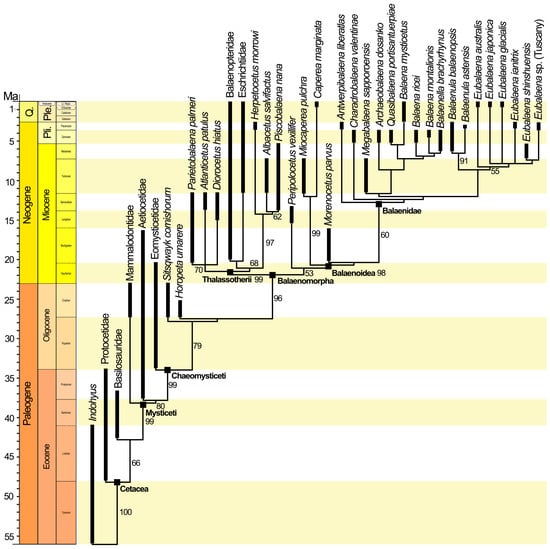
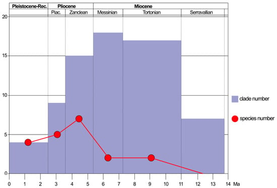
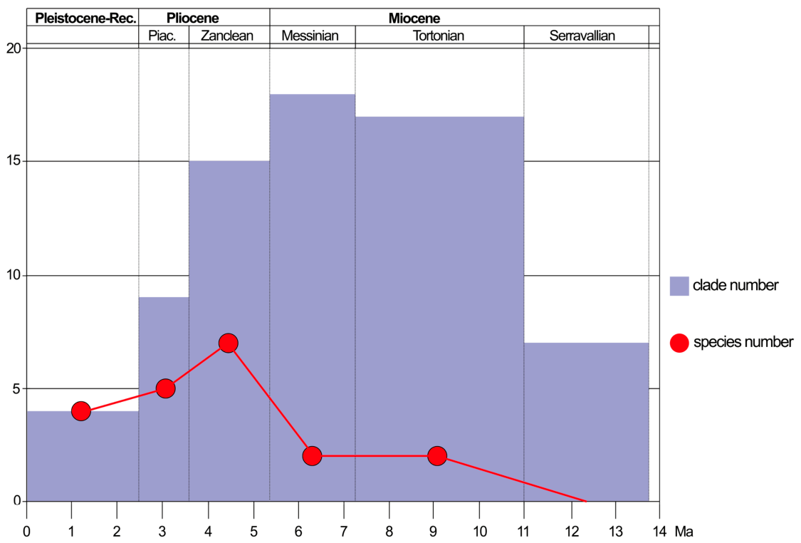
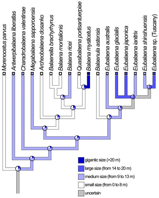
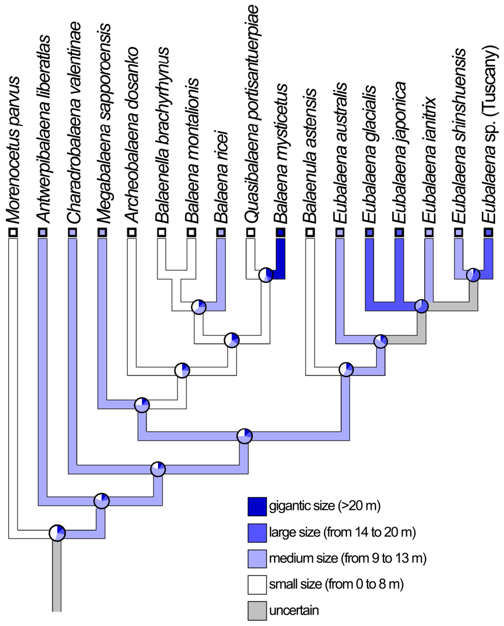

Disclaimer/Publisher’s Note: The statements, opinions and data contained in all publications are solely those of the individual author(s) and contributor(s) and not of MDPI and/or the editor(s). MDPI and/or the editor(s) disclaim responsibility for any injury to people or property resulting from any ideas, methods, instructions or products referred to in the content. |
© 2026 by the authors. Licensee MDPI, Basel, Switzerland. This article is an open access article distributed under the terms and conditions of the Creative Commons Attribution (CC BY) license.