


Abstract
The widespread use and improper disposal of plastics have made them ubiquitous contaminants that can potentially adversely affect both the environment and human health. The impact of microplastics (MPs)—plastic particles less than 5 mm in size—has been widely studied in marine and terrestrial environments; however, their potential effects on human health have only recently begun to receive attention. Although once considered inert materials, MPs exposure in laboratory animals is now linked to inflammation, immunological responses, lipid and energy metabolism alterations, mitochondrial damage, and other disorders. Since clearance-associated organs such as the liver, kidneys, spleen, lungs, and the gastrointestinal tract play a critical role in processing, metabolizing, filtering, and excreting toxins and foreign substances, they are particularly vulnerable to MPs exposure and accumulation. The increasing intake of plastics, primarily via ingestion and inhalation, raises concerns regarding their fate and retention in these organs, potentially leading to long-term health impacts. This review provides an overview of recent findings regarding MPs accumulation in clearance-associated organs and possible health impacts, and discusses future outlook and potential strategies for mitigating the health risks associated with nano- and microplastics exposure.
1. Introduction
Plastics have become an unavoidable component of modern life, permeating almost every aspect of our daily activities due to their flexibility, versatility, convenience, and a lack of viable alternatives. To meet rising demand, global plastic production surged from 1.7 megatons annually in the 1950s to almost 475 megatons in 2022 [1,2,3] and is expected to nearly triple by 2060 [4]. From the initiation of plastic production, an estimated 8300 megatons of plastic have been manufactured up to 2015, generating a cumulative plastic waste of about 6300 megatons. Out of this, only 9% was recycled, 12% was incinerated, and 79% was accumulated in landfills or the natural environment [5]. According to Earth Action, the 5th of September 2025 was considered Plastic Overshoot Day 2025, when the production of plastic waste exceeded the planet’s ability to manage it sustainably in one year. As plastic waste continues to accumulate in the environment without adequate management, it has slowly started to break down into smaller particles, which gradually find their way into the human body. Therefore, there is a critical need for studies that assess MPs exposure and accumulation in tissues and its possible effects on the body.
Plastics, being manmade, have varied compositions influenced by their type, source, uses, and additives [6,7]. Microplastics (MPs) are classified into primary and secondary MPs. Primary MPs are intentionally manufactured in the micron range for use in products like personal care items and cleaning agents and can enter the environment during their lifecycle [8,9]. In contrast, secondary MPs result from the fragmentation of larger plastics due to degradation processes or friction and are more prevalent in the environment [10]. Generally, particles with diameters from 5 mm to 1 µm are considered MPs, while nanoplastics (NPs) are defined as smaller than 1 µm in dimension [11,12]. Although regulatory bodies such as the European Union’s Marine Strategy Framework Directive: Good Environment Status (MSFD GES) [13], the International Organization for Standardization (ISO) [14], and the National Oceanic and Atmospheric Administration (NOAA) [15] have classified plastic debris based on particle size, the size ranges for MPs and NPs classification vary considerably across the literature (Table 1). To prevent this conflation of MPs with NPs, a standardized classification method for plastic contaminants is urgently needed; indeed, efforts have been made recently to develop a standardized plastics classification scheme not just based on size but also based on properties such as shape, color, solubility, and composition, for ease of comparison of the literature on MPs [16,17]. In this article, we have used the ISO24187 [18] naming system to categorize plastic contaminants reported in the literature. As plastic debris breaks down to the micro- and nanoscale, their surface-area-to-volume ratio increases, enabling them to navigate through tight spaces and thus easily penetrate biological barriers and tissues. MPs and NPs tend to absorb harmful chemicals such as pesticides, heavy metals, and persistent organic pollutants from the environment, which raises additional concerns about potential health risks once these accumulate in the tissues [19,20,21].
Table 1.
A comparison of how plastic debris is categorized in the published literature and in widely used classifications such as MSFD GES, ISO, and NOAA.
Per the most recent estimates, humans consume between 95 and 5348 million MPs in their lifetime, of which an estimated 59% accumulates in the body, while 41% is excreted [30]. This exposure occurs through contaminated drinks (bottled and tap water, soft drinks, milk), food such as fish, fruits and vegetables, salt, sugar, and other dietary products [31,32,33,34,35,36,37,38,39]. Notably, some MPs exposure from drinks and food items comes from the plastic packaging used for the products [40]. MPs contamination in the air has also been documented, primarily through shedding of synthetic fibers from clothing, tire erosion (automobiles and trucks), waste incineration, landfills, sewage sludge, household objects, abrasive products, 3D printing, and suspension of polymer fragments from urban air [41,42,43]. MPs made of polyester, PET, rayon, epoxy resin, poly(vinyl acetate), polyurethane, Teflon, and poly(N-methyl acrylamide) have been detected in the atmosphere [44]. Atmospheric fallout in urban areas is the most significant cause of MPs generation [45,46]. Since about 89% of the population in modern society engages in indoor activities, recent studies have also reported on indoor MPs contamination primarily from textile microfibers [42,47]. The indoor microfiber deposition concentration was reported in the range of 475 to 19,600 MPs/m2/day [45,48,49]. Dris et al. (2022) performed a study to estimate the amount of MPs present in both the indoor and outdoor environment and concluded that 0.3–1.5 particles/m3 are present in outdoor air, while indoor air was determined to contain about 0.4–56.5 particles m−3, comprising about 33% of polymers [50]. Since the gravity of airbone MPs (specific gravity as low as ≤1) is lower than that of sea water, these MPs have also been found to be easily transported on the sea-surface microlayer with the potential to get transferred to the air due to factors such as wind, sea spray, or wave breaking [51]. Once airborne, particle properties such as aerodynamic diameter, size, and shape, together with wind, can influence their persistence and atmospheric transport. About 7% of total MPs from the ocean are estimated to be transported airborne [52]. MPs as small as 5 µm have been detected in air samples [43]. Dermal contact is a less significant route of exposure since only particles smaller than 100 nm in diameter can penetrate through the stratum corneum [53]. However, few studies have studied dermal exposure to MPs [54]. In this review, ingestion and inhalation routes are the main routes of focus, as they represent two of the most common methods by which MPs can enter the body.
The processing, filtering, and excretion of ingested or inhaled MPs occur primarily via clearance-associated organs such as the liver, kidneys, spleen, lungs, and intestines. The liver performs the critical task of detoxifying and metabolizing various substances in the bloodstream, and recent research indicates that MPs could potentially be an emerging threat to liver health [55]. More than 90% of the ingested MPs, particularly those larger than 150 μm, are reported to be excreted in feces. In contrast, particles smaller than 150 μm can penetrate the gastrointestinal lining, and particles smaller than 2.5 µm can enter systemic circulation through endocytosis. NPs less than 0.1 μm in size have been found to cross the blood–brain barrier, while MPs up to 10 μm in size have been observed in the placenta [48,56,57,58]. Although there are emerging reports on the accumulation of MPs in humans, most in vivo investigations on the effects of this long-term accumulation have been carried out in animal models. In this review, we will discuss recent findings on MPs retention in animal clearance-associated organs, the impacted biological pathways, and the potential implications for human health.
2. Search Methodology
This narrative review focused on assessing and discussing the health impacts of microplastics exposure and accumulation in key clearance-associated organs, including the liver, kidneys, lungs, spleen, and gastrointestinal tract. A comprehensive search for the relevant scientific literature was conducted using PubMed, Embase, Scopus, and Google Scholar. Since this is a narrative review and focuses on microplastics’ effects on multiple organs, specific search terms were not pre-defined, and reviewers identified and selected literature based on its relevance to the scope and objectives of the review. The initial search was carried out in November 2022 and was updated in April 2025, June 2026, and August 2026 to include the most recent findings from this rapidly evolving area of research.
3. Composition of MPs in the Environment
Recent studies show that non-fiber plastic contaminants typically consist of 93% polymer resin and 7% additives by weight [5]. The main types of non-fiber plastic polymers include polyethylene (PE) at 36%, polypropylene (PP) at 21%, and polyvinyl chloride (PVC) at 12%, with polyethylene terephthalate (PET), polyurethane (PU), and polystyrene (PS) making up less than 10% each. Together, these seven polymer types represent 92% of global plastic production [5,59]. Notably, 42% of common non-fiber plastic contaminants, i.e., PE, PP, and PET, are used for packaging, while 19% of non-fiber plastics, particularly PVC, are used in construction. MPs, due to their small size and surface energy, can act as vectors that adsorb other pollutants, particularly heavy metals and hydrophobic organic chemicals (HOCs) [7,60]. When organisms are exposed to these MPs containing organic pollutants, both the MPs and the pollutants can be transferred into the organisms, increasing the accumulation and downstream effects of both. Plastics may also contain additives to enhance their properties, such as pigments to modify colors, fillers/reinforcement to enhance mechanical properties, antioxidants/stabilizers to provide resistance to heat and gaining, UV stabilizers to provide resistance to light degradation, flame retardants to improve flame/fire resistance, recycling additives to improve processing characteristics, and several performance-specific additives (plasticizers, anti-static/conductive additives, lubricants, surfactants, mold release agents, preservatives, blowing agents) [61]. Many plastic products contain monomers and oligomers, such as bisphenol A (BPA) and phthalates, which can pose health risks, particularly reproductive toxicity. BPA, found in polycarbonate and epoxy resins, is known to impact reproduction in aquatic organisms and can enter human systems through inhalation, skin contact, and ingestion. Phthalates, like di-n-octyl phthalate (DnOP) and di-2-ethylhexyl phthalate (DEHP), are linked to abnormal human development and reproductive health issues [62,63]. Additionally, flame retardants like polybrominated diphenyl ether (PBDE) and tetrabromobisphenol A (TBBPA) disrupt thyroid hormone balance and can have antiandrogenic effects [64]. These plastic additives present an additional risk as they, together with MPs, can be taken in by terrestrial and aquatic life (Figure 1), eventually accumulating in humans through different pathways [64,65].
Figure 1.
Main sources/routes of MPs/NPs exposure in humans. Arrows indicate movement of MPs/NPs from the different sources into the environment or into living organisms.
4. Consequences of MPs Accumulation in Clearance-Associated Organs
The accumulation of PVC MPs ranging in size from 5 to 110 µm in animal models such as rats, dogs, and pigs has been reported as early as 1970 [57,66]. As mentioned earlier, MPs intake can occur through ingestion, inhalation or, to a lesser extent, through the skin. Among these routes, ingestion is the most common path through which MPs exposure occurs. MPs entering the GI via contaminated food or water can be absorbed by the small intestine. These MPs pass through the intestinal epithelium by various mechanisms, including transcytosis, internalization by Microfold cells (M cells) and other cell types near Payer’s patch, paracellular transport, persorption through gaps in the villi, and uptake by migratory phagocytes. MPs less than 10 µm in diameter are mostly found in intestinal villi around Peyer’s patches. Once in Peyer’s patches, particles can be captured by macrophages and transported to lymph nodes or systemic circulation, potentially bypassing the liver, or they can be carried by the portal blood first to the liver and then to systemic circulation. Particles > 10 µm may be taken up by persorption or by migratory phagocytes. Particle characteristics such as size, hydrophobicity, and protein corona formation, as well as the patient’s GI health, can influence internalization rates. Smaller and hydrophobic particles tend to be absorbed more efficiently. In vitro studies show that PS NPs with a protein corona have increased translocation rates, influenced by protein quantity and composition [67]. Individuals with diseases such as inflammatory bowel disease and ulcerative colitis are more susceptible to MPs retention in the GI tract [68]. While more research is needed on whether MPs themselves cause GI disease, the current literature confirms the significant presence of MPs in feces worldwide [69], indicating considerable exposure of the GI tract to these ubiquitous contaminants. A recent study in Amazonian fish species showed that nearly 91.5% of ingested MPs were in the intestine, while about 8.5% of MPs were in the stomach [70].
MPs that translocate through the small intestine may eventually accumulate in the liver. As the primary organ involved in detoxification, the liver receives significant exposure to plastics and other foreign materials, which can compromise its function. The kidneys are similarly involved in filtering and removing waste products, including plastics. Like the liver, the spleen also lacks enzymatic capabilities to break down plastics and hence cannot clear them.
In cases of respiratory exposure, MPs can deposit in the airways by impaction, interception, and sedimentation. Alveolar deposition primarily occurs in particles smaller than 5 µm, while larger particles have a significantly lower deposition rate (about 0.5–5%) [71]. Particles that enter the respiratory airways are removed by mucociliary transport, which involves the movement of mucus toward the pharynx to be swallowed, aided by the beating of cilia, and then transportation to the digestive system. The lungs also contain alveolar macrophages that can phagocytose MPs for clearance.
Plastics that are less than 20 µm in size can easily penetrate biological membranes and accumulate in tissues [57], where they then cause inflammation, oxidative stress, cell injury, altered membrane functions, and immunotoxicity [72,73]. This prolonged presence may adversely affect organ function and overall health, especially since the body lacks the enzymes needed to break them down. Table 2 summarizes the major findings from different animal models on how clearance-associated organs are impacted by MPs of different types and sizes. Current knowledge on how MPs and NPs impact clearance-associated organs is summarized in the sections below.
Table 2.
Summary of organ-specific consequences of MPs exposure in non-human species from the literature.
4.1. Liver
The liver is the largest internal organ in the body and plays a vital role in detoxification. Hepatocytes, which filter blood entering the liver, form about 80% of the total liver volume. The liver also contains non-parenchymal cells such as Kupffer cells, hepatic stellate cells (HSCs), and liver sinusoidal endothelial cells (LSECs). Kupffer cells and LSECs are primarily responsible for maintaining immune tolerance in the liver against any foreign material arriving in the liver [100,101]. HSCs are usually quiescent and serve as depots for vitamin A storage, but they become activated during liver injury and contribute towards the hepatic fibrotic response [102]. Due to the liver’s role in metabolism, the liver cells frequently encounter materials that are to be eliminated from the body, including alcohol, drugs, fat, pathogens, and plastics, which can induce oxidative stress, disrupt cellular processes, and trigger inflammatory responses. In addition, MP-induced gut inflammation can lead to greater leakage of MPs and toxins from the gut into the liver, leading to detrimental effects as explained below.
The exposure of the liver to MPs likely occurs primarily via ingestion. MPs first arrive in the gastrointestinal (GI) tract through the consumption of contaminated food and beverages, from where they eventually arrive in the liver and other clearance-associated organs (Figure 2) [52,103]. Under normal physiological conditions, the mucus layer in the GI tract acts as a barrier to prevent the translocation of gut microbiota and their metabolites into the liver [104]. However, when this barrier is weakened by xenobiotic substances such as plastics and alcohol [105,106], bacteria and their metabolic byproducts like lipopolysaccharides (LPS) can enter the blood via the portal vein and then proceed to the liver [105,107]. The LPS then triggers inflammatory pathways and proinflammatory cytokine production by liver cells, initiating the development of liver disease. MP-induced hepatic inflammation has been reported in different animal models [95,104,108,109]. For example, Yin et al. [110] observed the induction of liver inflammation, lipid deposition, and the development of macrophage extracellular traps upon delivery of 5 µm PS microbeads in 1-week-old male and female chicks. The PS MP-induced macrophage extracellular traps were found to promote hepatocyte inflammation. Daily oral administration of 0.5 µm NPs at 0.5 mg/day in C57/BL6J mice was found to increase liver weight, promote the influx of natural killer cells and macrophages, increase serum aspartate aminotransferase (AST), alanine transaminase (ALT) and total bilirubin (TBIL) levels indicative of liver disease, and upregulate the expressions of proinflammatory cytokines interferon-gamma (IFN-γ), tumor necrosis factor-α (TNF-α), and interleukins (IL) -1β, -6, and -33 mRNA in the liver [111]. These findings were corroborated by Lee et al. [112], who reported greater liver accumulation and lipid droplet formation, and increased production of AST, ALT, glucose, triglyceride, superoxide dismutase, 4-hydroxy-2-nonenal-mercapturic acid (HNE-MA), and IL-6 in female C57BL/6 mice; these effects were greater with 0.5 µm NPs than with 5 µm MPs.
Figure 2.
Summary of the possible physiological effects/pathways that can be impacted in the liver, kidneys, spleen, and GI due to exposure to MPs/NPs by ingestion, based on findings from in vivo animal studies. Arrows indicate movement of MPs/NPs into the different organs following ingestion.
Other research groups, including ours, have also observed similar size-dependent effects of MPs and NPs on the liver and other organs. 0.1 µm NPs were found to enter hepatocytes more easily than 1 µm MPs and cause nuclear and mitochondrial DNA damage and activation of signaling pathways that lead to liver fibrosis [113]. The effects of size on MPs accumulation in zebrafish liver were assessed by Lu et al. [76]; histopathological analysis showed that fluorescent PS beads of size 5 µm and 70 nm accumulated in the gills, liver, and gut of zebrafish after 7 days of MPs exposure and induced inflammation and lipid accumulation. No liver accumulation was observed for the larger 20 µm-sized MPs. This size-dependent accumulation could be attributed to the fact that submicron-sized particles can enter the circulatory system more easily and accumulate in the liver and spleen, whereas larger-sized particles may not pass through gastrointestinal barriers as easily and may instead get eliminated in the feces. Furthermore, there is a greater number of particles per unit volume for 5 μm PS MPs than for 20 μm PS MPs, leading to the observed differences in accumulation.
4.1.1. Effects of MPs/NPs on Hepatic Lipid Metabolism and Energy Regulation
The accumulation of MPs has also been associated with changes in energy metabolism and lipid synthesis in the liver, which increases the risk of developing metabolic-dysfunction-associated steatotic liver disease (MASLD). Chiu et al. [114] investigated the hepatic effects of PS MPs sized 0.08–100 μm in C57BL/6J mice following oral exposure. Mice were gavaged with PS MPs at doses of 0.5, 5, and 50 mg/kg/day for 28 days. Liver tissue analysis revealed a dose-dependent increase in PS MPs accumulation, increased ROS, and significant reductions in triglyceride levels. Transcriptomic and pathway analysis indicated that PS MPs disrupted lipid metabolism via upregulation of the NR4A1-AMPK signaling axis and induction of mitochondrial dysfunction. siRNA-mediated knockdown of NR4A1 in hepatocytes suppressed the induction of AMPK and lipid-metabolism-related proteins upon PS MPs exposure, further implicating NR4A1 as a key regulator of MP-induced hepatic lipid dysregulation. Lu et al. [109] reported that PS MPs induced hepatic lipid metabolism disorder and significantly reduced the richness and diversity of the gut microbiota in male ICR (Institute of Cancer Research, China) mice treated with these MPs. Li et al. [115] treated ICR male mice with PS NPs or MPs (80 nm, 0.5 µm, and 5 µm) at 1 mg/L via drinking water and observed significant liver inflammation and lipid deposition for the smaller-sized particles. They identified three key hub genes–Acot3, Abcc3 and Nr1i3—that can be implicated in liver fibrosis development upon MPs/NPs exposure. In a recent study, Del Piano et al. [116] treated gilthead seabream with sub-chronic levels of PS MPs (1–20 µm, 0.5–5% of the feed) for 21 days through contaminated food and observed upregulation in the gene expression of markers involved in liver metabolism such as peroxisome proliferator-activated receptors (PPARγ) and sterol regulatory element-binding protein (Srebp-1c). Severe hepatic congestion, inflammation, and necrosis were also observed. Cui et al. [117] examined the hepatic effects of pristine and UV-aged PE MPs (5–30 μm) in male C57BL/6J mice. Animals were orally exposed to 0.01 or 1 mg/day of either pristine or aged MPs. Both forms disrupted plasma metabolites linked to lipid metabolism and digestion, but aged PE MPs induced more severe alterations. These included reductions in hepatic glutathione, increased ROS, downregulation of proteins involved in fatty acid metabolism such as glutathione S-transferase A, and elongation of very-long-chain fatty acid protein 5 (ELOVL5). Changes in gut microbiota, particularly the reduced abundance of Akkermansia and Lactobacillus, were also reported, suggesting that hepatic metabolic dysfunction may be driven by gut–liver axis disruption upon aged MPs exposure. Furthermore, MPs accumulation in the liver led to a significant reduction in triglyceride and total cholesterol levels, suggesting an imbalance in lipid metabolism [117]. Lu et al. [95] treated mice with 0.5 and 50 μm PS MPs at concentrations of 100 and 1000 μg/L and reported the development of hepatic lipid disorder post-treatment. With MPs treatment, both hepatic triglyceride and total cholesterol levels decreased by 12.7% in the 1000 μg/L 0.5 μm PS MPs-treated group and by 13.9% in 1000 μg/L 50 μm PS MPs-treated group, confirming hepatic lipid metabolism disorder in mice. Zhou et al. [118] observed that zebrafish treated with PS MPs (~490 nm) in tank water had greater lipid droplet accumulation in the liver as well as greater triglyceride and cholesterol levels and decreased free fatty acids and total bile acids compared with the control untreated group. Significantly higher levels of the proinflammatory cytokine tumor necrosis factor-α (TNF-α) and ROS, as well as marked changes in the gut microbiome, were also observed in the zebrafish following MPs treatment. Deng et al. [108] studied changes in the metabolism profile in mice treated with 0.01, 0.1, and 0.5 mg of MPs/day. Both 20 µm and 5 µm MPs dispersed in Milli-Q water were used for testing. For MPs of both sizes given at a 0.5 mg/day dosage, a 60% increase in lactate dehydrogenase (LDH) activity was observed in the liver compared with control untreated mice. The study concluded that MPs exposure results in energy deficiency, as evidenced by a significant decrease in the weight of the liver and a significant increase in food intake in the mice, especially at high MP doses [108,119]. Luo et al. [120] treated mice with 1000 µg/L of PS MPs during pregnancy, and the results showed significant changes in glucose transport, glycolysis, synthesis of fatty acids, and oxidation of fatty acids. Moreover, this study also confirmed that MPs could impair lipid metabolism by significantly reducing the production of hepatic adenosine triphosphate (ATP). Furthermore, this study reported that MPs could impair energy and lipid metabolism by increasing LDH activity in the F1 and F2 offspring of the mice. Work by Shi and colleagues [121] studied the long-term impact of PS MPs on liver health using BALB/c mice administered 10 mg/L of either unmodified or amine-modified PS MPs (unmodified or amine-terminated, both 5 µm) through drinking water for a duration of 6 months. The treatment led to notable liver injury, with significantly elevated serum levels of ALT, AST, and ALP, along with hepatic lipid droplet accumulation and histopathological signs of hepatocyte degeneration. Proinflammatory cytokines, including TNF-α, IL-6, IL-1β, and IFN-γ, were upregulated, particularly in the amine-modified MPs group, which exhibited stronger hepatotoxic effects. The study also reported increased hepatocyte senescence and ferroptosis, with enhanced lipid peroxidation, iron accumulation, and downregulation of GPX4. Mechanistically, these effects were mediated via activation of the HO-1/Nrf2 signaling pathway and were significantly attenuated by co-treatment with ferroptosis inhibitor ferrostatin-1 or HO-1 inhibitor zinc protoporphyrin, suggesting that chronic PS MPs exposure can induce MASLD through ferroptosis-induced hepatocyte senescence.
4.1.2. MP/NP-Induced Hepatic Oxidative Stress and ROS Production
Several studies have reported the induction of oxidative stress in the liver following MPs exposure. A recent study by Yu et al. [122] reported that PS MPs exposure in crabs (Eriocheir sinensis) led to oxidative damage and inflammatory response in the liver. The increased ROS activity due to MPs exposure activated the cellular antioxidative defense system in crabs. They reported an elevation in glutathione peroxidase (GPx), superoxide dismutase (SOD), catalase (CAT), and glutathione s-transferase (GST) activity at lower PS MPs concentrations and a reduction in these enzyme activities at higher PS MPs concentrations. These antioxidant enzymes are usually associated with mitigating oxidative stress by eliminating ROS [123]. Significant changes in oxidative-stress-linked metabolites were also reported in mice. In particular, a significant reduction in lysine, pyruvate, and threonine was observed due to exposure to MPs [108]. These oxidative-stress-linked metabolites are endogenous antioxidants that provide protection against xenobiotic substances [124,125]. Overall, the results suggest that MPs exposure creates an imbalance in antioxidant defense mechanisms in mice [108].
4.1.3. MP/NP-Induced Liver Fibrosis
Emerging reports also point towards the role of MPs in liver fibrosis initiation and progression. Shen et al. [113] demonstrated that the activation of the cGAS/STING (cyclic guanosine monophosphate-adenosine monophosphate synthase-stimulator of interferon gene) pathway can explain liver fibrosis caused by exposure to MPs. The cGAS/STING signaling pathway activates a cascade reaction to pass an inflammatory signal by identifying fragments of endogenous and exogenous double-stranded DNA in the cytoplasm [126,127]. They also observed free mitochondrial DNA (mtDNA) in the cytoplasm, which indicates mitochondrial damage [113]. Chung et al. [128] established the link between the presence of mtDNA in the cytoplasm, the cGAS/STING signaling pathway, and organ fibrosis. Based on these findings, Shen et al. concluded that the cGAS/STING signaling pathway plays a critical role in MP-induced fibrosis and damage in the liver [113].
4.1.4. Effects of Non-Polystyrene MPs on the Liver
Although the majority of available preclinical MPs research into the liver uses pristine PS beads, there are also reports on PVC MP-induced toxicity resulting in hepatocyte death and functional disorder in the liver [129,130]. Studies have established that exposure to vinyl chloride—the precursor of PVC—elevates the risk of hepatic angiosarcoma by 11- to 16-fold [131,132] Chen et al. [129] observed a decrease in probiotics and an increase in pathogenic bacteria in the gut of mice exposed to PVC MPs. Male mice exposed to a chronic PVC MPs dosage (0.5 mg/day) for 60 days had significant hepatic injury, leading to increases in serum ALT and AST levels and decreases in serum triglyceride and total bile acid (TBA) levels. This hepatic inflammation follows apoptosis-related phosphoinositide 3 kinase (PI3K)/protein kinase B (Akt) and oxidative stress activation-related Nrf2/HO-1 signaling pathways. In addition, PVC MPs exposure caused gut microbiota dysbiosis in mice similar to PS MPs exposure [129]. Moreover, activation of Akt in the signaling pathway stabilizes and causes the accumulation of apoptosis-related protein p53. p53 has been shown in previous studies to cause hepatic apoptosis and associated injury [133,134,135]. Yu et al. [122] reported increased expression of the gene encoding p38 in juvenile Eriocheir sinensis in the mitogen-activated protein kinase (MAPK) signaling pathway, resulting in a significant reduction in the expression of extracellular signal-regulated kinase (ERK), mitogen-activated protein kinase (MEK), and AKT, which can significantly impede growth. Fan et al. [136] investigated the effects of orally administered PLA MPs (2–5 μm, 50 mg/kg/day) on the gut–liver axis in BALB/c mice over 7-day and 28-day exposure periods. The particles underwent in vivo degradation into nanoscale fragments (<200 nm), accompanied by increased fecal lactic acid levels, indicating a mechanical breakdown in the gut. Short-term exposure-induced proinflammatory responses in both serum and the liver, with elevated TNF-α and IL-6 levels, while long-term exposure led to time-dependent metabolic reprogramming. In the liver, early-phase activation of glutathione and vitamin B6 pathways transitioned into suppressed TCA cycle activity and altered bile acid biosynthesis with prolonged exposure. Parallel changes in jejunal and hepatic purine/pyrimidine metabolism and persistent upregulation of glycerophospholipid-related metabolites, such as phosphatidylserine and lysophosphatidylcholine, suggested inter-organ crosstalk. Dysbiosis of the gut microbiota was also observed, with early increases in potentially pathogenic genera (Staphylococcus, Streptococcus) and later enrichment of Lactobacillus and Lachnospiraceae. A 2026 transcriptomics study by Jung et al. [137] showed that PE—one of the most understudied MP types—caused hepatic dysfunction in mice given standard or metabolic dysfunction-associated steatohepatitis (MASH)-inducing diets. The downstream effects of PE MPs and NPs accumulation in the liver therefore need to be studied further.
4.1.5. Age-Related Effects of MPs on the Liver
There is also some evidence of age-related detrimental effects of MPs on the liver. Aging can lead to decreases in liver weight and blood flow to the liver, a decreased number of hepatocytes, and an overall decrease in detoxification capabilities [138], which could lead to prolonged residence time of toxins like MPs in the organ. Recent studies by Xie et al. on 62-week-old male KM mice (approximately equal to 60 human years) showed dose-dependent inflammation, vacuolization, and damage to the liver upon MPs exposure [139]. These findings point towards the need for more detailed investigations into older/aged animals and comparing the data obtained with the liver tissues of younger animals.
4.1.6. Emerging Evidence on Human Liver Exposure to MPs/NPs
Although most of the literature studying MP effects on the liver has been conducted on animals, data on MPs accumulation and potential effects on human livers have now started to come to the forefront. Horvatitis et al. [140] analyzed 11 human liver tissue samples from 6 patients with chronic liver diseases and 5 individuals without liver-related ailments for MPs detection. Based on their analysis, 100% of patients with liver cirrhosis tested positive for MPs (4–30 µm), and 100% of individuals without liver disease tested negative for MPs in their liver, kidney, and spleen. They found commonly available plastics, such as PS, PVC, PET, PMMA, PP, and polyoxymethylene (POM) in the cirrhotic liver tissues. While the sample size in this study was small, the data indicate that chronic liver disease and liver injury may play a key role in facilitating MPs accumulation in the liver. Complementing this, Schwenger et al. [141] examined the levels of fecal MPs in MASLD. In their study of 23 adults, comprising healthy, obese, and MASH patients, MP levels in feces were positively correlated with hepatic portal macrophage infiltration, natural killer cell abundance, and the expression of 19 hepatic genes related to immune response and apoptosis. Higher fecal MP fiber content was also linked to reduced abundance of Lachnospiraceae and greater levels of fecal Bifidobacterium. Importantly, among MASH patients who were followed longitudinally for 12 months after bariatric surgery, those with persistent liver disease showed significantly higher fecal MP fragments than those with normalized histology.
4.1.7. Co-Exposure of Liver to MPs/NPs and Other Environmental Pollutants
A relatively recent finding is that MPs, due to their high hydrophobicity, can work synergistically with other environmental pollutants such as heavy metal ions [142], flame retardants [143], bisphenol A, and nonylphenol to cause liver damage [144]. This phenomenon is called the “Trojan horse effect” of MPs. Recent studies have begun to investigate the potential for synergistic toxicity when MPs are co-administered with environmental contaminants, particularly in the gut–liver axis. Li et al. exposed zebrafish to aged PS MPs (10 μm, 200 μg/L) and cadmium (10 μg/L) for 21 days and observed greater hepatic cadmium accumulation and damage compared with either pollutant alone. Co-exposure increased hepatic ROS, SOD, MDA, CAT, TNF-α, triglycerides (TG), and total cholesterol, alongside metabolic disruption of the sphingolipid and pentose phosphate pathways. Gut barrier proteins ZO-1 and MUC2 were also suppressed, indicating enhanced systemic toxicity via gut leakage [145]. Fan et al. [146] similarly reported that broilers exposed to PS MPs (0.5, 5, and 50 μm; 100 mg/L) and cadmium (140 mg/kg) exhibited hepatocyte swelling, vacuolation, inflammatory infiltration, and degeneration. Mechanistically, this damage was linked to activation of the TLR4/MyD88/NF-κB signaling pathway and disruption of intestinal tight junction proteins. Xiao et al. [147] demonstrated that mice orally exposed to 0.5 μm MPs (10 mg/kg/day) and bisphenol A (1 mg/kg/day) for 90 days developed liver fibrosis, with histological evidence of extracellular matrix accumulation. This was attributed to the co-exposure to MPs and bisphenol A, leading to suppression of the BMAL1/E-cadherin signaling pathway. These conclusions were supported by gene editing experiments in Bmal1−/− mice and AML12 hepatocytes. Finally, Li et al. [148] showed that zebrafish treated with 5 μm PS MPs and 17α-methyltestosterone (0.4 mg/L each) for 21 days exhibited transcriptomic and metabolomic evidence of liver and brain lipid metabolic dysfunction, with upregulation of genes such as CYP1A, CYP3A, and PPARα, and histopathological changes indicative of damage across the liver-brain axis. This is a critical direction for research in the future, as there is an urgent unmet need for understanding how MPs accumulation may impact human health—either directly or by enhancing the effects of other pollutants—through interactions with them in the environment.
4.2. Kidneys
The impact of MPs on the kidneys is of great importance owing to their role in clearance, especially of small-sized particles via urine. Glomerular filtration precludes the clearance of particles of a size greater than approximately 10 nm through urine [149,150,151]. Indeed, there is evidence in extant literature that MPs in mammals are mostly found to be excreted through feces [152,153,154]. However, the lack of glomerular filtration alone may not be responsible for the renal accumulation of MPs. A complex interplay of systemic biodistribution, vascular retention, and phagocytosis by resident macrophages can dictate the accumulation of MPs in the kidneys [155]. Given the key role the renal system plays in homeostasis, the effect of MPs and their accumulation within the kidneys may have systemic consequences and therefore merits some attention.
Several works have focused on the phenomenon of MPs accumulation within the kidneys. Huang and colleagues provide some of the most comprehensive work in this area. Administering PS MPs of varying sizes (5, 50, 100, 200 μm) orally to mice once a day for 10 weeks, they observed only the smallest of the particles accumulating within the kidneys. This is supported by the observations of other groups [81]. A study of internal tissues such as the kidney, liver, lungs, and ileum in companion animals (24 cats and 25 dogs) in the Porto metropolitan area resulted in a median of 0 MP/g after blank corrections, indicating the need for larger sample sizes and more robust sample processing and identification methods for MPs to ensure limited background contamination [85]. Meng and coworkers administered nano- and micro-scale PS to chickens (50 nm, 300 nm, 600 nm, and 4 μm) and examined tissue distribution after 24 h. Their results showed that 50 and 300 nm particles concentrated within the cells of the kidneys such as glomerular mesangial cells, macula densa, renal tubules, and tubular epithelial cells, as well as in the pericellular space, as did the 600 nm particles. By contrast, 4 μm PS MPs tended to either stay within the intracellular spaces or reside in the lumen of the renal tubule, indicating that at least some of these micron-scale particles are capable of penetrating (or inducing penetration through) the renal tubule [83]. This is an interesting result that appears confirmatory to a phenomenon of albumin leakage some groups have observed, which is described below. An inverse relationship between MPs size and depth of tissue penetration was observed, as is to be expected.
A pair of linked studies by Deng et al. and Yang et al. [108,156] delved deeper still into the toxicokinetics of MPs in the form of 5 and 20 μm PS MPs, administered orally to mice. They found that steady-state renal concentrations of both sizes were achieved within 14 days of initial administration, with maximal concentrations being 0.946 ± 0.093 mg/g and 0.783 ± 0.084 mg/g for 5 and 20 μm particles, respectively, and steady-state bioconcentration factors of 5.57 and 4.60, respectively. The trend of smaller particles accumulating more notably within the kidneys is thus reflected in these works as well. In addition, the mean residence time of these particles within the kidneys was found to be approximately 17 days. Yang and colleagues’ work is an excellent companion piece of literature that delves into work that complements the works summarized in this section. Of note here is a role postulated by Huang and colleagues for the rough surface of PS microparticles playing a key role in the histopathological perturbations they observed [81].
4.2.1. BUN/Creatinine Levels on MPs/NPs Exposure
The most common metrics for assessing the knock-on systemic effects of MP-induced renal disruption are the standard blood urea nitrogen (BUN) and serum creatinine (CRE) levels. In mice, significantly elevated BUN and CRE levels were observed upon exposure to PS MPs; Huang and colleagues’ work showed that both dietary fat and MPs exposure played a roughly equal role in the elevation of BUN levels, but also indicated a markedly heavier MPs influence on serum CRE levels (relative to the impact of diet) [81]. Yang et al. [156] performed more in-depth work in this area. Noting a significantly lower level of serum CRE over a 4- (0.2 mg MPs/day: 0.19 ± 0.01 mg/dL, 0.4 mg/day: 0.20 ± 0.01 mg/dL, control: 0.24 ± 0.02 mg/dL) and 8-week period (0.2 mg/day: 0.42 ± 0.01 mg/dL, 0.4 mg/day: 0.38 ± 0.02 mg/dL, control: 0.45 ± 0.01 mg/dL) and reasoning that typical kidney failure results in an increase in these levels, they examined muscle mass as a potential factor for the discrepancy. Their findings were of interest: muscle mass was found to be reduced in mice treated with PS MPs. They also showed the presence of proteins in the urine of mice treated with PS MPs, particularly albumin, which, when combined with the tubular lesions they observed, highlighted a possible link between MPs and chronic kidney disease. Work by Meng et al. [83] was less conclusive: mice treated with 600 nm PS particles showed higher serum BUN and CRE levels (relative to sham controls), but not those treated with 4 μm MPs. Furthermore, serum albumin was lower in mice treated with NPs than in controls, but not in those treated with MPs—a curious result given the apparent potential of MPs of that size to cause urinary albumin leakage demonstrated in other studies. Ahmed et al. [84] showed a PS NP exposure-dependent increase in serum BUN, CRE, and malondialdehyde in rats, while an inverse relationship was observed between exposure and serum glutathione.
4.2.2. MP/NP-Induced Kidney Oxidative Stress and ROS Production
Similar to the liver, mitochondrial dysfunction and associated oxidative stress are some of the most commonly associated pathophysiological features of renal MPs exposure. Levels of OPA1, Mfn1, and Mfn2 (three key proteins involved in mitochondrial fusion) were seen to decrease with increasing doses of PS MPs in chicken kidneys (with an associated decrease in respective gene expression), while levels of Drp1 (a driver of mitochondrial fission) were found to be significantly increased at doses of 100 mg/L. Curiously, the levels of Drp1 mRNA were not observed to change with exposure up until the highest level tested (100 mg/L), when a roughly 5-fold increase in transcription compared with all other groups (control, low/environmental exposure, and medium exposure) was detected. In the same study, levels of antioxidant enzymes (as measured in renal tissue supernatants) were found to be dysregulated upon MPs exposure: GPx underwent dose-dependent increases, while levels of CAT and SOD were reduced in a similar manner. MDA levels also underwent a dose-dependent increase. Total antioxidant capacity was found to be significantly decreased at all exposure levels compared with untreated controls [82]. In mice, PS NPs (600 nm) reduced CAT activity by approximately half, while 4 μm particles reduced such activity by only a quarter-fold relative to controls. Levels of T-SOD, GSH-px, and MDA were also observed to follow a general pattern of size-dependent increase [83]. Similar trends were observed in rats, where PS NP dose-dependent increases in MDA and decreases in renal GSH were observed [73]. These increases in MDA levels, in particular, point to possible lipid peroxidation within renal cell membranes as a result of exposure. A germane observation is one made by Huang and coworkers, who found that 5 μm PS microparticles that accumulated in the kidneys of animals in their work—combined with a normal chow diet (NCD)—had the same impact as an MP-less high-fat diet (HFD) on 5-week-old male mice. An HFD + MP combo only served to worsen these symptoms. These may serve to illustrate a possible microparticle–lipid axis at work in exposure-related renal dysfunction. Work in rats, however, revealed significant downregulation in the expression of both Nrf2 and GPx within renal tissue, conflicting somewhat with work in mice [84]. In vitro work with HEK293 cells showed that exposure to PS MPs up to 300 ng/mL for 24 h did not significantly elevate levels of HO-1, but did cause marked increases in ROS accumulation, likely through the inhibition (or downregulation) of antioxidant enzymes [157]. Similar results were observed by Goodman et al. [158], where 50 μg/mL concentrations of 1 μm PS MPs induced significantly elevated ROS build-up within HEK293 cells following just 2 h of exposure (although interestingly, 100 μg/mL exposure did not result in as much ROS accumulation within the first 6 h post-treatment). MPs exposure was shown to be capable of suppressing SOD2 and CAT expression over 72 h; intriguingly, 5 μg/mL exposure was more effective at this than 100 μg/mL. HK-2 cells treated with 0.2–0.8 mg/mL PS MPs were shown to have significantly higher mitochondrial ROS accumulation (up to a 3-fold increase at the highest tested concentration), accompanied by significantly higher levels of Bad (B-cell lymphoma-2 (BCL-2) Associated Agonist of Cell Death) with increasing time (5–60 min), as well as a significant decrease in Bcl2 levels after 10 min of MPs exposure; levels of Bax (BCL-2 associated X protein) tended to stay almost unchanged over the 60 min period evaluated. MitoTEMPO treatment was capable of rescuing cells from ROS accumulation to some degree, also serving to lower levels of Bad significantly. These, together with changes in levels of inflammation- and autophagy-related markers (see below), paint a complex picture of mitochondria-induced apoptosis in vitro [159]. A complicated interplay between the size of MPs, level of exposure, and the degree of oxidative stress generated seems apparent.
4.2.3. MP/NP-Induced Kidney Inflammation
The infiltration of proinflammatory immune cells into the kidneys and the issue of oxidative stress discussed above bring with them the ability to induce widespread inflammation in renal tissue. Thus, inflammation is another common pathophysiological feature of renal MPs exposure. Work with chicken kidneys revealed that exposure at environmental (1 mg/L) or higher concentrations of PS MPs would result in inflammation via activation of the NF-κB pathway and upregulation of both transcription and expression of NF-κB p65, its upstream signaling partner IκBα, IL1B (both pro and mature), TNFα, and iNOs. This was found to give way to necroptosis via the RIP1/RIP3/MLKL pathway (all downstream of TNFα), all of which were shown to be both transcribed and expressed increasingly in a dose-dependent manner, while levels of caspase 8 showed a marked decrease with higher PS MPs exposure. Levels of HSPs (60, 70, and 90) were also found to increase in chicken renal tissue with increasing MPs exposure, driven partly by a combination of oxidative stress and inflammation [82]. The impact of the size of PS-based MPs in mice also showed levels of IL-6 and IL-10 increasing by approximately 2- and 4-fold, respectively (relative to controls) upon exposure to 600 nm particles (with far more moderate increases induced by 4 μm particles), while levels of Monocyte Chemoattractant Protein-1(MCP-1) and TNFα were markedly higher in tissue exposed to 600 nm particles (7- and 2-fold increases relative to controls, respectively), with more modest increases in animals exposed to 4 μm particles [72]. Chen and colleagues examined the inflammatory pathways induced by MPs in HEK293 cells and found that, at least in vitro, inflammation was not driven by NF-κB (or a ROCK-dependent pathway), but that levels of NLRP3 were reduced following 24 h treatment with 300 ng/mL PS MPs. This led them to posit MP-induced autophagy as a cause. A panel of inflammatory cytokines were monitored, and the results were both interesting and confirmatory of their proposed mechanism: a low-level exposure (3 ng/mL, 24 h) was found to result in the upregulation of 33 different cytokines (and downregulation of MCP-1 and eotaxin), while high-level exposure (300 ng/mL, 24 h) caused a marked reduction in all cytokines, with some going lower than in untreated controls, most likely through the removal of inflammasome complexes through exposure-induced autophagy [157]. Levels of Alpha-1 antitrypsin (AAT) were also shown to be reduced in MP-treated cells, impairing renoprotection from oxidative stress. Work with HK-2 cells by Wang et al. [159] added more depth to the cellular-level effects of PS MPs and MPs in general. The 24 h exposure to MPs resulted in upregulation of IRE1α, implicated heavily in ER stress. In addition, rapid increases in levels of p-ERK1/2, p-p38, and JNK were observed, increasing after just 5–20 min of exposure, indicating rapid activation of the MAPK pathway post-exposure. p-mTOR and p-AKT were observed to decrease with increasing PS MPs exposure, indicating suppression of the PI3K/AKT/mTOR pathway in these cells. cPLA2 and COX-1 levels were also found to be elevated at higher (0.4 and 0.8 mg/mL, respectively) exposure levels, while autophagy-associated LC3 and Beclin 1 were upregulated upon exposure as well. As mentioned previously, these cells were also treated with MitoTEMPO, which resulted in significant decreases in both IRE1α and LC3, while also increasing levels of p-mTOR. Given the MAPK pathway’s involvement in inflammation and the AKT/mTOR pathway’s role in suppressing autophagy, Wang and colleagues’ work serves to demonstrate a close interrelationship between ER stress, mitochondrial-driven oxidative stress and contributions to inflammatory processes, and general proinflammatory pathways activated by exposure of H-2 cells to MPs in the form of PS MPs. Ahmed and colleagues observed a significantly higher presence of COX-2 in rats treated with PS NPs, particularly within the renal cortex and cells of the collecting tubules within the medulla, and transcription of Cytc and CASP3 were found to be significantly elevated in rat renal tissue upon treatment with both concentrations of PS NPs tested [84].
PS MPs were also found to be cytotoxic to HEK293 cells, with cell viability dropping to <80% after 24 h exposure to 300 ng/mL. The mechanism behind this was shown to be a mixture of MP-induced autophagy and apoptosis [157]. The same study found significantly lower levels of ZO-2 (a regulator of renal barrier integrity) in PS MP-treated cells. Conversely, no reduction in HEK293 viability was observed by Ahmed and coworkers [158] at concentrations of up to 100 μg/mL of 1 μm PS MPs over 72 h. They did observe a marked decrease in cell proliferation rates at >24 h. Similarly, no significant reduction in cell viability was observed in HK-2 cells upon treatment with up to 0.8 mg/mL of PS MPs over several days [159]. A need exists for further confirmatory studies in this area.
Finally, it is also worth noting an interesting line of research pursued by Xu et al. [86]: investigating the effect of varying PS terminal groups (standard, carboxyl-terminated, and amine-terminated) on mice (male, 6 wks., 1 mg NPs/day) over 28 days, using 100 nm PS NPs. Their work found that all three types of NPs accumulated in the kidneys but elicited no significant effect on serum BUN or CRE levels (albeit some slight reduction in serum CRE was observed in the case of both carboxyl- and amine-terminated PS NPs). At the cellular level, TNFα was elevated in PS-exposed tissue, while all three varieties of PS induced elevated IL-6 levels within renal tissue.
4.2.4. Emerging Evidence on Human Kidneys’ Exposure to MPs/NPs
MPs accumulation and effects on human renal tissue are also now coming to the forefront. Using MicroRaman spectroscopy methods, Massardo et al. [160] confirmed the presence of PE MPs and PS MPs in the range of 1 to 20 µm in the kidneys and 3 to 13 µm in the urine of human nephrectomy samples. Wang et al. [161] observed MPs contamination in donor kidneys (sample size = 28) for transplantation, with PE and PVC MPs being the most abundant contaminants. Significant MP levels were also observed in the ureter. They also observed that specific MP types such as natural rubber NR, PC, PP, and butadiene rubber were associated with early postoperative blood pressure changes in transplant patients, highlighting the need to monitor vascular function changes during and after transplantation as MPs become increasingly detected in human organs. Assessment of organs from six fetuses in a recent study by Zhang et al. [162] showed the presence of predominantly PE MPs in the kidneys and liver, with sizes varying from 29.5 to 64.2 µm.
4.3. Lungs
As one of the primary sites of the body that is directly accessed by foreign objects like MPs and NPs via inhalation, and due to its role in the clearance of foreign objects via mucociliary clearance, alveolar-macrophage-associated phagocytosis, and lymphatic transport, it is essential to study the short-term and long-term effects of MPs accumulation in the lungs. Most studies to date have emphasized investigating the effects of ingested MPs on human health [163]. However, the pulmonary route has emerged as the second most likely route of human exposure to MPs [164] (Figure 3).
Figure 3.
Summary of the possible physiological effects/pathways impacted in the lungs due to the interaction of MPs/NPs with lung cells, based on findings from in vivo animal studies. The inset shows macrophage (purple) uptake and highlights detrimental effects of the MPs/NPs in the alveolar tissue. Arrows indicate movement of MPs/NPs into the lungs after inhalation.
A large fraction of inhaled MPs encounter mucociliary clearance as the first line of defense; they are either expelled via coughing or taken to the mouth, from where they pass to the GI. Smaller MPs and NPs are able to bypass this clearance to accumulate in the lung tissue [165,166,167]. MPs accumulation in different regions of the lungs occurs in a size- and flow-rate-dependent manner [168]. The alveolar region of the lungs is lined by lung surfactant (LS) secreted by type II alveolar epithelial cells, which play an important role in regulating alveolar surface tension, thus preventing alveolar collapse [169]. The inhaled MPs deposited in the alveolar region can interact with LS components, thus affecting alveolar surface tension and also inhibiting protection from foreign exogenous materials [170,171]. Inhaled MPs biopersistence is largely dependent on pulmonary clearance mechanisms and MPs durability [172]. In vitro experiments have been performed to study the characteristics of inhaled MPs in synthetic extracellular pulmonary fluid. Various MP fibers composed of polycarbonate (PC), PE, and PP fibers were found to have their surface area intact and undergo almost no dissolution even after 180 days, confirming the durability of inhaled MPs [173]. A study was also conducted on workers in the textile industry handling various fibers such as polyester, polyolefin, acrylic, and polyamide. Histopathological analysis of lung biopsies from these workers demonstrated pulmonary granulomatous lesions that are suspected to have been caused by the textile fiber MPs [174].
Studies also show that long-term exposure to MPs may result in cytotoxic effects in the lungs, even causing cell death. Dong et al. [164] studied the effects of PS MPs on human lung epithelial BEAS-2B cells, wherein cytotoxic and inflammatory effects were observed due to the production of ROS. BEAS-2B cells demonstrated dose-dependent cytotoxicity when treated with different concentrations of PS MPs (0–1000 µg/cm2) for 24 and 48 h, wherein the cells treated with the highest MPs concentration (1000 µg/cm2) displayed approximately 35–40% cytotoxicity. They also caused a decrease in transepithelial electrical resistance in the cells by decreasing zonula occludens proteins, which, in turn, may increase the risk for chronic obstructive pulmonary disease. Zi Fan et al. [88] investigated the effects of inhaled PS MPs on Sprague Rats, wherein the rats were intra-tracheally instilled with different sizes of PS MPs (100 nm, 500 nm, 1 μm, and 2.5 μm) for 3 days followed by instillation of different concentrations of 100 nm PS MPs every 2 days for 2 weeks. The inhaled MPs induced pulmonary inflammation and injury in all rats, displaying an upregulation of proinflammatory cytokines, namely IL-6, TNF-α, and IL-1β. Inhaled MPs with sizes as small as 100 nm were found to be deposited in the lungs, disrupting the epithelial barrier integrity of the alveolar region. They also observed alterations in the expression of long noncoding RNA (lncRNA) and circular RNA (circRNA) post-MPs treatment in all rat groups, both of which are believed to play a role in lung inflammation. Similar observations have been made in mouse lungs following MPs exposure, where PVC MPs, in particular, activated ROS signaling, leading to the induction of senescence in lung cells [175]. Domestic and fetal pigs in their natural environment were found to have MP fibers in their lung tissue; polyamide, a major component of nylon, was observed in domestic pig lungs, while PC fibers were more common in fetal pigs [176].
There are emerging reports of MPs accumulation in human lung cells and bronchoalveolar fluid. Baeza-Martínez et al. [177] detected the presence of MPs in bronchoalveolar lavage fluid sampled from the lower respiratory tract of humans. With the help of micro Fourier transform interferometer (µ-FTIR), scanning electron microscopy, and energy-dispersive X-ray spectroscopy (SEM-EDS) analysis of the samples, they were able to detect polyester as well as other non-synthetic fibers like rayon, cellulose, and cotton fibers in human patients, and they observed a higher percentage of MPs in smokers compared with the non-smoker group. Tokito et al. [178] also observed MPs in the bronchoalveolar lavage of 30 patients with diffuse lung disease diagnosis; the majority of MPs were smaller than 10 µm in size, and about 93.5% of MPs were particulate, while 6.5% were fiber. Wang et al. [179] reported that the lung tissue samples of 12 non-smoking patients evaluated by them showed the presence of MPs between 20 and 100 µm in size, with a median concentration of 2.19 particles/g. They observed that females and patients residing within about 300 m of the main road had greater MPs accumulation in lung tissues, possibly due to factors such as traffic-generated MPs and the washing of textiles. Another study analyzed the deposition of MPs in human lung tissues, wherein 5 out of 10 sample individuals were found to possess MPs in their lungs, with a size of less than 10 µm [180]. Studies performed at coroner autopsies have also detected the presence of inhaled MPs in 20 adult cadavers (non-smoking) from São Paulo, belonging to a wide range of ages (48–94 years), of whom 13 were females and 7 were males. With the aid of Raman spectroscopy, polymeric particles (n = 33) with a size smaller than 5.5 µm and fibers (n = 4) measuring between 8.12 and 16.8 µm were present in greater than 50% of the analyzed samples, indicating that MPs accumulation does occur in human lungs [181]. There have been reports of some workers in the flock, vinyl chloride, and textile industries developing interstitial and airway lung diseases due to occupational exposure to airborne MPs [6,52]. Momeni et al. reported the presence of MPs in the sputum, pleural fluid, and bronchoalveolar lavage of 34 patients, with PE being the most predominant MP observed [182]. Although several studies have been performed to confirm the presence and accumulation of MPs in the pulmonary region, more detailed studies analyzing its cellular internalization, fate, and impact on human health are needed and are now being explored by various research groups.
4.4. Gastrointestinal (GI) System
The GI system maintains the overall health of an individual and participates in clearance via the removal of solid and semi-solid waste from the body. The large community of microorganisms available in the GI tract contributes to various essential functions for host physiology, such as the breakdown of undigested food and participation in the metabolism of drugs, toxins, and xenobiotics [183,184]. As mentioned earlier, since the main pathway for MPs to enter the organism is ingestion [185], the GI system acts as a first line of defense against MPs exposure. The potential interactions between MPs and gut microbes, the intestinal epithelium, and mucus need to be studied further to understand the consequences of MPs accumulation. Several studies reporting MPs accumulation and its effects on the GI tract were performed on marine and freshwater creatures [186]. Fish in a south-central Saskatchewan creek were found to have MPs in their GI tract that mainly comprised fibers and fragments [187]. In another study conducted in Jakarta, Indonesia, these same types of MPs, fragments and fibers, were found in the GI tract of local fish. It is also reported that bigger fish had more MPs than smaller fish because several large fish are predators of smaller fish [188].
The first reported study pertaining to MPs exposure demonstrated that 2 µm PS beads accumulated in the intestinal tissues after 5 min of ingestion by rats, suggesting a relatively fast uptake process [189]. Most of the particles were reportedly in the luminal and intervillous compartments of the small intestine. Interestingly, most of these particles disappeared after 5 weeks, and few particles persisted in the region [183,189]. Depending on the size of MPs, different routes may dominate during MPs uptake by the GI tract [184]. Generally, larger particles move down the GI tract more quickly than smaller particles, resulting in lower uptake and, consequently, a reduced health risk from the larger particles. MPs adherence to gastric mucus can result in quick clearance and lower absorption due to the low adherence of the mucus layer to the intestine [190]. Although mucus is available as the first barrier to prevent MPs exposure, MPs can easily cross the mucus barrier due to their physico-chemical transformation after contact with intestinal components. The specialized M cells overlying Peyer’s patches, which belong to mucosa-associated lymph node tissues are the primary site for MPs (~10 µm) absorption in the intestine. Jin and coworkers [96] observed a reduction in intestinal mucus secretion and damage to intestinal barrier function after exposing mice to 5µm PS MPs. Another study also reported a reduction in mucus secretion after mice had oral exposure to PS MPs for 5 weeks [95]. Qiao et al. [191] reported that the shape of MPs (beads, fragments, and fibers) can influence accumulation and toxicity in the GI. Studies in zebrafish showed greater accumulation of fibers than fragments and beads in the gut. The fibers caused mucosal damaged increased gut permeability and inflammation, and also disrupted metabolism in addition to inducing microbiota dysbiosis in the intestines.
Several in vitro studies have been conducted to observe the consequences of MPs exposure in the GI tract. Zhang and coworkers studied the effect of different sizes of PS MPs on human colonic epithelial cells (CCD841CoN) and small intestinal epithelial cells (HIEC-6). The concentrations of four different sizes (0.1, 0.5, 1, 5 µm) of PS particles were 12.5, 25, 50, and 100 µg/mL, and there was low toxicity in both CCD841CoN and HIEC-6 cells. However, PS MPs of 0.1 µm size showed increased intracellular ROS levels, which led to oxidative stress in cells. Further, 5 µm size PS MPs induced a high level of mitochondrial depolarization [185], which can result in mitochondrial damage and cell apoptosis due to prolonged exposure to PS MPs [73]. In addition, HEIC-6 took up more PS MPs compared with CCD841CoN, resulting in a higher toxic effect [185]. In a recent study, a mucosal artificial colon (M-ARCOL) model was developed to investigate the repeated exposure of PE MPs on the human gut microbiota and intestinal barrier, using a co-culture of intestinal epithelial and mucus-secreting cells. Fecal samples from four healthy adults were collected, and four different M-ARCOL bioreactors were inoculated with these samples. The fermentations were performed for 22 days and, to mimic human exposure, the system was treated with 21 mg of PE MPs every day for 14 days. Human colon adenocarcinoma Caco-2 and mucus-secreting colon adenocarcinoma HT29-MTX co-cultures treated with the M-ARCOL supernatants from day 8 and day 22 showed no toxicity. However, PE MPs showed individual-dependent effects on luminal and mucosal gut microbiota in terms of composition and metabolic activity [184]. The same findings were seen in another study conducted in Indonesia on human stool samples. Interestingly, they reported the presence of genes encoding plastic-degrading enzymes in the gut microbiome following exposure [192]. Microbiota-dependent uptake of plastic particles has also been observed; Lachnospiraceae can internalize PS NPs, and their EVs can suppress MUC-13 expression, which can lead to intestinal dysfunction [193]. Another major concern regarding MP interactions with the gut microbiota is that this can potentially disrupt the microbiota-gut–brain axis and induce a false sense of satiety. The subsequent decreased food intake can lead to serious malnutrition [194,195]. In addition, MPs can hinder the absorption of nutrients. There is also the possibility that the MPs could damage the structure of the gut via abrasions, perforations, or even blockages [196]. Huang et al. [119] demonstrated that treatment with 5 µm sized PS microspheres led to the disruption of homeostasis in gut microbiota, dose-dependent weight gain, and changes in lipid metabolism in a dose-dependent manner up to a concentration of 0.1 µg/mL. A recent study by Liu et al. [197] showed that PS NPs with COOH and NH2 functional groups were taken up by Caco-2 cells and demonstrated dose-dependent cytotoxicity as well as transcriptomic changes, including differential expression of nearly 125 genes, including CYP1A1 and CYP1B1, known to mediate ROS production, and GBP3, known to mediate inflammasome activation and innate immunity. Finally, the Trojan horse effects of MPs in the gut need to be studied in detail. Hu et al. [198] reported severe toxic effects in the intestine from co-exposure to MPs and cadmium, resulting in microbiota dysbiosis and severe liver and intestinal damage. Jiang et al. [199] conducted studies on Caco-2 cells, where they showed increased internalization of the perfluorooctanoic acid (PFOA) pollutant when co-delivered with PS and PVC MPs and NPs, leading to increased oxidative and DNA damage and activation of pro-survival signaling.
4.5. Spleen
The spleen is a critical organ for hematopoiesis, immunity, and clearance of particulate matter from the blood [200]. As the largest lymphatic organ in mammals, the spleen contains a large number of macrophages and lymphocytes. Due to these reasons, the spleen can absorb and phagocytose foreign particles in the blood, which can be further supported by the recent finding of a higher amount of MPs in the spleen compared with other organs [201,202,203]. Wang and coworkers [204] studied mice who were fed 5 µg/mL of PS MP-containing water for 4 weeks. They reported downregulation of S100A8, the most abundant danger-associated molecular pattern protein that responds to an external stimulus to regulate local inflammation and immune response, implying MP-induced spleen damage and immune suppression. Spleen damage due to the downregulation of S100A8 was further confirmed using hematoxylin and eosin (H&E) staining. Further, proteoglycan 2 (PRG2) was 10% higher in the MP-treated group than in the control mice group, while the expression of metallothionein-1 (MT1), S100A8, and S100A9 was reduced. Notably, S100A8 and S100A9 were downregulated by nearly 50% in the MP-treated group. These expressions were involved in pathways that regulate metabolism and immune suppression. The significant downregulation of S100A8/A9 indicated immune suppression in the spleen. In addition, the higher CD4+/CD8+ T-cell ratio and lower spleen weight indicated suppressed immunity in MP-treated mice. They also reported that the administration of PS MPs changed the pathways involved in toll-like receptor (TLR) binding, Receptor for Advanced Glycation End-products (RAGE) binding, and arachidonic acid binding. TLR4 is a pattern recognition receptor that plays a key role in inflammation in infectious diseases. The RAGE receptor is known to be crucial for regulating metabolic changes and inflammation [205,206]. S100A8, as a key player in immune suppression, downregulates TLR4 and RAGE. Therefore, blocking TLR4 and RAGE activity can provide protection to mice. These findings indicate that TLR4 and RAGE pathways play an important role in inflammation and tissue damage [202]. A more recent study by Guo et al. [90] in chickens confirmed mitochondrial damage in the spleen following PS MPs (5 µm diameter) exposure, leading to redox imbalance in the body, greater lipid peroxidation, and triggering ferroptosis, causing damage to spleen tissue. Oxidative stress and apoptosis of cells in the spleen were also noted by Zhang et al. [89] in 1-week-old Japanese quail (Coturnix japonica) chicks, which were exposed to 0.02 mg/kg, 0.4 mg/kg, and 8 mg/kg PS MPs through their feed for 5 weeks. The spleens of the quails following MPs exposure had loose cell arrangement and greater vacuolation with significant mitochondrial swelling and breakage. The splenic cells of quails exposed to MPs also demonstrated inflammatory responses through the activation of p38 MAPK and TNF signaling pathways, leading to greater cell apoptosis in the spleen. The recurring association between MPs, mitochondrial dysfunction, and ROS production in the spleen in in vivo studies underscores the urgent need for a comprehensive evaluation of MPs accumulation and its potential impact on spleen function.
5. Recent Insights into Intracellular Fate and Clearance of MP/NPs
Since MP/NP research is an evolving area, there is much to understand about how they are internalized by cells, the pathways impacted by this internalization, and MPs/NPs retention or clearance from tissues. Preliminary evidence from 2025 indicates that PS NPs (100 nm in size) accumulate in the lysosomes of macrophages upon uptake, adsorb lysosomal proteins such as cathepsin D, and then undergo Ca2+-mediated exocytosis [207]. A similar endo-lysosomal uptake pathway was observed in the osteoblast-lineage cell line MC3T3-E1. Here, PS NPs (100 nm) were internalized via the Rab5-Rab7-lysosome axis and caused lysosomal membrane permeabilization, leading to the release of lysosomal contents into the cytosol and the induction of mitochondrial dysfunction due to mTOR signaling inhibition and increased mitophagy [208]. Furthermore, PS NPs (50 nm) have been shown to be internalized by human neurons by clathrin-mediated endocytosis and micropinocytosis and to be localized in the lysosomes, although this accumulation did not appear to have any effect on cytotoxicity, neurite outgrowth, and oxidative stress [209]. The above data therefore indicate that plastic particles follow similar uptake pathways as nanoparticle-drug delivery systems, eventually accumulating in lysosomes. However, more studies using human-relevant MP/NP types and exposure concentrations are needed to assess potential downstream effects and MPs/NPs fate after this accumulation. Cellular expulsion and intracellular translocation have recently been recognized as important pathways for MPs/NPs clearance. A recent study in astrocytes by Adamiak et al. [210] showed that PS NPs (25 nm) taken up by astrocytes into endosomes and lysosomes are then packaged into extracellular vesicles, which are released by the cells; NPs were also found to affect the protein composition of the EV cargo. NP-containing EVs can also cause barrier disruption, as reported by Hsu et al. [193], who observed alterations in intestinal microRNA in the extracellular vesicles derived from goblet-like cells upon treatment with 100 nm carboxylate-modified PS NPs, which compromises the ZO-1 tight junction protein and increases intestinal permeability. The intracellular fates of MPs/NPs may be strongly modulated by the protein corona when they are exposed to biological fluids—an area that is now receiving greater attention. Dawson et al. [211] highlighted this by using quantitative proteomics to confirm protein corona formation on PE beads (50 and 500 µm), PA fibers (100 µm), PE terephthalate fibers (500 µm), and pristine and leached textile microfibers following 24h incubation in bovine serum albumin. It is critical to characterize these protein corona layers in different types of MPs and NPs, as the components can potentially dictate where the plastic contaminants can accumulate in the body and how they are internalized and cleared. Rapid intestinal epithelial turnover and shedding of microplastic contaminants into the intestinal lumen are some of the key clearance pathways for MPs and NPs [71].
6. Future Outlook and Conclusions
The demand for and consumption of plastic products have risen significantly in the last 50 years, making MPs pollution a major concern, given the uncertainty regarding its adverse effects on the global environment and human health. Due to improper waste management and the inherent degradation behavior of plastics, much of the plastic waste generated in urban areas are spread across aquatic environments and the atmosphere, resulting in their accumulation and persistence in almost all corners of the world. MPs have already penetrated various stages of the food chain, and MPs accumulation has been confirmed in humans and fetuses. The accumulation and retention of MPs are dependent on their type, size, shape, and concentration. As summarized above, recent studies suggest that MPs less than 20 µm in diameter can penetrate organs, and MPs less than 10 µm in diameter can penetrate cell membranes and even cross the placental barrier in cells or laboratory animals. As the consumption of MPs rises, their accumulation and persistence in clearance-associated organs also increases, leading to damage to these organs. MPs can potentially also degrade upon contact with digestive enzymes in the body, enabling them to penetrate through the GI tract and accumulate in other organs, including clearance-associated organs. As discussed in this review, most of the data available so far are based on animal studies; therefore, more comprehensive research into human tissue samples is required to understand MPs accumulation, persistence, and potential health impacts. The most common pathways of tissue damage identified to date include oxidative stress, MAPK-pathway-induced inflammation, and gut microbiota dysbiosis.
Going forward, there are multiple areas of research that deserve greater focus to adequately study the uptake, fate, and toxic effects of MPs in the body. First, we currently lack methods for modeling human-relevant exposures to MPs in the laboratory. Most in vitro investigations reported to date involve short-term exposure of cells to a large concentration of MPs/NPs under controlled conditions. However, MPs/NPs accumulation in the body builds up over time through multiple exposures to relatively smaller doses. Indeed, recent data show that MPs and NPs detected in post-mortem human organs (Table 3) are much lower than what has been tested with preclinical models (Table 2), although the sample size for human tissue evaluations is very small. Even within human organs, there are significant variations in the MPs or NPs detected in each study, perhaps dependent on the location and activities of the subjects. There is a need for modeling human exposure realistically in the laboratory so that the toxic effects induced by MPs persistence can be predicted correctly.
Table 3.
Human evidence table showing concentrations of plastic contaminants reported in human clearance-associated organs according to recent literature.
Secondly, many studies use pristine polymer beads, often made of polystyrene, of uniform sizes for their investigations; however, MPs exist in different shapes and sizes in the environment, and humans are usually exposed to mixtures of possibly weathered plastics with or without additives; this exposure needs to be mimicked more closely during in vitro and in vivo experimentation. The dominance of PS microbeads in MPs research is a major concern, and other MP types must be implemented in ongoing research to understand the accumulation and downstream effects of other persistent MP types in the environment. Third, more robust methods of isolation and quantification of MPs/NPs from tissues are needed. Digestion reagents used must be compatible with different compositions of MPs and must not impact their size and surface properties [218]. Methods like Raman and Fourier Transform Infrared Spectroscopy (FTIR), including nano-FTIR, are used today for MPs quantification [219]. SEM can be used to provide morphological details [220]. Recently, submicron techniques such as atomic force microscopy-based infrared spectroscopy (AFM-IR) and optical photothermal infrared spectroscopy (O-PTIR) have also been used for nanoscale chemical analysis of small NPs that are <1 µm in diameter [221]. Dynamic light scattering can provide information on particle diameters and polydispersity; however, tissue digestion methods must be optimized to minimize tissue interference in the readings. In addition, since plastics have become ubiquitous in our daily life, it is important to ensure that the assessment of MPs/NPs accumulation in organs is not compromised by background contamination of research materials by plastics. Although the digestive system plays an important role as the first line of defense against MPs exposure, relatively few detailed studies have investigated the long-term effects of MPs, especially on the GI tract and GI membrane integrity. Therefore, more experimental studies should be designed to analyze the GI effects of MPs exposure. Since gut microbiota can form biofilms on the MPs present in the GI tract, the effects of these interactions on MPs residence time, biodiversity of the gut microbiota over time, and any consequent inflammatory or digestive issues in the GI tract must be explored in more detail. Airborne MPs and their accumulation and effects on human lungs are a relatively understudied area. Humans may inhale more MPs and microfibers through respiration than through water and food, but long-term effects of the accumulation of these MPs of different shapes and sizes in the lungs have not been studied and are urgently needed. Finally, MPs can further break down into smaller-sized NPs. Due to their smaller size and larger surface area, NPs can penetrate through biological barriers more easily than larger particles. However, in comparison to MPs, NPs have received relatively less attention, and hence, the exposure routes and concentration in the environment have not been properly investigated. Human samples must be investigated where possible to understand the accumulation, barrier penetration, and fate of MPs and NPs following exposure.
The findings over the last 5 years have led to preliminary research into methods of safely eliminating MPs accumulated in the body. Teng et al.’s. [222] recent work shows that Lacticaseibacillus paracasei DT66 and Lactiplantibacillus plantarum DT88 probiotic bacteria are promising for adsorption and elimination of MPs. Male SPF C57 mice treated with PS MPs and given an oral gavage of prebiotics showed a >34% excretion rate and a nearly 67% reduction in the retention of PS MPs in the intestine. Casella et al. [223] reported that 0.8 g of chitosan derived from Procambarus clarkii (PCC), when given immediately before a standardized meal to ten healthy volunteers, led to increased excretion of nine MPs—PA, PE, PP, PS, polyester (PES), polyacrylonitrile (PAN), polyacrylamide (PAM), polyethyelene terephthalate (PET), and rayon (RA). The cationic chitosan can promote gastrointestinal sequestration of negatively charged MPs so that they can be eliminated through the feces. Dietary antioxidants such as vitamin C, quercetin, or curcumin have also been explored to overcome MP-induced gut inflammation and oxidative stress [224]. As the world starts to become increasingly aware of the ubiquity and persistence of plastics in the environment and in the body, we expect to see more rigorous investigations into the long-term effects of these pollutants on organs within the body as well as methods of reducing exposure to them. Several research groups, as reported above, are already leading these critical efforts.
Author Contributions
J.M.U.: methodology, visualization, writing—original draft, writing—review and editing; K.D.C.P.: visualization, writing—original draft; A.G.: writing—original draft; S.D.: writing—original draft; J.U.M.: conceptualization, supervision, writing—review and editing, funding acquisition. All authors have read and agreed to the published version of the manuscript.
Funding
This work was supported in part by the National Cancer Institute and the National Heart, Lung, and Blood Institute of the National Institutes of Health under grant numbers R37CA283937 and R01HL182828 respectively, awarded to J.U.M. We also acknowledge support from the the URI Plastics seed grant from the Rhode Island Institutional Development Award (IDeA) Network of Biomedical Research Excellence from the National Institute of General Medical Sciences of the National Institutes of Health under grant number P20GM103430.
Data Availability Statement
No new data were created or analyzed in this study.
Acknowledgments
The graphical abstract, Figure 1, Figure 2 and Figure 3 were created in Biorender (https://www.biorender.com/, accessed on 18 September 2026, Menon, J., 2026), licensed under CC BY 4.0 and incorporates an image by brgfx on Magnific/Freepik (https://www.magnific.com/, accessed on 18 September 2026).
Conflicts of Interest
The authors declare no conflict of interest.
References
- Henry, B.; Laitala, K.; Klepp, I.G. Microfibres from apparel and home textiles: Prospects for including microplastics in environmental sustainability assessment. Sci. Total Environ. 2019, 652, 483–494. [Google Scholar] [CrossRef] [Scilit] [PubMed]
- Landrigan, P.J.; Dunlop, S.; Treskova, M.; Raps, H.; Symeonides, C.; Muncke, J.; Spring, M.; Stegeman, J.; Almroth, B.C.; Chiles, T.C.; et al. The Lancet Countdown on health and plastics. Lancet 2025, 406, 1044–1062, Erratum in Lancet 2025, 406, 1008. https://doi.org/10.1016/S0140-6736(25)01772-6. Erratum in Lancet 2025, 406, 1952. https://doi.org/10.1016/S0140-6736(25)02125-7. [Google Scholar] [CrossRef] [Scilit] [PubMed]
- Sridharan, S.; Kumar, M.; Singh, L.; Bolan, N.S.; Saha, M. Microplastics as an emerging source of particulate air pollution: A critical review. J. Hazard. Mater. 2021, 418, 126245. [Google Scholar] [CrossRef] [Scilit] [PubMed]
- Landrigan, P.J.; Raps, H.; Cropper, M.; Bald, C.; Brunner, M.; Canonizado, E.M.; Charles, D.; Chiles, T.C.; Donohue, M.J.; Enck, J.; et al. The Minderoo-Monaco Commission on Plastics and Human Health. Ann. Glob. Health 2023, 89, 23, Erratum in Ann Glob Health 2023, 89, 71. https://doi.org/10.5334/aogh.4331. [Google Scholar] [CrossRef] [Scilit] [PubMed]
- Geyer, R.; Jambeck, J.R.; Law, K.L. Production, use, and fate of all plastics ever made. Sci. Adv. 2017, 3, e1700782. [Google Scholar] [CrossRef] [Scilit] [PubMed]
- Hale, R.C.; Seeley, M.E.; La Guardia, M.J.; Mai, L.; Zeng, E.Y. A global perspective on microplastics. J. Geophys. Res. Oceans 2020, 125, e2018JC014719. [Google Scholar] [CrossRef] [Scilit]
- Hoellein, T.J.; Shogren, A.J.; Tank, J.L.; Risteca, P.; Kelly, J.J. Microplastic deposition velocity in streams follows patterns for naturally occurring allochthonous particles. Sci. Rep. 2019, 9, 3740. [Google Scholar] [CrossRef] [Scilit] [PubMed]
- Hayes, A.; Kirkbride, K.P.; Leterme, S.C. Variation in polymer types and abundance of microplastics from two rivers and beaches in Adelaide, South Australia. Mar. Pollut. Bull. 2021, 172, 112842. [Google Scholar] [CrossRef] [Scilit] [PubMed]
- Udovicki, B.; Andjelkovic, M.; Cirkovic-Velickovic, T.; Rajkovic, A. Microplastics in food: Scoping review on health effects, occurrence, and human exposure. Int. J. Food Contam. 2022, 9, 7. [Google Scholar] [CrossRef] [Scilit]
- Sharma, S.; Chatterjee, S. Microplastic pollution, a threat to marine ecosystem and human health: A short review. Environ. Sci. Pollut. Res. Int. 2017, 24, 21530–21547. [Google Scholar] [CrossRef] [Scilit] [PubMed]
- Ho, K.T.; Bjorkland, R.; Burgess, R.M. Comparing the definitions of microplastics based on size range: Scientific and policy implications. Mar. Pollut. Bull. 2024, 207, 116907. [Google Scholar] [CrossRef] [Scilit] [PubMed]
- Arif, Y.; Mir, A.R.; Zielinski, P.; Hayat, S.; Bajguz, A. Microplastics and nanoplastics: Source, behavior, remediation, and multi-level environmental impact. J. Environ. Manag. 2024, 356, 120618. [Google Scholar] [CrossRef] [Scilit] [PubMed]
- Bellou, N.; Gambardella, C.; Karantzalos, K.; Monteiro, J.G.; Canning-Clode, J.; Kemna, S.; Arrieta-Giron, C.A.; Lemmen, C. Global assessment of innovative solutions to tackle marine litter. Nat. Sustain. 2021, 4, 516–524. [Google Scholar] [CrossRef] [Scilit]
- Brožová, K.; Heviánková, S.; Halfar, J.; Čabanová, K.; Valigůrová, A. Plastic degradation in aquatic environments: A review of challenges and the need for standardized experimental approaches. Front. Environ. Sci. 2025, 13, 1677793. [Google Scholar] [CrossRef] [Scilit]
- Lippiatt, S.; Opfer, S.; Arthur, C. Marine Debris Monitoring and Assessment: Recommendations for Monitoring Debris Trends in the Marine Environment. 2013. Available online: https://repository.library.noaa.gov/view/noaa/2681/noaa_2681_DS1.pdf (accessed on 28 August 2026).
- Bermudez, J.R.; Swarzenski, P.W. A microplastic size classification scheme aligned with universal plankton survey methods. MethodsX 2021, 8, 101516. [Google Scholar] [CrossRef] [Scilit] [PubMed]
- Hartmann, N.B.; Huffer, T.; Thompson, R.C.; Hassellov, M.; Verschoor, A.; Daugaard, A.E.; Rist, S.; Karlsson, T.; Brennholt, N.; Cole, M.; et al. Are We Speaking the Same Language? Recommendations for a Definition and Categorization Framework for Plastic Debris. Environ. Sci. Technol. 2019, 53, 1039–1047. [Google Scholar] [CrossRef] [Scilit] [PubMed]
- ISO 24187:2023; Principles for the Analysis of Microplastics Present in the Environment. ISO: Geneva, Switzerland, 2023.
- Jeong, E.; Lee, J.-Y.; Redwan, M. Animal exposure to microplastics and health effects: A review. Emerg. Contam. 2024, 10, 100369. [Google Scholar] [CrossRef] [Scilit]
- Okoye, C.O.; Addey, C.I.; Oderinde, O.; Okoro, J.O.; Uwamungu, J.Y.; Ikechukwu, C.K.; Okeke, E.S.; Ejeromedoghene, O.; Odii, E.C. Toxic chemicals and persistent organic pollutants associated with micro-and nanoplastics pollution. Chem. Eng. J. Adv. 2022, 11, 100310. [Google Scholar] [CrossRef] [Scilit]
- Xiang, Y.; Jiang, L.; Zhou, Y.; Luo, Z.; Zhi, D.; Yang, J.; Lam, S.S. Microplastics and environmental pollutants: Key interaction and toxicology in aquatic and soil environments. J. Hazard. Mater. 2022, 422, 126843. [Google Scholar] [CrossRef] [Scilit] [PubMed]
- ISO/TR 21960:2020; Plastics—Environmental Aspects—State of Knowledge and Methodologies. ISO: Geneva, Switzerland, 2020.
- Tursi, A.; Baratta, M.; Easton, T.; Chatzisymeon, E.; Chidichimo, F.; De Biase, M.; De Filpo, G. Microplastics in aquatic systems, a comprehensive review: Origination, accumulation, impact, and removal technologies. RSC Adv. 2022, 12, 28318–28340. [Google Scholar] [CrossRef] [Scilit] [PubMed]
- Wagner, M.; Scherer, C.; Alvarez-Munoz, D.; Brennholt, N.; Bourrain, X.; Buchinger, S.; Fries, E.; Grosbois, C.; Klasmeier, J.; Marti, T.; et al. Microplastics in freshwater ecosystems: What we know and what we need to know. Environ. Sci. Eur. 2014, 26, 12. [Google Scholar] [CrossRef] [Scilit] [PubMed]
- Lambert, S.; Sinclair, C.; Boxall, A. Occurrence, degradation, and effect of polymer-based materials in the environment. Rev. Environ. Contam. Toxicol. 2014, 227, 1–53. [Google Scholar] [CrossRef] [Scilit] [PubMed]
- EFSA Panel on Contaminants in the Food Chain (CONTAM). Presence of microplastics and nanoplastics in food, with particular focus on seafood. EFSA J. 2016, 14, e04501. [Google Scholar] [CrossRef] [Scilit] [PubMed]
- Desforges, J.P.; Galbraith, M.; Dangerfield, N.; Ross, P.S. Widespread distribution of microplastics in subsurface seawater in the NE Pacific Ocean. Mar. Pollut. Bull. 2014, 79, 94–99. [Google Scholar] [CrossRef] [Scilit] [PubMed]
- Koelmans, A.A.; Kooi, M.; Law, K.L.; Van Sebille, E. All is not lost: Deriving a top-down mass budget of plastic at sea. Environ. Res. Lett. 2017, 12, 114028. [Google Scholar] [CrossRef] [Scilit]
- Browne, M.A.; Crump, P.; Niven, S.J.; Teuten, E.; Tonkin, A.; Galloway, T.; Thompson, R. Accumulation of microplastic on shorelines woldwide: Sources and sinks. Environ. Sci. Technol. 2011, 45, 9175–9179. [Google Scholar] [CrossRef] [Scilit] [PubMed]
- Akhtar, A.; Xu, H.; Gulzar, S.; Wei, J.; Liu, L.; Yang, M.; Batool, U.; Bakar, A.; Nawaz, M.; Sun, J. Lifetime exposure to microplastics: From consumption to distribution in the body. J. Hazard. Mater. Adv. 2026, 21, 101077. [Google Scholar] [CrossRef] [Scilit]
- Ossmann, B.E.; Sarau, G.; Holtmannspotter, H.; Pischetsrieder, M.; Christiansen, S.H.; Dicke, W. Small-sized microplastics and pigmented particles in bottled mineral water. Water Res. 2018, 141, 307–316. [Google Scholar] [CrossRef] [Scilit] [PubMed]
- Tong, H.; Jiang, Q.; Hu, X.; Zhong, X. Occurrence and identification of microplastics in tap water from China. Chemosphere 2020, 252, 126493. [Google Scholar] [CrossRef] [Scilit] [PubMed]
- Cox, K.D.; Covernton, G.A.; Davies, H.L.; Dower, J.F.; Juanes, F.; Dudas, S.E. Human Consumption of Microplastics. Environ. Sci. Technol. 2019, 53, 7068–7074, Erratum in Environ. Sci. Technol. 2020, 54, 10974. https://doi.org/10.1021/acs.est.0c04032. [Google Scholar] [CrossRef] [Scilit] [PubMed]
- Liebezeit, G.; Liebezeit, E. Origin of synthetic particles in honeys. Pol. J. Food Nutr. Sci. 2015, 65, 143–147. [Google Scholar] [CrossRef] [Scilit]
- Oliveri Conti, G.; Ferrante, M.; Banni, M.; Favara, C.; Nicolosi, I.; Cristaldi, A.; Fiore, M.; Zuccarello, P. Micro- and nano-plastics in edible fruit and vegetables. The first diet risks assessment for the general population. Environ. Res. 2020, 187, 109677. [Google Scholar] [CrossRef] [Scilit] [PubMed]
- Prata, J.C.; Paco, A.; Reis, V.; da Costa, J.P.; Fernandes, A.J.S.; da Costa, F.M.; Duarte, A.C.; Rocha-Santos, T. Identification of microplastics in white wines capped with polyethylene stoppers using micro-Raman spectroscopy. Food Chem. 2020, 331, 127323. [Google Scholar] [CrossRef] [Scilit] [PubMed]
- Kim, J.S.; Lee, H.J.; Kim, S.K.; Kim, H.J. Global Pattern of Microplastics (MPs) in Commercial Food-Grade Salts: Sea Salt as an Indicator of Seawater MP Pollution. Environ. Sci. Technol. 2018, 52, 12819–12828. [Google Scholar] [CrossRef] [Scilit] [PubMed]
- Kim, S.-K.; Song, N.-S. Microplastics in edible salt: A literature review focusing on uncertainty related with measured minimum cutoff sizes. Curr. Opin. Food Sci. 2021, 41, 16–25. [Google Scholar] [CrossRef] [Scilit]
- Liebezeit, G.; Liebezeit, E. Non-pollen particulates in honey and sugar. Food Addit. Contam. Part A 2013, 30, 2136–2140. [Google Scholar] [CrossRef] [Scilit] [PubMed]
- Ranjan, V.P.; Joseph, A.; Goel, S. Microplastics and other harmful substances released from disposable paper cups into hot water. J. Hazard. Mater. 2021, 404, 124118. [Google Scholar] [CrossRef] [Scilit] [PubMed]
- Amato-Lourenco, L.F.; Dos Santos Galvao, L.; de Weger, L.A.; Hiemstra, P.S.; Vijver, M.G.; Mauad, T. An emerging class of air pollutants: Potential effects of microplastics to respiratory human health? Sci. Total Environ. 2020, 749, 141676. [Google Scholar] [CrossRef] [Scilit] [PubMed]
- Catarino, A.I.; Macchia, V.; Sanderson, W.G.; Thompson, R.C.; Henry, T.B. Low levels of microplastics (MP) in wild mussels indicate that MP ingestion by humans is minimal compared to exposure via household fibres fallout during a meal. Environ. Pollut. 2018, 237, 675–684. [Google Scholar] [CrossRef] [Scilit] [PubMed]
- Gasperi, J.; Wright, S.L.; Dris, R.; Collard, F.; Mandin, C.; Guerrouache, M.; Langlois, V.; Kelly, F.J.; Tassin, B. Microplastics in air: Are we breathing it in? Curr. Opin. Environ. Sci. Health 2018, 1, 1–5. [Google Scholar] [CrossRef] [Scilit]
- Enyoh, C.E.; Verla, A.W.; Verla, E.N.; Ibe, F.C.; Amaobi, C.E. Airborne microplastics: A review study on method for analysis, occurrence, movement and risks. Environ. Monit. Assess. 2019, 191, 668. [Google Scholar] [CrossRef] [Scilit] [PubMed]
- O’Brien, S.; Rauert, C.; Ribeiro, F.; Okoffo, E.D.; Burrows, S.D.; O’Brien, J.W.; Wang, X.; Wright, S.L.; Thomas, K.V. There’s something in the air: A review of sources, prevalence and behaviour of microplastics in the atmosphere. Sci. Total Environ. 2023, 874, 162193. [Google Scholar] [CrossRef] [Scilit] [PubMed]
- Dris, R.; Gasperi, J.; Saad, M.; Mirande, C.; Tassin, B. Synthetic fibers in atmospheric fallout: A source of microplastics in the environment? Mar. Pollut. Bull. 2016, 104, 290–293. [Google Scholar] [CrossRef] [Scilit] [PubMed]
- Zhang, Q.; Zhao, Y.; Du, F.; Cai, H.; Wang, G.; Shi, H. Microplastic Fallout in Different Indoor Environments. Environ. Sci. Technol. 2020, 54, 6530–6539. [Google Scholar] [CrossRef] [Scilit] [PubMed]
- Dewika, M.; Markandan, K.; Irfan, N.A.; Mohd Abdah, M.A.A.; Ruwaida, J.N.; Sara, Y.Y.; Khalid, M. Review of microplastics in the indoor environment: Distribution, human exposure and potential health impacts. Chemosphere 2023, 324, 138270. [Google Scholar] [CrossRef] [Scilit] [PubMed]
- Zuri, G.; Karanasiou, A.; Lacorte, S. Microplastics: Human exposure assessment through air, water, and food. Environ. Int. 2023, 179, 108150. [Google Scholar] [CrossRef] [Scilit] [PubMed]
- Jenner, L.C.; Rotchell, J.M.; Bennett, R.T.; Cowen, M.; Tentzeris, V.; Sadofsky, L.R. Detection of microplastics in human lung tissue using muFTIR spectroscopy. Sci. Total Environ. 2022, 831, 154907. [Google Scholar] [CrossRef] [Scilit] [PubMed]
- Boucher, J.; Friot, D. Primary Microplastics in the Oceans: A Global Evaluation of Sources; Iucn: Gland, Switzerland, 2017; Volume 10.
- Prata, J.C.; da Costa, J.P.; Lopes, I.; Duarte, A.C.; Rocha-Santos, T. Environmental exposure to microplastics: An overview on possible human health effects. Sci. Total Environ. 2020, 702, 134455. [Google Scholar] [CrossRef] [Scilit] [PubMed]
- Revel, M.; Châtel, A.; Mouneyrac, C. Micro (nano) plastics: A threat to human health? Curr. Opin. Environ. Sci. Health 2018, 1, 17–23. [Google Scholar] [CrossRef] [Scilit]
- Aristizabal, M.; Jimenez-Orrego, K.V.; Caicedo-Leon, M.D.; Paez-Cardenas, L.S.; Castellanos-Garcia, I.; Villalba-Moreno, D.L.; Ramirez-Zuluaga, L.V.; Hsu, J.T.S.; Jaller, J.; Gold, M. Microplastics in dermatology: Potential effects on skin homeostasis. J. Cosmet. Dermatol. 2024, 23, 766–772. [Google Scholar] [CrossRef] [Scilit] [PubMed]
- Unagolla, J.M.; Das, S.; Flanagan, R.; Oehler, M.; Menon, J.U. Targeting chronic liver diseases: Molecular markers, drug delivery strategies and future perspectives. Int. J. Pharm. 2024, 660, 124381. [Google Scholar] [CrossRef] [Scilit] [PubMed]
- Campanale, C.; Massarelli, C.; Savino, I.; Locaputo, V.; Uricchio, V.F. A Detailed Review Study on Potential Effects of Microplastics and Additives of Concern on Human Health. Int. J. Environ. Res. Public Health 2020, 17, 1212. [Google Scholar] [CrossRef] [Scilit] [PubMed]
- Kannan, K.; Vimalkumar, K. A Review of Human Exposure to Microplastics and Insights Into Microplastics as Obesogens. Front. Endocrinol. 2021, 12, 724989. [Google Scholar] [CrossRef] [Scilit] [PubMed]
- Ragusa, A.; Svelato, A.; Santacroce, C.; Catalano, P.; Notarstefano, V.; Carnevali, O.; Papa, F.; Rongioletti, M.C.A.; Baiocco, F.; Draghi, S.; et al. Plasticenta: First evidence of microplastics in human placenta. Environ. Int. 2021, 146, 106274. [Google Scholar] [CrossRef] [Scilit] [PubMed]
- Jones, J.I.; Vdovchenko, A.; Cooling, D.; Murphy, J.F.; Arnold, A.; Pretty, J.L.; Spencer, K.L.; Markus, A.A.; Vethaak, A.D.; Resmini, M. Systematic Analysis of the Relative Abundance of Polymers Occurring as Microplastics in Freshwaters and Estuaries. Int. J. Environ. Res. Public Health 2020, 17, 9304. [Google Scholar] [CrossRef] [Scilit] [PubMed]
- Li, Y.; Tao, L.; Wang, Q.; Wang, F.; Li, G.; Song, M. Potential Health Impact of Microplastics: A Review of Environmental Distribution, Human Exposure, and Toxic Effects. Environ. Health 2023, 1, 249–257. [Google Scholar] [CrossRef] [Scilit] [PubMed]
- Fries, E.; Dekiff, J.H.; Willmeyer, J.; Nuelle, M.T.; Ebert, M.; Remy, D. Identification of polymer types and additives in marine microplastic particles using pyrolysis-GC/MS and scanning electron microscopy. Environ. Sci. Process Impacts 2013, 15, 1949–1956. [Google Scholar] [CrossRef] [Scilit] [PubMed]
- Xu, P.; Peng, G.; Su, L.; Gao, Y.; Gao, L.; Li, D. Microplastic risk assessment in surface waters: A case study in the Changjiang Estuary, China. Mar. Pollut. Bull. 2018, 133, 647–654. [Google Scholar] [CrossRef] [Scilit] [PubMed]
- Yuan, Z.; Nag, R.; Cummins, E. Human health concerns regarding microplastics in the aquatic environment-From marine to food systems. Sci. Total Environ. 2022, 823, 153730. [Google Scholar] [CrossRef] [Scilit] [PubMed]
- Andrady, A.L. Microplastics in the marine environment. Mar. Pollut. Bull. 2011, 62, 1596–1605. [Google Scholar] [CrossRef] [Scilit] [PubMed]
- Thompson, R.C.; Moore, C.J.; vom Saal, F.S.; Swan, S.H. Plastics, the environment and human health: Current consensus and future trends. Philos. Trans. R. Soc. Lond. B Biol. Sci. 2009, 364, 2153–2166. [Google Scholar] [CrossRef] [Scilit] [PubMed]
- Volkheimer, G. Hematogenous dissemination of ingested polyvinyl chloride particles. Ann. N. Y. Acad. Sci. 1975, 246, 164–171. [Google Scholar] [CrossRef] [Scilit] [PubMed]
- Ali, N.; Katsouli, J.; Marczylo, E.L.; Gant, T.W.; Wright, S.; Bernardino de la Serna, J. The potential impacts of micro-and-nano plastics on various organ systems in humans. eBioMedicine 2024, 99, 104901. [Google Scholar] [CrossRef] [Scilit] [PubMed]
- Tan, Y.H.; Mokhtar, N.; Raja Ali, R.A.; Gew, L.T. Microplastics in the Gastrointestinal Tract: A Systematic Review. J. Gastroenterol. Hepatol. 2026, 41, 465–476. [Google Scholar] [CrossRef] [Scilit] [PubMed]
- Perez-Guevara, F.; Kutralam-Muniasamy, G.; Shruti, V.C. Critical review on microplastics in fecal matter: Research progress, analytical methods and future outlook. Sci. Total Environ. 2021, 778, 146395. [Google Scholar] [CrossRef] [Scilit] [PubMed]
- Souza, M.T.V.; Silva, G.S.D.; Pinto, I.A.; Manzato, L.; Val, A.L. Occurrence of microplastics in the gastrointestinal tract of Amazonian fish species with different feeding habits. Acad. Bras. Cienc. 2025, 97, e20241204. [Google Scholar] [CrossRef] [Scilit] [PubMed]
- Prata, J.C. Microplastics and human health: Integrating pharmacokinetics. Crit. Rev. Environ. Sci. Technol. 2023, 53, 1489–1511. [Google Scholar] [CrossRef] [Scilit]
- Schirinzi, G.F.; Perez-Pomeda, I.; Sanchis, J.; Rossini, C.; Farre, M.; Barcelo, D. Cytotoxic effects of commonly used nanomaterials and microplastics on cerebral and epithelial human cells. Environ. Res. 2017, 159, 579–587. [Google Scholar] [CrossRef] [Scilit] [PubMed]
- Wu, B.; Wu, X.; Liu, S.; Wang, Z.; Chen, L. Size-dependent effects of polystyrene microplastics on cytotoxicity and efflux pump inhibition in human Caco-2 cells. Chemosphere 2019, 221, 333–341. [Google Scholar] [CrossRef] [Scilit] [PubMed]
- Wang, C.; Hou, M.; Shang, K.; Wang, H.; Wang, J. Microplastics (Polystyrene) Exposure Induces Metabolic Changes in the Liver of Rare Minnow (Gobiocypris rarus). Molecules 2022, 27, 584. [Google Scholar] [CrossRef] [Scilit] [PubMed]
- Iheanacho, S.C.; Odo, G.E. Dietary exposure to polyvinyl chloride microparticles induced oxidative stress and hepatic damage in Clarias gariepinus (Burchell, 1822). Environ. Sci. Pollut. Res. Int. 2020, 27, 21159–21173. [Google Scholar] [CrossRef] [Scilit] [PubMed]
- Lu, Y.; Zhang, Y.; Deng, Y.; Jiang, W.; Zhao, Y.; Geng, J.; Ding, L.; Ren, H. Uptake and Accumulation of Polystyrene Microplastics in Zebrafish (Danio rerio) and Toxic Effects in Liver. Environ. Sci. Technol. 2016, 50, 4054–4060. [Google Scholar] [CrossRef] [Scilit] [PubMed]
- Hu, J.; Zuo, J.; Li, J.; Zhang, Y.; Ai, X.; Zhang, J.; Gong, D.; Sun, D. Effects of secondary polyethylene microplastic exposure on crucian (Carassius carassius) growth, liver damage, and gut microbiome composition. Sci. Total Environ. 2022, 802, 149736. [Google Scholar] [CrossRef] [Scilit] [PubMed]
- Li, X.; Huang, Y.; Li, W.; Deng, C.; Cao, W.; Yao, Y. Effects of Polystyrene Microplastic Exposure on Liver Cell Damage, Oxidative Stress, and Gene Expression in Juvenile Crucian Carp (Carassius auratus). Toxics 2025, 13, 53. [Google Scholar] [CrossRef] [Scilit] [PubMed]
- Missawi, O.; Jeddou, I.B.; Venditti, M.; Zitouni, N.; Zaouali, M.A.; Abdennebi, H.B.; Messaoudi, I.; Reiter, R.J.; Minucci, S.; Banni, M. Environmental microplastic accumulation exacerbates liver ischemia-reperfusion injury in rat: Protective effects of melatonin. Sci. Total Environ. 2023, 860, 160155. [Google Scholar] [CrossRef] [Scilit] [PubMed]
- Mu, Y.; Sun, J.; Li, Z.; Zhang, W.; Liu, Z.; Li, C.; Peng, C.; Cui, G.; Shao, H.; Du, Z. Activation of pyroptosis and ferroptosis is involved in the hepatotoxicity induced by polystyrene microplastics in mice. Chemosphere 2022, 291, 132944. [Google Scholar] [CrossRef] [Scilit] [PubMed]
- Huang, D.; Zhang, Y.; Long, J.; Yang, X.; Bao, L.; Yang, Z.; Wu, B.; Si, R.; Zhao, W.; Peng, C.; et al. Polystyrene microplastic exposure induces insulin resistance in mice via dysbacteriosis and pro-inflammation. Sci. Total Environ. 2022, 838, 155937. [Google Scholar] [CrossRef] [Scilit] [PubMed]
- Meng, X.; Yin, K.; Zhang, Y.; Wang, D.; Lu, H.; Hou, L.; Zhao, H.; Xing, M. Polystyrene microplastics induced oxidative stress, inflammation and necroptosis via NF-kappaB and RIP1/RIP3/MLKL pathway in chicken kidney. Toxicology 2022, 478, 153296. [Google Scholar] [CrossRef] [Scilit] [PubMed]
- Meng, X.; Zhang, J.; Wang, W.; Gonzalez-Gil, G.; Vrouwenvelder, J.S.; Li, Z. Effects of nano- and microplastics on kidney: Physicochemical properties, bioaccumulation, oxidative stress and immunoreaction. Chemosphere 2022, 288, 132631. [Google Scholar] [CrossRef] [Scilit] [PubMed]
- Ahmed, Y.H.; El-Naggar, M.E.; Rashad, M.M.; Youssef, A.M.; Galal, M.K.; Bashir, D.W. Screening for polystyrene nanoparticle toxicity on kidneys of adult male albino rats using histopathological, biochemical, and molecular examination results. Cell Tissue Res. 2022, 388, 149–165. [Google Scholar] [CrossRef] [Scilit] [PubMed]
- Prata, J.C.; Silva, A.L.P.; da Costa, J.P.; Dias-Pereira, P.; Carvalho, A.; Fernandes, A.J.S.; da Costa, F.M.; Duarte, A.C.; Rocha-Santos, T. Microplastics in Internal Tissues of Companion Animals from Urban Environments. Animals 2022, 12, 1979. [Google Scholar] [CrossRef] [Scilit] [PubMed]
- Xu, D.; Ma, Y.; Han, X.; Chen, Y. Systematic toxicity evaluation of polystyrene nanoplastics on mice and molecular mechanism investigation about their internalization into Caco-2 cells. J. Hazard. Mater. 2021, 417, 126092. [Google Scholar] [CrossRef] [Scilit] [PubMed]
- Usman, S.; Abdull Razis, A.F.; Shaari, K.; Amal, M.N.A.; Saad, M.Z.; Mat Isa, N.; Nazarudin, M.F. Polystyrene Microplastics Exposure: An Insight into Multiple Organ Histological Alterations, Oxidative Stress and Neurotoxicity in Javanese Medaka Fish (Oryzias javanicus Bleeker, 1854). Int. J. Environ. Res. Public Health 2021, 18, 9449. [Google Scholar] [CrossRef] [Scilit] [PubMed]
- Fan, Z.; Xiao, T.; Luo, H.; Chen, D.; Lu, K.; Shi, W.; Sun, C.; Bian, Q. A study on the roles of long non-coding RNA and circular RNA in the pulmonary injuries induced by polystyrene microplastics. Environ. Int. 2022, 163, 107223. [Google Scholar] [CrossRef] [Scilit] [PubMed]
- Zhang, Q.; Zhang, Y.; Jing, L.; Zhao, H. Microplastics induced inflammation in the spleen of developmental Japanese quail (Coturnix japonica) via ROS-mediated p38 MAPK and TNF signaling pathway activation(1). Environ. Pollut. 2024, 341, 122891. [Google Scholar] [CrossRef] [Scilit] [PubMed]
- Guo, T.; Geng, X.; Zhang, Y.; Hou, L.; Lu, H.; Xing, M.; Wang, Y. New insights into the spleen injury by mitochondrial dysfunction of chicken under polystyrene microplastics stress. Poult. Sci. 2024, 103, 103674. [Google Scholar] [CrossRef] [Scilit] [PubMed]
- Lahnsteiner, F.; Hampuwo, B.M.; Lahnsteiner, E.; Dunser, A. Changes in haematology, metabolic rate, and cellular structure of spleen and head kidney of brown trout, Salmo trutta, after exposure to polystyrene microplastic particles. Aquat. Toxicol. 2026, 290, 107653. [Google Scholar] [CrossRef] [Scilit] [PubMed]
- Lin, W.; Zhou, X.; Zeng, Y.; Li, X.; Qu, F.; Tang, J. Microplastics as vectors for microcystin-LR: Enhanced bioavailability, oxidative damage, and immune dysregulation in zebrafish spleen. Fish Shellfish Immunol. 2025, 167, 110883. [Google Scholar] [CrossRef] [Scilit] [PubMed]
- Ding, R.; Chen, Y.; Shi, X.; Li, Y.; Yu, Y.; Sun, Z.; Duan, J. Size-dependent toxicity of polystyrene microplastics on the gastrointestinal tract: Oxidative stress related-DNA damage and potential carcinogenicity. Sci. Total Environ. 2024, 912, 169514. [Google Scholar] [CrossRef] [Scilit] [PubMed]
- Hasegawa, Y.; Okamura, T.; Ono, Y.; Ichikawa, T.; Saijo, Y.; Nakanishi, N.; Sasano, R.; Hamaguchi, M.; Takano, H.; Fukui, M. Oral exposure to high concentrations of polystyrene microplastics alters the intestinal environment and metabolic outcomes in mice. Front. Immunol. 2024, 15, 1407936. [Google Scholar] [CrossRef] [Scilit] [PubMed]
- Lu, L.; Wan, Z.; Luo, T.; Fu, Z.; Jin, Y. Polystyrene microplastics induce gut microbiota dysbiosis and hepatic lipid metabolism disorder in mice. Sci. Total Environ. 2018, 631, 449–458. [Google Scholar] [CrossRef] [Scilit] [PubMed]
- Jin, Y.; Lu, L.; Tu, W.; Luo, T.; Fu, Z. Impacts of polystyrene microplastic on the gut barrier, microbiota and metabolism of mice. Sci. Total Environ. 2019, 649, 308–317. [Google Scholar] [CrossRef] [Scilit] [PubMed]
- Guo, X.; Lv, M.; Li, J.; Ding, J.; Wang, Y.; Fu, L.; Sun, X.; Han, X.; Chen, L. The distinct toxicity effects between commercial and realistic polystyrene microplastics on microbiome and histopathology of gut in zebrafish. J. Hazard. Mater. 2022, 434, 128874. [Google Scholar] [CrossRef] [Scilit] [PubMed]
- Xu, J.; Zhang, Q.; Wang, Y.; Cheng, Z.; Zhu, H.; Zhao, H.; Yao, Y.; Hua, L.; Qiao, B.; Zhao, L.; et al. Polyethylene microplastics impair chicken growth through gut microbiota-induced hepatic fatty acid metabolism dysfunction. J. Hazard. Mater. 2025, 493, 138335. [Google Scholar] [CrossRef] [Scilit] [PubMed]
- Xing, Y.F.; Zhu, X.Y.; Huang, J.H.; Nan, Y.X.; Duan, Y.F.; Zhang, J.S. Toxic effects of microplastics and nitrite exposure on intestinal histology, digestion, immunity, and microbial community of shrimp Litopenaeus vannamei. Mar. Pollut. Bull. 2024, 200, 116077. [Google Scholar] [CrossRef] [Scilit] [PubMed]
- Dunnington, K. Drug Metabolism; BoD–Books on Demand: London, UK, 2021. [Google Scholar]
- Shetty, S.; Lalor, P.F.; Adams, D.H. Liver sinusoidal endothelial cells-gatekeepers of hepatic immunity. Nat. Rev. Gastroenterol. Hepatol. 2018, 15, 555–567. [Google Scholar] [CrossRef] [Scilit] [PubMed]
- Weiskirchen, R.; Friedman, S.L. Hepatic Stellate Cells; Springer: New York, NY, USA, 2023. [Google Scholar]
- Salim, S.Y.; Kaplan, G.G.; Madsen, K.L. Air pollution effects on the gut microbiota: A link between exposure and inflammatory disease. Gut Microbes 2014, 5, 215–219. [Google Scholar] [CrossRef] [Scilit] [PubMed]
- Wen, S.; Zhao, Y.; Liu, S.; Chen, Y.; Yuan, H.; Xu, H. Polystyrene microplastics exacerbated liver injury from cyclophosphamide in mice: Insight into gut microbiota. Sci. Total Environ. 2022, 840, 156668. [Google Scholar] [CrossRef] [Scilit] [PubMed]
- Mohammed, M.J.; Tadros, M.G.; Michel, H.E. Geraniol protects against cyclophosphamide-induced hepatotoxicity in rats: Possible role of MAPK and PPAR-gamma signaling pathways. Food Chem. Toxicol. 2020, 139, 111251. [Google Scholar] [CrossRef] [Scilit] [PubMed]
- Zheng, H.; Wang, J.; Wei, X.; Chang, L.; Liu, S. Proinflammatory properties and lipid disturbance of polystyrene microplastics in the livers of mice with acute colitis. Sci. Total Environ. 2021, 750, 143085. [Google Scholar] [CrossRef] [Scilit] [PubMed]
- Luissint, A.C.; Parkos, C.A.; Nusrat, A. Inflammation and the Intestinal Barrier: Leukocyte-Epithelial Cell Interactions, Cell Junction Remodeling, and Mucosal Repair. Gastroenterology 2016, 151, 616–632. [Google Scholar] [CrossRef] [Scilit] [PubMed]
- Deng, Y.; Zhang, Y.; Lemos, B.; Ren, H. Tissue accumulation of microplastics in mice and biomarker responses suggest widespread health risks of exposure. Sci. Rep. 2017, 7, 46687. [Google Scholar] [CrossRef] [Scilit] [PubMed]
- Yong, C.Q.Y.; Valiyaveettil, S.; Tang, B.L. Toxicity of microplastics and nanoplastics in mammalian systems. Int. J. Environ. Res. Public Health 2020, 17, 1509. [Google Scholar] [CrossRef] [Scilit] [PubMed]
- Yin, K.; Wang, D.; Zhang, Y.; Lu, H.; Hou, L.; Guo, T.; Zhao, H.; Xing, M. Polystyrene microplastics promote liver inflammation by inducing the formation of macrophages extracellular traps. J. Hazard. Mater. 2023, 452, 131236. [Google Scholar] [CrossRef] [Scilit] [PubMed]
- Zhao, L.; Shi, W.; Hu, F.; Song, X.; Cheng, Z.; Zhou, J. Prolonged oral ingestion of microplastics induced inflammation in the liver tissues of C57BL/6J mice through polarization of macrophages and increased infiltration of natural killer cells. Ecotoxicol. Environ. Saf. 2021, 227, 112882. [Google Scholar] [CrossRef] [Scilit] [PubMed]
- Lee, S.H.; Lin, T.A.; Yan, Y.H.; Chien, C.C.; Cheng, T.J. Hepatic and metabolic outcomes induced by sub-chronic exposure to polystyrene microplastics in mice. Arch. Toxicol. 2024, 98, 3811–3823. [Google Scholar] [CrossRef] [Scilit] [PubMed]
- Shen, R.; Yang, K.; Cheng, X.; Guo, C.; Xing, X.; Sun, H.; Liu, D.; Liu, X.; Wang, D. Accumulation of polystyrene microplastics induces liver fibrosis by activating cGAS/STING pathway. Environ. Pollut. 2022, 300, 118986. [Google Scholar] [CrossRef] [Scilit] [PubMed]
- Chiu, H.W.; Chu, C.W.; Huang, C.C.; Chia, Z.C.; Wang, Y.L.; Lee, Y.H. Polystyrene microplastics induce hepatic lipid metabolism and energy disorder by upregulating the NR4A1-AMPK signaling pathway. Environ. Pollut. 2025, 369, 125850. [Google Scholar] [CrossRef] [Scilit] [PubMed]
- Li, Q.; Zhu, K.; Huang, L.; Niu, X.; Li, L.; Gao, L.; Xia, Z. Polystyrene microplastics induce liver fibrosis and lipid deposition in mice through three hub genes revealed by the RNA-seq. Sci. Rep. 2025, 15, 2583. [Google Scholar] [CrossRef] [Scilit] [PubMed]
- Del Piano, F.; Almroth, B.C.; Lama, A.; Piccolo, G.; Addeo, N.F.; Paciello, O.; Martino, G.; Esposito, S.; Mercogliano, R.; Pirozzi, C.; et al. Subchronic oral exposure to polystyrene microplastics affects hepatic lipid metabolism, inflammation, and oxidative balance in gilthead seabream (Sparus aurata). Ecotoxicol. Environ. Saf. 2024, 279, 116455. [Google Scholar] [CrossRef] [Scilit] [PubMed]
- Cui, H.; Jiang, X.; Cao, J.; Yang, W.; Yang, B.; Li, M. Comparative Analysis of Metabolic Dysfunctions Associated with Pristine and Aged Polyethylene Microplastic Exposure via the Liver-Gut Axis in Mice. ACS Nano 2025, 19, 14272–14283. [Google Scholar] [CrossRef] [Scilit] [PubMed]
- Zhou, W.; Shi, W.; Du, X.; Han, Y.; Tang, Y.; Ri, S.; Ju, K.; Kim, T.; Huang, L.; Zhang, W.; et al. Assessment of Nonalcoholic Fatty Liver Disease Symptoms and Gut-Liver Axis Status in Zebrafish after Exposure to Polystyrene Microplastics and Oxytetracycline, Alone and in Combination. Environ. Health Perspect. 2023, 131, 47006. [Google Scholar] [CrossRef] [Scilit] [PubMed]
- Huang, H.; Wei, F.; Qiu, S.; Xing, B.; Hou, J. Polystyrene microplastics trigger adiposity in mice by remodeling gut microbiota and boosting fatty acid synthesis. Sci. Total Environ. 2023, 890, 164297. [Google Scholar] [CrossRef] [Scilit] [PubMed]
- Luo, T.; Wang, C.; Pan, Z.; Jin, C.; Fu, Z.; Jin, Y. Maternal Polystyrene Microplastic Exposure during Gestation and Lactation Altered Metabolic Homeostasis in the Dams and Their F1 and F2 Offspring. Environ. Sci. Technol. 2019, 53, 10978–10992. [Google Scholar] [CrossRef] [Scilit] [PubMed]
- Shi, Y.; Hong, R.; Fan, Z.; Huan, R.; Gao, Y.; Ma, M.; Liu, T.; Pan, C. Chronic environmental exposure to polystyrene microplastics increases the risk of nonalcoholic fatty liver disease. Toxicology 2025, 511, 154067. [Google Scholar] [CrossRef] [Scilit] [PubMed]
- Yu, P.; Liu, Z.; Wu, D.; Chen, M.; Lv, W.; Zhao, Y. Accumulation of polystyrene microplastics in juvenile Eriocheir sinensis and oxidative stress effects in the liver. Aquat. Toxicol. 2018, 200, 28–36. [Google Scholar] [CrossRef] [Scilit] [PubMed]
- Deng, Y.; Zhang, Y.; Zhang, R.; Wu, B.; Ding, L.; Xu, K.; Ren, H. Mice in vivo toxicity studies for monohaloacetamides emerging disinfection byproducts based on metabolomic methods. Environ. Sci. Technol. 2014, 48, 8212–8218. [Google Scholar] [CrossRef] [Scilit] [PubMed]
- Levine, R.L.; Mosoni, L.; Berlett, B.S.; Stadtman, E.R. Methionine residues as endogenous antioxidants in proteins. Proc. Natl. Acad. Sci. USA 1996, 93, 15036–15040. [Google Scholar] [CrossRef] [Scilit] [PubMed]
- Liu, J.; Yeo, H.C.; Overvik-Douki, E.; Hagen, T.; Doniger, S.J.; Chyu, D.W.; Brooks, G.A.; Ames, B.N. Chronically and acutely exercised rats: Biomarkers of oxidative stress and endogenous antioxidants. J. Appl. Physiol. 2000, 89, 21–28. [Google Scholar] [CrossRef] [Scilit] [PubMed]
- Li, Q.; Cao, Y.; Dang, C.; Han, B.; Han, R.; Ma, H.; Hao, J.; Wang, L. Inhibition of double-strand DNA-sensing cGAS ameliorates brain injury after ischemic stroke. EMBO Mol. Med. 2020, 12, e11002. [Google Scholar] [CrossRef] [Scilit] [PubMed]
- Motwani, M.; Pesiridis, S.; Fitzgerald, K.A. DNA sensing by the cGAS-STING pathway in health and disease. Nat. Rev. Genet. 2019, 20, 657–674. [Google Scholar] [CrossRef] [Scilit] [PubMed]
- Chung, K.W.; Dhillon, P.; Huang, S.; Sheng, X.; Shrestha, R.; Qiu, C.; Kaufman, B.A.; Park, J.; Pei, L.; Baur, J.; et al. Mitochondrial Damage and Activation of the STING Pathway Lead to Renal Inflammation and Fibrosis. Cell Metab. 2019, 30, 784–799.e5. [Google Scholar] [CrossRef] [Scilit] [PubMed]
- Chen, X.; Zhuang, J.; Chen, Q.; Xu, L.; Yue, X.; Qiao, D. Chronic exposure to polyvinyl chloride microplastics induces liver injury and gut microbiota dysbiosis based on the integration of liver transcriptome profiles and full-length 16S rRNA sequencing data. Sci. Total Environ. 2022, 839, 155984. [Google Scholar] [CrossRef] [Scilit] [PubMed]
- Jiang, B.; Kauffman, A.E.; Li, L.; McFee, W.; Cai, B.; Weinstein, J.; Lead, J.R.; Chatterjee, S.; Scott, G.I.; Xiao, S. Health impacts of environmental contamination of micro- and nanoplastics: A review. Environ. Health Prev. Med. 2020, 25, 29. [Google Scholar] [CrossRef] [Scilit] [PubMed]
- Gennaro, V.; Ceppi, M.; Crosignani, P.; Montanaro, F. Reanalysis of updated mortality among vinyl and polyvinyl chloride workers: Confirmation of historical evidence and new findings. BMC Public Health 2008, 8, 21. [Google Scholar] [CrossRef] [Scilit] [PubMed]
- Zarus, G.M.; Muianga, C.; Brenner, S.; Stallings, K.; Casillas, G.; Pohl, H.R.; Mumtaz, M.M.; Gehle, K. Worker studies suggest unique liver carcinogenicity potential of polyvinyl chloride microplastics. Am. J. Ind. Med. 2023, 66, 1033–1047. [Google Scholar] [CrossRef] [Scilit] [PubMed]
- Derdak, Z.; Lang, C.H.; Villegas, K.A.; Tong, M.; Mark, N.M.; de la Monte, S.M.; Wands, J.R. Activation of p53 enhances apoptosis and insulin resistance in a rat model of alcoholic liver disease. J. Hepatol. 2011, 54, 164–172. [Google Scholar] [CrossRef] [Scilit] [PubMed]
- Liu, M.; Ai, W.; Sun, L.; Fang, F.; Wang, X.; Chen, S.; Wang, H. Triclosan-induced liver injury in zebrafish (Danio rerio) via regulating MAPK/p53 signaling pathway. Comp. Biochem. Physiol. C Toxicol. Pharmacol. 2019, 222, 108–117. [Google Scholar] [CrossRef] [Scilit] [PubMed]
- Yan, H.X.; Wu, H.P.; Zhang, H.L.; Ashton, C.; Tong, C.; Wu, H.; Qian, Q.J.; Wang, H.Y.; Ying, Q.L. p53 promotes inflammation-associated hepatocarcinogenesis by inducing HMGB1 release. J. Hepatol. 2013, 59, 762–768. [Google Scholar] [CrossRef] [Scilit] [PubMed]
- Fan, J.; Qu, Y.; Qu, L.; Shen, D.; Liu, H.; Nie, Z. Oral exposure to PLA microplastics induces time-dependent nanotoxicity via the gut-liver axis. J. Hazard. Mater. 2025, 495, 138931. [Google Scholar] [CrossRef] [Scilit] [PubMed]
- Jung, W.; Abushukair, H.; Patil, N.Y.; Ampadu, F.; Firouzi, M.; Rus, I.; Choi, J.; Muthukrishnan, S.D.; Shukla, S.; Tarantini, S.; et al. Spatial transcriptome mapping identifies Ppara-Anxa2 cross-talk in microplastic-induced hepatotoxicity. Sci. Adv. 2026, 12, eaec8681. [Google Scholar] [CrossRef] [Scilit] [PubMed]
- Kamimura, K.; Sakamaki, A.; Kamimura, H.; Setsu, T.; Yokoo, T.; Takamura, M.; Terai, S. Considerations of elderly factors to manage the complication of liver cirrhosis in elderly patients. World J. Gastroenterol. 2019, 25, 1817–1827. [Google Scholar] [CrossRef] [Scilit] [PubMed]
- Xie, P.; Li, P.; Zhu, X.; Chen, D.; Ommati, M.M.; Wang, H.; Han, L.; Xu, S.; Sun, P. Hepatotoxic of polystyrene microplastics in aged mice: Focus on the role of gastrointestinal transformation and AMPK/FoxO pathway. Sci. Total Environ. 2024, 917, 170471. [Google Scholar] [CrossRef] [Scilit] [PubMed]
- Horvatits, T.; Tamminga, M.; Liu, B.; Sebode, M.; Carambia, A.; Fischer, L.; Püschel, K.; Huber, S.; Fischer, E.K. Microplastics detected in cirrhotic liver tissue. eBioMedicine 2022, 82, 104147. [Google Scholar] [CrossRef] [Scilit] [PubMed]
- Schwenger, K.J.P.; Ghorbani, Y.; Bharatselvam, S.; Chen, L.; Chomiak, K.M.; Tyler, A.C.; Eddingsaas, N.C.; Fischer, S.E.; Jackson, T.D.; Okrainec, A.; et al. Links between fecal microplastics and parameters related to metabolic dysfunction-associated steatotic liver disease (MASLD) in humans: An exploratory study. Sci. Total Environ. 2024, 953, 176153. [Google Scholar] [CrossRef] [Scilit] [PubMed]
- Guo, X.; Wang, J. Projecting the sorption capacity of heavy metal ions onto microplastics in global aquatic environments using artificial neural networks. J. Hazard. Mater. 2021, 402, 123709. [Google Scholar] [CrossRef] [Scilit] [PubMed]
- Yu, Y.; Ma, R.; Qu, H.; Zuo, Y.; Yu, Z.; Hu, G.; Li, Z.; Chen, H.; Lin, B.; Wang, B.; et al. Enhanced adsorption of tetrabromobisphenol a (TBBPA) on cosmetic-derived plastic microbeads and combined effects on zebrafish. Chemosphere 2020, 248, 126067. [Google Scholar] [CrossRef] [Scilit] [PubMed]
- Koelmans, A.A.; Besseling, E.; Foekema, E.M. Leaching of plastic additives to marine organisms. Environ. Pollut. 2014, 187, 49–54. [Google Scholar] [CrossRef] [Scilit] [PubMed]
- Li, X.; Jing, K.; Song, P.; Yu, J. Aged polystyrene microplastics exacerbate cadmium-induced hepatotoxicity in zebrafish through gut-liver axis metabolic dysregulation. Environ. Chem. Ecotoxicol. 2025, 7, 859–871. [Google Scholar] [CrossRef] [Scilit]
- Fan, R.; Tian, W.; Qin, C.; Li, P.; Sun, Y.; Long, M.; Yang, S. Polystyrene Microplastics and Cadmium Drive the Gut-Liver Axis Through the TLR4/MyD88/NF-kappaB Signaling Pathway to Cause Toxic Effects on Broilers. Toxics 2025, 13, 248. [Google Scholar] [CrossRef] [Scilit] [PubMed]
- Xiao, B.; Yang, W.; Dong, H.; Liu, T.; Li, C.; Wang, Y.; Gao, D.; Han, G.; Kiran, F.; Wang, A.; et al. Co-Exposure to Polystyrene Microplastics and Bisphenol A Contributes to the Formation of Liver Fibrosis in Mice through Inhibition of the BMAL1/E-Cad Signaling Pathway. J. Agric. Food Chem. 2025, 73, 7405–7422. [Google Scholar] [CrossRef] [Scilit] [PubMed]
- Li, T.; Jia, Y.; Cao, L.; Zhao, H.; Xiong, Z.; Hu, C.; Li, S.; Liu, Q.; Wang, W.; Song, J.; et al. Polystyrene microplastics and 17alpha-Methyltestosterone induce sphingolipid metabolic disruption, leading to liver and brain damage via the liver-brain axis in zebrafish (Danio rerio). Free Radic. Biol. Med. 2025, 238, 387–401. [Google Scholar] [CrossRef] [Scilit] [PubMed]
- Wang, J.; Liu, G. Imaging Nano-Bio Interactions in the Kidney: Toward a Better Understanding of Nanoparticle Clearance. Angew. Chem. Int. Ed. Engl. 2018, 57, 3008–3010. [Google Scholar] [CrossRef] [PubMed]
- Choi, C.H.J.; Zuckerman, J.E.; Webster, P.; Davis, M.E. Targeting kidney mesangium by nanoparticles of defined size. Proc. Natl. Acad. Sci. USA 2011, 108, 6656–6661. [Google Scholar] [CrossRef] [Scilit] [PubMed]
- Du, B.; Jiang, X.; Das, A.; Zhou, Q.; Yu, M.; Jin, R.; Zheng, J. Glomerular barrier behaves as an atomically precise bandpass filter in a sub-nanometre regime. Nat. Nanotechnol. 2017, 12, 1096–1102. [Google Scholar] [CrossRef] [Scilit] [PubMed]
- Mohamed Nor, N.H.; Kooi, M.; Diepens, N.J.; Koelmans, A.A. Lifetime Accumulation of Microplastic in Children and Adults. Environ. Sci. Technol. 2021, 55, 5084–5096. [Google Scholar] [CrossRef] [Scilit] [PubMed]
- Schwabl, P.; Koppel, S.; Konigshofer, P.; Bucsics, T.; Trauner, M.; Reiberger, T.; Liebmann, B. Detection of Various Microplastics in Human Stool: A Prospective Case Series. Ann. Intern. Med. 2019, 171, 453–457. [Google Scholar] [CrossRef] [Scilit] [PubMed]
- Zhang, J.; Wang, L.; Kannan, K. Polyethylene Terephthalate and Polycarbonate Microplastics in Pet Food and Feces from the United States. Environ. Sci. Technol. 2019, 53, 12035–12042. [Google Scholar] [CrossRef] [Scilit] [PubMed]
- Augey, L.; Berdougo-Tritz, J.; Mbarek, A.; Jadoul, M.; Massy, Z.; Deray, G.; Liabeuf, S. Effects of microplastics and nanoplastics on the kidneys. Nephrol. Dial. Transpl. 2026, 41, 1412–1420. [Google Scholar] [CrossRef] [Scilit] [PubMed]
- Yang, Y.F.; Chen, C.Y.; Lu, T.H.; Liao, C.M. Toxicity-based toxicokinetic/toxicodynamic assessment for bioaccumulation of polystyrene microplastics in mice. J. Hazard. Mater. 2019, 366, 703–713. [Google Scholar] [CrossRef] [Scilit] [PubMed]
- Chen, Y.C.; Chen, K.F.; Lin, K.A.; Chen, J.K.; Jiang, X.Y.; Lin, C.H. The nephrotoxic potential of polystyrene microplastics at realistic environmental concentrations. J. Hazard. Mater. 2022, 427, 127871. [Google Scholar] [CrossRef] [Scilit] [PubMed]
- Goodman, K.E.; Hua, T.; Sang, Q.A. Effects of Polystyrene Microplastics on Human Kidney and Liver Cell Morphology, Cellular Proliferation, and Metabolism. ACS Omega 2022, 7, 34136–34153. [Google Scholar] [CrossRef] [Scilit] [PubMed]
- Wang, Y.L.; Lee, Y.H.; Hsu, Y.H.; Chiu, I.J.; Huang, C.C.; Huang, C.C.; Chia, Z.C.; Lee, C.P.; Lin, Y.F.; Chiu, H.W. The Kidney-Related Effects of Polystyrene Microplastics on Human Kidney Proximal Tubular Epithelial Cells HK-2 and Male C57BL/6 Mice. Environ. Health Perspect. 2021, 129, 57003. [Google Scholar] [CrossRef] [Scilit] [PubMed]
- Massardo, S.; Verzola, D.; Alberti, S.; Caboni, C.; Santostefano, M.; Eugenio Verrina, E.; Angeletti, A.; Lugani, F.; Ghiggeri, G.M.; Bruschi, M.; et al. MicroRaman spectroscopy detects the presence of microplastics in human urine and kidney tissue. Environ. Int. 2024, 184, 108444. [Google Scholar] [CrossRef] [Scilit] [PubMed]
- Wang, T.; Li, N.; Zhao, Z.; Zhang, Y.; Lin, L.; Ding, Y.; Feng, R.; Yang, Z.; Du, W.; Zeng, Y. Microplastic contamination in kidney transplant donor tissues and its association with early post-transplant blood pressure changes in recipients: A multi-modal analytical study. J. Hazard. Mater. 2025, 497, 139728. [Google Scholar] [CrossRef] [Scilit] [PubMed]
- Zhang, R.; Zhou, Y.I.; Zhao, X.; Chen, Q.; Li, X.; Chen, Y.; Xi, J.; Yang, R.; Xie, B.O.; Yang, Y.; et al. Identification and analysis of microplastics in main organs of human fetus. Ecotoxicol. Environ. Saf. 2026, 314, 120078. [Google Scholar] [CrossRef] [Scilit] [PubMed]
- Galloway, T.S. Micro-and nano-plastics and human health. In Marine Anthropogenic Litter; Springer: Cham, Switzerland, 2015; pp. 343–366. [Google Scholar]
- Dong, C.D.; Chen, C.W.; Chen, Y.C.; Chen, H.H.; Lee, J.S.; Lin, C.H. Polystyrene microplastic particles: In vitro pulmonary toxicity assessment. J. Hazard. Mater. 2020, 385, 121575. [Google Scholar] [CrossRef] [Scilit] [PubMed]
- Talau, S.A.; Chialda, M.; Ichim, C.; Dura, H.; Tanasescu, C. Tracing Microplastics in the Human Body: From Detection to Disease Mechanisms. Diagnostics 2025, 15, 2971. [Google Scholar] [CrossRef] [Scilit] [PubMed]
- Gou, Z.; Wu, H.; Li, S.; Liu, Z.; Zhang, Y. Airborne micro- and nanoplastics: Emerging causes of respiratory diseases. Part Fibre Toxicol. 2024, 21, 50. [Google Scholar] [CrossRef] [Scilit] [PubMed]
- Abad Lopez, A.P.; Trilleras, J.; Arana, V.A.; Garcia-Alzate, L.S.; Grande-Tovar, C.D. Atmospheric microplastics: Exposure, toxicity, and detrimental health effects. RSC Adv. 2023, 13, 7468–7489. [Google Scholar] [CrossRef] [Scilit] [PubMed]
- Huang, X.; Saha, S.C.; Saha, G.; Francis, I.; Luo, Z. Transport and deposition of microplastics and nanoplastics in the human respiratory tract. Environ. Adv. 2024, 16, 100525. [Google Scholar] [CrossRef] [Scilit]
- Fang, Q.; Zhao, Q.; Chai, X.; Li, Y.; Tian, S. Interaction of industrial smelting soot particles with pulmonary surfactant: Pulmonary toxicity of heavy metal-rich particles. Chemosphere 2020, 246, 125702. [Google Scholar] [CrossRef] [Scilit] [PubMed]
- Baoukina, S.; Tieleman, D.P. Computer simulations of lung surfactant. Biochim. Biophys. Acta 2016, 1858, 2431–2440. [Google Scholar] [CrossRef] [Scilit] [PubMed]
- Sosnowski, T.R.; Kubski, P.; Wojciechowski, K. New experimental model of pulmonary surfactant for biophysical studies. Colloids Surf. A Physicochem. Eng. Asp. 2017, 519, 27–33. [Google Scholar] [CrossRef] [Scilit]
- Greim, H.; Borm, P.; Schins, R.; Donaldson, K.; Driscoll, K.; Hartwig, A.; Kuempel, E.; Oberdorster, G.; Speit, G. Toxicity of fibers and particles. Report of the workshop held in Munich, Germany, 26-27 October 2000. Inhal. Toxicol. 2001, 13, 737–754. [Google Scholar] [CrossRef] [Scilit] [PubMed]
- Law, B.; Bunn, W.; Hesterberg, T. Solubility of polymeric organic fibers and manmade vitreous fibers in gambles solution. Inhal. Toxicol. 1990, 2, 321–339. [Google Scholar] [CrossRef] [Scilit]
- Pimentel, J.C.; Avila, R.; Lourenco, A.G. Respiratory disease caused by synthetic fibres: A new occupational disease. Thorax 1975, 30, 204–219. [Google Scholar] [CrossRef] [Scilit] [PubMed]
- Jin, W.; Zhang, W.; Tang, H.; Wang, P.; Zhang, Y.; Liu, S.; Qiu, J.; Chen, H.; Wang, L.; Wang, R.; et al. Microplastics exposure causes the senescence of human lung epithelial cells and mouse lungs by inducing ROS signaling. Environ. Int. 2024, 185, 108489. [Google Scholar] [CrossRef] [Scilit] [PubMed]
- Li, H.; Yang, Z.; Jiang, F.; Li, L.; Li, Y.; Zhang, M.; Qi, Z.; Ma, R.; Zhang, Y.; Fang, J.; et al. Detection of microplastics in domestic and fetal pigs’ lung tissue in natural environment: A preliminary study. Environ. Res. 2023, 216, 114623. [Google Scholar] [CrossRef] [Scilit] [PubMed]
- Baeza-Martinez, C.; Olmos, S.; Gonzalez-Pleiter, M.; Lopez-Castellanos, J.; Garcia-Pachon, E.; Masia-Canuto, M.; Hernandez-Blasco, L.; Bayo, J. First evidence of microplastics isolated in European citizens’ lower airway. J. Hazard. Mater. 2022, 438, 129439. [Google Scholar] [CrossRef] [Scilit] [PubMed]
- Tokito, T.; Kido, T.; Nagafuchi, O.; Tomoshige, K.; Nakazawa, K.; Shinozuka, K.; Sato, S.; Kido, K.; Noguchi, Y.; Matsumoto, T.; et al. Microplastics in Human Bronchoalveolar Lavage Fluid. Respirology 2025, 30, 1141–1152. [Google Scholar] [CrossRef] [Scilit] [PubMed]
- Wang, S.; Lu, W.; Cao, Q.; Tu, C.; Zhong, C.; Qiu, L.; Li, S.; Zhang, H.; Lan, M.; Qiu, L.; et al. Microplastics in the Lung Tissues Associated with Blood Test Index. Toxics 2023, 11, 759. [Google Scholar] [CrossRef] [Scilit] [PubMed]
- Amato-Lourenco, L.F.; Oliveira, R.C.; Junior, G.R.; Galvao, L.D.S.; Ando, R.A.; Mauad, T. Microplastics inhalation: Evidence in human lung tissue. Eur. Respir. J. 2021, 58, PA1792. [Google Scholar] [CrossRef] [Scilit]
- Amato-Lourenco, L.F.; Carvalho-Oliveira, R.; Junior, G.R.; Dos Santos Galvao, L.; Ando, R.A.; Mauad, T. Presence of airborne microplastics in human lung tissue. J. Hazard. Mater. 2021, 416, 126124. [Google Scholar] [CrossRef] [Scilit] [PubMed]
- Momeni, M.K.; Taghipour, H.; Ghayebzadeh, M.; Mohammadi, M.; Keikhaee, R. Isolation and characterization of microplastics from the human respiratory system: Sputum, broncho-alveolar lavage fluid, and pleural fluid simultaneously. Environ. Pollut. 2025, 365, 125389. [Google Scholar] [CrossRef] [Scilit] [PubMed]
- Fournier, E.; Etienne-Mesmin, L.; Grootaert, C.; Jelsbak, L.; Syberg, K.; Blanquet-Diot, S.; Mercier-Bonin, M. Microplastics in the human digestive environment: A focus on the potential and challenges facing in vitro gut model development. J. Hazard. Mater. 2021, 415, 125632. [Google Scholar] [CrossRef] [Scilit] [PubMed]
- Fournier, E.; Leveque, M.; Ruiz, P.; Ratel, J.; Durif, C.; Chalancon, S.; Amiard, F.; Edely, M.; Bezirard, V.; Gaultier, E.; et al. Microplastics: What happens in the human digestive tract? First evidences in adults using in vitro gut models. J. Hazard. Mater. 2023, 442, 130010. [Google Scholar] [CrossRef] [Scilit] [PubMed]
- Zhang, Y.; Wang, S.; Olga, V.; Xue, Y.; Lv, S.; Diao, X.; Zhang, Y.; Han, Q.; Zhou, H. The potential effects of microplastic pollution on human digestive tract cells. Chemosphere 2022, 291, 132714. [Google Scholar] [CrossRef] [Scilit] [PubMed]
- Cedervall, T.; Hansson, L.A.; Lard, M.; Frohm, B.; Linse, S. Food chain transport of nanoparticles affects behaviour and fat metabolism in fish. PLoS ONE 2012, 7, e32254. [Google Scholar] [CrossRef] [Scilit] [PubMed]
- Campbell, S.H.; Williamson, P.R.; Hall, B.D. Microplastics in the gastrointestinal tracts of fish and the water from an urban prairie creek. Facets 2017, 2, 395–409. [Google Scholar] [CrossRef] [Scilit]
- Azizah, N.; Saragih, G. Microplastics in digestive tracts of fishes from Jakarta Bay. In Proceedings of the IOP Conference Series. Earth and Environmental Science; IOP Publishing: Bristol, UK, 2019. [Google Scholar]
- Carr, K.E.; Smyth, S.H.; McCullough, M.T.; Morris, J.F.; Moyes, S.M. Morphological aspects of interactions between microparticles and mammalian cells: Intestinal uptake and onward movement. Prog. Histochem. Cytochem. 2012, 46, 185–252. [Google Scholar] [CrossRef] [Scilit] [PubMed]
- Ensign, L.M.; Cone, R.; Hanes, J. Oral drug delivery with polymeric nanoparticles: The gastrointestinal mucus barriers. Adv. Drug Deliv. Rev. 2012, 64, 557–570. [Google Scholar] [CrossRef] [Scilit] [PubMed]
- Qiao, R.; Deng, Y.; Zhang, S.; Wolosker, M.B.; Zhu, Q.; Ren, H.; Zhang, Y. Accumulation of different shapes of microplastics initiates intestinal injury and gut microbiota dysbiosis in the gut of zebrafish. Chemosphere 2019, 236, 124334. [Google Scholar] [CrossRef] [Scilit] [PubMed]
- Nugrahapraja, H.; Sugiyo, P.W.W.; Putri, B.Q.; Huang, L.; Hafza, N.; Götz, F.; Santoso, H.; Wibowo, A.T.; Luqman, A. Effects of microplastic on human gut microbiome: Detection of plastic-degrading genes in human gut exposed to microplastics—Preliminary study. Environments 2022, 9, 140. [Google Scholar] [CrossRef] [Scilit]
- Hsu, W.H.; Chen, Y.Z.; Chiang, Y.T.; Chang, Y.T.; Wang, Y.W.; Hsu, K.T.; Hsu, Y.Y.; Wu, P.T.; Lee, B.H. Polystyrene nanoplastics disrupt the intestinal microenvironment by altering bacteria-host interactions through extracellular vesicle-delivered microRNAs. Nat. Commun. 2025, 16, 5026. [Google Scholar] [CrossRef] [Scilit] [PubMed]
- Liu, H.; You, X.; Peng, Y.; Wei, G.; Kong, W.; Huang, C. The Microplastic-Gut Microbiota Axis: A Unifying Framework Linking Environmental Pollution to Undernutrition, Obesity, and Metabolic Dysfunction. Curr. Dev. Nutr. 2026, 10, 109445. [Google Scholar] [CrossRef] [Scilit] [PubMed]
- Lourenco, A.L.A.; Olivatto, G.P.; de Souza, A.J.; Tornisielo, V.L. Effects Caused by the Ingestion of Microplastics: First Evidence in the Lambari Rosa (Astyanax altiparanae). Animals 2023, 13, 3363. [Google Scholar] [CrossRef] [Scilit] [PubMed]
- Fackelmann, G.; Sommer, S. Microplastics and the gut microbiome: How chronically exposed species may suffer from gut dysbiosis. Mar. Pollut. Bull. 2019, 143, 193–203. [Google Scholar] [CrossRef] [Scilit] [PubMed]
- Liu, X.; Wang, J.; Borghi, A.; Xiao, S.; Caracciolo, G.; Pozzi, D.; Marchini, C.; Miceli, C. Functionalized polystyrene nanoplastics induce distinct toxicity and transcriptomic changes in human intestinal Caco-2 cells. Microplastics Nanoplastics 2025, 6, 11. [Google Scholar] [CrossRef] [Scilit]
- Hu, L.; Feng, X.; Lan, Y.; Zhang, J.; Nie, P.; Xu, H. Co-exposure with cadmium elevates the toxicity of microplastics: Trojan horse effect from the perspective of intestinal barrier. J. Hazard. Mater. 2024, 466, 133587. [Google Scholar] [CrossRef] [Scilit] [PubMed]
- Jiang, Q.; Liu, G.; Zeng, Z.; Liu, X.; Xu, F.; Wang, W.; Long, C.; Zhang, P.; Feng, B.; Qing, T. Microplastics exacerbate the toxicity of perfluorooctanoic acid to human cells via cell membrane damage and internalization increases. J. Environ. Chem. Eng. 2026, 14, 123740. [Google Scholar] [CrossRef] [Scilit]
- Dale, D.C. Editorial: The mysteries of the spleen. J. Leukoc. Biol. 2016, 100, 249–251. [Google Scholar] [CrossRef] [Scilit] [PubMed]
- Liu, Z.; Zhuan, Q.; Zhang, L.; Meng, L.; Fu, X.; Hou, Y. Polystyrene microplastics induced female reproductive toxicity in mice. J. Hazard. Mater. 2022, 424, 127629. [Google Scholar] [CrossRef] [Scilit] [PubMed]
- Wang, Q.; Duan, X.; Huang, F.; Cheng, H.; Zhang, C.; Li, L.; Ruan, X.; He, Q.; Yang, H.; Niu, W.; et al. Polystyrene nanoplastics alter virus replication in orange-spotted grouper (Epinephelus coioides) spleen and brain tissues and spleen cells. J. Hazard. Mater. 2021, 416, 125918. [Google Scholar] [CrossRef] [Scilit] [PubMed]
- Yang, X.; Jiang, J.; Wang, Q.; Duan, J.; Chen, N.; Wu, D.; Xia, Y. Gender difference in hepatic AMPK pathway activated lipid metabolism induced by aged polystyrene microplastics exposure. Ecotoxicol. Environ. Saf. 2022, 245, 114105. [Google Scholar] [CrossRef] [Scilit] [PubMed]
- Wang, J.; Wang, X.; Zhang, C.; Zhou, X. Microplastics induce immune suppression via S100A8 downregulation. Ecotoxicol. Environ. Saf. 2022, 242, 113905. [Google Scholar] [CrossRef] [Scilit] [PubMed]
- Otazu, G.K.; Dayyani, M.; Badie, B. Role of RAGE and Its Ligands on Inflammatory Responses to Brain Tumors. Front. Cell Neurosci. 2021, 15, 770472. [Google Scholar] [CrossRef] [Scilit] [PubMed]
- Zhang, Y.; Li, F.; Chen, C.; Li, Y.; Xie, W.; Huang, D.; Zhai, X.; Yu, W.; Wan, J.; Li, P. RAGE-mediated T cell metabolic reprogramming shapes T cell inflammatory response after stroke. J. Cereb. Blood Flow. Metab. 2022, 42, 952–965. [Google Scholar] [CrossRef] [Scilit] [PubMed]
- Liu, Z.; Yu, Y.; Zheng, Y.; Sheng, C.; Li, A.; Li, W.; Zhao, Z.; Ning, Y.; Zhou, R.; Hou, W.; et al. Lysosomal dependent transcytosis of polystyrene nanoplastics within macrophages. Food Chem. Toxicol. 2025, 206, 115771. [Google Scholar] [CrossRef] [Scilit] [PubMed]
- Sanjaya, S.S.; Karunarathne, W.A.H.M.; Park, J.; Jin, C.-Y.; Choi, Y.H.; Kim, G.-Y. Polystyrene microplastics disrupt osteogenic differentiation via lysosome-mediated mitochondrial dysfunction: Protective role of mTOR signaling. Environ. Chem. Ecotoxicol. 2025, 8, 521–536. [Google Scholar] [CrossRef] [Scilit]
- Ito, T.; Ikuno, Y.; Udagawa, O.; Tanaka, K.; Kurokawa, Y.; Kakeyama, M.; Maekawa, F. Human neurons are susceptible to the internalization of small-sized nanoplastics. Environ. Toxicol. Pharmacol. 2025, 118, 104776. [Google Scholar] [CrossRef] [Scilit] [PubMed]
- Adamiak, K.; Sidoryk-Węgrzynowicz, M.; Dąbrowska-Bouta, B.; Sulkowski, G.; Strużyńska, L. Plastic nanoparticles interfere with extracellular vesicle pathway in primary astrocytes. Ecotoxicol. Environ. Saf. 2024, 286, 117180. [Google Scholar] [CrossRef] [Scilit] [PubMed]
- Dawson, A.L.; Bose, U.; Ni, D.; Nelis, J.L.D. Unravelling protein corona formation on pristine and leached microplastics. Microplastics Nanoplastics 2024, 4, 9. [Google Scholar] [CrossRef] [Scilit]
- Nihart, A.J.; Garcia, M.A.; El Hayek, E.; Liu, R.; Olewine, M.; Kingston, J.D.; Castillo, E.F.; Gullapalli, R.R.; Howard, T.; Bleske, B.; et al. Bioaccumulation of microplastics in decedent human brains. Nat. Med. 2025, 31, 1114–1119, Erratum in Nat. Med. 2025, 31, 1367. https://doi.org/10.1038/s41591-025-03675-x. [Google Scholar] [CrossRef] [Scilit] [PubMed]
- Dzierzynski, E.; Blicharz-Grabias, E.; Komaniecka, I.; Panek, R.; Forma, A.; Gawlik, P.J.; Puzniak, D.; Flieger, W.; Choma, A.; Susniak, K.; et al. Post-mortem evidence of microplastic bioaccumulation in human organs: Insights from advanced imaging and spectroscopic analysis. Arch. Toxicol. 2025, 99, 4051–4066. [Google Scholar] [CrossRef] [Scilit] [PubMed]
- Cao, Z.; Lu, Y.; Yang, Q.; Luo, A.; Gou, S.; Zhou, L.; Li, H.; Wang, Y.; Ding, T. Mapping the hidden journey of microplastics: Multi-organ deposition patterns and organ-specific health risks revealed by AI-driven analysis. Innovation 2026, 7, 101031. [Google Scholar] [CrossRef] [Scilit] [PubMed]
- Radu, E.T.; Palade, N.A.; Solomon, C.C.; Somcutian, O.G.; Pumnea, M.P.; Totan, M.; Mihut, C.M.; Georgescu, C.; Frum, A.; Dobrea, C.M.; et al. Screening for Suspected Microplastic-like Particles in Post-Mortem Human Liver: Correlating Histopathological Alterations with Clinical Profiles. Life 2026, 16, 1315. [Google Scholar] [CrossRef] [Scilit] [PubMed]
- Li, H.; Yang, J.; Zhong, L.; Wong, G.W.K.; Huang, H.; Xu, Y.; Ma, W.; Lv, X.; Peng, L.; Liu, D.; et al. Microplastic exposure in the lungs of young children and its associations with allergic rhinitis: A cross-sectional study in China. Eco Environ. Health 2025, 4, 100193. [Google Scholar] [CrossRef] [Scilit] [PubMed]
- Zhu, L.; Kang, Y.; Ma, M.; Wu, Z.; Zhang, L.; Hu, R.; Xu, Q.; Zhu, J.; Gu, X.; An, L. Tissue accumulation of microplastics and potential health risks in human. Sci. Total Environ. 2024, 915, 170004. [Google Scholar] [CrossRef] [Scilit] [PubMed]
- Di Fiore, C.; Ishikawa, Y.; Wright, S.L. A review on methods for extracting and quantifying microplastic in biological tissues. J. Hazard. Mater. 2024, 464, 132991. [Google Scholar] [CrossRef] [Scilit] [PubMed]
- Zuri, G.; Bedia, C.; Spitieri, C.S.; Vassilatou, V.; Karanasiou, A.; Lacorte, S. Quantification of microplastics in bottled water by Pyr-GC-Orbitrap-MS, human exposure, and in vitro hepatotoxicity assessment. Food Control 2025, 178, 111489. [Google Scholar] [CrossRef] [Scilit]
- Kurisingal Cleetus, M.C.; Fabbricino, M.; Pontoni, L.; Locascio, A. Challenges in the analysis of microplastics and nanoplastics extracted from marine biological tissues: A review. Environ. Chem. Lett. 2026, 24, 485–510. [Google Scholar] [CrossRef] [Scilit]
- Belontz, S.L.; Brahney, J.; Caplan, C.E.; Dillon, E.; Yan, T.; Dominguez, G. Combining Submicron Spectroscopy Techniques (AFM-IR and O-PTIR) To Detect and Quantify Microplastics and Nanoplastics in Snow from a Utah Ski Resort. Environ. Sci. Technol. 2025, 59, 13362–13373. [Google Scholar] [CrossRef] [Scilit] [PubMed]
- Teng, X.; Zhang, T.; Rao, C. Novel probiotics adsorbing and excreting microplastics in vivo show potential gut health benefits. Front. Microbiol. 2024, 15, 1522794, Erratum in Front. Microbiol. 2026, 16, 1760123. https://doi.org/10.3389/fmicb.2025.1760123. [Google Scholar] [CrossRef] [Scilit] [PubMed]
- Casella, C.; Cornelli, U.; Ballaz, S.; Recchia, M.; Zanoni, G.; Ramos-Guerrero, L. Preliminary study on PCC-chitosan’s ability to enhance microplastic excretion in human stools from healthy volunteers. Foods 2025, 14, 2190. [Google Scholar] [CrossRef] [Scilit] [PubMed]
- Wang, Z.; Wang, Y.; Zhang, J.; Feng, G.; Miao, S.; Lu, R.; Tian, X.; Ye, Y. Antioxidant Intervention Against Microplastic Hazards. Antioxidants 2025, 14, 797. [Google Scholar] [CrossRef] [Scilit] [PubMed]
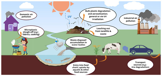
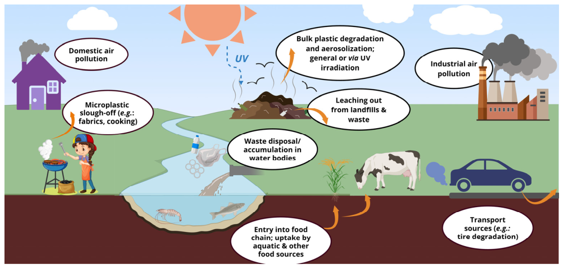
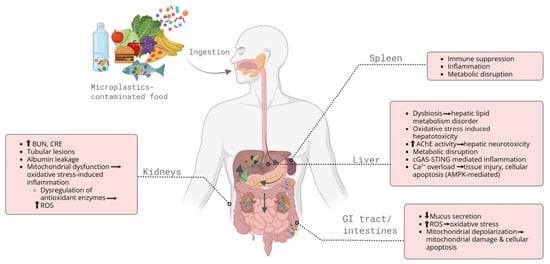
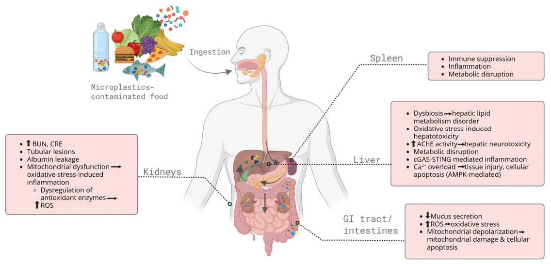
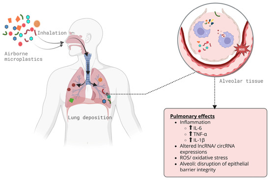
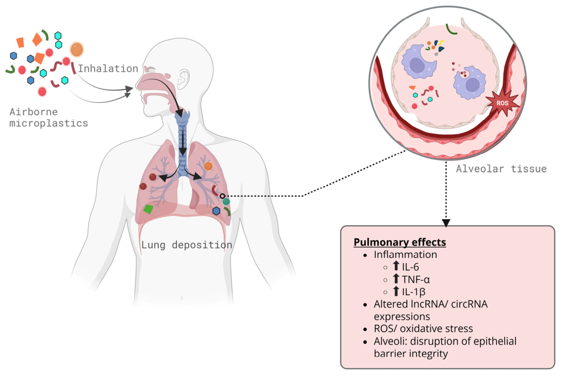

Disclaimer/Publisher’s Note: The statements, opinions and data contained in all publications are solely those of the individual author(s) and contributor(s) and not of MDPI and/or the editor(s). MDPI and/or the editor(s) disclaim responsibility for any injury to people or property resulting from any ideas, methods, instructions or products referred to in the content. |
© 2026 by the authors. Licensee MDPI, Basel, Switzerland. This article is an open access article distributed under the terms and conditions of the Creative Commons Attribution (CC BY) license.