


Abstract
Feline calicivirus (FCV) is a genetically diverse and rapidly evolving positive-sense RNA virus that remains a major cause of infectious disease in domestic cats worldwide. FCV is classically associated with upper respiratory tract disease and oral ulceration; however, the emergence of highly virulent systemic FCV (VS-FCV) strains has expanded its clinical significance owing to severe multisystemic disease, increased mortality, and frequent outbreaks in shelters, breeding colonies, and veterinary healthcare facilities. Continuous viral evolution driven by mutation, recombination, and immune selection promotes antigenic diversity, prolonged asymptomatic shedding, vaccine escape, and the emergence of novel variants, thereby complicating diagnosis, prevention, and disease control. This review summarizes recent advances in FCV molecular biology, viral evolution, host–virus interactions, pathogenesis, epidemiology, clinical manifestations, diagnostics, vaccination, and emerging antiviral and immunomodulatory therapies. The review also examines the available epidemiological evidence on FCV in India, highlighting reported regional findings, limitations in geographic and genomic coverage, and the need for coordinated surveillance. Within a One Health framework, FCV is considered primarily as an important companion-animal pathogen and a model for strengthening veterinary surveillance, biosecurity, genomic monitoring, and outbreak preparedness, rather than as a confirmed zoonotic threat. By integrating current scientific evidence with identified surveillance and research gaps, this review outlines priorities for improving FCV surveillance, prevention, and control and for strengthening preparedness for emerging companion-animal viral diseases.
1. Introduction
Emerging and re-emerging RNA viruses continue to represent a major challenge to global animal and public health because of their rapid evolutionary rates, extensive genetic diversity, and remarkable ability to adapt to changing host and environmental conditions. Although many RNA viruses of public-health concern are zoonotic, companion-animal pathogens such as feline calicivirus (FCV) are primarily important because of their effects on animal health, welfare, and veterinary healthcare systems. A One Health perspective recognizes the interconnectedness of companion animal health, environmental management, veterinary practice, and human activities. The growing companion animal population, increasing international movement of pets, expansion of animal shelters and breeding facilities, and changing patterns of urbanization can facilitate the persistence and dissemination of infectious pathogens within animal populations. Accordingly, strengthening surveillance, biosecurity, and outbreak preparedness for companion-animal diseases can improve veterinary disease control and laboratory capacity, while also providing infrastructure that may be applicable to the surveillance of other emerging pathogens [1].
Among viral pathogens affecting domestic cats, feline calicivirus (FCV) remains one of the most widespread, genetically diverse, and evolutionarily dynamic RNA viruses worldwide. FCV is a non-enveloped, positive-sense single-stranded RNA virus belonging to the genus Vesivirus within the family Caliciviridae. Similar to many RNA viruses, the lack of proofreading activity in its RNA-dependent RNA polymerase results in a high mutation rate that continuously generates genetically heterogeneous viral populations. Together with homologous recombination and immune-mediated selection, these evolutionary processes facilitate viral persistence, antigenic diversification, vaccine escape, and the emergence of novel variants capable of adapting to changing host populations [2,3,4].
FCV exhibits substantial genetic and antigenic diversity, and infection may result in prolonged viral shedding, allowing continued transmission within multi-cat populations. Although vaccination reduces clinical disease, protection against infection and shedding is incomplete, contributing to continued circulation of genetically diverse field viruses [3,5]. Molecular epidemiological studies have demonstrated extensive strain diversity within and between feline populations, emphasizing the importance of continued genomic surveillance and characterization of circulating variants [6,7].
Clinically, FCV is recognized as a major cause of feline upper respiratory tract disease, oral ulceration, conjunctivitis, polyarthritis, and chronic gingivostomatitis. However, the emergence of virulent systemic feline calicivirus (VS-FCV) has significantly altered the perception of FCV from a relatively mild respiratory pathogen to one capable of causing severe multisystemic disease characterised by vasculitis, facial oedema, cutaneous ulceration, pneumonia, hepatic dysfunction, and mortality exceeding 50% during outbreaks. Such outbreaks have been reported in veterinary hospitals, shelters, rescue centres, breeding colonies, and multi-cat households, where high animal density, environmental contamination, and prolonged viral shedding facilitate rapid transmission and continuous viral evolution [3,4,8,9].
One of the greatest challenges in FCV control is its remarkable genetic and antigenic diversity. Unlike genetically stable viruses, FCV circulates as heterogeneous quasi-species that continually evolve under host immune pressure. Consequently, naturally acquired and vaccine-induced immunity often provide incomplete cross-protection, allowing vaccinated animals to become infected and shed virus despite reduced clinical severity. Persistent carrier cats further contribute to viral maintenance and act as evolutionary reservoirs in which genetically distinct variants may emerge over time. These characteristics closely resemble evolutionary processes observed in several medically important RNA viruses and highlight FCV as an informative model for understanding viral adaptation, persistence, and outbreak dynamics in companion animal populations [10,11].
Recent advances in molecular diagnostics and whole-genome sequencing have improved the understanding of FCV epidemiology and viral evolution. Molecular epidemiological studies have demonstrated substantial genetic diversity among circulating FCV strains, while sequence-based investigations have identified geographically distributed lineages and recombination events. These findings highlight the limitations of relying solely on conventional epidemiological investigations and underscore the growing importance of genomic epidemiology for monitoring viral evolution, characterizing circulating variants, and evaluating vaccine–field strain relationships [2,6,7,8,12,13,14,15].
These challenges are particularly relevant in developing countries such as India, where the rapidly expanding companion animal population, increasing establishment of shelters and breeding facilities, and limited molecular surveillance provide favourable conditions for sustained viral circulation and evolution. Although sporadic molecular studies have reported genetically distinct Indian FCV isolates, comprehensive nationwide epidemiological data, whole-genome surveillance, and systematic evaluation of circulating strains remain limited. Addressing these knowledge gaps is essential for strengthening veterinary preparedness and developing evidence-based prevention and control strategies.
This review provides a comprehensive and updated overview of FCV biology, with emphasis on viral evolution, molecular epidemiology, host–virus interactions, clinical manifestations, diagnostics, vaccination, and emerging antiviral approaches. Particular attention is given to recent advances in genomic surveillance, outbreak preparedness, and One Health strategies, together with the current status of FCV in India and future research priorities. By integrating contemporary advances in virology with modern surveillance and preparedness concepts, this review aims to provide a practical framework for improving the prevention and control of FCV while illustrating the broader significance of companion animal RNA viruses in global infectious disease preparedness.
2. Global Importance of Feline Calicivirus as an Emerging Companion Animal RNA Virus
Feline calicivirus (FCV) is one of the most prevalent and evolutionarily dynamic viral pathogens affecting domestic cats worldwide. Although traditionally recognized as an important cause of feline upper respiratory tract disease, increasing evidence indicates that FCV should be regarded as an emerging companion animal RNA virus because of its remarkable genetic plasticity, broad clinical spectrum, capacity for persistent infection, and ability to generate highly virulent outbreak-associated variants. Similar to many medically important RNA viruses, FCV evolves continuously through mutation, recombination, and immune-mediated selection, enabling rapid adaptation to changing host populations and facilitating long-term persistence despite widespread vaccination [2,3,11].
The global expansion of companion animal ownership has substantially altered the epidemiology of infectious diseases affecting domestic cats. Urbanization, international movement of pets, commercial breeding, rescue organizations, and increasing numbers of multicat households have created favourable conditions for the introduction, maintenance, and dissemination of contagious pathogens. In these environments, FCV is efficiently transmitted through direct contact with oral and respiratory secretions as well as indirect exposure to contaminated fomites. Furthermore, prolonged viral shedding by clinically recovered or asymptomatic carrier cats enables continuous circulation within feline populations, making eradication virtually impossible [10,16,17].
Unlike many respiratory viruses that cause self-limiting infections, FCV exhibits considerable clinical and genetic heterogeneity. While most infections result in mild respiratory disease and oral ulceration, the emergence of virulent systemic feline calicivirus (VS-FCV) has demonstrated the virus’s capacity to cause severe multisystemic disease characterised by vasculitis, cutaneous ulceration, facial oedema, pneumonia, hepatic dysfunction, and mortality rates exceeding 50% during outbreaks. Such outbreaks have been reported in veterinary hospitals, animal shelters, breeding colonies, and rescue centres across North America, Europe, Asia, and Australia, emphasising the global significance of FCV and the importance of rapid outbreak recognition and containment [4,8,9].
A defining feature of FCV is its extraordinary antigenic diversity. The error-prone RNA-dependent RNA polymerase continuously generates genetically heterogeneous viral populations, while selective pressure imposed by host immunity promotes the emergence of variants capable of escaping neutralizing antibodies. Consequently, currently available vaccines generally reduce disease severity but do not consistently prevent infection, viral shedding, or transmission. Persistent infection further accelerates viral evolution by providing opportunities for within-host diversification, allowing chronically infected cats to serve both as reservoirs of infection and as sources of novel viral variants [2,3]. These evolutionary characteristics closely resemble those observed in several rapidly evolving RNA viruses of human and veterinary importance, making FCV an informative model for studying viral adaptation, antigenic drift, and outbreak emergence.
Recent advances in molecular diagnostics and whole-genome sequencing have transformed the understanding of FCV epidemiology. High-resolution genomic surveillance has demonstrated that genetically distinct FCV variants frequently circulate simultaneously within individual shelters or veterinary hospitals, whereas closely related viruses may be detected across geographically distant regions owing to international movement of companion animals. These findings highlight the limitations of relying solely on traditional epidemiological investigations and underscore the growing importance of genomic epidemiology for identifying transmission pathways, monitoring viral evolution, and evaluating vaccine relevance [11].
Although FCV has no confirmed zoonotic potential, its importance extends beyond feline medicine. The virus exemplifies many of the evolutionary and epidemiological principles that govern emerging RNA viruses, including rapid mutation, recombination, immune escape, persistent infection, and outbreak-associated evolution. Within the One Health framework, surveillance of FCV contributes to strengthening veterinary diagnostic capacity, improving infection-control practices, supporting antimicrobial stewardship, and enhancing preparedness for future emerging companion animal pathogens [1]. Consequently, FCV should be viewed not only as a significant feline pathogen but also as a valuable model for understanding the emergence, surveillance, and control of rapidly evolving RNA viruses in companion animal populations.
FCV continues to circulate worldwide in household, shelter, breeding and free-roaming cat populations, posing substantial challenges to companion animal health through its extensive genetic diversity, efficient transmission and persistent carrier states (Figure 1).
Figure 1.
Worldwide distribution of feline calicivirus (FCV) infection. The map illustrates the reported geographical occurrence of FCV in domestic cats across different continents, highlighting the widespread distribution of the virus globally. Created in BioRender. Publication License Details: https://BioRender.com/r9vygl1.
Epidemiological Status of Feline Calicivirus in India
Information on the epidemiology of feline calicivirus (FCV) in India remains relatively limited and geographically fragmented, with available studies differing in sampling populations, clinical presentation, diagnostic approaches, and molecular characterization. Recent Indian investigations nevertheless provide evidence of ongoing FCV circulation among domestic cats. A retrospective cross-sectional study conducted at the ICAR–Indian Veterinary Research Institute, Bareilly, Uttar Pradesh, evaluated 675 feline clinical samples collected between November 2023 and November 2025. FCV was detected by PCR in 1 of 675 samples (0.14%), whereas feline panleukopenia virus and feline herpesvirus-1 were detected at higher frequencies. The authors emphasized the continued circulation of feline viral pathogens despite vaccination and highlighted the need for sustained molecular surveillance and optimization of vaccination strategies [18].
Recent molecular evidence from Northeast India further indicates regional circulation and genetic variation of FCV. In a 2025 study from Assam, 56 vaccinated cats presenting with upper respiratory tract disease were investigated using RT-PCR targeting the FCV VP1 gene. FCV was detected in 5 of 56 cats (8.93%) when FCV-positive animals, including co-infections, were considered, while three animals showed FCV as a standalone infection (5.36%). The study also identified FCV/FHV-1 co-infections and examined the partial VP1 sequence of an FCV-positive sample.
Importantly, phylogenetic analysis of the Assam FCV isolate (PZ727510) showed clustering within an Asian lineage, with the isolate grouping closely with Chinese and South Korean field strains and occupying a branch distinct from the commercial vaccine strains F9 and 2024 included in the analysis. These findings provide preliminary evidence that FCV circulating in Indian cats may be genetically related to regional Asian field viruses, although the interpretation is limited by the analysis of a single partial VP1 sequence [18].
The marked difference between the detection frequencies reported in Bareilly (0.14%) and Assam (8.93% overall FCV positivity; 5.36% standalone FCV detection) should not be interpreted as a direct estimate of regional prevalence. The studies examined different populations and clinical settings: the Bareilly investigation used a broader retrospective collection of feline clinical samples, whereas the Assam investigation specifically enrolled cats presenting with upper respiratory tract disease. Differences in sampling strategy, disease status, geographic location, diagnostic methodology, and population structure may therefore contribute to the observed variation.
Despite these emerging observations, India currently lacks a coordinated nationwide FCV surveillance framework integrating standardized molecular diagnostics, systematic sampling across geographic regions, sequence-based epidemiology, and longitudinal monitoring of circulating strains. Available evidence is concentrated in individual institutions or regions, limiting assessment of the national distribution, genetic diversity, evolutionary dynamics, and relationship between field strains and vaccine strains. Future studies should therefore incorporate multicentric sampling from owned, free-roaming, shelter, breeding, and clinically affected cat populations; standardized RT-PCR/RT-qPCR-based detection; capsid gene sequencing and, where feasible, whole-genome sequencing; and phylogenetic and recombination analyses. Such coordinated surveillance would help establish an evidence-based epidemiological baseline for FCV in India and support evaluation of vaccine cross-protection, outbreak investigation, and development of regionally relevant prevention and control strategies.
3. Molecular Evolution and Genomic Epidemiology of Feline Calicivirus
Feline calicivirus (FCV) is a non-enveloped, positive-sense single-stranded RNA virus belonging to the genus Vesivirus within the family Caliciviridae. Its approximately 7.5–7.7 kb genome comprises three open reading frames (ORF1–ORF3), encoding non-structural proteins involved in viral replication and two structural proteins, VP1 and VP2, responsible for virion assembly and infectivity [3,19]. Although structurally compact, the FCV genome exhibits remarkable functional plasticity, enabling efficient replication, rapid adaptation, and continuous genetic diversification. These characteristics underpin the virus’s persistence in feline populations and its ability to generate antigenically distinct variants capable of causing endemic disease and sporadic outbreaks.
The viral capsid is primarily composed of the major structural protein VP1, which contains conserved shell (S) and variable protruding (P) domains. The hypervariable P2 subdomain is exposed on the virion surface and mediates receptor binding and antibody recognition. Consequently, this region accumulates mutations more rapidly than other genomic regions and represents the principal determinant of antigenic diversity and vaccine escape. In contrast, the minor structural protein VP2, although present in small quantities, plays an essential role in virion stability and genome delivery during viral entry [20,21].
FCV initiates infection by binding to feline junctional adhesion molecule-A (fJAM-A) expressed on epithelial cells of the oral and upper respiratory tract. Receptor engagement induces conformational rearrangements within the viral capsid, facilitating clathrin-mediated endocytosis and release of the viral RNA genome into the cytoplasm. Following entry, the genomic RNA functions directly as messenger RNA for translation of the ORF1 polyprotein, which is subsequently cleaved into functional non-structural proteins, including the viral protease, VPg, helicase, and RNA-dependent RNA polymerase (RdRp). These proteins coordinate viral RNA synthesis, intracellular membrane remodeling, and assembly of replication complexes within the cytoplasm [3,19]. The genome organization and intracellular replication strategy of FCV are summarized in Figure 2.
Figure 2.
Genome organization of feline calicivirus (FCV). Schematic representation of the FCV genome organization. The figure also illustrates the structural and non-structural proteins involved in viral replication, assembly, and release following receptor-mediated cell entry. Created in BioRender. Publication License Details: https://BioRender.com/2xzxj8t.
A defining molecular characteristic of FCV is the absence of proofreading activity in the viral RNA-dependent RNA polymerase, resulting in a high mutation rate during replication. Continuous accumulation of nucleotide substitutions generates genetically heterogeneous viral populations, commonly referred to as quasispecies, which provide a substrate for natural selection [22]. Under host immune pressure, variants possessing enhanced replication efficiency, altered receptor affinity, or reduced susceptibility to neutralizing antibodies are preferentially maintained. This dynamic evolutionary process promotes antigenic drift, persistent infection, and adaptation to vaccinated populations, thereby complicating long-term disease control [11].
In addition to point mutations, homologous recombination contributes significantly to FCV evolution. Recombination occurs when genetically distinct viruses co-infect the same host cell, allowing exchange of genomic segments and generation of novel viral lineages with altered biological characteristics. Most recombination events have been reported near the ORF1–ORF2 junction, although recombination within capsid-associated regions has also been described. Together with mutation, recombination accelerates viral diversification and contributes to the emergence of epidemiologically distinct strains [10,11].
The major viral, host, and environmental determinants responsible for FCV evolution, persistence, epidemiological spread and outbreak amplification are summarized in Table 1.
Table 1.
Determinants of Feline Calicivirus Evolution, Persistence, Transmission and Outbreak Amplification.
Persistent infection further amplifies viral evolution. Approximately 10–20% of infected cats continue to shed infectious virus for prolonged periods despite apparent clinical recovery. Continuous replication within these carrier animals provides opportunities for accumulation of adaptive mutations under immune selection, allowing chronically infected cats to function as both reservoirs of transmission and evolutionary incubators for novel variants [10]. This characteristic distinguishes FCV from many acute respiratory viruses and contributes substantially to its global persistence.
Despite extensive genomic diversity, no universally conserved molecular marker has been identified that reliably differentiates virulent systemic FCV (VS-FCV) from classical field strains. Comparative genomic studies indicate that highly virulent viruses emerge independently through multiple evolutionary pathways rather than from dissemination of a single hypervirulent lineage. Disease severity therefore appears to result from complex interactions among viral genotype, host immune responses, tissue tropism, and environmental factors rather than specific nucleotide substitutions alone [4,8]. Consequently, diagnosis of VS-FCV requires integration of clinical findings, epidemiological investigation, and genomic analysis rather than reliance on molecular detection alone.
Advances in structural biology and next-generation sequencing have substantially improved understanding of FCV molecular evolution. Whole-genome sequencing has demonstrated that genetically distinct variants frequently coexist within individual shelters and veterinary hospitals, while closely related viruses may circulate across geographically distant regions through movement of companion animals. Such observations highlight the limitations of partial capsid sequencing and emphasise the importance of comprehensive genomic surveillance for outbreak investigation, transmission tracking, and evaluation of vaccine–field strain relationships [11].
Collectively, the compact genome, error-prone replication machinery, capacity for recombination, and prolonged persistence make FCV one of the most evolutionarily dynamic RNA viruses affecting companion animals. These molecular characteristics not only explain the remarkable diversity observed among circulating field strains but also provide the biological basis for viral persistence, diagnostic challenges, vaccine escape, and the continuing emergence of novel variants. A detailed understanding of these mechanisms is therefore fundamental for the development of improved molecular diagnostics, broadly protective vaccines, and effective surveillance strategies.
4. Host–Virus Interactions and Pathogenesis
The pathogenesis of feline calicivirus (FCV) is determined by the interaction between viral genetic characteristics, cellular receptor usage, innate and adaptive immune responses, tissue tropism, and host susceptibility. This complex relationship explains the broad spectrum of outcomes ranging from asymptomatic infection and localized oral or respiratory disease to persistent infection and fatal virulent systemic feline calicivirus (VS-FCV). Importantly, disease severity cannot be attributed to a single viral mutation; rather, it reflects the combined effects of viral replication, immune evasion, inflammatory injury, and host-related factors [2,3].
4.1. Cellular Attachment, Entry, and Replication
FCV infection begins with attachment of the VP1 capsid protein to feline junctional adhesion molecule-A (fJAM-A), an immunoglobulin-superfamily receptor expressed at epithelial tight junctions. Receptor binding is mediated predominantly by the protruding P2 domain of VP1 and induces conformational rearrangements within the viral capsid. These structural changes expose the minor capsid protein VP2, which assembles into a portal-like structure that facilitates transfer of the viral genome across the endosomal membrane into the cytoplasm [20,21]. The functions of the major viral proteins, their interactions with host cellular pathways, and their potential exploitation as therapeutic targets are summarized in Table 2.
Table 2.
Viral Proteins, Host–Virus Interactions and Therapeutic Targets. Integrated summary of the major FCV proteins involved in host entry, replication, immune evasion and their potential exploitation for antiviral intervention.
Following predominantly clathrin-mediated endocytosis and endosomal acidification, the positive-sense RNA genome is released into the cytoplasm and translated directly. Viral non-structural proteins reorganize intracellular membranes to form replication complexes in which genomic and subgenomic RNAs are synthesized. Viral protease-mediated polyprotein processing and RNA-dependent RNA polymerase activity support rapid viral amplification, whereas suppression of cellular protein synthesis redirects host resources toward viral replication. Productive infection culminates in marked cytopathic effects and cell lysis, particularly within oral and respiratory epithelia [3,19].
4.2. Innate Immune Modulation and Inflammatory Injury
Early recognition of FCV RNA by intracellular pattern-recognition receptors would normally activate type I interferon and other antiviral pathways. However, FCV has evolved mechanisms that suppress these responses, thereby permitting efficient replication before effective adaptive immunity develops. Viral non-structural proteins interfere with interferon-associated signalling, host translation, autophagy, and cellular stress responses. In particular, the FCV p39 protein has been reported to promote autophagy-dependent degradation of retinoic acid-inducible gene I, thereby reducing antiviral signalling and favouring viral replication [51].
Although immune evasion facilitates replication, excessive host inflammation is an equally important determinant of tissue injury. FCV infection induces inflammatory mediators, including interleukin-1β, interleukin-6, tumour necrosis factor-α, chemokines, and interferons, which recruit neutrophils and macrophages to affected tissues. Controlled inflammation may contribute to viral clearance, whereas dysregulated responses can intensify epithelial necrosis, ulceration, vascular leakage, oedema, and secondary tissue damage. Thus, FCV pathogenesis reflects both direct virus-mediated cytolysis and immune-mediated injury.
4.3. Tissue Tropism and Clinical Correlates
The oral cavity and upper respiratory tract are the principal sites of FCV replication. Infection and destruction of lingual, palatal, and oropharyngeal epithelial cells produce the characteristic oral ulcers associated with acute disease. Replication in nasal and respiratory epithelia contributes to fever, sneezing, nasal discharge, conjunctivitis, and upper respiratory inflammation. Disruption of mucosal barriers may also predispose affected cats to secondary bacterial infection.
Certain strains exhibit broader tissue tropism and may involve the lungs, synovial tissues, skin, liver, pancreas, and vascular endothelium. Pulmonary infection may cause bronchointerstitial pneumonia, alveolar injury, and respiratory distress, particularly in young or immunocompromised cats. Transient polyarthritis and limping syndrome are thought to involve immune-complex deposition and inflammatory synovitis rather than extensive direct viral destruction of joints. Persistent viral antigen stimulation has also been associated with feline chronic gingivostomatitis, although FCV is unlikely to act as the sole cause of this multifactorial inflammatory disorder [2].
4.4. Persistence and Immune Escape
A proportion of infected cats continue to shed FCV after clinical recovery. Persistent shedding is supported by incomplete immune clearance, ongoing viral replication, antigenic variation, and selection of escape variants. Within-host viral populations may progressively diverge under antibody pressure, allowing FCV to remain detectable for months and, in some animals, considerably longer [10].
Carrier cats are epidemiologically important because they maintain infection within multicat populations and may introduce virus into shelters, breeding colonies, or households without displaying clinical signs. Persistent infection also provides an extended evolutionary environment in which mutations can accumulate, potentially generating variants with altered antigenicity, transmission efficiency, or tissue tropism. Vaccination generally reduces clinical severity but does not consistently prevent infection or shedding, further permitting circulation of FCV in vaccinated populations. The sequential events involved in FCV pathogenesis and the principal mechanisms of immune evasion are summarized in Figure 3.
Figure 3.
Pathogenesis and immune evasion of feline calicivirus (FCV). Sequential overview of FCV pathogenesis from viral entry and tissue tropism to host immune responses and immune evasion mechanisms. The figure summarizes the key events leading to persistent infection, systemic dissemination, and disease progression. Created in BioRender. Publication License Details: https://BioRender.com/4jdabmh.
4.5. Pathogenesis of Virulent Systemic FCV
VS-FCV represents the most severe clinical outcome of infection and is characterised by fever, facial and limb oedema, ulcerative skin lesions, pneumonia, jaundice, hepatic dysfunction, coagulopathy, and multiorgan failure. Pathological findings commonly include epithelial necrosis, vasculitis, endothelial injury, oedema, and widespread inflammatory lesions. These features indicate that systemic disease is driven by expanded tissue tropism together with severe vascular and inflammatory dysfunction [9,55].
Recent experimental evidence suggests that functional divergence among FCV genotypes may contribute to differences in pathogenicity. Two major FCV genotypes, GI and GII, have been identified, with the GII isolate WX9 exhibiting higher mortality and broader tissue tropism than the GI isolate WH0531 in experimental infection. GII was also associated with distinct codon usage patterns and VP1 structural evolution, suggesting that genotype-associated capsid variation may influence viral fitness and disease severity. Nevertheless, GII cannot be considered a universal molecular marker of VS-FCV, as pathogenicity is influenced by complex interactions among viral genetic variation, host responses, and tissue tropism [56]. Recent clinical and pathological evidence further supports the complex relationship between FCV genetic variation and disease phenotype. A fatal FCV-VSD infection in a young kitten was associated with severe pneumonia and footpad oedema, with FCV RNA detected in multiple organs and the highest viral RNA load observed in the lungs. Immunohistochemical analysis confirmed viral antigen in pulmonary tissue, with alveolar macrophages identified as the principal target cells and potential site of viral replication. Notably, phylogenetic analysis of the ORF2 gene showed that the highly virulent virus was not genetically distinguishable from FCV strains associated with upper respiratory tract disease. These findings indicate that severe systemic disease may arise from genetically diverse FCV populations and further support the view that VS-FCV pathogenicity is influenced by the interaction of viral genetic variation, tissue tropism, host factors, and environmental conditions rather than by a single molecular signature [57].
Comparative sequencing has not identified a single mutation or conserved genomic signature that reliably distinguishes all VS-FCV isolates from classical strains. Most systemic outbreaks appear to arise independently and involve genetically distinct viruses, suggesting that virulence can emerge through multiple evolutionary pathways. Alterations in capsid structure may influence receptor engagement or tissue tropism, whereas changes in non-structural proteins may affect replication kinetics and immune evasion. Nevertheless, these viral properties interact with host immunity, age, vaccination status, concurrent infection, and environmental stress to determine clinical outcome [4,8].
5. Clinical Spectrum and Diagnostic Approaches
5.1. Clinical Manifestations and Disease Spectrum
Feline calicivirus (FCV) infection exhibits remarkable clinical diversity, ranging from inapparent infection and mild upper respiratory disease to chronic inflammatory disorders and highly fatal systemic illness. Disease outcomes are influenced by viral strain, host age, immune status, vaccination history, concurrent infections, and environmental stress. Consequently, FCV should be regarded as a disease continuum rather than a single clinical entity. Because asymptomatic viral shedding is common and clinical signs frequently overlap with other pathogens of the feline upper respiratory disease complex (FURDC), clinical examination alone is insufficient for definitive diagnosis [2,3].
The majority of naturally infected cats develop acute upper respiratory disease, particularly kittens, shelter cats, and animals housed in multi-cat environments. Following an incubation period of approximately 2–5 days, affected cats typically present with pyrexia, lethargy, anorexia, sneezing, serous to mucopurulent nasal discharge, conjunctivitis, hypersalivation, and oral ulceration. Ulcerative lesions involving the tongue, hard palate, lips, and gingiva are considered among the most characteristic manifestations of FCV infection and often contribute to dysphagia, reduced food intake, and dehydration. While uncomplicated infections are generally self-limiting, secondary bacterial infections may prolong recovery, particularly in young or immunocompromised animals [3,16].
Although the respiratory tract represents the principal site of viral replication, FCV demonstrates considerable tissue tropism. Infection of pulmonary epithelial cells may result in bronchointerstitial pneumonia, especially in neonatal kittens, geriatric cats, or animals with compromised immunity. Some strains are also associated with limping syndrome, a transient febrile polyarthritis characterised by acute lameness, joint pain, and reluctance to move. These musculoskeletal manifestations are believed to result primarily from immune-mediated inflammation rather than extensive viral destruction of synovial tissues and generally resolve without permanent joint damage [3,58].
Persistent infection represents a distinctive biological feature of FCV. A proportion of clinically recovered cats continue to shed infectious virus for months or even years despite the absence of overt disease. These carrier animals play a critical epidemiological role by maintaining viral circulation within households, breeding colonies, shelters, and veterinary hospitals. Continuous viral replication under host immune pressure also facilitates antigenic diversification, allowing emergence of variants capable of evading pre-existing immunity [2,10]. Persistent FCV infection has additionally been implicated as one of several contributing factors in feline chronic gingivostomatitis, a severe immune-mediated inflammatory disorder characterised by painful proliferative oral lesions and chronic oral discomfort. Although FCV is unlikely to represent the sole etiological agent, viral persistence may amplify local immune dysregulation and perpetuate chronic inflammation [59].
The most severe manifestation of FCV infection is virulent systemic feline calicivirus (VS-FCV), an uncommon but devastating syndrome associated with high morbidity and mortality. Unlike classical respiratory disease, VS-FCV is characterised by widespread viral dissemination and systemic inflammatory injury. Affected cats commonly develop high fever, facial and limb oedema, ulcerative dermatitis, jaundice, dyspnoea, pneumonia, hepatic dysfunction, pancreatitis, coagulopathy, and multiorgan failure. Mortality frequently exceeds 50% during institutional outbreaks, particularly in veterinary hospitals and animal shelters where close animal contact facilitates rapid transmission [4,9,55]. Importantly, genomic studies have demonstrated that VS-FCV strains do not share a single conserved virulence marker, indicating that systemic disease results from complex interactions between viral evolution and host responses rather than a unique genetic lineage [8,11].
From a clinical perspective, recognition of disease severity is essential for early intervention and outbreak control. Mild respiratory disease generally requires supportive care, whereas the occurrence of rapidly progressive oedema, cutaneous ulceration, jaundice, unexplained pneumonia, or increased mortality within multicat populations should immediately raise suspicion of VS-FCV and prompt laboratory confirmation and implementation of strict infection-control measures. Thus, appreciation of the full clinical spectrum of FCV not only facilitates appropriate case management but also provides the foundation for selecting suitable diagnostic tests and guiding epidemiological investigations. The distinguishing clinical features of classical and virulent systemic FCV are summarized in Table 3.
Table 3.
Clinical spectrum of classical feline calicivirus (FCV) and virulent systemic FCV (VS-FCV).
5.2. Laboratory Diagnosis and Emerging Diagnostic Technologies
Accurate laboratory diagnosis is essential for confirming feline calicivirus (FCV) infection, differentiating it from other pathogens within the feline upper respiratory disease complex (FURDC), identifying persistent carriers, and rapidly detecting outbreaks of virulent systemic feline calicivirus (VS-FCV). Because clinical manifestations vary considerably and asymptomatic viral shedding is common, diagnosis should integrate clinical findings, epidemiological information, and laboratory testing. No single diagnostic assay is universally applicable, and selection of appropriate methods depends on the clinical presentation, stage of infection, specimen quality, and intended diagnostic objective [2,3].
The key features of conventional and emerging diagnostic technologies for feline calicivirus (FCV), including their turnaround time, major advantages, limitations, and primary applications, are compared in Table 4.
Table 4.
Comparison of current and emerging diagnostic technologies.
Appropriate sample selection is fundamental for reliable diagnosis. Oropharyngeal, oral, conjunctival, and nasal swabs collected during the acute phase generally contain the highest viral loads and are therefore preferred for molecular detection. In cats with lower respiratory involvement, bronchoalveolar lavage fluid may improve diagnostic sensitivity, whereas tissue samples from the liver, lungs, spleen, or skin collected during necropsy are valuable for investigating suspected VS-FCV outbreaks. Because viral shedding may continue long after clinical recovery, positive molecular results should always be interpreted in conjunction with clinical signs and epidemiological evidence rather than being regarded as definitive proof of active disease [3,10].
Historically, virus isolation in feline cell cultures was considered a diagnostic reference method because it confirmed the presence of infectious virus and enabled antigenic characterisation. However, isolation is labor-intensive, requires specialized laboratory facilities, and may fail when viral titres are low or specimens have been improperly transported. Consequently, virus isolation is now primarily reserved for epidemiological investigations, vaccine studies, and characterisation of emerging strains rather than routine clinical diagnosis [3].
Molecular techniques have become the cornerstone of FCV diagnosis. Conventional reverse transcription polymerase chain reaction (RT-PCR) and real-time quantitative RT-PCR (RT-qPCR) provide rapid, sensitive, and highly specific detection of viral RNA directly from clinical specimens. Real-time assays additionally permit quantification of viral load and reduce contamination risk associated with post-amplification processing. Nevertheless, the extensive genetic diversity of FCV presents an important diagnostic challenge because mutations within primer- and probe-binding regions may reduce assay sensitivity. Accordingly, diagnostic laboratories should periodically evaluate and update primer sets using contemporary sequence data to maintain analytical performance [11,66].
Multiplex molecular assays capable of simultaneously detecting FCV together with feline herpesvirus type 1, Chlamydia felis, Mycoplasma felis, and other respiratory pathogens have substantially improved the diagnosis of feline respiratory disease. These assays reduce turnaround time, conserve clinical specimens, and facilitate recognition of mixed infections, which are common in shelters and multi-cat environments. Interpretation, however, should remain cautious because molecular detection of FCV does not necessarily distinguish active infection from asymptomatic viral carriage [75].
Growing demand for rapid point-of-care diagnosis has stimulated development of several isothermal nucleic acid amplification technologies, including loop-mediated isothermal amplification (RT-LAMP), recombinase polymerase amplification (RPA), and recombinase-aided amplification (RAA). These methods eliminate the need for thermal cycling, substantially reduce assay time, and can be performed using portable equipment, making them attractive for field investigations and resource-limited veterinary settings. When combined with colorimetric detection or lateral-flow readouts, isothermal assays offer practical alternatives to conventional PCR, although broader multi-centre validation and standardization remain necessary before routine implementation [67,76].
Recent advances in molecular biosensing have introduced CRISPR-Cas-based diagnostics for FCV detection. CRISPR-Cas12 and Cas13 systems combine isothermal amplification with sequence-specific recognition, enabling rapid detection with high analytical sensitivity while minimizing false-positive results. Although these assays remain largely experimental, they represent promising next-generation diagnostic platforms capable of supporting decentralized testing during disease outbreaks [77]. Similarly, integration of microfluidics, nanotechnology, smartphone-assisted image analysis, and biosensor-based detection is expected to accelerate the development of affordable point-of-care devices suitable for veterinary practice and shelter surveillance.
Whole-genome sequencing (WGS) has become increasingly important for understanding FCV epidemiology. Unlike routine diagnostic PCR, WGS enables comprehensive analysis of viral evolution, recombination, transmission pathways, and vaccine–field strain relationships. Genomic surveillance has demonstrated that genetically distinct FCV variants often circulate simultaneously within individual shelters and veterinary hospitals, whereas closely related viruses may spread across geographically distant regions through movement of companion animals. During suspected VS-FCV outbreaks, sequencing therefore provides valuable information for outbreak reconstruction and monitoring viral evolution, although no conserved molecular marker currently distinguishes all systemic strains from classical FCV isolates [4,11].
Despite substantial technological progress, several diagnostic challenges remain unresolved. Persistent viral shedding, extensive antigenic diversity, intermittent virus excretion, and absence of genotype-specific virulence markers complicate interpretation of laboratory findings. Consequently, laboratory diagnosis should not rely solely on detection of viral RNA but should incorporate clinical assessment, vaccination history, epidemiological context, and, when appropriate, genomic investigation. Future diagnostic strategies will likely combine multiplex molecular detection, portable isothermal amplification, CRISPR-based biosensing, whole-genome sequencing, and artificial intelligence-assisted interpretation to enable rapid identification of emerging variants and strengthen outbreak preparedness.
Collectively, advances in molecular diagnostics have transformed FCV detection from conventional laboratory confirmation to comprehensive genomic surveillance. Continued development of rapid, affordable, and field-deployable diagnostic platforms, together with coordinated international surveillance, will be essential for improving disease control, monitoring viral evolution, and supporting evidence-based prevention strategies for this continuously evolving companion animal pathogen.
6. Vaccination, Vaccine Escape, and Future Vaccination Strategies
Vaccination remains the cornerstone of feline calicivirus (FCV) prevention and has substantially reduced the severity of clinical disease worldwide. Nevertheless, complete control of FCV remains challenging because of extensive genetic and antigenic diversity, persistent viral shedding, incomplete sterilizing immunity, and the continual emergence of novel variants. Unlike many viral diseases where vaccination effectively prevents infection, current FCV vaccines primarily reduce disease severity, viral shedding, and mortality rather than completely preventing infection or transmission. Consequently, vaccinated cats may still become infected and contribute to viral circulation, particularly in multicat environments such as shelters, breeding colonies, and veterinary hospitals [3,16].
Current commercial vaccines consist predominantly of modified-live virus (MLV) and inactivated (killed) vaccines, either as monovalent formulations or combined with other core feline vaccines against feline herpesvirus-1 (FHV-1) and feline panleukopenia virus (FPV). Modified-live vaccines generally induce rapid mucosal, humoral, and cell-mediated immune responses and often provide broader cross-protection than inactivated vaccines because limited viral replication better mimics natural infection. However, MLV vaccines should be used cautiously in severely immunocompromised animals and are generally avoided in situations where vaccine virus shedding could present theoretical concerns. Inactivated vaccines exhibit excellent safety profiles but usually require booster immunizations and adjuvants to achieve comparable immune protection [16,78].
Protective immunity against FCV depends on both virus-neutralizing antibodies and cellular immune responses. Neutralizing antibodies directed primarily against the hypervariable P2 domain of the VP1 capsid protein reduce viral attachment and spread, whereas T-cell responses contribute to clearance of infected cells and may provide broader protection against antigenically distinct strains. Although vaccination induces durable immunological memory, antibody titres do not always correlate with protection because antigenically divergent field isolates may partially escape neutralization despite previous vaccination [2,3].
One of the greatest challenges facing FCV vaccination is the extraordinary genetic plasticity of the virus. The error-prone RNA-dependent RNA polymerase continuously generates mutations during viral replication, producing highly diverse quasi-species that accumulate under immune selection pressure. The capsid VP1 P2 domain, which contains the principal neutralizing epitopes and receptor-binding regions, exhibits particularly high mutation rates. Amino acid substitutions within these epitopes can reduce antibody recognition and diminish cross-neutralization by vaccine-induced antibodies without necessarily altering viral fitness. Consequently, field strains frequently demonstrate variable susceptibility to vaccine-induced immunity, contributing to sporadic vaccine breakthrough infections [10,11].
Unlike influenza viruses, however, FCV does not evolve through globally synchronized antigenic drift involving dominant seasonal lineages. Instead, multiple genetically distinct strains circulate simultaneously within local cat populations, creating a complex antigenic landscape. This extensive diversity complicates vaccine design because no single vaccine strain adequately represents the global FCV population. Although most commercially available vaccines are based on historical isolates such as F9 or FCV-255, these strains remain capable of providing substantial clinical protection against many contemporary viruses despite relatively limited sequence similarity. This apparent paradox reflects the contribution of conserved neutralizing epitopes and cellular immunity in addition to strain-specific antibodies [3,79].
The emergence of virulent systemic feline calicivirus (VS-FCV) has further highlighted the limitations of existing vaccination strategies. Numerous outbreaks have occurred in well-vaccinated cat populations, demonstrating that current vaccines cannot always prevent systemic disease caused by highly divergent strains. Importantly, vaccination generally reduces mortality and disease severity even during VS-FCV outbreaks, emphasising that vaccine failure often represents incomplete cross-protection rather than complete absence of immunity. Since no unique molecular signature consistently predicts systemic virulence, routine vaccine updates based solely on genetic sequence remain difficult to implement [4,9]. The characteristics of currently available and emerging vaccine platforms, together with the principal mechanisms responsible for vaccine escape and future vaccine development strategies, are summarized in Table 5.
Table 5.
Vaccination Strategies and Mechanisms of Vaccine Escape in Feline Calicivirus. Integrated overview of currently available and emerging FCV vaccines together with the major biological mechanisms responsible for reduced vaccine efficacy and future vaccination strategies.
Future vaccine development is therefore shifting toward broader and more durable protective immunity rather than strain-specific protection. One promising strategy involves multivalent vaccines containing antigenically diverse FCV isolates to expand neutralizing antibody breadth. Another approach focuses on identifying conserved structural epitopes capable of inducing broadly neutralizing antibodies that remain effective across genetically divergent viruses. Advances in structural biology, particularly cryo-electron microscopy studies of the VP1–fJAM-A interaction, provide valuable insights into conserved receptor-binding interfaces that may serve as rational vaccine targets [21].
Next-generation vaccine platforms are also being actively explored. Recombinant viral-vectored vaccines, virus-like particles (VLPs), DNA vaccines, messenger RNA (mRNA) vaccines, nanoparticle-based delivery systems, and mucosal immunization strategies have demonstrated encouraging results in experimental studies. Among these, mRNA technology offers particular promise because vaccine sequences can be rapidly updated to match emerging variants, as demonstrated during the COVID-19 pandemic. Likewise, nanoparticle formulations and intranasal vaccines may enhance local mucosal immunity, potentially reducing viral shedding and transmission more effectively than conventional injectable vaccines. Although these approaches remain largely experimental for FCV, they represent important directions for future vaccine innovation [11].
Increasingly, genomic surveillance is becoming an essential component of vaccine development. Continuous monitoring of circulating FCV strains through whole-genome sequencing enables identification of emerging variants, characterisation of antigenic evolution, and evaluation of vaccine–field strain relationships. Integration of genomic epidemiology with structural modelling, antigenic cartography, and artificial intelligence-based prediction of epitope evolution may facilitate periodic optimization of vaccine formulations and improve preparedness for future outbreaks. Similar surveillance-driven approaches have transformed influenza and SARS-CoV-2 vaccine strategies and are likely to become increasingly relevant for rapidly evolving companion animal RNA viruses [8,11].
Ultimately, vaccination should be regarded as one component of an integrated disease-control strategy rather than a standalone intervention. Optimal prevention requires appropriate vaccination schedules, strict hygiene, quarantine of newly introduced animals, environmental disinfection, early molecular diagnosis, genomic surveillance, and rapid outbreak response. Continued advances in immunology, structural virology, and vaccine engineering are expected to generate broader, longer-lasting, and more adaptable vaccines capable of addressing the remarkable evolutionary capacity of FCV while strengthening preparedness against future emergence of highly virulent variants.
8. One Health, Outbreak Preparedness and Future Perspectives
Although FCV is not considered zoonotic, its rapid evolution, persistence, and outbreak dynamics provide a useful veterinary model for understanding evolutionary processes relevant to emerging RNA virusesHigh mutation rates, extensive quasispecies diversity, persistent infection, recombination, and rapid adaptation under immune selection mirror evolutionary processes observed in several medically important RNA viruses, including influenza virus, norovirus, and SARS-CoV-2. Consequently, FCV serves as a valuable model for investigating viral emergence, immune escape, and outbreak dynamics within densely populated animal communities [3,11].
The increasing global population of companion animals has substantially altered the epidemiology of infectious diseases. International movement of pets, expanding shelter populations, intensive breeding facilities, and frequent animal movement through veterinary hospitals provide ideal conditions for sustained viral transmission and emergence of novel FCV variants. These interconnected animal populations resemble complex transmission networks capable of supporting rapid viral evolution. Consequently, surveillance strategies should increasingly extend beyond individual clinical cases toward population-level genomic monitoring.
A One Health perspective recognizes the interconnectedness of companion animal health, environmental management, and human activities. Although FCV has no confirmed zoonotic potential, FCV outbreaks can generate important animal welfare concerns, economic costs, and demands on veterinary healthcare systems. Surveillance of FCV can also strengthen veterinary laboratory capacity, molecular diagnostic infrastructure, genomic sequencing capability, and outbreak response systems. These capacities may provide broader preparedness benefits by strengthening surveillance infrastructure applicable to other emerging animal pathogens, including those with zoonotic potential.
Rapid outbreak recognition remains fundamental for disease control. Veterinary hospitals, shelters, rescue organizations, and breeding colonies should implement standardized biosecurity protocols including vaccination, quarantine of newly admitted animals, isolation of clinically affected cats, rigorous environmental disinfection, and appropriate personal protective equipment for personnel. Early molecular diagnosis combined with rapid epidemiological investigation can substantially reduce transmission during institutional outbreaks, particularly when supported by whole-genome sequencing to identify transmission pathways.
Advances in genomic epidemiology are increasingly useful for FCV outbreak investigation. Portable sequencing technologies can enable real-time characterisation of viral genomes, facilitating identification of transmission clusters, monitoring of viral evolution, and differentiation between persistent circulation and repeated viral introduction. Integration of genomic data with geographical information systems, digital epidemiology, and mathematical transmission models will further improve understanding of FCV dissemination across local, national, and international animal populations. The transmission ecology of FCV, together with the major epidemiological drivers and integrated control strategies, is illustrated in Figure 4.
Figure 4.
Transmission ecology and epidemiological drivers of feline calicivirus (FCV). Major transmission routes, epidemiological drivers and intervention strategies involved in FCV circulation. The figure highlights the role of surveillance, vaccination, biosecurity, and coordinated veterinary disease-control measures in FCV prevention and outbreak management. Created in BioRender. Publication License Details: https://BioRender.com/t37ngns.
Artificial intelligence (AI) is expected to play an increasingly important role in companion animal infectious disease surveillance. Machine learning algorithms may assist prediction of viral evolution, identification of conserved vaccine targets, optimization of diagnostic primers, automated interpretation of sequencing data, and early recognition of outbreak clusters from electronic veterinary health records. Coupled with cloud-based genomic databases, AI-supported surveillance platforms could provide near real-time monitoring of emerging FCV variants and facilitate evidence-based public health decision-making.
Several important research gaps remain. The molecular determinants responsible for systemic virulence are still incompletely understood, and reliable biomarkers capable of predicting disease severity have not been identified. Likewise, improved understanding of mucosal immunity, long-term viral persistence, and host genetic susceptibility is required for development of broadly protective vaccines and targeted therapeutics. Future studies integrating comparative genomics, transcriptomics, proteomics, metabolomics, and structural biology will provide a more comprehensive understanding of FCV-host interactions.
International collaboration will be essential for addressing these challenges. Standardized genomic surveillance, harmonized diagnostic protocols, open-access sequence databases, and coordinated vaccine evaluation studies will substantially enhance preparedness against future FCV emergence. Because companion animal infectious diseases increasingly reflect global patterns of pathogen evolution, collaborative surveillance networks should become integral components of veterinary public health infrastructure.
Overall, FCV exemplifies how rapidly evolving RNA viruses continue to challenge conventional disease-control strategies despite decades of vaccination. Continued investment in molecular surveillance, innovative diagnostics, next-generation vaccines, antiviral development, and interdisciplinary One Health collaboration will be critical for reducing disease burden and strengthening preparedness against future companion animal viral outbreaks.
9. Conclusions
Feline calicivirus remains one of the most important viral pathogens of companion cats because of its exceptional genetic diversity, capacity for persistent infection, and continual emergence of antigenically distinct variants. Advances in structural virology, molecular epidemiology, and genomic surveillance have substantially improved understanding of viral evolution, host–pathogen interactions, and mechanisms underlying vaccine escape. Nevertheless, significant challenges remain, including the absence of reliable molecular markers of virulence, incomplete sterilizing immunity following vaccination, persistent viral shedding, and limited availability of virus-specific therapeutics.
Recent progress in whole-genome sequencing, isothermal molecular diagnostics, CRISPR-based detection platforms, artificial intelligence-assisted genomic analysis, and rational vaccine design is transforming the approach to FCV prevention and control. Future strategies should emphasise integrated surveillance systems capable of rapidly identifying emerging variants, supporting outbreak investigation, and informing evidence-based vaccine updates. Simultaneously, advances in antiviral drug discovery and host-directed immunomodulation offer promising opportunities to improve management of severe and systemic disease. In India, the currently available molecular evidence remains geographically limited, with recent reports from northern and northeastern regions demonstrating FCV detection and highlighting the need for broader, standardized surveillance across diverse feline populations.
Beyond its importance in feline medicine, FCV represents a valuable comparative model for understanding the evolution of rapidly mutating RNA viruses. Its capacity for immune escape, adaptation, and persistence illustrates fundamental biological principles shared by many emerging viral pathogens. Continued interdisciplinary collaboration among veterinarians, virologists, immunologists, molecular epidemiologists, and computational scientists will therefore be essential for translating fundamental discoveries into improved diagnostics, therapeutics, and vaccines. Sustained genomic surveillance, technological innovation, and international cooperation can strengthen veterinary capacity to monitor and respond to future companion-animal viral diseases.
Author Contributions
Conceptualization: R.P., N.M., G.D.R. and P.M.; Methodology: R.P., R.S.D., S.S., F.N., V.V., D.S., T.P., L.G. and A.N.; Literature acquisition and data curation: R.P., R.S.D., S.S., F.N., V.V., L.G., A.N., S.G. and R.D.; Visualization (figures and tables): R.P., S.S., F.N., V.V., L.G. and D.S.; Writing—original draft preparation: R.P., R.S.D., S.S., F.N., V.V., L.G. and D.S.; Writing—review and editing: T.P., S.G., R.D., P.M., M.S., G.D.R. and N.M.; Supervision: P.M., G.D.R. and N.M.; Project administration: R.P. and N.M. All authors have read and agreed to the published version of the manuscript.
Funding
This research received no external funding.
Institutional Review Board Statement
Not applicable.
Informed Consent Statement
Not applicable.
Data Availability Statement
No new datasets were generated or analyzed during the current study. All data discussed in this review are available in the cited published literature.
Acknowledgments
The authors gratefully acknowledge Ramasamy Selvarani, Department of Biochemistry and Physiology, University of Oklahoma Health Sciences, Oklahoma City, Oklahoma, USA, for her valuable technical assistance and support in the preparation of scientific illustrations using BioRender (https://www.biorender.com/).
Conflicts of Interest
The authors declare no conflicts of interest.
Abbreviations
The following abbreviations are used in this manuscript:
| CRISPR | Clustered Regularly Interspaced Short Palindromic Repeats |
| FCV | Feline Calicivirus |
| fJAM-A | Feline Junctional Adhesion Molecule-A |
| FURDC | Feline Upper Respiratory Disease Complex |
| HRM | High-Resolution Melting |
| IFN | Interferon |
| IL | Interleukin |
| NTPase | Nucleoside Triphosphatase |
| ORF | Open Reading Frame |
| PCR | Polymerase Chain Reaction |
| RdRp | RNA-Dependent RNA Polymerase |
| RIG-I | Retinoic Acid-Inducible Gene I |
| RNA | Ribonucleic Acid |
| RT-LAMP | Reverse Transcription Loop-Mediated Isothermal Amplification |
| RT-qPCR | Reverse Transcription Quantitative Polymerase Chain Reaction |
| TNF-α | Tumor Necrosis Factor Alpha |
| VP1 | Viral Capsid Protein 1 |
| VP2 | Viral Capsid Protein 2 |
| VPg | Virus Protein, Genome-Linked |
| VS-FCV | Virulent Systemic Feline Calicivirus |
References
- Destoumieux-Garzón, D.; Mavingui, P.; Boetsch, G.; Boissier, J.; Darriet, F.; Duboz, P.; Fritsch, C.; Giraudoux, P.; Le Roux, F.; Morand, S.; et al. The One Health Concept: 10 Years Old and a Long Road Ahead. Front. Vet. Sci. 2018, 5, 14. [Google Scholar] [CrossRef] [Scilit] [PubMed]
- Hofmann-Lehmann, R.; Hosie, M.J.; Hartmann, K.; Egberink, H.; Truyen, U.; Tasker, S.; Belák, S.; Boucraut-Baralon, C.; Frymus, T.; Lloret, A.; et al. Calicivirus Infection in Cats. Viruses 2022, 14, 937. [Google Scholar] [CrossRef] [Scilit] [PubMed]
- Radford, A.D.; Addie, D.; Belák, S.; Boucraut-Baralon, C.; Egberink, H.; Frymus, T.; Gruffydd-Jones, T.; Hartmann, K.; Hosie, M.J.; Lloret, A.; et al. Feline Calicivirus Infection. ABCD Guidelines on Prevention and Management. J. Feline Med. Surg. 2009, 11, 556–564. [Google Scholar] [CrossRef] [Scilit] [PubMed]
- Willi, B.; Spiri, A.M.; Meli, M.L.; Samman, A.; Hoffmann, K.; Sydler, T.; Cattori, V.; Graf, F.; Diserens, K.A.; Padrutt, I.; et al. Molecular Characterization and Virus Neutralization Patterns of Severe, Non-Epizootic Forms of Feline Calicivirus Infections Resembling Virulent Systemic Disease in Cats in Switzerland and in Liechtenstein. Vet. Microbiol. 2016, 182, 202–212. [Google Scholar] [CrossRef] [Scilit] [PubMed]
- Feline Calicivirus-PubMed. Available online: https://pubmed.ncbi.nlm.nih.gov/17296159/ (accessed on 27 July 2026).
- Hou, J.; Sánchez-Vizcaíno, F.; McGahie, D.; Lesbros, C.; Almeras, T.; Howarth, D.; O’Hara, V.; Dawson, S.; Radford, A.D. European Molecular Epidemiology and Strain Diversity of Feline Calicivirus. Vet. Rec. 2016, 178, 114–115. [Google Scholar] [CrossRef] [Scilit] [PubMed]
- Afonso, M.M.; Pinchbeck, G.L.; Smith, S.L.; Daly, J.M.; Gaskell, R.M.; Dawson, S.; Radford, A.D. A Multi-National European Cross-Sectional Study of Feline Calicivirus Epidemiology, Diversity and Vaccine Cross-Reactivity. Vaccine 2017, 35, 2753–2760. [Google Scholar] [CrossRef] [Scilit] [PubMed]
- Duclos, A.A.; Guzmán Ramos, P.J.; Mooney, C.T. Virulent Systemic Feline Calicivirus Infection: A Case Report and First Description in Ireland. Ir. Vet. J. 2024, 77, 1. [Google Scholar] [CrossRef] [Scilit] [PubMed]
- Pedersen, N.C.; Elliott, J.B.; Glasgow, A.; Poland, A.; Keel, K. An Isolated Epizootic of Hemorrhagic-like Fever in Cats Caused by a Novel and Highly Virulent Strain of Feline Calicivirus. Vet. Microbiol. 2000, 73, 281–300. [Google Scholar] [CrossRef] [Scilit] [PubMed]
- Coyne, K.P.; Jones, B.R.D.; Kipar, A.; Chantrey, J.; Porter, C.J.; Barber, P.J.; Dawson, S.; Gaskell, R.M.; Radford, A.D. Lethal Outbreak of Disease Associated with Feline Calicivirus Infection in Cats. Vet. Rec. 2006, 158, 544–550. [Google Scholar] [CrossRef] [Scilit] [PubMed]
- Wei, Y.; Zeng, Q.; Gou, H.; Bao, S. Update on Feline Calicivirus: Viral Evolution, Pathogenesis, Epidemiology, Prevention and Control. Front. Microbiol. 2024, 15, 1388420. [Google Scholar] [CrossRef] [Scilit] [PubMed]
- Radford, A.D.; Bennett, M.; McArdle, F.; Dawson, S.; Turner, P.C.; Glenn, M.A.; Gaskell, R.M. The Use of Sequence Analysis of a Feline Calicivirus (FCV) Hypervariable Region in the Epidemiological Investigation of FCV Related Disease and Vaccine Failures. Vaccine 1997, 15, 1451–1458. [Google Scholar] [CrossRef] [Scilit] [PubMed]
- Lee, S.-Y.; Kim, Y.-K.; Kim, Y.-S.; Na, E.-J.; Kim, Y.-J.; Oem, J.-K. Intergenic Recombination in Feline Calicivirus Associated with a Hemorrhagic-like Disease in the Republic of Korea. Acta Virol. 2021, 65, 232–236. [Google Scholar] [CrossRef] [Scilit] [PubMed]
- Kim, S.; Cheng, Y.; Fang, Z.; Liu, X.; Zhongqi, Q.; Weidong, Y.; Yilmaz, A.; Yilmaz, H.; Umar, S. Molecular Epidemiology and Phylogenetic Analysis of Feline Calicivirus in Kunshan, China. Virol. J. 2024, 21, 50. [Google Scholar] [CrossRef] [Scilit] [PubMed]
- Frontiers | Classification of Genotypes Based on the VP1 Gene of Feline Calicivirus and Study of Cross-Protection between Different Genotypes. Available online: https://www.frontiersin.org/journals/microbiology/articles/10.3389/fmicb.2023.1226877/full (accessed on 19 September 2026).
- Coyne, K.P.; Gaskell, R.M.; Dawson, S.; Porter, C.J.; Radford, A.D. Evolutionary Mechanisms of Persistence and Diversification of a Calicivirus within Endemically Infected Natural Host Populations. J. Virol. 2007, 81, 1961–1971. [Google Scholar] [CrossRef] [Scilit] [PubMed]
- Day, M.J.; Horzinek, M.C.; Schultz, R.D.; Squires, R.A. Vaccination Guidelines Group (VGG) of the World Small Animal Veterinary Association (WSAVA) WSAVA Guidelines for the Vaccination of Dogs and Cats. J. Small Anim. Pract. 2016, 57, E1–E45. [Google Scholar] [CrossRef] [Scilit] [PubMed]
- Khandelwal, S.; Ranjeet, P.; Kumar, P.M.; Singh, K.; Dixit, S.; Gopinathan, A.; De, U.K.; Parveen, H.; Mahajan, S. A Retrospective Cross-Sectional Study on the Molecular Prevalence of Viral Infections in Domestic Canines and Felines in Bareilly, India. Indian J. Comp. Microbiol. Immunol. Infect. Dis. 2025, 46, 180–188. [Google Scholar] [CrossRef] [Scilit]
- Incidence and Characterization of Feline Alphaherpesvirus 1 and Feline Calicivirus from Domestic Cats in Assam, India | Request PDF. Available online: https://www.researchgate.net/publication/413600139_Incidence_and_Characterization_of_Feline_Alphaherpesvirus_1_and_Feline_Calicivirus_from_Domestic_Cats_in_Assam_India (accessed on 19 September 2026).
- Sosnovtsev, S.V.; Belliot, G.; Chang, K.-O.; Onwudiwe, O.; Green, K.Y. Feline Calicivirus VP2 Is Essential for the Production of Infectious Virions. J. Virol. 2005, 79, 4012–4024. [Google Scholar] [CrossRef] [Scilit] [PubMed]
- Bhella, D.; Gatherer, D.; Chaudhry, Y.; Pink, R.; Goodfellow, I.G. Structural Insights into Calicivirus Attachment and Uncoating. J. Virol. 2008, 82, 8051–8058. [Google Scholar] [CrossRef] [Scilit] [PubMed]
- Conley, M.J.; McElwee, M.; Azmi, L.; Gabrielsen, M.; Byron, O.; Goodfellow, I.G.; Bhella, D. Calicivirus VP2 Forms a Portal-like Assembly Following Receptor Engagement. Nature 2019, 565, 377–381. [Google Scholar] [CrossRef] [Scilit] [PubMed]
- Quasispecies Evolution of a Hypervariable Region of the Feline Calicivirus Capsid Gene in Cell Culture and in Persistently Infected Cats-PubMed. Available online: https://pubmed.ncbi.nlm.nih.gov/9460915/ (accessed on 28 July 2026).
- Wang, Z. Feline Calicivirus (FCV): From Molecular Characteristics to Vaccine Development Prospects. Int. J. Mol. Vet. Res. 2023, 13, 1–10. [Google Scholar]
- Coyne, K.P.; Christley, R.M.; Pybus, O.G.; Dawson, S.; Gaskell, R.M.; Radford, A.D. Large-Scale Spatial and Temporal Genetic Diversity of Feline Calicivirus. J. Virol. 2012, 86, 11356–11367. [Google Scholar] [CrossRef] [Scilit] [PubMed]
- Coyne, K.P.; Dawson, S.; Radford, A.D.; Cripps, P.J.; Porter, C.J.; McCracken, C.M.; Gaskell, R.M. Long-Term Analysis of Feline Calicivirus Prevalence and Viral Shedding Patterns in Naturally Infected Colonies of Domestic Cats. Vet. Microbiol. 2006, 118, 12–25. [Google Scholar] [CrossRef] [Scilit] [PubMed]
- Porter, C.J.; Radford, A.D.; Gaskell, R.M.; Ryvar, R.; Coyne, K.P.; Pinchbeck, G.L.; Dawson, S. Comparison of the Ability of Feline Calicivirus (FCV) Vaccines to Neutralise a Panel of Current UK FCV Isolates. J. Feline Med. Surg. 2008, 10, 32–40. [Google Scholar] [CrossRef] [Scilit] [PubMed]
- Localized Adaptive Evolution in VP1 Drives Antigenic Divergence of Feline Calicivirus despite High Sequence Conservation | Veterinary Research | Springer Nature Link. Available online: https://link.springer.com/article/10.1186/s13567-026-01778-y (accessed on 19 September 2026).
- Zhang, Y.; Cheng, Z.; Guo, C.; Yang, T.; Zhao, W.; Qian, J.; Xia, X.; Bi, J.; Zhou, D.; Xu, S.; et al. Discovery of a Novel Genogroup Feline Calicivirus through Molecular Evolution in Group-Housed Cats in China. Sci. Rep. 2025, 15, 19059. [Google Scholar] [CrossRef] [Scilit] [PubMed]
- Wardley, R.C. Feline Calicivirus Carrier State. A Study of the Host/Virus Relationship. Arch. Virol. 1976, 52, 243–249. [Google Scholar] [CrossRef] [Scilit] [PubMed]
- Bannasch, M.J.; Foley, J.E. Epidemiologic Evaluation of Multiple Respiratory Pathogens in Cats in Animal Shelters. J. Feline Med. Surg. 2005, 7, 109–119. [Google Scholar] [CrossRef] [Scilit] [PubMed]
- Zicola, A.; Saegerman, C.; Quatpers, D.; Viandier, J.; Thiry, E. Feline Herpesvirus 1 and Feline Calicivirus Infections in a Heterogeneous Cat Population of a Rescue Shelter. J. Feline Med. Surg. 2009, 11, 1023–1027. [Google Scholar] [CrossRef] [Scilit] [PubMed]
- Binns, S.H.; Dawson, S.; Speakman, A.J.; Cuevas, L.E.; Hart, C.A.; Gaskell, C.J.; Morgan, K.L.; Gaskell, R.M. A Study of Feline Upper Respiratory Tract Disease with Reference to Prevalence and Risk Factors for Infection with Feline Calicivirus and Feline Herpesvirus. J. Feline Med. Surg. 2000, 2, 123–133. [Google Scholar] [CrossRef] [Scilit] [PubMed]
- Doultree, J.C.; Druce, J.D.; Birch, C.J.; Bowden, D.S.; Marshall, J.A. Inactivation of Feline Calicivirus, a Norwalk Virus Surrogate. J. Hosp. Infect. 1999, 41, 51–57. [Google Scholar] [CrossRef] [Scilit] [PubMed]
- Duizer, E.; Bijkerk, P.; Rockx, B.; De Groot, A.; Twisk, F.; Koopmans, M. Inactivation of Caliciviruses. Appl. Environ. Microbiol. 2004, 70, 4538–4543. [Google Scholar] [CrossRef] [Scilit] [PubMed]
- Clay, S.; Maherchandani, S.; Malik, Y.S.; Goyal, S.M. Survival on Uncommon Fomites of Feline Calicivirus, a Surrogate of Noroviruses. Am. J. Infect. Control 2006, 34, 41–43. [Google Scholar] [CrossRef] [Scilit] [PubMed]
- A Feline Calicivirus Strain Confers Broad Immunity | Animal Diseases | Springer Nature Link. Available online: https://link.springer.com/article/10.1186/s44149-026-00268-0 (accessed on 19 September 2026).
- Lu, Z.; Ledgerwood, E.D.; Hinchman, M.M.; Dick, R.; Parker, J.S.L. Conserved Surface Residues on the Feline Calicivirus Capsid Are Essential for Interaction with Its Receptor Feline Junctional Adhesion Molecule A (fJAM-A). J. Virol. 2018, 92, e00035-18. [Google Scholar] [CrossRef] [Scilit] [PubMed]
- Makino, A.; Shimojima, M.; Miyazawa, T.; Kato, K.; Tohya, Y.; Akashi, H. Junctional Adhesion Molecule 1 Is a Functional Receptor for Feline Calicivirus. J. Virol. 2006, 80, 4482–4490. [Google Scholar] [CrossRef] [Scilit] [PubMed]
- Bhella, D.; Goodfellow, I.G. The Cryo-Electron Microscopy Structure of Feline Calicivirus Bound to Junctional Adhesion Molecule A at 9-Angstrom Resolution Reveals Receptor-Induced Flexibility and Two Distinct Conformational Changes in the Capsid Protein VP1. J. Virol. 2011, 85, 11381–11390. [Google Scholar] [CrossRef] [Scilit] [PubMed]
- Goodfellow, I.; Chaudhry, Y.; Gioldasi, I.; Gerondopoulos, A.; Natoni, A.; Labrie, L.; Laliberté, J.-F.; Roberts, L. Calicivirus Translation Initiation Requires an Interaction between VPg and eIF 4 E. EMBO Rep. 2005, 6, 968–972. [Google Scholar] [CrossRef] [Scilit] [PubMed]
- Chaudhry, Y.; Nayak, A.; Bordeleau, M.-E.; Tanaka, J.; Pelletier, J.; Belsham, G.J.; Roberts, L.O.; Goodfellow, I.G. Caliciviruses Differ in Their Functional Requirements for eIF4F Components. J. Biol. Chem. 2006, 281, 25315–25325. [Google Scholar] [CrossRef] [Scilit] [PubMed]
- Willcocks, M.M.; Carter, M.J.; Roberts, L.O. Cleavage of Eukaryotic Initiation Factor eIF4G and Inhibition of Host-Cell Protein Synthesis during Feline Calicivirus Infection. J. Gen. Virol. 2004, 85, 1125–1130. [Google Scholar] [CrossRef] [Scilit] [PubMed]
- Bailey, D.; Kaiser, W.J.; Hollinshead, M.; Moffat, K.; Chaudhry, Y.; Wileman, T.; Sosnovtsev, S.V.; Goodfellow, I.G. Feline Calicivirus P32, P39 and P30 Proteins Localize to the Endoplasmic Reticulum to Initiate Replication Complex Formation. J. Gen. Virol. 2010, 91, 739–749. [Google Scholar] [CrossRef] [Scilit] [PubMed]
- Frontiers | Calicivirus Non-Structural Proteins: Potential Functions in Replication and Host Cell Manipulation. Available online: https://www.frontiersin.org/journals/microbiology/articles/10.3389/fmicb.2021.712710/full (accessed on 19 September 2026).
- Wei, L.; Huhn, J.S.; Mory, A.; Pathak, H.B.; Sosnovtsev, S.V.; Green, K.Y.; Cameron, C.E. Proteinase-Polymerase Precursor as the Active Form of Feline Calicivirus RNA-Dependent RNA Polymerase. J. Virol. 2001, 75, 1211–1219. [Google Scholar] [CrossRef] [Scilit] [PubMed]
- Humoud, M.N.; Doyle, N.; Royall, E.; Willcocks, M.M.; Sorgeloos, F.; van Kuppeveld, F.; Roberts, L.O.; Goodfellow, I.G.; Langereis, M.A.; Locker, N. Feline Calicivirus Infection Disrupts Assembly of Cytoplasmic Stress Granules and Induces G3BP1 Cleavage. J. Virol. 2016, 90, 6489–6501. [Google Scholar] [CrossRef] [Scilit] [PubMed]
- Sosnovtseva, S.A.; Sosnovtsev, S.V.; Green, K.Y. Mapping of the Feline Calicivirus Proteinase Responsible for Autocatalytic Processing of the Nonstructural Polyprotein and Identification of a Stable Proteinase-Polymerase Precursor Protein. J. Virol. 1999, 73, 6626–6633. [Google Scholar] [CrossRef] [Scilit] [PubMed]
- Di Profio, F.; Carnevale, M.; Marsilio, F.; Pellegrini, F.; Martella, V.; Di Martino, B.; Sarchese, V. Feline Calicivirus Infection: Current Understanding and Implications for Control Strategies. Animals 2025, 15, 2009. [Google Scholar] [CrossRef] [Scilit] [PubMed]
- Mao, J.; Ye, S.; Deng, J.; Song, J.; Wang, Z.; Chen, A.; Zhou, P.; Li, S. Feline Calicivirus P39 Inhibits Innate Immune Responses by Autophagic Degradation of Retinoic Acid Inducible Gene I. Int. J. Mol. Sci. 2023, 24, 5254. [Google Scholar] [CrossRef] [Scilit] [PubMed]
- Yumiketa, Y.; Narita, T.; Inoue, Y.; Sato, G.; Kamitani, W.; Oka, T.; Katayama, K.; Sakaguchi, T.; Tohya, Y. Nonstructural Protein P39 of Feline Calicivirus Suppresses Host Innate Immune Response by Preventing IRF-3 Activation. Vet. Microbiol. 2016, 185, 62–67. [Google Scholar] [CrossRef] [Scilit] [PubMed]
- Liu, Y.; Liu, X.; Kang, H.; Hu, X.; Liu, J.; Tian, J.; Qu, L. Identification of Feline Interferon Regulatory Factor 1 as an Efficient Antiviral Factor against the Replication of Feline Calicivirus and Other Feline Viruses. Biomed. Res. Int. 2018, 2018, 2739830. [Google Scholar] [CrossRef] [Scilit] [PubMed]
- Tian, J.; Kang, H.; Huang, J.; Li, Z.; Pan, Y.; Li, Y.; Chen, S.; Zhang, J.; Yin, H.; Qu, L. Feline Calicivirus Strain 2280 P30 Antagonizes Type I Interferon-Mediated Antiviral Innate Immunity through Directly Degrading IFNAR1 mRNA. PLoS Pathog. 2020, 16, e1008944. [Google Scholar] [CrossRef] [Scilit] [PubMed]
- DGIST Scholar: Evaluation of Antiviral Efficacy of Recombinant Feline Interferon Lambda-1 Against Calicivirus. Available online: https://scholar.dgist.ac.kr/handle/20.500.11750/57802 (accessed on 19 September 2026).
- Foley, J.; Hurley, K.; Pesavento, P.A.; Poland, A.; Pedersen, N.C. Virulent Systemic Feline Calicivirus Infection: Local Cytokine Modulation and Contribution of Viral Mutants. J. Feline Med. Surg. 2006, 8, 55–61. [Google Scholar] [CrossRef] [Scilit] [PubMed]
- Pesavento, P.A.; Chang, K.-O.; Parker, J.S.L. Molecular Virology of Feline Calicivirus. Vet. Clin. N. Am. Small Anim. Pract. 2008, 38, 775–786. [Google Scholar] [CrossRef] [Scilit] [PubMed]
- Yan, Q.; Ye, M.; Zhao, Z.; Li, W.; Ye, Z.; Li, L.; Zhang, Y.; Xiong, J.; Lei, Z.; Cao, S. Functional Divergence of Feline Calicivirus Genotypes Shapes Virulence and Antigenic Diversity. Vet. Microbiol. 2026, 321, 111183. [Google Scholar] [CrossRef] [Scilit] [PubMed]
- Magliocca, M.; Mandrioli, L.; Battilani, M.; Bacci, B.; Ballotta, G.; Anjomanibenisi, M.; Urbani, L.; Martella, L.; Facile, V.; Scarpellini, R.; et al. Description of a Virulent Systemic Feline Calicivirus Infection in a Kitten with Footpads Oedema and Fatal Pneumonia. Pathogens 2025, 14, 1183. [Google Scholar] [CrossRef] [Scilit] [PubMed]
- Hurley, K.F.; Sykes, J.E. Update on Feline Calicivirus: New Trends. Vet. Clin. N. Am. Small Anim. Pract. 2003, 33, 759–772. [Google Scholar] [CrossRef] [Scilit] [PubMed]
- Dowers, K.L.; Hawley, J.R.; Brewer, M.M.; Morris, A.K.; Radecki, S.V.; Lappin, M.R. Association of Bartonella Species, Feline Calicivirus, and Feline Herpesvirus 1 Infection with Gingivostomatitis in Cats. J. Feline Med. Surg. 2010, 12, 314–321. [Google Scholar] [CrossRef] [Scilit] [PubMed]
- Hurley, K.E.; Pesavento, P.A.; Pedersen, N.C.; Poland, A.M.; Wilson, E.; Foley, J.E. An Outbreak of Virulent Systemic Feline Calicivirus Disease. J. Am. Vet. Med. Assoc. 2004, 224, 241–249. [Google Scholar] [CrossRef] [Scilit] [PubMed]
- Balboni, A.; Verin, R.; Buldrini, I.; Zamagni, S.; Morini, M.; Terrusi, A.; Gallina, L.; Urbani, L.; Dondi, F.; Battilani, M. Natural Cases of Polyarthritis Associated with Feline Calicivirus Infection in Cats. Vet. Res. Commun. 2022, 46, 613–619. [Google Scholar] [CrossRef] [Scilit] [PubMed]
- Lanave, G.; Buonavoglia, A.; Pellegrini, F.; Di Martino, B.; Di Profio, F.; Diakoudi, G.; Catella, C.; Omar, A.H.; Vasinioti, V.I.; Cardone, R.; et al. An Outbreak of Limping Syndrome Associated with Feline Calicivirus. Animals 2023, 13, 1778. [Google Scholar] [CrossRef] [Scilit] [PubMed]
- Schorr-Evans, E.M.; Poland, A.; Johnson, W.E.; Pedersen, N.C. An Epizootic of Highly Virulent Feline Calicivirus Disease in a Hospital Setting in New England. J. Feline Med. Surg. 2003, 5, 217–226. [Google Scholar] [CrossRef] [Scilit] [PubMed]
- Reynolds, B.S.; Poulet, H.; Pingret, J.-L.; Jas, D.; Brunet, S.; Lemeter, C.; Etievant, M.; Boucraut-Baralon, C. A Nosocomial Outbreak of Feline Calicivirus Associated Virulent Systemic Disease in France. J. Feline Med. Surg. 2009, 11, 633–644. [Google Scholar] [CrossRef] [Scilit] [PubMed]
- Deschamps, J.-Y.; Topie, E.; Roux, F. Nosocomial Feline Calicivirus-Associated Virulent Systemic Disease in a Veterinary Emergency and Critical Care Unit in France. JFMS Open Rep. 2015, 1, 2055116915621581. [Google Scholar] [CrossRef] [Scilit] [PubMed]
- Abd-Eldaim, M.; Potgieter, L.; Kennedy, M. Genetic Analysis of Feline Caliciviruses Associated with a Hemorrhagic-Like Disease. J. Vet. Diagn. Investig. 2005, 17, 420–429. [Google Scholar] [CrossRef] [Scilit] [PubMed]
- Khamsingnok, P.; Rapichai, W.; Rattanasrisomporn, A.; Rungsuriyawiboon, O.; Choowongkomon, K.; Rattanasrisomporn, J. Comparison of PCR, Nested PCR, and RT-LAMP for Rapid Detection of Feline Calicivirus Infection in Clinical Samples. Animals 2024, 14, 2432. [Google Scholar] [CrossRef] [Scilit] [PubMed]
- Mu, H.Y.; Zhou, H.L.; Jing, Z.; Lu, W.; Zhang, J.W. An Exo Probe-Based Reverse Transcription and Recombinase Polymerase Amplification for Rapid Detection of Feline Calicivirus in Cats. Biomed. Environ. Sci. 2024, 37, 325–330. [Google Scholar] [CrossRef] [Scilit] [PubMed]
- Thieulent, C.J.; Carossino, M.; Peak, L.; Wolfson, W.; Balasuriya, U.B.R. Development and Validation of Multiplex One-Step qPCR/RT-qPCR Assays for Simultaneous Detection of SARS-CoV-2 and Pathogens Associated with Feline Respiratory Disease Complex. PLoS ONE 2024, 19, e0297796. [Google Scholar] [CrossRef] [Scilit] [PubMed]
- Phongroop, K.; Rattanasrisomporn, J.; Tangtrongsup, S.; Rungsipipat, A.; Piewbang, C.; Techangamsuwan, S. High-Resolution Melting Analysis for Simultaneous Detection and Discrimination between Wild-Type and Vaccine Strains of Feline Calicivirus. Vet. Q. 2023, 43, 2272188. [Google Scholar] [CrossRef] [Scilit] [PubMed]
- Zhang, Z.; Li, J.; Zhang, C.; Bai, X.; Zhang, T. Rapid Detection of Feline Calicivirus Using Lateral Flow Dipsticks Based on CRISPR/Cas13a System. Animals 2024, 14, 3663. [Google Scholar] [CrossRef] [Scilit] [PubMed]
- Zhou, Y.; Wu, D.; Tang, M.; Ye, Z.; Feng, E.; Zhang, H.; Luo, G.; Wang, Z.; Wang, C.; Liu, L.; et al. Development and Validation of a Droplet Digital PCR Assay for Detection of Feline Herpesvirus Type-1. Vet. Sci. 2025, 12, 1107. [Google Scholar] [CrossRef] [Scilit] [PubMed]
- Frontiers | Whole-Genome Sequencing of Feline Calicivirus in Domestic Cats, South Korea, 2023. Available online: https://www.frontiersin.org/journals/veterinary-science/articles/10.3389/fvets.2025.1570761/full (accessed on 19 September 2026).
- Zhang, W.; Li, L.; Deng, X.; Kapusinszky, B.; Pesavento, P.A.; Delwart, E. Faecal Virome of Cats in an Animal Shelter. J. Gen. Virol. 2014, 95, 2553–2564. [Google Scholar] [CrossRef] [Scilit] [PubMed]
- Isolation and Phylogenetic Analysis of Feline Calicivirus Strains from Various Region of China | Animal Diseases | Springer Nature Link. Available online: https://link.springer.com/article/10.1186/s44149-022-00047-7 (accessed on 28 July 2026).
- Complete Genome Sequence of Feline Calicivirus Strain HRB-SS from a Cat in Heilongjiang Province, Northeastern China-PubMed. Available online: https://pubmed.ncbi.nlm.nih.gov/25189577/ (accessed on 28 July 2026).
- Huang, J.; Liu, Y.; He, Y.; Yang, X.; Li, Y. CRISPR-Cas13a Based Visual Detection Assays for Feline Calicivirus Circulating in Southwest China. Front. Vet. Sci. 2022, 9, 913780. [Google Scholar] [CrossRef] [Scilit] [PubMed]
- Schultz, R.D.; Thiel, B.; Mukhtar, E.; Sharp, P.; Larson, L.J. Age and Long-Term Protective Immunity in Dogs and Cats. J. Comp. Pathol. 2010, 142, S102–S108. [Google Scholar] [CrossRef] [Scilit] [PubMed]
- Poulet, H.; Jas, D.; Lemeter, C.; Coupier, C.; Brunet, S. Efficacy of a Bivalent Inactivated Non-Adjuvanted Feline Calicivirus Vaccine: Relation between in Vitro Cross-Neutralization and Heterologous Protection in Vivo. Vaccine 2008, 26, 3647–3654. [Google Scholar] [CrossRef] [Scilit] [PubMed]
- Spiri, A.M.; Novacco, M.; Meli, M.L.; Stirn, M.; Riond, B.; Fogle, J.E.; Boretti, F.S.; Herbert, I.; Hosie, M.J.; Hofmann-Lehmann, R. Modified-Live Feline Calicivirus Vaccination Elicits Cellular Immunity against a Current Feline Calicivirus Field Strain in an Experimental Feline Challenge Study. Viruses 2021, 13, 1736. [Google Scholar] [CrossRef] [Scilit] [PubMed]
- Spiri, A.M.; Meli, M.L.; Riond, B.; Herbert, I.; Hosie, M.J.; Hofmann-Lehmann, R. Environmental Contamination and Hygienic Measures After Feline Calicivirus Field Strain Infections of Cats in a Research Facility. Viruses 2019, 11, 958. [Google Scholar] [CrossRef] [Scilit] [PubMed]
- Cao, L.; Liu, J.; Li, Y.; Xie, D.; Yan, Q.; Li, Q.; Cao, Y.; Du, W.; Li, J.; Ye, Z.; et al. Biological Characteristics of Feline Calicivirus Epidemic Strains in China and Screening of Broad-Spectrum Protective Vaccine Strains. Vaccines 2023, 11, 1858. [Google Scholar] [CrossRef] [Scilit] [PubMed]
- Addie, D.; Poulet, H.; Golder, M.C.; McDonald, M.; Brunet, S.; Thibault, J.-C.; Hosie, M.J. Ability of Antibodies to Two New Caliciviral Vaccine Strains to Neutralise Feline Calicivirus Isolates from the UK. Vet. Rec. 2008, 163, 355–357. [Google Scholar] [CrossRef] [Scilit] [PubMed]
- DACVIM, M.R.L. PhD Feline Vaccination: Guidelines, Indications, and Risks (Proceedings) | Dvm360. Available online: https://www.dvm360.com/view/feline-vaccination-guidelines-indications-and-risks-proceedings (accessed on 19 September 2026).
- Phongroop, K.; Rattanasrisomporn, J.; Piewbang, C.; Tangtrongsup, S.; Rungsipipat, A.; Techangamsuwan, S. Molecular Epidemiology and Strain Diversity of Circulating Feline Calicivirus in Thai Cats. Front. Vet. Sci. 2024, 11, 1377327. [Google Scholar] [CrossRef] [Scilit] [PubMed]
- Poulet, H.; Brunet, S.; Leroy, V.; Chappuis, G. Immunisation with a Combination of Two Complementary Feline Calicivirus Strains Induces a Broad Cross-Protection against Heterologous Challenges. Vet. Microbiol. 2005, 106, 17–31. [Google Scholar] [CrossRef] [Scilit] [PubMed]
- Yang, Y.; Liu, Z.; Chen, M.; Feng, K.; Qi, R.; Kang, H.; Jiang, Q.; Qu, L.; Liu, J. An Effectively Protective VLP Vaccine Candidate for Both Genotypes of Feline Calicivirus. Front. Immunol. 2024, 15, 1515342. [Google Scholar] [CrossRef] [Scilit] [PubMed]
- Tang, A.; Zhu, M.; Zhu, J.; Zhang, D.; Zhu, S.; Meng, C.; Li, C.; Liu, G. The Recombinant Feline Herpesvirus 1 Expressing Feline Calicivirus VP1 Protein Is Safe and Effective in Cats. Vaccine 2024, 42, 126468. [Google Scholar] [CrossRef] [Scilit] [PubMed]
- Cubillos-Zapata, C.; Angulo, I.; Almanza, H.; Borrego, B.; Zamora-Ceballos, M.; Castón, J.R.; Mena, I.; Blanco, E.; Bárcena, J. Precise Location of Linear Epitopes on the Capsid Surface of Feline Calicivirus Recognized by Neutralizing and Non-Neutralizing Monoclonal Antibodies. Vet. Res. 2020, 51, 59. [Google Scholar] [CrossRef] [Scilit] [PubMed]
- Zhang, M.-D.; Xie, Z.-F.; Li, X.-H.; Huang, W.; Qian, Q.-Y.; Cao, H.-T.; Liu, J.-W.; Zhang, Y.-Q.; Wang, B.; Qin, Y.; et al. Engineered VP1 mRNA Vaccine Induces Immunity and Complete Protection Against Feline Calicivirus in Cats. Transbound. Emerg. Dis. 2026, 2026, 9499266. [Google Scholar] [CrossRef] [Scilit] [PubMed]
- Li, P.; Jiang, Z.; Shi, J.; Sha, H.; Yu, Z.; Zhao, Y.; Han, S.; Ma, L. A Self-Assembled Nanoparticle Vaccine Elicits Effective Neutralizing Antibody Response against EBV Infection. Front. Immunol. 2025, 15, 1530364. [Google Scholar] [CrossRef] [Scilit] [PubMed]
- Marif, H.; Raoof, H.; Ali, K.; Muhamad, S.; Ali, B.; Sulaiman, R.; Nasrullah, O. Feline Calicivirus: A Comprehensive Review. Basrah J. Vet. Res. 2025, 24, 141–165. [Google Scholar] [CrossRef] [Scilit]
- Fumian, T.M.; Tuipulotu, D.E.; Netzler, N.E.; Lun, J.H.; Russo, A.G.; Yan, G.J.H.; White, P.A. Potential Therapeutic Agents for Feline Calicivirus Infection. Viruses 2018, 10, 433. [Google Scholar] [CrossRef] [Scilit] [PubMed]
- Fujita, S.; Koba, R.; Tohya, Y. Identification of Amino Acid Substitutions Escaping from a Broadly Neutralizing Monoclonal Antibody of Feline Calicivirus. Virus Res. 2022, 318, 198848. [Google Scholar] [CrossRef] [Scilit] [PubMed]
- McDonagh, P.; Sheehy, P.A.; Fawcett, A.; Norris, J.M. Antiviral Effect of Mefloquine on Feline Calicivirus in Vitro. Vet. Microbiol. 2015, 176, 370–377. [Google Scholar] [CrossRef] [Scilit] [PubMed]
- Suppression of Feline Calicivirus Replication Using Small Interfering RNA Targeted to Its Polymerase Gene. Available online: https://www.jstage.jst.go.jp/article/bio/17/2/17_87/_article/-char/en (accessed on 19 September 2026).
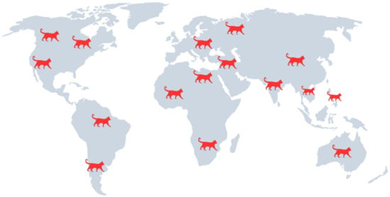
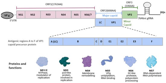
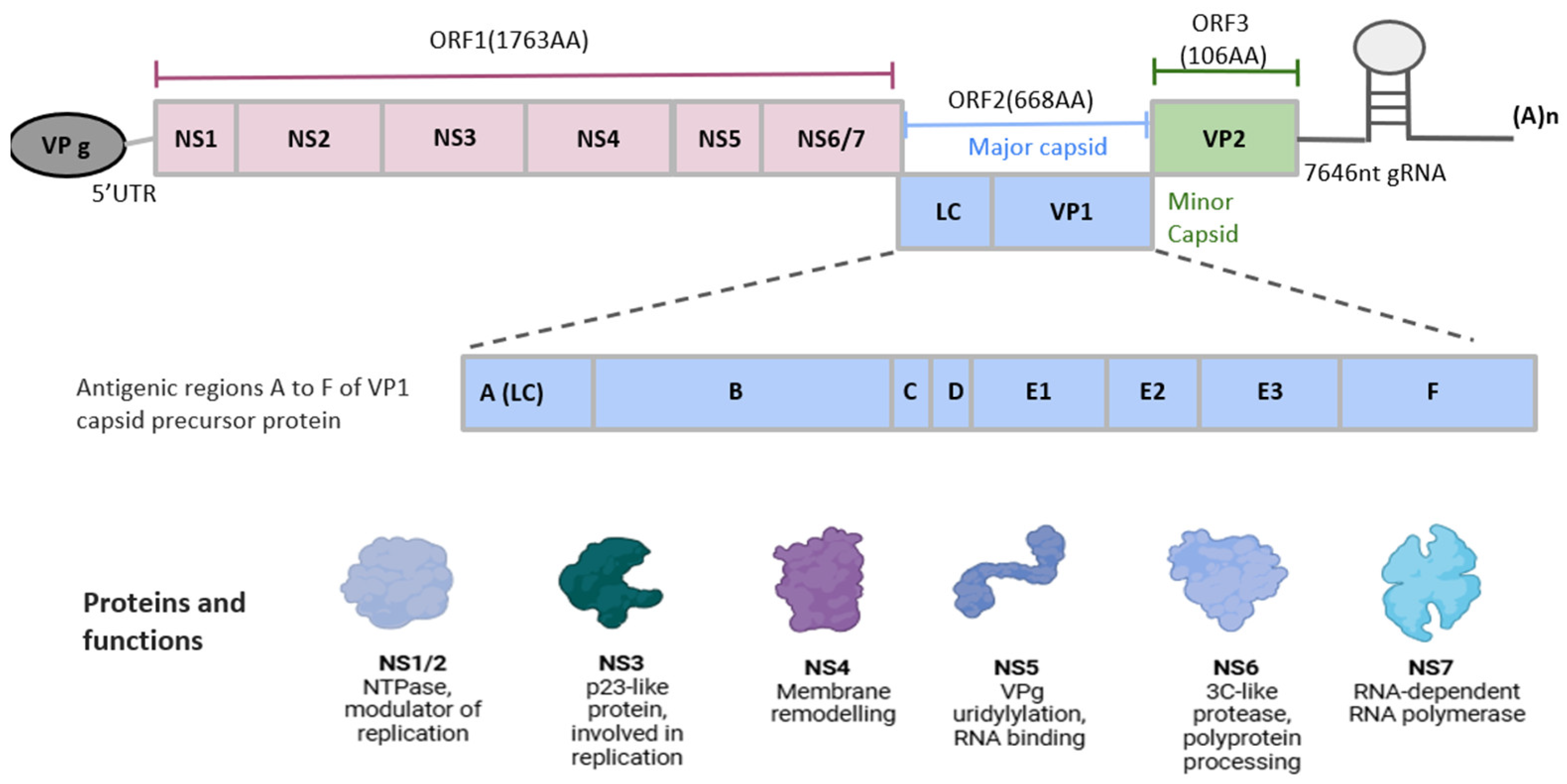
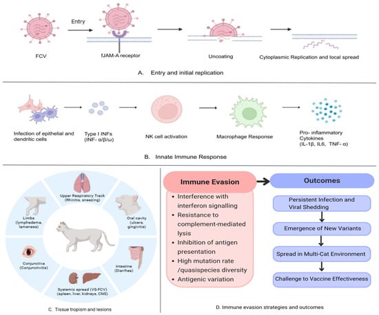
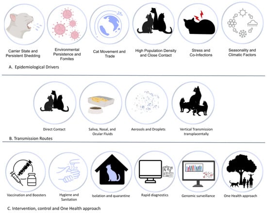

Disclaimer/Publisher’s Note: The statements, opinions and data contained in all publications are solely those of the individual author(s) and contributor(s) and not of MDPI and/or the editor(s). MDPI and/or the editor(s) disclaim responsibility for any injury to people or property resulting from any ideas, methods, instructions or products referred to in the content. |
© 2026 by the authors. Licensee MDPI, Basel, Switzerland. This article is an open access article distributed under the terms and conditions of the Creative Commons Attribution (CC BY) license.