


Abstract
Carbapenem-resistant Enterobacterales (CRE) are a major public health concern, yet genomic data from Bulgaria remain limited. We investigated the genomic epidemiology and genomic characteristics of clinically significant CRE recovered at University Hospital “Lozenetz”, Sofia, between 2022 and 2025. Antimicrobial susceptibility testing, phenotypic carbapenemase characterization, and PCR were combined with whole-genome sequencing of 23 purposively selected isolates chosen to capture temporal, species, carbapenemase, specimen, ward, and resistance-phenotype diversity. The proportion of CRE among all Enterobacterales remained broadly stable over the study period, and Klebsiella pneumoniae was the predominant species throughout. NDM-type carbapenemases were the most frequent overall, although VIM-, KPC-, and OXA-48-like enzymes were also detected. Among the sequenced isolates K. pneumoniae ST6260 (n = 16) was predominant. The remaining isolates included single K. pneumoniae isolates belonging to ST11, ST101, and ST258; two Enterobacter hormaechei ST114; one Proteus mirabilis ST93; and one Providencia stuartii ST46. All ST6260 isolates carried chromosomal blaNDM-5 within a conserved multidrug-resistance module with variable downstream regions, whereas blaOXA-232 was situated on a small ColKP3 plasmid within a defined subcluster. Overall, the data indicate that ST6260 is an emerging hospital-associated multidrug-resistant lineage.
1. Introduction
Carbapenem-resistant Gram-negative bacteria have emerged as a major public health problem over the past two decades. Among them, carbapenem-resistant Enterobacterales (CRE), together with Acinetobacter baumannii, and Pseudomonas aeruginosa are classified by the World Health Organization as critical priority pathogens for which new antimicrobial agents are urgently needed [1]. Despite international and national guidance for surveillance, infection prevention, and antimicrobial stewardship [2,3,4,5], these organisms continue to spread globally and remain one of the most significant challenges in healthcare [6,7,8,9,10].
Members of the Enterobacterales order are normal constituents of the intestinal microbiota, yet they are also among the most common causes of both community-acquired and healthcare-associated infections. They cause a broad spectrum of infections, including bloodstream infections, urinary tract infections, lower respiratory tract infections and wound infections, particularly in hospitalized patients and individuals with severe underlying diseases [11,12]. These infections are associated with prolonged hospitalization, increased costs, and poorer clinical outcomes [13]. Among Enterobacterales, Klebsiella pneumoniae is the species most frequently associated with carbapenem resistance, followed by Escherichia coli and Enterobacter spp. [14,15].
Carbapenem resistance in Enterobacterales is most often mediated by carbapenemase production. However, resistance may also arise through mutations that modify outer membrane permeability and alter efflux pump activity, resulting in reduced intracellular antibiotic concentrations. Carbapenemase genes are often carried on plasmids, integrons, and insertion sequence-rich regions, allowing them to spread efficiently between strains and across species. Several major carbapenemase groups have been described, including class A enzymes such as KPC (K. pneumoniae carbapenemase), class B metallo-β-lactamases including NDM (New Delhi metallo-β-lactamase) and VIM (Verona integron-encoded metallo-β-lactamase), and class D enzymes such as the OXA-48-like carbapenemases. The type of carbapenemase has direct clinical implications, as it determines both susceptibility patterns and available treatment options. For example, many KPC-producing isolates remain susceptible to some of the newer β-lactam/β-lactamase inhibitor combinations, whereas infections caused by metallo-β-lactamase producers, particularly NDM-producing strains, are often much more difficult to treat [16,17,18,19,20,21,22,23].
In Europe, carbapenemase-producing Enterobacterales are increasingly associated with the spread of NDM-type metallo-β-lactamases and OXA-48-like enzymes [24,25,26]. Previous studies have linked their epidemiology to the expansion of dominant clones and to the dissemination of mobile genetic elements carrying resistance determinants [9,27,28]. International organizations such as ECDC and WHO also coordinate surveillance efforts to track and limit the spread of these pathogens [25,26,29]. By contrast, data on the genomic epidemiology of carbapenem-resistant Enterobacterales in Bulgaria remain limited.
Although a considerable amount of information on CRE epidemiology is available from neighbouring countries [15,28,30], published data from Bulgaria addressing this problem have only begun to emerge in the 2020s. A recent regional review of carbapenem-resistant K. pneumoniae [31] summarized reports from Balkan countries, including Bulgaria, and described isolates carrying carbapenemases such as KPC, NDM-1, VIM-1 and OXA-48-like enzymes. More recent Bulgarian studies suggest that the local epidemiology may be becoming more complex, with additional resistance mechanisms and new genetic backgrounds appearing among circulating isolates. These studies reported carbapenemase-producing Enterobacterales carrying enzymes such as NDM-1, NDM-5, OXA-48, OXA-232, VIM-1, VIM-4 and VIM-86 [32,33,34].
The aim of this study was to characterize the genomic epidemiology of CRE at University Hospital “Lozenetz” in Sofia between 2022 and 2025. We assessed the occurrence of CRE during this period and performed whole-genome sequencing on selected isolates to investigate mechanisms of carbapenem and other antimicrobial resistance, as well as associated virulence determinants. A central focus of the study was the emergence and spread of K. pneumoniae ST6260 carrying blaNDM-5.
2. Materials and Methods
2.1. Study Setting
The study was conducted at the University Hospital “Lozenetz” in Sofia, Bulgaria, a 189-bed tertiary-care hospital including a 12-bed intensive care unit (ICU). During the study period, the hospital had a median of 44,936 bed-days annually. CRE isolates recovered between 1 January 2022 and 31 December 2025 were included in the study. For this study, clinically significant CRE were defined as the first CRE isolate per patient recovered from a diagnostic clinical specimen in the context of a clinically proven/highly suspected infection. Rectal screening or other surveillance isolates and isolates considered to represent colonization without clinical infection were excluded.
2.2. Identification and Antimicrobial Susceptibility Testing
Species identification was performed by matrix-assisted laser desorption/ionization time-of-flight mass spectrometry (MALDI-TOF MS; Bruker, Bremen, Germany). Antimicrobial susceptibility testing (AST) was performed by disc diffusion. Colistin susceptibility was determined by broth microdilution (Erba Lachema, Brno, Czech Republic). For cefiderocol, disc diffusion was used initially; isolates with inhibition-zone diameters within the EUCAST area of technical uncertainty (ATU; 18–21 mm) were retested by the Bruker UMC broth microdilution method using iron-depleted cation-adjusted Mueller–Hinton broth supplied as part of the commercial test system. For a subset of 30 nearly consecutive CRE isolates recovered in 2022–2023, MIC50 and MIC90 values for selected newer agents were determined by gradient diffusion (E-tests, Liofilchem, Roseto degli Abruzzi, Italy). The subset was selected purposively to include different specimen types, hospital wards, clinical diagnoses, bacterial species, and carbapenemase phenotypes and comprised 26 K. pneumoniae, one P. mirabilis, one P. stuartii, one Enterobacter xianfangensis, and one E. cloacae isolate. Phenotypic carbapenemase testing available at the time indicated 27 metallo-β-lactamase producers, two KPC producers, and one OXA-48-like producer. The sample size of 30 represented the accepted minimum used locally for descriptive MIC50/MIC90 estimation and was also constrained by the available E-test strip format and study funding. MIC50 and MIC90 were estimated from cumulative MIC distributions plotted on semilogarithmic graph paper: cumulative percentages were plotted on the x-axis and MIC values on the logarithmic y-axis, and the fitted line was used to read the MICs corresponding to 50% and 90% inhibition. Susceptibility results were interpreted using EUCAST clinical breakpoints, version 15.0 (2025). Plazomicin was interpreted according to CLSI M100, 33rd edition (2023), because EUCAST clinical breakpoints were not available for this agent.
2.3. DNA Extraction
Genomic DNA was extracted from bacterial cultures grown on MacConkey agar (35 °C, 18–24 h) using a Proteinase K-based lysis and alcohol precipitation method. An inoculum was prepared by picking 10–30 colonies from each culture and suspending them in 400 µL ATL lysis buffer (QIAGEN, Hilden, Germany), followed by incubation at 80 °C for 10 min. Subsequently, 20 µL Proteinase K (20 mg/mL) was added, and samples were incubated at 56 °C for 30 min. Then, 180 µL of 6 M sodium chloride was added, samples were vigorously mixed for 15 s, and centrifuged at 14,500× g for 15 min. The supernatant was transferred to a fresh tube, and genomic DNA was precipitated by adding an equal volume of 100% isopropanol, followed by gentle mixing by inversion and incubation at room temperature for 10 min. After centrifugation at 6000× g for 15 min, the supernatant was discarded, and the DNA pellet was washed three times with 1.5 mL cold 70% ethanol, each wash followed by centrifugation at 6000× g for 15 min. Samples were then air-dried and resuspended in 100 µL sterile TE buffer.
DNA concentration was measured using a NanoDrop 2000 Spectrophotometer (Thermo Fisher Scientific), and working dilutions were prepared to a final concentration of 30 ng/µL for downstream PCR applications. For long-read sequencing, 1 µg of DNA from the stock solution was used per sample.
2.4. Detection of Carbapenemases and Other Antimicrobial Resistance Genes
Carbapenemase production was investigated using the phenotypic methods available during the study period, including the combined disc test (MASTDISCS™ Combi Carbaplus, Mast Diagnostics, Bootle, UK), MBL strips (HiMedia Laboratories, Mumbai, India), and the rapid lateral-flow assay O.K.N.V.I. RESIST-5 (Coris BioConcept, Gembloux, Belgium). These complementary methods were used to distinguish major serine carbapenemases from metallo-β-lactamases and to provide rapid presumptive categorization for routine clinical management. The lateral-flow assay was introduced later in the study period because implementation depended on local procurement cycles and reagent availability.
Carbapenemase genes (blaOXA-48-like, blaNDM, blaKPC, and blaVIM) were confirmed by multiplex PCR in 67 isolates. This subset comprised the isolates for which PCR confirmation was performed prospectively under the available financial and reagent constraints and was selected across study years, hospital wards, clinical diagnoses, and bacterial species. Primer sequences and PCR conditions are provided in Supplementary Table S1. Following amplification, 5 µL of each PCR product was mixed with runSAFE 6× loading dye/DNA stain (Thistle Scientific, Glasgow, UK) at a ratio of one part stain to five parts DNA, separated on a 1% agarose gel, and visualized using blue or UV illumination according to the manufacturer’s instructions. Amplicon sizes corresponding to the expected fragment lengths were used to determine the presence of the target carbapenemase genes.
Additional antimicrobial resistance genes were investigated using the Bac Multi-Resist Real-TM multiplex real-time PCR assay (Sacace Biotechnologies, Como, Italy) according to the manufacturer’s instructions.
2.5. Whole-Genome Sequencing
Whole-genome sequencing was performed on 23 purposively selected CRE isolates. Selection was not random and was designed to maximize diversity across study year, bacterial species, carbapenemase category, hospital ward, specimen type, resistance phenotype, clinical severity, and isolate availability and DNA quality, while giving priority to recent highly resistant isolates and isolates from serious infections. Routine duplicate isolates from the same patient were excluded. Two isolate pairs from the same patients were retained because they represented clearly distinct colony populations with markedly different mucoid phenotypes and were therefore considered biologically non-identical. The number sequenced was determined by the available project funding rather than by a formal statistical sample-size calculation; consequently, the genomic subset was intended to provide focused molecular characterization and should not be interpreted as a proportionally representative sample of the entire CRE collection.
Sequencing libraries were prepared using the Rapid Barcoding Kit (SQK-RBK114.24; Oxford Nanopore Technologies plc., Oxford, UK) according to the manufacturer’s instructions, except that 1 µg of input genomic DNA per isolate was used. Barcoded samples were pooled (6–8 isolates per run) and sequenced on a MinION Mk1C device using R10.4.1 flow cells (Oxford Nanopore Technologies plc., Oxford, UK). Basecalling was performed using the Super Accuracy (SUP) model implemented in MinKNOW (v25.05.14), and barcode demultiplexing was carried out automatically.
2.6. Bioinformatic Analysis
Raw reads were quality-checked with NanoPlot v1.43.0 [35], filtered with Filtlong v0.2.1 (https://github.com/rrwick/Filtlong, accessed on 20 April 2026), and adapter-trimmed using Porechop_ABI v0.5.0 [36]. Genome assemblies were generated with Autocycler v0.5.2 [37], polished with Medaka v2.1.1 (https://github.com/nanoporetech/medaka, accessed on 20 April 2026), and reoriented using Dnaapler v1.2.0 [38]. Assembly quality was assessed with BUSCO v6.0.0 [39] and QUAST v5.3.0 [40]. Assemblies were evaluated for contamination with CheckM2 v1.1.0 [41]. Genome annotation was performed using the NCBI Prokaryotic Genome Annotation Pipeline (PGAP) v2025-05-06 [42]. Species identification and multilocus sequence type (MLST) assignment were performed automatically through the PubMLST/BIGSdb API [43]. Antimicrobial resistance genes were identified with AMRFinderPlus v4.0.23 [44], while K. pneumoniae isolates were further characterized with Kleborate v3.1.3 [45]. Virulence-associated genes were screened using ABRicate v1.0.1 (https://github.com/tseemann/abricate, accessed on 20 April 2026) against the core VFDB dataset [46]. Plasmid replicons were investigated using PlasmidFinder v2.1.6 [47]. Unless otherwise stated, analyses were performed using default parameters and thresholds.
Soft-core genome SNP phylogeny for the ST6260 K. pneumoniae isolates was inferred using a Snippy-based workflow applied to the high-quality polished assemblies. Per-isolate variant calling was performed with Snippy v4.6.0 (https://github.com/tseemann/snippy, accessed on 20 April 2026), using the --ctgs parameter, with the chromosome of the earliest complete K. pneumoniae ST6260 genome from Bulgaria deposited in GenBank (NZ_CP186365) as the reference. The resulting variants were combined with snippy-core to generate a whole-genome alignment, which was subsequently cleaned with snippy-clean_full_aln. Recombinant regions were identified and filtered using Gubbins v3.4 [48], and the alignment was further processed with Core-SNP-filter v0.2.0 [49] to produce the final 95% soft-core SNP alignment. Pairwise SNP distances were calculated with snp-dists v0.8.2 (https://github.com/tseemann/snp-dists, accessed on 20 April 2026) and a maximum-likelihood phylogenetic tree was reconstructed using IQ-TREE 2 v2.4.0 [50] with polytomies allowed. Phylogenetic trees and associated genomic metadata were visualized using Microreact [51].
Annotated genomes were subsequently used to investigate the genetic context of carbapenemase loci using Clinker v0.0.27 [52]. The blaOXA-232-carrying plasmid was visualized using Plasmid-Map (v0.2.0).
2.7. Statistical Analysis
Temporal differences in the annual proportion of CRE were evaluated using the chi-square test. Resistance proportions in the combined 2022–2023 and 2024–2025 periods were compared using two-sided Fisher’s exact tests because of the modest group sizes and sparse cells. A two-sided p value < 0.05 was considered statistically significant. These analyses were descriptive and were not adjusted for multiple comparisons or potential changes in isolate mix and testing practice over time.
3. Results
3.1. Distribution of CRE Isolates over the Study Period
The proportion of CRE among Klebsiella, Enterobacter and Proteus-Providencia-Morganella spp. isolates from blood, urine, respiratory specimens, bile and wounds was 34.1% in 2022, 27.3% in 2023, 35.1% in 2024, and 35.7% in 2025 (Table 1); the difference across the four years was not statistically significant (chi-square test, p = 0.410). K. pneumoniae was the predominant species, accounting for 178 of 202 (88%) CRE isolates overall. Other CRE species were detected less frequently and included Enterobacter cloacae complex (n = 14), Proteus mirabilis (n = 7), Providencia stuartii (n = 1), and Morganella morganii (n = 2). CRE were recovered from a range of clinical specimens, most commonly urine (n = 80) and wound specimens (n = 47), followed by blood (n = 35), respiratory specimens (n = 30), and bile (n = 10).
Table 1.
Distribution of CRE isolates by year and specimen category, 2022–2025.
3.2. Carbapenemase Characterization and Antimicrobial Susceptibility of the CRE Isolates
Among the 67 CRE isolates subjected to multiplex PCR confirmation, NDM-type carbapenemases were the predominant group, while VIM, OXA-48-like, and KPC were identified less often (Table 2). These 67 isolates represent the prospectively PCR-tested subset rather than the complete collection of 202 CRE isolates. Isolates co-harboring NDM and OXA-48-like carbapenemases were observed only among K. pneumoniae.
Table 2.
Distribution of carbapenemase categories among tested CRE isolates, 2022–2025.
Antimicrobial susceptibility testing showed extensive resistance to most routinely used antimicrobial agents. Among isolates tested in 2022–2023 (n = 37), resistance was observed in 24/37 (64.9%) for gentamicin, 30/37 (81.1%) for amikacin, 27/37 (73.0%) for trimethoprim-sulfamethoxazole, and 6/37 (16.2%) for colistin. Among isolates tested in 2024–2025 (n = 122), the corresponding values were 99/122 (81.1%), 110/122 (90.2%), 116/122 (95.1%), and 61/122 (50.0%), respectively. Compared with 2022–2023, resistance was significantly higher in 2024–2025 for gentamicin (Fisher’s exact test, p = 0.046), trimethoprim-sulfamethoxazole (p < 0.001), and colistin (p < 0.001), but not for amikacin (p = 0.152). These comparisons should be interpreted cautiously because isolate composition and routine testing practices changed over time.
Among the newer agents, cefiderocol showed the most favorable in vitro activity. In the 2022–2023 subset tested by MIC gradient methods (n = 30), cefiderocol had an MIC50 of 0.21 mg/L and an MIC90 of 1.8 mg/L, while plazomicin showed an MIC50 of 1.00 mg/L and an MIC90 of 128.00 mg/L. In contrast, imipenem-relebactam, meropenem-vaborbactam, and ceftazidime-avibactam showed limited activity (Table 3).
Table 3.
MIC50 and MIC90 values (mg/L) of selected newer antimicrobial agents (tested by gradient diffusion) against a subset of carbapenem-resistant Enterobacterales (n = 30) isolated in 2022–2023.
In the later period (2024–2025), when newer agents were tested mainly by disc diffusion, cefiderocol again showed the greatest activity, although 25% of tested isolates were non-susceptible. Imipenem-relebactam, meropenem-vaborbactam and ceftolozane-tazobactam showed minimal in vitro activity, while ceftazidime-avibactam remained active only against a minority of isolates, with a resistance rate of 82% (Table 4).
Table 4.
Resistance rates among tested CRE isolates for selected newer antimicrobial agents (2024–2025).
3.3. Genomic Characterization of Sequenced CRE Isolates
Whole-genome sequencing was performed on 23 purposively selected CRE isolates, including K. pneumoniae (n = 19), E. hormaechei (n = 2), P. mirabilis (n = 1), and P. stuartii (n = 1). The selection enriched for recent, highly resistant, clinically relevant isolates and was designed to capture biological diversity rather than to reproduce the proportional species or ward distribution of the full 202-isolate collection. All sequenced isolates yielded assemblies that passed the study quality-control criteria and were suitable for downstream genomic analysis; detailed read and assembly metrics, including coverage-related and contiguity measures, are provided in Supplementary Table S2. Isolate-level species, sequence type, carbapenemase genes, and key metadata are provided in Supplementary Table S3.
Within the K. pneumoniae subset, 16/19 isolates belonged to ST6260, while the remaining three were assigned to ST11, ST101, and ST258. The two E. hormaechei isolates belonged to ST114, whereas the P. mirabilis and P. stuartii isolates were assigned to ST93 and ST46, respectively.
The carbapenemases identified in the sequenced collection were blaNDM-1 (n = 3), blaNDM-5 (n = 16), blaVIM-1 (n = 1), blaVIM-86 (n = 1), blaOXA-232 (n = 5), blaOXA-48 (n = 1), blaKPC-2 (n = 1), and blaKPC-3 (n = 1). All blaNDM-5 and blaOXA-232 were restricted to the K. pneumoniae ST6260 lineage, although blaOXA-232 was present only in a subset of those isolates. The ST11 isolate co-harbored blaNDM-1 and blaOXA-48, while the ST258 and ST101 isolates carried blaKPC-2 and blaKPC-3, respectively. Among the non-Klebsiella isolates, blaVIM-1 was detected in P. mirabilis, blaVIM-86 in P. stuartii, and blaNDM-1 in both E. hormaechei isolates.
3.4. Phylogenomic Structure and Genomic Features of K. pneumoniae ST6260
The ST6260 lineage was first identified in 2022 and remained present through 2025. Isolates were recovered predominantly from urine (n = 10), but also from blood (n = 3), central venous catheter (n = 1), bronchoalveolar lavage (n = 1), and wound specimen (n = 1). ST6260 isolates originated from various clinical settings, including intensive care, cardiac surgery, nephrology, cardiology, and ambulatory care.
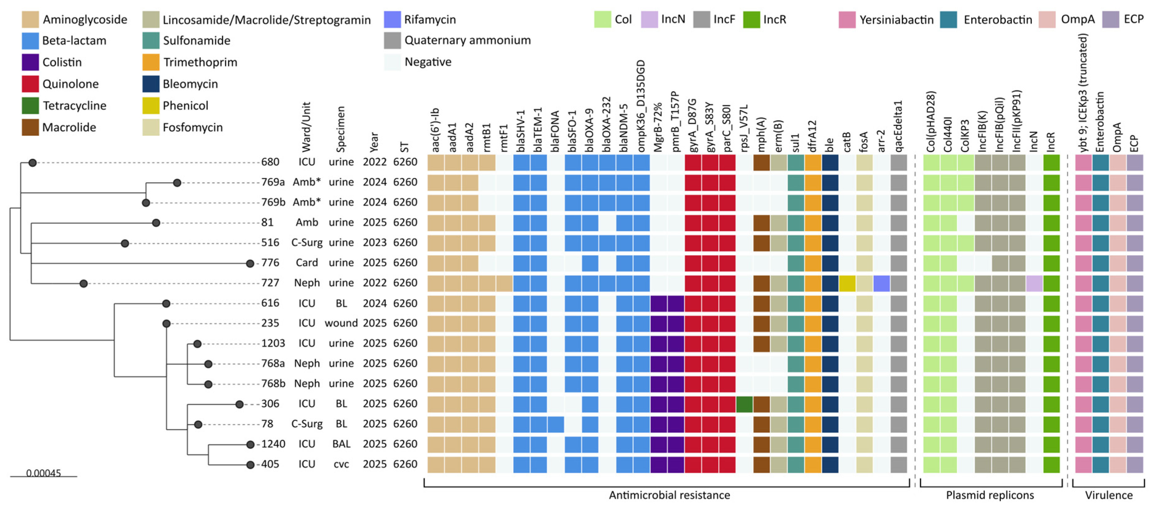
Pairwise SNP distances among the 16 ST6260 isolates ranged from 3 to 46 SNPs (complete distance matrix is provided in Supplementary Table S5). Phylogenomic analysis identified two main clusters within ST6260, with the closest isolates from the two clusters differing by 17 SNPs (Figure 1). Cluster 1 comprised seven isolates, including the 2022–2023 representatives, the closely related outpatient pair recovered in 2024, and two isolates from 2025. This cluster contained all the blaOXA-232-positive isolates. Cluster 2 comprised nine colistin-resistant isolates recovered in 2024–2025, all of which showed mgrB truncation and the pmrBT157P substitution. Six isolates in Cluster 2 originated from the ICU, with two from nephrology and one from cardiac surgery.
Figure 1.
Maximum-likelihood phylogeny based on the 95% soft-core SNP alignment of the 16 K. pneumoniae ST6260 isolates. Metadata tracks indicate hospital ward, specimen type, year of isolation, sequence type (ST), selected resistance determinants, plasmid replicons, and virulence-associated loci. The tree shows the division of ST6260 into two major clusters, with Cluster 1 including all blaOXA-232-positive isolates and Cluster 2 defined by mgrB truncation together with the pmrBT157P substitution. BL, blood; BAL, bronchoalveolar lavage; CVC, central venous catheter; Amb, ambulatory; Amb *, ambulatory patient who had undergone urological surgery at another hospital 2 weeks earlier; C-Surg, cardiac surgery; ICU, intensive care unit; Neph, nephrology; Card, cardiology. Colored cells indicate the presence of the corresponding resistance determinant, plasmid replicon, or virulence-associated locus; colors correspond to the functional categories shown in the figure key, whereas blank cells indicate absence.
All 16 ST6260 isolates shared a common multidrug-resistance profile that included blaNDM-5, blaSHV-1, blaTEM-1, blaOXA-9, aadA1, aadA2, aac(6′)-Ib, dfrA12, sul1, ble, fosA, and qacEdelta1. All isolates also carried the quinolone resistance-associated substitutions gyrAS83Y and gyrAD87G, and parCS80I. In addition, all ST6260 isolates shared the same ompK36D135DGD insertion and the plasmid replicons IncR, IncFIB(pQil), IncFII(pKP91), Col(pHAD28), and Col440I.
Variation beyond this shared background was observed across several resistance classes. blaSFO-1 and rmtB1 were each detected in 13/16 isolates, although they did not consistently co-occur. By contrast, mph(A) and erm(B) consistently co-occurred and were each present in 11/16 isolates. blaOXA-232 was present in 5/16 isolates, always together with the ColKP3 replicon, and was restricted to Cluster 1. A single isolate from Cluster 1 also carried rmtF1 together with an IncN plasmid replicon. As described above, all Cluster 2 isolates showed mgrB truncation together with the pmrBT157P substitution. Rare additional features included catB, arr-2, and rpsJV57L in individual isolates.
Virulence-associated determinants were also assessed. In K. pneumoniae ST6260, these included OmpA, which has established roles in adherence, invasion, and biofilm-associated persistence; the siderophores enterobactin; fyuA and a truncated ybt9/ICEKp3 yersiniabactin element; and E. coli common pilus (ECP)-associated adherence genes. Genes classically associated with hypervirulent K. pneumoniae, including the major hypermucoidy and high-virulence siderophore markers, were not detected. Capsule locus and O-antigen assignments and the full virulence-gene profiles are provided in the supplementary isolate-level data where available. Because this analysis was based on gene presence/absence, no direct conclusions regarding biofilm production, serum resistance, siderophore expression, or in vivo virulence can be drawn.
3.5. Chromosomal blaNDM-5 Resistance Region and Accessory Plasmid-Mediated Determinants in ST6260
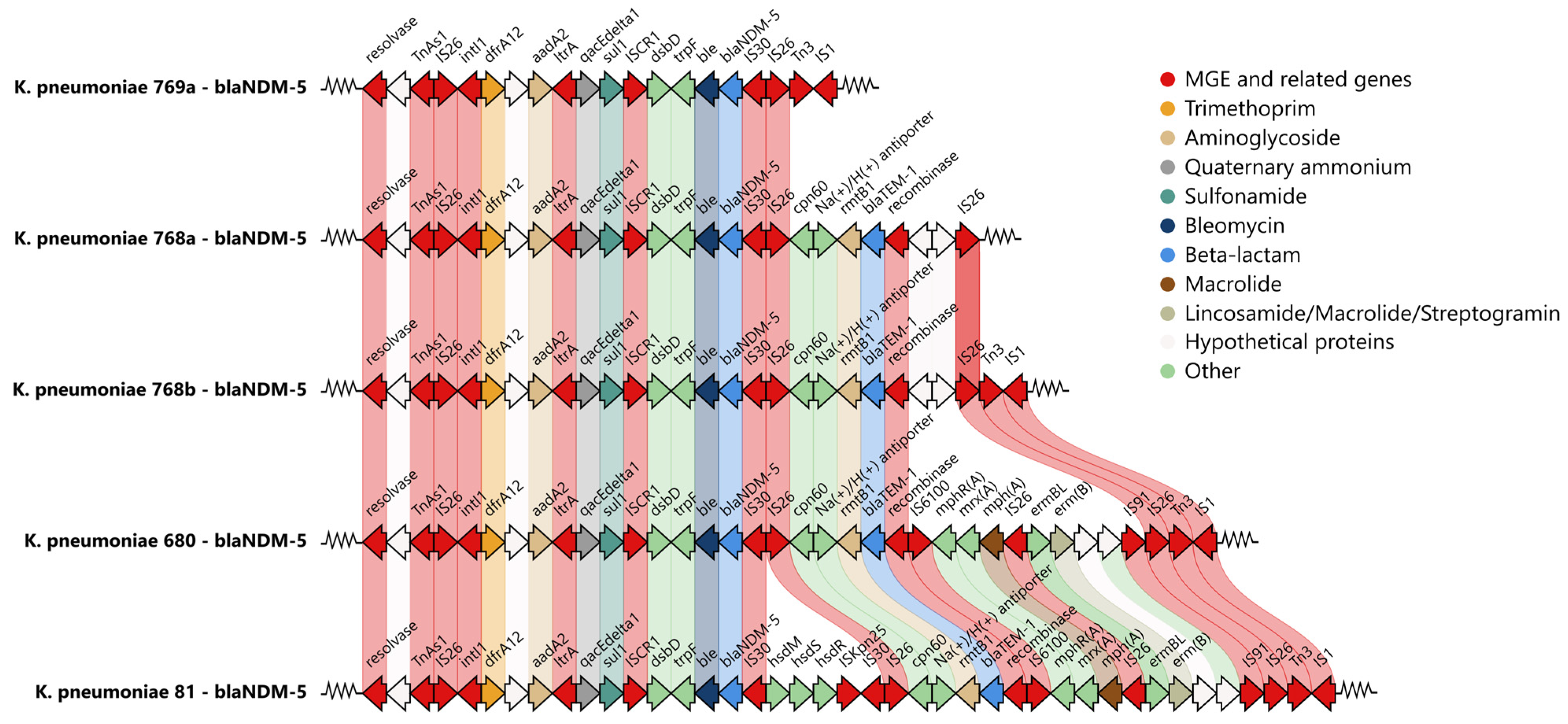
Comparative analysis of representative blaNDM-5-containing regions identified a conserved chromosomal multidrug-resistance segment shared across ST6260 isolates (Figure 2). The core structure comprised TnAs1, IS26, intI1, dfrA12, aadA2, ltrA, qacEdelta1, sul1, ISCR1, ble, blaNDM-5, IS30, and a downstream IS26 element. Beyond this conserved core, structural variation was observed in the downstream region, including additional resistance determinants, extra IS26 copies, other insertion-sequence elements, and adjacent non-AMR genes. In the extended downstream segments, rmtB1 co-occurred with blaTEM-1, while mph(A) and erm(B) were located further downstream and were present only in some of the larger configurations. This variation was in line with the uneven distribution of several accessory resistance determinants across the ST6260 isolates.
Figure 2.
Comparative genomic organization of representative chromosomal blaNDM-5-containing resistance regions in K. pneumoniae ST6260 isolates. The figure shows a conserved shared core surrounding blaNDM-5 across the ST6260 lineage, together with variable downstream extensions containing additional IS26 copies and accessory resistance modules. This comparison illustrates the overall conservation of the chromosomal blaNDM-5 region and the structural variation affecting its more distal segments. Arrows represent annotated coding sequences and indicate their transcriptional orientation; arrow colors correspond to the functional categories shown in the figure key, and shaded connecting regions indicate homologous sequence blocks between compared loci.
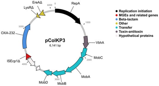
In contrast, blaOXA-232 was carried on a compact 6141 bp ColKP3 plasmid detected in a subset of ST6260 isolates (Figure 3). The plasmid backbone comprised repA, a mobABCD mobilization module, and vbhA, while blaOXA-232 was located adjacent to a truncated ISEcp1 element and alongside truncated lysR and ereA genes.
Figure 3.
Circular map of the blaOXA-232-carrying ColKP3 plasmid identified in a subset of K. pneumoniae ST6260 isolates. The plasmid comprises a compact ColKP3 backbone with repA, a mobABCD mobilization module, and vbhA, while blaOXA-232 is located adjacent to a truncated ISEcp1 element and truncated lysR and ereA genes. Arrows represent annotated coding sequences and indicate their transcriptional orientation; colors correspond to the functional categories shown in the figure key, and dashed leader lines connect gene labels to the corresponding plasmid features.
3.6. Genomic Characteristics of Non-ST6260 K. pneumoniae and Other CRE
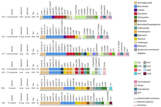
Outside the dominant K. pneumoniae ST6260 lineage, the remaining sequenced CRE isolates showed marked genomic diversity (Figure 4).
Figure 4.
Genomic diversity of the non-ST6260 sequenced carbapenem-resistant Enterobacterales isolates. Annotation tracks indicate species, sequence type (ST), year of isolation, specimen type, selected resistance determinants, plasmid replicons, and virulence-associated loci. Amb, ambulatory; V-Surg, Vascular surgery; Surg, Surgery; Orth, Orthopedy; Urol, Urology. Colored cells indicate the presence of the corresponding resistance determinant, plasmid replicon, or virulence-associated locus; colors correspond to the functional categories shown in the figure key, whereas blank cells indicate absence.
Among the non-ST6260 K. pneumoniae isolates, the ST11 isolate co-harbored blaNDM-1 and blaOXA-48. blaOXA-48 was located on an IncL plasmid, whereas blaNDM-1 was carried on a multi-replicon IncFIB(K)/IncFII(K)/IncR plasmid with additional resistance determinants: blaCTX-M-15, qnrS1, aac(6′)-Ib-cr5, and the blaSHV_C-112A promoter mutation. The ST258 isolate carried blaKPC-2 on an IncFIB(K)/IncFII(K) plasmid, whereas blaCTX-M-15, qnrB9, and aac(6′)-Ib-cr5 were located on a separate IncX3 plasmid. In the ST101 isolate, blaKPC-3 was found on a multi-replicon IncFIA(HI1)/IncFII(K)/IncR plasmid, together with armA.
Both E. hormaechei ST114 isolates carried blaNDM-1 on an IncFII(pKPX1)/repB(R1701) plasmid, along with chromosomal blaCTX-M-15, qnrB1, and aac(6′)-Ib-cr5. The P. mirabilis ST93 isolate harbored chromosomal blaVIM-1, blaCMY-99, and armA. Finally, the P. stuartii ST46 isolate carried blaVIM-86, blaCMY-4, armA, and aac(6′)-Ib-cr5 on an IncC plasmid.
The chromosomal and plasmid localization of the carbapenemase loci identified across the sequenced isolates is detailed in Supplementary Table S4.
4. Discussion
Carbapenem resistance continues to be a significant problem in Europe, and current epidemiological trends suggest that the European Union is not on track to meet its 2030 targets for reducing Klebsiella pneumoniae infection rate [53].
Across 2022–2025, the proportion of clinically significant CRE among all Enterobacterales remained stable and K. pneumoniae was the dominant species throughout. NDM-type carbapenemases were the most frequently detected overall, but VIM-, KPC-, and OXA-48-like enzymes were also identified in other species and sequence types. Recent Bulgarian studies from other hospitals in Sofia have documented Enterobacterales carrying NDM, VIM, KPC, and OXA-48-like carbapenemases [33,34], consistent with the resistance backgrounds observed in the present study. Our ST11 K. pneumoniae isolate carried blaOXA-48 on an IncL plasmid, matching previous reports of blaOXA-48 on conserved pOXA-48-like IncL plasmids [54,55], while plasmid-associated blaKPC-2 and blaKPC-3 in our ST258 and ST101 isolates were in line with the KPC-producing K. pneumoniae isolates reported by Markovska et al. [33]. Similar overlap with previous Bulgarian findings was also seen in the non-Klebsiella isolates, particularly the P. stuartii ST46 isolate carrying blaVIM-86 on an IncC plasmid [34]. K. pneumoniae ST6260, carrying blaNDM-5 and blaOXA-232 was recently reported as an emerging multi-drug-resistant lineage in Bulgaria [33,56]. In this context, ST6260 was the predominant lineage among our sequenced K. pneumoniae isolates, accounting for 16 of 19 isolates. They were recovered over multiple years, from different specimen types and across several hospital wards, indicating that this lineage was not confined to a single short-term cluster.
Pairwise distances of 3 to 46 SNPs across the 16 isolates, together with recovery from multiple wards over several years, are consistent with sustained local circulation within the hospital. The lineage was divided into two phylogenetic clusters with distinct temporal patterns and genetic features. Cluster 1 comprised earlier and more diverse isolates and included all blaOXA-232-positive strains, whereas Cluster 2 consisted predominantly of more recent colistin-resistant isolates, characterized by mgrB truncation together with the pmrBT157P substitution. The closest isolates from the two clusters differed by 17 SNPs, which supports their separation into distinct sublineages. For outbreak investigation, isolates differing by ≤10 SNPs were considered strong candidates for recent transmission, while distances of 11–15 SNPs suggested possible but less certain epidemiological linkage. The absence of inter-cluster distances below 17 SNPs is indicative of separate transmission networks rather than a single continuous outbreak. SNP thresholds proposed for other K. pneumoniae lineages, such as the 21-SNP value discussed for ST258/512 [57], may serve as a general reference, but are not directly applicable to ST6260 because such cutoffs are lineage- and dataset-dependent. Overall, the observed patterns are consistent with local diversification of the ST6260 lineage within the hospital.
Clinically, ST6260 was recovered from urinary, bloodstream, catheter-associated, respiratory, and wound specimens and from several wards, including the ICU, cardiac surgery, nephrology, cardiology, and ambulatory care. This broad distribution is compatible with adaptation to the hospital environment, although patient-level outcome data, prior antimicrobial exposure, duration of ICU stay, and comorbidities were not available for systematic analysis. Unlike globally established high-risk K. pneumoniae clones such as ST258, ST11, ST147, and ST307, ST6260 remains sparsely represented in international genomic datasets. Nevertheless, its persistence across years, chromosomal blaNDM-5, accumulation of accessory resistance determinants, and emergence of a colistin-resistant sublineage identify features that may support future high-risk-clone behavior.
The chromosomal blaNDM-5 region in ST6260 showed a conserved core structure with variable downstream extensions. The shared core contained TnAs1, IS26, intI1, dfrA12, aadA2, ltrA, qacEdelta1, sul1, ISCR1, ble, blaNDM-5, IS30, and a downstream IS26 element, whereas the more distal segments differed by the presence of additional IS26 copies and accessory resistance modules. This organization is consistent with an IS26-rich multidrug-resistance locus undergoing structural variation. IS26 is well recognized as a major contributor to the assembly and rearrangement of complex resistance regions in Enterobacterales, including insertion, deletion, inversion, and amplification events [58,59]. Although the exact sequence of events that gave rise to these configurations cannot be resolved from our data, the observed structure indicates that the chromosomal region carrying blaNDM-5 remains structurally dynamic rather than fixed. At the same time, the chromosomal localization may favor stable persistence of blaNDM-5 while permitting continued accumulation and reorganization of nearby accessory resistance determinants, driven by IS26 mobilization.
The co-occurrence of mgrB truncation and the pmrB T157P substitution in all Cluster 2 isolates provides a plausible genomic basis for the colistin-resistant phenotype. Loss of functional MgrB can derepress PhoPQ signaling and promote lipid A modification, whereas alterations in PmrB may further modify PmrAB-dependent regulation. Because the contribution of the specific PmrB T157P substitution was not tested experimentally, it should be regarded as a candidate contributing alteration rather than definitive proof of causality. The uniform presence of these changes within Cluster 2 is nevertheless consistent with clonal expansion of a colistin-resistant sublineage under local antimicrobial selection pressure.
Virulence-associated loci in ST6260 were limited to common fitness- and colonization-associated determinants and iron accumulation with a possibility of biofilm formation. Thus, the clinical relevance of this lineage was driven mainly by multidrug resistance and restricted treatment options rather than by a classical hypervirulent genotype. Although no classical hypervirulence-associated determinants were identified, the combination of adhesion- and colonization-associated loci may contribute to prolonged persistence in the hospital environment. Functional assays evaluating biofilm formation, serum resistance, and siderophore production were beyond the scope of the present study and should be addressed in future investigations. OmpA, adherence-associated loci and siderophores could plausibly contribute to surface attachment, biofilm formation, environmental persistence, and colonization of medical devices. However, biofilm formation, serum resistance, siderophore production, and other functional virulence phenotypes were not measured, and gene detection alone cannot establish the magnitude of these traits.
Beyond Bulgaria, published information on ST6260 remains limited, but the available literature suggests that this lineage is not unique to our hospital setting or to Bulgaria. In addition to the recent Bulgarian reports, ST6260 isolates have also been described from India and Nepal, which points to a broader, although still sparsely documented, distribution [60,61].
Across both study periods, therapeutic options for CRE infections remained limited. Some isolates remained susceptible to fosfomycin, but intravenous fosfomycin was not available locally. Colistin was used extensively in clinical practice for urinary, bloodstream, and respiratory infections, yet resistance increased substantially throughout the study period. Treatment was further complicated by the late introduction of cefiderocol and the absence of aztreonam-avibactam. Among the newer agents tested, cefiderocol showed the most favorable in vitro activity in 2022–2023, although non-susceptibility became more common by 2024–2025. Bulgarian data from another Sofia hospital similarly identified cefiderocol as one of the few remaining active options against blaNDM-5/blaOXA-232-positive ST6260 isolates [33]. Potential mechanisms of reduced cefiderocol susceptibility include alterations in siderophore-receptor-mediated uptake, changes in permeability or efflux, and the combined effect of metallo-β-lactamases and additional β-lactamases. These mechanisms were not investigated experimentally in the present collection. The 25% non-susceptibility rate should therefore be interpreted as a clinically important phenotypic observation rather than as evidence for a single defined mechanism. Recent studies indicate that cefiderocol activity against NDM-producing Enterobacterales is variable and often lower than against VIM producers [62,63]. These limitations make aztreonam-avibactam particularly relevant because of its high in vitro activity against many contemporary MBL-producing isolates, although this agent was not available locally during the study period [64].
This study has several limitations. Whole-genome sequencing was performed on a purposively selected, funding-limited subset rather than the complete CRE collection; the sequenced isolates therefore provide focused genomic insight but are not a statistically representative sample of all species, wards, or years. Phenotypic comparisons between the earlier and later periods were constrained by changes in the testing algorithm, isolate composition, and the later introduction of several newer antimicrobial agents into routine laboratory workflow. Patient-level data on prior antimicrobial exposure, comorbidities, ICU duration, treatment, and outcomes were not available for systematic analysis. Plasmid conjugation or mobility experiments were not performed, so the transfer potential of blaOXA-232-bearing ColKP3 and other plasmids was inferred only from sequence structure. Likewise, biofilm formation, serum resistance, siderophore production, and other functional virulence phenotypes were not evaluated. Although phylogenomic analysis supports local transmission and persistence of ST6260, it cannot definitively reconstruct all transmission events, environmental reservoirs, or inter-ward routes. Finally, the observed structure of the chromosomal blaNDM-5 region does not resolve the exact sequence of events that generated the different configurations.
The persistence of ST6260 across multiple wards emphasizes the need for coordinated infection-prevention measures, including rapid recognition of carbapenemase-producing isolates, contact precautions, enhanced environmental cleaning, screening targeted to epidemiologically linked high-risk patients, and ward-to-ward communication during patient transfer. The available data cannot identify a specific environmental reservoir or reconstruct individual transmission routes. Nonetheless, repeated recovery from the ICU and other departments is compatible with inter-ward dissemination through patient movement, shared healthcare personnel, equipment, or unrecognized colonization. Prospective genomic surveillance integrated with conventional epidemiology would be required to distinguish these possibilities and to evaluate the effect of control interventions. Compared with globally established high-risk clones such as ST11, ST258, ST147, and ST307, ST6260 remains only sporadically reported worldwide. Nevertheless, its sustained persistence over four consecutive years, recovery from multiple hospital wards and specimen types, and stable association with chromosomal blaNDM-5 suggest successful adaptation to the local hospital environment. Continued genomic surveillance will be important to determine whether this lineage remains locally confined or acquires broader regional or international epidemiological significance.
In conclusion, the proportion of clinically significant CRE among the Enterobacterales groups analyzed remained broadly stable over the study period, while whole-genome sequencing identified a multidrug-resistant K. pneumoniae ST6260 lineage that emerged in 2022 and persisted through 2025. This lineage carried chromosomal blaNDM-5, whereas blaOXA-232 was confined to a defined subcluster on a small ColKP3 plasmid. Although other carbapenemase-producing isolates were also present, ST6260 was the dominant lineage among the purposively sequenced K. pneumoniae isolates. Its persistence across years and wards, together with a dynamic IS26-rich chromosomal multidrug-resistance region and the emergence of a colistin-resistant sublineage, indicates epidemiological success and genomic adaptability. Routine integration of genomic surveillance with infection-control investigation and antimicrobial stewardship is warranted to detect such lineages early, guide targeted interventions, and support coordinated CRE monitoring across Bulgaria and the wider Balkan region.
Supplementary Materials
The following supporting information can be downloaded at: https://www.mdpi.com/article/10.3390/amh71030032/s1, Table S1, primers and PCR conditions for detection of major carbapenemase genes; Table S2, assembly quality metrics for the 23 sequenced carbapenem-resistant Enterobacterales isolates; Table S3, isolate-level overview of the sequenced carbapenem-resistant Enterobacterales collection, including species, sequence type, carbapenemase genes, and key metadata; Table S4, chromosomal and plasmid localization of carbapenemase loci in the sequenced isolates; Table S5, pairwise SNP distance matrix for the 16 K. pneumoniae ST6260 isolates.
Author Contributions
Conceptualization, E.K. and R.V.; methodology, E.K., I.N.I., R.V. and T.T.; software, I.S., T.T. and R.V.; validation, I.S., I.N.I., D.H., R.V. and T.T.; formal analysis, I.S., I.N.I., E.K., R.V. and T.T.; investigation, I.S., I.N.I. and D.H.; resources, R.V., I.N.I. and E.K.; data curation, I.S., T.T. and D.H.; writing—original draft preparation, E.K., I.S. and T.T.; writing—review and editing, I.S., I.N.I., R.V., T.T., E.K. and D.H.; visualization, I.S., R.V. and T.T.; supervision, E.K., R.V. and I.N.I.; project administration, T.T.; funding acquisition, R.V. All authors have read and agreed to the published version of the manuscript.
Funding
This research was funded by the Scientific Fund of Sofia University “St. Kliment Ohridski”, grant number 80-10-132/2023. Project No. BG16RFPR002-1.014-0017 Center of Competence, “Fundamental, Translational and Clinical Investigations of Infections and Immunity”, funded by “Scientific, Research, Innovations and Digitalization for Intelligent Transformations 2021–2027” Program; Project No. KP-06-N43/8 of 8 December 2020, “Innate immune response to RNA viral pathogens: Evaluation of the role of variants in genes involved in RLR/TLR signaling pathways of the host using a SARS-CoV-2 invasion model”, funded under the “Fundamental Scientific Research—2020” program of the Bulgarian National Science Fund.
Institutional Review Board Statement
Not applicable.
Informed Consent Statement
Not applicable.
Data Availability Statement
Raw sequences and genome assemblies of all sequenced CRE isolates have been deposited in the National Center for Biotechnology Information (NCBI) database under BioProject accession number PRJNA1452497. All other data generated or analyzed during this study are included in this article and its Supplementary Materials. Additional supporting materials are available from the corresponding author upon reasonable request.
Acknowledgments
Not applicable.
Conflicts of Interest
The authors declare no conflicts of interest.
References
- World Health Organization. WHO Bacterial Priority Pathogens List, 2024—Bacterial Pathogens of Public Health Importance to Guide Research, Development and Strategies to Prevent and Control Antimicrobial Resistance; World Health Organization: Geneva, Switzerland, 2024; pp. 1–72. [Google Scholar]
- Center for Diseases Control and Prevention. Guidance for Control of Carbapenem-Resistant Enterobacteriaceae (CRE); 2012 CRE Toolkit; Center for Diseases Control and Prevention: Atlanta, GA, USA, 2012.
- Tacconelli, E.; Cataldo, M.A.; Dancer, S.J.; De Angelis, G.; Falcone, M.; Frank, U.; Kahlmeter, G.; Pan, A.; Petrosillo, N.; Rodríguez-Baño, J.; et al. ESCMID Guidelines for the Management of the Infection Control Measures to Reduce Transmission of Multidrug-Resistant Gram-Negative Bacteria in Hospitalized Patients. Clin. Microbiol. Infect. 2014, 20, 1–55. [Google Scholar] [CrossRef] [Scilit] [PubMed]
- Magiorakos, A.P.; Burns, K.; Rodríguez Baño, J.; Borg, M.; Daikos, G.; Dumpis, U.; Lucet, J.C.; Moro, M.L.; Tacconelli, E.; Simonsen, G.S.; et al. Infection Prevention and Control Measures and Tools for the Prevention of Entry of Carbapenem-Resistant Enterobacteriaceae into Healthcare Settings: Guidance from the European Centre for Disease Prevention and Control. Antimicrob. Resist. Infect. Control 2017, 6, 113. [Google Scholar] [CrossRef] [Scilit] [PubMed]
- World Health Organization. Guidelines for the Prevention and Control of Carbapenem-Resistant Enterobacteriaceae, Acinetobacter Baumannii and Pseudomonas Aeruginosa in Health Care Facilities; World Health Organization: Geneva, Switzerland, 2017. [Google Scholar]
- Almadhoon, H.; Ahmad, I.; Lee, W.W.Y.; Matthews, A.; Carter, E.R.; Otter, J.A.; Lecky, D.M.; Holmes, A.H.; Zhu, N.J. Prevalence of Carbapenem-Resistant Enterobacterales in Healthcare and Community Settings in the UK: A Systematic Review and Meta-Analysis. J. Hosp. Infect. 2025, 165, 19–31. [Google Scholar] [CrossRef] [Scilit] [PubMed]
- Saito, S.; Sakurai, A.; Matsumura, Y.; Uemura, K.; Hase, R.; Kato, H.; Itoh, N.; Hashimoto, T.; Matono, T.; Yu, J.; et al. Molecular Epidemiology and Patient Outcome of Carbapenem-Resistant Enterobacterales, Pseudomonas Aeruginosa and Acinetobacter Baumannii in Japan: A Multicenter Study from MultiDrug-Resistant Organisms Clinical Research Network. JAC. Antimicrob. Resist. 2025, 7, dlaf027. [Google Scholar] [CrossRef] [Scilit] [PubMed]
- Alvisi, G.; Curtoni, A.; Fonnesu, R.; Piazza, A.; Signoretto, C.; Piccinini, G.; Sassera, D.; Gaibani, P. Epidemiology and Genetic Traits of Carbapenemase-Producing Enterobacterales: A Global Threat to Human Health. Antibiotics 2025, 14, 141. [Google Scholar] [CrossRef] [Scilit] [PubMed]
- González-Gómez, I.; Ruíz-Carrascoso, G.; Herruzo-Cabrera, R. A Decade of Epidemiology and Incidence of Carbapenemase-Producing Bacteria in a Tertiary Hospital in Southern Europe. Infect. Prev. Pract. 2025, 7, 100469. [Google Scholar] [CrossRef] [Scilit] [PubMed]
- Durante-Mangoni, E.; Bertolino, L.; Mastroianni, C.; Viale, P.; Bassetti, M.; Citton, R.; Gómez-Ulloa, D.; Roset, M.; McCann, E. Complicated Carbapenem-Resistant Infections: A Treatment Pathway Analysis in Italian Sites. Infez. Med. 2021, 29, 434–449. [Google Scholar] [CrossRef] [Scilit] [PubMed]
- ECDC. Carbapenem-Resistant Enterobacterales—Third Update; ECDC: Solna, Sweden, 2025. [CrossRef]
- Silvotti, M.G.; Scaltriti, E.; Bolzoni, L.; Zerbi, B.; Tocci, G.; Zappavigna, A.; Lamberti, G.; Donati, F.; Federici, F.; Pongolini, S.; et al. Outbreak of Carbapenem Resistant Klebsiella Pneumoniae in a Neurorehabilitation Unit: Genomic Epidemiology Reveals Complex Transmission Pattern in a Tertiary Care Hospital. J. Glob. Antimicrob. Resist. 2025, 41, 195–201. [Google Scholar] [CrossRef] [Scilit] [PubMed]
- Cassini, A.; Högberg, L.D.; Plachouras, D.; Quattrocchi, A.; Hoxha, A.; Simonsen, G.S.; Colomb-Cotinat, M.; Kretzschmar, M.E.; Devleesschauwer, B.; Cecchini, M.; et al. Attributable Deaths and Disability-Adjusted Life-Years Caused by Infections with Antibiotic-Resistant Bacteria in the EU and the European Economic Area in 2015: A Population-Level Modelling Analysis. Lancet Infect. Dis. 2019, 19, 56–66. [Google Scholar] [CrossRef] [Scilit] [PubMed]
- Budia-Silva, M.; Kostyanev, T.; Ayala-Montaño, S.; Bravo-Ferrer Acosta, J.; Garcia-Castillo, M.; Cantón, R.; Goossens, H.; Rodriguez-Baño, J.; Grundmann, H.; Reuter, S. International and Regional Spread of Carbapenem-Resistant Klebsiella Pneumoniae in Europe. Nat. Commun. 2024, 15, 5092. [Google Scholar] [CrossRef] [Scilit] [PubMed]
- Papadimitriou-Olivgeris, M.; Bartzavali, C.; Georgakopoulou, A.; Kolonitsiou, F.; Mplani, V.; Spiliopoulou, I.; Christofidou, M.; Fligou, F.; Marangos, M. External Validation of INCREMENT-CPE Score in a Retrospective Cohort of Carbapenemase-Producing Klebsiella Pneumoniae Bloodstream Infections in Critically Ill Patients. Clin. Microbiol. Infect. 2021, 27, 915.e1–915.e3. [Google Scholar] [CrossRef] [Scilit] [PubMed]
- Falcone, M.; Giordano, C.; Leonildi, A.; Galfo, V.; Lepore, A.; Suardi, L.R.; Riccardi, N.; Barnini, S.; Tiseo, G. Clinical Features and Outcomes of Infections Caused by Metallo-β-Lactamase–Producing Enterobacterales: A 3-Year Prospective Study From an Endemic Area. Clin. Infect. Dis. 2024, 78, 1111–1119. [Google Scholar] [CrossRef] [Scilit] [PubMed]
- Harada, S.; Aoki, K.; Nomura, Y.; Ohama, Y.; Araoka, H.; Hayama, B.; Sakurai, T.; Ueda, A.; Ishii, Y.; Tsutsumi, T. Transmission of Global Clones of NDM-Producing Enterobacterales and Interspecies Spread of IncX3 Plasmid Harbouring BlaNDM-5 in Tokyo. J. Glob. Antimicrob. Resist. 2024, 38, 309–316. [Google Scholar] [CrossRef] [Scilit] [PubMed]
- Kanj, S.S.; Kantecki, M.; Arhin, F.F.; Gheorghe, M. Epidemiology and Outcomes Associated with MBL-Producing Enterobacterales: A Systematic Literature Review. Int. J. Antimicrob. Agents 2025, 65, 107449. [Google Scholar] [CrossRef] [Scilit] [PubMed]
- Lee, Y.P.; Liao, Y.C.; Chen, F.J.; Lin, T.W.; Huang, H.P.; Pan, H.H.; Liu, P.Y. Prevalence, Clinical Characteristics, and Mortality Impact of Metallo-β-Lactamase-Producing Enterobacter Cloacae Complex Infections: A Retrospective Cohort Study. J. Glob. Antimicrob. Resist. 2025, 45, 77–83. [Google Scholar] [CrossRef] [Scilit] [PubMed]
- Pilmis, B.; Dortet, L. Epidemiology, Treatment and Outcomes of Bloodstream Infections Due to MBL-Producing Enterobacterales in France: A Retrospective Study. Int. J. Antimicrob. Agents 2025, 66, 107630. [Google Scholar] [CrossRef] [Scilit] [PubMed]
- Bassetti, M.; Di Pilato, V.; Giani, T.; Vena, A.; Rossolini, G.M.; Marchese, A.; Giacobbe, D.R. Treatment of Severe Infections Due to Metallo-β-Lactamases-Producing Gram-Negative Bacteria. Future Microbiol. 2020, 15, 1489–1505. [Google Scholar] [CrossRef] [Scilit] [PubMed]
- Paul, M.; Carrara, E.; Retamar, P.; Tängdén, T.; Bitterman, R.; Bonomo, R.A.; de Waele, J.; Daikos, G.L.; Akova, M.; Harbarth, S.; et al. European Society of Clinical Microbiology and Infectious Diseases (ESCMID) Guidelines for the Treatment of Infections Caused by Multidrug-Resistant Gram-Negative Bacilli (Endorsed by European Society of Intensive Care Medicine). Clin. Microbiol. Infect. 2022, 28, 521–547. [Google Scholar] [CrossRef] [Scilit] [PubMed]
- Tamma, P.D.; Aitken, S.L.; Bonomo, R.A.; Mathers, A.J.; van Duin, D.; Clancy, C.J. Infectious Diseases Society of America Guidance on the Treatment of Extended-Spectrum β-Lactamase Producing Enterobacterales (ESBL-E), Carbapenem-Resistant Enterobacterales (CRE), and Pseudomonas Aeruginosa with Difficult-to-Treat Resistance (DTR-P. aeruginosa). Clin. Infect. Dis. 2021, 72, e169–e183. [Google Scholar] [CrossRef] [Scilit] [PubMed]
- Peirano, G.; Pitout, J.D.D. Rapidly Spreading Enterobacterales with OXA-48-like Carbapenemases. J. Clin. Microbiol. 2025, 63, e0151524. [Google Scholar] [CrossRef] [Scilit] [PubMed]
- Albiger, B.; Glasner, C.; Struelens, M.J.; Grundmann, H.; Monnet, D.L.; Koraqi, A.; Bino, S.; Hartl, R.; Apfalter, P.; Glupczynski, Y.; et al. Carbapenemase-Producing Enterobacteriaceae in Europe: Assessment by National Experts from 38 Countries, May 2015. Eurosurveillance 2015, 20, 30062. [Google Scholar] [CrossRef] [Scilit] [PubMed]
- Brolund, A.; Lagerqvist, N.; Byfors, S.; Struelens, M.J.; Monnet, D.L.; Albiger, B.; Kohlenberg, A.; Apfalter, P.; Hartl, R.; Glupczynski, Y.; et al. Worsening Epidemiological Situation of Carbapenemase-Producing Enterobacteriaceae in Europe, Assessment by National Experts from 37 Countries, July 2018. Eurosurveillance 2019, 24, 1900123. [Google Scholar] [CrossRef] [Scilit] [PubMed]
- Brkic, S.; Cirkovic, I. Carbapenem-Resistant Enterobacterales in the Western Balkans: Addressing Gaps in European AMR Surveillance Map. Antibiotics 2024, 13, 895. [Google Scholar] [CrossRef] [Scilit] [PubMed]
- Koumaki, V.; Voudanta, E.; Michelaki, A.; Orfanidou, M.; Vagiakou, E.; Vrioni, G.; Tsakris, A. Changing Epidemiology of Carbapenemases Among Carbapenem-Resistant Enterobacterales in a Greek Tertiary Care Hospital in Athens, 2020 to 2023. Antibiotics 2025, 14, 239. [Google Scholar] [CrossRef] [Scilit] [PubMed]
- Kohlenberg, A.; Linkevicius, M.; Alm, E.; Robesyn, E.; Svartström, O.; Palm, D.; Monnet, D.L.; Plachouras, D.; Albiger, B. From Structured Surveys to Outbreak Investigations: Advancing Genomic Surveillance of Carbapenem-Resistant Enterobacterales within the European Antimicrobial Resistance Genes Surveillance Network. Front. Public Health 2025, 13, 1671769. [Google Scholar] [CrossRef] [Scilit] [PubMed]
- Tryfinopoulou, K.; Linkevicius, M.; Pappa, O.; Alm, E.; Karadimas, K.; Svartström, O.; Polemis, M.; Mellou, K.; Maragkos, A.; Brolund, A.; et al. Emergence and Persistent Spread of Carbapenemase-Producing Klebsiella Pneumoniae High-Risk Clones in Greek Hospitals, 2013 to 2022. Eurosurveillance 2023, 28, 2300571. [Google Scholar] [CrossRef] [Scilit] [PubMed]
- Chatzidimitriou, M.; Kavvada, A.; Kavvadas, D.; Kyriazidi, M.A.; Eleftheriadis, K.; Varlamis, S.; Papaliagkas, V.; Mitka, S. Carbapenem-Resistant Klebsiella Pneumoniae in the Balkans: Clonal Distribution and Associated Resistance Determinants. Acta Microbiol. Immunol. Hung. 2024, 71, 10–24. [Google Scholar] [CrossRef] [Scilit] [PubMed]
- Linkevicius, M.; Bonnin, R.A.; Alm, E.; Svartstrom, O.; Apfalter, P.; Hartl, R.; Hasman, H.; Roer, L.; Raisanen, K.; Dortet, L.; et al. Rapid Cross-Border Emergence of NDM-5-Producing Escherichia Coli in the European Union/European Economic Area, 2012 to June 2022. Eurosurveillance 2023, 28, 2300209. [Google Scholar] [CrossRef] [Scilit] [PubMed]
- Markovska, R.; Stankova, P.; Popivanov, G.; Gergova, I.; Mihova, K.; Mutafchiyski, V.; Boyanova, L. Emergence of BlaNDM-5 and BlaOXA-232 Positive Colistin- and Carbapenem-Resistant Klebsiella Pneumoniae in a Bulgarian Hospital. Antibiotics 2024, 13, 677. [Google Scholar] [CrossRef] [Scilit] [PubMed]
- Sabtcheva, S.; Stoikov, I.; Ivanov, I.N.; Donchev, D.; Lesseva, M.; Georgieva, S.; Teneva, D.; Dobreva, E.; Christova, I. Genomic Characterization of Carbapenemase-Producing Enterobacter Hormaechei, Serratia Marcescens, Citrobacter Freundii, Providencia Stuartii, and Morganella Morganii Clinical Isolates from Bulgaria. Antibiotics 2024, 13, 455. [Google Scholar] [CrossRef] [Scilit] [PubMed]
- De Coster, W.; Rademakers, R. NanoPack2: Population-Scale Evaluation of Long-Read Sequencing Data. Bioinformatics 2023, 39, btad311. [Google Scholar] [CrossRef] [Scilit] [PubMed]
- Bonenfant, Q.; Noe, L.; Touzet, H. Porechop_ABI: Discovering Unknown Adapters in Oxford Nanopore Technology Sequencing Reads for Downstream Trimming. Bioinform. Adv. 2022, 3, vbac085. [Google Scholar] [CrossRef] [Scilit] [PubMed]
- Wick, R.R.; Howden, B.P.; Stinear, T.P. Autocycler: Long-Read Consensus Assembly for Bacterial Genomes. Bioinformatics 2025, 41, btaf474. [Google Scholar] [CrossRef] [Scilit] [PubMed]
- Bouras, G.; Grigson, S.R.; Papudeshi, B.; Mallawaarachchi, V.; Roach, M.J. Dnaapler: A Tool to Reorient Circular Microbial Genomes. J. Open Source Softw. 2024, 9, 5968. [Google Scholar] [CrossRef] [Scilit]
- Manni, M.; Berkeley, M.R.; Seppey, M.; Simão, F.A.; Zdobnov, E.M. BUSCO Update: Novel and Streamlined Workflows along with Broader and Deeper Phylogenetic Coverage for Scoring of Eukaryotic, Prokaryotic, and Viral Genomes. Mol. Biol. Evol. 2021, 38, 4647. [Google Scholar] [CrossRef] [Scilit] [PubMed]
- Gurevich, A.; Saveliev, V.; Vyahhi, N.; Tesler, G. QUAST: Quality Assessment Tool for Genome Assemblies. Bioinformatics 2013, 29, 1072. [Google Scholar] [CrossRef] [Scilit] [PubMed]
- Parks, D.H.; Imelfort, M.; Skennerton, C.T.; Hugenholtz, P.; Tyson, G.W. CheckM: Assessing the Quality of Microbial Genomes Recovered from Isolates, Single Cells, and Metagenomes. Genome Res. 2015, 25, 1043. [Google Scholar] [CrossRef] [Scilit] [PubMed]
- Tatusova, T.; Dicuccio, M.; Badretdin, A.; Chetvernin, V.; Nawrocki, E.P.; Zaslavsky, L.; Lomsadze, A.; Pruitt, K.D.; Borodovsky, M.; Ostell, J. NCBI Prokaryotic Genome Annotation Pipeline. Nucleic Acids Res. 2016, 44, 6614–6624. [Google Scholar] [CrossRef] [Scilit] [PubMed]
- Jolley, K.A.; Bray, J.E.; Maiden, M.C.J. Open-Access Bacterial Population Genomics: BIGSdb Software, the PubMLST.Org Website and Their Applications. Wellcome Open Res. 2018, 3, 124. [Google Scholar] [CrossRef] [Scilit] [PubMed]
- Feldgarden, M.; Brover, V.; Gonzalez-Escalona, N.; Frye, J.G.; Haendiges, J.; Haft, D.H.; Hoffmann, M.; Pettengill, J.B.; Prasad, A.B.; Tillman, G.E.; et al. AMRFinderPlus and the Reference Gene Catalog Facilitate Examination of the Genomic Links among Antimicrobial Resistance, Stress Response, and Virulence. Sci. Rep. 2021, 11, 12728. [Google Scholar] [CrossRef] [Scilit] [PubMed]
- Lam, M.M.C.; Wick, R.R.; Watts, S.C.; Cerdeira, L.T.; Wyres, K.L.; Holt, K.E. A Genomic Surveillance Framework and Genotyping Tool for Klebsiella Pneumoniae and Its Related Species Complex. Nat. Commun. 2021, 12, 4188. [Google Scholar] [CrossRef] [Scilit] [PubMed]
- Zhou, S.; Liu, B.; Zheng, D.; Chen, L.; Yang, J. VFDB 2025: An Integrated Resource for Exploring Anti-Virulence Compounds. Nucleic Acids Res. 2025, 53, D871–D877. [Google Scholar] [CrossRef] [Scilit] [PubMed]
- Carattoli, A.; Zankari, E.; Garciá-Fernández, A.; Larsen, M.V.; Lund, O.; Villa, L.; Aarestrup, F.M.; Hasman, H. In Silico Detection and Typing of Plasmids Using PlasmidFinder and Plasmid Multilocus Sequence Typing. Antimicrob. Agents Chemother. 2014, 58, 3895–3903. [Google Scholar] [CrossRef] [Scilit] [PubMed]
- Croucher, N.J.; Page, A.J.; Connor, T.R.; Delaney, A.J.; Keane, J.A.; Bentley, S.D.; Parkhill, J.; Harris, S.R. Rapid Phylogenetic Analysis of Large Samples of Recombinant Bacterial Whole Genome Sequences Using Gubbins. Nucleic Acids Res. 2015, 43, e15. [Google Scholar] [CrossRef] [Scilit] [PubMed]
- Taouk, M.L.; Featherstone, L.A.; Taiaroa, G.; Seemann, T.; Ingle, D.J.; Stinear, T.P.; Wick, R.R. Exploring SNP Filtering Strategies: The Influence of Strict vs Soft Core. Microb. Genom. 2025, 11, 001346. [Google Scholar] [CrossRef] [Scilit] [PubMed]
- Minh, B.Q.; Schmidt, H.A.; Chernomor, O.; Schrempf, D.; Woodhams, M.D.; Von Haeseler, A.; Lanfear, R. IQ-TREE 2: New Models and Efficient Methods for Phylogenetic Inference in the Genomic Era. Mol. Biol. Evol. 2020, 37, 1530–1534. [Google Scholar] [CrossRef] [Scilit] [PubMed]
- Argimón, S.; Abudahab, K.; Goater, R.J.E.; Fedosejev, A.; Bhai, J.; Glasner, C.; Feil, E.J.; Holden, M.T.G.; Yeats, C.A.; Grundmann, H.; et al. Microreact: Visualizing and Sharing Data for Genomic Epidemiology and Phylogeography. Microb. Genom. 2016, 2, e000093. [Google Scholar] [CrossRef] [Scilit] [PubMed]
- Gilchrist, C.L.M.; Chooi, Y.H. Clinker & Clustermap.Js: Automatic Generation of Gene Cluster Comparison Figures. Bioinformatics 2021, 37, 2473–2475. [Google Scholar] [CrossRef] [Scilit] [PubMed]
- Antimicrobial Resistance in the EU/EEA (EARS-Net)—Annual Epidemiological Report for 2024; ECDC: Solna, Sweden, 2025.
- Sabtcheva, S.; Ivanov, I.N.; Todorova, B.; Simeonov, Y.; Dobreva, E.; Ivanova, K.; Velinov, T.; Kantardjiev, T. Detection and Characterization of OXA-48-Producing Klebsiella Pneumoniae Originated in Bulgaria. J. Chemother. 2016, 28, 450–453. [Google Scholar] [CrossRef] [Scilit] [PubMed]
- David, S.; Cohen, V.; Reuter, S.; Sheppard, A.E.; Giani, T.; Parkhill, J.; the European Survey of Carbapenemase-Producing Enterobacteriaceae (EuSCAPE) Working Group; the ESCMID Study Group for Epidemiological Markers (ESGEM); Rossolini, G.M.; Feil, E.J.; et al. Genomic Analysis of Carbapenemase-Encoding Plasmids from Klebsiella Pneumoniae across Europe Highlights Three Major Patterns of Dissemination. bioRxiv 2019. bioRxiv:2019.12.19.873935. [Google Scholar] [CrossRef] [Scilit]
- Sabtcheva, S.; Stoikov, I.; Georgieva, S.; Donchev, D.; Hodzhev, Y.; Dobreva, E.; Christova, I.; Ivanov, I.N. Genomic Characterization of 16S RRNA Methyltransferase-Producing Enterobacterales Reveals the Emergence of Klebsiella Pneumoniae ST6260 Harboring RmtF, RmtB, BlaNDM-5, BlaOXA-232 and BlaSFO-1 Genes in a Cancer Hospital in Bulgaria. Antibiotics 2024, 13, 950. [Google Scholar] [CrossRef] [Scilit] [PubMed]
- David, S.; Reuter, S.; Harris, S.R.; Glasner, C.; Feltwell, T.; Argimon, S.; Abudahab, K.; Goater, R.; Giani, T.; Errico, G.; et al. Epidemic of Carbapenem-Resistant Klebsiella Pneumoniae in Europe Is Driven by Nosocomial Spread. Nat. Microbiol. 2019, 4, 1919–1929. [Google Scholar] [CrossRef] [Scilit] [PubMed]
- Harmer, C.J.; Hall, R.M. IS 26 and the IS 26 Family: Versatile Resistance Gene Movers and Genome Reorganizers. Microbiol. Mol. Biol. Rev. 2024, 88, e00119-22. [Google Scholar] [CrossRef] [Scilit] [PubMed]
- Tang, N.; Wei, D.; Zeng, Y.; Zhang, G.; Wang, C.; Feng, J.; Song, Y. Understanding the Rapid Spread of Antimicrobial Resistance Genes Mediated by IS26. mLife 2024, 3, 101–109. [Google Scholar] [CrossRef] [Scilit] [PubMed]
- Sharma, A.; Vats, N.; Rati, R.; Sharma, L.; Chatterjee, N.; Jaggi, N. Genotypic Diversity and Virulence Factors of Klebsiella Pneumoniae in a North Indian Tertiary Care Hospital. BMC Infect. Dis. 2024, 24, 1477. [Google Scholar] [CrossRef] [Scilit] [PubMed]
- Thapa, S.; Shrestha, B.; Joshi, D.R.; Tuladhar, R.; Getino, M.; Shrestha, M.; Pokhrel, Y.; Jauneikaite, E. Draft Genome Sequence of Carbapenem-Resistant Klebsiella Pneumoniae ST6260 Isolated from the Catheter Tip of a Female Patient in Nepal. Microbiol. Resour. Announc. 2025, 14, e0014525. [Google Scholar] [CrossRef] [Scilit] [PubMed]
- Markovska, R.; Stankova, P.; Stoeva, T.; Keuleyan, E.; Mihova, K.; Boyanova, L. In Vitro Antimicrobial Activity of Five Newly Approved Antibiotics against Carbapenemase-Producing Enterobacteria—A Pilot Study in Bulgaria. Antibiotics 2024, 13, 81. [Google Scholar] [CrossRef] [Scilit] [PubMed]
- Baicus, A.; Sisu, V.D.; Tatar, A.D.; Gherghel, A.; Iacob, D.G.; Pop, C.S.; Cirstoiu, C.F. In Vitro Activity of Cefiderocol Against Multi-Drug-Resistant Gram-Negative Clinical Isolates in Romania. Antibiotics 2025, 14, 1113. [Google Scholar] [CrossRef] [Scilit] [PubMed]
- Emeraud, C.; Dutkiewicz, M.; Dortet, L. Comparative in Vitro Efficacy of Aztreonam-Avibactam and Other Alternatives (Cefepime-Taniborbactam, Cefepime-Zidebactam, and Cefiderocol) against Metallo-β-Lactamase-Producing Enterobacterales Isolated in 2024 in France. Antimicrob. Agents Chemother. 2026, 70, e0017626. [Google Scholar] [CrossRef] [Scilit] [PubMed]

Disclaimer/Publisher’s Note: The statements, opinions and data contained in all publications are solely those of the individual author(s) and contributor(s) and not of MDPI and/or the editor(s). MDPI and/or the editor(s) disclaim responsibility for any injury to people or property resulting from any ideas, methods, instructions or products referred to in the content. |
© 2026 by the authors. Published by MDPI on behalf of the Hellenic Society for Microbiology. Licensee MDPI, Basel, Switzerland. This article is an open access article distributed under the terms and conditions of the Creative Commons Attribution (CC BY) license.