


Abstract
Genitourinary infections caused by Candida spp. and other yeasts have increased in incidence, and the emergence of resistant isolates to commonly prescribed antifungals is becoming more frequent. Thus, the purpose of this study was to characterize the physiological characteristics of 38 yeast isolates (Candida albicans [n = 32], Candida tropicalis [n = 3], and Nakaseomyces glabratus [n = 3]) recovered from genitourinary infections to better understand the diversity of their physiological profiles, their virulence factors, and their role in pathogenicity. In addition, an experimental study was conducted to determine the minimum inhibitory concentration (MIC) of the isolates using the M27-A3 microdilution method described by the Clinical Laboratory and Standards Institute. Clinical isolates of Candida spp. studied showed in vitro susceptibility to both fluconazole and clotrimazole, the latter having greater antifungal activity due to its lower MIC50. Statistically significant differences were found between the MICs obtained for fluconazole and clotrimazole, with the latter showing the highest in vitro activity. Therefore, the clinical use of clotrimazole is recommended, as is the ongoing need for this type of analysis to monitor changes in susceptibility profiles over time.
1. Introduction
Among the constituents of the human skin and mucous membrane microbiota are yeasts of the genus Candida and other related yeast-like fungi [1]. Under normal conditions, these microorganisms pose no risk to humans; however, they can act as opportunistic pathogens if predisposing factors exist in the host, causing the expression of virulence factors in yeast [2]. In this case, they can be responsible for a wide range of clinical conditions; among which, vulvovaginal and urinary tract infections are particularly common [3]. Despite the high incidence of these infections, their pathogenesis is still not fully understood, and the availability of therapeutic alternatives is limited. Furthermore, a trend toward increased resistance among isolates has been reported [4,5] due to self-medication and the indiscriminate use of treatments, which poses a high risk of developing isolates with acquired resistance and recurrent infections. This poses a higher risk of complicated candidiasis and the need to expand knowledge on the subject. In this regard, in 2023, the National Reference Center for Bacteriology of the Costa Rican Institute for Research and Education in Nutrition and Health (INCIENSA, from the Spanish Instituto Costarricense de Investigación y Enseñanza en Nutrición y Salud) opened a Mycology section and issued an alert for azole-resistant isolates in vulvovaginal candidiasis. This finding had already been described in other countries, but the circulation of yeasts with this resistance profile in Costa Rica has been scarcely documented so far; thus, any information on autochthonous strains is useful for the physicians to take into consideration when treating their patients.
Therefore, the purpose of this study was to physiologically characterize (growth at 37 °C, hemolyzing and melanin production and the excretion of proteases, phospholipases and ureases), and analyze the in vitro susceptibility patterns to fluconazole and clotrimazole of Candida spp. isolates and other related yeasts obtained from genitourinary infections deposited in the Fungal Collection of the School of Microbiology of the University of Costa Rica (B7732) in the period between 2020 and 2022. And thus contribute, as a preliminary study, to a better understanding of their diverse physiological profiles, their virulence factors, and their possible involvement in pathogenicity; contribute to an improvement in treatment by knowing the effectiveness of antifungals against these species; and contribute to epidemiological surveillance.
2. Materials and Methods
2.1. Isolates Analyzed
Thirty-eight Candida spp. isolates were processed, with the following distribution: Candida tropicalis (n = 3), Nakaseomyces glabratus (n = 3), and Candida albicans (n = 32). These isolates were identified by mass spectroscopy with Maldi Tof technology (MS Vitek® [bioMérieux, Marcy L’Étoile, France] and Maldi Biotyper® [Bruker, Billerica, MA, USA]) and deposited in the Fungal Collection of the School of Microbiology (B7732) of the Universidad de Costa Rica (UCR, University of Costa Rica) between 2020 and 2022 and preserved in test tubes containing Sabouraud Glucose Medium (SGM) with a layer of mineral oil and stored at room temperature (RT) (ca. [20–30] °C). The isolates came from different patients diagnosed with urogenital candidiasis. Isolate purity was assessed by analyzing the macroscopic and microscopic characteristics of the specimens in SGM. Macroscopic characteristics were observed visually directly from the tube (description of the colonial morphology of the anverse and reverse were made) and microscopic characteristics were described by observation in lactophenol cotton-blue with light microscopy at 40× [6]. Each isolate was subcultured in SGM and incubated for 48 h at RT before performing each of the tests to have fresh cultures and standardize the growth conditions prior to each analysis.
2.2. Phenotypic Tests
For all isolates and for the various determinations, tests were performed in triplicate using a blastospore suspension at a 0.5 McFarland turbidity. C. albicans strain ATCC 90028 was used as a positive control.
To determine growth at 37 °C, 10 µL of the suspension was placed on Petri dishes with SGM. These plates were incubated for 10 days, with growth radius readings in millimeters after three, five, and ten days.
To determine hemolysin production, blood agar plates, without added glucose, were used, and the presence or absence of hemolytic zones was recorded. A Streptococcus sp. strain was used as a positive control for the test.
To determine the presence of proteinases, casein agar was used, observing the presence or absence of a clear halo around the inoculum, and the halo size was measured in millimeters.
For phospholipases, egg yolk agar was used, where a positive result was observed as a precipitation halo around the inoculum. The halo size was measured in millimeters.
To determine urease production, each isolate was subcultured on urea agar in test tubes and the tubes were incubated at 37 °C for five days. A color change of the medium to fuchsia was considered as a positive result for the presence of ureases; if there was no color change, it was considered negative. A strain of Cryptococcus neoformans WM 148 was used as a positive control for the test.
Melanin production was determined by inoculating L-Dopa agar plates. Black colonies were considered positive and white colonies negative. C. neoformans WM 148 was used as positive control for the test.
2.3. Preparation of Microplates for Antifungal Susceptibility
The Minimal Inhibitory Concentration (MIC) was determined according to the Clinical and Laboratory Standards Institute (CLSI) document [7]. A stock solution of each antifungal was prepared at a concentration 100 times greater than the desired final concentration: 1600 μg/mL for clotrimazole (Sigma-Aldrich, St. Louis, MO, USA) and 12,800 μg/mL for fluconazole (Laboratorios Stein SA, Cartago, Costa Rica), using dimethyl sulfoxide (DMSO) (Sigma Chemicals, Co., St. Louis, MO, USA) as the diluent. From the stock solutions, nine serial 1:2 dilutions were made using DMSO as the diluent. A 1:50 dilution was then prepared using 200 μL from each tube and transferred to another tube containing 9.8 mL of RPMI 1640 (Thermo Fisher Scientific, Waltham, MA, USA). Separately, 200 µL of DMSO was added to two 9.8 mL RPMI tubes labeled No. 11 (growth control) and No. 12 (reagent blank and sterility control). From the above dilutions, 100 µL were loaded per well in the microplates as follows: the contents of tube No. 1 were poured into the wells of column No. 1 (1A–1H), the contents of tube No. 2 into the wells of column No. 2, and so on, up to column No. 12.
The plates were then sealed with Parafilm® M (Bemis Manufacturing Company, Sheboygan Falls, WI, USA), and stored at −80 °C. The final concentrations of the antifungals were from 0.03 to 16 µg/mL for clotrimazole and 0.25 to 128 µg/mL for fluconazole.
A suspension of the clinical isolates of Candida spp. and the control strains Candida parapsilosis ATCC 22019 and Candida krusei ATCC 6258 were prepared (24 h subcultures in SGM incubated at RT) to a turbidity of 0.5 on the McFarland scale (concentration 1–5 × 10⁶ blastospores/mL) in 0.85% sterile saline. A 1:1000 dilution was then made with RPMI (concentration 1–5 × 10³ blastospores/mL), which was used to inoculate the microplates with each antifungal agent, resulting in a final blastospore concentration in the wells of (0.5–2.5) × 103 blastospores/mL. One hundred microliters of the spore suspensions were added to each well from columns 1 to 11, and 100 µL of RPMI was added to the wells in column 12. Plates were incubated at 35 °C without shaking for 24 h for C. albicans and C. tropicalis and for 48 h for N. glabratus.
After incubation, the results were read visually. The MIC reported was the lowest concentration that produced 50% growth inhibition in the case of the fungistatic treatments. Valid results were considered if there was growth in the growth control and no growth in the reagents control.
2.4. Statistical Analysis
SPSS for Windows, version 20 (SPSS Inc., Chicago, IL, USA) was used to perform statistical analysis of the results. The geometric mean and range of MICs, as well as the MIC50 and MIC90 (50th and 90th percentiles), were calculated for each antifungal tested. The Kolmogorov–Smirnov test was performed to determine if the distribution of the MICs of both antifungals followed a normal distribution. As the distribution was not found to be normal, the non-parametric Mann–Whitney U test was employed to determine if the MICs obtained for fluconazole and clotrimazole were statistically different.
3. Results
The 38 isolates analyzed came from clinical samples from male (10.5%; n = 4) and female (89.5%; n = 34) patients. Regarding the pathologies presented by the patients, 39.5% (n = 15) had urinary tract infection and 60.5% (n = 23) vulvovaginal infection. Of the urinary tract infections, 73.3% (n = 11) affected women. The species that was found most frequently was C. albicans in 84.2% (n = 32) of the infections, followed by N. glabratus in 7.9% (n = 3) and C. tropicalis in 7.9% (n = 3). C. albicans caused 95.7% (n = 22) of the vulvovaginal infections and 66.7% (n = 10) of candidurias.
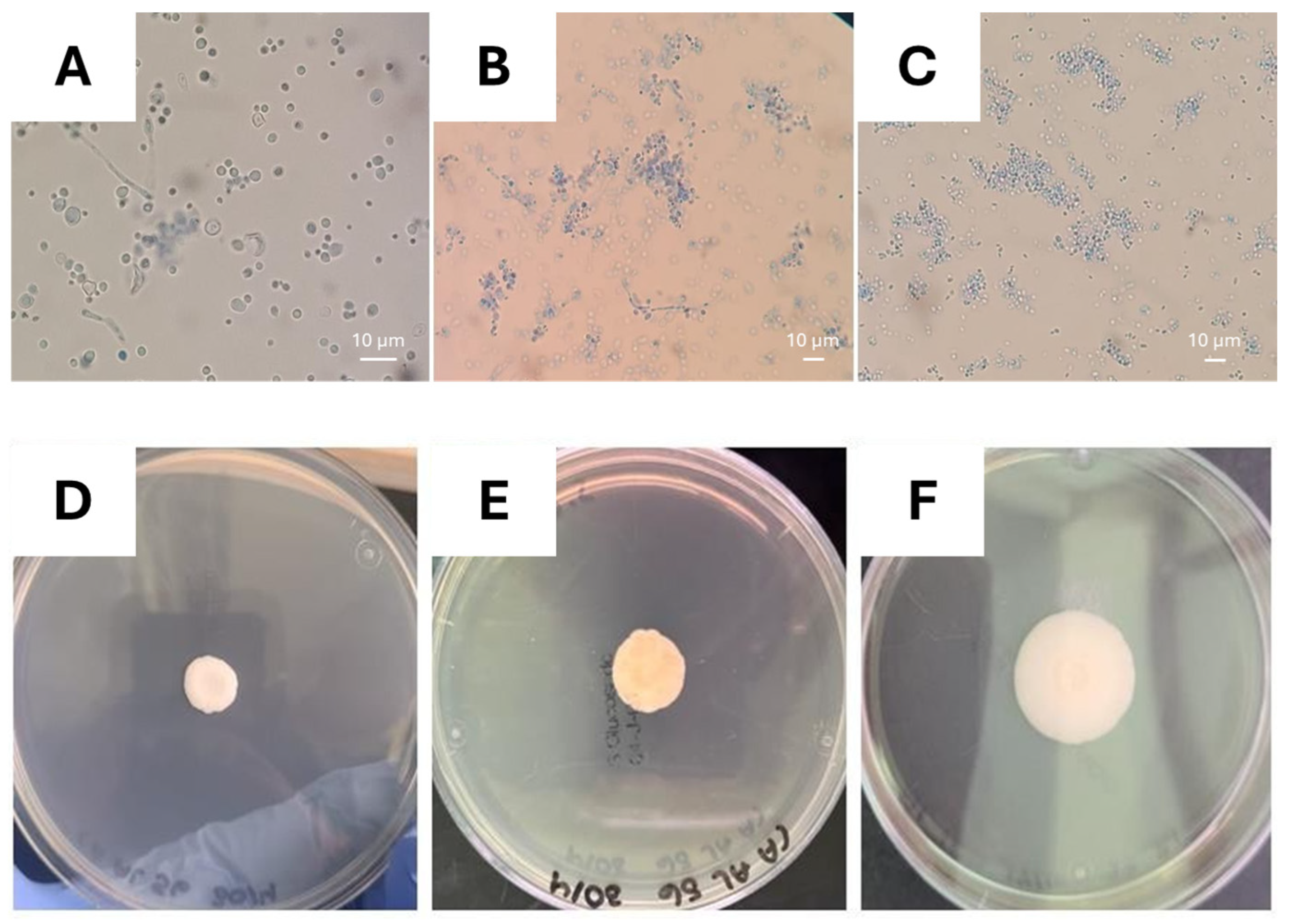
The expected macroscopic and microscopic characteristics were observed, showing the purity of the isolates (Figure 1). The SGM tubes with Candida isolates presented creamy-white, slightly raised, and creamy-looking colonies and C. albicans and C. tropicalis with mycelium submerged in the agar, a characteristic absent in N. glabratus. Thus, microscopically C. albicans and C. tropicalis presented blastospores and septate hyaline mycelium; while in N. glabratus there was only hyaline blastospores.
Figure 1.
Photographs of the microscopic and macroscopic morphology of Candida spp. isolated from genitourinary samples. Septate hyaline mycelium and blastospores of (A) C. albicans and (B) C. tropicalis and (C) blastospores of N. glabratus. Wet mounts were made with lactophenol cotton-blue reagent and observed at 40×. Example of colony growth of Candida sp. on SGM incubated at 37 °C are presented in (D) colony after three days, (E) after five days, and (F) after ten days.
3.1. Growth at 37 °C
The 38 isolates analyzed grew when inoculated with SGM and incubated at 37 °C for ten days. The growth was quite similar in all isolates, and their colony size for day three averaged (10.96 ± 1.26) mm; for day five, (15.53 ± 1.80) mm; and for day ten, (20.22 ± 1.86) mm. At the species level, C. albicans had an average growth on day ten of (20.14 ± 2.25) mm, N. glabratus of (19.89 ± 1.97) mm, and C. tropicalis of (20.00 ± 1.26) mm. Figure 1 shows the growth progression on days three, five, and ten, while Table 1 shows the average colony size obtained on day ten.
Table 1.
Growth at 37 °C and phospholipases and proteinases activity of Candida spp. isolates obtained from genitourinary samples after 10 days of incubation.
3.2. Hemolysin Production
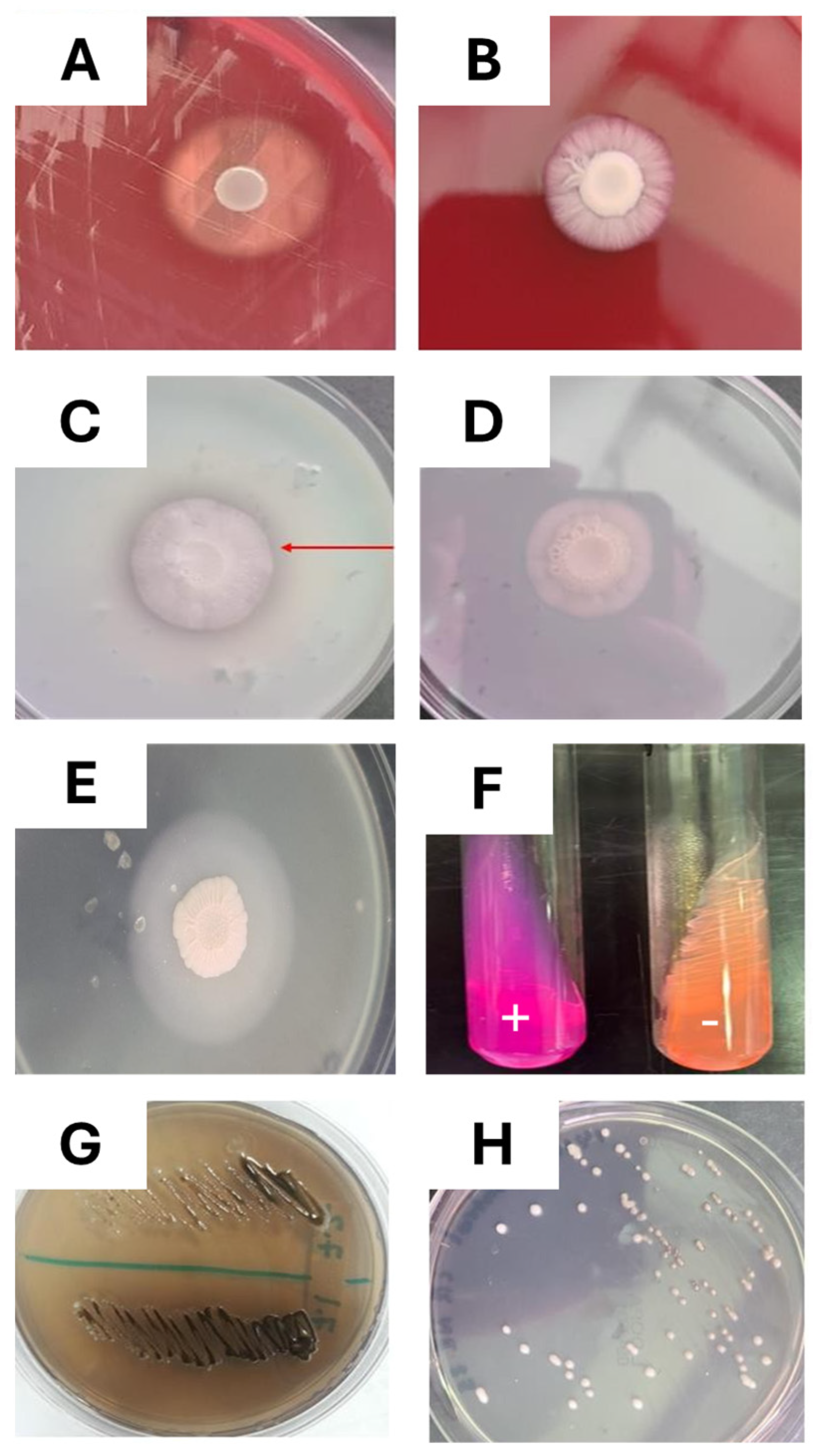
All isolates grew on the blood agar plates; however, none of the isolates produced a hemolysis halo. Therefore, all the isolates tested were negative for hemolysins, under the conditions tested. In addition, colony size was similar for all isolates and were the smallest when compared to the other agars tested (SGM, casein agar, and egg yolk agar), with an average of (7.33 ± 1.13) mm after three days, (9.22 ± 1.46) mm after five days, and (12.52 ± 2.52) mm after ten days. Figure 2A,B shows the positive control of the test and an isolate of Candida sp.
Figure 2.
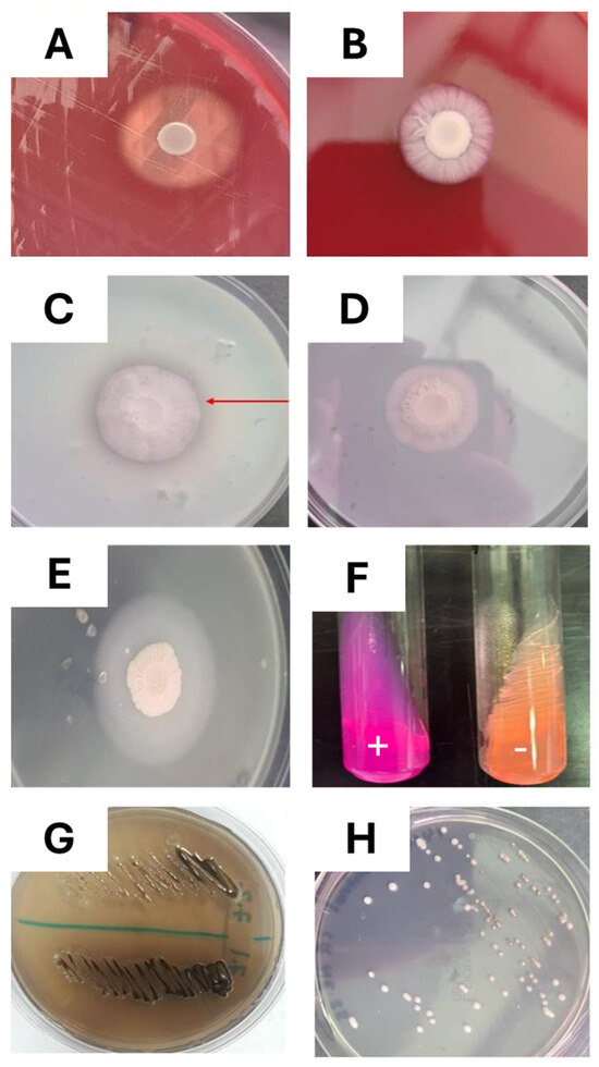
Enzymatic activity of Candida spp. The hemolysin production is presented in panels (A), positive control of the Streptococcus sp., and (B), Candida sp. isolated negative for hemolysins. Proteinase production on casein agar, incubated at 37 °C, is presented in panels (C), positive results where a clear halo is observed around the colony and is marked with a red arrow, and (D), negative results. Panel (E) presents a Candida sp. isolate grown on agar egg yolk with precipitate halo indicating the presence of phospholipases. Urease production is presented in panel (F). A positive control of Cryptococcus neoformans and a negative control of Candida sp. inoculated on urea agar tubes. Finally, melanin production is presented in panels (G), positive control C. neoformans with dark pigment development, and (H), Candida sp. isolate with no pigmentation.
3.3. Proteinase Production
All the isolates analyzed were able to grow on casein agar. An average growth of (12.22 ± 1.80) mm was observed after three days, (16.90 ± 3.01) mm at five days, and (25.37 ± 4.95) mm at ten days. Of these, 45.9% (n = 17) were positive for proteinases and the average halo diameter was (35.00 ± 6.78) mm at ten days. For C. albicans, a total of 17 positive results were obtained after ten days, for a percentage of 53.1%, and had an average halo diameter of (34.64 ± 6.78) mm. N. glabratus and C. tropicalis did not yield positive results. Regarding the site of infection, vulvovaginal candidiasis isolates were 40.9% positive for proteinases; while of the candidurias, 36.4% were positive. Figure 2C,D shows the comparison of Candida spp. isolates and Table 1 shows the summary of positive results in the production of proteinases.
3.4. Phospholipases Production
All isolates grew on egg yolk agar, the average growth diameter was (10.45 ± 1.25) mm after three days, (12.94 ± 1.98) mm after five days, and (16.33 ± 2.11) mm after ten days. As can be seen in Figure 2E, 92.1% (n = 35) were positive for phospholipases and the average halo size was (30.85 ± 6.71) mm. The negative isolates were of C. tropicalis, in which no precipitation halo was observed. The phospholipase activity results of C. albicans were all positive, with an average halo diameter of (31.20 ± 6.88) mm. The three isolates of N. glabratus obtained positive results, with an average halo diameter of (27.11 ± 0.84) mm. Finally, Table 1 presents these results.
3.5. Urease Production
All the isolates grew on the urea agar tubes; however, none of these changed the medium’s color (yellow to fuchsia), so all were considered negative. Figure 2F shows a comparison between the growth of the positive control C. neoformans and an isolate of Candida sp.
3.6. Melanin Production
White colonies were obtained on the L-Dopa agar plates that were inoculated with all isolates tested (Figure 2G,H), indicating that they do not produce melanin under the experimental conditions used.
3.7. Antifungal Susceptibility
Clinical isolates of Candida spp. retrieved from urogenital candidiasis were susceptible to the two antifungals analyzed in the study. Total or partial inhibition of growth was observed in vitro against fluconazole and clotrimazole, the results are presented in terms of MIC that inhibits 50% of the isolates (MIC50), MIC that inhibits 90% of the isolates (MIC90), and the values of the MIC range (minimum–maximum) for the Candida species and antifungal drugs used (Table 2).
Table 2.
In vitro susceptibility patterns of Costa Rican clinical isolates of Candida spp. (obtained from urogenital infections); C. albicans (n = 32), C. glabratus (n = 3), and C. tropicalis (n = 3).
Statistically significant differences were found between the means of the MICs of the antifungals (p < 0.05), with clotrimazole having the highest antifungal activity (lower MIC) since 100% of the isolates tested had MICs ≤ 0.50 μg/mL. In the case of fluconazole, it had a lower antifungal activity compared to the previous one, since MICs ≤ 0.50 μg/mL corresponded only to 55.2%. Also, it is worth noting that 2.6% (n = 1) of the isolates had a MIC of 16 μg/mL, representing the highest MIC reported in the study.
Following the cut-off points present in the M27-A3 document of CLSI, 97.3% of the isolates tested were sensitive to fluconazole with the remaining isolate (2.6%) being dose-dependent. Since there are no defined cut-off points for clotrimazole, all that can be interpreted is that the isolates were susceptible to this antifungal agent.
4. Discussion
Of the Candida species isolated from human beings, C. albicans is the most prevalent, both in healthy people and in patients with infections. It accounts for more than 80% of isolates of all forms of human candidiasis [8]. In vulvovaginal candidiasis, (85–95)% are due to C. albicans, of the remaining species that have been reported to cause this infection, the second most common is N. glabratus, followed by C. parapsilosis, C. tropicalis, and C. krusei [9]. The species that cause candiduria can vary, with C. albicans accounting for 50% to 70% of all Candida-related urinary isolates, followed by N. glabratus and C. tropicalis, which is the third most common species [10].
In this study, C. albicans was found to be the most frequent causative agent of the genitourinary candidiasis analyzed, accounting for 86.2%, followed by N. glabratus and C. tropicalis, each with 7.9%. More specifically, C. albicans caused 95.7% of vulvovaginal candidiasis and 66.7% of candidurias. The results are very similar to those previously reported. Genitourinary candidiasis is more frequent in women, this is because vulvovaginal candidiasis is very common; it is estimated that it affects 75% of women of reproductive age at least once in their lives [9]. In addition, urinary tract infections are more common in women, in fact, the female sex is considered a risk factor for candiduria [11]. Of the cases of genitourinary candidiasis studied, 89.5% affected women, showing that they are more susceptible to these infections.
Candida has the ability to change its morphology from yeast to filamentous. Such transformation to the hyphal form is considered the transition to the pathogenic form. This polymorphism gives it greater virulence, the ability to invade the host’s tissues, evade the immune system and suppress the host’s proinflammatory response. There are several factors that can promote growth in the form of yeast or hyphae, including temperature, pH, CO2 concentration, and glucose concentration. At 37 °C, both fungal morphologies of Candida spp. can be found. In contrast, N. glabratus is not polymorphic, growing only as blastospores [8,12].
In the evaluation of the microscopic characteristics with lactophenol cotton-blue wet mounts, the isolates of C. albicans and C. tropicalis showed both fungal morphologies, blastospores, and hyphae, while N. glabratus showed only blastospores, as it is not polymorphic.
A person’s normal body temperature is 37 °C, so the ability to grow at that temperature is necessary for pathogenic microorganisms, especially those that can cause invasive infections. Of all the species belonging to the genus Candida, it is known that a large part of them cannot grow at a temperature of 37 °C, which prevents them from being successful pathogens or even commensal to humans [8]. All the isolates studied, retrieved from genitourinary candidiasis, were able to grow abundantly at 37 °C, which proves their pathogenic potential. The ability to grow at this temperature gives these Candida species the possibility of generating various clinical entities, including candidemias, although other predisposing factors must always be considered since Candida is an opportunistic pathogen.
The production of hydrolytic enzymes favors the pathogenic capacity of Candida spp., as they are involved in the processes of adhesion to the host’s tissues, in the rupture of the cell membranes of the host, the invasion of mucosal surfaces and blood vessels, and the evasion of the immune response of the host [13]. The role of these enzymes as virulence factors needs to be studied in depth to better understand the aspects that determine the shift from commensal to pathogenic and whether this enzyme activity can establish the recurrence of an infection.
Clinical and experimental evidence demonstrated that secretory aspartyl proteinases (Sap) play multiple roles in C. albicans vaginitis. Its expression can be differentially regulated by changes in the microenvironment such as pH, the presence of a certain morphotype, and the stage of the infectious process. It has been observed that the vaginal fluid of women with vulvovaginal candidiasis and recurrent vulvovaginal candidiasis have a higher concentration of one or more Saps. Clinical isolates from these patients also produced higher amounts of these enzymes compared to isolates from asymptomatic patients, as reported from in vitro assays. Similarly, the Sap activity of oral candidiasis isolates was significantly higher than that of isolates of healthy Candida carriers in the oral cavity. Proteolytic activity has also been found in vitro in most isolates of other pathogenic Candida species, including Candida dubliniensis, C. tropicalis, and C. parapsilosis. The proteolytic activity of other non-pathogenic Candida species is generally lower, suggesting that virulence is correlated with the level of Sap production [9,14].
The results of the percentage of Candida isolates with proteinase production are variable, this may be related to differences in the experimental conditions and characteristics of the strains under study. Oksuz and colleagues [14] evaluated protease production in bovine serum albumin agar plates incubated at 37 °C for seven days. Eleven species of clinical isolates of Candida retrieved from different anatomical sites were analyzed. The isolates of C. tropicalis had 75.0% proteinase production, C. albicans had 56.7%, and N. glabratus had 11.1%. In relation to the colonization of anatomical sites, isolates from the urogenital system were the second most frequent of protease production with 55.1%, only behind those from the skin with 58.8% [15].
Al-Dabagh [16] detected extracellular proteinases in several Candida species, using casein agar that was inoculated and then incubated for 48 h at 37 °C. Proteinase activity was evidenced in 48.7% of the isolates of C. albicans, 39.4% of C. tropicalis, and 4.1% of N. glabratus. In addition, C. albicans and C. tropicalis seemed to be more efficient in the production of proteinases, as they presented larger hydrolysis zones [16].
Proteinase activity is highly variable and is affected by experimental conditions and the strains used. When the isolates were seeded with casein agar, a positive result was obtained only for C. albicans in 53.1%, which is consistent with the results of previous studies for this species. The number of isolates of N. glabratus and C. tropicalis were scarce, so it is not a very significant sample, in these cases the result were negative. The studies described above also reported lower proteinase activity in N. glabratus; however, the results for C. tropicalis were very different and further research is required. Overall, a proteinase positivity frequency of 45.9% was observed in genitourinary candidiasis, slightly lower than that reported by Oksuz and colleagues [15]. When analyzed more specifically, in vulvovaginal candidiasis there is a higher percentage of positive results, which suggests greater relevance of proteinase activity for this type of infection.
Phospholipases cut off the fatty acids from phospholipids, thereby destabilizing the membranes of mammalian cells. Evidence that phospholipases act as virulence factors was obtained by comparing Candida isolates obtained from patients with candidemia and those obtained from the oral cavity of healthy individuals, where it was shown that isolates from candidemias had greater phospholipase activity [13].
The functions of phospholipases during C. albicans infections are not precisely known, but they have been implicated in penetration of host cells, adhesion to epithelial cells, and perhaps interaction with host signal transduction pathways. The data indicates that phospholipases may be required for gastrointestinal and hematogenous dissemination of C. albicans. One study showed that the use of substances that inhibit phospholipase activity prevented the death of mice inoculated with lethal doses of C. albicans, showing the important role of these enzymes as virulence factors [14].
The production of phospholipase enzymes seems to be related to the virulence of Candida, this varies according to the species, anatomical site from which the isolation is obtained, and state of colonization or infection. This is shown by the study by Oksuz and colleagues in which egg yolk agar plates were inoculated and incubated at 37 °C for eight days [15]. The following percentages of phospholipase activity were obtained: C. albicans 53.8%, C. tropicalis 12.5%, and N. glabratus 0.0%. Phospholipase positivity was found more frequently in oral colonization isolates (59.0%), while the lowest frequency was found in those of the urogenital tract (24.1%).
Kantarcioglu and Yücel [17] conducted a similar experiment. They used the same methodology to evaluate the phospholipase activity of clinical isolates from various anatomical sites. It was found that 93.3% of C. albicans, 25.0% of N. glabratus, and 0.0% of C. tropicalis had phospholipase activity. Overall, of the 95 isolates of Candida species, 62.1% were positive with phospholipase [17].
The detection of phospholipase activity is highly variable, it will be affected by the anatomical site where the samples came from, the experimental conditions, and whether the isolates are colonization or infection type. Of the 38 Candida spp. isolated from genitourinary samples, 92.1% were positive for phospholipases, much higher than that reported by previous studies, which may be due to multiple factors. In this case, the cultures were left to incubate for a total of ten days. However, they were also monitored at three and five days. It was noted that some strains took longer than others to generate the halo. By extending the incubation time, the detection of phospholipases in isolates that produce them more slowly may be increased. In addition, there is great variability in the percentages of phospholipase positivity by species reported by [15,17] and those presented here. In the current study, the isolates of N. glabratus and C. tropicalis were very few and that greatly skews the results; thus research with a greater number of samples and from different species is needed for the results to be more robust.
Iron is an essential cofactor for several proteins and, therefore, for biochemical processes such as respiration and cellular metabolism, oxygen transport and DNA synthesis. In a Candida infection, iron absorption is considered an attribute of virulence and even colonization and proliferation are only possible if the fungus has access to enough iron. To acquire iron, C. albicans has three systems for acquiring high-affinity iron: (1) a reducing system responsible for the exploitation of iron from transferrin, ferritin, or the environment; (2) a steel-bearing system responsible for the procurement of iron from a series of siderophores produced by other bodies; and (3) a heme iron absorption and degradation system capable of acquiring iron from hemoglobin and probably heme proteins. In humans, 66.0% of total body iron circulates in the blood along with hemoglobin. To take advantage of this iron, Candida is thought to bind to erythrocytes and possess a hemolytic factor, probably a mannoprotein attached to the cell surface of the fungus. Although extracellular hydrolases may be involved, the mechanism by which C. albicans cause hemolysis and the molecular basis of this mechanism remain unknown. Upon release, hemoglobin uptake is mediated by specific hemoglobin receptors on the surface of C. albicans [18].
Multiple authors have studied the hemolytic activity of Candida and variable results have been reported. Negri and colleagues used glucose-supplemented sheep blood agar and incubated for 48 h at 37 °C, their results showed total hemolysis in all isolates retrieved from infections; while in the colonization isolates there was no hemolysis or only partial hemolysis [19]. In a similar experiment, Morales-López and colleagues [20] tested hemolysis on commercial blood agar and on dextrose potato agar supplemented with glucose and ram’s blood. Hemolysis was obtained in all isolates in the supplemented potato dextrose agar, 50% showed complete hemolysis, but in the commercial blood agar none of the isolates generated hemolysis [20].
Hemolytic activity may be affected or inhibited if the culture is incubated at temperatures other than 37 °C or if there is no glucose in the culture medium used. This was shown by the study in [21] where strains of C. albicans were incubated in trypticase soy agar plates with different added sugars and in blood agar plates with glucose or maltose. Hemolytic activity was demonstrated in plates containing glucose or maltose, but this was not the case in plaques containing sucrose, lactose, or galactose. These data indicate that hemolytic factor production can be regulated by the presence of glucose in the culture medium. In this study, blood agar made from tryptone soy and cow’s blood was used, so it does not contain glucose. This glucose-free culture medium may be the cause of no isolated study presenting hemolysis. It is likely that the absence of glucose inhibits the synthesis of hemolysins and hinders the growth of the fungus, since a smaller growth diameter was observed in it.
The fact that the presence of glucose significantly favors the development of Candida in vitro could explain why diabetes is such an important risk factor for the development of candidiasis. In this regard, Flores (2024) mentioned that hyperglycemia in diabetics can promote the growth of Candida in the vagina because glucose serves as a food source [22]. This availability of glucose can also make it easier for you to obtain iron. In addition, it should be taken into account that diabetes mellitus generates low immunity, which predisposes to candidiasis [23]. In cases of relapsed genitourinary candidiasis, predisposing factors for immunosuppression such as diabetes or HIV should be investigated, as they can lead to therapeutic failure [24].
Urea is a source of nitrogenous nutrition used by various microorganisms, which produce an enzyme (urease) that degrades the urea contained in the environment. The urease test is used as part of biochemical testing for the identification of bacteria and fungi. Urea agar contains phenol red as an indicator of pH change, so that if urea is hydrolyzed it breaks down into ammonium and alkalizes the medium, turning the agar from yellow to deep pink. This test is widely used for differentiation between the genera Cryptococcus and Candida, as they are urease positive and negative, respectively. However, these results may vary depending on the species or strain, in addition to the possible appearance of pathogenic strains whose biochemical or enzymatic behavior is not usual [25]. Abdulla and Mustafa (2020) inoculated colonies of Candida sp. in urea agar and found that the isolates of C. albicans, N. glabratus, and C. tropicalis were urease-negative, while C. krusei was positive for this test [26]. As expected after the review of previous studies and given the species studied, all isolates were urease-negative. The urease test is therefore very useful for the differentiation between Cryptococcus and Candida; however, a greater variety of Candida species should be investigated, especially since the isolation of non-albicans Candida species is now more frequent.
Melanins are present in most fungi, being found in almost all pathogenic fungi. They are hydrophobic pigments, typically dark brown or black, formed by the oxidative polymerization of phenolic and/or indole compounds. Two main types of melanin are found in the fungal kingdom, the DNH melanins, synthesized from acetyl-coenzyme A (CoA), and eumelanins, synthesized from L-Dopa through laccase (polyphenol oxidase). DHN-melanins have been reported in opportunistic fungi of the genera Cladosporium, Fonsecaea, Phialophora, and Wangiella, and in the conidia of Aspergillus fumigatus and Aspergillus niger, while eumelanins have been reported in C. albicans, Coccidioides posadasii, C. neoformans, and Histoplasma capsulatum, among others [27].
Melanin has been postulated to be involved in a variety of properties associated with virulence, including interactions with host cells; protection against oxidative stress, ultraviolet light and hydrolytic enzymes; resistance to antifungal agents; iron-fixing activities; and even the use of ionizing radiation in contaminated soils [28].
Walker and colleagues [28] demonstrated melanin production by inoculating 2 × 10⁶ cells/mL of synchronized cultured C. albicans into a defined minimum liquid medium with or without L-Dopa at 35 °C for five days in the dark in a rotary stirrer at 200 rpm. Under these conditions, in the medium containing L-Dopa, C. albicans produced visible brown pigment. No dark pigment visible during the same period was detected in the absence of L-Dopa. It was concluded that many strains of C. albicans form melanin under Dopa stimulation, although some do so more easily than others and some do not seem to produce it, such variability may be due to the fact that individual yeast cells grow semisynchronously and the time of appearance of melanin can be highly variable [28].
In a very similar experiment, Almeida-Paes and colleagues [29] induced the melanin production of N. glabratus by culturing 1 × 10⁶ blastospores/mL in a defined minimum medium with and without L-Dopa, these cultures were incubated in a rotary shaker at 200 rpm, protected from light, for seven days at 30 °C. All the clinical isolates used generated melanin in the medium supplemented with L-Dopa, which was evidenced by the turning black of the culture; while growths without L-Dopa remained white. The authors suggest that the variability in the percentage of strains that showed melanization in other studies may be due to differences in nutrients present in the culture media used [29]. It is worth noting that the authors used a liquid medium and we used agar plates, a factor that can explain the difference in the results.
Smith and colleagues found that C. auris strains melanized to a greater extent at 37 °C than at 30 °C when cultured with L-Dopa [30]. This has relevance for pathogenesis in human beings, since in vivo the conditions would be ideal for the melanization of C. auris. In addition, it was observed that C. auris required a high cell density (107 cells/mL) to synthesize melanin, at an intermediate density (106 cells/mL) only some strains were able to produce the pigment and in a delayed way, at a low density (105 cells/mL) none produced visible pigment. This could indicate the need for a quorum-sensing molecule to trigger effective melanization. This differs from that presented by C. neoformans, since the synthesis of melanin of the latter under conditions of low cell density was equally or more effective than under conditions of high density.
The production of melanin from compounds such as L-Dopa in Candida spp. is not sufficiently studied to be able to predict the behavior of different species and the effect of different experimental conditions. In this study, all isolates were negative for melanin synthesis. This may be because different conditions were used and the experiment was not as standardized as previous studies that showed positive results. The culture medium had a different composition, agar was used instead of liquid medium, a specific density of yeasts was not inoculated, and it was not incubated in strict darkness. More studies are required in which the experimental conditions are varied and a greater variety of species is analyzed to learn more about what the ideal conditions are and what factors inhibit melanin production and why.
With regard to the worldwide increase in azole resistance in Candida non-albicans species, it has been observed that although C. albicans continues to have a low incidence of resistance to fluconazole (0.5–2%), species such as C. tropicalis (4–9%) and N. glabratus (11–13%) accumulate higher percentages [4,5]. This can be explained, in part, by nonsense point mutations that have been described in C. tropicalis and N. glabratus which generate an overexpression of the PDH1, CDR1, and MDR1 genes, responsible for encoding drug transporters [31,32].
This leads to a constant need to measure the antifungal susceptibility of isolation and thus determine the success or selection of a better therapeutic approach. In order for the results obtained to be compared with similar research, it is a requirement that they are in the same category of study of susceptibility to antifungals, which can be classified into three; microdilution in broth, based on agar and automated systems such as E-test® (bioMérieux, Marcy-l’Étoile, France) and Vitek®2 (bioMérieux, Marcy-l’Étoile, France). Currently, the CLSI M27-A4 document [7] and the European Committee on Antimicrobial Susceptibility Testing (EUCAST) [33] the measurement and interpretation of MICs of important antifungal drugs used against different yeast species in broth microdilution testing. Prior to the development of this method, antifungal susceptibility studies showed inconsistent results with poor reproducibility. Although both methodologies are similar, they show some differences, such as the composition of the medium or the method of reading, so the interpretations of the cut-off points are not comparable [34,35].
Determining the cut-off points of azoles is complicated since they are fungistatic, so although some isolates persist in high concentrations of the drug, they are still susceptible to these antifungals. To overcome this phenomenon, both CLSI and EUCAST have established the endpoint MIC for azoles as the lowest concentration resulting in a 50% decrease in growth compared to control [35,36].
This leads to another problem since it is particularly difficult to visually determine this decrease in growth in the well, which can become very subjective and generate an erroneous classification of a resistant isolation as susceptible or vice versa, which represents one of the greatest sources of variability both intra and interlaboratory. For this reason, it is necessary to place each sample in duplicate, add growth and sterility controls in each plate, and ensure that all procedures, together with the final readings, are performed by the same individual without changing the light intensity and angle of view of the reading [37].
A crucial point to highlight in this study, as well as with other similar ones that evaluate the in vitro susceptibility of isolates of pathogenic microorganisms, is that a large part of the in vivo biological variables that exist inter individually and the pharmacokinetic arrangement of the specific colonization zone are not being taken into account. An example of the above is a topical treatment, although the distribution of the concentration of the compound greatly exceeds the MIC values recorded, variables such as low solubility in water, acidic environment of the vagina, among others, cause the antifungal effect observed for clotrimazole to be lower than expected [38,39,40].
This lack of ranges is because, unlike antimicrobial susceptibility tests, antifungal tests still lack a higher level of development in terms of sensitivity and resistance criteria, as well as cut-off points in the treatment of yeasts, the current development is due to the support of in vitro assay standardization committees [35].
In this methodology, the classification of an isolate as susceptible or resistant to an antifungal agent is determined by correlating the in vitro MIC of a particular drug with in vivo efficacy results from clinical or animal studies, including pharmacokinetics and pharmacodynamics, to help predict therapeutic outcome. Cut-off points are specific MIC values that allow isolates to be classified into the following categories; sensitive, intermediate, and resistant [41].
Unfortunately, there is not enough clinical trial data correlating the in vitro susceptibility results of broth microdilution methodologies against clinical outcomes for most fungal pathogens, the cut-off points proposed by CLSI and EUCAST are only available for mycosis of some of the Candida and Cryptococcus species [39].
CLSI M27-A3 classifies Candida MICs against fluconazole according to the following scale; ≤8 μg/mL should be classified as sensitive, 16–32 mg/mL is the dose-dependent susceptible range (SDD/I), and ≥64 μg/mL is classified as resistant. It is worth mentioning that the CLSI was based on clinical trials of oropharyngeal candidiasis and candidemias for the preparation of the cut-off points, so it corresponds to a different etiology from that of the urogenital isolates used in the present study, another problem is the lack of cut-off points for topical azoles such as clotrimazole and miconazole, which is also related to the anatomical area since for the treatment of oropharyngeal mycoses and hematogenous dissemination, oral or intravenous antifungals must be administered [39].
The cut-off points to determine whether a urogenital Candida isolate is sensitive or resistant to the different antifungals has not been reported, this limits the usefulness of these assays to predict therapeutic success when analyzing clinical isolates of this type, but even so, the data collected using this standardized antifungal susceptibility test allow epidemiological information to be obtained and generate relevant results for the design of antifungal susceptibility schemes, treatment, and monitoring of the resistance of circulating strains in the country [34,42].
Although the etiology of candidiasis is not the same, the cut-off points of fluconazole for genitourinary isolates can still be extrapolated since this treatment is transmitted hematogenously throughout the body, unlike clotrimazole, which is a topical of local action. Our results showed that 37 isolates of Candida spp. fall into the definition of sensitive by presenting a MIC ≥ 0.5 μg/mL, except for one dose-dependent isolate, corresponding to C. albicans. This corresponds to an isolated case since the highest MIC50 reported in the study corresponds to N. glabratus, although given the limited number of samples and the lack of isolates of other non-albicans species such as C. krusei and C. parapsilosis, it is not possible to compare results such as MIC90 and the susceptibility profile of other species causing urogenital symptoms. The results regarding the MIC of N. glabratus to fluconazole are consistent with similar studies such as that of [43] and [44], with a MIC50 of 8.0 μg/mL and 16.0 μg/mL, respectively.
A particular fact also happens in the previously mentioned research, and that is that, although C. albicans does not have the highest MIC50, they have maxima in their intervals which are classified in an intermediate and/or resistant isolation, which indicates that despite the fact that C. albicans presents one of the lowest resistances to azoles, there are some specific isolates which have already generated a high resistance to them.
Regarding clotrimazole, there are currently no cut-off points established in the CLSI methodology, but susceptibility patterns similar to those observed in this study have been reported. In a study of 287 isolates of vaginal candidiasis in Argentina, clotrimazole was reported to have the lowest MIC values (geometrical mean [GM] = 0.02 μg/mL, MIC90 = 0.03 μg/mL) compared to fluconazole and itraconazole [45]. Similarly, in a susceptibility study of 145 isolates of vaginal discharge carried out in Chile, it was found that clotrimazole had the highest antifungal activity, reporting a MIC50 of 0.03 μg/mL for the species C. albicans and 0.25 μg/mL for C. glabratus, both being lower than those reported for fluconazole and nystatin [43].
However, the tentative cut-off points proposed by [46] of CMI ≤ 0.5 μg/mL to be classified as sensitive for C. albicans can be used and extrapolated for the other species, obtaining that the 38 isolates fell into the classification of sensitive to clotrimazole. This cut-off point was obtained from the correlation between in vitro MICs using the M27-A standard and clinical evidence, which corresponds to 87 isolates of oropharyngeal candidiasis in immunocompromised patients. It is a study that is currently more than 20 years old since its publication, but it is still the most recent in this field, due in part to the low priority of this azole due to its limited use in invasive candidiasis, as it is not an antifungal for systemic use. CLSI still does not add cut-off points for the complete repertoire of antifungals and pathogenic species that cause infections today, so the work of Pelletier et al. represents a way to give greater meaning to the MICs obtained through the broth microdilution methodology. But there is the disadvantage of not being validated by the CLSI and of being carried out with an outdated M27-A standard, it also presents the same problem previously discussed about the etiological origin of the isolates analyzed, which may not be related to other types of isolates of Candida infections.
5. Conclusions
Candidiasis is quite frequent; specifically, genitourinary infections affect many women worldwide. To carry out its role as an opportunistic pathogen, Candida has multiple virulence factors that can determine its ability to infect, spread, and evade the immune system, as well as its drug resistance. However, studies have shown great variability in the expression of virulence factors due to differences in experimental conditions as well as the diversity of strains that may show different phenotypes. This study contributes to the understanding of the diversity of the physiological profiles of Candida spp. and its possible involvement in pathogenicity.
The growth at 37 °C of all clinical isolates was demonstrated, proving their capacity for colonization and infection in humans. The production of hydrolytic enzymes in most isolates, occurring more frequently in C. albicans, gives this species greater pathogenic capacity and possibly for this reason it is isolated more frequently. It was not possible to observe hemolysis or melanin in the cultures, although these characteristics have been described for Candida, probably due to additional nutritional considerations in the experimental model that allow one to more accurately resemble the parasitic environment. In the case of hemolysin production, the absence of glucose in the culture medium is considered to inhibit hemolytic factor or its synthesis. Thus, further studies are necessary. With respect to melanin production, it is not so clear how certain factors can affect it, but important differences were found with respect to the methodology used in those studies that did achieve pigment synthesis. Finally, the pathogenic strains analyzed were urease-negative, so this test is still very useful for the biochemical differentiation of the genera Cryptococcus and Candida.
C. albicans is the most prevalent and the most studied species, although there has been an increase in infections generated by Candida non-albicans species, so it is necessary to study these species further, as there are considerable differences in the expression of virulence factors and resistance to antifungal agents. In this regard we included the physiological characterization and the susceptibility patterns of C. tropicalis and N. glabratus. We are conscious of the fact that the number of isolates presented is limited and therefore further studies with a larger number of isolates are necessary. However, we included them in the study to emphasize the importance of identifying the etiological agent when treating individual patients.
Likewise, the experimental conditions that affect enzymatic activity must be deepened, since it must correlate with in vivo conditions and explain pathogenicity. This would allow experiments to be standardized, reproducibility to be improved, and to allow comparisons between studies to be appropriate. Also, more epidemiological research is recommended in which these virulence factors are related to the severity and recurrence of the pathology.
The use of standardized antifungal susceptibility tests are important for monitoring the resistance of clinical isolates. However, a comparative clinical trial is needed to correlate the cut-off points obtained on the basis of in vitro MICs with the success or failure of the patient’s treatment.
The susceptibility patterns of C. albicans, N. glabratus, and C. tropicalis were documented for fluconazole and clotrimazole, out of which the most effective treatment, in vitro, was clotrimazole. However, we also recommend that further studies are conducted to monitor the susceptibility patterns of other azole-based compounds like itraconazole, as it could also be used to treat these infections.
It is necessary that microdilution standards continue to venture into the criteria and sensitivity ranges of yeast isolates from mycoses of different species, as well as expand the current repertoire of antifungals to adapt to their use in different anatomical areas.
Author Contributions
Conceptualization, D.J.-V., A.I.V.-V. and S.L.-A.; methodology, Á.F.M.-R., R.A.N.-A., D.J.-V., A.I.V.-V. and S.L.-A.; software, Á.F.M.-R., R.A.N.-A. and D.J.-V.; validation, Á.F.M.-R., R.A.N.-A. and D.J.-V.; formal analysis, Á.F.M.-R., R.A.N.-A. and D.J.-V.; investigation, Á.F.M.-R. and R.A.N.-A.; resources, D.J.-V., A.I.V.-V. and S.L.-A.; data curation, Á.F.M.-R., R.A.N.-A., D.J.-V., A.I.V.-V. and S.L.-A.; writing—original draft preparation, Á.F.M.-R. and R.A.N.-A.; writing—review and editing, Á.F.M.-R., R.A.N.-A., D.J.-V., A.I.V.-V. and S.L.-A.; visualization, D.J.-V., A.I.V.-V. and S.L.-A.; supervision, D.J.-V., A.I.V.-V. and S.L.-A.; project administration, D.J.-V.; funding acquisition, D.J.-V. All authors have read and agreed to the published version of the manuscript.
Funding
This research was funded by Vicerrectoría de Investigación of the Universidad de Costa Rica, grant number B7732.
Data Availability Statement
Data will be available upon request.
Acknowledgments
The authors would like to thank Alejandra Gómez Arrieta for her technical support in maintaining the isolates.
Conflicts of Interest
The authors declare no conflicts of interest.
Abbreviations
The following abbreviations are used in this manuscript:
| CLSI | Clinical Laboratory and Standards Institute |
| EUCAST | European Committee on Antimicrobial Susceptibility Testing |
| INCIENSA | Instituto Costarricense de Investigación y Enseñanza en Nutrición y Salud |
| MIC | Minimal Inhibitory Concentration |
| Sap | Secretory aspartyl proteinases |
| SGM | Sabouraud Glucosate Medium |
References
- Frej-Madrzak, M.; Golec, S.; Wlodarczyk, K.; Choroszy-Król, I.; Nawrot, U. Susceptibility to clotrimazole of Candida spp. isolated from the genitourinary system- A single center study. Pathogens 2021, 10, 1142. [Google Scholar] [CrossRef] [Scilit]
- Soliman, S.S.M. Candida spp.-transmission, pathogenesis, host-pathogen interaction, prevention, and treatment. Front. Microbiol. 2023, 14, 1258837. [Google Scholar] [CrossRef] [Scilit]
- Ferrer, J. Vaginal candidosis: Epidemiological and etiological factors. Int. J. Gynecol. Obstet. 2000, 71, 21–27. [Google Scholar] [CrossRef] [Scilit]
- Cleveland, A.A.; Farley, M.M.; Harrison, L.H.; Stein, B.; Hollick, R.; Lockhart, S.R.; Magill, S.S.; Derado, G.; Park, B.J.; Chiller, T.M. Changes in Incidence and Antifungal Drug Resistance in Candidemia: Results From Population-Based Laboratory Surveillance in Atlanta and Baltimore, 2008–2011. Clin. Infect. Dis. 2012, 55, 1352–1361. [Google Scholar] [CrossRef] [Scilit] [PubMed]
- Pfaller, M.A.; Rhomberg, P.R.; Messer, S.A.; Jones, R.N.; Castanheira, M. Isavuconazole, micafungin, and 8 comparator antifungal agents’ susceptibility profiles for common and ancommon opportunistic fungi collected in 2013: Temporal analysis of antifungal drug resistance using CLSI species-specific clinical breakpoints and proposed epidemiological cutoff values. Diagn. Microbiol. Infect. Dis. 2015, 82, 303–313. [Google Scholar] [CrossRef] [Scilit] [PubMed]
- Gross, N.; Salas, I. Métodos Diagnósticos en Micología Médica, 1st ed.; Editorial UCR: San José, Costa Rica, 2012; pp. 20–23. [Google Scholar]
- CLSI Standard M27Reference Method for Broth Dilution Antifungal Susceptibility Testing of Yeasts, 4th ed.; CLSI: Malvern, PA, USA, 2017.
- Silva, S.; Negri, M.; Henriques, M.; Oliveira, R.; Williams, D.W.; Azeredo, J. Candida glabrata, Candida parapsilosis and Candida tropicalis: Epidemiology, pathogenicity and antifungal resistance. FEMS Microbiol. Rev. 2012, 36, 288–305. [Google Scholar] [CrossRef] [Scilit] [PubMed]
- Miró, M.S.; Rodríguez, E.; Vigezzi, C.; Icely, P.A.; Gonzaga de Freitas Araújo, M.; Riera, F.O.; Vargas, L.; Abiega, C.; Caeiro, J.P.; Sotomayor, C.E. Candidiasis vulvovaginal: Una antigua enfermedad con nuevos desafíos. Rev. Iberoam. Micol. 2017, 34, 65–71. [Google Scholar] [CrossRef] [Scilit]
- Achkar, J.M.; Fries, B.C. Candida infections of the genitourinary tract. Clin. Microbiol. Rev. 2010, 23, 253–273. [Google Scholar] [CrossRef] [Scilit]
- Jiménez-Guerra, G.; Moreno-Torres, I.C.; Gutiérrez-Soto, M.; Vázquez-Alonso, F.; Sorlózano-Puerto, A.; Navarro-Mari, J.M.; Gutiérrez-Fernández, J. Candiduria en pacientes hospitalizados: Etiología, sensibilidad a los fármacos antifúngicos y factores de riesgo. Off. J. Span. Soc. Chemother. 2018, 31, 323–328. [Google Scholar]
- De la Calle-Rodríguez, N.; Santa-Vélez, C.; Cardona-Castro, N. Factores de virulencia para la infección de tejidos queratinizados por Candida albicans y hongos dermatofitos. Rev. CES Med. 2012, 26, 43–55. [Google Scholar]
- Wibawa, T. The role of virulence factors in Candida albicans pathogenicity. J. Med. Sci. 2016, 48, 58–68. [Google Scholar] [CrossRef] [Scilit]
- Schaller, M.; Borelli, C.; Korting, H.C.; Hube, B. Hydrolytic enzymes as virulence factors of Candida albicans. Mycoses 2005, 48, 365–377. [Google Scholar] [CrossRef] [Scilit] [PubMed]
- Oksuz, S.; Sahin, I.; Yildirim, M.; Gulcan, A.; Yavuz, T.; Kaya, D.; Koc, A.N. Phospholipase and proteinase activities in different Candida species isolated from anatomically distinct sites of healthy adults. JPN J. Infect. Dis. 2007, 60, 280–283. [Google Scholar] [CrossRef] [Scilit]
- Al-Dabagh, N.N. Detection of enzymatic activities of Candida species isolated from hospitalized patients in Hilla. J. Pharm. Biol. Sci. 2015, 10, 44–47. [Google Scholar]
- Kantarcioglu, A.S.; Yücel, A. Phospholipase and protease activities in clinical Candida isolates with reference to the sources of strains. Mycoses 2002, 45, 160–165. [Google Scholar] [CrossRef] [Scilit]
- Almeida, R.S.; Wilson, D.; Hube, B. Candida albicans iron acquisition within the host. FEMS Yeast Res. 2009, 9, 1000–1012. [Google Scholar] [CrossRef] [Scilit]
- Negri, M.; Faria, M.; Guilhermetti, E.; Alves, A.; Paula, C.; Sviddzinski, T. Hemolytic activity and production of germ tubes related to pathogenic potential of clinical isolates of Candida albicans. Rev. Cienc. Farm. Básicas Apl. 2010, 31, 89–93. [Google Scholar]
- Morales-López, S.; Ustate, K.; Pedrozo, Z.; Torres, Y. Tipificación bioquímica y evaluación de la patogenicidad de aislamientos vulvovaginales del complejo Candida albicans. Biomédica 2023, 43, 194–205. [Google Scholar] [CrossRef] [Scilit] [PubMed]
- Manns, J.M.; Mosser, D.M.; Buckley, H.R. Production of a hemolytic factor by Candida albicans. Infect. Immun. 1994, 62, 5154–5156. [Google Scholar] [CrossRef] [Scilit]
- Flores, J. Consecuencias de la Candidiasis Vulvovaginal Recurrente en Mujeres de Edad Fértil. Bachelor’s Thesis, Universidad Técnica de Ambato, Ambato, Ecuador, 2024. Available online: https://repositorio.uta.edu.ec/jspui/handle/123456789/41637 (accessed on 2 June 2024).
- Basso, R.; Lopes da Silva, N.; Braccini, K.; Mezzari, A.; Meneghello, A. Etiología de la candidiasis vulvovaginal recidivante en la Atención Primaria de Salud en Santa Catarina, Brasil. Acta Bioquim. Clin. Latinoam. 2012, 46, 405–412. [Google Scholar]
- Enache-Angoulvant, A. Reglas de interpretación de las infecciones por Candida. Acta Bioquim. Clin. Latinoam. 2007, 41, 587–593. [Google Scholar]
- Pérez, C.; Goitía, K.; Mata, S.; Hartung, C.; Colella, M.T.; Reyes, H.; Hernández, C.; Villarroel, M.; Ontiveros, J.; Magaldy, S.; et al. Utilización del caldo de urea de Stuart para el test de la ureasa, como prueba en el diagnóstico de las levaduras. Rev. Soc. Ven. Microbiol. 2002, 22, 136–140. [Google Scholar]
- Abdulla, H.; Mustafa, E. Rapid Detection of Candida species Isolated from Denture Stomatitis Patients using Phenotypic methods and Chromogenic agar media. Al-Rafidain Dent. J. 2020, 20, 125–133. [Google Scholar] [CrossRef] [Scilit]
- Niño-Vega, G. La pared celular de hongos patógenos: Su importancia en la supervivencia del hongo y el establecimiento de las infecciones fúngicas. Rev. Soc. Ven. Microbiol. 2021, 41, 5–11. [Google Scholar]
- Walker, C.A.; Gómez, B.L.; Mora-Montes, H.M.; Mackenzie, K.S.; Munro, C.A.; Brown, A.J.P.; Gow, N.A.R.; Kibbler, C.C.; Odds, F.C. Melanin externalization in Candida albicans depends on cell wall chitin structures. Eukaryot. Cell 2010, 9, 329–1342. [Google Scholar] [CrossRef] [Scilit] [PubMed]
- Almeida-Paes, R.; Figueiredo-Carvalho, M.H.G.; da Silva, L.B.R.; Gerfen, G.S.; Araújo, G.R.; de Frases, S.; Zancopé-Oliveira, R.M.; Nosanchuk, J.D. Candida glabrata produces a melanin-like pigment that protects against stress conditions encountered during parasitism. Future Microbiol. 2021, 16, 509–520. [Google Scholar] [CrossRef] [Scilit]
- Smith, D.F.Q.; Mudrak, N.J.; Zamith-Miranda, D.; Honorato, L.; Nimrichter, L.; Chrissian, C.; Smith, B.; Gerfen, G.; Stark, R.E.; Nosanchuk, J.D.; et al. Melanization of Candida auris is associated with alteration of extracellular pH. J. Fungi 2022, 8, 1068. [Google Scholar] [CrossRef] [Scilit]
- Fan, X.; Xiao, M.; Zhang, D.; Huang, J.J.; Wang, H.; Hou, X.; Zhang, L.; Kong, F.; Chen, S.C.; Tong, Z.H.; et al. Molecular mechanisms of azole resistance in Candida tropicalis isolates causing invasive candidiasis in China. Clin. Microbiol. Infect. 2018, 25, 885–891. [Google Scholar] [CrossRef] [Scilit]
- Whaley, S.G.; Rogers, P.D. Azole Resistance in Candida glabrata. Curr. Infect. Dis. Rep. 2016, 18, 41. [Google Scholar] [CrossRef] [Scilit]
- EUCAST. EUCAST Antifungal MIC Method for Yeasts E.Def 7.3 Standard Guide; EUCAST: Växjö, Sweden, 2020. [Google Scholar]
- De Sousa, E.S.O.; Cortez, A.C.A.; de Souza Carvalho Melhem, M.; Frickmann, H.; de Souza, J.V.B. Factors influencing susceptibility testing of antifungal drugs: A critical review of document M27-A4 from the Clinical and Laboratory Standards Institute (CLSI). Braz. J. Microbiol. 2020, 51, 1791–1800. [Google Scholar] [CrossRef] [Scilit]
- Kidd, S.E.; Crawford, L.C.; Halliday, C.L. Antifungal Susceptibility Testing and Identification. Infect. Dis. Clin. N. Am. 2021, 35, 313–339. [Google Scholar] [CrossRef] [Scilit]
- Coenye, T.; De Vos, M.; Vandenbosch, D.; Nelis, H. Factors influencing the trailing endpoint observed in Candida albicans susceptibility testing using the CLSI procedure. Clin. Microbiol. Infect. 2008, 14, 495–497. [Google Scholar] [CrossRef] [Scilit]
- Cantón, E.; Martín, E.; Espinel-Ingroff, A. Métodos estandarizados por el CLSI para el estudio de la sensibilidad a los antifúngicos (documentos M27-A3, M38- A y M44-A). In Guía Práctica de Identificación y Diagnóstico en Micología Clínica, 2nd ed.; Pemán, J., Martín-Mazuelos, E., Rubio-Calvo, M.C., Eds.; Revista Iberoamericana de Micología: Bilbao, Spain, 2007; pp. 15a1–15b6. Available online: http://www.guia.reviberoammicol.com/Capitulo15.pdf (accessed on 1 April 2016).
- Debruyne, D. Clinical Pharmacokinetics of Fluconazole in Superficial and Systemic Mycoses. Clin. Pharmacokinet. 1997, 33, 52–77. [Google Scholar] [CrossRef] [Scilit]
- Grimling, B.; Karolewicz, B.; Nawrot, U.; Włodarczyk, K.; Górniak, A. Physicochemical and Antifungal Properties of Clotrimazole in Combination with High-Molecular Weight Chitosan as a Multifunctional Excipient. Mar. Drugs 2020, 18, 591. [Google Scholar] [CrossRef] [Scilit]
- Spitzer, M.; Wiederhold, N.P. Reduced Antifungal Susceptibility of Vulvovaginal Candida Species at Normal Vaginal pH Levels. J. Low. Genit. Tract Dis. 2018, 22, 152–158. [Google Scholar] [CrossRef] [Scilit]
- Perlin, D.S.; Wiederhold, N.P. Culture-Independent Molecular Methods for Detection of Antifungal Resistance Mechanisms and Fungal Identification. J. Infect. Dis. 2017, 216, S458–S465. [Google Scholar] [CrossRef] [Scilit]
- Choukri, F.; Benderdouche, M.; Sednaoui, P. In vitro susceptibility profile of 200 recent clinical isolates of Candida spp. to topical antifungal treatments of vulvovaginal candidiasis, the imidazoles and nystatin agents. J. Mycol. Médicale 2014, 24, 303–307. [Google Scholar] [CrossRef] [Scilit] [PubMed]
- Díaz-Jarabrán, M.C.; Camponovo, R.; Araya, I.; Cerda, A.; Santander, M.P.; Carrillo-Muñoz, A.J. Identificación y sensibilidad antifúngica in vitro de Candida spp. de origen vaginal a fluconazol, clotrimazol y nistatina. Rev. Esp. Quim 2016, 29, 151–154. [Google Scholar]
- Hacioglu, M.; Guzel, C.B.; Savage, P.B.; Tan, A. Antifungal susceptibilities, in vitro production of virulence factors and activities of ceragenins against Candida spp. isolated from vulvovaginal candidiasis. Med. Mycol. 2019, 57, 291–299. [Google Scholar] [CrossRef] [Scilit] [PubMed]
- Theill, L.; Dudiuk, C.; Morano, S.; Gamarra, S.; Nardin, M.E.; Méndez, E.; Garcia-Effron, G. Prevalence and antifungal susceptibility of Candida albicans and its related species Candida dubliniensis and Candida africana isolated from vulvovaginal simples in a hospital of Argentina. Rev. Argent. Microbiol. 2016, 48, 43–49. [Google Scholar] [CrossRef] [Scilit]
- Pelletier, R.; Peter, J.; Antin, C.; Gonzalez, C.; Wood, L.; Walsh, T.J. Emergence of resistance of Candida albicans to clotrimazole in human immunodeficieney virus-infected children: In vitro and clinical correlations. J. Clin. Microbiol. 2000, 38, 1563–1568. [Google Scholar] [CrossRef] [Scilit] [PubMed]


Disclaimer/Publisher’s Note: The statements, opinions and data contained in all publications are solely those of the individual author(s) and contributor(s) and not of MDPI and/or the editor(s). MDPI and/or the editor(s) disclaim responsibility for any injury to people or property resulting from any ideas, methods, instructions or products referred to in the content. |
© 2026 by the authors. Published by MDPI on behalf of the Hellenic Society for Microbiology. Licensee MDPI, Basel, Switzerland. This article is an open access article distributed under the terms and conditions of the Creative Commons Attribution (CC BY) license.