


Abstract
Micro- and nanoplastics (MNPs) are increasingly recognized as frequent environmental pollutants with growing evidence of tissue-specific lipid disruption in exposed organisms. MNP exposure is unavoidable and has attracted global attention due to its potential public health and ecological security risks. Unlike earlier studies that emphasize oxidative stress and inflammation, recent findings show that lipids are among the earliest and most sensitive molecular targets of MNP exposure. Lipidomics investigations across animal models reveal consistent patterns of lipidome remodeling, including altered phospholipid composition, disrupted sphingolipid balance, increased neutral-lipid storage, and mitochondrial lipid damage in metabolically active tissues such as the liver, kidney, lung, adipose tissue, and brain. Mechanistically, MNPs perturb membrane bilayer organization, induce MUFA and PUFA peroxidation, and destabilize lysosomal and mitochondrial function. These alterations trigger cardiolipin oxidation, ceramide accumulation, lipid droplet biogenesis, and impaired lipophagy, which collectively promote metabolic stress, energy imbalance, and neurotoxic or hepatotoxic phenotypes. Despite the growing number of tissue-specific studies, a major gap remains in understanding systemic MNP toxicity. The present review uniquely emphasizes tissue-resolved lipidomic signatures to identify convergent pathways of lipid disruption and proposes a conceptual framework, the “Lipid–Stress Axis”, to explain how localized lipidome perturbations may propagate into broader physiological dysfunction. By integrating lipidomics with metabolomics, imaging, and systems-biology approaches, we highlight key lipid-based biomarkers, mechanistic insights, and research needs essential for improving risk assessment and developing mitigation strategies against MNP-induced lipid dysregulation.
1. Introduction
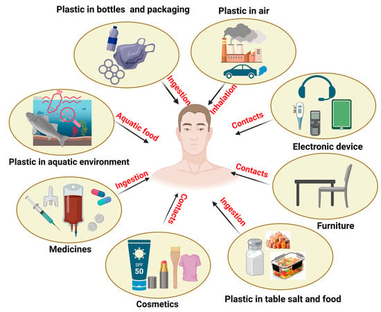
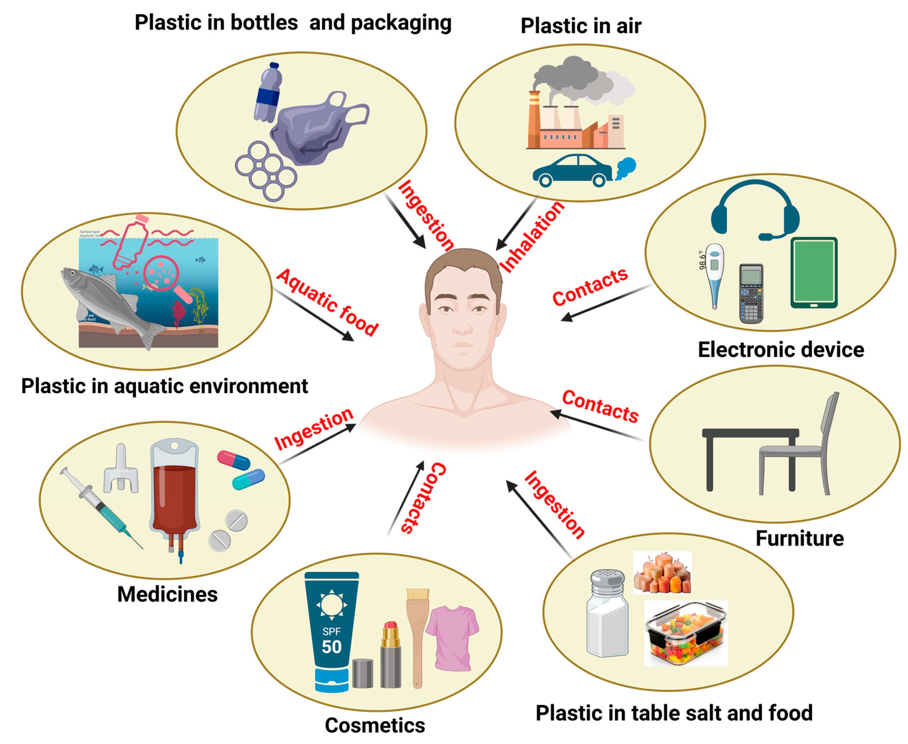
In the modern era, plastic pollution is currently one of the most significant environmental challenges that affects human lives [1]. Plastics such as microplastics (MPs) (size < 5 mm) and nanoplastics (NPs) (size < 100 nm) are now detected in soil, water, air, and food chains, making human exposure unavoidable. Recent studies have reported the widespread presence of MPs and NPs in freshwater and groundwater systems, highlighting water as a major vector for environmental dissemination and human exposure [2]. In addition, MNPs have been detected in fruits and vegetables, indicating dietary exposure routes beyond processed food products [3]. Current estimates suggest that more than 13 billion tons of plastic waste may accumulate in global ecosystems by 2050 [4]. Among the dozens of polymer types—such as polypropylene (PP), polyethylene (PE), polystyrene (PS), and polyethylene terephthalate (PET)—many degrade into micro- and nanosized particles capable of entering biological systems through ingestion, inhalation, and dermal contact (Figure 1) [5].
Figure 1.
Overview of major human exposure routes to MNPs, including ingestion (contaminated food, water, and packaging), inhalation (airborne particles), dermal contact (cosmetics, textiles, and personal care products), occupational exposure, and medical applications. Created in PowerPoint and BioRender. Smith, J. (2025). https://BioRender.com/c248457.
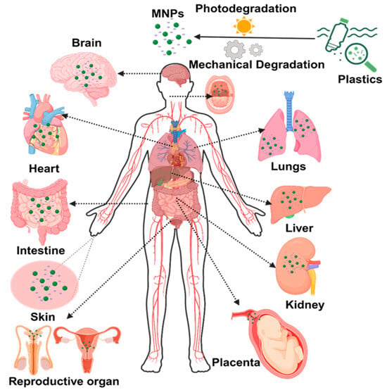
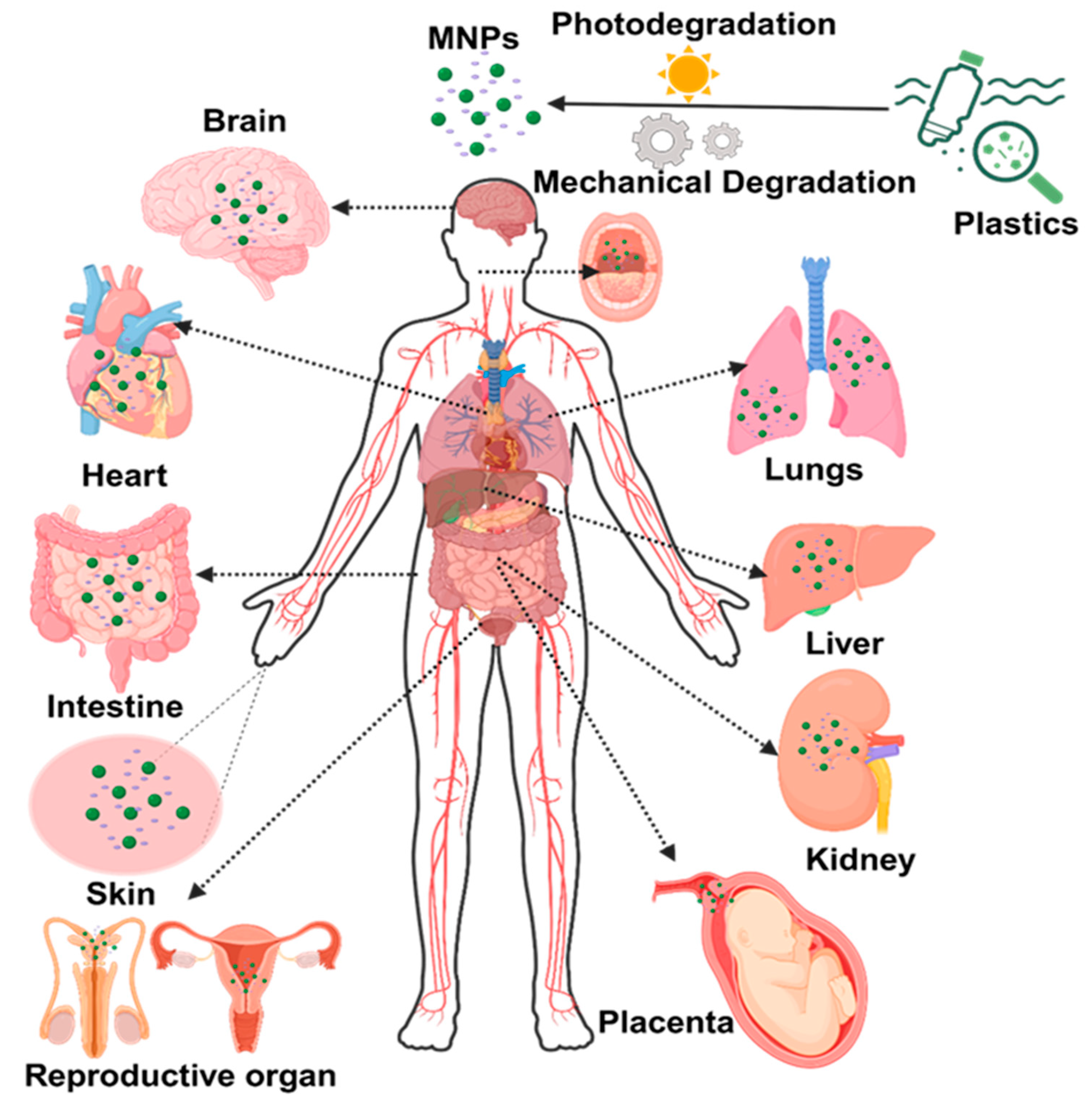
Evidence from animal studies and human autopsy samples indicates that these particles can cross biological barriers and accumulate in metabolically active organs, including the liver, kidney, adipose tissue, lung, and brain (Figure 2) [6]. Several studies investigated the neurotoxic effects of PS, PE, and MNPs in invertebrates such as nematodes, bivalves, and crustaceans, either in the presence or absence of co-exposure to other compounds [7]. The European Union recommends limited use of plastics, but there are no standards for food contamination with MPs. The European Food Safety Authority (EFSA) recommends systematic monitoring of MNPs in food. Beyond their intrinsic toxicity, MNPs can also act as vectors for transporting other environmental contaminants, including metals, persistent organic pollutants, and chemical additives, thereby amplifying their combined toxicological effects in exposed organisms [8].
Figure 2.
Schematic representation of the biodistribution of MNPs in the human body following environmental exposure via ingestion, inhalation, and dermal contact, and their potential accumulation in multiple organs. Created in BioRender. Smith, J. https://BioRender.com/c248457.
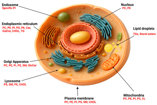
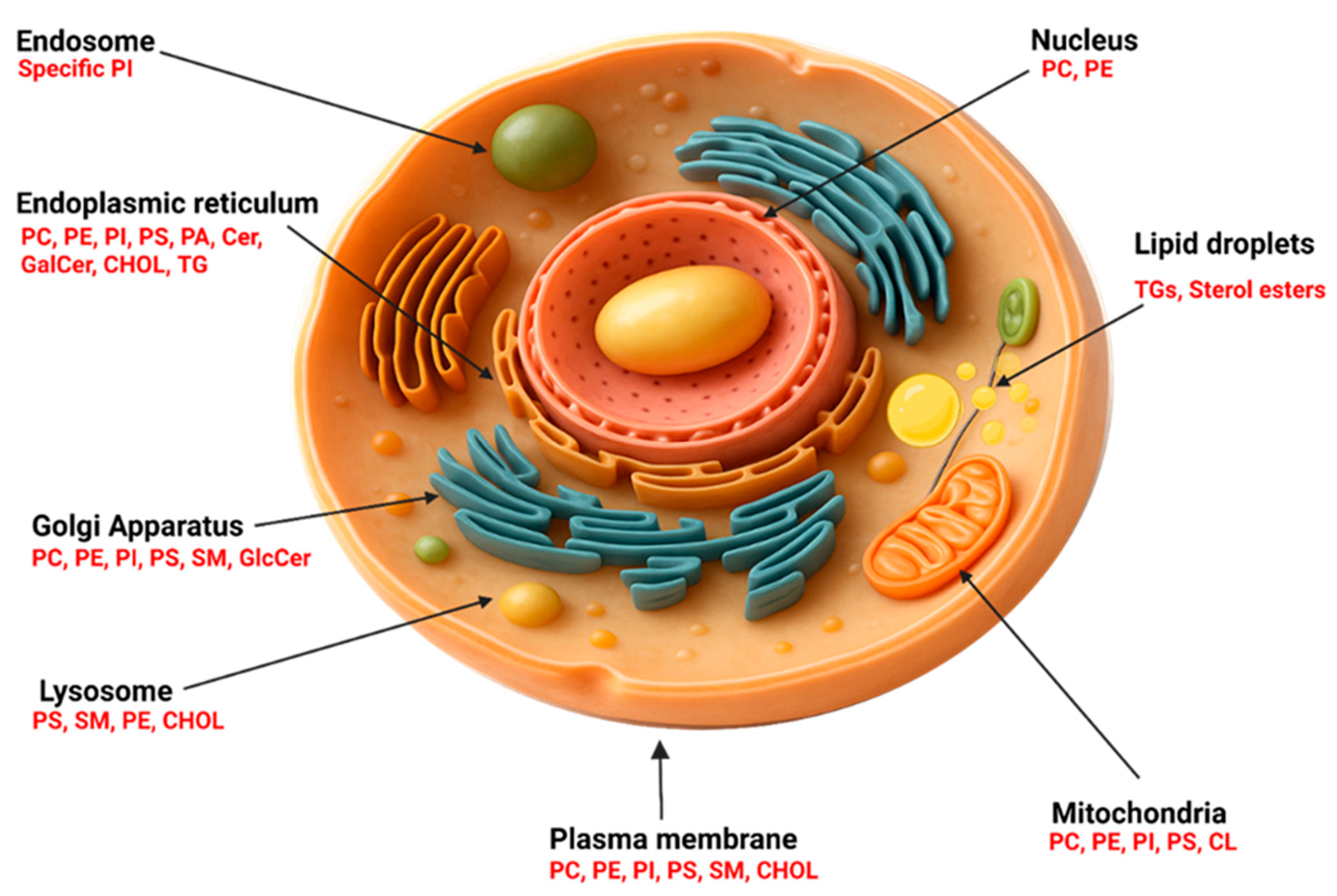
Although early research largely focused on oxidative stress and inflammation, it is now increasingly clear that lipids are among the earliest and most vulnerable molecular targets of MNP exposure. While oxidative stress represents a broader cellular response involving mitochondrial and inflammatory pathways, considerable evidence suggests that lipid remodeling is among the earliest and most sensitive molecular events following MNP exposure [9]. Lipids form the structural backbone of membranes, support energy metabolism, and regulate essential signaling pathways. Their distribution across organelles such as the phospholipid-rich endoplasmic reticulum, sphingolipid-enriched Golgi, or cardiolipin (CL)-dense mitochondria defines organelle identity and function as shown in Figure 3 [10]. Even subtle disturbances in lipid homeostasis can alter membrane fluidity, mitochondrial respiration, vesicular trafficking, and inflammatory signaling. Environmental toxicants, including MNP, disrupt this delicate balance, leading to phospholipid imbalance, sphingolipid dysregulation, sterol oxidation, and lipid droplet accumulation. Such lipid-centered responses are increasingly recognized as common features of cellular stress and are closely linked to metabolic impairment and cell survival [11,12], given that lipid-rich organs such as the liver, brain, and adipose tissue exhibit heightened susceptibility to lipid perturbations. Lipidomics has emerged as a powerful tool to investigate early biochemical changes induced by MNPs. High-resolution lipidomics enables the detection of tissue-specific lipid alterations, offering insights into how MNPs interact with cellular membranes, rewiring metabolic pathways, and destabilizing organelle function.
Figure 3.
Schematic representation of the subcellular distribution and diversity of major lipid classes across key organelles in a eukaryotic cell. Figure created using Canva and PowerPoint.
This review highlights current evidence on tissue-specific lipidomic alterations triggered by MNP exposure, identifies mechanistic pathways that underlie lipid disruption, and highlights key gaps that must be addressed to understand the lipid-mediated toxicity of MNPs. In particular, the lack of comparative, tissue-resolved lipidomic analyses across different polymer types and exposure scenarios represents a major gap in current MNP toxicology research.
2. Mechanism of MNP Toxicity at Lipidome Level
Lipidomics, as a specialized branch of metabolomics, enables high-resolution profiling of lipid species and pathways altered under toxicant exposure. Using advanced analytical platforms such as LC–MS and GC-MS, lipidomics has revealed that MNPs are potent disruptors of cellular and organellar lipid homeostasis [13]. Because most MNPs possess hydrophobic surfaces, they readily interact with lipid-rich structures, cross biological membranes, and initiate lipid-centered stress responses across multiple tissues [14].
2.1. Membrane Interaction and Disruption of Lipid Bilayer Architecture
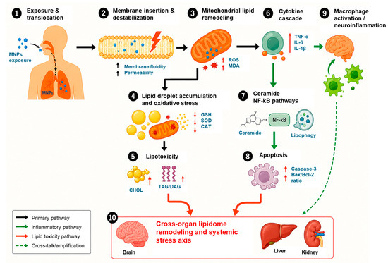
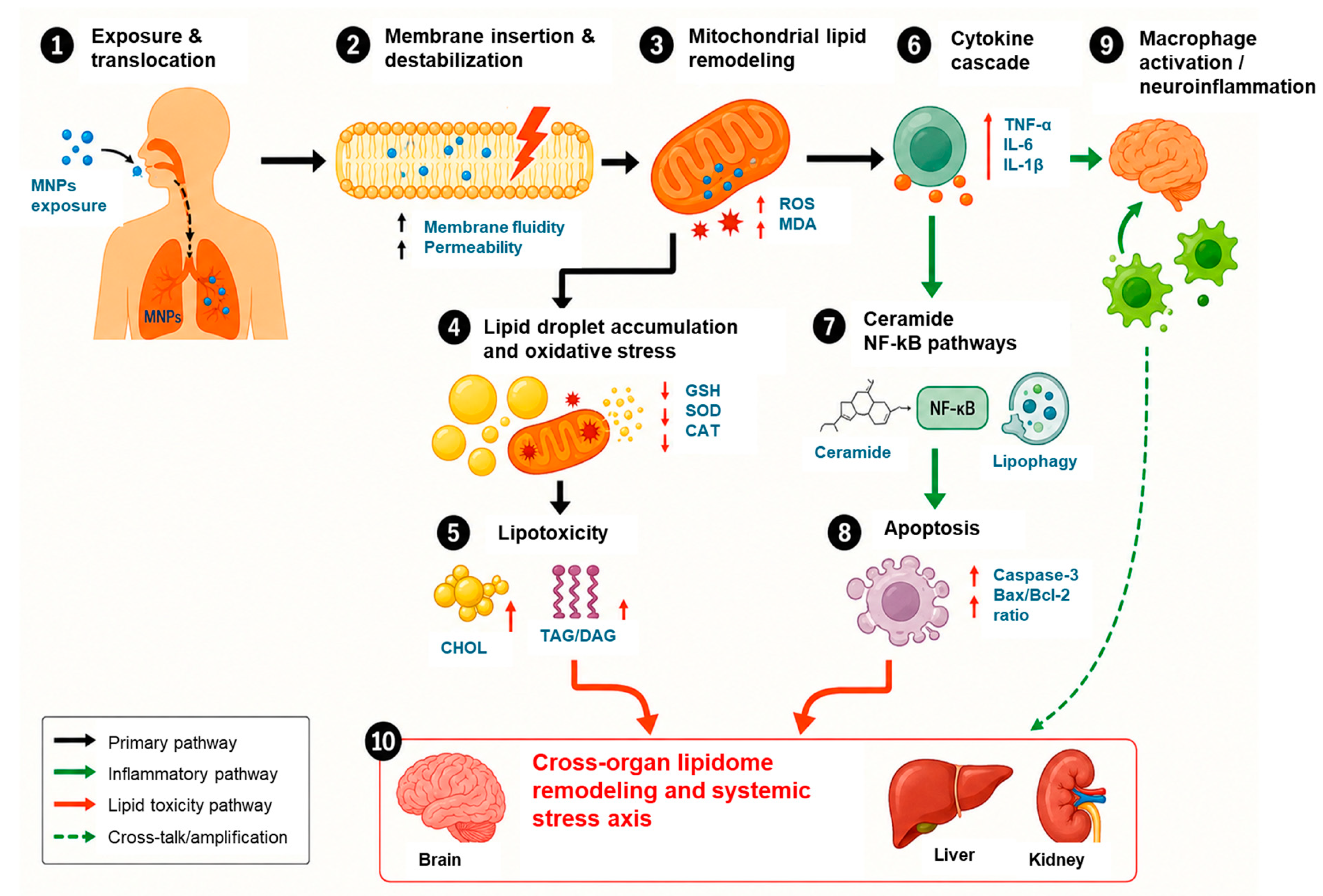
A primary mechanism of MNP toxicity involves direct physical interaction with cellular membranes. NPs can insert into or adsorb onto phospholipid bilayers, increasing membrane tension, altering membrane fluidity, and causing local curvature stress, as shown in Figure 4. These interactions destabilize lipid rafts, disrupt membrane barrier integrity, and promote increased ion leakage, particularly calcium, thereby initiating downstream oxidative and inflammatory signaling [15]. Importantly, plastic-derived oligomers, which originate from environmental weathering, thermal degradation, or fragmentation of larger polymers, represent a highly mobile and lipid-reactive fraction of MNPs. Molecular dynamics simulations provide evidence that PET and PE oligomers, including monomers and short-chain oligomers (tetramers), can embed between phospholipid acyl chains, resulting in bilayer thickening, reduced lipid mobility, and altered packing density. Such perturbations can occur even in the absence of nanoparticle internalization, highlighting membrane lipid reorganization as an early and primary toxic event [16,17]. Collectively, these studies indicate that cell membrane destabilization is a conserved, early event in MNP toxicity, governed by particle size, surface chemistry, and polymer composition (Table 1).
Figure 4.
Schematic overview of the proposed mechanisms underlying MNP-induced systemic lipidome remodeling across metabolically active tissues. Created in PowerPoint and BioRender. Smith, J. (2025). https://BioRender.com/c248457.
2.2. Oxidative Damage and Peroxidation of PUFA-Rich Lipids
MNP exposure consistently triggers the generation of reactive oxygen species (ROS), which disproportionately affect polyunsaturated phospholipids in membranes. ROS-induced peroxidation generates [18] oxidized phospholipids (OxPC, OxPE) and lysophospholipids (LPC, LPE), both of which compromise membrane stability. Beyond lipid peroxidation, excessive ROS production also contributes to mitochondrial dysfunction, redox imbalance, and pro-inflammatory signaling, thereby amplifying lipid-mediated cellular injury [19,20,21]. This oxidative remodeling is a hallmark of tissues with high PUFA content, such as the liver and brain. Increased LPC/LPE ratios reported in mice, zebrafish, and Drosophila models reflect ongoing membrane breakdown and inflammatory lipid signaling [22,23]. Thus, ROS-driven peroxidation of PUFA-rich lipids represents a critical intersection between oxidative stress and lipidome remodeling during MNP exposure (Table 1).
Table 1.
Mechanism of MNP toxicity at the lipidome level.
2.3. Mitochondrial Lipid Remodeling and Energetic Collapse
The mitochondrial inner membrane is enriched in CL, a phospholipid essential for the electron transport chain. Multiple MNP studies report decreased CL levels, reduced PC/PE ratios, and neutral-lipid accumulation, indicating mitochondrial membrane stress [27]. CL oxidation impairs mitochondrial respiration, reduces ATP production, and promotes excessive ROS generation, creating a self-amplifying cycle of lipid damage. These features mirror mitochondrial dysfunction observed in classical neurotoxicant models such as rotenone (Table 1) [24,25].
2.4. Neutral-Lipid Accumulation and Lipophagy Impairment
Across various animal models, PS-, PP-, and PE-derived MNPs induce a convergent phenotype characterized by triacylglycerol (TAG) accumulation, enlarged lipid droplets, and reduced phospholipid abundance. This shift toward storage lipids reflects disrupted fatty acid oxidation, impaired lipophagy, and compensatory sequestration of peroxidized lipids. Downregulation of autophagy-related genes (e.g., ATG5) further contributes to lipid droplet persistence and metabolic stress [20]. Among all tested materials, PS, PE, and PP are the most frequently studied and exhibit a convergent pattern of TAG accumulation and glycerophospholipid depletion, including Phosphatidylcholine (PC), Phosphatidylethanolamine (PE), and CL. This lipid remodeling reflects oxidative-stress-driven peroxidation of membrane lipids, compensatory lipid droplet formation, and impaired lipophagy. For example, PS-NPs in Drosophila and zebrafish consistently lower PC/PE ratios and increase neutral lipid levels, mirroring the mitochondrial lipid imbalance observed in rotenone-based neurotoxic models [26,27].
In contrast, PET and polymethyl methacrylate (PMMA), which are less degradable and have strong surface polarity, primarily disrupt membrane architecture rather than bulk storage lipids. Molecular dynamics simulations show PET oligomers inserting between phospholipid acyl chains, thickening the bilayer and altering phospholipid arrangement. Such physicochemical perturbation may increase membrane permeability and calcium influx, initiating oxidative cascades without requiring internalization [16,30]. Polyvinyl chloride (PVC) and polyamide (nylon) induce lipid stress primarily in aquatic models, where they elevate neutral lipids and deplete phospholipids, a pattern associated with energy imbalance and feeding suppression. Chlorine atoms in PVC may catalyze the generation of ROS, thereby enhancing lipid peroxidation [31].
2.5. Sphingolipid Dysregulation and Ceramide-Driven Cell Stress
MNP exposure alters sphingolipid metabolism, particularly increasing ceramide levels while decreasing sphingomyelin. Ceramide accumulation promotes mitochondrial outer membrane permeabilization, activates pro-apoptotic signaling pathways (Bax/Bcl-2), and contributes to neuroinflammation and metabolic dysfunction [28]. Elevated ceramide levels observed in MNP-exposed macrophages, liver cells, and zebrafish tissues highlight sphingolipid imbalance as a central mediator of MNP toxicity, as mentioned in Table 1.
2.6. Polymer Chemistry Determines the Mode of Lipid Disruption
Hydrophobic polymers such as PS, PE, and PP are readily internalized due to their strong affinity for lipid membranes and induce TAG accumulation, depletion of membrane phospholipids, and pronounced oxidative lipid remodeling [32]. These effects are frequently associated with lipid droplet expansion, mitochondrial stress, and altered fatty acid metabolism across multiple biological models. In contrast, more polar polymers such as PET and PMMA predominantly disrupt membrane architecture by altering lipid packing, bilayer thickness, and membrane fluidity, rather than inducing large-scale accumulation of neutral lipids. PVC and polyamide (nylon) particles primarily induce oxidative-stress-driven lipid damage, particularly in aquatic organisms, where reductions in phospholipid abundance and increased lipid peroxidation have been reported [33]. This polymer-specific hierarchy indicates that surface chemistry, polarity, molecular weight, and degradation potential critically govern lipid–membrane interactions and downstream lipidomic outcomes, and should therefore be considered when predicting MNP toxicity [34,35]. Studies reporting MNP-induced tissue-specific lipidomic alterations across various biological model systems are summarized in Table 2.
Table 2.
Lipidome alterations induced by size-dependent MNP polymers across biological models.
Collectively, these findings demonstrate that MNPs exert toxicity predominantly through polymer-chemistry-dependent lipidome remodeling, beginning with membrane destabilization and progressing through oxidative lipid damage, mitochondrial dysfunction, impaired lipophagy, and ceramide-driven stress responses.
3. Tissue-Specific Lipidome Dysregulation Induced by MNPs
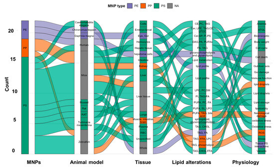
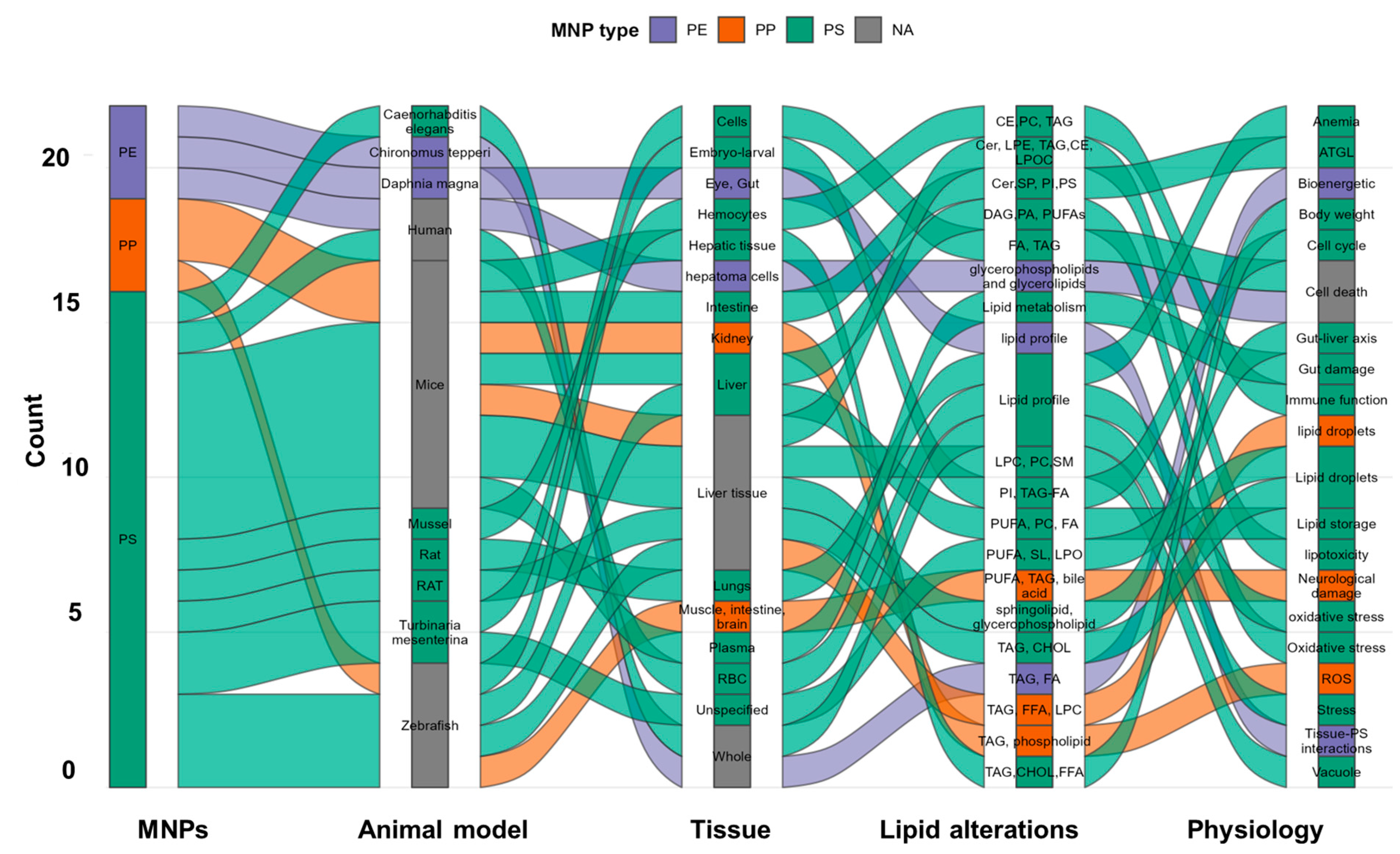
MNPs accumulate in multiple organs and biological fluids, including blood, breast milk, placenta, lung tissue, liver, kidney, adipose tissue, and brain, highlighting their widespread distribution in exposed organisms [44,45,46]. Increasing evidence from lipidomics shows that MNPs do not exert uniform toxicity across the body; instead, they trigger distinct lipidome remodeling patterns in each tissue, reflecting differences in lipid composition, metabolic roles, and cellular vulnerability, as shown in the Sankey plot (Figure 5) based on previously reported MNP-induced lipidome studies. The following sections summarize key lipidomic alterations reported in major organs.
Figure 5.
Integrated Sankey diagram summarizing the relationships among MNP polymer types, experimental models, tissue-specific lipidome alterations, and associated physiological outcomes across studies. Figure created using R Studio 4.5.1 (Posit, Boston, MA, USA).
3.1. Dysregulation of Liver Tissue Lipidome Associated with MNPs
The liver is one of the most vulnerable organs to MNP accumulation due to its central role in lipid metabolism, detoxification, and nutrient processing. Multiple animal studies show that MNP exposure profoundly alters hepatic lipid homeostasis.
The main ways humans are exposed to MNPs are through cutaneous absorption, inhalation of airborne particles, and consumption of watery foods. Among these routes, ingestion is the primary entry route. After entering the body, MNPs accumulate in organs or tissues. According to various studies, the liver is particularly vulnerable to MNP exposure, which may cause abnormalities in hepatic lipid metabolism. Previous research revealed that BALB C57 mice exposed to NPs for two weeks developed hepatic steatosis. The alteration of lipids and their metabolites throughout the body is known as lipid metabolic disorder [47,48,49]. According to a recent study, male mice exposed to NPs may develop obesity and hepatic fat accumulation. Lipid metabolites were shown to be the most significantly changed metabolic class following NP exposure [44]. In mice exposed to MPs, a lipidomic study revealed substantial changes in hepatic lipid species, particularly in free fatty acids (FFAs) and TAGs. In addition, the liver transcriptional profile showed that lipid metabolism pathways were enriched in MP-exposure-induced differentially expressed genes (DEGs) [50]. The liver is the organ responsible for nutrient metabolism and biotransformation. A previous study discovered that lipid metabolism is impacted by PP MPs [38]. An interesting finding reveals that PP MPs have great potential to alter the molecular landscape of the liver, cause complications associated with lipid metabolism, and alter the transcriptome of mitochondrial protein translation and their expression regulation. Nine differentially expressed genes linked to lipid biomarkers were found to be involved in cell differentiation (Bbc3, Lrrc15, and Gdf15), lipid catabolism (Etnppl and lipg), tRNA methyltransferase (Trmt112), and mitochondrial transcription (Mrps12 and Mrpl53) [51]. These changes drive hepatic steatosis, impaired lipophagy, and mitochondrial dysfunction, even at early exposure stages. Across multiple polymer types (PS, PP, PE), a conserved pattern of TAG accumulation and phospholipid depletion emerges, indicating a shared mechanism of lipidome disruption in the liver. These findings show a conserved disruption of liver lipid homeostasis with implications for metabolic disease.
3.2. Dysregulation of Lung Tissue Lipidome Associated with MNPs
Humans can directly inhale airborne MPs because of their tiny size. Inhaling plastic fibers and particles frequently causes respiratory problems, particularly in exposed workers [52]. MNPs, particularly PS-NPs, disrupt the biophysical function of pulmonary surfactants by adsorbing key phospholipid components [53]. This alters the phase behavior, surface tension, and membrane structure of the surfactant, which is crucial for reducing alveolar surface tension and preventing lung collapse. The collapse or thinning of this surfactant layer can reduce the lung’s defense and increase susceptibility to respiratory dysfunction. Very limited research has been conducted on lung lipidomics. Such changes compromise alveolar expansion, impair gas exchange, and sensitize the lung to inflammation. Given that the lung surfactant system is uniquely lipid-rich, MNP–lipid interactions may have disproportionate physiological consequences even at low exposure.
3.3. Dysregulation of Kidney Tissue Lipidome Associated with MNPs
The kidneys play a central role in lipid handling, filtration, and metabolic regulation. MNP exposure alters renal lipid composition in ways that resemble early stages of lipid-driven renal injury [54]. Lipid metabolic abnormalities, such as fatty acid oxidation, are linked to intracellular lipid accumulation and, ultimately, fibrosis and loss of kidney function.
According to a lipidomics study, PP-MPs altered the kidney lipidome by raising the amount of TAG and influencing the degree of unsaturation and phosphatidyl ethanolamines. The detrimental effects of PP-MPs on kidney functions, as demonstrated by the foot process fusion, ROS imbalance, and lipid reprogramming, were validated by correlation analysis between the expression of important genes linked to lipid metabolism, ROS imbalance, and Raman peak intensities [55]. These detrimental effects of MPs on human kidney cells imply that consuming MPs may cause toxicological issues with cell metabolism and intercellular interaction. One of the important findings highlights the possible negative consequences of MPs on human health since exposing human kidney cells to them causes morphological, metabolic, proliferative, and cellular stress [56]. Correlation analyses reveal that these lipid changes align with morphological damage, including podocyte foot process fusion and cytoskeletal distortion. In human kidney cell lines, MNP exposure induces morphological abnormalities, impaired proliferation, and mitochondrial stress, confirming that renal lipid dysregulation is a direct toxic outcome of MNP–cell interactions
3.4. Dysregulation of Brain Tissue Lipidome Associated with MNPs
Brain tissue is remarkably lipid-rich (50% of brain dry weight), with diversity in lipid classes, regional distribution, and cell-type specialization. Lipids are vital for myelin structure, neuronal membrane formation, and cell–cell communication. Recent studies demonstrate a trend of increasing MNP concentrations in the brain. The majority of MNPs detected in tissues are composed of PE and predominantly appear as NP flakes. In normal decedent brain samples, MNP concentrations were 7–30 times higher than those observed in livers or kidneys, with brain samples from dementia cases exhibiting even greater MNP accumulation [57]. A brain lipidomics study demonstrated that NPs disrupt brain lipidomic profiles, leading to neurotoxic effects and mitochondrial dysfunction [58]. These lipidomic findings position the brain as a critical target of MNP-induced lipid disruption, with potential implications for neurological disease progression.
Across tissues, MNPs consistently induce lipid-centered toxic responses, but each organ displays distinct susceptibility patterns reflecting its lipid composition and metabolic role. These tissue-specific signatures form the basis for understanding systemic lipid dysfunction and guide the need for future multi-organ lipidomics studies
These lipidomic disruptions converge on multiple physiological outcomes, including oxidative stress, impaired immune function, mitochondrial bioenergetic decline, lipid droplet accumulation, lipotoxicity, cell death, gut–liver axis disturbances, neurological damage, and altered body weight. The visualization captures how polymer type, organismal biology, tissue susceptibility, and lipid-class remodeling interact to determine the systemic toxicological landscape of MNP exposure.
A consolidated comparison of reported MP-induced lipid alterations across different tissues and model systems is presented in Table 3. This table highlights conserved and tissue-specific lipidomic responses, including TAG accumulation, phospholipid depletion, and altered fatty acid composition across species.
Table 3.
Reported studies of only MP-associated lipid alterations across tissues of model systems.
4. Mechanistic Pathways Linking MNP-Driven Lipid Disruption to Organ Dysfunction
The lipidomic alterations described earlier represent the initial molecular events triggered by MNP exposure. However, these early lipid changes do not remain localized; they translate into progressive functional impairment across tissues. Below, we outline how MNP-induced lipid disruptions propagate into organ-specific physiological and pathological outcomes, forming a cascade of metabolic, inflammatory, and structural defects
4.1. From Membrane Perturbation to Organellar Failure
Once MNPs disturb membrane architecture, cells experience loss of membrane-bound enzyme activity, defective receptor signaling, compromised ion gradients, impaired vesicular trafficking, and altered nutrient transport. Such membrane-level disturbances have been increasingly recognized as early upstream events that precede organellar stress in toxicant-induced cellular injury [61]. These abnormalities weaken organelle coordination. In hepatocytes and neurons, even minor membrane perturbations disrupt ER–mitochondria contact sites (MAMs), impairing calcium signaling, lipid shuttling, and apoptotic regulation [62]. Thus, membrane disruption evolves into global organelle communication failure.
4.2. Lipid Droplet Overload and Metabolic Reprogramming
Across experimental models, MPs and NPs consistently stimulate TAG accumulation and lipid droplet expansion in the liver, adipose tissue, and invertebrate larvae. For example, Liu et al. noted lipid droplet accumulation in mouse liver upon PP-MP exposure and MPs decreased polyunsaturated phospholipids, while NPs increased ether-linked phosphatidylcholines (PC-Ps/PCOs). Gene expression analysis showed that high concentrations of MPs downregulated the expression of fatty-acid-synthesis-related genes, and significantly downregulated the microsomal triglyceride transfer protein (mtp) gene, indicating a perturbation in lipid storage metabolism, which was not observed for NP exposure. In contrast, NPs induced a dose-dependent accumulation of lipids, suggesting increased lipid droplet formation and an activation of ceramide-mediated apoptosis pathway [63,64].
4.3. Oxidative Lipid Damage Propagates, Inflammation and Cell Injury
The accumulation of oxidized phospholipids and lysophospholipids functions not only as a marker of oxidative stress but as a bioactive driver of inflammation, activating macrophages and microglia, stimulating NF-κB-dependent inflammatory cascades, and disrupting endothelial integrity. MNP exposure leads to ROS generation and, in tissues with high PUFA content (brain & liver), results in peroxidation of phospholipids. The reduction in unsaturated PLs and rise in lysophospholipids (LPC LPE) reflect membrane breakdown [65]. For instance, mouse liver exposed to PP-MPs showed decreased unsaturated phospholipids [21].
This oxidative–inflammatory loop promotes steatohepatitis in the liver, alveolar inflammation in the lung, and neuroinflammation in the brain, linking lipid damage to tissue pathology.
4.4. Mitochondrial Dysfunction as the Central Driver of Organ Impairment
The mitochondrial consequences of lipid disruption, particularly CL oxidation and altered PC/PE balance, are significant. Phospholipids like CL in the inner mitochondrial membrane are essential for electron transport chain (ETC) integrity. Though direct CL data in MP studies are scarce, the observed phospholipid disruption implies mitochondrial vulnerability. Nevertheless, CL oxidation and altered PC/PE ratios are well-established hallmarks of mitochondrial vulnerability under oxidative and toxic stress [66]. Consequent ATP depletion, ROS amplification, and mitochondrial fragmentation may follow. These energetic deficits explain hepatic metabolic failure, kidney tubular vulnerability, reduced neuronal firing and synaptic plasticity, and compromised lung epithelial repair [67].
Thus, mitochondria serve as the critical convergence point where molecular lipid damage transforms into organ-wide dysfunction.
4.5. Ceramide Signaling, Apoptosis, and Inflammatory Cell Death
MNP-induced ceramide accumulation acts as a master regulator of cell fate. Ceramide-mediated apoptosis has been widely implicated in metabolic, neurodegenerative, and inflammatory diseases, reinforcing its relevance as a conserved stress effector [68]. Elevated ceramide levels activate apoptotic pathways (Bax/Bcl-2 imbalance), increase membrane permeability, drive endothelial dysfunction, and propagate inflammatory cell death (pyroptosis-like features) [69].
This pathway is intimately linked to hepatic fibrosis, renal injury, and neuronal degeneration, making ceramide a central mediator connecting lipidomic change to tissue pathology.
4.6. Sterol and Oxysterol Imbalance Drives Signaling Collapse
Altered cholesterol metabolism and oxysterol accumulation affect membrane raft stability, neurotransmitter receptor localization, vesicle fusion in neurons, immune cell activation, and endocrine signaling [70]. Such sterol-driven signaling disturbances are particularly detrimental in lipid-rich tissues such as the brain, where membrane microdomain organization is essential for synaptic function [71]. Oxysterols (e.g., 7-ketocholesterol) are potent neuroinflammatory agents and may contribute to cognitive deficits and synaptic dysfunction following MNP exposure.
4.7. System-Level Impact: The Lipid–Stress Axis
Although current evidence is derived mainly from isolated tissue studies, recurring lipidomic signatures across organs indicate that MNP-induced lipid disturbances do not remain confined to individual tissues. Instead, they appear to form part of a broader, interconnected toxicological process that we define as the Lipid–Stress Axis [72]. In this model, the earliest events, such as membrane destabilization, phospholipid depletion, PUFA peroxidation, and ceramide accumulation, set the stage for organellar dysfunction characterized by mitochondrial collapse, ER stress, disrupted ER–mitochondria contact sites, and impaired lipophagy. As these cellular failures intensify, they manifest as organ-level pathologies, including hepatic steatosis, neuroinflammation, renal metabolic stress, and surfactant impairment in the lung, reflecting the distinct lipid requirements of each tissue. Over time, these localized disturbances may propagate into systemic metabolic imbalance through altered circulating lipid profiles, disrupted inter-organ lipid trafficking, and broader endocrine or inflammatory dysregulation. Together, these interconnected processes provide a unified mechanistic framework explaining how primary lipidomic injury evolves into multi-organ physiological dysfunction, even though comprehensive cross-organ lipidomics datasets are not yet available. Conceptual frameworks linking lipid dysregulation to systemic metabolic disease have also been proposed in other toxicological and metabolic contexts [73].
5. Analytical Advances and Lipidomics Approaches in MNP Toxicology
Lipidomics has become an essential tool for uncovering early biochemical disturbances induced by MNPs. Recent methodological advances in separation technologies, mass spectrometry, and computational analysis have significantly improved the ability to detect subtle, tissue-specific lipid alterations. This section highlights key developments in analytical lipidomics and discusses current challenges in applying these methods to MNP toxicology. Recent advances have not only improved lipid detection sensitivity but have also enabled mechanistic linkage between lipidome alterations and functional toxicity endpoints [74].
5.1. High-Resolution Lipidomics Platforms
The most substantial progress in MNP toxicology has come from the adoption of high-resolution mass spectrometry-based lipidomics, which allows sensitive and comprehensive profiling of hundreds to thousands of lipid species. These platforms are particularly well-suited to detecting early lipid perturbations that precede overt cytotoxicity in MNP exposure models.
Untargeted LC–MS/MS is well suited for broad lipidome coverage and accurate mass detection, shotgun lipidomics for rapid, high-throughput characterization of intact lipids, GC–MS for fatty acids, sterols, and oxidation products, Raman and FTIR spectroscopy for detecting membrane lipid order and nanoscale interactions, and MS imaging (MALDI, DESI) to capture spatial lipid distribution in tissues [75,76]. Together, these techniques enable identification of phospholipid remodeling, sphingolipid disruption, CL oxidation, and neutral lipid accumulation, core signatures of MNP-induced lipid toxicity [50]. Each platform offers complementary strengths, with LC–MS/MS providing broad lipidome coverage, shotgun lipidomics enabling rapid class-level screening, and imaging approaches resolving spatial lipid heterogeneity [77].
5.2. Data Processing, Annotation, and Network-Level Interpretation
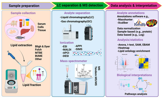
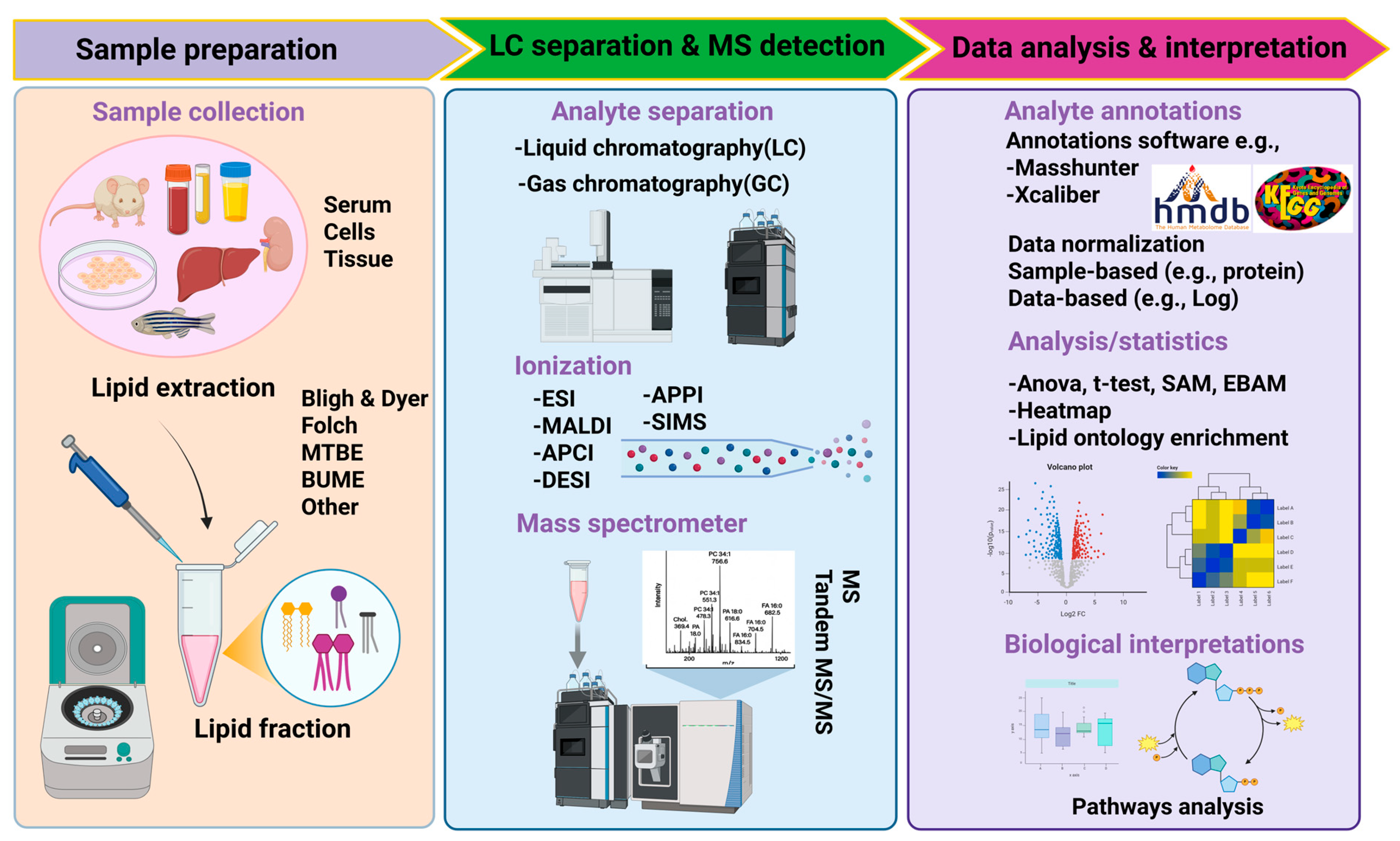
Advances in computational lipidomics have improved structural annotation, quantification, and pathway-level interpretation of lipidomic datasets. Common tools include MS-Dial, LipidSearch, LipidMatch, and LIPID MAPS for automated lipid identification, and XCMS, MZmine, and Progenesis QI for peak detection and alignment. These computational approaches enable the transition from descriptive lipid profiling to systems-level interpretation of coordinated lipid pathway disruptions [78]. KEGG, Reactome, and LipidMaps enable pathway enrichment for mechanistic insights. Network-based analyses (lipid–lipid correlation networks, lipid ontology enrichment) are used to identify coordinated lipid changes. Multivariate statistics (PCA, PLS-DA, OPLS) are used for biomarker discovery, as shown in Figure 6 [79]. Such integrative analyses are critical for translating experimental lipidomic signatures into potential biomarkers in humans [80]. These approaches reveal patterns including the coordinated downregulation of PC/PE clusters, the accumulation of ceramide-rich networks, and the reorganization of mitochondrial lipid modules upon MNP exposure.
Figure 6.
Schematic overview of the lipidomics workflow, illustrating sample preparation, chromatographic separation, and mass spectrometric detection, followed by data analysis and biological interpretation. Created in BioRender. Smith, J. (2025). https://BioRender.com/c248457.
5.3. Current Challenges and Knowledge Gaps in MNP–Lipidomics Research
Despite major technological advances, several challenges remain in applying lipidomics to MNP toxicology:
5.3.1. Analytical and Technical Challenges
Ion suppression and isomeric overlap complicate the accurate quantification of low-abundance lipids (e.g., oxidized phospholipids and CL species).
Interference by MNP particles during sample extraction, especially in lipid-rich tissues, can reduce lipid recovery or alter extraction efficiency.
There is limited standardization in extraction methods for combined MNP and lipid analyses [81,82].
5.3.2. Biological and Interpretation Challenges
Spatial heterogeneity within tissues (e.g., brain regions, hepatic zones) remains poorly explored due to the limited use of MS imaging.
While MNPs have been detected in human blood, placenta, lung, and brain, integrated lipidomics datasets are largely absent.
Scarcity of flux-based lipidomics: Most studies rely on static lipid measurements; isotope tracers (13C-acetate, 13C-palmitate, 13C-glucose) are needed to resolve lipid synthesis and turnover under MNP exposure.
There is an incomplete linkage between lipidomics and functional assays, such as mitochondrial respiration, lipid droplet imaging, or organelle ultrastructure.
These limitations highlight the need for multi-omics integration, standardized workflows, and in vivo fluxomics to fully characterize lipid-mediated MNP toxicity [83]. The absence of integrated human lipidomics datasets represents a major barrier to risk assessment and regulatory translation [84].
5.4. Future Opportunities in Analytical Lipidomics for MNP Research
Future opportunities include single-cell lipidomics to capture cell-type-specific responses in complex tissues, organelle-resolved lipidomics (mitochondrial, ER, lysosomal lipids), and MALDI imaging for spatial mapping of MNP-induced lipid changes [85]. Emerging analytical innovations are expected to bridge current gaps between exposure assessment, lipid dynamics, and functional outcomes. Other future opportunities include integrated lipidomics–proteomics–transcriptomics frameworks for mechanistic clarity, standard reference materials and inter-laboratory harmonization, and advanced bioinformatics using machine learning for lipid pattern prediction. These innovations will allow more accurate detection of MNP-induced lipid perturbations and provide mechanistic insights into early biochemical events [86,87].
6. Translational and Therapeutic Implications of MNP-Induced Lipidome Disruption
Growing evidence that MNPs disrupt lipid metabolism across multiple tissues raises important opportunities for translational research. Lipidomics not only reveals early molecular signatures of exposure but also provides a framework for identifying biomarkers, designing targeted interventions, and refining environmental risk assessment [88]. Such lipid-centered translational approaches are increasingly being explored in environmental and metabolic toxicology to bridge molecular injury with disease risk [89]. The following subsections summarize emerging translational insights based on current lipidomics findings.
6.1. Lipid-Based Biomarkers of MNP Exposure
Although lipidomics data across tissues are still evolving, several lipid classes consistently emerge as sensitive indicators of MNP-induced stress. These lipid signatures can potentially serve as biomarkers of exposure or early toxicity. Promising lipid biomarker candidates include:
- Increased TAGs and neutral lipids, reflecting lipid droplet expansion and impaired lipophagy;
- Elevated lysophospholipids (LPC, LPE) are associated with membrane breakdown and phospholipase activation;
- Reduced unsaturated phospholipids (PC, PE), indicating oxidative loss of PUFA-containing species;
- Altered sphingolipid balance, particularly increased ceramide and decreased sphingomyelin;
- Enhanced sterol and oxysterol levels, especially in the liver and kidney;
- Disrupted PC/PE and SM/Cer ratios, which reflect mitochondrial stress and apoptotic signaling.
Similar lipid classes have been proposed as early biomarkers in other environmental and metabolic stress conditions, supporting their translational relevance [90]. These lipid shifts are consistent across diverse animal models, polymer types, and exposure regimens, suggesting their potential as cross-tissue molecular fingerprints of MNP toxicity. Future validation in human biofluids (serum, urine, CSF) could enable minimally invasive biomonitoring. This is particularly relevant given recent reports of MNP detection in human blood, placenta, lung, and brain tissues [91].
6.2. Therapeutic or Mitigative Strategies Targeting Lipid Stress
Understanding lipid-centered mechanisms opens avenues for interventions to restore membrane stability, reduce oxidative lipid injury, and support organellar function.
6.2.1. Nutritional and Dietary Interventions
Omega-3 PUFAs (DHA/EPA) may replenish oxidized PUFA pools, stabilize membranes, and counteract peroxidation [92].
Plant-derived bioactives such as curcumin, quercetin, and polyphenols support mitochondrial health and have lipid-protective effects [93].
6.2.2. Pharmacological Approaches
Ceramide-synthase inhibitors may reduce ceramide-driven apoptosis and inflammation [94].
Lipophagy enhancers (e.g., AMPK activators) could promote clearance of damaged or expanded lipid droplets [95].
Mitochondria-targeted antioxidants (CoQ10, MitoQ, SS-31) can mitigate CL oxidation and restore respiratory chain function [96].
6.2.3. Lifestyle and Exposure-Reduction Strategies
Minimizing dietary and inhalation exposure to MNPs can reduce lipidome burden.
Exercise and metabolic activation may enhance lipid turnover and organelle resilience.
Regular lipidomics-based monitoring in high-risk populations (industrial workers, coastal communities, individuals with metabolic disease) may help detect early lipid perturbations.
These approaches remain conceptual but provide a framework for targeted mitigation once lipid biomarkers are validated.
6.3. Implications for Risk Assessment and Regulatory Evaluation
Integrating lipidomics into environmental toxicology has the potential to transform how MNP hazards are assessed. There is growing recognition that omics-based endpoints can complement traditional toxicological assays in regulatory decision-making [97]. Traditional endpoints, such as oxidative stress, inflammation, or histopathology, capture late-stage injury, whereas lipidomics reveals subcellular disturbances long before functional decline becomes measurable.
The Proposed Lipid–Stress Axis Offers a Useful Systems-Level Perspective
The Lipid–Stress Axis connects early membrane and mitochondrial lipid damage to downstream organ dysfunction.
It highlights vulnerable tissues (liver, brain, kidney, lung) based on lipid composition and metabolic load.
It provides mechanistic endpoints (e.g., PC/PE imbalance, CL oxidation, ceramide accumulation) that can be incorporated into regulatory screening frameworks.
Adopting lipidomics as a routine component of environmental risk evaluation would allow regulators to identify high-risk polymers, set exposure thresholds, and develop early-warning biomarkers of toxicity. Comparable systems-level frameworks have been proposed for metabolic, neurodegenerative, and mitochondrial toxicants [98]. This approach is particularly important given the widespread human presence of MNPs and the lack of long-term exposure data.
7. Future Directions and Knowledge Gaps
Despite rapid progress in understanding MNP-induced lipidome disruption, several important gaps limit our ability to fully interpret the biological relevance of these findings. Addressing these gaps will require methodological refinement, multi-organ integration, and translational validation as mentioned in Table 4. A major limitation is the scarcity of human lipidomics data under confirmed MNP exposure. Although MNPs have been detected in human blood, placenta, lung, and brain, comprehensive lipidomic profiling of exposed individuals is lacking. Future work should integrate MP quantification with plasma, serum, or tissue lipidomics to establish human-specific lipid signatures of exposure.
Another critical gap is the absence of lipid flux information. Current studies primarily measure static lipid levels, which cannot distinguish between altered synthesis, degradation, or redistribution. Incorporating stable-isotope tracers (13C-acetate, 13C-palmitate, 13C-glucose) into animal models will help elucidate dynamic changes in phospholipid turnover, TAG cycling, and sphingolipid metabolism during MNP exposure. Tissue heterogeneity represents another challenge. Most lipidomics studies analyze whole organs, masking region-specific vulnerability. Advanced methods such as MALDI-MS imaging, DESI imaging, and laser microdissection lipidomics will allow mapping of localized lipid disruptions, for example, across different brain regions or hepatic zones to identify precise structural targets of MNP toxicity. Furthermore, the downstream effects of lipidomic alterations remain poorly integrated. While individual tissues show characteristic lipid shifts, the mechanisms by which these changes propagate to other organs remain unresolved. Systems-biology approaches combining lipidomics, metabolomics, transcriptomics, and mitochondrial assays in coordinated multi-tissue studies are needed to verify and refine the proposed Lipid–Stress Axis.
Finally, therapeutic interventions targeting lipid repair pathways are largely untested. Controlled animal intervention studies using nutritional lipids, mitochondrial stabilizers, or ceramide-modulating compounds, combined with lipidomics and functional readouts, could provide the first proof-of-concept strategies for mitigating MNP-induced lipid stress.
Table 4.
This table shows the future directions and knowledge gaps for MNP-induced lipid remodeling.
Collectively, these future directions will deepen mechanistic understanding, enhance translational relevance, and refine risk assessment frameworks for MNP exposure.
8. Conclusions
MNPs have rapidly emerged as pervasive contaminants that disrupt lipid homeostasis across multiple metabolically active organs. Evidence from animal models demonstrates that MNPs consistently remodel the lipidome by altering phospholipid composition, elevating ceramide and lysophospholipid levels, driving neutral-lipid accumulation, and impairing mitochondrial lipid architecture. These early lipid perturbations form the foundation of a broader Lipid–Stress Axis, through which membrane instability, organellar dysfunction, and metabolic collapse translate into tissue-level outcomes such as hepatic steatosis, neuroinflammation, renal metabolic stress, and compromised pulmonary surfactant function.
Lipidomics offers a uniquely sensitive window into these early events, revealing biochemical changes long before conventional markers detect toxicity. Integrating lipidomic biomarkers into mechanistic toxicology and risk assessment will be critical for identifying vulnerable tissues, distinguishing polymer-specific hazards, and predicting long-term health consequences of chronic MNP exposure. Future progress will require coordinated human biomonitoring, multi-tissue lipidomics, stable-isotope flux studies, and targeted intervention trials that test whether restoring lipid balance can mitigate the adverse effects of MNPs.
Taken together, the emerging evidence positions lipidome disruption as a central mechanism of MNP toxicity. Establishing lipid-based biomarkers and mechanistic pathways not only advances our understanding of MNP-induced pathology but also provides a foundation for developing prevention-oriented strategies and informing regulatory policies in an era of escalating plastic pollution.
Author Contributions
Conceptualization, methodology, investigation, resources, writing—review and editing, supervision, project administration, R.C. Writing—original draft preparation, visualization, P.R., A.K.T., and D.N.K. reviewed and edited the manuscript. All authors have read and agreed to the published version of the manuscript.
Funding
The authors acknowledge the Department of Biotechnology, Government of India, for the Ramalingaswami grant (BT/RLF/Re-entry/21/2020), and the Council of Scientific and Industrial Research (CSIR), Government of India, for providing funding and fellowships in support of this work.
Institutional Review Board Statement
Not applicable. An institutional review and an ethics committee were not involved in this study as the work did not involve humans, animals, or their samples.
Informed Consent Statement
Not applicable as the present study did not involve any human subjects.
Data Availability Statement
No new data were created or analyzed in this study.
Acknowledgments
The authors would like to thank the Department of Biotechnology, Government of India, for the Ramalingaswami grant (BT/RLF/Re-entry/21/2020), and the Council of Scientific and Industrial Research (CSIR), Government of India, for providing funding and fellowships in support of this work. Priya Rathor thanks the UGC-SRF fellowship, and Ashutosh thanks the CSIR fellowship for PhD work.
Conflicts of Interest
The authors declare no conflicts of interest.
Abbreviations
The following abbreviations are used in this manuscript:
| MPs | Microplastics |
| NPs | Nanoplastics |
| TAG | Triacylglycerol |
| FFAs | Free fatty acids |
| LPC | Lysophosphatidylcholine |
| LPE | Lysophosphatidylethanolamine |
| PC | Phosphatidylcholine |
| PE | Phosphatidylethanolamine |
| UFAs | Unsaturated fatty acids |
| TFAs | Total fatty acids |
| CL | Cardiolipin |
References
- Thompson, R.C.; Olsen, Y.; Mitchell, R.P.; Davis, A.; Rowland, S.J.; John, A.W.G.; McGonigle, D.; Russell, A.E. Lost at Sea: Where Is All the Plastic? Science 2004, 304, 838. [Google Scholar] [CrossRef] [Scilit]
- Chandra, S.; Walsh, K.B. Microplastics in Water: Occurrence, Fate and Removal. J. Contam. Hydrol. 2024, 264, 104360. [Google Scholar] [CrossRef] [Scilit]
- Lazăr, N.N.; Călmuc, M.; Milea, Ș.A.; Georgescu, P.L.; Iticescu, C. Micro and Nano Plastics in Fruits and Vegetables: A Review. Heliyon 2024, 10, e28291. [Google Scholar] [CrossRef] [Scilit]
- Barboza, L.G.A.; Vieira, L.R.; Branco, V.; Figueiredo, N.; Carvalho, F.; Carvalho, C.; Guilhermino, L. Microplastics Cause Neurotoxicity, Oxidative Damage and Energy-Related Changes and Interact with the Bioaccumulation of Mercury in the European Seabass, Dicentrarchus labrax (Linnaeus, 1758). Aquat. Toxicol. 2018, 195, 49–57. [Google Scholar] [CrossRef] [Scilit]
- Li, P.; Wang, X.; Su, M.; Zou, X.; Duan, L.; Zhang, H. Characteristics of Plastic Pollution in the Environment: A Review. Bull. Environ. Contam. Toxicol. 2021, 107, 577–584. [Google Scholar] [CrossRef] [Scilit]
- Irfan, H.; Irfan, H.; Khan, M.A.; Inanc, O.; Al Hasibuzzaman, M. Microplastics and Nanoplastics: Emerging Threats to Cardiovascular Health—A Comprehensive Review. Ann. Med. Surg. 2025, 87, 209–216. [Google Scholar] [CrossRef] [Scilit]
- Lei, L.; Liu, M.; Song, Y.; Lu, S.; Hu, J.; Cao, C.; Xie, B.; Shi, H.; He, D. Polystyrene (Nano) Microplastics Cause Size-Dependent Neurotoxicity, Oxidative Damage and Other Adverse Effects in Caenorhabditis elegans. Environ. Sci. Nano 2018, 5, 2009–2020. [Google Scholar] [CrossRef] [Scilit]
- Tumwesigye, E.; Felicitas Nnadozie, C.; C Akamagwuna, F.; Siwe Noundou, X.; William Nyakairu, G.; Odume, O.N. Microplastics as Vectors of Chemical Contaminants and Biological Agents in Freshwater Ecosystems: Current Knowledge Status and Future Perspectives. Environ. Pollut. 2023, 330, 121829. [Google Scholar] [CrossRef] [Scilit] [PubMed]
- Ma, Q.; Lei, J.; Pang, Y.; Shen, Y.; Zhang, T. Neurotoxicity of Micro- and Nanoplastics: A Comprehensive Review of Central Nervous System Impacts. Environ. Health 2026, 4, 4–24. [Google Scholar] [CrossRef] [Scilit]
- Collins, F.S.; Tabak, L.A. NIH Public Access. Nature 2014, 1811, 637–647. [Google Scholar]
- Jarc, E.; Petan, T. Lipid Droplets and the Management of Cellular Stress. Yale J. Biol. Med. 2019, 92, 435–452. [Google Scholar] [PubMed]
- Okazaki, Y.; Saito, K. Roles of Lipids as Signaling Molecules and Mitigators during Stress Response in Plants. Plant J. 2014, 79, 584–596. [Google Scholar] [CrossRef] [Scilit]
- Yahaya, T.O.; Ibrahim, A.B.; Kalgo, A.S.; Adewale, M.K. Microplastics Exposure Altered Hematological and Lipid Profiles as Well as Liver and Kidney Function Parameters in Albino Rats (Rattus norvegicus). Environ. Anal. Health Toxicol. 2024, 39, e2024021. [Google Scholar] [CrossRef] [Scilit]
- Alqahtani, S.; Alqahtani, S.; Saquib, Q. Toxicological Impact of Microplastics and Nanoplastics on Humans: Understanding the Mechanistic Aspect of the Interaction. Front. Toxicol. 2023, 5, 1193386. [Google Scholar] [CrossRef] [Scilit] [PubMed]
- Fleury, J. Microplastics Destabilize Lipid Membranes by Mechanical Stretching. Proc. Natl. Acad. Sci. USA 2021, 118, e2104610118. [Google Scholar] [CrossRef] [Scilit]
- Järvenpää, J.; Perkkiö, M.; Laitinen, R.; Kakkonen, M.L. Open PE and PET Oligomers’ Interplay with Membrane Bilayers. Sci. Rep. 2022, 12, 2234. [Google Scholar] [CrossRef] [Scilit]
- Wang, W.; Zhang, J.; Qiu, Z.; Cui, Z.; Li, N.; Li, X.; Wang, Y.; Zhang, H.; Zhao, C. Effects of Polyethylene Microplastics on Cell Membranes: A Combined Study of Experiments and Molecular Dynamics Simulations. J. Hazard. Mater. 2022, 429, 128323. [Google Scholar] [CrossRef] [Scilit]
- Wu, H.; Xu, T.; Yang, N.; Xu, S. Polystyrene-Microplastics and Emamectin Benzoate Co-Exposure Induced Lipid Remodeling by Suppressing PPARα Signals to Drive ACSL4-Dependent Ferroptosis and Carp Splenic Injury. Pestic. Biochem. Physiol. 2025, 210, 106396. [Google Scholar] [CrossRef] [Scilit] [PubMed]
- Jeong, C.-B.; Kang, H.-M.; Lee, M.-C.; Kim, D.-H.; Han, J.; Hwang, D.-S.; Souissi, S.; Lee, S.-J.; Shin, K.-H.; Park, H.G.; et al. Adverse Effects of Microplastics and Oxidative Stress-Induced MAPK/Nrf2 Pathway-Mediated Defense Mechanisms in the Marine Copepod Paracyclopina Nana. Sci. Rep. 2017, 7, 41323. [Google Scholar] [CrossRef] [Scilit]
- Nnoruka, U.C.; Okonkwo, C.J.; Ilechukwu, I.; Okonkwo, C.J.; Belonwu, D.C. Impact of Polystyrene Microplastic Exposure on Lipid Profile and Oxidative Stress Status of Male and Female Wistar Rats. Environ. Anal. Health Toxicol. 2022, 37, e2022024-0. [Google Scholar] [CrossRef] [Scilit]
- Mahmud, F.; Sarker, D.B.; Jocelyn, J.A.; Sang, Q.-X.A. Molecular and Cellular Effects of Microplastics and Nanoplastics: Focus on Inflammation and Senescence. Cells 2024, 13, 1788. [Google Scholar] [CrossRef] [Scilit]
- Wang, T.; Perelló Amorós, M.; Porte, C. Lipidome and Gene Expression Profiling in Zebrafish Liver Spheroids: A 3D Model for Environmental Toxicology Applications. Environ. Sci. Technol. 2025, 59, 12543–12553. [Google Scholar] [CrossRef] [Scilit] [PubMed]
- Fasteen, T.D.; Hernandez, M.R.; Policastro, R.A.; Sterrett, M.C.; Zenter, G.E.; Tennessen, J.M. The Drosophila Estrogen-Related Receptor Promotes Triglyceride Storage within the Larval Fat Body. bioRxiv 2024. [Google Scholar] [CrossRef] [Scilit] [PubMed]
- Tiwari, A.K.; Rathor, P.; Patel, R.P.; Jha, P.K.; Birse, N.; Ratnasekhar, C.H. Comprehensive lipidomics reveal Rotenone exposure induces redox-driven lipid droplet accumulation, systemic lipidome perturbation and differential metabolic prioritization linked to Parkinson’s disease. Neurotoxicology 2026, 114, 103425. [Google Scholar] [CrossRef] [Scilit]
- Luo, S.; Zhao, X.; Wang, Y.; Jiang, M.; Cao, Y. Oral Exposure to Nanoplastics Altered Lipid Profiles in Mouse Intestine. Food Chem. Toxicol. Int. J. Publ. Br. Ind. Biol. Res. Assoc. 2025, 197, 115304. [Google Scholar] [CrossRef] [Scilit]
- Rehman, A.; Huang, F.; Zhang, Z.; Habumugisha, T.; Yan, C.; Shaheen, U.; Zhang, X. Nanoplastic Contamination: Impact on Zebrafish Liver Metabolism and Implications for Aquatic Environmental Health. Environ. Int. 2024, 187, 108713. [Google Scholar] [CrossRef] [Scilit]
- Florance, I.; Chandrasekaran, N.; Gopinath, P.M.; Mukherjee, A. Exposure to Polystyrene Nanoplastics Impairs Lipid Metabolism in Human and Murine Macrophages In Vitro. Ecotoxicol. Environ. Saf. 2022, 238, 113612. [Google Scholar] [CrossRef] [Scilit]
- Mignard, V.; Dubois, N.; Lanoé, D.; Joalland, M.-P.; Oliver, L.; Pecqueur, C.; Heymann, D.; Paris, F.; Vallette, F.M.; Lalier, L. Sphingolipid Distribution at Mitochondria-Associated Membranes (MAMs) upon Induction of Apoptosis. J. Lipid Res. 2020, 61, 1025–1037. [Google Scholar] [CrossRef] [Scilit]
- Nanoplastics in the Human Brain and Their Change in Abundance over Time. Nat. Med. 2025, 31, 1077–1078. [CrossRef] [Scilit]
- Chul, K.; Soon, S. Molecular Dynamics Simulation Study on the Structural Properties of Poly (Ethylene Terephthalate) under Uniaxial Extension and Thermal Shrinkage Processes. Curr. Appl. Phys. 2018, 18, 19–26. [Google Scholar] [CrossRef] [Scilit]
- Soliman, A.M.; Mohamed, A.S.; Abdel-khalek, A.A. Impact of Polyvinyl Chloride Nano- Plastics on the Biochemical Status of Oreochromis niloticus under a Predicted Global Warming Scenario. Sci. Rep. 2025, 15, 3671. [Google Scholar] [CrossRef] [Scilit]
- Menéndez-Pedriza, A.; Jaumot, J.; Bedia, C. Lipidomic Analysis of Single and Combined Effects of Polyethylene Microplastics and Polychlorinated Biphenyls on Human Hepatoma Cells. J. Hazard. Mater. 2022, 421, 126777. [Google Scholar] [CrossRef] [Scilit] [PubMed]
- Xia, X.; Sun, M.; Zhou, M.; Chang, Z.; Li, L. Polyvinyl Chloride Microplastics Induce Growth Inhibition and Oxidative Stress in Cyprinus carpio Var. Larvae. Sci. Total Environ. 2020, 716, 136479. [Google Scholar] [CrossRef] [Scilit]
- Yang, Z.; DeLoid, G.M.; Zarbl, H.; Baw, J.; Demokritou, P. Micro- and Nanoplastics (MNPs) and Their Potential Toxicological Outcomes: State of Science, Knowledge Gaps and Research Needs. NanoImpact 2023, 32, 100481. [Google Scholar] [CrossRef] [Scilit]
- Bochicchio, D.; Panizon, E.; Monticelli, L.; Rossi, G. Interaction of Hydrophobic Polymers with Model Lipid Bilayers. Sci. Rep. 2017, 7, 6357. [Google Scholar] [CrossRef] [Scilit]
- Li, X.; He, E.; Chen, G.; Cao, X.; Zhao, L.; Xu, X.; Fu, Z.; Qiu, H. Intergenerational Neurotoxicity of Polystyrene Nanoplastics in Offspring Mice Is Mediated by Dysfunctional Microbe-Gut-Brain Axis. Environ. Int. 2024, 192, 109026. [Google Scholar] [CrossRef] [Scilit]
- Yang, B.; Han, Y.; Hu, S.; Xie, X.; Zhu, X.; Yuan, L. Polystyrene Microplastics Induce Depression-like Behavior in Zebrafish via Neuroinflammation and Circadian Rhythm Disruption. Sci. Total Environ. 2025, 959, 178085. [Google Scholar] [CrossRef] [Scilit]
- Liu, M.; Mu, J.; Wang, M.; Hu, C.; Ji, J.; Wen, C.; Zhang, D. Impacts of Polypropylene Microplastics on Lipid Profiles of Mouse Liver Uncovered by Lipidomics Analysis and Raman Spectroscopy. J. Hazard. Mater. 2023, 458, 131918. [Google Scholar] [CrossRef] [Scilit]
- Lu, H.C.; Melvin, S.D.; Ziajahromi, S.; Neale, P.A.; Leusch, F.D.L.; Kumar, A. Polyethylene Microplastics Induced Lipidomic Responses in Chironomus tepperi: A Two-Generational Exploration. Sci. Total Environ. 2024, 919, 170837. [Google Scholar] [CrossRef] [Scilit]
- Järvenpää, J.P. Dynamics Behavior of PE and PET Oligomers in Lipid Bilayer Simulations. Micropl. Nanopl. 2023, 3, 27. [Google Scholar] [CrossRef] [Scilit]
- Jawdhari, A.; Mihăilescu, D.F.; Stan, M.S.; Bălănescu, M.-V.; Vlăsceanu, R.-I.; Staicu, C.A.; Crăciun, N.; Deák, G. Ingestion of Polyvinylchloride Powder Particles Induces Oxidative Stress and Hepatic Histopathological Changes in Oreochromis niloticus (Nile Tilapia)—A Preliminary Study. Sustainability 2023, 15, 6494. [Google Scholar] [CrossRef] [Scilit]
- Boran, T.; Zengin, O.S.; Seker, Z.; Akyildiz, A.G.; Kara, M.; Oztas, E.; Özhan, G. An Evaluation of a Hepatotoxicity Risk Induced by the Microplastic Polymethyl Methacrylate (PMMA) Using HepG2/THP-1 Co-Culture Model. Environ. Sci. Pollut. Res. Int. 2024, 31, 28890–28904. [Google Scholar] [CrossRef] [Scilit]
- Cole, M.; Coppock, R.; Lindeque, P.K.; Altin, D.; Reed, S.; Pond, D.W.; Sørensen, L.; Galloway, T.S.; Booth, A.M. Effects of Nylon Microplastic on Feeding, Lipid Accumulation, and Moulting in a Coldwater Copepod. Environ. Sci. Technol. 2019, 53, 7075–7082. [Google Scholar] [CrossRef] [Scilit] [PubMed]
- Chen, L.; Sui, G.; Wu, J.; Li, N.; Zhang, Z.; Du, Y.; Lü, M.; Yan, X.; Pan, G. Untargeted Metabolomics and Transcriptomics Joint Analysis of the Effects of Polystyrene Nanoplastics on Lipid Metabolism in the Mouse Liver. Lipids Health Dis. 2025, 24, 195. [Google Scholar] [CrossRef] [Scilit]
- Zhang, H.; Lan, M.; Qiu, L.; Li, X.; Liu, Y.; Zhou, Y.; Liu, J. Microplastics in the Lung Tissues Associated with Blood Test Index. Toxics 2023, 11, 759. [Google Scholar] [CrossRef] [Scilit]
- Ragusa, A.; Svelato, A.; Santacroce, C.; Catalano, P.; Notarstefano, V.; Carnevali, O.; Papa, F.; Ciro, M.; Rongioletti, A.; Baiocco, F.; et al. Plasticenta: First Evidence of Microplastics in Human Placenta. Environ. Int. 2021, 146, 106274. [Google Scholar] [CrossRef] [Scilit]
- Yang, X.; Jiang, J.; Wang, Q.; Duan, J.; Chen, N.; Wu, D. Ecotoxicology and Environmental Safety Gender Difference in Hepatic AMPK Pathway Activated Lipid Metabolism Induced by Aged Polystyrene Microplastics Exposure. Ecotoxicol. Environ. Saf. 2022, 245, 114105. [Google Scholar] [CrossRef] [Scilit]
- Li, Q.; Zhu, K.; Huang, L.; Niu, X.; Li, L.; Gao, L.; Xia, Z. Polystyrene Microplastics Induce Liver Fibrosis and Lipid Deposition in Mice through Three Hub Genes Revealed by the RNA-Seq. Sci. Rep. 2025, 15, 2583. [Google Scholar] [CrossRef] [Scilit] [PubMed]
- Xiao, W.; Xiang, P.; Liao, W.; Xiong, Z.; Peng, L.; Zou, L.; Liu, B.; Li, Q. Effects of Polystyrene Microplastics on the Growth and Metabolism of Highland Barley Seedlings Based on LC-MS. Front. Plant Sci. 2024, 15, 1477605. [Google Scholar] [CrossRef] [Scilit] [PubMed]
- Wang, Q.; Wu, Y.; Zhang, W.; Shen, T.; Li, H.; Wu, J.; Zhang, L.; Qin, L.; Chen, R.; Gu, W.; et al. Lipidomics and Transcriptomics Insight into Impacts of Microplastics Exposure on Hepatic Lipid Metabolism in Mice. Chemosphere 2022, 308, 136591. [Google Scholar] [CrossRef] [Scilit]
- Wang, M.; Wang, J.; Sun, X.; Zhang, K.; Gao, J.; Xu, X.; Wu, J.; Tao, F.; Zhang, D.; Liu, M. Molecular Landscape Remodeling Unravels the Cross-Links of Microplastics-Induced Lipidomic Fluctuations, Nutrient Disorders and Energy Disarrangements. Environ. Health 2025, 4, 448–461. [Google Scholar] [CrossRef] [Scilit] [PubMed]
- Lu, K.; Zhan, D.; Fang, Y.; Li, L.; Chen, G.; Chen, S.; Wang, L. Microplastics, Potential Threat to Patients with Lung Diseases. Front. Toxicol. 2022, 4, 958414. [Google Scholar] [CrossRef] [Scilit] [PubMed]
- Allen, H.C. Nanoplastic–lipid interactions at marine relevant interfaces: Implications for atmospheric chemistry. Environ. Sci. 2025, 5, 1180–1194. [Google Scholar] [CrossRef] [Scilit]
- Miller, L.M.; Rifkin, D.; Lee, A.K.; Kurella Tamura, M.; Pajewski, N.M.; Weiner, D.E.; Al-Rousan, T.; Shlipak, M.; Ix, J.H. Association of Urine Biomarkers of Kidney Tubule Injury and Dysfunction with Frailty Index and Cognitive Function in Persons with CKD in SPRINT. Am. J. Kidney Dis. Off. J. Natl. Kidney Found. 2021, 78, 530–540.e1. [Google Scholar] [CrossRef] [Scilit]
- Liu, M.; Wang, M.; Sun, X.; Mu, J.; Teng, T.; Jin, N.; Song, J.; Li, B.; Zhang, D. Chemosphere Polypropylene Microplastics Triggered Mouse Kidney Lipidome Reprogramming Combined with ROS Stress as Revealed by Lipidomics and Raman Biospectra. Chemosphere 2025, 370, 143926. [Google Scholar] [CrossRef] [Scilit]
- Goodman, K.E.; Hua, T.; Sang, Q.A. Effects of Polystyrene Microplastics on Human Kidney and Liver Cell Morphology, Cellular Proliferation, and Metabolism. ACS Omega 2022, 7, 34136–34153. [Google Scholar] [CrossRef] [Scilit]
- Nihart, A.J.; Garcia, M.A.; El Hayek, E.; Liu, R.; Olewine, M.; Kingston, J.D.; Castillo, E.F.; Gullapalli, R.R.; Howard, T.; Bleske, B.; et al. Bioaccumulation of Microplastics in Decedent Human Brains. Nat. Med. 2025, 31, 1114–1119. [Google Scholar] [CrossRef] [Scilit]
- Rathor, P.; Tiwari, A.K.; Patel, R.P.; Verma, A.; Singh, S.P.; Ch, R. Brain Lipidomics Identifies Mitochondrial Redox Dysfunction and Metabolic Trade-Offs 1 Associated with Parkinson’s Disease-like Pathology Induced by Nanoplastics Exposure. Free. Radic. Biol. Med. 2026, 249, 526–547. [Google Scholar] [CrossRef] [Scilit] [PubMed]
- Moon, H.; Jeong, D.; Choi, J.W.; Jeong, S.; Kim, H.; Song, B.W.; Lim, S.; Kim, I.K.; Lee, S.; Kim, S.W. Microplastic Exposure Linked to Accelerated Aging and Impaired Adipogenesis in Fat Cells. Sci. Rep. 2024, 14, 23920. [Google Scholar] [CrossRef] [Scilit]
- Lu, H.-C.; Kumar, A.; Melvin, S.D.; Ziajahromi, S.; Neale, P.A.; Leusch, F.D.L. Metabolomic responses in freshwater benthic invertebrate, Chironomus tepperi, exposed to polyethylene microplastics: A two-generational investigation. J. Hazard. Mater. 2023, 459, 132097. [Google Scholar] [CrossRef] [Scilit]
- Wang, C.; Ji, X.; Wang, X.; Song, Y.; Pan, C.; Qian, M.; Jin, Y. The Endoplasmic Reticulum-Mitochondrial Crosstalk Involved in Nanoplastics and Di (2-Ethylhexyl) Phthalate Co-Exposure Induced Damage to Mouse Mammary Epithelial Cells. Environ. Pollut. 2025, 372, 126014. [Google Scholar] [CrossRef] [Scilit]
- Duraes, A.D.S.; Jiao, E.L.; Zhang, W. Effects of Nanoplastics on Lipid Membranes and Vice Versa: Insights from All-Atom Molecular Dynamics Simulations. J. Phys. Chem. B 2025, 129, 3385–3395. [Google Scholar] [CrossRef] [Scilit]
- Llao, G.L.; Porte, C.; Wang, T. Distinctive Lipidomic Responses Induced by Polystyrene Micro- and Nano-Plastics in Zebrafish Liver Cells. Aquat. Toxicol. 2025, 281, 107291. [Google Scholar] [CrossRef] [Scilit] [PubMed]
- Zhang, Z.; Gao, Q.; Guo, Z.; Xie, H.; Li, Y.-F.; Guan, Y.; Wang, Y.; Cardoso, M.B.; Baimanov, D.; Zhang, Z.; et al. Polyvinyl Chloride Nanoplastics Induce Lipid Metabolism Reprogramming of Macrophages. Nano Res. 2025, 18, 94908009. [Google Scholar] [CrossRef] [Scilit]
- Fu, P.; Birukov, K.G. Oxidized Phospholipids in Control of Inflammation and Endothelial Barrier. Transl. Res. 2009, 153, 166–176. [Google Scholar] [CrossRef] [Scilit] [PubMed]
- Fox, C.A.; Romenskaia, I.; Dagda, R.K.; Ryan, R.O. Cardiolipin Nanodisks Confer Protection against Doxorubicin-Induced Mitochondrial Dysfunction. Biochim. Biophys. Acta-Biomembr. 2022, 1864, 183984. [Google Scholar] [CrossRef] [Scilit]
- Fàbrega, L.; Fernández-Checa, J.C.; Conde de la Rosa, L.; Garcia-Ruiz, C. Impact of Mitochondrial Lipid Alterations on Liver Disease Mechanisms and Progression. Explor. Dig. Dis. 2024, 3, 382–413. [Google Scholar] [CrossRef] [Scilit]
- Yabu, T.; Shiba, H.; Shibasaki, Y.; Nakanishi, T.; Imamura, S.; Touhata, K.; Yamashita, M. Stress-Induced Ceramide Generation and Apoptosis via the Phosphorylation and Activation of NSMase1 by JNK Signaling. Cell Death Differ. 2015, 22, 258–273. [Google Scholar] [CrossRef] [Scilit]
- Tohyama, J.; Oya, Y.; Ezoe, T.; Vanier, M.T.; Nakayasu, H.; Fujita, N.; Suzuki, K. Ceramide Accumulation Is Associated with Increased Apoptotic Cell Death in Cultured Fibroblasts of Sphingolipid Activator Protein-Deficient Mouse but Not in Fibroblasts of Patients with Farber Disease. J. Inherit. Metab. Dis. 1999, 22, 649–662. [Google Scholar] [CrossRef] [Scilit]
- Töröcsik, D.; Szanto, A.; Nagy, L. Oxysterol Signaling Links Cholesterol Metabolism and Inflammation via the Liver X Receptor in Macrophages. Mol. Asp. Med. 2009, 30, 134–152. [Google Scholar] [CrossRef] [Scilit]
- Delac, L.; Maioli, S. Cholesterol Metabolism and Oxysterols in Neurodegenerative Disorders: Spotlight on Alzheimer’s Disease. Curr. Opin. Endocr. Metab. Res. 2025, 41, 100590. [Google Scholar] [CrossRef] [Scilit]
- Liu, Y.; Zou, M.; Wang, Y. The Lipid-Oxidative Stress Axis: Novel Therapeutic Targets for Podocytopathy. J. Inflamm. Res. 2025, 18, 12505–12532. [Google Scholar] [CrossRef] [Scilit] [PubMed]
- Ertunc, M.E.; Hotamisligil, G.S. Lipid Signaling and Lipotoxicity in Metaflammation: Indications for Metabolic Disease Pathogenesis and Treatment. J. Lipid Res. 2016, 57, 2099–2114. [Google Scholar] [CrossRef] [Scilit]
- Aristizabal-Henao, J.J.; Ahmadireskety, A.; Griffin, E.K.; Ferreira Da Silva, B.; Bowden, J.A. Lipidomics and Environmental Toxicology: Recent Trends. Curr. Opin. Environ. Sci. Health 2020, 15, 26–31. [Google Scholar] [CrossRef] [Scilit]
- Slijkhuis, N.; Towers, M.; Claude, E.; van Soest, G. MALDI versus DESI Mass Spectrometry Imaging of Lipids in Atherosclerotic Plaque. Rapid Commun. Mass. Spectrom. 2025, 39, e9927. [Google Scholar] [CrossRef] [Scilit]
- Genova, J.; Petrov, M.; Bivas, I.; Rafailov, P.; Naradikian, H.; Katranchev, B. Fourier-Transform Infrared and Raman Characterization of Bilayer Membranes of the Phospholipid SOPC and Its Mixtures with Cholesterol. Colloids Surf. A Physicochem. Eng. Asp. 2018, 557, 85–93. [Google Scholar] [CrossRef] [Scilit]
- Wu, Z.; Shon, J.C.; Liu, K.-H. Mass Spectrometry-Based Lipidomics and Its Application to Biomedical Research. J. Lifestyle Med. 2014, 4, 17–33. [Google Scholar] [CrossRef] [Scilit]
- Li, W.; Wang, J.; Zhang, C.; Wang, N.; Zhang, C.; Chen, W.; Wu, T. Using an Integrated Feature-Based Molecular Network and Lipidomics Approach to Reveal the Differential Lipids in Yak Shanks and Flanks. Food Chem. 2023, 403, 134352. [Google Scholar] [CrossRef] [Scilit]
- Züllig, T.; Trötzmüller, M.; Köfeler, H.C. Lipidomics from Sample Preparation to Data Analysis: A Primer. Anal. Bioanal. Chem. 2020, 412, 2191–2209. [Google Scholar] [CrossRef] [Scilit]
- Hornburg, D.; Wu, S.; Moqri, M.; Zhou, X.; Contrepois, K.; Bararpour, N.; Traber, G.M.; Su, B.; Metwally, A.A.; Avina, M.; et al. Dynamic Lipidome Alterations Associated with Human Health, Disease, and Ageing. Nat. Metab. 2023, 5, 1578–1594. [Google Scholar] [CrossRef] [Scilit]
- Wang, J.; Han, X. Analytical Challenges of Shotgun Lipidomics at Different Resolution of Measurements. Trends Anal. Chem. 2019, 121, 115697. [Google Scholar] [CrossRef] [Scilit]
- von Gerichten, J.; Saunders, K.; Bailey, M.J.; Gethings, L.A.; Onoja, A.; Geifman, N.; Spick, M. Challenges in Lipidomics Biomarker Identification: Avoiding the Pitfalls and Improving Reproducibility. Metabolites 2024, 14, 461. [Google Scholar] [CrossRef] [Scilit]
- Shih, L.-M.; Tang, H.-Y.; Lynn, K.-S.; Huang, C.-Y.; Ho, H.-Y.; Cheng, M.-L. Stable Isotope-Labeled Lipidomics to Unravel the Heterogeneous Development of Lipotoxicity. Molecules 2018, 23, 2826. [Google Scholar] [CrossRef] [Scilit]
- Xu, H.; Yu, Z.; Xie, Y. Quantitative Human Biomonitoring of Micro- and Nanoplastics: Exposure Profiles, Mechanistic Insights, and Health Implications. J. Hazard. Mater. 2026, 502, 141054. [Google Scholar] [CrossRef] [Scilit] [PubMed]
- Li, D.; Ouyang, Z.; Ma, X. Mass Spectrometry Imaging for Single-Cell or Subcellular Lipidomics: A Review of Recent Advancements and Future Development. Molecules 2023, 28, 2712. [Google Scholar] [CrossRef] [Scilit]
- von Gerichten, J.; Saunders, K.D.G.; Spick, M.; Bailey, M.J. Single-Cell Lipidomics: Performance Evaluation across Four Liquid Chromatography Mass Spectrometry (LC-MS) Systems. Analyst 2025, 150, 4525–4534. [Google Scholar] [CrossRef] [Scilit]
- Chin, S.-T.; Hoerlendsberger, G.; Wong, K.W.; Li, S.; Bong, S.H.; Whiley, L.; Wist, J.; Masuda, R.; Greeff, J.; Holmes, E.; et al. Targeted Lipidomics Coupled with Machine Learning for Authenticating the Provenance of Chicken Eggs. Food Chem. 2023, 410, 135366. [Google Scholar] [CrossRef] [Scilit]
- Stephenson, D.J.; Hoeferlin, L.A.; Chalfant, C.E. Lipidomics in Translational Research and the Clinical Significance of Lipid-Based Biomarkers. Transl. Res. 2017, 189, 13–29. [Google Scholar] [CrossRef] [Scilit] [PubMed]
- Koelmel, J.P.; Napolitano, M.P.; Ulmer, C.Z.; Vasiliou, V.; Garrett, T.J.; Yost, R.A.; Prasad, M.N.V.; Godri Pollitt, K.J.; Bowden, J.A. Environmental Lipidomics: Understanding the Response of Organisms and Ecosystems to a Changing World. Metabolomics 2020, 16, 56. [Google Scholar] [CrossRef] [Scilit]
- Waqas, W.; Yuan, Y.; Xue, Y.; Wang, L.; Chen, Y.; Zhang, J.; Dildar, T.; Ikhwanuddin, M.; Ma, H. Lipidomics: An Emerging Tool in Aquatic Toxicology Research. J. Hazard. Mater. 2025, 494, 138777. [Google Scholar] [CrossRef] [Scilit]
- Wang, T.; Yi, Z.; Tan, Y.; Huang, Y.; Li, T.; Lu, W.; Zhuang, W.; Guo, S. Detection of Micro- and Nanoplastics in Cerebrospinal Fluid and Blood: Implications for Brain Diseases. Ecotoxicol. Environ. Saf. 2026, 309, 119614. [Google Scholar] [CrossRef] [Scilit] [PubMed]
- Rathor, P.; Ratnasekhar, C.H. Fatty acid metabolic dysfunction in Parkinson’s disease. In Biology of Fatty Acids; Academic Press: Cambridge, MA, USA, 2026; pp. 487–518. [Google Scholar] [CrossRef] [Scilit]
- Panickar, K.S.; Anderson, R.A. Effect of Polyphenols on Oxidative Stress and Mitochondrial Dysfunction in Neuronal Death and Brain Edema in Cerebral Ischemia. Int. J. Mol. Sci. 2011, 12, 8181–8207. [Google Scholar] [CrossRef] [Scilit]
- Shen, X.; Feng, R.; Zhou, R.; Zhang, Z.; Liu, K.; Wang, S. Ceramide as a Promising Tool for Diagnosis and Treatment of Clinical Diseases: A Review of Recent Advances. Metabolites 2025, 15, 195. [Google Scholar] [CrossRef] [Scilit]
- Jin, M.; Yu, H.; Deng, Q.; Wang, Z.; Liang, H. AMPK Affects the Development of Early-Stage NAFLD by Activating Autophagy and Fatty Acid Oxidation. Sci. Rep. 2025, 16, 1425. [Google Scholar] [CrossRef] [Scilit] [PubMed]
- Rocha, M.; Esplugues, J.V.; Hernández-Mijares, A.; Víctor, V.M. Mitochondrial-Targeted Antioxidants and Oxidative Stress: A Proteomic Prospective Study. Curr. Pharm. Des. 2009, 15, 3052–3062. [Google Scholar] [CrossRef] [Scilit]
- Smith, K.F.; Pochon, X.; Melvin, S.D.; Wheeler, T.T.; Tremblay, L.A. Integration of “Omics”-Based Approaches in Environmental Risk Assessment to Establish Cause and Effect Relationships: A Review. Toxics 2025, 13, 714. [Google Scholar] [CrossRef] [Scilit]
- Sturla, S.J.; Boobis, A.R.; FitzGerald, R.E.; Hoeng, J.; Kavlock, R.J.; Schirmer, K.; Whelan, M.; Wilks, M.F.; Peitsch, M.C. Systems Toxicology: From Basic Research to Risk Assessment. Chem. Res. Toxicol. 2014, 27, 314–329. [Google Scholar] [CrossRef] [Scilit]
- Wang, Y.; Xie, F.; Li, W.; Ji, L.; Guan, G.; Abudula, A.; Yang, Z.; Gao, F. Impact of Microplastics on Growth and Lipid Accumulation in Scenedesmus Quadricauda. Fermentation 2025, 11, 56. [Google Scholar] [CrossRef] [Scilit]
- Cuypers, E.; Navarro-martín, L.; Men, A.; Molina-mill, L.; Garcia, K.; Gual, M.; Barata, C.; Cillero-pastor, B.; Heeren, R.M.A. Mass Spectrometry Imaging Enables Detection of MPs and Their Effects in Daphnia Magna Following Acute Exposure. Aquat. Toxicol. 2025, 280, 107253. [Google Scholar] [CrossRef] [Scilit]
Disclaimer/Publisher’s Note: The statements, opinions and data contained in all publications are solely those of the individual author(s) and contributor(s) and not of MDPI and/or the editor(s). MDPI and/or the editor(s) disclaim responsibility for any injury to people or property resulting from any ideas, methods, instructions or products referred to in the content. |
© 2026 by the authors. Licensee MDPI, Basel, Switzerland. This article is an open access article distributed under the terms and conditions of the Creative Commons Attribution (CC BY) license.