


Abstract
Energy thriftiness and metabolic adaptations have had a crucial role in the emergence and spreading of the Homo lineage in the world. A higher-energy demand was required not only for the growing body mass, encephalization and human proliferation, but also for the survival adaptations to the environmental stresses. Because lean body mass lacks the energy-storage capacity required to supply the body’s demands, dedicated fat-storing cells originated. To feed such fat stores, the hominid evolution developed “meat-adaptive” genes to detect, digest and metabolize higher fat diets, and body-fat stores can be affected by lifestyle through hormonal-controlled daily energy balance. In energy surplus conditions, hypertrophy and hyperplasia of adipocytes can occur, with hypertrophic adipocyte signaling both a neo-adipocyte differentiation (leading to hyperplasia) and a local macrophage density (resident + infiltrated macrophages) for fat surplus scavenging. Adiposity-induced inflammation is caused by fat-overstored (hypertrophied) adipocytes that may operate as an overactive endocrine organ secreting an array of pro-inflammatory adipokines that, in combination with resident-macrophage activity and infiltrated blood-recruited, monocyte-derived macrophages, amplify the inflammatory process by spurting pro-inflammatory cytokines into the bloodstream. From an evolutionary perspective, obese humans represent a natural selection overexpressing the “thrifty” genes evolved for efficient food collection and fat deposition intended to help in survival in prolonged periods of famine. However, genetically speaking, obesity is a polygenic multifactorial disorder. Considering the rapidity of obesity-epidemic growth worldwide, epigenetic sets forth the key assumption of the mismatch between our human genome molded over thousands of generations, coping with the unprecedented dietary and physical conditions. Consequently, obesity would be due to our evolutionary-adapted polygenic-charge expressed by a deteriorated lifestyle characterized by high energy-dense food intake coupled with a reduction in caloric expenditure stemming from new mobility-reducing technologies. As a model of lifestyle change (LiSM), our 28-year on-going longitudinal study (“Moving for Health”) has shown effectiveness in the reduction not only of obesity but especially of its comorbidities, in a (10 week to 3 year) length-dependent LiSM. However, a disappointing progressive decrease in compliance with the study has been observed and attributed to the resistance of people to change their actual “obesogenic” lifestyle, basically represented by the individuals’ demand for labor-saving technologies and convenient, affordable, palatable foods.
1. Introduction
1.1. Survival Evolutionary Energy-Thriftiness
Environmental adaptation that yields a natural selection is a core organizing concept in biology; it shapes organisms to their functioning within a particular set of environmental conditions [1]. Humans share a common ancestor with the chimpanzee and bonobo primates; they all lived in East Africa about 6 million years ago. At the genetic level, humans are very similar to chimpanzees, but humans clearly differ from other primates in many aspects, specifically with regard to anthropometrics and behavior [2]. The evolution of H. erectus in Africa is widely viewed as a “major adaptive shift” in human evolution. Hominins have since H. erectus experienced a huge body weight increase and brain growth, and have taken an upright position [3]. In their evolution, humans grew in body and brain sizes, but the most important of their evolutionary features is the high level of encephalization (large brain/body mass ratio). It is often taken for granted that the benefit of a larger brain yields a beef up of “intelligence” that makes us stand out amongst other mammals, including our nearest relatives, the primates [4]. The man’s central nervous system accounts for only 2% of the whole body mass but expends 23% of our daily energy. Over the course of the last three million years, hominines’ brain size grew threefold, and the first function of a larger brain would be to provide the energy for the extra mass [5]. The high energy expenditure of the human brain highlights the importance of the strong selective benefits stemming from a larger brain and enhanced cognitive abilities across human evolution [6]. Large brains are rare and occur only when animals come under strong selection. For example, the trigger for brain enlargement in marine mammals was probably environmental, specifically due to the cooling of the southern oceans [7] and to a major food supply. In the case of humans, two environmental pressure situations materialized; they were associated with the two major periods of human brain expansion. The first occurred when bipedal running began. The mobility, the freeing of the hands as man evolved to an erect position, and the stereoscopic vision most certainly contributed greatly to man’s survival [8]. In the framework of Darwin’s theory of evolution, we can assume that the process of natural selection favored individuals who had the capacity of gathering available food.
The second period of human brain expansion is coincident with the mastery of fire and the introduction of the cooking process [5,9]. Thus, although dietary change was not the sole factor responsible for the evolution of a larger brain size, the exploitation of high-quality foods likely fueled the energetic expenditure of larger brains and required more complex behaviors. Overall, human brain expansion was associated with changes in diet, foraging, and energy metabolism [5].
The brain is one of the most energetically expensive organs in the vertebrate body; therefore, the large amount of energy required to maintain the brain tissue will impose serious constraints on the brain size evolution [10]. The energy cost of evolutionary encephalization could be explained based on three sustained hypotheses: the direct metabolic constraints, the expensive tissue trade-off, and the energy trade-off [5,11]. Primitive humans experiencing brain enlargement became more efficient hunters/food gatherers and hence gained greater access to more nutritious and easily digestible food (e.g., fruits, nuts, and meat). Consequently, they no longer needed a large gastrointestinal tract. The “expensive-tissue hypothesis” suggests that the massive expansion of the neocortex in humans came at the expense of the gastrointestinal tract [9]. Along with the diet quality improvement, other factors such as allomaternal subsidies, cognitive buffering [through earlier weaning and longer juvenile periods], locomotion reduced costs, cooperative behavior, and reduced allocation to production, all operated simultaneously, thus allowing for the extraordinary brain enlargement in our lineage [5]. Thus, although the dietary change was not the sole factor causing the growth of the brain size, the exploitation of high-quality foods likely fueled the energetic expenditure of larger brains and demanded more complex selected behaviors for dealing with greater brain size.
Along with higher body mass and encephalization, our ancestors experienced an extension of their lifespan and therefore, for millions of years, they were often faced with survival stresses, including famine, infectious diseases, trauma and physical stress [12]. Specifically, the history of human populations, since the late Paleolithic era, has been characterized by a struggle for survival in the face of food shortage, epidemics and changes in climate encountered during migrations [13]. However, lifespan extension meant greater environmental risks; this explains why the metabolic and immune systems are crucial in the animal kingdom [14,15]. An activated immune system usually requires substantial energy compared to a quiescent state. The energy requirement rises during an active inflammation phase. In an infection, every Celsius degree rise in body temperature means 15% more energy expenditure! [16]. Hence, the molecular systems regulating the metabolic processes and the immune response have co-evolved, and today they mutually regulate each other. In order to face an infection challenge and other environmental stresses, an activated immune system has a pressing need for energy-rich substrates that must be allocated from internal and external energy storage compounds (glycogen, proteins, triglycerides, or free fatty acids). Hence, it is not surprising that metabolic and immune pathways have evolved to be closely linked and interdependent and that the genes controlling metabolic and pathogen-sensing systems have been highly conserved in lower-level organisms all the way to mammals [14,17]. Under normal conditions, the integration of the metabolic and immune systems is fundamental for the maintenance of good health [18]. Survival of multicellular organisms depends on the body’s ability to adapt to stressful conditions, such as a high-energy-demand process to control infections [19]. Energy metabolism is crucial during chronic inflammation, not only in terms of energy supply, but also in the control of the immune response through metabolic signals [20]. It is now apparent that critical proteins are necessary for regulating energy metabolism, such as peroxisome proliferator-activated receptors (PPAR γ), toll-like receptors, and fatty acid-binding proteins. These critical proteins also act as links between nutrient metabolism and inflammatory pathway activation in immune cells [21,22]. Considering that, in ancient times, food supply was seldom consistent, it is contended that the ancient hunter-gatherer experienced cycles of feast and famine, and because of the periods of famine, certain genes evolved to regulate efficient intake and utilization of the stored “fuel”. On the other hand, during famine periods, individuals carrying the thrifty genotype would exhibit a survival advantage because they relied on larger amounts of previously stored energy to maintain homeostasis [23,24]. The notion of a thrifty genotype was initially proposed by Neel [25], who argued that certain genotypes were selected into the human genome because of their selective advantage over the less thrifty genes. Neel [25] defined a thrifty genotype as “being exceptionally efficient in the intake and/or utilization of food”. Hence, the thrifty genes would have been favored in human populations that had been exposed to continuous fluctuations of feast and famine. The arguments were that those periods of gorging alternated with periods of greatly reduced food availability, and the supposed advantages of thrifty genes would help cope with periods of famine in our distant past [25]. In the general language, the word thrifty implies some degree of prosperity alternating with the earlier frugality and careful management of resources. This indicates that metabolic adaptations played a crucial role in the emergence of the present-day Homo sapiens lineage, in particular in buffering reproduction based on ecological stochasticity [26].
1.1.1. The Energy Thriftiness of Body Adiposity
The evolutionary “higher quality” diet means that man can eat less food and still be able to draw from that food the energy and the nutrients our body demands. While the dietary change might not be the prime factor responsible for the evolution of a large-sized human brain, improvements in dietary quality and increased consumption of dietary fat appear to have been a necessary condition for fostering the evolution of the human lineage [5]. Comparative analyses of primate dietary patterns show that the high energy expenditures of large human brains are partially supported by diets that are rich in energy and fat. Besides yielding energy, dietary fat also contains functional fatty acids and vitamins essential for growth, development, and maintenance of good health. In man’s diet, energy-yielding nutrients, such as fatty acids, provide double the kcal/g compared to carbohydrates or protein. In a 70 kg man, nearly 135,000 calories are stored as fat, and only about 1% of this amount is stored as carbohydrate [27].
Adipocytes are the main cell component of the adipose tissue, and they store excess calories in the form of triglycerides; they also secrete compounds that regulate systemic nutrient and energy homeostasis within the whole body [28,29]. Body-fat in the adipocytes is stored anhydrously either in the intracellular or in the interorgan spaces, insulating parenchymal organs and functioning as a subcutaneous cushion [3]. Because of their caloric reservoir capacity, adipocytes are able to expand in response to overnutrition and release lipids in response to energy deficit [28]. White adipose tissue (composed of the classical fat cells), is found in visceral and subcutaneous adipose depots with the potential for growth and expansion depending on energy intake and the host’s metabolic demand [28].
It has been suggested that a “thrifty” (meat-adaptive) gene helped fat storage under conditions of improved availability of nutrients and allowed its reutilization under starvation. The thrifty gene was under positive selection pressure in ancestral life when seasonal and climatic conditions resulted in fluctuating food availability. Bouchard et al. [30] suggested five broad types of thrifty genes categorized by variability: (a) metabolic rate or thermogenesis; (b) predisposition for physical activity; (c) efficacy of fat oxidation; (d) appetite, and (e) adipocyte lipid storage capacity [31].
With the evolution of Homo erectus, our ancestors began a new lifestyle oriented around a combination of meat eating and food gathering. Key genetic mutations were critical to promoting the enhanced lipid metabolism necessary for subsisting on diets with greater levels of animal material. Compared to large-bodied apes, humans have an enhanced capacity to digest and metabolize fat-dense food. Our gastrointestinal tract, with its expanded small intestine and reduced colon, is quite different from that of chimpanzees and gorillas and is consistent with the consumption of a high-quality diet with large amounts of animal food. Therefore, it has been shown that the evolution of key “meat-adaptive” genes in hominid evolution was critical in promoting enhanced lipid metabolism necessary for subsisting on diets containing greater levels of animal material [32]. Along with the critical selection for key “meat-adaptive” genes allowing our hominid ancestors to more effectively exploit diets with higher contents of animal fat, there are evidences highlighting the remarkable capacity of the human brain and sensory system to accurately assess the potential energy content of food items [33]. Thus, associated with the evolution of man’s high-quality diet, the need for an energy-rich diet also appears to have shaped our ability to detect and metabolize high-fat foods. That is why we show a strong preference for lipid-rich foods. It has been shown that these preferences are based on the smell, texture, and taste of fatty foods [34,35,36].
In sum, the thriftiness to effectively detect, metabolize, and store fats likely provided a huge selective advantage to our hominid ancestors, allowing them to expand their dwelling into the diverse ecosystems around the world [37].
1.1.2. Energy Balance, Fat Storage and Body Composition
Body size is one of the most important characteristics of any animal because it affects a range of behavioral, ecological, and physiological traits, including energy requirements, food selection, reproductive strategies, predation risk, range size, and locomotor style [38]. Body composition refers to the amount and distribution of body fat and fat-free tissues. Hence, body mass is greatly affected by variations in the size of adipose depots [39]. Body fatness had a crucial role in the emergence of present-day Homo sapiens lineage, in particular in buffering their reproduction rate from ecological stochasticity [26]. Particularly in women, a fatty body rather than a thin body was the fertility symbol in ancient populations, and before the advent of the “Hollywood stars”, a fatty-robust female was the common symbol of beauty, as well.
In general, the development of body composition is shaped by the very high metabolic demands of brain metabolism early in life [40]. Humans have higher levels of body fat than other primate species, and, with about 15–16% body fat, human infants have the highest body fat levels of any mammalian species [41]. In order to provide energy reserves for the high metabolic demands of a large, rapidly growing brain, human infants are born with high body fat levels and continue to gain fat during the first year of postnatal life [42]. Human infants are born altricially (relatively underdeveloped for their age), and unlike other primates, continue to experience rapid brain growth into their early postnatal life [40]. Thus, humans appear to have developed important adaptations in fat metabolism to be able to respond to the brain’s high energy demands early in life [37]. Additionally, mammals have developed mechanisms to store energy during periods of plenty, which helps them to ensure survival during periods of drought and famine. Therefore, the human organism has extensive “fuel” reserves in the adipose tissue, which can meet energy demands during substantial periods. Normally, variable amounts of fat are safely stored subcutaneously, either locally in the hips and thighs (in adults) or spread over the total body subcutaneous tissue (in children). Such a fat layer confers a metabolically protective reserve for both genders and helps support pregnancy and lactation in females [43].
In humans, the food-derived energy is stored mainly as fat and in healthy adults, body weight (and fat mass) is tightly regulated despite day-to-day variations in food intake and in energy expenditure. Following high-fat intake, a diet-induced thermogenesis ensues, including the increased expression of uncoupling proteins (UCPs), which act as a defense mechanism against weight gain. The increased heat generated by UCPs helps not only to balance out the growth of energy expenditure but also promotes satiety, thereby establishing an energy balance. However, in the long term, positive energy balance creates the need for surplus fat storage [44]. People gain weight if they expend less energy than the amount they take in.
1.1.3. Hypertrophic and Hyperplasic Adipocytes
Because the tissues of the lean body mass lack the storage capacity to meet the “fuel” demands imposed by famine, dedicated fat-storing cells were evolutionarily necessary to extend survival during the recurrent famine cycles [24]. As to the adipocytes, white fat cells are specialized for the storage of chemical energy as triglycerides, while brown fat cells dissipate chemical energy in the form of heat. Additionally, adipocytes appear to protect against the lipotoxic damage to lean tissues that occurs during lipoatrophic conditions such as famine and fasting [45]. Overall, the adipocyte is thought to be both a static storage depot for calories as triglycerides and an endocrine organ to secrete many hormonal factors, including lipid mediators, stress kinases and pro-inflammatory cytokines and chemokines. These molecules participate in the regulation of energy metabolism, lipid storage, and inflammatory responses [12].
Excess energy is mostly stored in the adipocytes that constitute the adipose tissue, causing them to become enlarged. Both hypertrophy and hyperplasia of adipocytes occur, but hypertrophy is predominant during the initial stages of adiposity. Because the capacity to hypertrophy is limited, once adipocytes reach a critical size, neo-adipocytes differentiate, and the action of growth factors secreted by the hypertrophied adipocytes is observed [46], leading to hyperplasia. The long- term storage of surplus amounts of lipid can have a negative impact on health [12].
1.1.4. Controlling Fat Stores
The control of fat stores in the body is mediated mainly by the adipocyte hormones leptin [47] and adiponectin [48,49]. When caloric intake is equal to caloric expenditure, the liporegulatory system is at rest, and the lean tissues contain little or no unmetabolized lipids, and the leptin levels are low. However, if such an individual chronically consumes more calories than are needed to meet his/her caloric expenditure, the adipocytes will expand, and the leptin levels will rise in proportion to the degree of lipid overload [45].
Physiologically, the triglyceride levels of both adipose and lean tissues are substantially reduced by a direct anti-steatotic effect of endogenous hyperleptinemia. For though leptin presents two major effects, a hypothalamic effect in the (down) regulation of food intake and another, peripherally, by enhancing lipid oxidation and inhibiting lipogenesis in tissues, both effects occur via activation of AMP-activated protein kinase (AMPK). Therefore, ectopic accumulation of surplus lipids is minimal, and partitioning of body fat is well maintained [4]. It is known that subcutaneous adipocytes supply much, if not most, of the body’s hyperleptinemia. In contrast, the visceral adipocytes provide a reduced amount of hyperleptinemia [50]. Complementary to the leptin activity, mechanisms of adiponectin action include the increase in lipid oxidation in the liver and skeletal muscles. Adiponectin action in the lipid oxidation of the liver and of the skeletal muscles occurs via activation of AMP-activated protein kinase (AMPK) and induction of peroxisome proliferator-activated receptor (PPAR) [51,52,53]. Adiponectin reduces lipogenesis by reducing the activity of enzymes involved in fatty acid synthesis, namely acetyl-CoA carboxylase and fatty acid synthase. Deficiency of and/or unresponsiveness to leptin prevents these protective events, resulting in ectopic accumulation of lipids. It is known that leptin resistance plays a role in the process where appetite overrules the message of satiety.
1.1.5. Adipocyte-Macrophage Interaction
Enlarged adipocytes secrete macrophage chemoattractant protein 1 (MCP-1), which attracts macrophages. Additionally, with increased adiposity, the fat-derived protein called angiopoietin-like protein 2 (Angptl2) rises in fat tissues. Angptl2 causes blood vessels to remodel and attract macrophages. An additional source of macrophages could be the transdifferentiation of pre-adipocytes to residual macrophages. Thus, adipocyte hypertrophy controls both the differentiation of neo-adipocytes (increased fat storage) and the local macrophage density (fat surplus scavenging) [54].
When expanded fat cells leak or break open, resident and embedded macrophages are mobilized into the adipose tissue for a clean-up by phagocytosis. The safe removal of apoptotic debris by macrophages—often referred to as efferocytosis—is crucial for maintaining tissue integrity and preventing self-immunity or tissue-damaging inflammation. Macrophages clear tissues of hazardous materials originating from dead cells and ultimately adopt a pro-resolving activation state. The signals that allow apoptotic cells to control macrophage immune functions are complex and involve metabolites released by the apoptotic cells and also metabolites produced by the actual macrophages during the digestion of apoptotic cell contents [55]. The removal of apoptotic adipocytes by the so-called adipose tissue macrophages occurs by their draining out through the lymphatic vascular system. It is well established that angiogenesis is closely associated with the outgrowth of adipose tissue, since the expanding adipose tissue requires a greater nutrient supply through the blood vessels. The results support a crosstalk between the lymphatic vascular system and the adipose tissue, thus linking the lymphatic function with metabolic diseases, obesity, and adipose tissue [56].
The death of adipocytes within the adipose tissue, the adipose tissue hypoxia and the production of chemotactic factors have all been envisaged as regulating the obesity-associated shift in the local macrophage cell profile (Mo and M1).
1.2. Inflammatory-Related Illness
1.2.1. Adiposity-Induced Inflammation
Adipocyte apoptosis is an inflammation-generating process, and the removal of apoptotic adipocytes by the so-called adipose tissue macrophages triggers a sequence of events that lead to meta-inflammation [55].
The thrifty genotype of inflammation was molded for defense against body aggression, as a stereotyped response to organic molecules (“danger signals”) of infection (Pathogen-Associated Molecular Patterns-PAMPs) and/or cell debris of damaged tissues (Damage-Associated Molecular Patterns-DAMPs). In the adipose tissue, macrophage scavenger actions are part of the cell defense system against overstored fat (hypertrophied) adipocytes. In orchestrated actions, the endothelial cells respond by increasing the expression of adhesion molecules, which, along with the chemokines, serve to recruit immune cells, including monocyte-derived macrophages, to the adipose tissue. Adiposity-induced inflammation is associated basically with the increased adipose tissue macrophage infiltration. In addition to the macrophage infiltration, hypertrophic adipocytes may produce pro-inflammatory cytokines and chemokines such as TNF-alpha, IL-6, leptin, resisting, MCP-1, and PAI-1. Production of these substances bears local effects. Endothelial cells respond through the increased expression of adhesion molecules, which, along with the chemokines, serve to recruit immune cells, including monocyte-derived macrophages, in the adipose tissue. TNF’s primary role is to regulate the immune cells and induce inflammation. White blood cells then assist by releasing more cytokines [57]. Subsequently, the White Adipose Tissue, which is an overactive endocrine organ, secretes an array of inflammatory adipokines, such as tumor necrosis factor (TNF-α), monocyte attractant protein 1 and interleukin 6 [58].
The inflammatory response may begin with the accumulation of lipids in the adipose tissue and the expansion of the fat mass, leading (as DAMP) to the initiation of an inflammatory process; this opens up the possibility that the resident macrophages may serve to amplify the signal by contributing to the production of pro-inflammatory cytokines. The hypertrophic adipocytes enhance the effects of the macrophage infiltration, acting as an overactive endocrine organ secreting an array of pro-inflammatory adipokines, cytokines and chemokines. These inflammatory cytokines may not only induce a chronic inflammatory process in the adipocyte tissue, but they also may be released into the circulatory blood system, thus spreading systemically, causing a systemic inflammatory stress together with an elevation of the circulating levels of insulin, leptin and IL-6 [59], that may represent a common compensatory mechanism to systemic inflammation [60]. Thus, the accumulation of lipids in the adipose tissue and the expansion of the fat mass lead to the initiation of an inflammatory signal amplified by the resident macrophages. Inflammatory cytokines may not only induce a chronic inflammatory process in the adipocyte tissue but may also be released into the circulatory blood system [61]. Therefore, the inflammatory signal is enhanced by the cross-talk among endothelial cells, adipocytes, and resident macrophages, which together contribute to the production of pro-inflammatory cytokines. It is possible that, as an inflammatory marker, leptin responds specifically to adipose-derived inflammatory cytokines. Moreover, when outside the cell, adipocytes-released saturated fatty acids may act as DAMP activating toll-like receptors (TLRs) and inflammasome, resulting in Non-Alcoholic Fatty-Liver Disease (NAFLD) [62].
Besides the inflammatory reaction, the excess intracellular nutrient intake can induce oxidative stress in the adipose tissue, which exerts significant effects on adipose tissue biology and can lead to dysregulation of the adipocyte function that manifests itself as inhibited adipocyte differentiation, enhanced immune cell infiltration into adipocytes and increased inflammatory cytokine secretion [63].
1.2.2. Lipotoxicity of Free-Fatty Acids Storage
Excess fat-energy is stored as triglycerides (TG), primarily in the adipose tissue but in other tissues as well. By allowing surplus “fuel” to be stored as TG during caloric abundance for subsequent retrieval during periods of caloric need, we help to extend survival [64]. Triglycerides are probably the least toxic form in which the lipid surplus can be sequestered and may, at least in the short term, actually protect against severe metabolic trauma. Therefore, the harmful lipids are quite probably not in the form of neutral fat [65]. However, if non-adipose tissues are exposed to an excess of long-chain fatty acids, unless leptin action increases their oxidation sufficiently, unoxidized fatty acids enter non-oxidative pathways. While initially they are sequestered as harmless neutral fat, ultimately, some will enter more toxic pathways. De novo ceramide formation is one of these pathways, and ceramide is probably the most damaging lipid [45]. Ceramides are a family of waxy lipid molecules, composed of sphingosine and a fatty acid. De novo ceramide synthesis occurs via condensation of unoxidized fattyacyl-CoA and serine, catalyzed by the enzyme serine palmitoyltransferase [66]. As a bioactive lipid, ceramide has been implicated in different physiological functions including apoptosis, cell growth arrest, differentiation, cell senescence, cell migration and adhesion [67]. The role of ceramide and of its downstream metabolites has been described in pathological conditions including obesity, diabetes, neural-degeneration, inflammation and cancer [68]. Ceramides induce insulin resistance by inhibiting normal IRS-1 tyrosine phosphorylation and downstream insulin signal transduction. Ceramide increases c-Jun NH2-terminal kinase (JNK) activity and, therefore, inhibits Akt/PKB signaling. The effects of JNK are through the serine phosphorylation of the insulin receptor substrate (IRS)-1 [69,70,71].
In mitochondria, ceramide suppresses the electron transport chain and induces the production of reactive oxygen species. However, in general, reactive oxygen species, endoplasmic reticulum stress, and ceramides are beefed up by adiposity [72]. All have been shown to activate both JNK and NF-kB. Overall, insulin signaling transduction can be inhibited through serine-kinase activation induced by the transcription factors NF-kB and JNK, activated in pro-inflammatory and pro-oxidant states, found in situations of intracellular ceramide formation [73].
The death of adipocytes within the adipose tissue, adipose tissue hypoxia, production of chemotactic factors, and increased free fatty acid (FFA) fluxes have all been suggested as regulating the obesity-associated shift in the local macrophage cell profile. All have also been shown to activate both JNK and NF-kB [74].
In non-adipocyte cells, there is a balance between uptake (and production) and oxidation of fatty acids. The lipid-induced dysfunction in the lean tissues is referred to as lipotoxicity [2], and lipid-induced programmed cell death is called lipoapoptosis [75]. Therefore, lipotoxicity is a syndrome that results from the accumulation of lipid intermediates in non-adipose tissue, leading to cellular dysfunction and death. The insulin resistance may be one example of cell lipotoxicity [76]. Lipotoxicity affects tissues such as the liver, skeletal muscles, heart and kidneys and results, when lipid is forced into organ cells, in significantly impaired function [77].
Expansion of visceral fat mass, as well as ectopic fat accumulation in the liver and skeletal muscles, results from the inability of the body to adequately store energy, a state that is driven by insulin resistance of the subcutaneous adipose tissue [78]. Storage of even a modest caloric surplus in lean tissue would ultimately be manifested clinically by hepatic steatosis (fatty liver), myosteatosis (fatty muscle), cardiomyo-lipodystrophy, renal lipodistrophy and, consequently, non-insulin-dependent diabetes mellitus and lipid-renal or cardiomyopathy [45].
1.2.3. Obesity Inflammation
Obesity is defined as a medical condition characterized by an imbalance between energy intake and expenditure that is associated with increased consumption of high-energy foods and decreased physical activity, leading to excessive accumulation and storage of adipose tissue. The imbalance between energy intake and dissipation triggered by overnutrition leads to a persistent, low-grade inflammatory state and an increased susceptibility to chronic diseases. This chronic inflammation is different from the classic immune response to an infectious agent and thus has been referred to as meta- or para-inflammation [79,80]. However, the same organs at the intersection of metabolism and immune function are involved in precipitating all of these different inflammatory responses [64]. It is important to distinguish the metabolically healthier fat storage patterns from other patterns often associated with degenerative diseases, such as upper-body obesity, fat around the abdominal organs or viscera, neck, and tongue [78].
Macrophage scavenger activity is part of the cell defense against fat-overstored (hypertrophied) adipocytes. Another important characteristic of obesity is the progressive infiltration of macrophages in the adipose tissue. Obesity usually presents fat-overloaded adipocytes, high-inflammatory response, higher levels of plasma pro-inflammatory cytokines, insulin resistance, dyslipidemia, blood hypertension, etc. [31].
1.3. Obesity Epigenetics and Obesogenic Lifestyle
Obesity has been expanding in epidemic proportions. In the last 50 years, it has more than doubled in adults and tripled in children and youth in western populations [81]. The swift obesity epidemic indicates that the disorder has an environmental cause [82]. Humans, in many situations, are challenged by environmental conditions that extend beyond their evolved capacity to adapt to new environments or to new challenges that have not been faced before in their evolution [83].
Any change in phenotype or gene expression caused by modifications independently of the genotype can be defined as epigenetics [84].
The epigenetic recognizes that our predisposition to obesity lies in our previous evolutionary scenarios and has the key assumption of the mismatch between the human genome molded over millions of years that has to cope with the modern dietary and physical environments. It is known that obese humans who underwent evolutionary selection present overexpression of thrifty genes developed for efficient food collection and fat deposition to help survive during periods of famine [31]. In humans, the polygenic heritage of obesity is estimated as being 40–70%, attributed to nearly 2,313 genes. Then, adiposity is our form of thrifty that emerged from frugality in energy expenditure or upregulation of appetite [31]. Although the influences that shape these regulatory systems are multifactorial and complex, many studies have shown that the sympathetic nervous system plays an important role by regulating adipose tissue storage [85], as well as modulating energy expenditure through diet-induced thermogenesis [86]. The ventromedial and lateral hypothalamus modulate hunger and the feeling of satiety through both branches of the autonomic nervous system [87].
Early experimental hypotheses pointed out that obesity should be accompanied by low sympathetic activity, since the latter would reduce thermogenesis and thus lead to weight gain [88]. However, human studies have shown that baseline sympathetic activity increases in obesity [89,90]. Although this issue has not yet been totally resolved, the general consensus is that obesity is associated with sympathetic overactivity, and this elevated sympathetic tone represents the body’s compensatory response aimed at achieving weight stabilization [91,92].
As an adaptation to an individual’s environment, obesity is especially prominent following prolonged periods of positive energy balance and may be most pronounced when foods are energy-dense and/or when a sedentary lifestyle is adopted. Consequently, the main theories regarding genetic predisposition to obesity are competing with each other, whether or not obesity is interpreted as a process of natural selection [31].
Shortly after the end of World War II, an unprecedented environmental change took place in the western part of the globe. For the first time in human experience, the eating habits of entire nations were altered by the marketing of energy-dense processed food, having in common a high carbohydrate and fat content, very low cost, and easy availability in supermarkets and fast-food restaurants. As a consequence of sustained overnutrition, obesity has become epidemic in industrialized countries and is increasingly common in developing countries worldwide. The prevalence rates continue to rise, most rapidly in developing countries, where obesity is occurring in all groups at younger ages. The obesogenic environment features aggressive promotion of the foods, together with a reduction in caloric expenditure resulting from new immobilizing technologies. This behavior changed the caloric balance of at least two recent generations. After half a century of this nutritional revolution, less than 40% of the American population displayed a normal body mass index [45].
In summary, the rise in obesity rates internationally can be considered the result of changes in the environment that have helped lower the cost of food production, lowered the time and monetary cost of food consumption, and increased the real cost of being physically active at work and at home. Moreover, there has been a health reduction as a consequence of obesity and the bringing about of a host of new drugs and devices to the market to better manage the adverse health effects originating from obesity. We may conclude that the changing obesogenic environment is largely a response to the consumer’s demand for labor-saving technology and for convenient, affordable food [31,93].
The Thriftiness of Physical Activity
Famine induces lipolysis, offering higher amounts of free-fatty acids for muscular beta-oxidation, reducing plasma glucose uptake, through allosteric reduction in phosphofructokinase by the citrate generated by FFA oxidation. Hence, this famine-induced insulin resistance may develop in order to conserve muscle glycogen storage through the oxidation of greater quantities of fatty acids. This ‘thrifty’ regulation allows skeletal muscle to consume enough energy. It also allowed our ancestors to continue their intense physical activities, hunting for food, despite prolonged fasting conditions [94]. The fact that both feast-famine and exercise-rest cycles produce oscillations in glycogen concentration implies that some of the glycogen cycling regulatory mechanisms for the above two hunter-gatherer situations could be common [95].
Ancient physicians believed in the value of physical activity for health; they called it the “exercise factor”. Current evidence suggests that the work-out protective effect may be ascribed mostly to its anti-inflammatory effects [96]. During the exercises, contracting skeletal muscles release anti-inflammatory myokines (IL-6, IL-10 and IL-Ra). IL-6 inhibits TNF-α production in adipose tissue and macrophages [97].
Independent of the fed state, prolonged endurance exercise elicits a variety of metabolic and morphological gene expression, including those related to mitochondrial biogenesis, fast-slow fiber-type transformation and substrate metabolism. In contrast, heavy resistance exercises stimulate the synthesis of contractile proteins responsible for muscle hypertrophy and maximal contractile force output [98].
The health benefits of regular physical exercise are well established. Nonetheless, the molecular mechanism(s) responsible for exercise-induced health benefits remain a topic of debate. Regular physical exercise-mediated health promotion may involve sirtuin-regulated pathways, including the antioxidant macromolecular damage repair, energy, mitochondrial function and neuronal plasticity-associated pathways [99]. Among sirtuins, one of the keycell-signaling candidates proposed to provide exercise-induced benefits is sirtuin 3 (SIRT3). SIRT3, an NAD+ dependent mitochondrial deacetylase, positively modulates many cellular processes, including energy metabolism, mitochondrial biogenesis, and protection against oxidative stress [100].
2. Results
Lifestyle-Change Results from the Dynamic Cohort Study “Moving for Health”
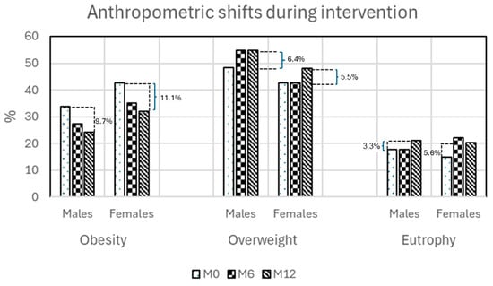
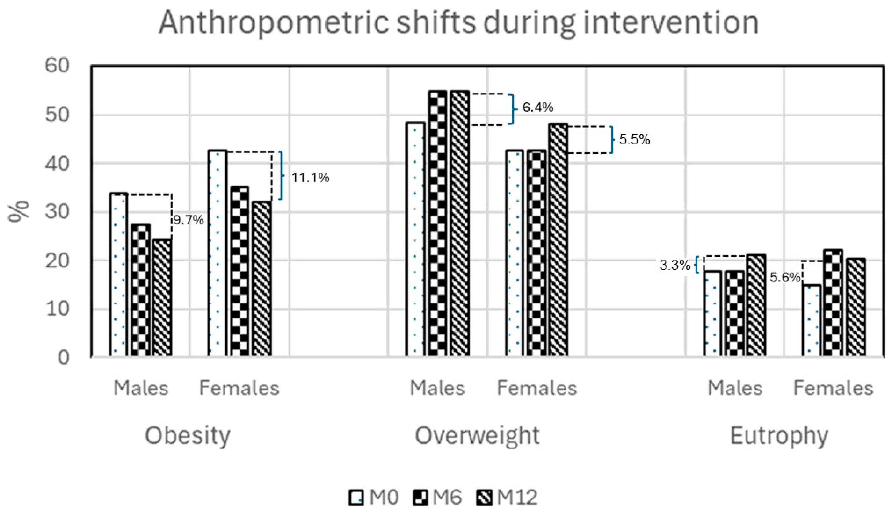
In our 1991–2019 NCDs surveillance relative to our dynamic cohort study (“Moving for Health”), we found in a sample of over a thousand individuals that 82.1% were overweight, 51.2% had abdominal obesity and 39% whole-body obesity [101]. Inflammation, oxidative stress and derived insulin resistance were detected in the sample patients underlying the metabolic (dis)stress of lipotoxicity and gluco-lipotoxicity. Those subjects were submitted to an existing multi-professional program (“Moving for health”) intended to foster lifestyle re-education through physical activity and dietary adequacy (LiSM). The LiSM led to a quality of life improvement through better diet, physical activity and fitness [101]. There was a growth of physical activity, mainly in the leisure domain, which, with dietary adequacy, led to anthropometric improvements that were greater in terms of fat reduction rather than in gaining muscle mass. Body-weight loss, along with obesity reduction and eutrophy achievement (from overweight individuals only), was due to LiSM’s long-lasting dependent intervention (Table 1); there was a slightly better achievement in females (Figure 1). The underlying positive effects of LiSM on the patients include reduced metabolic stress, acting as an anti-inflammatory and antioxidant factor [62,96,102,103]. Overall, the effectiveness of the 10-week LiSM on dietary adequacy, physical fitness, anthropometry and metabolic stress impacted NCDs positively by decreasing their prevalence from 67.5% (T2D) to 21.9% (MetS). Thus, by decreasing the rate of blood hypertension, inflammation, oxidative stress and insulin resistance, our established LiSM was shown to be effective in reducing obesity rates [31] and co-pathologies of T2D [104], blood hypertension [105,106], metabolic syndrome [107,108] and NAFLD [109,110].
Table 1.
Body weight and fatness decreased with lifestyle interventions.
Figure 1.
Gender anthropometric shifts during interventions (data from 108). M0: Baseline; M6: 6 mo. intervention; M12: 12 mo. intervention.
3. Conclusions
Humans have manifested the evolution of particular morphological and biological traits that distinguish them from most other mammals, and the evolution of the many distinctive human characteristics suggests major energetic and dietary shifts. This evolutionary “higher quality” diet means that we can eat less volume of food to obtain the energy and nutrients our body demands. Comparative analyses of primate dietary patterns indicate that human diets are rich in energy and fat. Besides yielding energy, dietary fat also contains functional fatty acids and vitamins essential for growth, development, and maintenance of good health. It is suggested that a group of “thrifty” genes helped fat storage under conditions of improved availability of nutrients and allowed reutilization under starvation conditions. Adipocytes are the main component of the adipose tissue and are responsible for the storage of excess calories in the form of triglycerides; adipocytes also produce secreted factors that regulate systemic nutrient and energy homeostasis within the whole body. Because of their caloric reservoir capacity, adipocytes are able to expand in response to overnutrition and release lipids in response to energy deficit [28]. White adipose tissue (the classical fat tissue) is found in visceral and subcutaneous adipose depots with the potential for growth and expansion depending on energy intake and the host’s metabolic demand. Surviving in this genetic scenario, obese humans are the consequence of an evolutionary selection consisting of overexpression of thrifty genes evolved for efficient food collection and fat deposition to survive periods of famine. Nowadays, this evolutionary-adapted polygenic charge is overexpressed in deteriorated lifestyle conditions.
Our data have revealed the lifestyle environment that includes reduced physical activity (and fitness) and reduced intake of raw vegetables and fruits, and a higher intake of refined oils and carbohydrates, which are strong risk factors for obesity in both school children and free-living adults [59,114]. Our existing multi-professional program (“Moving for health”), which fosters lifestyle re-education, led to an improvement of quality of life through a better diet, physical activity and fitness. At the baseline of the positive health effects of the intervention was the reduced metabolic stress, acting as an anti-inflammatory and anti-oxidant factor, impacting the obesity reduction processes and co-pathologies [101]. However, the implementation of the “Moving for health” program nationally has faced in and out resistance, a fact which seems hard to overcome currently.
4. Future Directions
To the classical statement that “Nothing in medicine makes sense, except in the light of evolution” [118] Carl Sagan’s quote reminds us that “Extinction is the rule. Survival is the exception”. So, we might understand that Evolutionary Biology has done a “good job” allowing the Homo lineage to survive under unfavorable environmental conditions. By helping vary the dietary pattern from omnivorous-natural (Paleolithic) to omnivorous-manufactured (Industrial-urban era), our ancestral Homo sapiens-sapiens expressing the energy-thrifty genome, increased his/her body weight and lifespan. However, in more recent times, featuring environmentally-rich nutrients, human body-fatness went out of control, expressing the inefficiency of the innate mechanisms to avoid fatty-overloaded body. At this point, we attested the inefficiency of our hypothalamic energy-balance control, either for reducing appetite or to increase satiety in positive energy-balance situations. Other failed responses are seen, with fatty-body becoming increasingly bigger due to lower (physiological) fat removal of food-induced overloaded fatty tissue. Interestingly, this phenomenon occurs in parallel with the full expression of inflammation, the thrifty genotype that evolved to scavenge the expanded fat cells that leak or those cells that break open. Thus, in human evolution, the thrifty genotype denoted efficiency in promoting fat-loaded adipocytes; this was sped by unsuccessful lifestyle-change trials resulting in malignancy of obesity-inflammation and its derived comorbidities, which shortened lifespan.
Overall, we should acknowledge that the existing energy-saving genotype for dietary adequacy, even with the feeding with contemporary manufactured food, has fulfilled its expected role of reducing worldwide undernourishment and allowed the individuals’ height increase and a longer lifespan. However, it also caused some unhealthy drawbacks and people worldwide, despite being aware of the noxious consequences, resist giving up their right to enjoy the current palatable energy-dense diet.
The rise in obesity rates internationally might be considered a result of changes in the environment, including the reduced cost of food production, the reduced time and monetary cost of food consumption, as well as the growth of the real cost of being physically active at work and at home. In fact, our 28-year-long dynamic cohort showed that obesity and its co-morbidities are significantly associated with an inadequate dietary intake and physical inactivity [31,101,119].
Leaving aside the obesity-esthetic aspects and focusing mainly on its major life-threatening aspect, the chronic inflammation, the contemporary “predator” of obese people, our 28-year-long dynamic-cohort study has shown the effectiveness of the supervised physical exercise protocols in reducing metabolic stress, mainly inflammation and insulin resistance, along with minor body weight changes [102]. So “fitness” enables “fatness” to become healthy! Physical activity could therefore be a natural remedy for recovering part of the imbalance caused by modern lifestyles—free of cost and without the side effects of many pharmacological treatments. In this connection, physical exercises may have profound implications for public health and be used as a preventive and/or complementary therapeutic strategy for our lifestyle-change protocols [96].
Funding
This research received no external funding.
Data Availability Statement
Data used in this review are stored in the CEMENUTRI files at this institution. Further inquiries can be directed to the author.
Conflicts of Interest
The author declares no conflict of interest.
References
- Neel, J.V.; Weder, A.B.; Julius, S. Type II diabetes, essential hypertension, and obesity as “syndromes of impaired genetic homeostasis”: The “thrifty genotype” hypothesis enters the 21st century. Perspect. Biol. Med. 1998, 42, 44–74. [Google Scholar] [CrossRef] [Scilit] [PubMed]
- Lee, Y.; Hirose, H.; Ohneda, M.; Johnson, J.H.; McGarry, J.D.; Unger, R.H. Beta-Cell lipotoxicity in the pathogenesis of non-insulin-dependent diabetes mellitus of obese rats: Impairment in adipocyte-Beta-cell relationships. Proc. Natl. Acad. Sci. USA 1994, 91, 10878–10882. [Google Scholar] [CrossRef] [Scilit] [PubMed]
- Antón, S.C. A natural history of Homo erectus. Am. J. Phys. Anthr. 2003, 46, 126–170. [Google Scholar] [CrossRef] [Scilit]
- White, T.D.; Asfaw, B.; DeGusta, D.; Gilbert, H.; Richards, G.D.; Suwa, G.; Howell, F.C. Pleistocene Homo sapiens from Middle Awash, Ethiopia. Nature 2003, 423, 742–747. [Google Scholar] [CrossRef] [Scilit] [PubMed]
- Burini, R.C.; Leonard, W.R. The evolutionary roles of nutrition selection and dietary quality in the human brain size and encephalization. Nutrire 2018, 43, 19. [Google Scholar] [CrossRef] [Scilit]
- Kotrschal, A.; Rogell, B.; Bundsen, A.; Svensson, B.; Zajitschek, S.; Brännström, I.; Immler, S.; Maklakov, A.A.; Kolm, N. The benefit of evolving a larger brain: Big-brained guppies perform better in a cognitive task. Anim. Behav. 2013, 86, e4–6. [Google Scholar] [CrossRef] [Scilit]
- Fordyce, R.E. Whale evolution and Oligocene southern-ocean environments. Palaeogeogr. Palaeoclim. Palaeoecol. 1980, 31, 319–336. [Google Scholar] [CrossRef] [Scilit]
- di Costanzo, J. Paleolithic nutrition: A model for the future? Curr. Opin. Clin. Nutr. Metab. Care 2000, 3, 87–92. [Google Scholar] [CrossRef] [Scilit]
- Aiello, L.C.; Wheeler, P. The expensive-tissue hypothesis—The brain and the digestive system in human and primate evolution. Curr. Anthropol. 1995, 36, 199–221. [Google Scholar] [CrossRef] [Scilit]
- Mink, J.W.; Blumenschine, R.J.; Adams, D.B. Ratio of central nervous-system to body metabolism in vertebrates: Its constancy and functional basis. Am. J. Phys. 1981, 241, R203–R212. [Google Scholar] [CrossRef] [Scilit]
- Tomas, E.; Tsao, T.S.; Saha, A.K.; Murrey, H.E.; Zhang, C.C.; Itani, S.I.; Lodish, H.F.; Ruderman, N.B. Enhanced muscle fat oxidation and glucose transport by ACRP30 globular domain: Acetyl-CoA carboxylase inhibition and AMP-activated protein kinase activation. Proc. Natl. Acad. Sci. USA 2002, 99, 16309–16313. [Google Scholar] [CrossRef] [Scilit] [PubMed]
- Johnson, A.R.; Milner, J.J.; Makowski, L. The inflammation highway: Metabolism accelerates inflammatory traffic in obesity. Immunol. Rev. 2012, 249, 218–238. [Google Scholar] [CrossRef] [Scilit] [PubMed]
- Bradbury, J. Ancient footsteps in our genes: Evolution and human disease. Gene variants selected during evolution may underlie many common diseases. Lancet 2004, 363, 952–953. [Google Scholar] [CrossRef] [Scilit] [PubMed]
- Sell, H.; Habich, C.; Eckel, J. Adaptive immunity in obesity and insulin resistance. Nat. Rev. Endocrinol. 2012, 8, 709–716. [Google Scholar] [CrossRef] [Scilit]
- Chandra, R.K. Nutrition, immunity and infection: From basic knowledge of dietary manipulation of immune responses to practical application of ameliorating suffering and improving survival. Proc. Natl. Acad. Sci. USA 1996, 93, 14304–14307. [Google Scholar] [CrossRef] [Scilit]
- Straub, R.H. Concepts of evolutionary medicine and energy regulation contribute to the etiology of systemic chronic inflammatory diseases. Brain, Behav. Immun. 2011, 25, 1–5. [Google Scholar] [CrossRef] [Scilit]
- Wellen, K.E.; Hotamisligil, G.S. Inflammation, stress, and diabetes. J. Clin. Investig. 2005, 115, 1111–1119. [Google Scholar] [CrossRef] [Scilit]
- Zhou, M.S.; Wang, A.; Yu, H. Link between insulin resistance and hypertension: What is the evidence from evolutionary biology? Diabetol. Metab. Syndr. 2014, 6, 12. [Google Scholar] [CrossRef] [Scilit]
- Blackburn, G.L. Pasteur’s Quadrant and malnutrition. Nature 2001, 409, 397–401. [Google Scholar] [CrossRef] [Scilit]
- Spies, C.M.; Straub, R.H.; Buttgereit, F. Energy metabolism and rheumatic diseases: From cell to organism. Arthritis Res. Ther. 2012, 14, 216. [Google Scholar] [CrossRef] [Scilit]
- Cipolletta, D.; Feuerer, M.; Li, A.; Kamei, N.; Lee, J.; Shoelson, S.E.; Benoist, C.; Mathis, D. PPAR-gamma is a major driver of the accumulation and phenotype of adipose tissue Treg cells. Nature 2012, 486, 549–553. [Google Scholar] [CrossRef] [Scilit] [PubMed]
- Fernandez-Real, J.M.; Pickup, J.C. Innate immunity, insulin resistance and type 2 diabetes. Diabetologia 2011, 55, 273–278. [Google Scholar] [CrossRef] [Scilit] [PubMed]
- Wells, J.C. Ethnic variability in adiposity, thrifty phenotypes and cardiometabolic risk: Addressing the full range of ethnicity, including those of mixed ethnicity. Obes. Rev. 2012, 13, 14–29. [Google Scholar] [CrossRef] [Scilit] [PubMed]
- Neel, J.V. The “thrifty genotype” in 1998. Nutrition Reviews. Nutr. Rev. 1999, S2–S9. [Google Scholar]
- Neel, J.V. Diabetes mellitus: A “thrifty” genotype rendered detrimental by “progress”? Am. J. Hum. Genet. 1962, 14, 353–362. [Google Scholar]
- Wells, J.C. Thrift: A guide to thrifty genes, thrifty phenotypes and thrifty norms. Int. J. Obes. 2009, 33, 1331–1338. [Google Scholar] [CrossRef] [Scilit]
- Cahill, G.F., Jr. Starvation in Man. N. Engl. J. Med. 1970, 282, 668–675. [Google Scholar] [CrossRef] [Scilit]
- Rutkowski, J.M.; Stem, J.H.; Scherer, P.E. The cell biology of fat expansion. J. Cell Biol. 2015, 208, 501–512. [Google Scholar] [CrossRef] [Scilit]
- Stem, J.H.; Rutkowski, J.M.; Scherer, P.E. Adiponectin, leptin, and fatty acids in the maintenance of metabolic homeostasis through adipose tissue crosstalk. Cell Metab. 2016, 23, 770–784. [Google Scholar] [CrossRef] [Scilit]
- Bouchard, C. The biological predisposition to obesity: Beyond the thrifty genotype scenario. Int. J. Obes. 2007, 31, 1337–1339. [Google Scholar] [CrossRef] [Scilit]
- Burini, R.C.; de Oliveira, E.P.; Michelin, E.; McLellan, K.C.P. Epidemic obesity: An evolutionary perspective on the modern obesity crisis to a rationale for a treatment. In Obesity Epidemic, 1st ed.; A Brazilian community-based data; iConcept Press: Hong Kong, China, 2013; pp. 20–38. [Google Scholar]
- Finch, C.E.; Stanford, C.B. Meat-adaptive genes and the evolution of slower aging in humans. Q. Rev. Biol. 2004, 79, 3–50. [Google Scholar] [CrossRef] [Scilit]
- Muskiet, F.A.J. Pathophysiology and Evolutionary Aspects of Dietary Fats and Long-Chain Polyunsaturated Fatty Acids across the Life Cycle. In Fat Detection: Taste, Texture, and Post Ingestive Effects; Montmayeur, J.P., le Coutre, J., Eds.; CRC Press/Taylor & Francis: Boca Raton, FL, USA, 2010; Chapter 2. [Google Scholar]
- Gaillard, D.; Passilly-Degrace, P.; Besnard, P. Molecular mechanisms of fat preference and overeating. Ann. N. Y. Acad. Sci. 2008, 1141, 163–175. [Google Scholar] [CrossRef] [Scilit]
- Le Coutre, J.; Schmitt, J.A.J. Food ingredients and cognitive performance. Curr. Opin. Clin. Nutr. Metab. Care 2008, 11, 706–710. [Google Scholar] [CrossRef] [Scilit]
- Sclafani, A. Psychobiology of food preferences. Int. J. Obes. 2001, 25, S13–S16. [Google Scholar] [CrossRef]
- Leonard, W.R.; Snodgrass, J.J.; Robertson, M.L. Evolutionary Perspectives on Fat Ingestion and Metabolism in Humans. In Fat Detection: Taste, Texture, and Post Ingestive Effects; Montmayeur, J.P., le Coutre, J., Eds.; CRC Press/Taylor & Francis: Boca Raton, FL, USA, 2010; Chapter 1. [Google Scholar]
- Aiello, L.C.; Key, C. Energetic consequences of being a Homo erectus female. Am. J. Hum. Biol. 2002, 14, 551–565. [Google Scholar] [CrossRef] [Scilit] [PubMed]
- Prado, C.M.; Maia, Y.L.; Ormsbee, M.; Sawyer, M.B.; Baracos, V.E. Assessment of nutritional status in cancer—The relationship between body composition and pharmacokinetics. Anti Cancer Agents Med. Chem. 2013, 13, 1197–1203. [Google Scholar] [CrossRef] [Scilit] [PubMed]
- Rosenberg, K.R. The evolution of modern human childbirth. Am. J. Phys. Anthr. 1992, 35, 89–124. [Google Scholar] [CrossRef] [Scilit]
- Kuzawa, C.W. Adipose tissue in human infancy and childhood: An evolutionary perspective. Am. J. Phys. Anthr. 1998, 41, 177–209. [Google Scholar] [CrossRef] [Scilit]
- Leonard, W.R.; Robertson, M.L.; Snodgrass, J.J.; Kuzawa, C.W. Metabolic correlates of hominid brain evolution. Comp. Biochem. Physiol. Part A 2003, 136, 5–15. [Google Scholar] [CrossRef] [Scilit]
- Norgan, N.G. The beneficial effects of body fat and adipose tissue in humans. Int. J. Obes. 1997, 21, 738–746. [Google Scholar] [CrossRef] [Scilit]
- Ruiz-Nunez, B.; Pruimboom, L.; Dijck-Brouwer, D.A.; Muskiet, F.A. Lifestyle and nutritional imbalances associated with Western diseases: Causes and consequences of chronic systemic low-grade inflammation in an evolutionary context. J. Nutr. Biochem. 2013, 24, 1183–1201. [Google Scholar] [CrossRef] [Scilit]
- Unger, R.H. Minireview: Weapons of Lean Body Mass Destruction: The Role of Ectopic Lipids in the Metabolic Syndrome. Endocrinology 2003, 144, 5159–5165. [Google Scholar] [CrossRef] [Scilit] [PubMed]
- Gregoire, F.M.; Smas, C.M.; Sul, H.S. Understanding adipocyte differentiation. Physiol. Rev. 1998, 78, 783–809. [Google Scholar] [CrossRef] [Scilit] [PubMed]
- Zhang, Y.; Proenca, R.; Maffei, M.; Barone, M.; Leopold, L.; Friedman, J.M. Positional cloning of the mouse obese gene and its human homologue. Nature 1994, 372, 425–432. [Google Scholar] [CrossRef] [Scilit] [PubMed]
- Scherer, P.E.; Williams, S.; Fogliano, M.; Baldini, G.; Lodish, H.F. A novel serum protein similar to C1q, produced exclusively in adipocytes. J. Biol. Chem. 1995, 270, 26746–26749. [Google Scholar] [CrossRef] [Scilit] [PubMed]
- Maeda, K.; Okubo, K.; Shimomura, I.; Funahashi, T.; Matsuzawa, Y.; Matsubara, K. cDNA cloning and expression of a novel adipose specific collagen-like factor, apM1 (adipose most abundant gene transcript 1). Biochem. Biophys. Res. Commun. 1996, 221, 286–289. [Google Scholar] [CrossRef] [Scilit]
- Wajchenberg, B.L.; Giannella-Neto, D.; Da Silva, M.E.; Santos, R.F. Depot-specific hormonal characteristics of subcutaneous and visceral adipose tissue and their relation to the metabolic syndrome. Horm. Metab. Res. 2002, 34, 616–621. [Google Scholar] [CrossRef] [Scilit]
- Kadowaki, T.; Yamauchi, T. Adiponectin and adiponectin receptors. Endocr. Rev. 2005, 26, 439–451. [Google Scholar] [CrossRef] [Scilit]
- Myeong, J.Y.; Gha, Y.L.; Jun-Jae, C.; Young, H.A.; Seung, H.H.; Jae, B.K. Adiponectin Increases Fatty Acid Oxidation in Skeletal Muscle Cells by Sequential Activation of AMP-Activated Protein Kinase, p38 Mitogen-Activated Protein Kinase, and Peroxisome Proliferator–Activated Receptor α. Diabetes 2006, 55, 2562–2570. [Google Scholar]
- Yamauchi, T.; Kamon, J.; Minokoshi, Y.; Ito, Y.; Waki, H.; Uchida, S.; Yamashita, S.; Noda, M.; Kita, S.; Ueki, K.; et al. Adiponectin stimulates glucose utilizationandfatty-acidoxidationbyactivating AMP-activatedproteinkinase. Nat. Med. 2002, 8, 1288–1295. [Google Scholar] [CrossRef] [Scilit]
- Rossner, S. Obesity: The disease of the twenty-first century. Int. J. Obes. 2002, 26, S2–S4. [Google Scholar] [CrossRef] [Scilit]
- Roszer, T. Adipose tissue immunometabolism and apoptotic cell clearance. Cells 2021, 10, 2288. [Google Scholar] [CrossRef] [Scilit] [PubMed]
- Escobedo, N.; Oliver, G. The lymphatic vasculature: Its role in adipose metabolism and obesity. Cell Metab. 2017, 26, 598–609. [Google Scholar] [CrossRef] [Scilit] [PubMed]
- Mohamed-Ali, V.; Goodrick, S.; Rawesh, A.; Katz, D.R.; Miles, J.M.; Yudkin, S.; Klein, S.; Coppack, S.W. Subcutaneous Adipose Tissue Releases Interleukin-6, But Not Tumor Necrosis Factor-α, in vivo. J. Clin. Endocrinol. Metab. 1997, 82, 4196–4200. [Google Scholar] [CrossRef] [Scilit] [PubMed]
- Lumeng, C.N.; Bodzin, J.L.; Saltiel, A.R. Obesity induces a phenotypic switch in adipose tissue macrophage polarization. J. Clin. Investig. 2007, 117, 175–184. [Google Scholar] [CrossRef] [Scilit]
- Burini, R.C.; Sormani, L.E.; Franz, H.P.B.; Correa, R.C. Inflammatory stress-related chronic Non-Communicable Diseases—Triggering factors and attenuation by a 10-wk lifestyle-change with physical exercises and dietary adequacy. Eur. J. Appl. Physiol. 2026. submitted. [Google Scholar]
- Burini, R.C. Understanding Contemporary Diseases Under Evolutionary Medicine View; Atena Editora: Ponta Grossa, Brazil, 2023; ISBN 978-65-258-1092-8. [Google Scholar] [CrossRef] [Scilit]
- Lumeng, C.N.; Saltiel, A.R. Inflammatory links between obesity and metabolic disease. J. Clin. Investig. 2011, 121, 2111–2117. [Google Scholar] [CrossRef] [Scilit]
- Burini, R.C.; Anderson, E.; Durstine, J.L.; Carson, J. Inflammation, physical activity, and chronic disease: An evolutionary perspective. Sports Med. Health Sci. 2020, 2, 1–6. [Google Scholar] [CrossRef] [Scilit]
- Kahn, S.E.; Hull, R.L.; Utzschneider, K.M. Mechanisms linking obesity to insulin resistance and type 2 diabetes. Nature 2006, 444, 840–846. [Google Scholar] [CrossRef] [Scilit]
- Shoelson, S.E.; Herrero, L.; Naaz, A. Obesity, inflammation, and insulin resistance. Gastroenterology 2007, 132, 2169–2180. [Google Scholar] [CrossRef] [Scilit]
- Listenberger, L.L.; Han, X.; Lewis, S.E.; Cases, S.; Farese, R.V., Jr.; Ory, D.S.; Schaffer, J.E. Triglyceride accumulation protects against fatty acid-induced lipotoxicity. Proc. Natl. Acad. Sci. USA 2003, 100, 3077–3082. [Google Scholar] [CrossRef] [Scilit]
- Weiss, B.; Stoffel, W. Human and murine serine-palmitoyl-CoA transferase–cloning, expression and characterization of the key enzyme in sphingolipid synthesis. Eur. J. Biochem. 1997, 249, 239–247. [Google Scholar] [CrossRef] [Scilit] [PubMed]
- Hannum, Y.A.; Obeid, L.M. Principles of bioactive lipid signaling: Lessons from sphingolipids. Nat. Rev. Mol. Cell Biol. 2008, 9, 139–150. [Google Scholar] [CrossRef] [Scilit] [PubMed]
- Zeidan, Y.H.; Hannum, Y.A. Translational aspects of sphingolipid metabolism. Trends Mol. Med. 2007, 13, 327–336. [Google Scholar] [CrossRef] [Scilit] [PubMed]
- Aguirre, V.; Uchida, T.; Yenush, L.; Davis, R.; White, M.F. The c-Jun NH(2)-terminal kinase promotes insulin resistance during association with insulin receptor substrate-1 and phosphorylation of Ser(307). J. Biol. Chem. 2000, 275, 9047–9054. [Google Scholar] [CrossRef] [Scilit]
- Chavez, J.A.; Siddique, M.M.; Wang, S.T.; Ching, J.; Shayman, J.A.; Summers, S.A. Ceramides and glucosylceramides are independent antagonists of insulin signaling. J. Biol. Chem. 2014, 289, 723–734. [Google Scholar] [CrossRef] [Scilit] [PubMed] [PubMed Central]
- Hirosumi, J.; Tuncman, G.; Chang, L.; Gorgun, C.Z.; Uysal, K.T.; Maeda, K.; Karin, M.; Hotamisligil, G.S. A central role for JNK in obesity and insulin resistance. Nature 2002, 420, 333–336. [Google Scholar] [CrossRef] [Scilit]
- Kogot-Levin, A.; Saada, A. Ceramide and the mitochondrial respiratory chain. Biochimie 2014, 100, 88–94. [Google Scholar] [CrossRef] [Scilit] [PubMed]
- Portero-Mclellan, K.C.; Manda, R.M.; Sloan, L.A.; Burini, R.C. Epigenetics of glucose metabolism and the basis for T2DM interventions. In Type 2 Diabetes; IntechOpen: London, UK, 2013; Chapter 3; pp. 52–70. [Google Scholar] [CrossRef] [Scilit]
- Sun, K.; Kusminski, C.M.; Scherer, P.E. Adipose tissue remodeling and obesity. J. Clin. Investig. 2011, 121, 2094–2101. [Google Scholar] [CrossRef] [Scilit]
- Shimabukuro, M.; Higa, M.; Zhou, Y.T.; Wang, M.Y.; Newgard, C.B.; Unger, R.H. Lipoapoptosis in Beta-cells of obese prediabeticfa/fa rats: Role of serine palmitoyltransferase overexpression. J. Biol. Chem. 1998, 273, 32487–32490. [Google Scholar] [CrossRef] [Scilit]
- Shulman, G.I. Cellular mechanisms of insulin resistance. J. Clin. Investig. 2000, 106, 171–176. [Google Scholar] [CrossRef] [Scilit]
- Unger, R.H. Reinventing Type 2 Diabetes: Pathogenesis, treatment and Prevention. JAMA 2008, 299, 1185–1187. [Google Scholar] [CrossRef] [Scilit]
- Despres, J.P.; Lemieux, I. Abdominal obesity and metabolic syndrome. Nature 2006, 444, 881–887. [Google Scholar] [CrossRef] [Scilit] [PubMed]
- Hotamisligil, G.S. Inflammation and metabolic disorders. Nature 2006, 444, 860–867. [Google Scholar] [CrossRef] [Scilit] [PubMed]
- Hotamisligil, G.S.; Erbay, E. Nutrient sensing and inflammation in metabolic disease. Nat. Rev. Immunol. 2008, 8, 923–934. [Google Scholar] [CrossRef] [Scilit] [PubMed]
- IBGE Instituto Brasileiro de Geografia e Estatística. Pesquisa de Orçamentos Familiares 2008–2009: Antropometria e Estado Nutricional de Crianças, Adolescentes e Adultos no Brasil; IBGE: Rio de Janeiro, Brazil, 2010. [Google Scholar]
- Speakman, J.R. A non-adaptive scenario explaining the genetic predisposition to obesity: The “predation release” hypothesis. Cell Metab. 2007, 6, 5–12. [Google Scholar] [CrossRef] [Scilit]
- Hanson, M.; Gluckman, P. Developmental origins of non-communicable disease: Population and public health implications. Am. J. Clin. Nutr. 2011, 94, S1754–S1758. [Google Scholar] [CrossRef] [Scilit]
- Tarry-Adkins, J.L.; Ozanne, S.E. Mechanisms of early life programming: Current knowledge and future directions. Am. J. Clin. Nutr. 2011, 94, S1765–S1771. [Google Scholar] [CrossRef] [Scilit]
- Penicau, L.; Cousin, B.; Leloup, C.; Lorsignol, A.; Casteilla, L. The autonomic nervous system, adipose tissue plasticity, and energy balance. Nutrition 2000, 16, 903–908. [Google Scholar] [CrossRef] [Scilit]
- Landsberg, L.; Young, J.B. The role of the sympathoadrenal system in modulating energy expenditure. Clin. Endocrinol. Metab. 1984, 13, 475–499. [Google Scholar] [CrossRef] [Scilit]
- Bray, G.A. Integration of energy intake and expenditure in animals and man: The autonomic and adrenal hypothesis. Clin. Endocrinol. Metab. 1984, 13, 521–546. [Google Scholar] [CrossRef] [Scilit]
- Peterson, H.R.; Rothschild, M.; Weinberg, C.R.; Fell, R.D.; McLeish, K.R.; Pfeifer, M.A. Body fat and the activity of the autonomic nervous system. N. Engl. J. Med. 1988, 318, 1077–1083. [Google Scholar] [CrossRef] [Scilit]
- Eikelis, N.; Schlich, M.; Aggarwal, A.; Kaye, D.; Esler, M. Interactions between leptin and the human sympathetic nervous system. Hypertension 2003, 41, 1072–1079. [Google Scholar] [CrossRef] [Scilit] [PubMed]
- Grassi, G.; Seravalle, G.; Cattaneo, B.M.; Bolla, G.B.; Lanfranchi, A.; Colombo, M.; Giannattasio, C.; Brunani, A.; Cavagnini, F.; Mancia, G. Sympathetic activation in obese normotensive subjects. Hypertension 1995, 25, 560–563. [Google Scholar] [CrossRef] [Scilit] [PubMed]
- Landsberg, L. Feast or famine: The sympathetic nervous system response to nutrient intake. Cell. Mol. Neurobiol. 2006, 26, 495–506. [Google Scholar] [CrossRef] [Scilit] [PubMed]
- Rumantir, M.S.; Vaz, M.; Jennings, G.L.; Collier, G.; Kaye, D.M.; Seals, D.R.; Wiesner, G.H.; Rocca, H.P.B.-L.; Esler, M.D. Neural mechanisms in human obesity-related hypertension. J. Hypertens. 1999, 17, 1125–1133. [Google Scholar] [CrossRef] [Scilit]
- Finkelstein, E.A.; Strombotne, K.L. The economics of obesity. Am. J. Clin. Nutr. 2010, 91, 1520S–1524S. [Google Scholar] [CrossRef] [Scilit]
- Booth, F.W.; Lees, S.J. Fundamental questions about genes, inactivity, and chronic diseases. Physiol. Genom. 2007, 28, 146–157. [Google Scholar] [CrossRef] [Scilit]
- Bertelli, M.; Alushi, B.; Veicsteina, A. Physical exercise to prevent multifactorial diseases: A warning written in our genes? Sport Sci. Health 2008, 4, 33–35. [Google Scholar] [CrossRef] [Scilit]
- Burini, R.C. Physical Exercises: From the Evolutionary Thriftiness to the Epigenetics of Contemporary Diseases and Current Therapeutics for Physical Inactivity Diseasomes, 1st ed; SM Group Open Access eBooks, 2017; volume 1, pp. 1–25. [Google Scholar]
- Szostak, J.; Laurant, P. The forgotten face of regular physical exercise: A ‘natural’ anti-atherogenic activity. Clin. Sci. 2011, 121, 91–106. [Google Scholar] [CrossRef] [Scilit]
- Coffey, V.G.; Hawley, J.A. The molecular bases of training adaptation. Sports Med. 2007, 37, 737–763. [Google Scholar] [CrossRef] [Scilit] [PubMed]
- Radak, Z.; Koltai, E.; Taylor, A.W.; Higuchi, M.; Kumagai, S.; Ohno, H.; Goto, S.; Boldogh, I. Redox-regulatingsirtuins in aging, caloricrestriction, andexercise. Free. Radic. Biol. Med. 2013, 58, 87–97. [Google Scholar] [CrossRef] [Scilit] [PubMed]
- Zhou, L.; Pinho, R.; Gu, Y.; Radak, Z. The Role of SIRT3 in Exercise and Aging. Cells 2022, 11, 2596. [Google Scholar] [CrossRef] [Scilit] [PubMed]
- Burini, R.C. The Brazilian Dynamic-Cohort Study “Moving for Health” as a Proactive model of Evolutionary/Behavioral –Medicine to Non-Communicable Diseases in a community-based subjects. Int. J. Health Sci. 2022, 2, 2–23. [Google Scholar] [CrossRef] [Scilit]
- Burini, R.C.; Burini, F.H.P.; Durstine, J.L.; Orsatti, F.L. The effectiveness of Lifestyle-Change Approach to the Non-Communicable Chronic Diseases. The Role of Physical Exercise to the Brazilian Dynamic-Cohort Study “Moving for Health”. Sports Med. Health Sci. 2026, in press. [Google Scholar]
- Burini, R.C.; Kano, H.T.; Nakagaki, M.S.; Frenhani, P.B.; McLellan, K.C.P. Behavioral factors of Abdominal Obesity and effects of lifestyle changes with Fiber Adequacy. New Insights Obes. Genet. Beyond 2017, 1, 14–22. [Google Scholar] [CrossRef] [Scilit]
- Mecca, M.S.; Moreto, F.; Burini, F.H.; Dalanesi, R.C.; McLellan, K.C.; Burini, R.C. Ten-week lifestyle changing program reduces several indicators for metabolic syndrome in overweight adults. Diabetol. Metab. Syndr. 2012, 4, 1. [Google Scholar] [CrossRef] [Scilit]
- Burini, R.C.; Kano, H.T.; Barros, L.P.; Grandini, N.A.; Correa, C.R. Plasma Fetuin-A changes in the gluco-lipotoxicity interplay between non-alcoholic fatty-liver disease and coronary-heart disease. J. Metab. Synd. 2023, 12, 1–7. [Google Scholar]
- Talon, L.d.C.; de Oliveira, E.P.; Moreto, F.; Portero-McLellan, K.C.; Burini, R.C. Omega-3 fatty acids supplementation decreases metabolic syndrome prevalence after lifestyle modification program. J. Funct. Foods 2015, 19, 922–928. [Google Scholar] [CrossRef] [Scilit]
- Nakagaki, M.S.; Michelin Portero-McLellan, K.C.; Burini, R.C. Gained Physical Activity And Fitness Are Sustained By Adults After Their Discontinuation In A Lifestyle Modification Program. In Proceedings of the ACSM Annual Meeting, World Congress on Exercise is Medicine and World Congress on the Role of Inflammation in Exercise, Health and Disease, Orlando, FL, USA, 27–31 May 2014; Volume 46, p. s952. [Google Scholar]
- da Silva, C.H.F.; Duarte, S.J.H.; Ribeiro, T.C.; Spiri, W.C.; de Paula Ravagnani, F.C.; de Faria Coelho-Ravagnani, C.; Burini, R.C. Longitudinal effect of a program to change lifestyle on health indicators in adults. Rev. Eletronica Gest. Saude 2011, 2, 415–426. [Google Scholar]
- Orsatti, F.L.; Nahas, E.A.; Maesta, N.; Nahas-Neto, J.; Burini, R.C. Plasma hormones, muscle mass and strength in resistance-trained postmenopausal women. Maturitas 2008, 59, 394–404. [Google Scholar] [CrossRef] [Scilit] [PubMed]
- Cunha, P.M.; Ribeiro, A.S.; Padilha, C.; Nunes, J.P.; Schoenfeld, B.J.; Cyrino, L.T.; Tomeleri, C.M.; Nascimento, M.A.; Antunes, M.; Fernandes, R.R.; et al. Improvement of Oxidative Stress in Older Women Is Dependent on Resistance Training Volume: Active Aging Longitudinal Study. J. Strength Cond. Res. 2020, 36, 1141–1146. [Google Scholar] [CrossRef] [Scilit] [PubMed]
- Burini, R.C.; Torezan, G.A.; McLellan, K.C.P. Behavioral risk factors and effects of lifestyle modification on adults with Diabetes: A Brazilian community-based study. In Emerging Issues in Medical Diagnosis and Treatment; Concept Press Ltd.: Braintree, UK, 2013; Volume 1. [Google Scholar]
- Burini, R.C.; Nakagaki, M.S.; Michelin, E.; Burini, F.H.P. Treating Blood Hypertension in a Brazilian Community: Moving from Reactive Homeostatic Model to Proactive Allostatic Healthcare. Ann. Clin. Hypertens. 2018, 2, 001–016. [Google Scholar] [CrossRef] [Scilit]
- Burini, R.C.; Michelin, E.; Burini, F.H.P.; Moreto, F.; Mclellan, K.C.P. Behavioral risk factors and effects of lifestyle-modification on the metabolic syndrome in adults. A Brazilian community-based study. In Handbook on Metabolic Syndrome: Classification, Risk Factors and Health Impact, 1st ed.; Lopez Garcia, C.M., Perez Gonzalez, P.A., Eds.; Nova Science Publishers: New York, NY, USA, 2012; pp. 1–20. [Google Scholar]
- Tomeleri, C.M.; Souza, M.F.; Burini, R.C.; Cavaglieri, C.R.; Ribeiro, A.S.; Antunes, M.; Nunes, J.P.; Venturini, D.; Barbosa, D.S.; Sardinha, L.B.; et al. Resistance training reduces metabolic syndrome and inflammatory markers in older women: A randomized controlled trial. J. Diabetes 2017, 10, 328–337. [Google Scholar] [CrossRef] [Scilit]
- Burini, R.C.; Burini, F.H.P.; Kano, H.T.; Barros, L.P.; Nunes, C.N.M.; Correa, C.R.; Nakagaki, M.S. Behavioral Determinants of Fatty Liver Index and of NAFLD Responsiveness to a 10-Week Lifestyle Modification Program with Supervised Physical Exercises and Dietary Counseling. J. Metab. Synd. 2023, 12. [Google Scholar] [CrossRef]
- Nakagaki, M.S.; Kano, H.T.; Burini, R.C. Metabolic Syndrome Response to Different Physical–Exercise Protocols. Am. J. Sports Sci. 2019, 7, 182. [Google Scholar] [CrossRef] [Scilit]
- Burini, R.C.; Simonetti, L.A.; Maesta, N.; Waib, P.H. Efficiency and Cost less of a Long-term Physical Exercise Program to Non-medicated Hypertensive Males. Adv. Stud. Med. Sci. 2013, 1, 111–123. Available online: https://goo.gl/80WAEp (accessed on 24 November 2025).
- Varki, A. Nothing in medicine makes sense, except in the light of evolution. J. Mol. Med. 2012, 90, 481–494. [Google Scholar] [CrossRef] [Scilit]
- Kano, H.T.; Burini, F.H.P.; Burini, R.C. Nutritional And Physical-Fitness Factors Coursing With Non-Communicable Diseases From Overweight To A Higher Risk Of Vascular And Liver Outcomes In A Community-Based Adults. Int. J. Health Sci. 2022, 2, 2–16. [Google Scholar] [CrossRef] [Scilit]

Disclaimer/Publisher’s Note: The statements, opinions and data contained in all publications are solely those of the individual author(s) and contributor(s) and not of MDPI and/or the editor(s). MDPI and/or the editor(s) disclaim responsibility for any injury to people or property resulting from any ideas, methods, instructions or products referred to in the content. |
© 2026 by the author. Licensee MDPI, Basel, Switzerland. This article is an open access article distributed under the terms and conditions of the Creative Commons Attribution (CC BY) license.