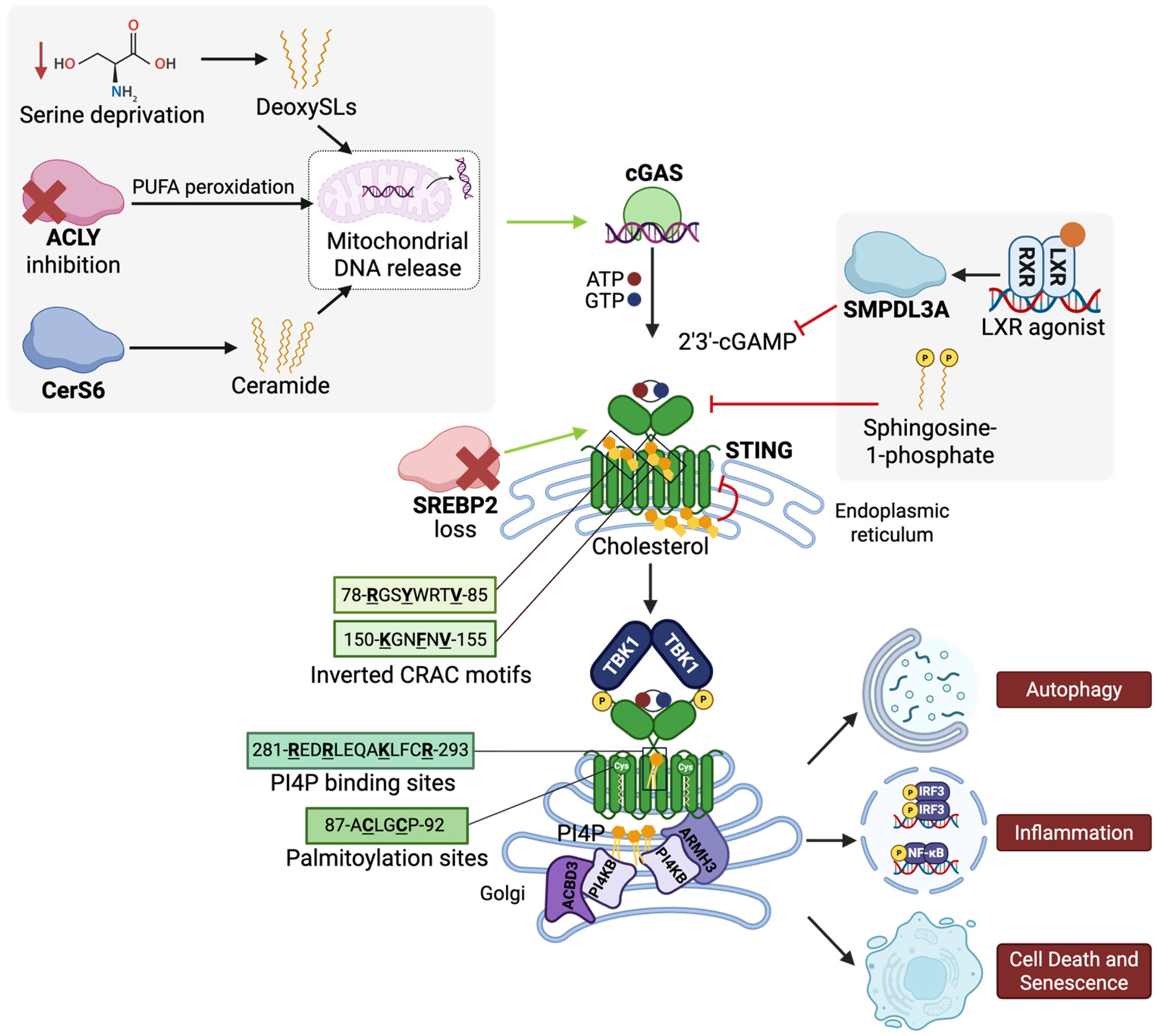

Lipids Meet Immunity: Metabolic Control of cGAS-STING


Abstract
1. Introduction
2. Fatty Acids and cGAS-STING Signaling
3. Glycerophospholipids and cGAS-STING Signaling
4. Sphingolipids and cGAS-STING Signaling
5. Sterols and cGAS-STING Signaling
6. Glycerolipids and cGAS-STING Signaling
7. Prenol Lipids in cGAS-STING Signaling
8. Saccharolipids in cGAS-STING Signaling
9. Polyketides in cGAS-STING Signaling
10. Therapeutic Implications of Lipid Regulation of cGAS-STING
11. Conclusions and Future Directions
Author Contributions
Funding
Data Availability Statement
Acknowledgments
Conflicts of Interest
Abbreviations
| ACBD3 | Golgi resident protein GCP60 |
| ACLY | ATP citrate lyase |
| AML | Acute myeloid leukemia |
| ARMH3 | Armadillo-like helical domain-containing protein 3 |
| cGAMP | Cyclic GMP-AMP |
| cGAS | Cyclic GMP-AMP synthase |
| CRAC | Cholesterol recognition/interaction amino acid consensus |
| DeoxySL | 1-Deoxysphingolipid |
| dsDNA | Double-stranded DNA |
| ER | Endoplasmic reticulum |
| FADS1 | Fatty acid desaturase 1 |
| HFD | High-fat diet |
| HMGCR | HMG-CoA reductase |
| HSV-1 | Herpes simplex virus 1 |
| ICAM-1 | Intercellular adhesion molecule 1 |
| IRF3 | Interferon regulatory factor 3 |
| ISG | Interferon-stimulated gene |
| LD | Lipid droplet |
| LPS | Lipopolysaccharide |
| LSD | Lysosomal storage disorder |
| LXR | Liver X receptor |
| MβCD | Methyl-beta-cyclodextrin |
| mtDNA | Mitochondrial DNA |
| MUFA | Monounsaturated fatty acid |
| NASH | Nonalcoholic steatohepatitis |
| NPC | Niemann–Pick disease type C |
| NF-κB | Nuclear factor kappa B |
| PD-L1 | Programmed death-ligand 1 |
| PIP | Phosphoinositide |
| PI(3,5)P2 | Phosphatidylinositol 3,5-bisphosphate |
| PI4KB | Phosphatidylinositol 4-kinase beta |
| PI4P | Phosphatidylinositol 4-phosphate |
| PIKFYVE | 1-phosphatidylinositol 3-phosphate 5-kinase |
| RAB22A | Ras-related protein Rab-22A |
| SAC1 | Phosphatidylinositol-4-phosphatase |
| SAVI | STING-associated vasculopathy with onset in infancy |
| SCD2 | Stearoyl-Coenzyme A desaturase 2 |
| S1P | Sphingosine-1-phosphate |
| SMPDL3A | Sphingomyelin phosphodiesterase acid like 3A |
| SOAT1 | Sterol O-acyltransferase 1 |
| SPHK2 | Sphingosine kinase 2 |
| SPT | Serine palmitoyltransferase |
| SPTLC2 | Serine palmitoyltransferase 2 |
| STING | Stimulator of interferon genes |
| TAG | Triacylglycerol |
| TBK1 | TANK-binding kinase 1 |
| TGN | Trans-Golgi network |
| VDAC1 | Voltage-dependent anion channel 1 |
| WT | Wild type |
References
- Samson, N.; Ablasser, A. The cGAS-STING pathway and cancer. Nat. Cancer 2022, 3, 1452–1463. [Google Scholar] [CrossRef] [Scilit] [PubMed]
- Oduro, P.K.; Zheng, X.; Wei, J.; Yang, Y.; Wang, Y.; Zhang, H.; Liu, E.; Gao, X.; Du, M.; Wang, Q. The cGAS-STING signaling in cardiovascular and metabolic diseases: Future novel target option for pharmacotherapy. Acta Pharm. Sin. B 2022, 12, 50–75. [Google Scholar] [CrossRef] [Scilit] [PubMed]
- Decout, A.; Katz, J.D.; Venkatraman, S.; Ablasser, A. The cGAS-STING pathway as a therapeutic target in inflammatory diseases. Nat. Rev. Immunol. 2021, 21, 548–569. [Google Scholar] [CrossRef] [Scilit]
- Paul, B.D.; Snyder, S.H.; Bohr, V.A. Signaling by cGAS-STING in Neurodegeneration, Neuroinflammation, and Aging. Trends Neurosci. 2021, 44, 83–96. [Google Scholar] [CrossRef] [Scilit] [PubMed]
- Dvorkin, S.; Cambier, S.; Volkman, H.E.; Stetson, D.B. New frontiers in the cGAS-STING intracellular DNA-sensing pathway. Immunity 2024, 57, 718–730. [Google Scholar] [CrossRef] [Scilit]
- Wang, M.M.; Zhao, Y.; Liu, J.; Fan, R.R.; Tang, Y.Q.; Guo, Z.Y.; Li, T. The role of the cGAS-STING signaling pathway in viral infections, inflammatory and autoimmune diseases. Acta Pharmacol. Sin. 2024, 45, 1997–2010. [Google Scholar] [CrossRef] [Scilit]
- Furman, D.; Campisi, J.; Verdin, E.; Carrera-Bastos, P.; Targ, S.; Franceschi, C.; Ferrucci, L.; Gilroy, D.W.; Fasano, A.; Miller, G.W.; et al. Chronic inflammation in the etiology of disease across the life span. Nat. Med. 2019, 25, 1822–1832. [Google Scholar] [CrossRef] [Scilit]
- Zhang, Z.; Zhang, C. Regulation of cGAS-STING signalling and its diversity of cellular outcomes. Nat. Rev. Immunol. 2025, 25, 425–444. [Google Scholar] [CrossRef] [Scilit]
- Zhang, B.; Xu, P.; Ablasser, A. Regulation of the cGAS-STING Pathway. Annu. Rev. Immunol. 2025, 43, 667–692. [Google Scholar] [CrossRef] [Scilit]
- Chen, Q.; Sun, L.; Chen, Z.J. Regulation and function of the cGAS-STING pathway of cytosolic DNA sensing. Nat. Immunol. 2016, 17, 1142–1149. [Google Scholar] [CrossRef] [Scilit]
- Balka, K.R.; De Nardo, D. Molecular and spatial mechanisms governing STING signalling. FEBS J. 2021, 288, 5504–5529. [Google Scholar] [CrossRef] [Scilit]
- Jeltema, D.; Abbott, K.; Yan, N. STING trafficking as a new dimension of immune signaling. J. Exp. Med. 2023, 220, e20220990. [Google Scholar] [CrossRef] [Scilit] [PubMed]
- Mukai, K.; Konno, H.; Akiba, T.; Uemura, T.; Waguri, S.; Kobayashi, T.; Barber, G.N.; Arai, H.; Taguchi, T. Activation of STING requires palmitoylation at the Golgi. Nat. Commun. 2016, 7, 11932. [Google Scholar] [CrossRef] [Scilit]
- Zhang, B.C.; Laursen, M.F.; Hu, L.; Hazrati, H.; Narita, R.; Jensen, L.S.; Hansen, A.S.; Huang, J.; Zhang, Y.; Ding, X.; et al. Cholesterol-binding motifs in STING that control endoplasmic reticulum retention mediate anti-tumoral activity of cholesterol-lowering compounds. Nat. Commun. 2024, 15, 2760. [Google Scholar] [CrossRef] [Scilit]
- Hou, Y.; Wang, Z.; Liu, P.; Wei, X.; Zhang, Z.; Fan, S.; Zhang, L.; Han, F.; Song, Y.; Chu, L.; et al. SMPDL3A is a cGAMP-degrading enzyme induced by LXR-mediated lipid metabolism to restrict cGAS-STING DNA sensing. Immunity 2023, 56, 2492–2507. [Google Scholar] [CrossRef] [Scilit] [PubMed]
- Minton, K. Lipid metabolism extinguishes cGAS-STING-induced inflammation. Nat. Rev. Immunol. 2023, 23, 785. [Google Scholar] [CrossRef] [Scilit] [PubMed]
- Akhmetova, K.; Balasov, M.; Chesnokov, I. Drosophila STING protein has a role in lipid metabolism. Elife 2021, 10, e67358. [Google Scholar] [CrossRef] [Scilit]
- Yang, Z.; Guo, G.; Li, J.; Sun, C. The Role of cGAS-STING Pathway in Chronic Liver Diseases: Mechanisms and Therapeutic Perspectives. FASEB J. 2025, 39, e71114. [Google Scholar] [CrossRef] [Scilit]
- Lercher, A.; Baazim, H.; Bergthaler, A. Systemic Immunometabolism: Challenges and Opportunities. Immunity 2020, 53, 496–509. [Google Scholar] [CrossRef] [Scilit]
- Wang, A.; Luan, H.H.; Medzhitov, R. An evolutionary perspective on immunometabolism. Science 2019, 363, eaar3932. [Google Scholar] [CrossRef] [Scilit]
- Triantafilou, M.; Ramanjulu, J.; Booty, L.M.; Jimenez-Duran, G.; Keles, H.; Saunders, K.; Nevins, N.; Koppe, E.; Modis, L.K.; Pesiridis, G.S.; et al. Human rhinovirus promotes STING trafficking to replication organelles to promote viral replication. Nat. Commun. 2022, 13, 1406. [Google Scholar] [CrossRef] [Scilit]
- Conroy, M.J.; Andrews, R.M.; Andrews, S.; Cockayne, L.; Dennis, E.A.; Fahy, E.; Gaud, C.; Griffiths, W.J.; Jukes, G.; Kolchin, M.; et al. LIPID MAPS: Update to databases and tools for the lipidomics community. Nucleic Acids Res. 2024, 52, D1677–D1682. [Google Scholar] [CrossRef] [Scilit] [PubMed]
- Liebisch, G.; Fahy, E.; Aoki, J.; Dennis, E.A.; Durand, T.; Ejsing, C.S.; Fedorova, M.; Feussner, I.; Griffiths, W.J.; Köfeler, H.; et al. Update on LIPID MAPS classification, nomenclature, and shorthand notation for MS-derived lipid structures. J. Lipid Res. 2020, 61, 1539–1555. [Google Scholar] [CrossRef] [Scilit] [PubMed]
- Hansen, A.L.; Buchan, G.J.; Rühl, M.; Mukai, K.; Salvatore, S.R.; Ogawa, E.; Andersen, S.D.; Iversen, M.B.; Thielke, A.L.; Gunderstofte, C.; et al. Nitro-fatty acids are formed in response to virus infection and are potent inhibitors of STING palmitoylation and signaling. Proc. Natl. Acad. Sci. USA 2018, 115, E7768–E7775. [Google Scholar] [CrossRef] [Scilit]
- Mao, Y.; Luo, W.; Zhang, L.; Wu, W.; Yuan, L.; Xu, H.; Song, J.; Fujiwara, K.; Abe, J.I.; LeMaire, S.A.; et al. STING-IRF3 Triggers Endothelial Inflammation in Response to Free Fatty Acid-Induced Mitochondrial Damage in Diet-Induced Obesity. Arterioscler. Thromb. Vasc. Biol. 2017, 37, 920–929. [Google Scholar] [CrossRef] [Scilit] [PubMed]
- Luteijn, R.D.; van Terwisga, S.R.; Ver Eecke, J.E.; Onia, L.; Zaver, S.A.; Woodward, J.J.; Wubbolts, R.W.; Raulet, D.H.; van Kuppeveld, F.J.M. The activation of the adaptor protein STING depends on its interactions with the phospholipid PI4P. Sci. Signal. 2024, 17, eade3643. [Google Scholar] [CrossRef] [Scilit]
- Fang, R.; Jiang, Q.; Jia, X.; Jiang, Z. ARMH3-mediated recruitment of PI4KB directs Golgi-to-endosome trafficking and activation of the antiviral effector STING. Immunity 2023, 56, 500–515. [Google Scholar] [CrossRef] [Scilit]
- Li, J.; Tan, J.X.; Chen, Z.J.; Zhang, X.; Bai, X.C. Regulation of STING activation by phosphoinositide and cholesterol. Nature 2026. [Google Scholar] [CrossRef] [Scilit]
- Tan, J.X.; Lv, B.; Li, J.; Li, T.; Du, F.; Chen, X.; Zhang, X.; Bai, X.C.; Chen, Z.J. PtdIns(3,5)P(2) is an endogenous ligand of STING in innate immune signalling. Nature 2026. [Google Scholar] [CrossRef] [Scilit]
- Kemmoku, H.; Takahashi, K.; Mukai, K.; Mori, T.; Hirosawa, K.M.; Kiku, F.; Uchida, Y.; Kuchitsu, Y.; Nishioka, Y.; Sawa, M.; et al. Single-molecule localization microscopy reveals STING clustering at the trans-Golgi network through palmitoylation-dependent accumulation of cholesterol. Nat. Commun. 2024, 15, 220. [Google Scholar] [CrossRef] [Scilit]
- Takahashi, K.; Niki, T.; Ogawa, E.; Fumika, K.; Nishioka, Y.; Sawa, M.; Arai, H.; Mukai, K.; Taguchi, T. A cell-free assay implicates a role of sphingomyelin and cholesterol in STING phosphorylation. Sci. Rep. 2021, 11, 11996. [Google Scholar] [CrossRef] [Scilit] [PubMed]
- Zhu, Z.; Cao, Y.; Jian, Y.; Hu, H.; Yang, Q.; Hao, Y.; Jiang, H.; Luo, Z.; Yang, X.; Li, W.; et al. CerS6 links ceramide metabolism to innate immune responses in diabetic kidney disease. Nat. Commun. 2025, 16, 1528. [Google Scholar] [CrossRef] [Scilit] [PubMed]
- Joshi, J.C.; Joshi, B.; Rochford, I.; Rayees, S.; Akhter, M.Z.; Baweja, S.; Chava, K.R.; Tauseef, M.; Abdelkarim, H.; Natarajan, V.; et al. SPHK2-Generated S1P in CD11b(+) Macrophages Blocks STING to Suppress the Inflammatory Function of Alveolar Macrophages. Cell Rep. 2020, 30, 4096–4109.e5. [Google Scholar] [CrossRef] [Scilit]
- Chu, T.T.; Tu, X.; Yang, K.; Wu, J.; Repa, J.J.; Yan, N. Tonic prime-boost of STING signalling mediates Niemann-Pick disease type C. Nature 2021, 596, 570–575. [Google Scholar] [CrossRef] [Scilit] [PubMed]
- Kanno, T.; Nakajima, T.; Yokoyama, S.; Asou, H.K.; Sasamoto, S.; Kamii, Y.; Hayashizaki, K.; Ouchi, Y.; Onodera, T.; Takahashi, Y.; et al. SCD2-mediated monounsaturated fatty acid metabolism regulates cGAS-STING-dependent type I IFN responses in CD4(+) T cells. Commun. Biol. 2021, 4, 820. [Google Scholar] [CrossRef] [Scilit]
- Yu, Y.; Liu, Y.; An, W.; Song, J.; Zhang, Y.; Zhao, X. STING-mediated inflammation in Kupffer cells contributes to progression of nonalcoholic steatohepatitis. J. Clin. Investig. 2019, 129, 546–555. [Google Scholar] [CrossRef] [Scilit]
- Luo, X.; Li, H.; Ma, L.; Zhou, J.; Guo, X.; Woo, S.L.; Pei, Y.; Knight, L.R.; Deveau, M.; Chen, Y.; et al. Expression of STING Is Increased in Liver Tissues From Patients With NAFLD and Promotes Macrophage-Mediated Hepatic Inflammation and Fibrosis in Mice. Gastroenterology 2018, 155, 1971–1984.e4. [Google Scholar] [CrossRef] [Scilit]
- Wang, X.T.; Zhu, X.; Lian, Z.H.; Liu, Q.; Yan, H.H.; Qiu, Y.; Ge, X.Y. AUP1 and UBE2G2 complex targets STING signaling and regulates virus-induced innate immunity. mBio 2025, 16, e0060225. [Google Scholar] [CrossRef] [Scilit]
- Ning, L.; Wei, W.; Wenyang, J.; Rui, X.; Qing, G. Cytosolic DNA-STING-NLRP3 axis is involved in murine acute lung injury induced by lipopolysaccharide. Clin. Transl. Med. 2020, 10, e228. [Google Scholar] [CrossRef] [Scilit]
- Liu, S.; Guan, L.; Peng, C.; Cheng, Y.; Cheng, H.; Wang, F.; Ma, M.; Zheng, R.; Ji, Z.; Cui, P.; et al. Mycobacterium tuberculosis suppresses host DNA repair to boost its intracellular survival. Cell Host Microbe 2023, 31, 1820–1836.e10. [Google Scholar] [CrossRef] [Scilit]
- Wang, C.; Zhang, R.; He, J.; Yu, L.; Li, X.; Zhang, J.; Li, S.; Zhang, C.; Kagan, J.C.; Karp, J.M.; et al. Ultrasound-responsive low-dose doxorubicin liposomes trigger mitochondrial DNA release and activate cGAS-STING-mediated antitumour immunity. Nat. Commun. 2023, 14, 3877. [Google Scholar] [CrossRef] [Scilit] [PubMed]
- Ogawa, E.; Mukai, K.; Saito, K.; Arai, H.; Taguchi, T. The binding of TBK1 to STING requires exocytic membrane traffic from the ER. Biochem. Biophys. Res. Commun. 2018, 503, 138–145. [Google Scholar] [CrossRef] [Scilit]
- Dobbs, N.; Burnaevskiy, N.; Chen, D.; Gonugunta, V.K.; Alto, N.M.; Yan, N. STING Activation by Translocation from the ER Is Associated with Infection and Autoinflammatory Disease. Cell Host Microbe 2015, 18, 157–168. [Google Scholar] [CrossRef] [Scilit] [PubMed]
- Gonugunta, V.K.; Sakai, T.; Pokatayev, V.; Yang, K.; Wu, J.; Dobbs, N.; Yan, N. Trafficking-Mediated STING Degradation Requires Sorting to Acidified Endolysosomes and Can Be Targeted to Enhance Anti-tumor Response. Cell Rep. 2017, 21, 3234–3242. [Google Scholar] [CrossRef] [Scilit] [PubMed]
- Terry, A.R.; Hay, N. Emerging targets in lipid metabolism for cancer therapy. Trends Pharmacol. Sci. 2024, 45, 537–551. [Google Scholar] [CrossRef] [Scilit]
- Wakil, S.J.; Abu-Elheiga, L.A. Fatty acid metabolism: Target for metabolic syndrome. J. Lipid Res. 2009, 50, S138–S143. [Google Scholar] [CrossRef] [Scilit]
- Grabner, G.F.; Xie, H.; Schweiger, M.; Zechner, R. Lipolysis: Cellular mechanisms for lipid mobilization from fat stores. Nat. Metab. 2021, 3, 1445–1465. [Google Scholar] [CrossRef] [Scilit]
- Su, X.; Abumrad, N.A. Cellular fatty acid uptake: A pathway under construction. Trends Endocrinol. Metab. 2009, 20, 72–77. [Google Scholar] [CrossRef] [Scilit]
- Currie, E.; Schulze, A.; Zechner, R.; Walther, T.C.; Farese, R.V., Jr. Cellular fatty acid metabolism and cancer. Cell Metab. 2013, 18, 153–161. [Google Scholar] [CrossRef] [Scilit]
- Farese, R.V., Jr.; Walther, T.C. Glycerolipid Synthesis and Lipid Droplet Formation in the Endoplasmic Reticulum. Cold Spring Harb. Perspect. Biol. 2023, 15, a041246. [Google Scholar] [CrossRef] [Scilit]
- Hishikawa, D.; Hashidate, T.; Shimizu, T.; Shindou, H. Diversity and function of membrane glycerophospholipids generated by the remodeling pathway in mammalian cells. J. Lipid Res. 2014, 55, 799–807. [Google Scholar] [CrossRef] [Scilit]
- Resh, M.D. Covalent lipid modifications of proteins. Curr. Biol. 2013, 23, R431–R435. [Google Scholar] [CrossRef] [Scilit]
- Kanefsky, J.; Basse, M.; Sokei, J.; di Martino, O.; Valin, L.; Jaspers, Y.; Martinez, E.; Huhn, J.; Di Marcantonio, D.; Magee, J.A.; et al. Disruption of polyunsaturated fatty acid biosynthesis drives STING-dependent acute myeloid leukemia cell maturation and death. J. Biol. Chem. 2024, 300, 107214. [Google Scholar] [CrossRef] [Scilit]
- Zaidi, N.; Swinnen, J.V.; Smans, K. ATP-citrate lyase: A key player in cancer metabolism. Cancer Res. 2012, 72, 3709–3714. [Google Scholar] [CrossRef] [Scilit]
- Xiang, W.; Lv, H.; Xing, F.; Sun, X.; Ma, Y.; Wu, L.; Lv, G.; Zong, Q.; Wang, L.; Wu, Z.; et al. Inhibition of ACLY overcomes cancer immunotherapy resistance via polyunsaturated fatty acids peroxidation and cGAS-STING activation. Sci. Adv. 2023, 9, eadi2465. [Google Scholar] [CrossRef] [Scilit]
- O’Connor, R.S. Checkmate: Metabolic flexibility with a STING in its tail. Sci. Adv. 2023, 9, eadm6816. [Google Scholar] [CrossRef] [Scilit]
- Gautam, J.; Wu, J.; Lally, J.S.V.; McNicol, J.D.; Fayyazi, R.; Ahmadi, E.; Oniciu, D.C.; Heaton, S.; Newton, R.S.; Rehal, S.; et al. ACLY inhibition promotes tumour immunity and suppresses liver cancer. Nature 2025, 645, 507–517. [Google Scholar] [CrossRef] [Scilit]
- Morrow, M.R.; Batchuluun, B.; Wu, J.; Ahmadi, E.; Leroux, J.M.; Mohammadi-Shemirani, P.; Desjardins, E.M.; Wang, Z.; Tsakiridis, E.E.; Lavoie, D.C.T.; et al. Inhibition of ATP-citrate lyase improves NASH, liver fibrosis, and dyslipidemia. Cell Metab. 2022, 34, 919–936. [Google Scholar] [CrossRef] [Scilit]
- Vance, J.E. Phosphatidylserine and phosphatidylethanolamine in mammalian cells: Two metabolically related aminophospholipids. J. Lipid Res. 2008, 49, 1377–1387. [Google Scholar] [CrossRef] [Scilit]
- Osawa, T.; Fujikawa, K.; Shimamoto, K. Structures, functions, and syntheses of glycero-glycophospholipids. Front. Chem. 2024, 12, 1353688. [Google Scholar] [CrossRef] [Scilit]
- Hammond, G.R.V.; Burke, J.E. Novel roles of phosphoinositides in signaling, lipid transport, and disease. Curr. Opin. Cell Biol. 2020, 63, 57–67. [Google Scholar] [CrossRef] [Scilit] [PubMed]
- Skwarek, L.C.; Boulianne, G.L. Great expectations for PIP: Phosphoinositides as regulators of signaling during development and disease. Dev. Cell 2009, 16, 12–20. [Google Scholar] [CrossRef] [Scilit] [PubMed]
- Dickson, E.J.; Hille, B. Understanding phosphoinositides: Rare, dynamic, and essential membrane phospholipids. Biochem. J. 2019, 476, 1–23. [Google Scholar] [CrossRef] [Scilit] [PubMed]
- Shaw, A.L.; Burke, J.E. Molecular insight on the role of the phosphoinositide PIP3 in regulating the protein kinases Akt, PDK1, and BTK. Biochem. Soc. Trans. 2025, 53, 737–749. [Google Scholar] [CrossRef] [Scilit]
- Mayer, I.A.; Arteaga, C.L. The PI3K/AKT Pathway as a Target for Cancer Treatment. Annu. Rev. Med. 2016, 67, 11–28. [Google Scholar] [CrossRef] [Scilit]
- Han, J.; Zhang, S.; Hou, Y.; Wang, Y.; Zhang, Z.; Yang, J.; Xu, Y.; Peng, Z.; Yin, H.; Chen, K.; et al. A chemical agonist and the Golgi-resident lipid PI4P activate STING by inducing transmembrane helix rearrangement. Immunity 2026, 59, 34–47. [Google Scholar] [CrossRef] [Scilit]
- Zheng, Y.; Zhang, X.; Gao, C. PI4P STimulatING innate immune activation: Beyond the Golgi. Cell Mol. Immunol. 2023, 20, 1399–1400. [Google Scholar] [CrossRef] [Scilit]
- Qin, C.; Feng, S.; Feng, P. STeerING PI4P for innate immune activation. Immunity 2023, 56, 463–465. [Google Scholar] [CrossRef] [Scilit]
- Graham, T.R.; Burd, C.G. Coordination of Golgi functions by phosphatidylinositol 4-kinases. Trends Cell Biol. 2011, 21, 113–121. [Google Scholar] [CrossRef] [Scilit]
- Hammond, G.R.; Schiavo, G.; Irvine, R.F. Immunocytochemical techniques reveal multiple, distinct cellular pools of PtdIns4P and PtdIns(4,5)P(2). Biochem. J. 2009, 422, 23–35. [Google Scholar] [CrossRef] [Scilit]
- Gao, Y.; Zheng, X.; Chang, B.; Lin, Y.; Huang, X.; Wang, W.; Ding, S.; Zhan, W.; Wang, S.; Xiao, B.; et al. Intercellular transfer of activated STING triggered by RAB22A-mediated non-canonical autophagy promotes antitumor immunity. Cell Res. 2022, 32, 1086–1104. [Google Scholar] [CrossRef] [Scilit] [PubMed]
- Mesmin, B.; Bigay, J.; Polidori, J.; Jamecna, D.; Lacas-Gervais, S.; Antonny, B. Sterol transfer, PI4P consumption, and control of membrane lipid order by endogenous OSBP. EMBO J. 2017, 36, 3156–3174. [Google Scholar] [CrossRef] [Scilit] [PubMed]
- Fuggetta, N.; Rigolli, N.; Magdeleine, M.; Hamaï, A.; Seminara, A.; Drin, G. Reconstitution of ORP-mediated lipid exchange coupled to PI4P metabolism. Proc. Natl. Acad. Sci. USA 2024, 121, e2315493121. [Google Scholar] [CrossRef] [Scilit]
- Kumagai, K.; Hanada, K. Structure, functions and regulation of CERT, a lipid-transfer protein for the delivery of ceramide at the ER-Golgi membrane contact sites. FEBS Lett. 2019, 593, 2366–2377. [Google Scholar] [CrossRef] [Scilit] [PubMed]
- Doyle, C.P.; Timple, L.; Hammond, G.R.V. OSBP is a Major Determinant of Golgi Phosphatidylinositol 4-Phosphate Homeostasis. Contact 2024, 7, 25152564241232196. [Google Scholar] [CrossRef] [Scilit]
- Wang, Y.J.; Wang, J.; Sun, H.Q.; Martinez, M.; Sun, Y.X.; Macia, E.; Kirchhausen, T.; Albanesi, J.P.; Roth, M.G.; Yin, H.L. Phosphatidylinositol 4 phosphate regulates targeting of clathrin adaptor AP-1 complexes to the Golgi. Cell 2003, 114, 299–310. [Google Scholar] [CrossRef] [Scilit]
- Mesmin, B.; Bigay, J.; Moser von Filseck, J.; Lacas-Gervais, S.; Drin, G.; Antonny, B. A four-step cycle driven by PI(4)P hydrolysis directs sterol/PI(4)P exchange by the ER-Golgi tether OSBP. Cell 2013, 155, 830–843. [Google Scholar] [CrossRef] [Scilit]
- De Matteis, M.A.; Fico, M.; Venditti, R. Regulation and function of PI4P at the Golgi complex. Biochim. Biophys. Acta Mol. Cell Biol. Lipids 2025, 1870, 159626. [Google Scholar] [CrossRef] [Scilit]
- Posor, Y.; Jang, W.; Haucke, V. Phosphoinositides as membrane organizers. Nat. Rev. Mol. Cell Biol. 2022, 23, 797–816. [Google Scholar] [CrossRef] [Scilit]
- Hannun, Y.A.; Obeid, L.M. Sphingolipids and their metabolism in physiology and disease. Nat. Rev. Mol. Cell Biol. 2018, 19, 175–191. [Google Scholar] [CrossRef] [Scilit]
- Kuo, A.; Hla, T. Regulation of cellular and systemic sphingolipid homeostasis. Nat. Rev. Mol. Cell Biol. 2024, 25, 802–821. [Google Scholar] [CrossRef] [Scilit] [PubMed]
- Hanada, K.; Kumagai, K.; Tomishige, N.; Kawano, M. CERT and intracellular trafficking of ceramide. Biochim. Biophys. Acta 2007, 1771, 644–653. [Google Scholar] [CrossRef] [Scilit] [PubMed]
- Liu, S.; Wang, A.; Chen, C.; Xu, P. Organelle-specific signaling of cGAS-STING. Trends Cell Biol. 2025. [Google Scholar] [CrossRef] [Scilit]
- von Blume, J.; Hausser, A. Lipid-dependent coupling of secretory cargo sorting and trafficking at the trans-Golgi network. FEBS Lett. 2019, 593, 2412–2427. [Google Scholar] [CrossRef] [Scilit]
- Deng, Y.; Rivera-Molina, F.E.; Toomre, D.K.; Burd, C.G. Sphingomyelin is sorted at the trans Golgi network into a distinct class of secretory vesicle. Proc. Natl. Acad. Sci. USA 2016, 113, 6677–6682. [Google Scholar] [CrossRef] [Scilit] [PubMed]
- Hirschenberger, M.; Lepelley, A.; Rupp, U.; Klute, S.; Hunszinger, V.; Koepke, L.; Merold, V.; Didry-Barca, B.; Wondany, F.; Bergner, T.; et al. ARF1 prevents aberrant type I interferon induction by regulating STING activation and recycling. Nat. Commun. 2023, 14, 6770. [Google Scholar] [CrossRef] [Scilit]
- Mukai, K.; Ogawa, E.; Uematsu, R.; Kuchitsu, Y.; Kiku, F.; Uemura, T.; Waguri, S.; Suzuki, T.; Dohmae, N.; Arai, H.; et al. Homeostatic regulation of STING by retrograde membrane traffic to the ER. Nat. Commun. 2021, 12, 61. [Google Scholar] [CrossRef] [Scilit]
- Huang, F.Q.; Wang, H.F.; Yang, T.; Yang, D.; Liu, P.; Alolga, R.N.; Ma, G.; Liu, B.; Pan, A.; Liu, S.J.; et al. Ceramides increase mitochondrial permeabilization to trigger mtDNA-dependent inflammation in astrocytes during brain ischemia. Metabolism 2025, 166, 156161. [Google Scholar] [CrossRef] [Scilit]
- Muthusamy, T.; Cordes, T.; Handzlik, M.K.; You, L.; Lim, E.W.; Gengatharan, J.; Pinto, A.F.M.; Badur, M.G.; Kolar, M.J.; Wallace, M.; et al. Serine restriction alters sphingolipid diversity to constrain tumour growth. Nature 2020, 586, 790–795. [Google Scholar] [CrossRef] [Scilit]
- Falcone, M.; Maddocks, O.D.K. SERIneALanine Killer: SPT promiscuity inhibits tumour growth via intra-tumoral deoxysphingolipid production. Signal Transduct. Target. Ther. 2020, 5, 274. [Google Scholar] [CrossRef] [Scilit]
- Saha, S.; Valezquez, F.N.; Dehpanah, F.; Mao, C.; Montrose, D.C. Deoxysphingolipids activate cGAS-STING in colon cancer cells and enhance tumor immunity. bioRxiv 2024. [Google Scholar] [CrossRef] [Scilit]
- Brown, A.J.; Sharpe, L.J.; Rogers, M.J. Oxysterols: From physiological tuners to pharmacological opportunities. Br. J. Pharmacol. 2021, 178, 3089–3103. [Google Scholar] [CrossRef] [Scilit]
- Sharpe, L.J.; Brown, A.J. Controlling cholesterol synthesis beyond 3-hydroxy-3-methylglutaryl-CoA reductase (HMGCR). J. Biol. Chem. 2013, 288, 18707–18715. [Google Scholar] [CrossRef] [Scilit] [PubMed]
- Qian, L.; Scott, N.A.; Capell-Hattam, I.M.; Draper, E.A.; Fenton, N.M.; Luu, W.; Sharpe, L.J.; Brown, A.J. Cholesterol synthesis enzyme SC4MOL is fine-tuned by sterols and targeted for degradation by the E3 ligase MARCHF6. J. Lipid Res. 2023, 64, 100362. [Google Scholar] [CrossRef] [Scilit]
- Chua, N.K.; Coates, H.W.; Brown, A.J. Squalene monooxygenase: A journey to the heart of cholesterol synthesis. Prog. Lipid Res. 2020, 79, 101033. [Google Scholar] [CrossRef] [Scilit] [PubMed]
- Luo, J.; Yang, H.; Song, B.L. Mechanisms and regulation of cholesterol homeostasis. Nat. Rev. Mol. Cell Biol. 2020, 21, 225–245. [Google Scholar] [CrossRef] [Scilit] [PubMed]
- Gill, S.; Stevenson, J.; Kristiana, I.; Brown, A.J. Cholesterol-dependent degradation of squalene monooxygenase, a control point in cholesterol synthesis beyond HMG-CoA reductase. Cell Metab. 2011, 13, 260–273. [Google Scholar] [CrossRef] [Scilit]
- Menzies, S.A.; Volkmar, N.; van den Boomen, D.J.; Timms, R.T.; Dickson, A.S.; Nathan, J.A.; Lehner, P.J. The sterol-responsive RNF145 E3 ubiquitin ligase mediates the degradation of HMG-CoA reductase together with gp78 and Hrd1. Elife 2018, 7, e40009. [Google Scholar] [CrossRef] [Scilit]
- Goldstein, J.L.; DeBose-Boyd, R.A.; Brown, M.S. Protein sensors for membrane sterols. Cell 2006, 124, 35–46. [Google Scholar] [CrossRef] [Scilit]
- Ford, I.; Villanueva, M.; Lee, M.S.; Zhou, Q.D.; Yuen, C.; Damoiseaux, R.; Bensinger, S.J.; Backus, K.M. Defining STING-sterol interactions with chemoproteomics. RSC Chem. Biol. 2025, 6, 1451–1464. [Google Scholar] [CrossRef] [Scilit]
- York, A.G.; Williams, K.J.; Argus, J.P.; Zhou, Q.D.; Brar, G.; Vergnes, L.; Gray, E.E.; Zhen, A.; Wu, N.C.; Yamada, D.H.; et al. Limiting Cholesterol Biosynthetic Flux Spontaneously Engages Type I IFN Signaling. Cell 2015, 163, 1716–1729. [Google Scholar] [CrossRef] [Scilit]
- Hong, C.; Tontonoz, P. Liver X receptors in lipid metabolism: Opportunities for drug discovery. Nat. Rev. Drug Discov. 2014, 13, 433–444. [Google Scholar] [CrossRef] [Scilit]
- Brown, A.J.; Jessup, W. Oxysterols and atherosclerosis. Atherosclerosis 1999, 142, 1–28. [Google Scholar] [CrossRef] [Scilit] [PubMed]
- Luu, W.; Sharpe, L.J.; Capell-Hattam, I.; Gelissen, I.C.; Brown, A.J. Oxysterols: Old Tale, New Twists. Annu. Rev. Pharmacol. Toxicol. 2016, 56, 447–467. [Google Scholar] [CrossRef] [Scilit]
- Calkin, A.C.; Tontonoz, P. Liver x receptor signaling pathways and atherosclerosis. Arterioscler. Thromb. Vasc. Biol. 2010, 30, 1513–1518. [Google Scholar] [CrossRef] [Scilit] [PubMed]
- Pham, P.T.; Fukuda, D.; Nishimoto, S.; Kim-Kaneyama, J.R.; Lei, X.F.; Takahashi, Y.; Sato, T.; Tanaka, K.; Suto, K.; Kawabata, Y.; et al. STING, a cytosolic DNA sensor, plays a critical role in atherogenesis: A link between innate immunity and chronic inflammation caused by lifestyle-related diseases. Eur. Heart J. 2021, 42, 4336–4348. [Google Scholar] [CrossRef] [Scilit] [PubMed]
- Sakai, C.; Ueda, K.; Goda, K.; Fujita, R.; Maeda, J.; Nakayama, S.; Sotomaru, Y.; Tashiro, S.; Yoshizumi, M.; Ishida, T.; et al. A possible role for proinflammatory activation via cGAS-STING pathway in atherosclerosis induced by accumulation of DNA double-strand breaks. Sci. Rep. 2023, 13, 16470. [Google Scholar] [CrossRef] [Scilit]
- Prabhu, A.V.; Luu, W.; Li, D.; Sharpe, L.J.; Brown, A.J. DHCR7: A vital enzyme switch between cholesterol and vitamin D production. Prog. Lipid Res. 2016, 64, 138–151. [Google Scholar] [CrossRef] [Scilit]
- Araldi, E.; Fernández-Fuertes, M.; Canfrán-Duque, A.; Tang, W.; Cline, G.W.; Madrigal-Matute, J.; Pober, J.S.; Lasunción, M.A.; Wu, D.; Fernández-Hernando, C.; et al. Lanosterol Modulates TLR4-Mediated Innate Immune Responses in Macrophages. Cell Rep. 2017, 19, 2743–2755. [Google Scholar] [CrossRef] [Scilit]
- Spann, N.J.; Garmire, L.X.; McDonald, J.G.; Myers, D.S.; Milne, S.B.; Shibata, N.; Reichart, D.; Fox, J.N.; Shaked, I.; Heudobler, D.; et al. Regulated accumulation of desmosterol integrates macrophage lipid metabolism and inflammatory responses. Cell 2012, 151, 138–152. [Google Scholar] [CrossRef] [Scilit]
- Xiao, J.; Li, W.; Zheng, X.; Qi, L.; Wang, H.; Zhang, C.; Wan, X.; Zheng, Y.; Zhong, R.; Zhou, X.; et al. Targeting 7-Dehydrocholesterol Reductase Integrates Cholesterol Metabolism and IRF3 Activation to Eliminate Infection. Immunity 2020, 52, 109–122.e6. [Google Scholar] [CrossRef] [Scilit]
- Zhang, X.; McDonald, J.G.; Aryal, B.; Canfrán-Duque, A.; Goldberg, E.L.; Araldi, E.; Ding, W.; Fan, Y.; Thompson, B.M.; Singh, A.K.; et al. Desmosterol suppresses macrophage inflammasome activation and protects against vascular inflammation and atherosclerosis. Proc. Natl. Acad. Sci. USA 2021, 118, e2107682118. [Google Scholar] [CrossRef] [Scilit]
- Coleman, R.A.; Mashek, D.G. Mammalian triacylglycerol metabolism: Synthesis, lipolysis, and signaling. Chem. Rev. 2011, 111, 6359–6386. [Google Scholar] [CrossRef] [Scilit]
- Yen, C.L.; Stone, S.J.; Koliwad, S.; Harris, C.; Farese, R.V., Jr. Thematic review series: Glycerolipids. DGAT enzymes and triacylglycerol biosynthesis. J. Lipid Res. 2008, 49, 2283–2301. [Google Scholar] [CrossRef] [Scilit]
- Kemper, C.; Sack, M.N. Linking nutrient sensing, mitochondrial function, and PRR immune cell signaling in liver disease. Trends Immunol. 2022, 43, 886–900. [Google Scholar] [CrossRef] [Scilit] [PubMed]
- Olzmann, J.A.; Carvalho, P. Dynamics and functions of lipid droplets. Nat. Rev. Mol. Cell Biol. 2019, 20, 137–155. [Google Scholar] [CrossRef] [Scilit] [PubMed]
- Luo, Y.; Chang, L.; Ji, Y.; Liang, T. ER: A critical hub for STING signaling regulation. Trends Cell Biol. 2024, 34, 865–881. [Google Scholar] [CrossRef] [Scilit]
- Goldstein, J.L.; Brown, M.S. Regulation of the mevalonate pathway. Nature 1990, 343, 425–430. [Google Scholar] [CrossRef] [Scilit] [PubMed]
- Nakagawa, K.; Hirota, Y.; Sawada, N.; Yuge, N.; Watanabe, M.; Uchino, Y.; Okuda, N.; Shimomura, Y.; Suhara, Y.; Okano, T. Identification of UBIAD1 as a novel human menaquinone-4 biosynthetic enzyme. Nature 2010, 468, 117–121. [Google Scholar] [CrossRef] [Scilit]
- Wilson, M.P.; Kentache, T.; Althoff, C.R.; Schulz, C.; de Bettignies, G.; Cabrera, G.M.; Cimbalistiene, L.; Burnyte, B.; Yoon, G.; Costain, G.; et al. A pseudoautosomal glycosylation disorder prompts the revision of dolichol biosynthesis. Cell 2024, 187, 3784. [Google Scholar] [CrossRef] [Scilit]
- Debier, C.; Larondelle, Y. Vitamins A and E: Metabolism, roles and transfer to offspring. Br. J. Nutr. 2005, 93, 153–174. [Google Scholar] [CrossRef] [Scilit] [PubMed]
- Turunen, M.; Olsson, J.; Dallner, G. Metabolism and function of coenzyme Q. Biochim. Biophys. Acta 2004, 1660, 171–199. [Google Scholar] [CrossRef] [Scilit] [PubMed]
- Welti, M. Regulation of dolichol-linked glycosylation. Glycoconj. J. 2013, 30, 51–56. [Google Scholar] [CrossRef] [Scilit]
- O’Connor, S.E. Reduction hits the sweet spot: A revised biosynthesis of dolichol. Cell 2024, 187, 3502–3503. [Google Scholar] [CrossRef] [Scilit]
- Lane, K.T.; Beese, L.S. Thematic review series: Lipid posttranslational modifications. Structural biology of protein farnesyltransferase and geranylgeranyltransferase type I. J. Lipid Res. 2006, 47, 681–699. [Google Scholar] [CrossRef] [Scilit]
- Wang, M.; Casey, P.J. Protein prenylation: Unique fats make their mark on biology. Nat. Rev. Mol. Cell Biol. 2016, 17, 110–122. [Google Scholar] [CrossRef] [Scilit] [PubMed]
- Bou Khalil, M.; Hou, W.; Zhou, H.; Elisma, F.; Swayne, L.A.; Blanchard, A.P.; Yao, Z.; Bennett, S.A.; Figeys, D. Lipidomics era: Accomplishments and challenges. Mass. Spectrom. Rev. 2010, 29, 877–929. [Google Scholar] [CrossRef] [Scilit]
- Raetz, C.R.; Whitfield, C. Lipopolysaccharide endotoxins. Annu. Rev. Biochem. 2002, 71, 635–700. [Google Scholar] [CrossRef] [Scilit]
- Ishikawa, E.; Ishikawa, T.; Morita, Y.S.; Toyonaga, K.; Yamada, H.; Takeuchi, O.; Kinoshita, T.; Akira, S.; Yoshikai, Y.; Yamasaki, S. Direct recognition of the mycobacterial glycolipid, trehalose dimycolate, by C-type lectin Mincle. J. Exp. Med. 2009, 206, 2879–2888. [Google Scholar] [CrossRef] [Scilit]
- Santamaria, C.; Biegas, K.J.; Lim, P.N.; Cabral, J.; Kim, C.Y.; Lee, J.R.; Gaidhane, I.V.; Papson, C.; Gomard-Henshaw, K.; Rothchild, A.C.; et al. Trehalose dimycolate inhibits phagosome maturation and promotes intracellular M. tuberculosis growth via noncanonical SNARE interactions. Proc. Natl. Acad. Sci. USA 2025, 122, e2423292122. [Google Scholar] [CrossRef] [Scilit]
- Ruhl, C.R.; Pasko, B.L.; Khan, H.S.; Kindt, L.M.; Stamm, C.E.; Franco, L.H.; Hsia, C.C.; Zhou, M.; Davis, C.R.; Qin, T.; et al. Mycobacterium tuberculosis Sulfolipid-1 Activates Nociceptive Neurons and Induces Cough. Cell 2020, 181, 293–305. [Google Scholar] [CrossRef] [Scilit] [PubMed]
- Brown, E.M.; Clardy, J.; Xavier, R.J. Gut microbiome lipid metabolism and its impact on host physiology. Cell Host Microbe 2023, 31, 173–186. [Google Scholar] [CrossRef] [Scilit] [PubMed]
- Wang, F.; Merces, G.; Alderson, D.; Wollman, A.J.M.; Geoghegan, M.; Chang, C.Y. Lipopolysaccharide clustering in colistin persistent and resistant bacteria. npj Antimicrob. Resist. 2025, 3, 88. [Google Scholar] [CrossRef] [Scilit]
- Huang, Z.; Xie, S.; Liu, R.-Z.; Xiang, C.; Yao, S.; Zhang, L. Plug-and-play engineering of modular polyketide synthases. Nat. Chem. Biol. 2025, 21, 1361–1367. [Google Scholar] [CrossRef] [Scilit]
- Yao, S.; Xie, S.; Liu, R.-Z.; Huang, Z.; Zhang, L. Expanding catalytic versatility of modular polyketide synthases for alcohol biosynthesis. Nat. Chem. Biol. 2025, 21, 1368–1375. [Google Scholar] [CrossRef] [Scilit]
- Cortes, J.; Haydock, S.F.; Roberts, G.A.; Bevitt, D.J.; Leadlay, P.F. An unusually large multifunctional polypeptide in the erythromycin-producing polyketide synthase of Saccharopolyspora erythraea. Nature 1990, 348, 176–178. [Google Scholar] [CrossRef] [Scilit] [PubMed]
- Zhang, W.; Ames, B.D.; Tsai, S.C.; Tang, Y. Engineered biosynthesis of a novel amidated polyketide, using the malonamyl-specific initiation module from the oxytetracycline polyketide synthase. Appl. Environ. Microbiol. 2006, 72, 2573–2580. [Google Scholar] [CrossRef] [Scilit]
- Caffrey, P.; Lynch, S.; Flood, E.; Finnan, S.; Oliynyk, M. Amphotericin biosynthesis in Streptomyces nodosus: Deductions from analysis of polyketide synthase and late genes. Chem. Biol. 2001, 8, 713–723. [Google Scholar] [CrossRef] [Scilit]
- Hutchinson, C.R.; Colombo, A.L. Genetic engineering of doxorubicin production in Streptomyces peucetius: A review. J. Ind. Microbiol. Biotechnol. 1999, 23, 647–652. [Google Scholar] [CrossRef] [Scilit]
- Campbell, C.D.; Vederas, J.C. Biosynthesis of lovastatin and related metabolites formed by fungal iterative PKS enzymes. Biopolymers 2010, 93, 755–763. [Google Scholar] [CrossRef] [Scilit]
- Huang, X.; Liang, N.; Zhang, F.; Lin, W.; Ma, W. Lovastatin-Induced Mitochondrial Oxidative Stress Leads to the Release of mtDNA to Promote Apoptosis by Activating cGAS-STING Pathway in Human Colorectal Cancer Cells. Antioxidants 2024, 13, 679. [Google Scholar] [CrossRef] [Scilit]
- Lippincott-Schwartz, J.; Yuan, L.; Tipper, C.; Amherdt, M.; Orci, L.; Klausner, R.D. Brefeldin A’s effects on endosomes, lysosomes, and the TGN suggest a general mechanism for regulating organelle structure and membrane traffic. Cell 1991, 67, 601–616. [Google Scholar] [CrossRef] [Scilit]
- Klausner, R.D.; Donaldson, J.G.; Lippincott-Schwartz, J. Brefeldin A: Insights into the control of membrane traffic and organelle structure. J. Cell Biol. 1992, 116, 1071–1080. [Google Scholar] [CrossRef] [Scilit] [PubMed]
- Broadfield, L.A.; Pane, A.A.; Talebi, A.; Swinnen, J.V.; Fendt, S.M. Lipid metabolism in cancer: New perspectives and emerging mechanisms. Dev. Cell 2021, 56, 1363–1393. [Google Scholar] [CrossRef] [Scilit]
- Tiwari, V.; Simons, M. Lipid metabolism and neuroinflammation: What is the link? J. Exp. Med. 2025, 222, e20241232. [Google Scholar] [CrossRef] [Scilit] [PubMed]
- Yoon, H.; Shaw, J.L.; Haigis, M.C.; Greka, A. Lipid metabolism in sickness and in health: Emerging regulators of lipotoxicity. Mol. Cell 2021, 81, 3708–3730. [Google Scholar] [CrossRef] [Scilit]
- Hornburg, D.; Wu, S.; Moqri, M.; Zhou, X.; Contrepois, K.; Bararpour, N.; Traber, G.M.; Su, B.; Metwally, A.A.; Avina, M.; et al. Dynamic lipidome alterations associated with human health, disease and ageing. Nat. Metab. 2023, 5, 1578–1594. [Google Scholar] [CrossRef] [Scilit]
- Yu, X.; Cai, L.; Yao, J.; Li, C.; Wang, X. Agonists and Inhibitors of the cGAS-STING Pathway. Molecules 2024, 29, 3121. [Google Scholar] [CrossRef] [Scilit] [PubMed]
- Zhang, S.; Zheng, R.; Pan, Y.; Sun, H. Potential Therapeutic Value of the STING Inhibitors. Molecules 2023, 28, 3127. [Google Scholar] [CrossRef] [Scilit]
- Shi, J.; Zhang, Y.; Zhao, N.; Seki, E.; Ma, L.; Kocic, G.; Li, X.; Terzić, J.; Zheng, T. Precision targeting of STING: Challenges, innovations, and clinical outlook for cancer therapy. Innov. 2026, 7, 101074. [Google Scholar] [CrossRef] [Scilit]
- Hällqvist, J.; Toomey, C.E.; Pinto, R.; Baldwin, T.; Doykov, I.; Wernick, A.; Al Shahrani, M.; Evans, J.R.; Lachica, J.; Pope, S.; et al. Multi-omic analysis reveals lipid dysregulation associated with mitochondrial dysfunction in parkinson’s disease brain. Nat. Commun. 2025, 16, 10490. [Google Scholar] [CrossRef] [Scilit]
- Yilmaz, A.; Ashrafi, N.; Ashrafi, R.; Akyol, S.; Saiyed, N.; Kerševičiūtė, I.; Gabrielaite, M.; Gordevicius, J.; Graham, S.F. Lipid profiling of Parkinson’s disease brain highlights disruption in Lysophosphatidylcholines, and triacylglycerol metabolism. npj Park. Dis. 2025, 11, 159. [Google Scholar] [CrossRef] [Scilit] [PubMed]
- Galper, J.; Dean, N.J.; Pickford, R.; Lewis, S.J.G.; Halliday, G.M.; Kim, W.S.; Dzamko, N. Lipid pathway dysfunction is prevalent in patients with Parkinson’s disease. Brain 2022, 145, 3472–3487. [Google Scholar] [CrossRef] [Scilit]
- Szego, E.M.; Malz, L.; Bernhardt, N.; Rösen-Wolff, A.; Falkenburger, B.H.; Luksch, H. Constitutively active STING causes neuroinflammation and degeneration of dopaminergic neurons in mice. Elife 2022, 11, e81943. [Google Scholar] [CrossRef] [Scilit]
- Gulen, M.F.; Samson, N.; Keller, A.; Schwabenland, M.; Liu, C.; Glück, S.; Thacker, V.V.; Favre, L.; Mangeat, B.; Kroese, L.J.; et al. cGAS-STING drives ageing-related inflammation and neurodegeneration. Nature 2023, 620, 374–380. [Google Scholar] [CrossRef] [Scilit] [PubMed]
- Wang, A.; Chen, C.; Mei, C.; Liu, S.; Xiang, C.; Fang, W.; Zhang, F.; Xu, Y.; Chen, S.; Zhang, Q.; et al. Innate immune sensing of lysosomal dysfunction drives multiple lysosomal storage disorders. Nat. Cell Biol. 2024, 26, 219–234. [Google Scholar] [CrossRef] [Scilit] [PubMed]
- Do, J.; McKinney, C.; Sharma, P.; Sidransky, E. Glucocerebrosidase and its relevance to Parkinson disease. Mol. Neurodegener. 2019, 14, 36. [Google Scholar] [CrossRef] [Scilit]
- Stirnemann, J.; Belmatoug, N.; Camou, F.; Serratrice, C.; Froissart, R.; Caillaud, C.; Levade, T.; Astudillo, L.; Serratrice, J.; Brassier, A.; et al. A Review of Gaucher Disease Pathophysiology, Clinical Presentation and Treatments. Int. J. Mol. Sci. 2017, 18, 441. [Google Scholar] [CrossRef] [Scilit]
- Wang, R.; Sun, H.; Cao, Y.; Zhang, Z.; Chen, Y.; Wang, X.; Liu, L.; Wu, J.; Xu, H.; Wu, D.; et al. Glucosylceramide accumulation in microglia triggers STING-dependent neuroinflammation and neurodegeneration in mice. Sci. Signal. 2024, 17, eadk8249. [Google Scholar] [CrossRef] [Scilit]
- Yue, B.; Gao, W.; Lovell, J.F.; Jin, H.; Huang, J. The cGAS-STING pathway in cancer immunity: Dual roles, therapeutic strategies, and clinical challenges. Essays Biochem. 2025, 69, 63–75. [Google Scholar] [CrossRef] [Scilit]
- Faubert, B.; Solmonson, A.; DeBerardinis, R.J. Metabolic reprogramming and cancer progression. Science 2020, 368, eaaw5473. [Google Scholar] [CrossRef] [Scilit] [PubMed]
- Huang, J.; Tsang, W.Y.; Fang, X.N.; Zhang, Y.; Luo, J.; Gong, L.Q.; Zhang, B.F.; Wong, C.N.; Li, Z.H.; Liu, B.L.; et al. FASN Inhibition Decreases MHC-I Degradation and Synergizes with PD-L1 Checkpoint Blockade in Hepatocellular Carcinoma. Cancer Res. 2024, 84, 855–871. [Google Scholar] [CrossRef] [Scilit]
- Zhao, D.; Wu, L.; Li, Y. Targeting lipid metabolism to enhance cancer immunotherapy. Biochim. Biophys. Acta Rev. Cancer 2025, 1880, 189416. [Google Scholar] [CrossRef] [Scilit] [PubMed]
- Zhu, L.; Tang, Z.; Jiang, W.; Dong, Y.; Li, X.; Huang, K.; Wu, T.; Xu, L.; Guo, W.; Gu, Y. Cholesterol biosynthesis induced by radiotherapy inhibits cGAS-STING activation and contributes to colorectal cancer treatment resistance. Exp. Mol. Med. 2025, 57, 1089–1105. [Google Scholar] [CrossRef] [Scilit] [PubMed]
- Bakhoum, S.F.; Ngo, B.; Laughney, A.M.; Cavallo, J.A.; Murphy, C.J.; Ly, P.; Shah, P.; Sriram, R.K.; Watkins, T.B.K.; Taunk, N.K.; et al. Chromosomal instability drives metastasis through a cytosolic DNA response. Nature 2018, 553, 467–472. [Google Scholar] [CrossRef] [Scilit]
- Beernaert, B.; Parkes, E.E. cGAS-STING signalling in cancer: Striking a balance with chromosomal instability. Biochem. Soc. Trans. 2023, 51, 539–555. [Google Scholar] [CrossRef] [Scilit]
- Meric-Bernstam, F.; Sweis, R.F.; Hodi, F.S.; Messersmith, W.A.; Andtbacka, R.H.I.; Ingham, M.; Lewis, N.; Chen, X.; Pelletier, M.; Chen, X.; et al. Phase I Dose-Escalation Trial of MIW815 (ADU-S100), an Intratumoral STING Agonist, in Patients with Advanced/Metastatic Solid Tumors or Lymphomas. Clin. Cancer Res. 2022, 28, 677–688. [Google Scholar] [CrossRef] [Scilit]
- Wang, B.; Yu, W.; Jiang, H.; Meng, X.; Tang, D.; Liu, D. Clinical applications of STING agonists in cancer immunotherapy: Current progress and future prospects. Front. Immunol. 2024, 15, 1485546. [Google Scholar] [CrossRef] [Scilit]


| Lipid Class | Lipid/Pathway Component | Effect on cGAS-STING Pathway | Mechanism | Experimental Model/Context | References |
|---|---|---|---|---|---|
| Fatty acids | Palmitoylation | Activation | STING palmitoylation on Cys88 and Cys91 enables Golgi signaling cluster | Cell models, SAVI mutations | [13] |
| Fatty acids | Nitro-fatty acids | Inhibition | Nitro-alkylation inhibits STING palmitoylation | Cell models, SAVI mutations | [24] |
| Fatty acids | Palmitate | Activation | Palmitate exposure induces mitochondrial stress and mtDNA release | Cell models, diet-induced obesity mice | [25] |
| Glycerophosphpolipids | PI4P | Activation | PI4P binds STING, enriches PI4P-binding proteins, maintains Golgi lipid environment for STING trafficking | Cell model, ARMH3-deficient mice | [26,27] |
| Glycerophosphpolipids | PI(3,5)P2 | Activation | PI(3,5)P2 binds between STING dimers to promote oligomerization during cGAMP-mediated activation of STING | Structural model, cell models, cell-free assays | [28,29] |
| Sphingolipids | Sphingomyelin | Activation | Supports membrane structural organization to promote clustering and activation | Cell-free assays, cell models | [30,31] |
| Sphingolipids | D-ceramide-C6 | Inhibition | Disrupts Golgi membrane order | Cell model | [13] |
| Sphingolipids | CerS6-derived ceramide | Activation | Induces mitochondrial pore formation through VDAC1 oligomerization | Cell model, diabetic mice | [32] |
| Sphingolipids | S1P | Inhibition | Direct binding to STING inhibits activity | Cell model, SPHK2-deficient mice | [33] |
| Sterols | Cholesterol | Inhibition/Activation | ER cholesterol binding to STING can prevent ER exit, lysosomal cholesterol accumulation can boost STING trafficking | Cell model, NPC1-deficient mice | [14,34] |
| Glycerolipids | TAG/LDs | Context-dependent modulation | TAG synthesis and LD formation may buffer lipotoxicity and limit mtDNA-driven cGAS activation; LD dynamics may influence STING trafficking though remains unproven | Cell model, steatosis, HFD mice | [35,36,37,38] |
| Prenol lipids | Prenylation, ubiquinone, dolichol | Unresolved/Potential indirect modulation | Prenylation regulates trafficking, ubiquinone supports mitochondrial energy production, dolichol affects glycosylation | No direct experimental evidence to date | |
| Saccharolipids | Bacterial envelope lipids (e.g., LPS, sulfolipid-1) | Context-dependent modulation | Host-pathogen interactions induce stress that may indirectly modulate cGAS-STING | Infection models | [39,40] |
| Polyketides | Doxorubicin | Activation | Induces mitochondrial stress and mtDNA release | Cell model, tumor model in mice | [41] |
| Polyketides | Brefeldin A | Inhibition | Blocks ER-to-Golgi trafficking of STING | Cell model | [42,43] |
| Polyketides | Bafilomycin A | Activation | Prevents STING lysosomal degradation and prolongs STING responses | Cell model, tumor model in mice | [44] |
Disclaimer/Publisher’s Note: The statements, opinions and data contained in all publications are solely those of the individual author(s) and contributor(s) and not of MDPI and/or the editor(s). MDPI and/or the editor(s) disclaim responsibility for any injury to people or property resulting from any ideas, methods, instructions or products referred to in the content. |
© 2026 by the authors. Licensee MDPI, Basel, Switzerland. This article is an open access article distributed under the terms and conditions of the Creative Commons Attribution (CC BY) license.
Share and Cite
Mah, C.Y.; Aw, X.Y.; Chua, N.K. Lipids Meet Immunity: Metabolic Control of cGAS-STING. Lipidology 2026, 3, 10. https://doi.org/10.3390/lipidology3010010
Mah CY, Aw XY, Chua NK. Lipids Meet Immunity: Metabolic Control of cGAS-STING. Lipidology. 2026; 3(1):10. https://doi.org/10.3390/lipidology3010010
Chicago/Turabian StyleMah, Chui Yan, Xuan Yuan Aw, and Ngee Kiat Chua. 2026. "Lipids Meet Immunity: Metabolic Control of cGAS-STING" Lipidology 3, no. 1: 10. https://doi.org/10.3390/lipidology3010010
APA StyleMah, C. Y., Aw, X. Y., & Chua, N. K. (2026). Lipids Meet Immunity: Metabolic Control of cGAS-STING. Lipidology, 3(1), 10. https://doi.org/10.3390/lipidology3010010