


Abstract
Lymphoid malignancies comprise a diverse group of hematologic cancers characterized by extensive genetic, epigenetic, and microenvironmental heterogeneity. Despite substantial advances in targeted therapies and immunotherapeutic approaches, disease relapse and therapeutic resistance remain major clinical challenges. Increasing evidence suggests that malignant lymphoid cells are highly dependent on protein homeostasis (proteostasis) networks to cope with the elevated proteotoxic stress imposed by oncogenic signaling, rapid proliferation, immunoglobulin synthesis, and microenvironmental stressors. This dependence, often referred to as proteostasis addiction, represents a critical vulnerability that can be therapeutically exploited. Proteostasis is maintained through an integrated network that regulates protein synthesis, folding, quality control, and degradation. In lymphoid malignancies, dysregulation of these pathways drives adaptive responses involving molecular chaperones, the unfolded protein response (UPR), the ubiquitin–proteasome system (UPS), and autophagy–lysosome pathways. These mechanisms collectively enable tumor cells to survive conditions that would otherwise induce proteotoxic collapse and cell death. Notably, the clinical success of proteasome inhibitors in plasma cell neoplasms has provided proof of concept that targeting proteostasis can yield meaningful therapeutic benefit. In this review, we discuss the major sources of proteotoxic stress in lymphoid malignancies and summarize the molecular mechanisms that sustain proteostasis addiction. We further examine current and emerging therapeutic strategies aimed at disrupting proteostasis networks, including proteasome inhibitors, UPR-targeted agents, chaperone-directed therapies, and novel targeted protein degradation technologies. Finally, we highlight the contribution of proteostasis remodeling to therapeutic resistance and discuss future opportunities for biomarker development and precision medicine. A deeper understanding of proteostasis dependencies may facilitate the identification of novel therapeutic vulnerabilities and improve outcomes for patients with lymphoid malignancies.
1. Introduction
Lymphoid malignancies comprise a diverse group of hematological cancers arising from the malignant transformation of B lymphocytes, T lymphocytes, or natural killer (NK) cells at different stages of differentiation [1]. This heterogeneous disease spectrum includes Hodgkin lymphoma, non-Hodgkin lymphoma, chronic lymphocytic leukemia (CLL), acute lymphoblastic leukemia (ALL), and plasma cell neoplasms such as multiple myeloma (MM) [2]. Although significant advances in molecular diagnostics, targeted therapies, and immunotherapeutic approaches have substantially improved clinical outcomes over the past two decades, relapse, drug resistance, and disease progression remain major challenges for many patients. Increasing evidence suggests that these clinical challenges are closely associated with the remarkable capacity of malignant lymphoid cells to adapt to diverse cellular stresses and maintain survival under adverse conditions.
One fundamental challenge faced by all cells is the maintenance of protein homeostasis, commonly referred to as proteostasis [3]. Proteostasis encompasses the coordinated regulation of protein synthesis, folding, trafficking, quality control, and degradation, thereby ensuring the integrity and functionality of the cellular proteome [4]. In healthy cells, proteostasis is maintained by an interconnected network involving molecular chaperones, endoplasmic reticulum (ER) quality control systems, the unfolded protein response (UPR), the ubiquitin–proteasome system (UPS), and autophagy–lysosome pathways [5]. These mechanisms collectively monitor protein quality, eliminate damaged or misfolded proteins, and enable cells to adapt to fluctuating environmental and metabolic conditions.
Cancer cells are frequently subjected to elevated proteotoxic stress resulting from oncogenic transformation. Aberrant activation of oncogenes, rapid cellular proliferation, chromosomal instability, oxidative stress, and metabolic reprogramming collectively increase the burden of protein synthesis and impair protein quality control [6]. Consequently, malignant cells become increasingly dependent on proteostasis networks to maintain cellular fitness and avoid proteotoxic collapse. This phenomenon has been described as “proteostasis addiction,” a state in which cancer cells exhibit a greater reliance on proteostasis mechanisms than their normal counterparts [7]. The concept is analogous to oncogene addiction and reflects a non-oncogenic vulnerability that can potentially be exploited therapeutically.
Proteostasis addiction appears to be particularly relevant in lymphoid malignancies [8]. Unlike many solid tumors, lymphoid cells are intrinsically specialized for extensive protein synthesis and secretion, especially during immune activation and differentiation. Plasma cells, for example, produce enormous quantities of immunoglobulins, imposing a substantial burden on ER protein-folding machinery [9]. Similarly, aggressive lymphomas frequently exhibit constitutive activation of oncogenic drivers such as MYC, which dramatically enhances global protein synthesis and metabolic activity [10]. These biological features generate persistent proteotoxic stress that necessitates robust adaptive responses to maintain proteome integrity. As a result, malignant lymphoid cells often become highly dependent on molecular chaperones, UPR signaling pathways, proteasomal degradation, and autophagy-mediated quality control systems.
The clinical success of proteasome inhibitors in multiple myeloma provides compelling evidence supporting the therapeutic relevance of proteostasis vulnerabilities [11]. Agents such as bortezomib have transformed the treatment landscape of plasma cell neoplasms by disrupting protein degradation pathways and inducing lethal proteotoxic stress [12]. More recently, growing interest has emerged in targeting additional components of the proteostasis network, including ER stress signaling pathways, heat shock proteins, autophagy regulators, and novel protein degradation technologies such as proteolysis-targeting chimeras (PROTACs) and molecular glues. These approaches highlight the potential of proteostasis-directed therapies to complement existing targeted and immune-based treatment strategies.
Importantly, proteostasis pathways are not merely passive stress-response mechanisms but actively contribute to disease progression, immune evasion, and therapeutic resistance. Tumor cells can dynamically remodel proteostasis networks in response to environmental challenges, therapeutic pressure, and interactions with the tumor microenvironment. Such adaptive responses frequently enable malignant cells to survive otherwise lethal insults and represent a major obstacle to durable treatment responses. Understanding how lymphoid malignancies establish, maintain, and exploit proteostasis addiction may therefore reveal new biomarkers and therapeutic opportunities.
In this review, we discuss the major sources of proteotoxic stress in lymphoid malignancies and summarize the molecular mechanisms that sustain proteostasis addiction. We further examine current and emerging therapeutic strategies targeting proteostasis networks, including proteasome inhibition, UPR modulation, chaperone-directed therapies, and targeted protein degradation technologies. Finally, we explore the role of proteostasis remodeling in therapeutic resistance and highlight future directions for translating proteostasis-based vulnerabilities into precision treatment approaches for lymphoid malignancies (Figure 1).
Figure 1.
Proteostasis addiction and its therapeutic exploitation in lymphoid malignancies. Oncogenic signaling, immunoglobulin production, and microenvironmental stress activate adaptive proteostasis networks that support tumor survival and create therapeutic vulnerabilities. Uparrows: The corresponding biological or molecular event such as Translation/Ribosome biogenesis/Unfolded protein is increased.
2. Sources of Proteotoxic Stress in Lymphoid Malignancies
A defining feature of proteostasis addiction is the persistent accumulation of proteotoxic stress [13]. Proteotoxic stress arises when the cellular capacity for protein folding, quality control, and degradation becomes insufficient to manage the burden imposed by protein synthesis, structural abnormalities, or environmental perturbations [14]. While normal cells encounter transient proteotoxic challenges during physiological adaptation, malignant cells are exposed to chronic and often escalating proteotoxic stress throughout tumor initiation, progression, and treatment [15]. To survive under these conditions, cancer cells must continuously engage proteostasis networks, rendering them particularly vulnerable to disruptions of protein quality-control mechanisms.
In lymphoid malignancies, proteotoxic stress originates from multiple sources, including oncogene-driven translational amplification [16], excessive immunoglobulin biosynthesis [17], and hostile microenvironmental conditions [18]. These stressors collectively reshape cellular proteostasis landscapes and establish a state of proteostasis dependency that supports malignant survival.
2.1. Oncogene-Driven Protein Synthesis Overload
One of the primary drivers of proteotoxic stress in lymphoid malignancies is dysregulated oncogenic signaling [19]. Oncogenes not only promote proliferation and survival but also profoundly alter cellular biosynthetic activity, particularly protein translation. Enhanced protein synthesis increases the probability of translational errors, protein misfolding, and aggregation, thereby overwhelming protein quality-control systems.
Among the oncogenic regulators implicated in lymphoid malignancies, MYC represents a central mediator of proteotoxic stress. MYC is frequently activated through chromosomal translocations, gene amplification, or aberrant signaling pathways in aggressive B-cell lymphomas, including Burkitt lymphoma and subsets of diffuse large B-cell lymphoma (DLBCL) [20]. As a master regulator of cellular growth, MYC stimulates ribosome biogenesis, nucleotide synthesis, amino acid metabolism, and global mRNA translation. Although these processes support rapid tumor expansion, they also impose a substantial burden on protein-folding machinery by dramatically increasing the production of nascent polypeptides.
The consequences of MYC-driven translational amplification extend beyond simple increases in protein abundance [21]. Accelerated protein synthesis can exceed the folding capacity of molecular chaperones and endoplasmic reticulum quality-control systems, resulting in the accumulation of unfolded or misfolded proteins. Furthermore, MYC-induced metabolic reprogramming frequently elevates reactive oxygen species (ROS) production, which can damage proteins through oxidation and further exacerbate proteotoxic stress [22]. Experimental studies have demonstrated that MYC-overexpressing tumor cells exhibit heightened sensitivity to disruptions of proteasome function and molecular chaperone activity, supporting the notion that oncogenic signaling creates therapeutically exploitable proteostasis vulnerabilities.
In addition to MYC, aberrant activation of the PI3K/AKT/mTOR pathway contributes significantly to proteotoxic stress in lymphoid malignancies. mTOR serves as a key regulator of mRNA translation by controlling ribosomal protein synthesis and translation initiation [23]. Constitutive mTOR signaling, frequently observed in lymphomas and lymphoid leukemias, promotes sustained anabolic growth and elevated protein production. Similar to MYC activation, excessive mTOR-driven translation increases the burden on proteostasis networks and necessitates adaptive mechanisms to maintain proteome integrity. Collectively, these observations suggest that oncogenic signaling pathways not only drive malignant transformation but also generate a chronic state of proteotoxic stress that fuels dependence on proteostasis mechanisms.
2.2. Immunoglobulin Biosynthesis and Endoplasmic Reticulum Burden
A distinctive source of proteotoxic stress in many lymphoid malignancies arises from the physiological functions of their cellular origins. B lymphocytes and plasma cells are specialized for the production and secretion of immunoglobulins, a process that requires extensive engagement of protein-folding and secretory machinery [24]. Malignant transformation preserves or even amplifies these biosynthetic programs, creating a unique form of proteostasis stress that is particularly evident in plasma cell neoplasms.
Multiple myeloma provides the most striking example of immunoglobulin-associated proteotoxic stress [25]. Malignant plasma cells continuously synthesize large quantities of monoclonal immunoglobulins or immunoglobulin light chains, imposing extraordinary demands on the endoplasmic reticulum [26]. Because immunoglobulins are structurally complex proteins requiring precise folding, assembly, and post-translational modification, even minor disruptions in ER quality-control systems can lead to the accumulation of unfolded proteins and activation of ER stress responses.
To cope with this burden, myeloma cells rely heavily on the unfolded protein response (UPR), a highly conserved adaptive signaling network that coordinates protein folding and degradation [27]. Key UPR regulators, including IRE1α, XBP1, and PERK, facilitate ER expansion, enhance chaperone expression, and promote protein quality control. Sustained activation of these pathways enables malignant plasma cells to tolerate otherwise lethal levels of secretory stress. Importantly, this dependence creates a therapeutic vulnerability, as demonstrated by the remarkable efficacy of proteasome inhibitors in multiple myeloma.
Although most extensively studied in myeloma, immunoglobulin-associated proteotoxic stress is not restricted to plasma cell malignancies. Activated B-cell-like DLBCL and chronic lymphocytic leukemia frequently exhibit elevated immunoglobulin synthesis and secretion, accompanied by increased ER stress signaling. Emerging evidence suggests that UPR activation contributes not only to tumor survival but also to disease progression, metabolic adaptation, and resistance to therapy in these malignancies. Thus, excessive immunoglobulin production represents a fundamental biological characteristic linking lymphoid differentiation programs to proteostasis addiction.
2.3. Microenvironmental and Metabolic Stress
Beyond intrinsic oncogenic and biosynthetic pressures, lymphoid malignancies are exposed to diverse microenvironmental stressors that further challenge proteostasis networks [28]. Tumor cells reside within complex ecosystems composed of stromal cells, immune cells, extracellular matrix components, and fluctuating nutrient and oxygen supplies. These conditions impose additional constraints on protein homeostasis and necessitate continuous adaptive responses.
Hypoxia is a common feature of lymphoid tumor microenvironments, particularly within rapidly growing lymphomas and heavily infiltrated bone marrow niches [29]. Reduced oxygen availability impairs oxidative protein folding in the endoplasmic reticulum and promotes the accumulation of unfolded proteins. Hypoxia also alters cellular metabolism and activates stress-responsive signaling pathways that intersect extensively with proteostasis networks. As a result, tumor cells frequently rely on enhanced chaperone activity and UPR signaling to survive under hypoxic conditions.
Nutrient deprivation constitutes another major source of proteotoxic stress [30]. Malignant lymphoid cells exhibit high metabolic demands and often compete with neighboring cells for glucose, amino acids, and other essential nutrients. Insufficient nutrient availability disrupts protein synthesis, compromises ATP-dependent protein folding, and impairs degradation pathways [31]. In response, tumor cells activate adaptive mechanisms such as autophagy, which recycles intracellular components to maintain metabolic homeostasis and proteome integrity.
Oxidative stress further contributes to the proteotoxic burden. Elevated ROS production can result from oncogenic signaling, mitochondrial dysfunction, inflammatory cytokines, and therapeutic interventions. Oxidized proteins are particularly prone to misfolding and aggregation, increasing the demand for molecular chaperones and protein degradation systems [32]. Persistent oxidative stress therefore reinforces dependence on proteostasis networks and may contribute to therapeutic resistance.
Importantly, the relative contribution of these stressors differs substantially among lymphoid malignancies. Multiple myeloma is dominated by immunoglobulin-driven secretory stress, which imposes sustained demands on ER folding, ER-associated degradation, and proteasome activity [33]. In contrast, Burkitt lymphoma and MYC-high subsets of DLBCL experience pronounced translational and oxidative stress resulting from MYC-driven ribosome biogenesis and protein synthesis [34]. ABC-DLBCL may additionally engage secretory and UPR programs downstream of chronic B-cell receptor and NF-κB signaling [35], whereas mantle cell lymphoma and CLL rely more strongly on chaperone-supported oncogenic signaling and microenvironmental adaptation [36]. In T-cell malignancies [37], NOTCH1, MYC, and mTOR activation can increase translational and metabolic stress, although these tumors generally lack the immunoglobulin-associated secretory burden characteristic of plasma-cell neoplasms. Thus, proteostasis addiction represents a spectrum of lineage- and genotype-dependent states rather than a uniform property of all lymphoid malignancies.
Taken together, oncogenic activation, excessive immunoglobulin biosynthesis, and microenvironmental stress cooperate to generate chronic proteotoxic stress in lymphoid malignancies (Figure 2). These converging pressures exceed the capacity of normal protein quality-control mechanisms and necessitate extensive proteostasis adaptation. Consequently, malignant lymphoid cells become highly dependent on molecular pathways that maintain proteome integrity, establishing the biological foundation for proteostasis addiction and its therapeutic exploitation.
Figure 2.
Major sources of proteotoxic stress in lymphoid malignancies. Oncogenic signaling, secretory ER burden, and microenvironmental stress converge to increase proteotoxic burden and promote dependence on proteostasis networks. Uparrows: The corresponding biological or molecular event such as Translation/Ribosome biogenesis/Unfolded protein is increased.
3. Proteostasis Adaptation Mechanisms
The persistent proteotoxic stress generated by oncogenic signaling, excessive immunoglobulin biosynthesis, and microenvironmental challenges necessitates robust adaptive responses in lymphoid malignancies. Failure to maintain protein quality control results in the accumulation of damaged proteins, organelle dysfunction, activation of apoptotic pathways, and ultimately cell death. To avoid proteotoxic collapse, malignant lymphoid cells engage an interconnected network of proteostasis mechanisms that collectively preserve proteome integrity and cellular fitness [38].
These adaptive pathways include molecular chaperone systems that facilitate protein folding, stress-responsive signaling networks that coordinate protein quality control, and degradation pathways that eliminate damaged proteins [39]. Together, these mechanisms enable tumor cells to tolerate conditions that would otherwise be incompatible with survival and contribute significantly to disease progression, therapeutic resistance, and clinical relapse.
3.1. Molecular Chaperones and Protein Folding Programs
Protein folding is an inherently error-prone process that becomes increasingly vulnerable under conditions of cellular stress. Newly synthesized polypeptides must acquire precise three-dimensional conformations to achieve biological functionality. However, elevated protein synthesis, oxidative stress, genetic mutations, and metabolic perturbations can disrupt protein folding, resulting in the accumulation of nonfunctional or aggregation-prone proteins. Molecular chaperones constitute the first line of defense against such proteotoxic insults.
Among the major chaperone families, heat shock proteins (HSPs) play a particularly important role in maintaining proteostasis [40]. HSPs assist nascent protein folding, stabilize partially folded intermediates, prevent protein aggregation, and facilitate the refolding or degradation of damaged proteins. Their expression is primarily regulated by heat shock factor 1 (HSF1), a master transcriptional regulator that is activated in response to diverse cellular stresses [41].
HSP90 is one of the most extensively studied chaperones in cancer biology [42]. Unlike general housekeeping chaperones, HSP90 preferentially stabilizes numerous signaling proteins that are essential for malignant growth and survival. These client proteins include kinases, transcription factors, and cell-cycle regulators involved in pathways such as MYC, PI3K/AKT, NF-κB, JAK/STAT, and B-cell receptor (BCR) signaling. In lymphoid malignancies, HSP90 supports the stability of multiple oncogenic drivers simultaneously, thereby functioning as a central hub linking proteostasis maintenance with oncogenic signaling networks.
In diffuse large B-cell lymphoma (DLBCL), mantle cell lymphoma, and chronic lymphocytic leukemia, increased HSP90 expression has been associated with enhanced tumor survival and adverse clinical outcomes [43]. Experimental inhibition of HSP90 destabilizes numerous client proteins, resulting in widespread disruption of oncogenic signaling and induction of apoptosis. These findings suggest that malignant lymphoid cells exploit chaperone networks not only to maintain proteome integrity but also to sustain oncogenic dependency programs.
HSP70 represents another critical component of adaptive proteostasis responses [44]. Unlike HSP90, which primarily stabilizes signaling proteins, HSP70 functions more broadly in protein folding, trafficking, and degradation. HSP70 expression is frequently elevated in hematologic malignancies and contributes to cellular resistance against proteotoxic stress by suppressing protein aggregation and inhibiting apoptosis [45]. Importantly, HSP70 can cooperate with HSP90 to coordinate the maturation and stabilization of oncogenic proteins, creating a multilayered defense system against proteotoxic damage.
Beyond individual chaperones, increasing evidence suggests that tumor cells activate global protein-folding programs in response to chronic stress. These adaptations involve coordinated transcriptional upregulation of chaperones, co-chaperones, and protein quality-control factors. Such responses enhance folding capacity and allow malignant cells to accommodate the increased protein burden associated with transformation. Consequently, molecular chaperones have emerged as critical mediators of proteostasis addiction and attractive therapeutic targets in lymphoid malignancies.
3.2. Unfolded Protein Response: Coordinating Cellular Adaptation to ER Stress
While molecular chaperones primarily function at the level of individual proteins, the unfolded protein response (UPR) operates as a broader adaptive signaling network that coordinates cellular responses to endoplasmic reticulum stress [46]. The ER serves as the principal site for protein folding and maturation of secretory and membrane proteins. When unfolded or misfolded proteins accumulate beyond the folding capacity of the ER, specialized stress sensors initiate the UPR to restore homeostasis.
The UPR is mediated by three canonical signaling branches: inositol-requiring enzyme 1 alpha (IRE1α), protein kinase RNA-like endoplasmic reticulum kinase (PERK), and activating transcription factor 6 (ATF6) [47]. Among these pathways, IRE1α and PERK appear to play particularly important roles in lymphoid malignancies and have received substantial attention as potential therapeutic targets.
3.2.1. IRE1α–XBP1 Signaling
The IRE1α pathway represents the most evolutionarily conserved branch of the UPR. Upon activation by ER stress, IRE1α undergoes oligomerization and autophosphorylation, leading to the unconventional splicing of X-box binding protein 1 (XBP1) mRNA. The resulting spliced XBP1 protein functions as a potent transcription factor that promotes ER expansion, lipid biosynthesis, protein folding, and degradation pathways [48].
The importance of the IRE1α–XBP1 axis is particularly evident in plasma cell biology [49]. XBP1 is essential for plasma cell differentiation and immunoglobulin secretion, processes that require massive expansion of ER capacity. Malignant plasma cells in multiple myeloma retain this dependency and frequently exhibit constitutive activation of the IRE1α–XBP1 pathway. By enhancing folding capacity and secretory function, XBP1 enables myeloma cells to tolerate extraordinary levels of immunoglobulin production.
Beyond multiple myeloma, activation of IRE1α–XBP1 signaling has also been reported in various B-cell malignancies, where it contributes to tumor growth, metabolic adaptation, and resistance to cellular stress. Importantly, prolonged activation of this pathway may facilitate malignant progression by promoting survival under conditions that would otherwise induce apoptosis.
3.2.2. PERK–eIF2α–ATF4 Signaling
The PERK pathway serves as a complementary mechanism for reducing ER stress. Upon activation, PERK phosphorylates eukaryotic initiation factor 2 alpha (eIF2α), leading to transient suppression of global protein synthesis [50]. This response decreases the influx of newly synthesized proteins into the ER, thereby reducing folding demand.
Paradoxically, while general translation is inhibited, selective translation of specific stress-responsive transcripts is enhanced. One of the most important targets is activating transcription factor 4 (ATF4) [51], which regulates genes involved in amino acid metabolism, redox homeostasis, autophagy, and stress adaptation.
In lymphoid malignancies, activation of the PERK–eIF2α–ATF4 axis provides several survival advantages [52]. First, suppression of global translation reduces proteotoxic burden during periods of acute stress. Second, ATF4-dependent transcription promotes metabolic flexibility and antioxidant responses that facilitate adaptation to hostile microenvironments. Third, PERK signaling can coordinate with autophagy pathways to remove damaged cellular components and maintain proteome integrity.
Notably, the PERK pathway exhibits a context-dependent duality. Moderate activation promotes survival and adaptation, whereas excessive or prolonged activation can induce apoptosis through downstream effectors such as C/EBP homologous protein (CHOP) [53]. Malignant lymphoid cells therefore require precise regulation of PERK signaling to balance adaptive and cytotoxic outcomes.
Collectively, the IRE1α–XBP1 and PERK–eIF2α–ATF4 pathways function as central coordinators of proteostasis adaptation. Their persistent activation in lymphoid malignancies reflects the chronic proteotoxic stress experienced by tumor cells and underscores their importance as therapeutic targets.
3.3. Protein Degradation Systems: Eliminating Damaged Proteins
Even with enhanced folding capacity and adaptive stress signaling, a substantial fraction of proteins inevitably becomes damaged, misfolded, or obsolete. Efficient degradation of these proteins is therefore essential for maintaining proteostasis. In lymphoid malignancies, two major degradation systems cooperate to prevent toxic protein accumulation: the ubiquitin–proteasome system (UPS) and the autophagy–lysosome pathway [54].
The UPS is the primary mechanism for selective degradation of short-lived, misfolded, or regulatory proteins. Proteins destined for destruction are tagged with ubiquitin chains through the coordinated action of E1, E2, and E3 enzymes [55]. Polyubiquitinated substrates are subsequently recognized and degraded by the 26S proteasome. In addition to maintaining protein quality control, the UPS regulates numerous biological processes, including cell-cycle progression, signal transduction, DNA repair, and apoptosis.
Lymphoid malignancies exhibit profound dependence on proteasome activity due to their elevated protein synthesis rates and chronic proteotoxic stress. This dependence is particularly pronounced in multiple myeloma, where continuous immunoglobulin production generates large quantities of defective proteins requiring degradation [56]. Inhibition of proteasome function results in rapid accumulation of misfolded proteins, overwhelming cellular stress responses and triggering apoptosis. The clinical success of proteasome inhibitors strongly validates the UPS as a critical proteostasis vulnerability in hematologic cancers.
Autophagy provides a complementary degradation mechanism that is particularly important during metabolic stress and nutrient deprivation. Unlike the UPS, which primarily targets individual proteins, autophagy can eliminate protein aggregates, damaged organelles, and large cytoplasmic structures [57]. During this process, cellular components are sequestered into double-membrane autophagosomes and delivered to lysosomes for degradation and recycling.
In lymphoid malignancies, autophagy serves both cytoprotective and context-dependent tumor-suppressive functions. Under conditions of proteotoxic stress, autophagy removes aggregated proteins and damaged mitochondria, thereby limiting cellular injury [58]. Moreover, autophagy supplies metabolic substrates that support survival during nutrient limitation and therapeutic stress. Emerging evidence indicates that inhibition of autophagy can enhance the efficacy of proteasome inhibitors and other anticancer agents, highlighting the functional interplay between these degradation pathways.
Importantly, the UPS and autophagy pathways do not operate independently. Extensive crosstalk exists between these systems, allowing tumor cells to compensate for disruption of one pathway through activation of the other [59]. Such compensatory mechanisms contribute to treatment resistance and represent a major challenge for proteostasis-targeted therapies.
Taken together, molecular chaperones, UPR signaling networks, and protein degradation systems form an integrated proteostasis adaptation program that enables lymphoid malignancies to withstand chronic proteotoxic stress. These pathways collectively establish the mechanistic foundation of proteostasis addiction and provide multiple opportunities for therapeutic intervention, which are discussed in the following section.
The relative utilization of these adaptive pathways is disease-dependent. Multiple myeloma cells preferentially engage the IRE1α–XBP1 axis, ER-associated degradation, and the proteasome to accommodate sustained immunoglobulin production [60]. MYC-driven lymphomas require chaperone and degradation capacity to buffer translational overload, whereas mantle cell lymphoma and CLL frequently exploit HSP90-dependent stabilization of BCR-associated and cell-cycle regulators. T-cell malignancies may rely on integrated stress-response and autophagy programs downstream of NOTCH1–MYC–mTOR signaling [37]. These differences may influence both therapeutic sensitivity and the compensatory pathways activated after treatment. The major proteotoxic stressors, adaptive mechanisms, candidate biomarkers, and therapeutic vulnerabilities across representative lymphoid malignancies are summarized in Table 1.
Table 1.
Disease-specific proteostasis adaptations and therapeutic vulnerabilities across lymphoid malignancies.
4. Therapeutic Exploitation of Proteostasis Addiction
The remarkable dependence of lymphoid malignancies on proteostasis networks creates a unique therapeutic opportunity [61]. Unlike normal cells, which generally maintain substantial proteostatic reserve capacity, malignant lymphoid cells often operate near the limits of their protein quality-control machinery due to persistent oncogenic, biosynthetic, and microenvironmental stress. Consequently, even modest perturbations in proteostasis pathways may shift the balance from adaptive survival to irreversible proteotoxic collapse.
The clinical success of proteasome inhibition in multiple myeloma established the principle that proteostasis addiction represents a therapeutically actionable vulnerability [62]. Building upon this foundation, increasing efforts have focused on targeting additional components of the proteostasis network, including unfolded protein response signaling, molecular chaperones, and emerging targeted protein degradation technologies. Collectively, these approaches seek to disrupt the adaptive mechanisms that enable malignant cells to tolerate chronic proteotoxic stress.
4.1. Proteasome Inhibition: The First Successful Proteostasis-Targeted Therapy
Among all proteostasis-directed strategies, inhibition of the ubiquitin–proteasome system (UPS) remains the most clinically validated approach [63]. The proteasome plays a central role in maintaining protein quality control by degrading misfolded, damaged, and short-lived regulatory proteins. Because lymphoid malignancies frequently exhibit elevated protein synthesis and substantial proteotoxic burden, they often display heightened dependence on proteasome activity.
Multiple myeloma provides the clearest example of this vulnerability. Malignant plasma cells continuously synthesize large quantities of immunoglobulins, generating an enormous burden of proteins requiring folding, quality control, and degradation. Under these conditions, proteasome inhibition rapidly induces the accumulation of ubiquitinated proteins, exacerbates ER stress, activates apoptotic pathways, and ultimately triggers cell death.
The introduction of proteasome inhibitors fundamentally transformed the therapeutic landscape of multiple myeloma. Bortezomib, the first-in-class proteasome inhibitor, demonstrated unprecedented clinical activity and became a cornerstone of modern myeloma treatment [64]. Mechanistically, bortezomib inhibits the chymotrypsin-like activity of the 26S proteasome, resulting in the accumulation of misfolded proteins and activation of terminal ER stress responses [65]. In addition to inducing proteotoxic stress, proteasome inhibition influences numerous signaling pathways involved in cell survival, inflammation, and apoptosis.
Subsequent generations of proteasome inhibitors, including carfilzomib and ixazomib, have further expanded therapeutic options. Carfilzomib irreversibly inhibits proteasome activity and exhibits enhanced potency in certain resistant settings [66], whereas ixazomib offers the convenience of oral administration [67]. Together, these agents have demonstrated that sustained disruption of protein degradation pathways can generate meaningful and durable clinical responses.
Beyond multiple myeloma, proteasome inhibitors have shown varying degrees of activity in mantle cell lymphoma [68], diffuse large B-cell lymphoma (DLBCL) [69], and other lymphoid malignancies. However, therapeutic responses in these diseases have generally been less dramatic than those observed in myeloma. This discrepancy likely reflects differences in baseline proteotoxic stress, immunoglobulin production, and proteostasis dependency among distinct lymphoid tumor types.
The greater sensitivity of multiple myeloma to proteasome inhibition is likely determined by the combination of high secretory output and limited proteostatic reserve. Immunoglobulin synthesis continuously generates incompletely folded or unassembled proteins that require ER-associated degradation and proteasomal clearance [70]. Consequently, malignant plasma cells frequently operate close to their maximal protein-folding and degradation capacity. Proteasome inhibition therefore produces rapid accumulation of polyubiquitinated proteins, severe ER stress, and terminal UPR activation. Most lymphomas lack a comparable constitutive secretory burden and may retain greater capacity to compensate through chaperone induction, reduced translation, or autophagy. Nevertheless, selected lymphoma subsets with high immunoglobulin secretion, MYC-driven translation, or immunoproteasome dependency may remain vulnerable, emphasizing the need for disease- and biomarker-specific patient selection.
Despite their clinical success, proteasome inhibitors face several limitations. Acquired resistance frequently develops through adaptive remodeling of proteostasis networks, alterations in proteasome subunits, enhanced stress-response pathways, and activation of compensatory autophagy mechanisms. These observations highlight the need for combination strategies that simultaneously target multiple nodes within the proteostasis network.
4.2. Targeting Unfolded Protein Response Signaling Pathways
Given the central role of the unfolded protein response (UPR) in maintaining adaptation to chronic ER stress, pharmacological targeting of UPR signaling has emerged as an attractive therapeutic strategy [71]. Unlike proteasome inhibitors, which disrupt protein degradation directly, UPR-targeted therapies interfere with the signaling pathways that enable cells to adapt to proteotoxic stress.
Among the three major UPR branches, the IRE1α–XBP1 pathway has attracted considerable interest [72]. As discussed previously, activation of this pathway promotes ER expansion, enhances folding capacity, and facilitates the degradation of misfolded proteins. In multiple myeloma and several B-cell malignancies, constitutive IRE1α–XBP1 signaling supports survival under conditions of intense secretory stress.
Experimental inhibition of IRE1α RNase activity reduces XBP1 splicing, impairs adaptive stress responses, and sensitizes tumor cells to ER stress-induced apoptosis. Preclinical studies have demonstrated that targeting the IRE1α–XBP1 axis can suppress tumor growth and enhance the activity of existing therapies [73]. Importantly, inhibition of this pathway may be particularly effective in tumors characterized by high secretory activity and pronounced dependence on ER function.
The PERK–eIF2α–ATF4 pathway represents another promising target. PERK activation allows tumor cells to transiently reduce global protein synthesis and limit further accumulation of unfolded proteins [74]. At the same time, ATF4 promotes metabolic adaptation, antioxidant defenses, and autophagy. These activities collectively enhance cellular resilience under stressful conditions.
Inhibition of PERK signaling has shown antitumor activity in several experimental models by disrupting adaptive stress responses and increasing sensitivity to proteotoxic insults. However, therapeutic targeting of PERK remains challenging because this pathway also plays important physiological roles in normal tissues. Consequently, selective modulation rather than complete inhibition may ultimately prove more clinically feasible.
A major challenge in UPR-directed therapy is the context-dependent nature of ER stress signaling. Moderate activation of UPR pathways frequently promotes survival, whereas excessive activation can trigger apoptosis. Therapeutic strategies must therefore carefully balance suppression of adaptive signaling against induction of terminal ER stress. Future studies will likely focus on identifying biomarkers that predict UPR dependency and guide patient selection.
4.3. Chaperone-Directed Therapies
Because molecular chaperones function as central regulators of protein folding and stability, they represent another attractive target for disrupting proteostasis addiction. Tumor cells frequently upregulate chaperone networks to accommodate elevated protein synthesis and maintain the stability of oncogenic proteins [75]. Consequently, inhibition of chaperone activity may simultaneously impair proteostasis and oncogenic signaling.
HSP90 has emerged as the most extensively investigated chaperone target in cancer [76]. A unique feature of HSP90 is its ability to stabilize a diverse array of oncogenic client proteins, including kinases, transcription factors, and signaling intermediates that drive malignant growth. In lymphoid malignancies, HSP90 supports pathways involving MYC, AKT, NF-κB, JAK/STAT, and BCR signaling.
Pharmacological inhibition of HSP90 induces degradation of multiple client proteins simultaneously, producing a broad antitumor effect [77]. Preclinical studies in lymphoma and leukemia models have demonstrated significant antitumor activity, including inhibition of proliferation, induction of apoptosis, and enhanced sensitivity to conventional therapies.
Despite the encouraging biological rationale, clinical development of HSP90 inhibitors has been challenging [78]. Dose-limiting toxicities, limited therapeutic windows, and induction of compensatory heat shock responses have constrained clinical efficacy. In particular, HSP90 inhibition frequently triggers upregulation of HSP70 and other stress-response proteins that partially restore proteostasis capacity and attenuate therapeutic effects.
HSP70 has therefore emerged as a complementary target. Unlike HSP90, HSP70 functions more broadly in protein folding and protection against stress-induced damage. Elevated HSP70 expression is associated with resistance to apoptosis and poor clinical outcomes in several hematologic malignancies [79]. Although selective HSP70 inhibitors remain largely in preclinical development, growing evidence suggests that dual targeting of HSP90 and HSP70 may produce synergistic disruption of proteostasis networks.
Future chaperone-directed strategies may benefit from rational combinations with proteasome inhibitors, immunotherapies, or targeted agents. By simultaneously destabilizing oncogenic proteins and impairing stress adaptation, such combinations could enhance therapeutic efficacy while reducing opportunities for compensatory resistance.
4.4. Emerging Targeted Protein Degradation Technologies
Recent advances in chemical biology have led to the development of targeted protein degradation technologies that extend the therapeutic exploitation of proteostasis beyond conventional inhibition strategies [80]. Rather than blocking protein function directly, these approaches harness endogenous degradation machinery to selectively eliminate disease-driving proteins.
Proteolysis-targeting chimeras (PROTACs) represent the most prominent example of this strategy. PROTAC molecules contain two functional domains connected by a linker: one binds the target protein, while the other recruits an E3 ubiquitin ligase [81]. This induced proximity promotes ubiquitination of the target protein and subsequent degradation by the proteasome.
Compared with traditional inhibitors, PROTACs offer several potential advantages. First, they eliminate the target protein rather than merely suppressing its activity. Second, catalytic degradation allows repeated utilization of individual PROTAC molecules. Third, degradation may overcome resistance mechanisms arising from target overexpression or non-catalytic protein functions [82].
Several PROTAC-based approaches are being investigated in lymphoid malignancies [83]. Degraders targeting Bruton’s tyrosine kinase (BTK) have demonstrated activity in models of B-cell malignancies, including settings characterized by resistance to conventional BTK inhibitors [84]. Similarly, efforts are underway to develop degraders targeting BCL6, MYC-associated pathways, and other critical oncogenic regulators.
Molecular glues represent a related but mechanistically distinct strategy [85]. These small molecules promote interactions between target proteins and E3 ligases, resulting in selective degradation. The clinical success of immunomodulatory drugs such as lenalidomide and pomalidomide has provided compelling proof of concept for this approach [86]. By inducing degradation of IKZF1 and IKZF3 transcription factors, these agents exert profound antitumor and immunomodulatory effects in multiple myeloma.
Autophagy-targeting chimeras (AUTACs) extend targeted degradation beyond the ubiquitin–proteasome system by directing intracellular cargo toward selective autophagy [87]. These bifunctional molecules contain a target-binding ligand linked to an autophagy-inducing degradation tag, enabling the lysosomal clearance of intracellular proteins, protein aggregates, or damaged organelles. AUTACs may provide an alternative strategy for eliminating substrates that are poorly processed by the proteasome. However, their potential application in lymphoid malignancies remains preclinical and requires careful evaluation because autophagy can also support tumor survival and therapeutic resistance.
Lysosome-targeting chimeras (LYTACs) are designed to degrade extracellular and membrane-associated proteins that are generally inaccessible to conventional PROTACs [88]. By simultaneously binding a target protein and a lysosome-shuttling receptor, LYTACs promote receptor-mediated internalization and subsequent lysosomal degradation. This strategy could potentially be applied to disease-associated cell-surface proteins in lymphoid malignancies. Nevertheless, its clinical translation will depend on tumor-selective receptor expression, efficient internalization and lysosomal trafficking, favorable pharmacokinetic properties, and limited effects on normal immune cells.
Importantly, targeted protein degradation technologies occupy a unique position at the intersection of oncogenic signaling and proteostasis regulation. By exploiting endogenous degradation machinery, these approaches directly leverage proteostasis pathways to eliminate disease-promoting proteins. As the field continues to evolve, targeted degradation is likely to become an increasingly important component of precision therapies for lymphoid malignancies.
4.5. From Proteostasis Disruption to Proteotoxic Collapse
Although proteasome inhibitors, UPR-targeted therapies, chaperone inhibitors, and targeted protein degradation technologies act through distinct molecular mechanisms, they ultimately converge on a common biological outcome: disruption of proteostasis adaptation. By impairing the ability of malignant lymphoid cells to manage chronic proteotoxic stress, these therapies push tumor cells beyond their proteostatic tolerance threshold, resulting in catastrophic accumulation of damaged proteins, activation of terminal stress responses, and apoptotic cell death.
This conceptual framework highlights proteostasis addiction as a unifying therapeutic vulnerability across diverse lymphoid malignancies. Continued efforts to identify proteostasis-dependent tumor subsets, develop predictive biomarkers, and design rational combination strategies will be essential for maximizing the clinical impact of proteostasis-targeted therapies.
Clinical development of proteostasis-targeted therapies has been highly uneven [89]. Proteasome inhibitors are clinically established in multiple myeloma and, to a more limited extent, mantle cell lymphoma. In contrast, HSP90 inhibitors have generally shown limited single-agent activity and have been constrained by toxicity, pharmacokinetic limitations, and compensatory induction of HSP70. Pharmacological inhibitors of IRE1α, PERK, HSP70, and autophagy remain predominantly preclinical or early-stage strategies. Targeted degradation has achieved clinical validation through CRBN-dependent molecular glues such as lenalidomide and pomalidomide, while next-generation CELMoDs and BTK degraders are undergoing clinical evaluation. AUTACs and LYTACs remain preclinical platforms without validated efficacy in lymphoid malignancies. The development status, major limitations, and reported or anticipated resistance mechanisms of representative agents are summarized in Table 2.
Table 2.
Representative proteostasis-targeting agents and development status in lymphoid malignancies.
5. Proteostasis and Therapeutic Resistance
Although proteostasis-targeted therapies have demonstrated substantial clinical efficacy, particularly in multiple myeloma and selected lymphoid malignancies, durable responses remain uncommon for many patients. Disease relapse and acquired drug resistance continue to limit long-term therapeutic success. Increasing evidence suggests that proteostasis networks themselves play a central role in therapeutic resistance by enabling malignant cells to adapt dynamically to treatment-induced stress [90]. Rather than functioning as static pathways, proteostasis mechanisms exhibit remarkable plasticity, allowing tumor cells to rewire protein quality-control programs and survive otherwise lethal insults. Understanding these adaptive responses is therefore essential for the development of more effective and durable proteostasis-based therapeutic strategies.
5.1. Adaptive Rewiring of Proteostasis Pathways
A fundamental mechanism of resistance involves compensatory activation of alternative proteostasis pathways following therapeutic intervention. Because proteostasis is maintained by an interconnected network of molecular chaperones, stress-response pathways, and degradation systems, inhibition of one component often triggers adaptive responses in others.
Proteasome inhibitor resistance provides a well-characterized example of such rewiring [91]. While initial proteasome inhibition induces accumulation of ubiquitinated proteins and severe proteotoxic stress, surviving tumor cells frequently activate compensatory stress-response programs that partially restore protein homeostasis. Enhanced expression of molecular chaperones, increased ER stress tolerance, and activation of adaptive unfolded protein response (UPR) signaling have all been implicated in resistance to proteasome-directed therapies.
In particular, persistent activation of the IRE1α–XBP1 and PERK–ATF4 pathways may increase cellular capacity to manage treatment-induced proteotoxic stress. By expanding ER function, promoting protein folding, and improving stress adaptation, these pathways can attenuate the cytotoxic consequences of proteasome inhibition [92]. Consequently, tumor cells that successfully upregulate adaptive UPR signaling may acquire a survival advantage during prolonged therapy.
Heat shock proteins represent another important component of adaptive resistance. Exposure to proteotoxic stress frequently induces activation of heat shock factor 1 (HSF1), resulting in transcriptional upregulation of HSP70, HSP90, and related chaperones. Elevated chaperone expression enhances protein-folding capacity and limits protein aggregation, thereby buffering the effects of therapeutic stress [93]. This phenomenon has been observed in multiple hematologic malignancies and may contribute to reduced sensitivity to both proteasome inhibitors and targeted therapies.
Resistance may also arise through direct remodeling of the ubiquitin–proteasome system. Alterations in PSMB5, which encodes the β5 catalytic proteasome subunit, can reduce inhibitor binding or alter catalytic activity [94]. Resistant cells may also change the abundance or composition of 19S, 20S, constitutive, or immunoproteasome complexes. Activation of the NFE2L1/NRF1-mediated proteasome recovery pathway can transcriptionally restore proteasome capacity following acute inhibition [95]. In addition, changes in E3 ubiquitin ligases, deubiquitinating enzymes, or substrate-recognition machinery can reduce the delivery of proteins to the proteasome. Such alterations are also relevant to targeted degraders: loss or dysfunction of CRBN can confer resistance to immunomodulatory molecular glues, whereas changes in the recruited E3 ligase or target protein may impair PROTAC-induced degradation.
Collectively, these observations highlight the remarkable flexibility of proteostasis networks and emphasize the limitations of single-agent approaches. Effective therapeutic strategies may require simultaneous targeting of multiple adaptive pathways to prevent compensatory survival responses.
5.2. Autophagy-Mediated Resistance
Among the various compensatory mechanisms activated during therapeutic stress, autophagy has emerged as one of the most important mediators of resistance [96]. As discussed previously, autophagy functions as a major protein and organelle quality-control pathway capable of degrading protein aggregates, damaged organelles, and other cytoplasmic components. Under conditions of treatment-induced proteotoxic stress, activation of autophagy can provide a powerful survival advantage.
Proteasome inhibition frequently results in increased autophagic activity. This response is thought to represent a compensatory attempt to remove proteins that can no longer be efficiently degraded through the ubiquitin–proteasome system [97]. By enhancing lysosomal degradation pathways, tumor cells can partially offset proteasome dysfunction and limit toxic protein accumulation.
Beyond protein quality control, autophagy also supports metabolic adaptation. Therapeutic stress often disrupts nutrient availability, mitochondrial function, and cellular bioenergetics [98]. Through recycling intracellular components, autophagy generates amino acids, lipids, and metabolic intermediates that help sustain cell survival during periods of stress. This metabolic support may be particularly important in aggressive lymphomas and heavily treated relapsed disease.
Metabolic rewiring can further reduce the cytotoxic consequences of proteostasis disruption [99]. Resistant cells may enhance antioxidant capacity through increased glutathione and NADPH production, thereby limiting the oxidative damage associated with protein accumulation and ER stress. Reprogramming of mitochondrial metabolism, amino-acid utilization, and lipid biosynthesis can also provide ATP and biosynthetic substrates required for chaperone activity, protein degradation, and membrane expansion. In multiple myeloma, acquisition of a less differentiated or less secretory phenotype may decrease immunoglobulin production and thereby reduce proteasome workload. These adaptations illustrate that resistance can emerge not only by increasing protein-clearance capacity but also by lowering the amount of proteotoxic stress that must be managed.
Preclinical studies have demonstrated that inhibition of autophagy can enhance the cytotoxic effects of proteasome inhibitors, ER stress-inducing agents, and certain targeted therapies [100]. These findings suggest that dual inhibition of the proteasome and autophagy pathways may represent a promising strategy for overcoming resistance. However, given the complex and context-dependent functions of autophagy in cancer biology, further investigation is needed to identify patient populations most likely to benefit from such approaches.
5.3. Microenvironment-Mediated Resistance
In addition to tumor cell-intrinsic adaptations, the tumor microenvironment plays a critical role in shaping proteostasis-dependent resistance mechanisms [101]. Lymphoid malignancies exist within highly specialized cellular ecosystems composed of stromal cells, immune cells, extracellular matrix components, cytokines, and growth factors. These interactions provide both survival signals and stress-buffering mechanisms that can diminish therapeutic efficacy.
Bone marrow stromal cells represent a particularly important source of protection in multiple myeloma and chronic lymphocytic leukemia. Through direct cell–cell interactions and secretion of soluble factors, stromal cells can activate signaling pathways that enhance protein quality control, suppress apoptosis, and promote adaptation to proteotoxic stress. Cytokines such as interleukin-6 and other microenvironment-derived signals have been shown to support survival under conditions of ER stress and proteasome inhibition [102].
Metabolic interactions within the microenvironment may further contribute to resistance. Tumor-associated stromal cells can supply nutrients and metabolic substrates that alleviate treatment-induced stress and facilitate recovery of cellular homeostasis [103]. Hypoxic niches may also promote activation of stress-response pathways, including UPR signaling and autophagy, thereby enhancing cellular resilience. As a result, microenvironmental support can effectively raise the threshold of proteotoxic stress required to induce tumor cell death.
These observations suggest that targeting tumor-intrinsic proteostasis mechanisms alone may be insufficient in certain settings. Therapeutic strategies capable of simultaneously disrupting microenvironmental support networks may therefore yield superior clinical outcomes.
5.4. Future Opportunities for Combination Therapies
The multifaceted nature of proteostasis-mediated resistance strongly supports the development of rational combination therapies. Rather than targeting a single node of the proteostasis network, future therapeutic approaches will likely focus on coordinated disruption of complementary adaptive pathways [104].
Several potential strategies have emerged. Combining proteasome inhibitors with autophagy inhibitors may prevent activation of compensatory degradation mechanisms and promote irreversible proteotoxic stress [105]. Similarly, simultaneous targeting of proteasome function and UPR signaling could impair both protein degradation and stress adaptation, thereby overwhelming cellular defense systems. Chaperone inhibitors may further enhance these effects by reducing protein-folding capacity and destabilizing oncogenic proteins.
Integration of proteostasis-targeted therapies with modern precision oncology approaches also represents a promising direction [89]. Proteostasis disruption may increase tumor susceptibility to B-cell receptor inhibitors, BCL2 antagonists, antibody-based therapies, and cellular immunotherapies. For example, enhanced ER stress and protein misfolding can influence antigen presentation, immune recognition, and apoptotic priming, creating opportunities for synergistic therapeutic interactions.
Ultimately, therapeutic resistance reflects the extraordinary adaptive capacity of proteostasis networks in malignant lymphoid cells. Proteasome adaptation, stress-response rewiring, alternative clearance pathways, metabolic reprogramming, and microenvironmental support collectively reduce treatment-induced proteotoxic stress and promote resistant tumor-cell survival (Figure 3). By identifying the molecular determinants of this plasticity and developing strategies that simultaneously target dominant compensatory pathways, future therapies may transform transient responses into durable disease control.
Figure 3.
Mechanisms of resistance to proteostasis-targeted therapies. Proteasome adaptation, stress-response rewiring, alternative clearance pathways, and metabolic or microenvironmental support collectively reduce proteotoxic stress and promote resistant tumor-cell survival.
6. Conclusions and Future Perspectives
Lymphoid malignancies are characterized by profound biological heterogeneity and remarkable adaptability to diverse cellular stresses. Accumulating evidence indicates that a central feature underlying this adaptability is the persistent reliance on proteostasis networks. Driven by oncogene activation, excessive immunoglobulin production, and microenvironmental stressors, malignant lymphoid cells experience continuous proteotoxic stress that exceeds the buffering capacity of normal protein quality-control systems. To survive under these conditions, tumor cells engage a coordinated set of adaptive mechanisms, including molecular chaperones, unfolded protein response (UPR) signaling, and protein degradation pathways. This state of heightened dependence on proteostasis mechanisms defines the concept of proteostasis addiction [7].
The therapeutic relevance of this vulnerability has been clearly demonstrated by the clinical success of proteasome inhibition in multiple myeloma and supported by emerging strategies targeting additional components of the proteostasis network. By disrupting protein degradation, impairing stress-response signaling, or interfering with molecular chaperone function, these approaches ultimately drive malignant cells beyond their proteotoxic tolerance threshold, resulting in catastrophic protein accumulation and cell death [106]. Importantly, the integration of proteostasis-targeted therapies with existing treatment modalities offers significant potential to enhance therapeutic efficacy and overcome resistance.
Despite these advances, several critical challenges remain [107]. The limited clinical translation of many proteostasis-targeting agents reflects several interconnected challenges. First, proteostasis pathways perform essential functions in normal hematopoietic, immune, pancreatic, neuronal, and other highly secretory tissues, resulting in narrow therapeutic windows. This problem has been particularly evident with HSP90 and PERK inhibitors. Second, inhibition of one proteostasis node frequently activates compensatory pathways; for example, HSP90 inhibition can trigger HSF1-dependent HSP70 induction, whereas proteasome inhibition can enhance UPR and autophagic activity. Third, early clinical trials have often enrolled biologically heterogeneous populations without selecting patients according to proteostasis dependency or target engagement. Finally, inadequate tumor exposure, incomplete pathway inhibition, and the plasticity of malignant and microenvironmental stress responses may further limit efficacy. These findings indicate that biological rationale alone is insufficient and that successful translation will require tumor-selective delivery, pharmacodynamic confirmation, biomarker-guided patient selection, and rational combination strategies.
The clinical experience with HSP90 inhibitors illustrates these translational challenges. In a phase II study of AUY922 in relapsed or refractory lymphoma, the overall response rate was only 10%, the median progression-free survival was approximately one month, and the study was terminated early because of limited activity and clinically significant toxicities [108]. Development of SNX-5422 was also restricted by ocular toxicity, despite evidence of HSP90 target engagement [109]. More broadly, induction of HSP70 in peripheral blood cells has frequently confirmed pharmacodynamic activity without reliably predicting tumor response. This discrepancy may reflect insufficient or intermittent intratumoral HSP90 inhibition, heterogeneity in the dependency of tumors on individual HSP90 client proteins, and compensatory HSF1-mediated induction of HSP70 and other chaperones. Because HSP90 is also required for proteome maintenance in normal tissues, increasing drug exposure to achieve sustained client-protein depletion may further narrow the therapeutic window. Early trials additionally enrolled heterogeneous, heavily pretreated populations without selecting tumors according to HSP90 dependency. Future development may therefore require tumor-based pharmacodynamic assessment, biomarker-defined patient selection, and more selective targeting of HSP isoforms, co-chaperones, or compensatory heat-shock responses rather than broad HSP90 inhibition alone.
Future progress in this field will likely depend on the integration of emerging technologies and systems-level approaches. Single-cell and spatial technologies may help resolve the heterogeneity that currently limits biomarker development. Single-cell proteomics and phosphoproteomics can identify subpopulations characterized by high UPR activity, chaperone dependence, proteasome activity, or autophagic adaptation and can track how these states change during treatment [110,111,112]. Spatial proteomics may determine whether proteostasis-resistant cells are enriched in hypoxic, nutrient-limited, stromal, or immune-protected niches within bone marrow and lymph-node microenvironments. Integration of these measurements with genomic, transcriptomic, metabolic, and clinical-response data may support the development of composite proteostasis-dependency scores. AI and machine learning approaches could assist in identifying multidimensional biomarker patterns and predicting rational drug combinations [113]; however, their clinical application will require interpretable models, independent validation cohorts, standardized sample processing, and prospective confirmation.
Candidate biomarkers should reflect both the source of proteotoxic stress and the adaptive capacity of malignant cells. XBP1 mRNA splicing, XBP1s abundance, ER expansion, and immunoglobulin output may indicate secretory and UPR dependency, particularly in plasma-cell neoplasms. HSF1 activation and HSP70/HSP90 expression may identify tumors with enhanced chaperone capacity, whereas proteasome catalytic activity, proteasome-subunit composition, polyubiquitinated-protein burden, and NFE2L1/NRF1 activation may reflect dependence on protein degradation. MYC expression, ribosome-biogenesis programs, and translational signatures could identify tumors exposed to oncogene-driven protein-synthesis stress. Additional markers, including p-eIF2α, ATF4, CHOP, BiP/GRP78, p62/SQSTM1, and dynamic autophagic flux, may provide complementary information on stress intensity and pathway adaptation. Because no single marker is likely to capture the complexity of proteostasis addiction, composite signatures will require prospective validation before they can be used as clinical companion diagnostics.
In conclusion, proteostasis represents a fundamental biological axis underlying both the survival and therapeutic vulnerability of lymphoid malignancies. A deeper understanding of proteostasis addiction and its regulatory networks holds significant promise for the development of more effective, durable, and precisely targeted therapeutic strategies in lymphoid cancers.
Author Contributions
Conceptualization, J.Z.; Writing—original draft preparation, T.Z., H.Z. and S.Z.; Writing—review and editing, T.Z. and J.Z.; Visualization, T.Z.; Supervision, J.Z. All authors have read and agreed to the published version of the manuscript.
Funding
This research received no external funding.
Institutional Review Board Statement
Not applicable.
Informed Consent Statement
Not applicable.
Data Availability Statement
No new data were created or analyzed in this study. Data sharing is not applicable to this article.
Conflicts of Interest
The authors declare no conflicts of interest.
References
- Bühler, M.M.; Martin-Subero, J.I.; Pan-Hammarström, Q.; Campo, E.; Rosenquist, R. Towards precision medicine in lymphoid malignancies. J. Intern. Med. 2022, 292, 221–242. [Google Scholar] [CrossRef] [Scilit] [PubMed]
- Dalla-Favera, R. Lymphoid malignancies: Many tumor types, many altered genes, many therapeutic challenges. J. Clin. Investig. 2012, 122, 3396–3397. [Google Scholar] [CrossRef] [Scilit] [PubMed]
- Borlepawar, A.; Neu, M.; Ma, Z.; Deshpande, A.; Bühringer, H.; Hajieva, P.; Frey, N.; Rangrez, A.Y. Emerging perspectives in proteostasis: Bridging mechanisms and therapeutics for human diseases. Signal Transduct. Target. Ther. 2026, 11, 224. [Google Scholar] [CrossRef] [Scilit] [PubMed]
- Venkitaraman, A.R. Proteostasis Deregulation by Metabolism Drives the Hallmarks of Cancer. Annu. Rev. Biochem. 2026, 95, 483–506. [Google Scholar] [CrossRef] [Scilit] [PubMed]
- Díaz-Villanueva, J.F.; Díaz-Molina, R.; García-González, V. Protein Folding and Mechanisms of Proteostasis. Int. J. Mol. Sci. 2015, 16, 17193–17230. [Google Scholar] [CrossRef] [Scilit] [PubMed]
- Lv, X.; Lu, X.; Cao, J.; Luo, Q.; Ding, Y.; Peng, F.; Pataer, A.; Lu, D.; Han, D.; Malmberg, E.; et al. Modulation of the proteostasis network promotes tumor resistance to oncogenic KRAS inhibitors. Science 2023, 381, eabn4180. [Google Scholar] [CrossRef] [Scilit] [PubMed]
- Dufey, E.; Urra, H.; Hetz, C. ER proteostasis addiction in cancer biology: Novel concepts. Semin. Cancer Biol. 2015, 33, 40–47. [Google Scholar] [CrossRef] [Scilit] [PubMed]
- Acosta-Alvear, D.; Cho, M.Y.; Wild, T.; Buchholz, T.J.; Lerner, A.G.; Simakova, O.; Hahn, J.; Korde, N.; Landgren, O.; Maric, I.; et al. Paradoxical resistance of multiple myeloma to proteasome inhibitors by decreased levels of 19S proteasomal subunits. eLife 2015, 4, e08153. [Google Scholar] [CrossRef] [Scilit] [PubMed]
- Ricci, D.; Gidalevitz, T.; Argon, Y. The special unfolded protein response in plasma cells. Immunol. Rev. 2021, 303, 35–51. [Google Scholar] [CrossRef] [Scilit] [PubMed]
- He, X.; Chen, Z.; Luo, J.; Chen, H.; Hou, Y.; Liu, D.; Liu, Y. Toosendanin suppresses acute myeloid leukemia by targeting DDX5/c-Myc axis to inhibit protein synthesis. J. Transl. Med. 2026, 24, 862. [Google Scholar] [CrossRef] [Scilit] [PubMed]
- Sedlacek, J. Impact of proteostasis workload on sensitivity to proteasome inhibitors in multiple myeloma. Clin. Exp. Med. 2025, 25, 176. [Google Scholar] [CrossRef] [Scilit] [PubMed]
- Stanford, B.L.; Zondor, S.D. Bortezomib treatment for multiple myeloma. Ann. Pharmacother. 2003, 37, 1825–1830. [Google Scholar] [CrossRef] [Scilit] [PubMed]
- Xia, D.; Ye, Y. In Search of a Cure for Proteostasis-Addicted Cancer: A AAA Target Revealed. Cancer Cell 2015, 28, 550–552. [Google Scholar] [CrossRef] [Scilit] [PubMed]
- Langin, G.; Raffeiner, M.; Biermann, D.; Franz, M.; Spinti, D.; Börnke, F.; Macek, B.; Üstün, S. Proteotoxic stress response is governed by ER-associated sorting of proteasome transcriptional activators. Mol. Cell 2026, 86, 1963–1978.e7. [Google Scholar] [CrossRef] [Scilit] [PubMed]
- Wang, Y.; Ma, A.; Song, N.J.; Shannon, A.E.; Amankwah, Y.S.; Chen, X.; Wu, W.; Wang, Z.; Saadey, A.A.; Yousif, A.; et al. Proteotoxic stress response drives T cell exhaustion and immune evasion. Nature 2025, 647, 1025–1035. [Google Scholar] [CrossRef] [Scilit] [PubMed]
- Jenkins, T.W.; Fitzgerald, J.E.; Park, J.; Wilson, A.M.; Berry, K.L.; Wong, K.S.; Houry, W.A.; Lee, I.; Maksimenko, A.V.; Panizzi, P.R.; et al. Highly specific Immunoproteasome inhibitor M3258 induces proteotoxic stress and apoptosis in KMT2A::AFF1 driven acute lymphoblastic leukemia. Sci. Rep. 2025, 15, 17284. [Google Scholar] [CrossRef] [Scilit] [PubMed]
- Marino, S.; Petrusca, D.N.; Bishop, R.T.; Anderson, J.L.; Sabol, H.M.; Ashby, C.; Layer, J.H.; Cesarano, A.; Davé, U.P.; Perna, F.; et al. Pharmacologic targeting of the p62 ZZ domain enhances both anti-tumor and bone-anabolic effects of bortezomib in multiple myeloma. Haematologica 2024, 109, 1501–1513. [Google Scholar] [CrossRef] [Scilit] [PubMed]
- Hockemeyer, K.; Sakellaropoulos, T.; Chen, X.; Ivashkiv, O.; Sirenko, M.; Zhou, H.; Gambi, G.; Battistello, E.; Avrampou, K.; Sun, Z.; et al. The stress response regulator HSF1 modulates natural killer cell anti-tumour immunity. Nat. Cell Biol. 2024, 26, 1734–1744. [Google Scholar] [CrossRef] [Scilit] [PubMed]
- Tubío-Santamaría, N.; Jayavelu, A.K.; Schnoeder, T.M.; Eifert, T.; Hsu, C.J.; Perner, F.; Zhang, Q.; Wenge, D.V.; Hansen, F.M.; Kirkpatrick, J.M.; et al. Immunoproteasome function maintains oncogenic gene expression in KMT2A-complex driven leukemia. Mol. Cancer 2023, 22, 196. [Google Scholar] [CrossRef] [Scilit] [PubMed]
- Alduaij, W.; Jiang, A.; Villa, D.; Collinge, B.; Ben-Neriah, S.; Boyle, M.; Meissner, B.; Hilton, L.K.; Farinha, P.; Slack, G.W.; et al. CNS relapse in high-grade B-cell lymphoma with MYC and BCL2 rearrangements and dark-zone signature-expressing DLBCL. Blood 2025, 145, 590–596. [Google Scholar] [CrossRef] [Scilit] [PubMed]
- Borgenvik, A.; Holmberg, K.O.; Bolin, S.; Zhao, M.; Savov, V.; Rosén, G.; Hutter, S.; Garancher, A.; Rahmanto, A.S.; Bergström, T.; et al. Dormant SOX9-Positive Cells Facilitate MYC-Driven Recurrence of Medulloblastoma. Cancer Res. 2022, 82, 4586–4603. [Google Scholar] [CrossRef] [Scilit] [PubMed]
- Wang, J.; Zhang, L.; Chen, K.; Wei, F.; Li, W.; Cui, C.; Chen, F.; Wei, B.; Huang, T.; Fang, H.; et al. c-Myc/GRPEL1 maintains fatty acid synthesis via FASN to support PDAC cell proliferation. Cell Death Dis. 2026, 17, 205. [Google Scholar] [CrossRef] [Scilit] [PubMed]
- Karatrasoglou, E.A.; Dimou, M.; Piperidou, A.; Lakiotaki, E.; Korkolopoulou, P.; Vassilakopoulos, T.P. The Role of mTOR in B Cell Lymphoid Malignancies: Biologic and Therapeutic Aspects. Int. J. Mol. Sci. 2023, 24, 14110. [Google Scholar] [CrossRef] [Scilit] [PubMed]
- Huang, Y.; Xu, Z.; Yan, Y.; Li, C.; Yan, J.; Wei, Y.; Li, J.; Zhang, L.; Zhang, N.; Bachert, C.; et al. Excessive Endoplasmic Reticulum Stress in B Cells Associates with the Local Immunoglobulin Production in Severe Type 2 Chronic Rhinosinusitis with Nasal Polyps. Clin. Exp. Allergy 2025, 55, 1194–1207. [Google Scholar] [CrossRef] [Scilit] [PubMed]
- Liu, J.C.; Zhang, K.; Zhang, X.; Guan, F.; Zeng, H.; Kubo, M.; Lee, P.; Candotti, F.; James, L.K.; Camara, N.O.S.; et al. Immunoglobulin class-switch recombination: Mechanism, regulation, and related diseases. MedComm 2024, 5, e662. [Google Scholar] [CrossRef] [Scilit] [PubMed]
- Drori, A.; Tirosh, B. Regulation of immunoglobulin synthesis, modification, and trafficking by the unfolded protein response a quantitative approach. Methods Enzymol. 2011, 491, 309–325. [Google Scholar] [CrossRef] [Scilit] [PubMed]
- Lezama, R.A.; Gudiño García, A.; López Ramírez, K.; Vela Ojeda, J.; Reyes Maldonado, E.; Victoria-Avila, Z.L. Mechanism regulation of endoplasmic reticulum stress in multiple myeloma cells: Modulation of the UPR, UPS, autophagy, HSPs, and potential therapeutic targets. Expert. Rev. Hematol. 2026, 19, 143–156. [Google Scholar] [CrossRef] [Scilit] [PubMed]
- Enssle, J.C.; Häupl, B.; Qoku, A.; Wang, B.; Wright, G.W.; Barrans, S.; Zhou, Y.; Care, M.A.; Burton, C.; Gribbin, C.; et al. Pathogenesis of diffuse large B cell lymphoma proteogenotypes. Cancer Cell 2026, 44, 1437–1457.e14. [Google Scholar] [CrossRef] [Scilit] [PubMed]
- Ma, J.; Al Moussawi, K.; Lou, H.; Chan, H.F.; Wang, Y.; Chadwick, J.; Phetsouphanh, C.; Slee, E.A.; Zhong, S.; Leissing, T.M.; et al. Deficiency of factor-inhibiting HIF creates a tumor-promoting immune microenvironment. Proc. Natl. Acad. Sci. USA 2024, 121, e2309957121. [Google Scholar] [CrossRef] [Scilit] [PubMed]
- Santiago, A.M.; Gonçalves, D.L.; Morano, K.A. Mechanisms of sensing and response to proteotoxic stress. Exp. Cell Res. 2020, 395, 112240. [Google Scholar] [CrossRef] [Scilit] [PubMed]
- Jamwal, R.S.; Sharma, B.; Minerva; Gupta, A.; Misri, S.; Shankaryan, R.; Shah, R.; Kumar, R. Protein misfolding and its dual role in neurodegeneration and cancer progression. Adv. Protein Chem. Struct. Biol. 2025, 148, 355–377. [Google Scholar] [CrossRef] [Scilit] [PubMed]
- Cui, X.; Zhang, Y.; Lu, Y.; Xiang, M. ROS and Endoplasmic Reticulum Stress in Pulmonary Disease. Front. Pharmacol. 2022, 13, 879204. [Google Scholar] [CrossRef] [Scilit] [PubMed]
- Kropp, E.M.; Matono, S.; Wang, O.Y.; Robida, A.M.; Kandarpa, M.; Grant, J.L.; Oleson, B.J.; Alt, A.; Talpaz, M.; Pianko, M.J.; et al. Small molecule screening identifies cytotoxic endoplasmic reticulum-associated degradation inhibitors in multiple myeloma. Cell Death Dis. 2026, 17, 303. [Google Scholar] [CrossRef] [Scilit] [PubMed]
- Molina, E.; García-Gutiérrez, L.; Junco, V.; Perez-Olivares, M.; de Yébenes, V.G.; Blanco, R.; Quevedo, L.; Acosta, J.C.; Marín, A.V.; Ulgiati, D.; et al. MYC directly transactivates CR2/CD21, the receptor of the Epstein-Barr virus, enhancing the viral infection of Burkitt lymphoma cells. Oncogene 2023, 42, 3358–3370. [Google Scholar] [CrossRef] [Scilit] [PubMed]
- Kang, H.; Maurer, L.M.; Cheng, J.; Smyers, M.; Klei, L.R.; Hu, D.; Hofstatter Azambuja, J.; Murai, M.J.; Mady, A.; Ahmad, E.; et al. A small-molecule inhibitor of BCL10-MALT1 interaction abrogates progression of diffuse large B cell lymphoma. J. Clin. Investig. 2025, 135, e164573. [Google Scholar] [CrossRef] [Scilit] [PubMed]
- Quaglia, F.M.; Gambino, S.; Galasso, M.; Vegliante, M.C.; Visco, C.; Scupoli, M.T. B-cell receptor signaling and microenvironment crosstalk in mantle cell lymphoma. Cell Commun. Signal. 2025, 23, 486. [Google Scholar] [CrossRef] [Scilit] [PubMed]
- Li, W.; Zhou, H.; Qin, D.; Lin, J.; Jia, S.; Weng, J.; Xu, B.; Zha, J. Therapeutic activation of PPARα contributes to NOTCH1 inhibition in T-cell acute lymphoblastic leukemia. BMC Med. 2026. Epub ahead of printing. [Google Scholar] [CrossRef] [Scilit] [PubMed]
- Ji, J.H.; Armstrong, W.R.; Li, J.H.; Lalani, A.; Lee, C.D.; Bharadwaj, V.; Ho, K.; Liang, J.; Muir, A.; O’Sullivan, T.E. The transcriptional repressor Fli1 inhibits proteostasis during nutrient stress to limit NK cell persistence in solid tumors. Immunity 2026, 59, 717–733.e10. [Google Scholar] [CrossRef] [Scilit] [PubMed]
- Hesterberg, R.S.; Davis, J.T.; Handoo, K.J.; Elmarsafawi, A.G.; Augello, A.C.; Cheng, C.H.; Atkins, R.; Lee, D.H.; Yang, C.; Yao, J.; et al. Lymphoma accelerates T cell and tissue aging. Cancer Cell 2025, 43, 1917–1936.e8. [Google Scholar] [CrossRef] [Scilit] [PubMed]
- Melikov, A.; Novák, P. Heat Shock Protein Network: The Mode of Action, the Role in Protein Folding and Human Pathologies. Folia Biol. 2024, 70, 152–165. [Google Scholar] [CrossRef] [Scilit] [PubMed]
- Dai, C. The heat-shock, or HSF1-mediated proteotoxic stress, response in cancer: From proteomic stability to oncogenesis. Philos. Trans. R. Soc. Lond. B Biol. Sci. 2018, 373, 20160525. [Google Scholar] [CrossRef] [Scilit] [PubMed]
- Whalen, K.M.; Freeman, B.C. Hsp90: A means to an end. Cell Stress. Chaperones 2026, 31, 100190. [Google Scholar] [CrossRef] [Scilit] [PubMed]
- Yan, F.; Jiang, V.; Jordan, A.; Che, Y.; Liu, Y.; Cai, Q.; Xue, Y.; Li, Y.; McIntosh, J.; Chen, Z.; et al. The HSP90-MYC-CDK9 network drives therapeutic resistance in mantle cell lymphoma. Exp. Hematol. Oncol. 2024, 13, 14. [Google Scholar] [CrossRef] [Scilit] [PubMed]
- Pustovaya, K.; Venediktov, A.; Soldatov, V.; Kuzmin, E.; Pokidova, K.; Gartzeva, V.; Payushina, O.; Tsytsarev, V.; Meglinski, I.; Piavchenko, G. Recent insights into HSP70: Proteostasis and beyond. Front. Mol. Biosci. 2026, 13, 1791536. [Google Scholar] [CrossRef] [Scilit] [PubMed]
- Zhang, H.; Song, T.; Song, Y.; Hu, Z.; Soliman, M.E.S.; Jiang, M.; Yin, F.; Yang, Z.; Wang, Z.; Zhang, Z. A kinase-independent Bcr-Abl function mediating an Hsp70-Bim protein-protein interaction in chronic myeloid leukemia. Int. J. Biol. Macromol. 2025, 310, 143249. [Google Scholar] [CrossRef] [Scilit] [PubMed]
- Gawlak-Socka, S.; Kowalczyk, E.; Wiktorowska-Owczarek, A. Unfolded Protein Response at the Crossroads: Integrating Endoplasmic Reticulum Stress with Cellular Stress Networks. Int. J. Mol. Sci. 2026, 27, 1986. [Google Scholar] [CrossRef] [Scilit] [PubMed]
- Mai, P.M.Q.; Truong, T.A.; Samala, S.K.; Lakshmisha, B.M.; Biswal, P.; Koushki, K.; Mallepaddi, P.C.; Vijay, G.; Krishnan, S. The Unfolded Protein Response-Novel Mechanisms, Challenges, and Key Considerations for Therapeutic Intervention. Cancers 2025, 17, 3639. [Google Scholar] [CrossRef] [Scilit] [PubMed]
- Xie, S.Z. IRE1α-XBP1 moonlighting to restrict leukemia. Nat. Immunol. 2025, 26, 157–158. [Google Scholar] [CrossRef] [Scilit] [PubMed]
- Kouda, H.; Nagata, K.; Saito, R.; Nishiyama, C. Role of the IRE1α-XBP1 Axis in IgE-Dependent Activation of Mast Cells. Int. J. Mol. Sci. 2026, 27, 4532. [Google Scholar] [CrossRef] [Scilit] [PubMed]
- Fels, D.R.; Koumenis, C. The PERK/eIF2alpha/ATF4 module of the UPR in hypoxia resistance and tumor growth. Cancer Biol. Ther. 2006, 5, 723–728. [Google Scholar] [CrossRef] [Scilit] [PubMed]
- Rozpedek, W.; Pytel, D.; Mucha, B.; Leszczynska, H.; Diehl, J.A.; Majsterek, I. The Role of the PERK/eIF2α/ATF4/CHOP Signaling Pathway in Tumor Progression During Endoplasmic Reticulum Stress. Curr. Mol. Med. 2016, 16, 533–544. [Google Scholar] [CrossRef] [Scilit] [PubMed]
- Liu, K.; Xin, H.; Meng, K.; Zhao, L.; Lin, S.; Chen, W.; Xie, Q.; Zhu, T. Pseudorabies virus infection induces lymphocyte depletion associated with cellular apoptosis and endoplasmic reticulum stress in the mouse spleen. BMC Vet. Res. 2025, 21, 673. [Google Scholar] [CrossRef] [Scilit] [PubMed]
- Wang, T.; Liu, W.; Lyu, C.; Luo, Y.; Tang, X.; Han, C.; Xu, R. Realgar transforming solution suppresses KG-1a-derived CD34(+)CD38(−) acute myeloid leukemia stem cell-like phenotypes in association with ER-mitochondrial stress and mitophagy-related alterations. J. Ethnopharmacol. 2026, 369, 121896. [Google Scholar] [CrossRef] [Scilit] [PubMed]
- Shukla, M.; Narayan, M. Proteostasis and Its Role in Disease Development. Cell Biochem. Biophys. 2025, 83, 1725–1741. [Google Scholar] [CrossRef] [Scilit] [PubMed]
- Liu, Z.; Lai, J.; Ma, Z.; Pan, J.; Yang, C.; Fu, R. Targeting the Ubiquitin-Proteasome System for Cancer. MedComm 2025, 6, e70391. [Google Scholar] [CrossRef] [Scilit] [PubMed]
- Lub, S.; Maes, K.; Menu, E.; De Bruyne, E.; Vanderkerken, K.; Van Valckenborgh, E. Novel strategies to target the ubiquitin proteasome system in multiple myeloma. Oncotarget 2016, 7, 6521–6537. [Google Scholar] [CrossRef] [Scilit] [PubMed]
- Hanley, S.E.; Cai, K.Q.; Willis, S.D.; Stieg, D.C.; Klein-Szanto, A.J.; Campbell, K.S.; Strich, R. Cyclin C promotes pancreatic developmentand suppresses cancer initiation by maintenance of the autophagy-lysosome pathway. iScience 2026, 29, 114949. [Google Scholar] [CrossRef] [Scilit] [PubMed]
- Chong, S.J.F.; Valentin, R.; Wang, J.; Zhu, F.; Gokhale, P.C.; Eschle, B.K.; Garbicz, F.; Iskandar, K.; Sewastianik, T.; Toh, B.C.Y.; et al. CD47 blockade-driven necroptosis complements BCL-2 inhibition-driven apoptosis in lymphoid malignancies. J. Hematol. Oncol. 2026, 19, 11. [Google Scholar] [CrossRef] [Scilit] [PubMed]
- Wu, Y.; Luo, H.; Pan, Z.; Chen, W.; Bi, L. Bidirectional crosstalk between ER stress and lipid metabolism: From proteostasis to tumor adaptation. Cell Death Discov. 2025, 12, 37. [Google Scholar] [CrossRef] [Scilit] [PubMed]
- Salimi, A.; Schroeder, K.M.; Schemionek-Reinders, M.; Vieri, M.; Maletzke, S.; Gezer, D.; Masouleh, B.K.; Appelmann, I. Targeting autophagy increases the efficacy of proteasome inhibitor treatment in multiple myeloma by induction of apoptosis and activation of JNK. BMC Cancer 2022, 22, 735. [Google Scholar] [CrossRef] [Scilit] [PubMed]
- Dai, X.; Lyu, R.; Ge, G. Targeting proteostasis pathways for cancer therapy. J. Pharm. Anal. 2025, 15, 101287. [Google Scholar] [CrossRef] [Scilit] [PubMed]
- Cumerlato, M.; Maccagno, M.; Labrador, M.; Luciano, B.; Mereu, E.; Porro, M.; Bandini, C.; Furlan, M.; Maisano, D.; D’Agostino, M.; et al. Bedaquiline amplifies proteasome inhibitor efficacy and overcomes resistance in multiple myeloma. Haematologica 2026. Epub ahead of printing. [Google Scholar] [CrossRef] [Scilit] [PubMed]
- Li, B.; Adam Eichhorn, P.J.; Chng, W.J. Targeting the ubiquitin pathway in lymphoid malignancies. Cancer Lett. 2024, 594, 216978. [Google Scholar] [CrossRef] [Scilit] [PubMed]
- Richardson, P.G.; Anderson, K.C. Bortezomib: A novel therapy approved for multiple myeloma. Clin. Adv. Hematol. Oncol. 2003, 1, 596–600. [Google Scholar] [PubMed]
- Sogbein, O.; Paul, P.; Umar, M.; Chaari, A.; Batuman, V.; Upadhyay, R. Bortezomib in cancer therapy: Mechanisms, side effects, and future proteasome inhibitors. Life Sci. 2024, 358, 123125. [Google Scholar] [CrossRef] [Scilit] [PubMed]
- Parlati, F.; Lee, S.J.; Aujay, M.; Suzuki, E.; Levitsky, K.; Lorens, J.B.; Micklem, D.R.; Ruurs, P.; Sylvain, C.; Lu, Y.; et al. Carfilzomib can induce tumor cell death through selective inhibition of the chymotrypsin-like activity of the proteasome. Blood 2009, 114, 3439–3447. [Google Scholar] [CrossRef] [Scilit] [PubMed]
- Moreau, P. Oral therapy for multiple myeloma: Ixazomib arriving soon. Blood 2014, 124, 986–987. [Google Scholar] [CrossRef] [Scilit] [PubMed]
- Holkova, B.; Grant, S. Proteasome inhibitors in mantle cell lymphoma. Best. Pract. Res. Clin. Haematol. 2012, 25, 133–141. [Google Scholar] [CrossRef] [Scilit] [PubMed]
- Delforoush, M.; Berglund, M.; Edqvist, P.H.; Sundström, C.; Gullbo, J.; Enblad, G. Expression of possible targets for new proteasome inhibitors in diffuse large B-cell lymphoma. Eur. J. Haematol. 2017, 98, 52–56. [Google Scholar] [CrossRef] [Scilit] [PubMed]
- Horzum, U.; Oberacher, H.; Haun, M.; Geley, S.; Roman-Trufero, M.; Auner, H.; Martowicz, A.; Untergasser, G.; Gunsilius, E.; Willenbacher, W.; et al. Targeting endoplasmic reticulum export disrupts metabolic resilience in multiple myeloma. Signal Transduct. Target. Ther. 2026, 11, 262. [Google Scholar] [CrossRef] [Scilit] [PubMed]
- Unal, B.; Saatcioglu, F. Targeting the unfolded protein response for cancer therapy: Mitigating tumor adaptation and immune suppression. Biomark. Res. 2025, 13, 156. [Google Scholar] [CrossRef] [Scilit] [PubMed]
- Morita, S.; Uraki, S.; Ariyasu, H.; Tsuji, T.; Doi, A.; Furuta, H.; Yamoto, T.; Nakao, N.; Akamizu, T.; Matsuoka, T.A. Profiling of Unfolded Protein Response Markers and Effect of IRE1α-specific Inhibitor in Pituitary Neuroendocrine Tumor. Endocrinology 2024, 165, bqae008. [Google Scholar] [CrossRef] [Scilit] [PubMed]
- Benedetti, R.; Arena, A.; Romeo, M.A.; Gilardini Montani, M.S.; Gonnella, R.; Santarelli, R.; Trivedi, P.; Cirone, M. Concomitant Inhibition of IRE1α/XBP1 Axis of UPR and PARP: A Promising Therapeutic Approach against c-Myc and Gammaherpesvirus-Driven B-Cell Lymphomas. Int. J. Mol. Sci. 2022, 23, 9113. [Google Scholar] [CrossRef] [Scilit] [PubMed]
- Bienvenue-Pariseault, J.; Sagrillo-Fagundes, L.; Wong-Yen, P.; Stakamatos, D.; Cohen, M.; Vaillancourt, C. Melatonin Induces PERK-ATF4 Unfolded Protein Response and Apoptosis in Human Choriocarcinoma Cells. J. Pineal Res. 2025, 77, e70072. [Google Scholar] [CrossRef] [Scilit] [PubMed]
- Berenger, E.; Maestri, A.; Vaz, D.R.; Kochetkova, E.; Norberg, E.; Kaminskyy, V.O.; Endicott, S.J.; Vakifahmetoglu-Norberg, H. Pharmacological strategies targeting chaperone-mediated autophagy. Trends Pharmacol. Sci. 2026, 47, 622–637. [Google Scholar] [CrossRef] [Scilit] [PubMed]
- Zhang, B.; Zou, X.; Zhou, J.; Sun, Q.; Chen, D. Heat Shock Protein 90: From Molecular Chaperone Function to Therapeutic Targeting in Malignancies. Adv. Sci. 2026, 13, e75895. [Google Scholar] [CrossRef] [Scilit] [PubMed]
- Yin, X.; Shu, H.; Wang, D.; You, Q.; Zhang, Q.; Wang, L. HSP90 inhibitors beyond oncology: Optimization and mechanism insights in non-oncological diseases. Drug Discov. Today 2025, 30, 104514. [Google Scholar] [CrossRef] [Scilit] [PubMed]
- Liu, Y.; Li, S.; Wang, K.; Wang, Y.; Wang, Y.; Zhang, C.; Wu, H.; Wang, G.; Qin, F.; Song, Z.; et al. Unveiling the HSP90 inhibitor mediated effects on endoplasmic reticulum stress and redox signaling:from a cancer inhibitor to retinal degeneration catalyst. Free Radic. Biol. Med. 2025, 237, 176–194. [Google Scholar] [CrossRef] [Scilit] [PubMed]
- Mouawad, N.; Capasso, G.; Ruggeri, E.; Martinello, L.; Severin, F.; Visentin, A.; Facco, M.; Trentin, L.; Frezzato, F. Is It Still Possible to Think about HSP70 as a Therapeutic Target in Onco-Hematological Diseases? Biomolecules 2023, 13, 604. [Google Scholar] [CrossRef] [Scilit] [PubMed]
- Tahir, A.; Khan, S.A.; Imtiaz, E.; Qamar, M.A. Molecular glue degraders versus PROTACs: A novel paradigm in targeted protein degradation. Ann. Med. Surg. 2026, 88, 3872–3873. [Google Scholar] [CrossRef] [Scilit] [PubMed]
- Lu, X.; Qin, J.; Dong, S.; Yuan, Y.; Zhang, Z.; Zhang, B.; Chen, X.; Gan, X.; Liang, H.; Liu, F. Proteolysis-targeting chimera (PROTAC) in cancer: Design principles and applications on “undruggable” targets. Biomark. Res. 2026, 14, 69. [Google Scholar] [CrossRef] [Scilit] [PubMed]
- Fan, G.; Zhang, Q.; Guo, W.; Liu, M.; Tan, D.; Tang, Z.; Yang, J. Proteolysis-targeting chimera (PROTAC): A promising senolytic strategy. Ageing Res. Rev. 2026, 115, 103027. [Google Scholar] [CrossRef] [Scilit] [PubMed]
- Wang, X.; Zhang, M.; Mei, H. Prospects and advances of PROTAC in the treatment of hematologic malignancies. Exp. Hematol. Oncol. 2026, 15, 32. [Google Scholar] [CrossRef] [Scilit] [PubMed]
- Bravo-Gonzalez, A.; Alasfour, M.; Soong, D.; Noy, J.; Pongas, G. Advances in Targeted Therapy: Addressing Resistance to BTK Inhibition in B-Cell Lymphoid Malignancies. Cancers 2024, 16, 3434. [Google Scholar] [CrossRef] [Scilit] [PubMed]
- Gray, J.L.; Xiao, Z.; Rogga, V.V.; Zhang, X.; Tate, E.W. From Serendipity to Strategy: Rationalizing Molecular Glue Discovery and Proximity-Induced Pharmacology through Chemical Biology. J. Am. Chem. Soc. 2026, 148, 4791–4814. [Google Scholar] [CrossRef] [Scilit] [PubMed]
- Glaza, P.; Pluta, R.; Odrzywół, K.E.; Klejnot, M.; Wieczorek, M.; Cottens, S.; Coppen, D.; Dobrzański, P.; Drmota, T.; Lis-Grześniak, J.; et al. Targeted degradation of GSPT1 and NEK7 by a molecular glue prodrug for treatment of HCC. Commun. Chem. 2025, 8, 247. [Google Scholar] [CrossRef] [Scilit] [PubMed]
- Takahashi, D.; Moriyama, J.; Nakamura, T.; Miki, E.; Takahashi, E.; Sato, A.; Akaike, T.; Itto-Nakama, K.; Arimoto, H. AUTACs: Cargo-Specific Degraders Using Selective Autophagy. Mol. Cell 2019, 76, 797–810.e10. [Google Scholar] [CrossRef] [Scilit] [PubMed]
- Banik, S.M.; Pedram, K.; Wisnovsky, S.; Ahn, G.; Riley, N.M.; Bertozzi, C.R. Lysosome-targeting chimaeras for degradation of extracellular proteins. Nature 2020, 584, 291–297. [Google Scholar] [CrossRef] [Scilit] [PubMed]
- Zhang, C.; Li, J.; Tang, Q.; Li, L.; Cao, D. Targeting proteostasis for cancer therapy: Current advances, challenges, and future perspectives. Mol. Cancer 2025, 24, 265. [Google Scholar] [CrossRef] [Scilit] [PubMed]
- Hosseinpourshirazi, F.; Mendes, U.D.; Aksoy, Z.B.; Aydos, D.; Sözer, M.; Aljaser, L.; Gundogdu, M.; Sık, S.; Tuncay, E.; Turan, B.; et al. Liraglutide Modulates Zinc Release and Improves Mitochondrial Function in Insulin-Resistant Senescent Cardiomyocytes. Cardiovasc. Toxicol. 2026, 26, 22. [Google Scholar] [CrossRef] [Scilit] [PubMed]
- Labrador, M.; Cozzubbo, S.; Porro, M.; Cumerlato, M.; Bandini, C.; Mereu, E.; Paradzik, T.; Donati, B.; Manicardi, V.; Ronchetti, D.; et al. MDM2 Drives Proteasome Inhibitor Resistance and Represents a TP53-Independent Therapeutic Vulnerability in Multiple Myeloma. Cells 2026, 15, 831. [Google Scholar] [CrossRef] [Scilit] [PubMed]
- Zeissig, M.N.; Hewett, D.R.; Mrozik, K.M.; Panagopoulos, V.; Wallington-Gates, C.T.; Spencer, A.; Dold, S.M.; Engelhardt, M.; Vandyke, K.; Zannettino, A.C.W. Expression of the chemokine receptor CCR1 decreases sensitivity to bortezomib in multiple myeloma cell lines. Leuk. Res. 2024, 139, 107469. [Google Scholar] [CrossRef] [Scilit] [PubMed]
- Li, N.; Wang, T.; Li, Z.; Ye, X.; Deng, B.; Zhuo, S.; Yao, P.; Yang, M.; Mei, H.; Chen, X.; et al. Dorsomorphin induces cancer cell apoptosis and sensitizes cancer cells to HSP90 and proteasome inhibitors by reducing nuclear heat shock factor 1 levels. Cancer Biol. Med. 2019, 16, 220–233. [Google Scholar] [CrossRef] [Scilit] [PubMed]
- Wang, Y.; Wang, Y.; Iriki, T.; Hashimoto, E.; Inami, M.; Hashimoto, S.; Watanabe, A.; Takano, H.; Motosugi, R.; Hirayama, S.; et al. The DYT6 dystonia causative protein THAP1 is responsible for proteasome activity via PSMB5 transcriptional regulation. Nat. Commun. 2025, 16, 1600. [Google Scholar] [CrossRef] [Scilit] [PubMed]
- Yoshida, Y.; Okada, M.; Arai, N.; Ando, C.; Kinoshita, D.; Nishito, Y.; Kametani, F.; Hasegawa, M.; Tanaka, K.; Endo, A. NFE2L1/Nrf1 forms a coactivator complex post-peptide:N-glycanase-mediated sequence editing and mitigates proteasome dysfunction. Proc. Natl. Acad. Sci. USA 2026, 123, e2517547123. [Google Scholar] [CrossRef] [Scilit] [PubMed]
- Mishra, S.D.; Mendonca, P.; Kaur, S.; Soliman, K.F.A. Targeting Autophagy to Overcome Chemoresistance and Immune Resistance in Triple-Negative Breast Cancer. Cancers 2026, 18, 1359. [Google Scholar] [CrossRef] [Scilit] [PubMed]
- Moriya, S.; Kazama, H.; Hino, H.; Takano, N.; Hiramoto, M.; Aizawa, S.; Miyazawa, K. Clarithromycin overcomes stromal cell-mediated drug resistance against proteasome inhibitors in myeloma cells via autophagy flux blockage leading to high NOXA expression. PLoS ONE 2023, 18, e0295273. [Google Scholar] [CrossRef] [Scilit] [PubMed]
- Bolandi, S.M.; Pakjoo, M.; Fernandez-Marrero, B.; Boskabadi, A.R.; Sephavand, E.M.; Khorashad, J.S.; Ghavami, S.; Eiring, A.M. Autophagy and Lipid Metabolism as a Therapeutic Target for Overcoming Drug Resistance in Acute Myeloid Leukemia. Life 2026, 16, 428. [Google Scholar] [CrossRef] [Scilit] [PubMed]
- Sheng, Y.; Zhang, H.; Yi, S. Functions of mitochondrial metabolism in hematopoiesis and hematopoietic malignancies. Blood Sci. 2026, 8, e00271. [Google Scholar] [CrossRef] [Scilit]
- Wang, Z.; Guo, Z.; Cao, C.; Wang, Z.; Huang, Z.; Zhang, X.; Zheng, Y.; She, D.; Zhu, H.; Yu, L.; et al. HSP90AB1-Mediated Ubiquitin-Proteasome Degradation of ITGBL1 Promotes Osteosarcoma Progression by Inhibiting Endoplasmic Reticulum Stress-Induced Autophagy. Adv. Sci. 2026, 13, e15651. [Google Scholar] [CrossRef] [Scilit] [PubMed]
- Guo, D.; Peng, Y.; Yu, Y. The proteostasis paradox: From systemic collapse in aging to pathway-specific addiction in prostate cancer. Front. Cell Dev. Biol. 2026, 14, 1755668. [Google Scholar] [CrossRef] [Scilit] [PubMed]
- Chen, F.; Fu, J.; Feng, H. IL-6 Promotes Muscle Atrophy by Increasing Ubiquitin-Proteasome Degradation of Muscle Regeneration Factors After Cerebral Infarction in Rats. Neuromol. Med. 2025, 27, 3. [Google Scholar] [CrossRef] [Scilit] [PubMed]
- Zhen, Y.; Chen, S.; Niu, B.; Wang, Y.; Leung, C.H.; Zhong, H.J. Unraveling MARCH6’s role in cancer progression and metabolism from protein homeostasis to oncogenesis. Pharmacol. Res. 2026, 229, 108236. [Google Scholar] [CrossRef] [Scilit] [PubMed]
- Zheng, L.C.; Wong, K.K.W.; Lam, S.S.Y.; Leung, G.M.K.; Li, C.; Tong, K.W.; Lam, W.; Zeng, X.Y.; Chan, K.C.; Chan, N.N.; et al. Quizartinib and omacetaxine mepesuccinate combination therapy in FLT3-ITD AML: A phase II trial. Nat. Commun. 2026, 17, 4902. [Google Scholar] [CrossRef] [Scilit] [PubMed]
- Chuang, Y.C.; Ou, J.h. Effects of cellular membranes and the precore protein on hepatitis B virus core particle assembly and DNA replication. mBio 2025, 16, e0397224. [Google Scholar] [CrossRef] [Scilit] [PubMed]
- Elshazly, A.M.; Hosseini, N.; Vangala, J.; Shen, S.; Neely, V.; Hu, X.; Pagare, P.P.; Harada, H.; Grant, S.; Radhakrishnan, S.K. Proteasome inhibition enhances lysosome-mediated targeted protein degradation. Cell Death Dis. 2026, 17, 614. [Google Scholar] [CrossRef] [Scilit] [PubMed]
- Dolloff, N.G. Emerging Therapeutic Strategies for Overcoming Proteasome Inhibitor Resistance. Adv. Cancer Res. 2015, 127, 191–226. [Google Scholar] [CrossRef] [Scilit] [PubMed]
- Oki, Y.; Younes, A.; Knickerbocker, J.; Samaniego, F.; Nastoupil, L.; Hagemeister, F.; Romaguera, J.; Fowler, N.; Kwak, L.; Westin, J. Experience with HSP90 inhibitor AUY922 in patients with relapsed or refractory non-Hodgkin lymphoma. Haematologica 2015, 100, e272–e274. [Google Scholar] [CrossRef] [Scilit] [PubMed]
- Rajan, A.; Kelly, R.J.; Trepel, J.B.; Kim, Y.S.; Alarcon, S.V.; Kummar, S.; Gutierrez, M.; Crandon, S.; Zein, W.M.; Jain, L.; et al. A phase I study of PF-04929113 (SNX-5422), an orally bioavailable heat shock protein 90 inhibitor, in patients with refractory solid tumor malignancies and lymphomas. Clin. Cancer Res. 2011, 17, 6831–6839. [Google Scholar] [CrossRef] [Scilit] [PubMed]
- Zhang, H.; Zhang, J. Comprehensive omics profiling in acute myeloid leukemia: From molecular landscape to clinical translation. Curr. Proteom. 2025, 22, 100028. [Google Scholar] [CrossRef] [Scilit]
- Wu, X.; Zhang, J. Multi-Omics Approaches to Understanding Therapy Resistance in Acute Lymphoblastic Leukemia. Lymphatics 2026, 4, 8. [Google Scholar] [CrossRef] [Scilit]
- Wu, X.; Zhang, J. Leukemia stem cell differentiation in acute myeloid leukemia: Molecular mechanisms and multi-omics insights. Curr. Proteom. 2026, 23, 100101. [Google Scholar] [CrossRef] [Scilit]
- Li, J.; Tan, X.; Chen, Y.; Fang, J.; Liu, H.; Ma, Y.; Wen, Q.; Yan, L.; Zhang, C.; Zhang, X.; et al. Transcriptomics-based multi-omics approach for optimizing risk stratification in acute myeloid leukemia. Blood Sci. 2026, 8, e00295. [Google Scholar] [CrossRef] [Scilit] [PubMed]
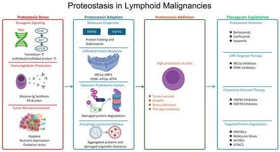
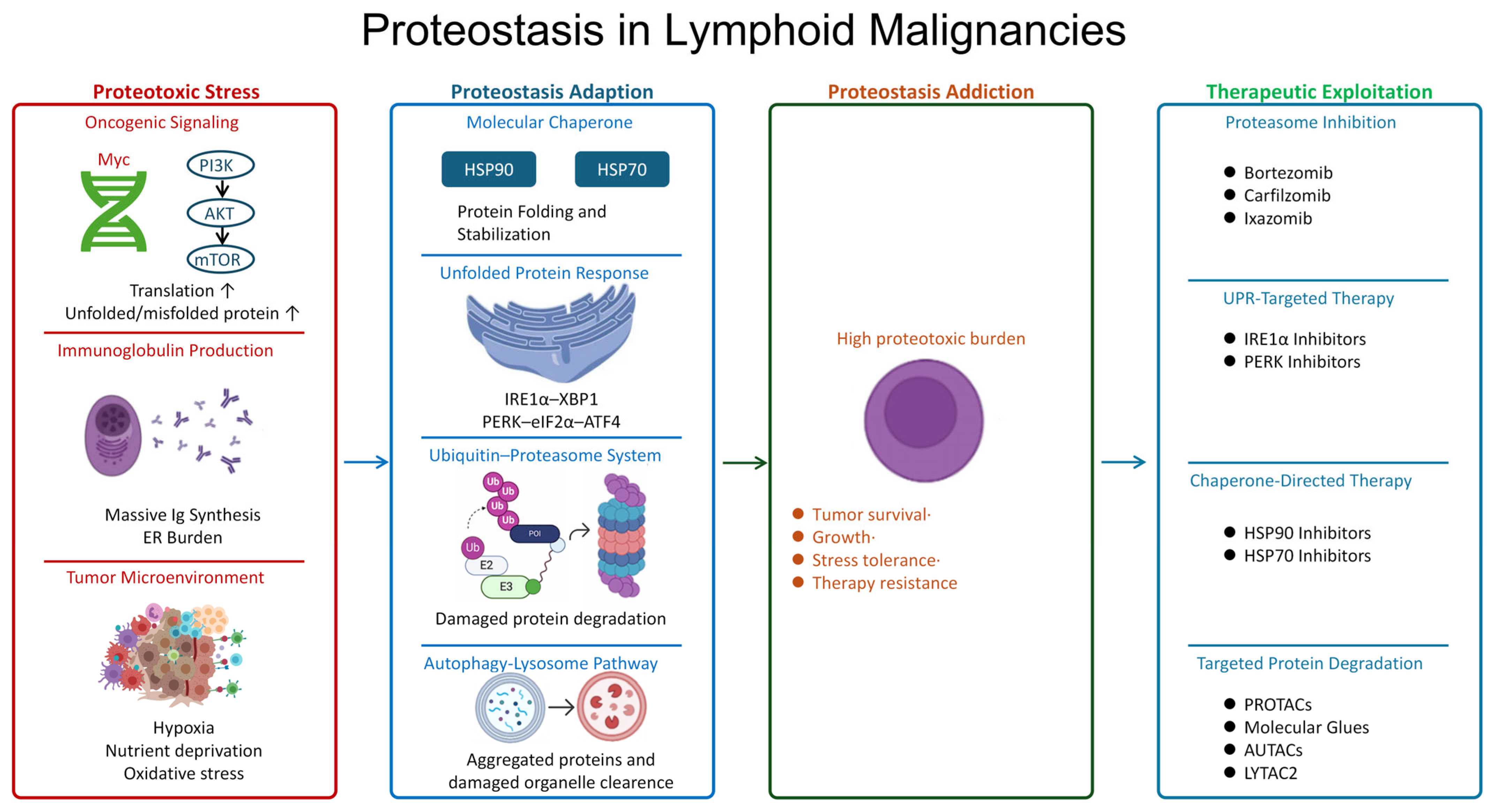
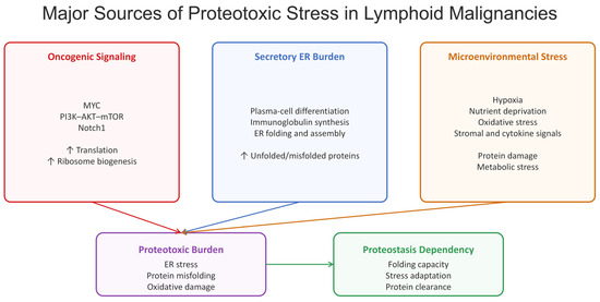
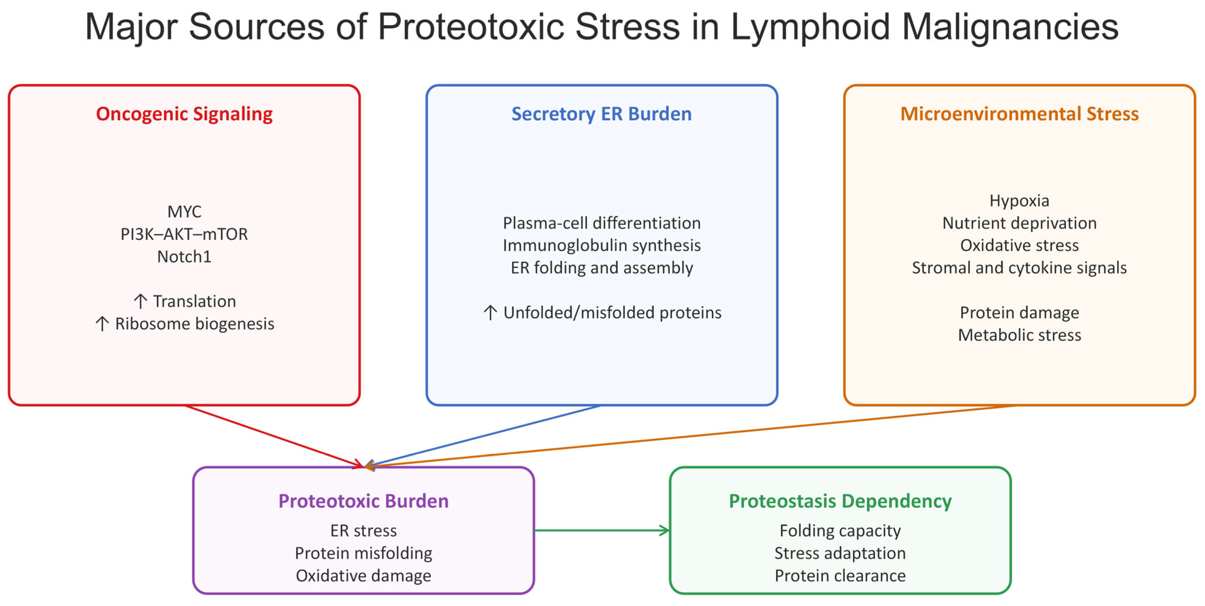
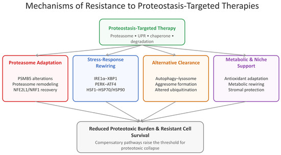
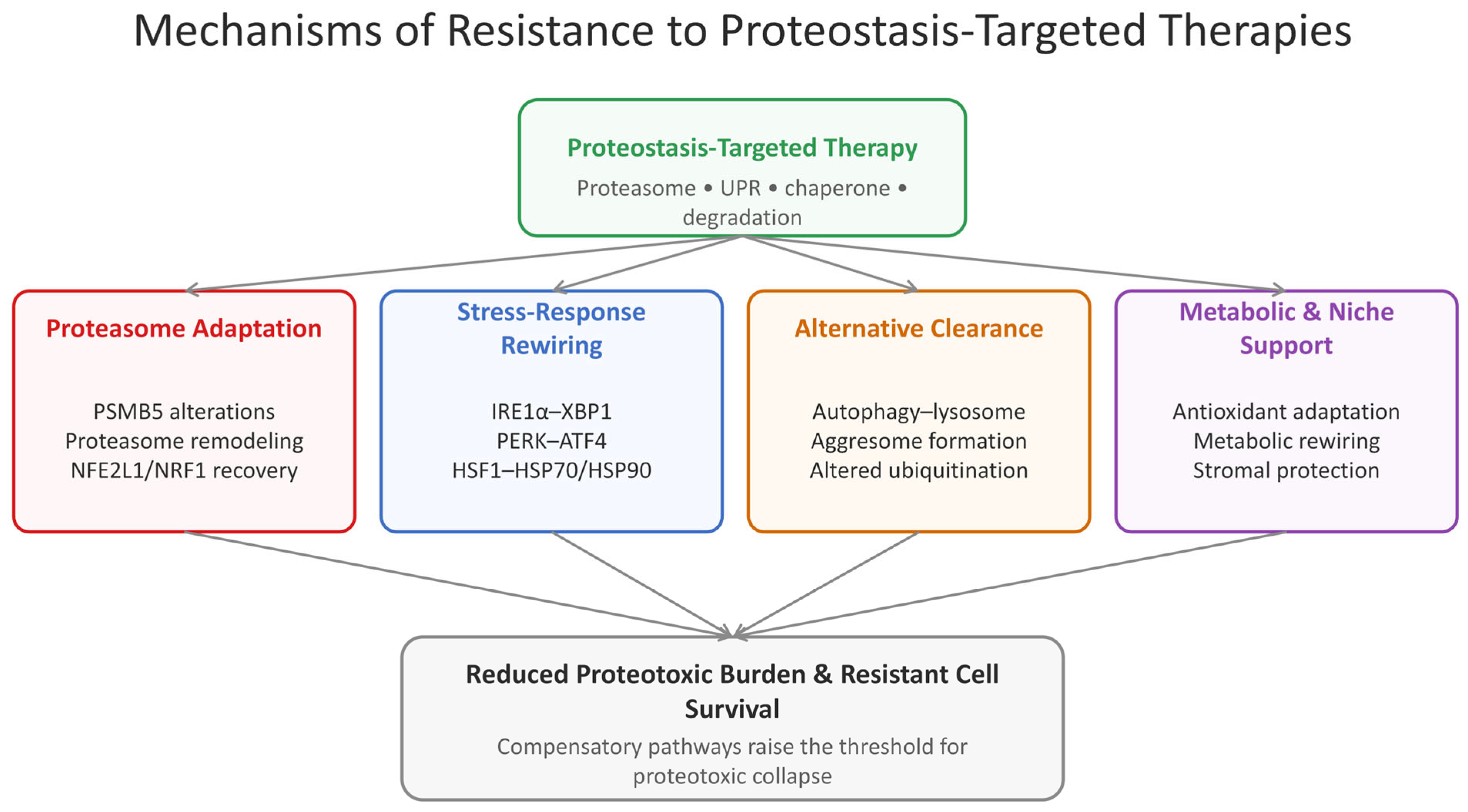

Disclaimer/Publisher’s Note: The statements, opinions and data contained in all publications are solely those of the individual author(s) and contributor(s) and not of MDPI and/or the editor(s). MDPI and/or the editor(s) disclaim responsibility for any injury to people or property resulting from any ideas, methods, instructions or products referred to in the content. |
© 2026 by the authors. Licensee MDPI, Basel, Switzerland. This article is an open access article distributed under the terms and conditions of the Creative Commons Attribution (CC BY) license.