


Abstract
The discovery of the glymphatic system and meningeal lymphatic vessels established that the brain possesses an organized lymphatic clearance network that drains to the deep cervical lymphatics. Disruption of this system has been suggested to contribute to the accumulation of pathogenic proteins implicated in neurodegeneration. Cervical lymphatic reconstruction has been associated with some neurological benefit in Alzheimer’s and Parkinson’s disease, and preclinical work has demonstrated that noninvasive manipulation of superficial cervical lymphatics can increase cerebrospinal fluid outflow. Whether noninvasive cervicofacial lymphedema therapy can produce comparable neurological effects remains unknown. We report two men with Parkinson’s disease (Hoehn and Yahr stages 2.5 and 3) treated with a standardized cervicofacial lymphedema therapy protocol targeting cervical and facial lymphatic pathways. Total MDS-UPDRS scores decreased by 19 points (34.5%) in Patient 1 and 5 points in Patient 2. The reduction in Patient 1 exceeded the published threshold for clinically meaningful total-score improvement. Benefits were reproducible and session-linked, and were accompanied by motor and non-motor gains in cognition, alertness, mood, energy, and sleep; interruption of therapy was associated with return toward baseline. Cervicofacial lymphedema therapy may represent a low-risk, noninvasive intervention hypothesized to influence brain lymphatic clearance in Parkinson’s disease.
1. Introduction
For much of modern neuroscience, the brain was considered exempt from the lymphatic system [1,2,3]. While peripheral organs depend on lymphatic drainage for metabolic waste removal, the central nervous system was thought to rely solely on cerebrospinal fluid circulation. This presumed exception reinforced the view that the brain operated under fundamentally different biological rules.
This understanding has evolved. The discovery of the glymphatic system in 2012, followed by the identification of meningeal lymphatic vessels in 2015, established that the brain possesses an organized lymphatic clearance network [1,2,3]. These pathways connect intracranial fluid transport to the deep cervical lymphatic system and play a central role in metabolic waste removal [2,3,4,5]. Disruption of this system has been implicated in neurodegeneration, largely on the basis of animal models and more recently human studies [4,5,6,7,8]. Preclinical findings are not uniform, and sustained meningeal lymphatic manipulation did not alter amyloid pathology in one experimental model [9].
Impaired glymphatic and meningeal lymphatic function is associated with aging, sleep disturbance, inflammation, headache, brain injury and vascular disease, and correlates with accumulation of pathogenic proteins implicated in Parkinson’s and Alzheimer’s disease [4,5,6,7,8]. In humans, supporting evidence is emerging but inconclusive. Impaired meningeal lymphatic drainage has been reported in idiopathic Parkinson’s disease, and glymphatic dysfunction has been associated with amyloid deposition, neurodegeneration, and clinical progression in Alzheimer’s disease [10,11]. Within this framework, neurodegenerative disorders can be understood, in part, as conditions of impaired lymphatic clearance and subsequent accumulation of pathogenic protein molecules leading to neuronal dysfunction and death [4,7,8,12,13].
Clinical observations outside the brain reinforce this concept. In the treatment of peripheral and truncal lymphedema, patients frequently report cognitive symptoms, including impaired concentration, memory, mental clarity, and fatigue [14,15]. Restoration of lymphatic flow through lymphatic reconstruction and lymphedema therapy has been associated with improved cognitive functions [14,15,16,17,18,19,20,21,22,23,24]. These observations suggest that lymphatic dysfunction may exert systemic effects on brain physiology and cognition [12,14,15].
Building on this experience, lymphatic reconstruction has been extended to patients with neurodegenerative disease. Reconstruction of cervical lymphatic pathways has been performed in patients with Alzheimer’s and Parkinson’s disease, primarily in Asia, with early reports describing neurological improvement [12,13,25,26,27,28,29,30]. Although preliminary, these findings provide proof of concept that modulating extracranial lymphatic outflow may influence central nervous system function [12,13,25,26,27,28,29,30].
If both lymphatic reconstruction and lymphedema therapy are effective in treating extracranial lymphatic dysfunction, and surgical approaches have shown preliminary benefit in neurodegenerative disease, an important question follows: can noninvasive lymphatic therapy produce similar effects in the brain [12,13,16,17,18,19,20,21,22,23,24]?
Here, we describe a case report of two patients with Parkinson’s disease treated with cervicofacial lymphedema therapy. Clinical course, symptom response, and objective measures were evaluated to characterize patients’ response to targeted lymphatic therapy.
2. Detailed Case Description
2.1. Staging and Outcome Measures
Parkinson disease severity was classified using the Hoehn and Yahr (H&Y) staging system (stages 1–5), where stage 1 indicates unilateral disease and stage 5 indicates bed-bound status [31]. Stage 2.5 indicates mild bilateral disease with recovery on the pull test, and stage 3 indicates bilateral disease with postural instability but preserved independence.
The primary outcome was change in the Movement Disorder Society–Unified Parkinson’s Disease Rating Scale (MDS-UPDRS) total score (Parts I–IV), with items scored from 0 to 4, where higher scores indicate greater impairment [32]. Clinically meaningful improvement was evaluated using published thresholds of 7.1 points for the total MDS-UPDRS and 3.25 points for Part III [33,34,35].
Secondary outcomes included the Patient Global Impression of Change (PGIC), a 7-point self-reported scale ranging from “very much improved” to “very much worse,” assessing the patient’s overall perceived change since treatment [36]. Missing or inconsistent data were recorded without imputation. Statistical analyses and data visualization were performed using R version 4.6.0 (R Foundation for Statistical Computing, Vienna, Austria).
2.2. Case Presentations
2.2.1. Patient 1
A 57-year-old man with Parkinson’s disease (Hoehn and Yahr stage 2.5) reported mild cognitive symptoms, including impaired concentration and brain fog, without impact on occupational performance. He was independently ambulatory without assistive devices and largely independent in activities of daily living, with mild difficulty in fine motor tasks including buttons and zippers. He remained physically active and employed full-time. No significant comorbidities were reported.
He underwent cervicofacial lymphedema therapy over 16 weeks beginning 12 February 2025, delivered 2–4 times per week for 48 sessions total, with sessions lasting 40–45 min. Medications included extended-release carbidopa/levodopa, amantadine, escitalopram, vitamin D, and melatonin.
2.2.2. Patient 2
A 56-year-old man with Parkinson’s disease (Hoehn and Yahr stage 3) had a history of hypertension and prior orthopedic and spinal procedures. He lived alone and was independent in basic activities of daily living but reported fatigue, tremor, stiffness, balance impairment, apathy, and mood symptoms limiting higher-level activity.
He underwent a 6-week course of cervicofacial lymphedema therapy beginning 19 March 2025, with outcome assessment performed at eight weeks. Treatment was administered 1–2 times per week by a certified therapist and daily via self-administration, totaling 42 sessions, with sessions lasting approximately 45 min. Medications included carbidopa/levodopa and cardiovascular agents.
2.3. Lymphedema Therapy Protocol
Each session followed a standardized sequence targeting cervical and facial lymphatic pathways [16]. Treatment began with supraclavicular lymph node stimulation, followed by stationary circular techniques along the medial and lateral internal jugular chains from the earlobe to the supraclavicular fossa, and then submandibular nodes. Facial treatment included preauricular and postauricular nodes and facial lymphatic regions. The sequence concluded with repeat submandibular, jugular, and supraclavicular stimulation. The treatment sequence is demonstrated in the video in Supplementary Materials.
The full sequence was repeated three times per session, for approximately 45 min total. No compression garments or adjunctive modalities were used.
2.4. Outcomes
2.4.1. Quantitative Outcomes
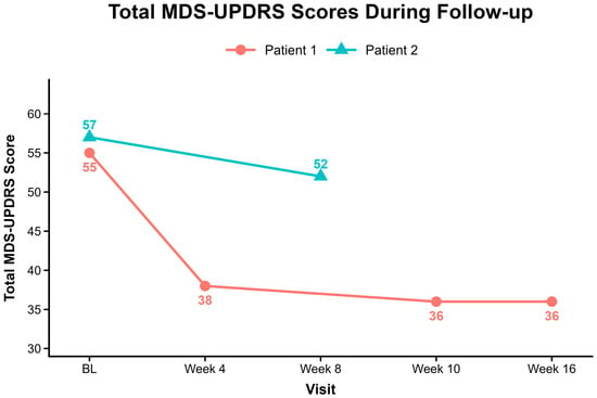
Total MDS-UPDRS scores improved in both patients after cervicofacial lymphedema therapy (Figure 1). Patient 1 decreased from 55 at baseline to 38 at four weeks and 36 at 10 and 16 weeks, representing a sustained 19-point reduction (34.5%) that exceeded the published threshold for clinically meaningful total-score improvement. Patient 2 decreased from 57 at baseline to 52 at eight weeks, representing a 5-point reduction.
Figure 1.
Total MDS-UPDRS scores during follow-up. Total scores are shown for both patients at baseline and subsequent assessments after initiation of cervicofacial lymphedema therapy. Lower scores indicate less impairment. Patient 1 was followed through week 16 and Patient 2 through week 8; only observed assessments are shown. The x-axis is categorical and represents the sequence of assessments rather than proportional elapsed time. Connecting lines aid visualization of within-patient trajectories and do not imply continuous change or linear interpolation between assessments.
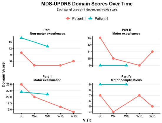
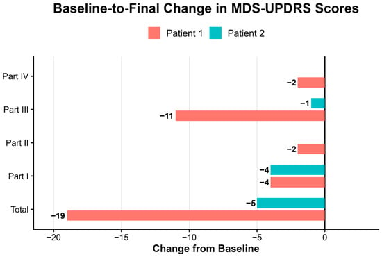
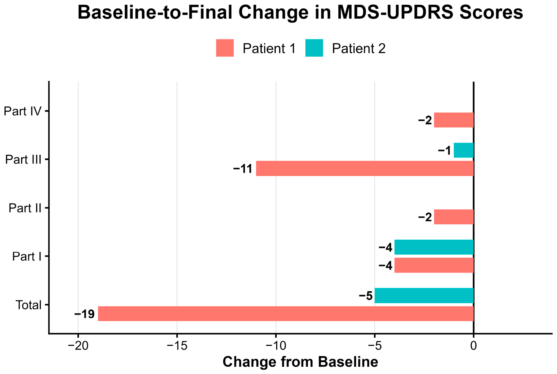
Domain-level changes differed between patients (Figure 2). In Patient 1, improvements were observed in Part I (−6 by week 10), Part II (−4 by week 10), Part III (−11 by week 16), and Part IV (−4 by week 4), with the 11-point Part III reduction exceeding the published threshold for clinically meaningful motor improvement. This suggests an apparent plateau in the total score after the initial month, although domain-specific trajectories differed: Part III continued to improve, while Parts I, II, and IV remained stable or fluctuated. In Patient 2, Part I improved by 4 points, Parts II and IV were unchanged, and Part III improved by 1 point.
Figure 2.
MDS-UPDRS domain scores over time. Scores for MDS-UPDRS Parts I–IV are shown for both patients. The domains represent non-motor experiences of daily living, motor experiences of daily living, motor examination, and motor complications, respectively. Each panel uses an independent y-axis scale. The x-axis is categorical and displays the sequence of observed assessments rather than proportional elapsed time. Connecting lines link observed measurements for visual clarity and do not imply continuous change or linear interpolation between assessments.
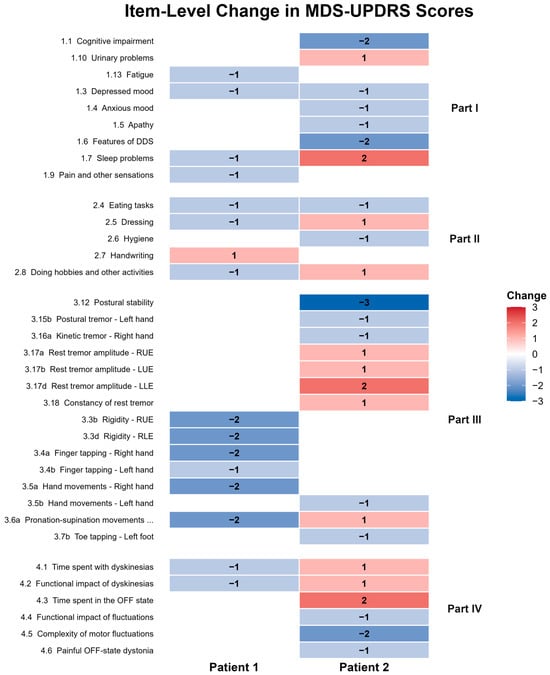
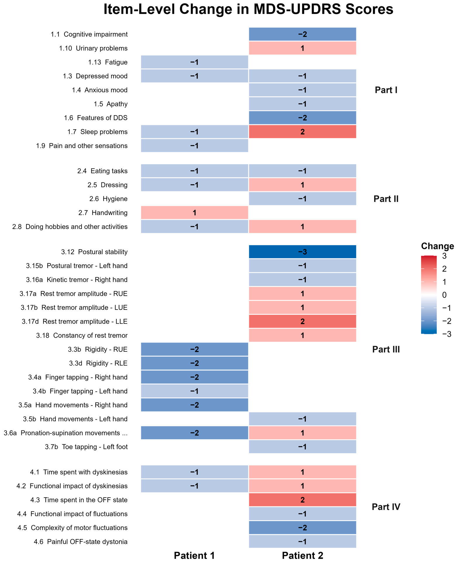
Baseline-to-final change scores are summarized in Figure 3. Patient 1 showed a larger final total-score reduction, driven mainly by motor improvement, whereas Patient 2 showed a smaller reduction driven mainly by non-motor improvement. Item-level changes are shown in Figure 4 and indicate mixed item-level effects, particularly in Patient 2, rather than uniform improvement across domains.
Figure 3.
Baseline-to-final change in MDS-UPDRS scores. Change from baseline to final assessment in total MDS-UPDRS and Part I–IV domain scores. Negative values indicate improvement.
Figure 4.
Item-level change in MDS-UPDRS scores. Baseline-to-final item-level change in MDS-UPDRS scores among items that changed in at least one patient. Negative values indicate improvement, positive values indicate worsening, and blank cells indicate no change.
Both patients reported PGIC scores of 6 at final assessment, corresponding to “better, definite improvement.” Interpretation of Patient 2 remains limited by shorter treatment duration, lower treatment frequency, and partial reliance on self-administration.
2.4.2. Caregiver-Reported Outcomes
Therapists observed session-linked motor improvements in both patients. In Patient 1, improvements in gait speed, stride length, arm swing, and facial expressivity were observed by session 4. These effects reportedly lasted several hours and were reproducible across sessions. In Patient 2, therapists reported modest improvements in postural stability and hand tremor, while gait remained unchanged during ambulation.
Non-motor changes were also reported in both patients. In Patient 1, cumulative improvements were noted in cognition, alertness, mood, energy, and sleep. Caregiver reports confirmed improved sleep and bed mobility. In Patient 2, cognition and mood improved, stiffness decreased, olfaction improved, and energy levels were near-normal.
During the treatment period, Patient 1 reported improved response to dopaminergic medication, reduced as-needed dosing, and had decreased levodopa–carbidopa dosage after neurologic evaluation.
3. Discussion
The glymphatic system was described in 2012 as a paravascular pathway for cerebrospinal fluid–interstitial fluid exchange and solute clearance [1]. Meningeal lymphatic vessels were identified in 2015, establishing central nervous system drainage to cervical lymphatics [2,3]. Impaired clearance has been linked to aging, sleep disturbance, inflammation, vascular disease, and pathogenic protein accumulation [4,5,6,7,8]. Neurodegeneration may therefore reflect, in part, impaired lymphatic clearance with toxic protein retention and neuronal dysfunction [4,7,8,12,13].
Peripheral lymphatic disease supports this relationship. Lymphedema causes swelling, inflammation, discomfort, and functional limitation [16,24]. Many patients also report impaired concentration, memory, energy, and mental clarity, which we have termed Lymphedema Related Dementia Syndrome (LRDS) [14,15]. Improvement after lymphatic reconstruction or lymphedema therapy is often accompanied by improvement in these symptoms, supporting lymphatic dysfunction as a systemic process with neurological manifestations [14,15,16,17,18,19,20,21,22,23,24].
These observations support impaired cerebral lymphatic clearance, or brain lymphedema, as a contributor to neurodegenerative disease. We discussed this concept as early as 2016, but clinical translation remained limited [12]. Proof of concept emerged in 2022, when cervical lymphatic reconstruction in Alzheimer’s disease was associated with neurological improvement, indicating that extracranial lymphatic modulation may influence central nervous system function [12,13,25,26,27,28,29,30].
Lymphatic surgery and lymphedema therapy both improve lymphatic drainage in extremity lymphedema [16,17,18,19,20,21,22,23,24]. Cervical lymphatic reconstruction has also been associated with preliminary neurological improvement [12,13]. Whether cervicofacial lymphatic therapy can produce similar neurological effects remains unknown.
Force-regulated mechanical manipulation of superficial cervical lymphatics through intact skin increased cerebrospinal fluid outflow to cervical lymph nodes and corrected age-related drainage impairment in mice [37]. The effect occurred through compression of superficial cervical lymphatics, with little effect on spontaneous lymphatic contractions [37]. These data indicate that nonoperative cervical lymphatic manipulation can increase cerebrospinal fluid outflow in a preclinical model [37].
In this case report, cervicofacial lymphedema therapy was associated with reproducible, session-dependent motor and non-motor changes. Patient 1 showed sustained improvement in total and motor MDS-UPDRS scores. Patient 2 showed a smaller 5-point reduction in total MDS-UPDRS score, driven mainly by non-motor improvement, with stable activities of daily living and motor complications and minimal motor examination change. Differences in treatment duration, frequency, and delivery likely contributed to the difference in response.
Aggregate UPDRS scores did not capture several clinically relevant changes. Both patients reported improved cognition, alertness, and daytime energy. Patient 1 also improved in sleep and mood. Patient 2 reported improved postural stability, reduced hand tremor, improved olfaction, and near-normal energy, despite persistent gait impairment and worsened sleep. These findings show the limits of total UPDRS scores in capturing domain-specific and fluctuating responses [32,33].
Some effects appeared treatment-dependent and reversible. Therapy interruption was associated with return toward baseline, followed by improvement after treatment resumed. This pattern is more consistent with an acute or subacute physiological effect than with delayed disease modification.
Medication changes add uncertainty. Patient 1 reduced medication without perceived loss of benefit, while Patient 2 had medication changes during treatment. These changes may reflect altered dopaminergic responsiveness but also confound interpretation.
The mechanism remains undefined. Rapid symptomatic changes after individual 45 min sessions are unlikely to reflect changes in chronic protein aggregation. A more plausible explanation is acute change in intracranial fluid handling, inflammatory signaling, or both [4,5,37]. Advanced neuroimaging can determine whether cervicofacial therapy changes cerebral fluid burden, perivascular flow, meningeal lymphatic drainage, or inflammatory markers. Over longer intervals, any enhancement of cerebral lymphatic outflow, if present, may influence chronic protein clearance [4,12,37]. However, abnormal protein accumulation may contribute to neurodegeneration without being its sole or primary driver, and reducing pathological burden does not necessarily translate into substantial clinical benefit. Accordingly, even if cerebral protein clearance were enhanced, its clinical effect would remain uncertain [38,39,40,41].
This study is limited by small sample size, expectancy effects, medication confounding, unblinded assessment, and variable treatment timing. The uncontrolled design precludes causal inference. A two-patient series cannot establish efficacy or generalizability. Outcomes relied on clinician and caregiver report without objective, instrumented measurement of motor function or sleep. Video-based tools now allow quantitative motor assessment and would strengthen future work [42]. Nonspecific effects of manual therapy and touch, therapeutic attention, patient expectation, repeated administration of rating scales, and natural symptom fluctuation may each contribute to the observed changes. Follow-up was limited to 16 weeks, and durability beyond this interval is unknown. Target engagement of glymphatic or lymphatic pathways was not measured.
The findings align with brain lymphatic biology, prior surgical observations, and preclinical evidence that cervical lymphatic manipulation can increase cerebrospinal fluid outflow [1,2,3,4,5,6,7,8,10,11,12,13,25,26,27,28,29,30,37]. Cervicofacial lymphedema therapy was associated with reproducible motor and non-motor improvement and may warrant further investigation as a low-risk strategy hypothesized to modulate brain lymphatic clearance. Objective imaging, biomarker endpoints, standardized treatment parameters, and stage-stratified cohorts are needed to define durability, cumulative effect, and optimal candidates. Controlled, blinded designs are required, including a sham or active comparator that targets an anatomically distinct region and a crossover structure to separate specific treatment effects from nonspecific ones.
4. Conclusions
Cervicofacial lymphedema therapy may represent a safe, potentially effective approach that could influence cerebral lymphatic function. If validated, a technique already familiar to lymphedema therapists could extend beyond edema management and emerge as a simple and accessible strategy for neurological disease.
Supplementary Materials
The following supporting information can be downloaded at https://www.mdpi.com/article/10.3390/lymphatics4030044/s1.
Author Contributions
Conceptualization, Y.M. and W.F.C.; methodology, Y.M., A.B., H.S. and W.F.C.; software, Y.M.; validation, W.P.; formal analysis, Y.M.; investigation, Y.M., A.B. and H.S.; resources, W.F.C.; data curation, Y.M., A.B. and H.S.; writing—original draft preparation, Y.M.; writing—review and editing, Y.M., W.P. and W.F.C.; visualization, Y.M., A.B. and H.S.; supervision, W.F.C.; project administration, Y.M. and W.F.C. All authors have read and agreed to the published version of the manuscript.
Funding
This research received no external funding.
Institutional Review Board Statement
Formal institutional review board approval was not required by the Cleveland Clinic Institutional Review Board for this retrospective clinical case report. Written informed consent was obtained from both patients and documented by signed consent forms authorizing inclusion in this report and publication of deidentified clinical information and accompanying deidentified material. Angela Bialorucki, an author and therapist who appears in the Supplementary video, also provided written informed consent authorizing publication of the Supplementary video with her identifiable appearance.
Informed Consent Statement
Informed consent was obtained from all subjects involved in the study.
Data Availability Statement
The data supporting the findings of this study are not publicly available due to patient privacy and confidentiality considerations. Data necessary to verify the reported findings may be made available to the journal and peer reviewers upon reasonable request to the corresponding author, subject to applicable ethical and institutional requirements.
Conflicts of Interest
The authors declare no conflicts of interest.
References
- Iliff, J.J.; Wang, M.; Liao, Y.; Plogg, B.A.; Peng, W.; Gundersen, G.A.; Benveniste, H.; Vates, G.E.; Deane, R.; Goldman, S.A.; et al. A paravascular pathway facilitates CSF flow through the brain parenchyma and the clearance of interstitial solutes, including amyloid beta. Sci. Transl. Med. 2012, 4, 147ra111. [Google Scholar] [CrossRef] [Scilit] [PubMed]
- Louveau, A.; Smirnov, I.; Keyes, T.J.; Eccles, J.D.; Rouhani, S.J.; Peske, J.D.; Derecki, N.C.; Castle, D.; Mandell, J.W.; Lee, K.S.; et al. Structural and functional features of central nervous system lymphatic vessels. Nature 2015, 523, 337–341, Erratum in Nature 2016, 533, 278. [Google Scholar] [CrossRef] [Scilit] [PubMed]
- Aspelund, A.; Antila, S.; Proulx, S.T.; Karlsen, T.V.; Karaman, S.; Detmar, M.; Wiig, H.; Alitalo, K. A dural lymphatic vascular system that drains brain interstitial fluid and macromolecules. J. Exp. Med. 2015, 212, 991–999. [Google Scholar] [CrossRef] [Scilit] [PubMed]
- Da Mesquita, S.; Louveau, A.; Vaccari, A.; Smirnov, I.; Cornelison, R.C.; Kingsmore, K.M.; Contarino, C.; Onengut-Gumuscu, S.; Farber, E.; Raper, D.; et al. Functional aspects of meningeal lymphatics in ageing and Alzheimer’s disease. Nature 2018, 560, 185–191, Erratum in Nature 2018, 564, E7. [Google Scholar] [CrossRef] [Scilit] [PubMed]
- Plog, B.A.; Nedergaard, M. The glymphatic system in CNS health and disease: Past, present and future. Annu. Rev. Pathol. 2018, 13, 379–394. [Google Scholar] [CrossRef] [Scilit] [PubMed]
- Xie, L.; Kang, H.; Xu, Q.; Chen, M.J.; Liao, Y.; Thiyagarajan, M.; O’Donnell, J.; Christensen, D.J.; Nicholson, C.; Iliff, J.J.; et al. Sleep drives metabolite clearance from the adult brain. Science 2013, 342, 373–377. [Google Scholar] [CrossRef] [Scilit] [PubMed]
- Nedergaard, M.; Goldman, S.A. Glymphatic failure as a final common pathway to dementia. Science 2020, 370, 50–56. [Google Scholar] [CrossRef] [Scilit] [PubMed]
- Buccellato, F.R.; D’Anca, M.; Fenoglio, C.; Scarpini, E.; Galimberti, D. The role of the glymphatic system in Alzheimer’s and Parkinson’s disease pathogenesis. Biomedicines 2022, 10, 2261. [Google Scholar] [CrossRef] [Scilit] [PubMed]
- Antila, S.; Chilov, D.; Nurmi, H.; Li, Z.; Näsi, A.; Gotkiewicz, M.; Sitnikova, V.; Jäntti, H.; Acosta, N.; Koivisto, H.; et al. Sustained meningeal lymphatic vessel atrophy or expansion does not alter Alzheimer’s disease-related amyloid pathology. Nat. Cardiovasc. Res. 2024, 3, 474–491. [Google Scholar] [CrossRef] [Scilit] [PubMed]
- Ding, X.B.; Wang, X.X.; Xia, D.H.; Liu, H.; Tian, H.Y.; Fu, Y.; Chen, Y.K.; Qin, C.; Wang, J.Q.; Xiang, Z.; et al. Impaired meningeal lymphatic drainage in patients with idiopathic Parkinson’s disease. Nat. Med. 2021, 27, 411–418. [Google Scholar] [CrossRef] [Scilit] [PubMed]
- Huang, S.Y.; Zhang, Y.R.; Guo, Y.; Du, J.; Ren, P.; Wu, B.; Feng, J.; Initiative, A.D.N.; Cheng, W.; Yu, J. Glymphatic system dysfunction predicts amyloid deposition, neurodegeneration, and clinical progression in Alzheimer’s disease. Alzheimer’s Dement. 2024, 20, 3251–3269. [Google Scholar] [CrossRef] [Scilit] [PubMed]
- Hong, J.P.; Chen, W.F.; Nguyen, D.H.; Xie, Q. A proposed role for lymphatic supermicrosurgery in the management of Alzheimer’s disease: A primer for reconstructive microsurgeons. Arch. Plast. Surg. 2025, 52, 96–103. [Google Scholar] [CrossRef] [Scilit] [PubMed]
- Lahmar, T.; Thuau, F.; Pinard, G.; Boutoleau-Bretonniere, C.; Perrot, P.; Lancien, U. Brain lymphatic drainage pathways, deep cervical lymphatic surgery, and current insights: A systematic review. J. Prev. Alzheimers Dis. 2026, 13, 100335. [Google Scholar] [CrossRef] [Scilit] [PubMed]
- Chen, W.F.; Jou, C.; Pandey, S.K.; Lo, S.L. Primary lymphedema: Anatomically isolated or a pervasive systemic disorder? Plast. Reconstr. Surg. Glob. Open 2024, 12, e6328. [Google Scholar] [CrossRef] [Scilit] [PubMed]
- Salman, M.; Lo, S.L.; Cheong, D.C.F.; Chen, W.F. Lymph and mind: A new perspective on cognitive dysfunction in lymphedema. Plast. Reconstr. Surg. Glob. Open 2025, 13, 15–16. [Google Scholar] [CrossRef] [Scilit]
- Executive Committee of the International Society of Lymphology. The diagnosis and treatment of peripheral lymphedema: 2023 consensus document of the International Society of Lymphology. Lymphology 2023, 56, 133–151. [Google Scholar] [CrossRef] [Scilit]
- Chen, W.F. How to get started performing supermicrosurgical lymphaticovenular anastomosis to treat lymphedema. Ann. Plast. Surg. 2018, 81, S15–S20. [Google Scholar] [CrossRef] [Scilit] [PubMed]
- Chen, W.F.; Yamamoto, T.; Fisher, M.; Liao, J.; Carr, J. The “octopus” lymphaticovenular anastomosis: Evolving beyond the standard supermicrosurgical technique. J. Reconstr. Microsurg. 2015, 31, 450–457. [Google Scholar] [CrossRef] [Scilit] [PubMed]
- Chen, W.F.; Tuncer, F.B.; Zeng, W. Advanced technical pearls for successful supermicrosurgical lymphaticovenular anastomosis. Ann. Plast. Surg. 2021, 86, S165–S172. [Google Scholar] [CrossRef] [Scilit] [PubMed]
- Pandey, S.K.; Fahradyan, V.; Orfahli, L.M.; Chen, W.F. Supermicrosurgical lymphaticovenular anastomosis vs vascularized lymph vessel transplant: Technical optimization and when to perform which. Plast. Aesthet. Res. 2021, 8, 47. [Google Scholar] [CrossRef] [Scilit]
- Bailey, E.A.; Kukreja-Pandey, S.; Chen, W.F. Advances in surgical lymphedema management: The emergence and refinement of lymph node-to-vein anastomosis (LNVA). Curr. Surg. Rep. 2024, 12, 83–88. [Google Scholar] [CrossRef] [Scilit]
- Chen, W.F.; Cheong, D.C.F.; Tedone Clemente, E.; Salman, M. Robot-assisted lymph node-to-vein anastomosis: Lessons from the first 22 cases at a high-volume lymphatic supermicrosurgery center. Curr. Oncol. 2025, 32, 377. [Google Scholar] [CrossRef] [Scilit] [PubMed]
- Pak, C.S.; Suh, H.P.; Kwon, J.G.; Cho, M.J.; Hong, J.P. Lymph node to vein anastomosis (LNVA) for lower extremity lymphedema. J. Plast. Reconstr. Aesthet. Surg. 2021, 74, 2059–2067. [Google Scholar] [CrossRef] [Scilit] [PubMed]
- Kukreja-Pandey, S.; Gaxiola-Garcia, M.A.; Moheeputh, N.; Chen, W.F. Current and future implications of lymphedema surgery in head and neck reconstruction. Oral Maxillofac. Surg. Clin. N. Am. 2024, 36, 567–574. [Google Scholar] [CrossRef] [Scilit] [PubMed]
- Lu, H.R.; Tan, Y.F.; Xie, Q.P. Preliminary observation of deep cervical lymphatic-venous anastomosis under off-eyepiece 3D microscope to treat an elderly patient with cognitive impairment. Chin. J. Microsurg. 2022, 45, 570–574. (In Chinese) [Google Scholar]
- Chen, J.Y.; Zhao, D.W.; Yin, Y.; Gui, L.; Chen, X.B.; Wang, X.M.; Wang, P.B.; Zuo, C.H.; Lai, Z.P.; Li, C.; et al. Deep cervical lymphovenous anastomosis for Alzheimer’s disease: Microsurgical procedure in a prospective cohort study. Int. J. Surg. 2025, 111, 4211–4221. [Google Scholar] [CrossRef] [Scilit] [PubMed]
- Ma, X.; Wang, F.; Wang, G.; Zhao, M.; Zheng, Y.; Guo, Y.; Wu, J.; Liu, Y.; Liu, Y.; He, G.; et al. A surgical therapy for Alzheimer’s disease with lymphaticovenular anastomosis. J. Alzheimer’s Dis. Rep. 2025, 9, 25424823251384244. [Google Scholar] [CrossRef] [Scilit] [PubMed]
- Tang, C.; Zheng, X.; Li, H.; Wang, B.; Zheng, Y.; Song, W.; Wu, A.; Xin, L.; Zhang, D.; Yang, R.; et al. Deep cervical lymphatic-venous anastomosis in dementia: A clinical and mechanistic evaluation. Int. J. Surg. 2026, 112, 9002–9014. [Google Scholar] [CrossRef] [Scilit] [PubMed]
- Fu, X.; Zhang, J.; Xiao, Q.; Li, T.; Li, Q.; Zhang, S.; Zeng, W.; Gong, M.; Cai, P.; Deng, Z.; et al. Deep cervical lymphatic-venous anastomosis attenuates cognitive dysfunction and biomarker abnormalities in severe Alzheimer’s disease: A prospective single-arm study. Alzheimer’s Dement. 2026, 22, e71150. [Google Scholar] [CrossRef] [Scilit] [PubMed]
- Hu, S.; Ge, J.; Wang, G.; Zhao, M.; Lv, P.; Wang, F.; Guo, F.; Huang, J.; Guan, H.; Ma, X.; et al. Plasma biomarker changes following deep cervical lymphatic venous anastomosis: An exploratory study in Alzheimer’s disease using single-molecule array technology. J. Alzheimer’s Dis. 2026, 111, 279–293. [Google Scholar] [CrossRef] [Scilit] [PubMed]
- Hoehn, M.M.; Yahr, M.D. Parkinsonism: Onset, progression and mortality. Neurology 1967, 17, 427–442. [Google Scholar] [PubMed]
- Goetz, C.G.; Tilley, B.C.; Shaftman, S.R.; Stebbins, G.T.; Fahn, S.; Martinez-Martin, P.; Poewe, W.; Sampaio, C.; Stern, M.B.; Dodel, R.; et al. Movement Disorder Society-sponsored revision of the Unified Parkinson’s Disease Rating Scale (MDS-UPDRS): Scale presentation and clinimetric testing results. Mov. Disord. 2008, 23, 2129–2170. [Google Scholar] [CrossRef] [Scilit] [PubMed]
- Makkos, A.; Kovács, M.; Aschermann, Z.; Harmat, M.; Janszky, J.; Karádi, K.; Kovács, N. Are the MDS-UPDRS-based composite scores clinically applicable? Mov. Disord. 2018, 33, 835–839. [Google Scholar] [CrossRef] [Scilit] [PubMed]
- Horváth, K.; Aschermann, Z.; Ács, P.; Deli, G.; Janszky, J.; Komoly, S.; Balázs, É.; Takács, K.; Karádi, K.; Kovács, N. Minimal clinically important difference on the Motor Examination part of MDS-UPDRS. Park. Relat. Disord. 2015, 21, 1421–1426. [Google Scholar] [CrossRef] [Scilit] [PubMed]
- Mishra, B.; Bhaumik, S.; Jha, M.; Kalra, R. A systematic review of minimum clinically important difference in Parkinson’s disease rating scales. Mov. Disord. Clin. Pract. 2024, 11, 707–721. [Google Scholar]
- Hurst, H.; Bolton, J. Assessing the clinical significance of change scores recorded on subjective outcome measures. J. Manip. Physiol. Ther. 2004, 27, 26–35. [Google Scholar] [CrossRef] [Scilit] [PubMed]
- Jin, H.; Yoon, J.H.; Hong, S.P.; Hwang, Y.S.; Yang, M.J.; Choi, J.; Kang, H.J.; Baek, S.E.; Jin, C.; Jung, J.; et al. Increased CSF drainage by non-invasive manipulation of cervical lymphatics. Nature 2025, 643, 755–767. [Google Scholar] [CrossRef] [Scilit] [PubMed]
- Bennett, D.A.; Schneider, J.A.; Arvanitakis, Z.; Kelly, J.F.; Aggarwal, N.T.; Shah, R.C.; Wilson, R.S. Neuropathology of older persons without cognitive impairment from two community-based studies. Neurology 2006, 66, 1837–1844. [Google Scholar] [CrossRef] [Scilit] [PubMed]
- Aiello Bowles, E.J.; Crane, P.K.; Walker, R.L.; Chubak, J.; LaCroix, A.Z.; Anderson, M.L.; Rosenberg, D.; Keene, C.D.; Larson, E.B. Cognitive resilience to Alzheimer’s disease pathology in the human brain. J. Alzheimer’s Dis. 2019, 68, 1071–1083. [Google Scholar] [CrossRef] [Scilit] [PubMed]
- van Dyck, C.H.; Swanson, C.J.; Aisen, P.; Bateman, R.J.; Chen, C.; Gee, M.; Kanekiyo, M.; Li, D.; Reyderman, L.; Cohen, S.; et al. Lecanemab in early Alzheimer’s disease. N. Engl. J. Med. 2023, 388, 9–21. [Google Scholar] [CrossRef] [Scilit] [PubMed]
- Sims, J.R.; Zimmer, J.A.; Evans, C.D.; Lu, M.; Ardayfio, P.; Sparks, J.; Wessel, A.M.; Shcherbinin, S.; Wang, H.; Nery, E.S.M.; et al. Donanemab in early symptomatic Alzheimer disease: The TRAILBLAZER-ALZ 2 randomized clinical trial. JAMA 2023, 330, 512–527. [Google Scholar] [PubMed]
- Acevedo, G.; Lange, F.; Calonge, C.; Peach, R.; Wong, J.K.; Guarin, D.L. VisionMD: An open-source tool for video-based analysis of motor function in movement disorders. npj Park. Dis. 2025, 11, 27. [Google Scholar] [CrossRef] [Scilit] [PubMed]
Disclaimer/Publisher’s Note: The statements, opinions and data contained in all publications are solely those of the individual author(s) and contributor(s) and not of MDPI and/or the editor(s). MDPI and/or the editor(s) disclaim responsibility for any injury to people or property resulting from any ideas, methods, instructions or products referred to in the content. |
© 2026 by the authors. Licensee MDPI, Basel, Switzerland. This article is an open access article distributed under the terms and conditions of the Creative Commons Attribution (CC BY) license.