Case Report

Hodgkin Lymphoma in a Patient with Down Syndrome: Case Report and Review of the Literature

by
Lucía Belén Queizan
1,*,
María José Serer
1,
Laura Galluzzo Mutti
2,
Hernán Zamaro
1,
María Sara Felice
1,
Pedro Zubizarreta
1 and
Elizabeth Alfaro
1
1
Department of Oncology, Juan P. Garrahan Hospital, Buenos Aires 1245, Argentina
2
Department of Pathology, Juan P. Garrahan Hospital, Buenos Aires 1245, Argentina
*
Author to whom correspondence should be addressed.
Lymphatics 2026, 4(3), 42; https://doi.org/10.3390/lymphatics4030042
Submission received: 6 June 2026 / Revised: 2 July 2026 / Accepted: 24 July 2026 / Published: 4 August 2026

Abstract

The association between Down syndrome (DS) and Hodgkin lymphoma (HL) is rare, with only a few cases reported in the literature. Here, we report the case of a pediatric patient with DS diagnosed with HL. In addition, a literature review was conducted, identifying only seven pediatric cases. We highlight treatment-related toxicity in this group of patients. Given the high survival rates of patients with HL, current strategies focus on individualizing treatment intensity based on the initial disease characteristics and the patient’s response. This consideration becomes even more important in patients with DS. The patient described in this report achieved complete metabolic remission after chemotherapy, experienced manageable grade 3 treatment-related toxicities, and remains in complete remission after 27 months of follow-up.

1. Introduction

Patients with Down syndrome (DS) have a 10- to 20-fold increased risk of developing acute leukemia compared to the general pediatric population [1]. This may be explained by altered hematopoiesis and a bone marrow microenvironment driven by the additional genetic material on chromosome 21, which promotes the production of increased numbers of immature hematopoietic precursors [1]. In contrast, this population has a low incidence of solid tumors [2]. The association between DS and Hodgkin lymphoma (HL) is rare, with only a few cases reported in the literature [3,4]. This case underscores the clinical challenges of treating HL in children with DS, particularly the need to balance treatment efficacy with increased susceptibility to therapy-related toxicity.
In addition to the case description, a literature review was conducted on 25 March 2025, using the MEDLINE/PubMed database of the National Library of Medicine, with the MeSH terms “Down syndrome” AND “Hodgkin disease”. Studies published in English or Spanish involving pediatric patients (0–18 years) were eligible for inclusion. Titles and abstracts were screened for relevance, followed by full-text review of potentially eligible articles. Studies involving adult populations, non-English or non-Spanish publications, conference abstracts without sufficient clinical data, review articles, and duplicate reports were excluded. The search was updated on 6 June 2026, to identify any additional eligible publications.

2. Case Description

A 7-year-old male patient with a postnatal diagnosis of Down syndrome (DS) presented to a tertiary care hospital with cervical masses, pallor, and weight loss. He also experienced fever and respiratory distress. The patient had irregular pediatric follow-up and had not undergone screening studies for conditions associated with his underlying syndrome.
The patient was in fair general condition and hemodynamically stable, with tachypnea. The patient required oxygen via nasal cannula at 1 L/min. He presented with generalized cutaneous and mucosal pallor. He exhibited marked hypoventilation in the right lung field with crackles in the middle and lower right lung zones. Dullness to percussion was noted in the spine at that level. Enlarged, non-tender bilateral cervical, supraclavicular, and axillary lymphadenopathies were present, with adherence to deeper tissues. Splenomegaly was also noted.
A complete blood count revealed leukopenia (3410/mm3), lymphopenia (272/mm3) and anemia (hemoglobin level: 7 g/dL). Chemistry and coagulation tests were within normal limits. Peripheral blood smear showed anisocytosis, microcytosis, and hypochromia in erythrocytes, with no pathological findings in the other cell lineages. A chest X-ray revealed a bulky mediastinal mass. Bone marrow examination showed no infiltration by atypical cells.
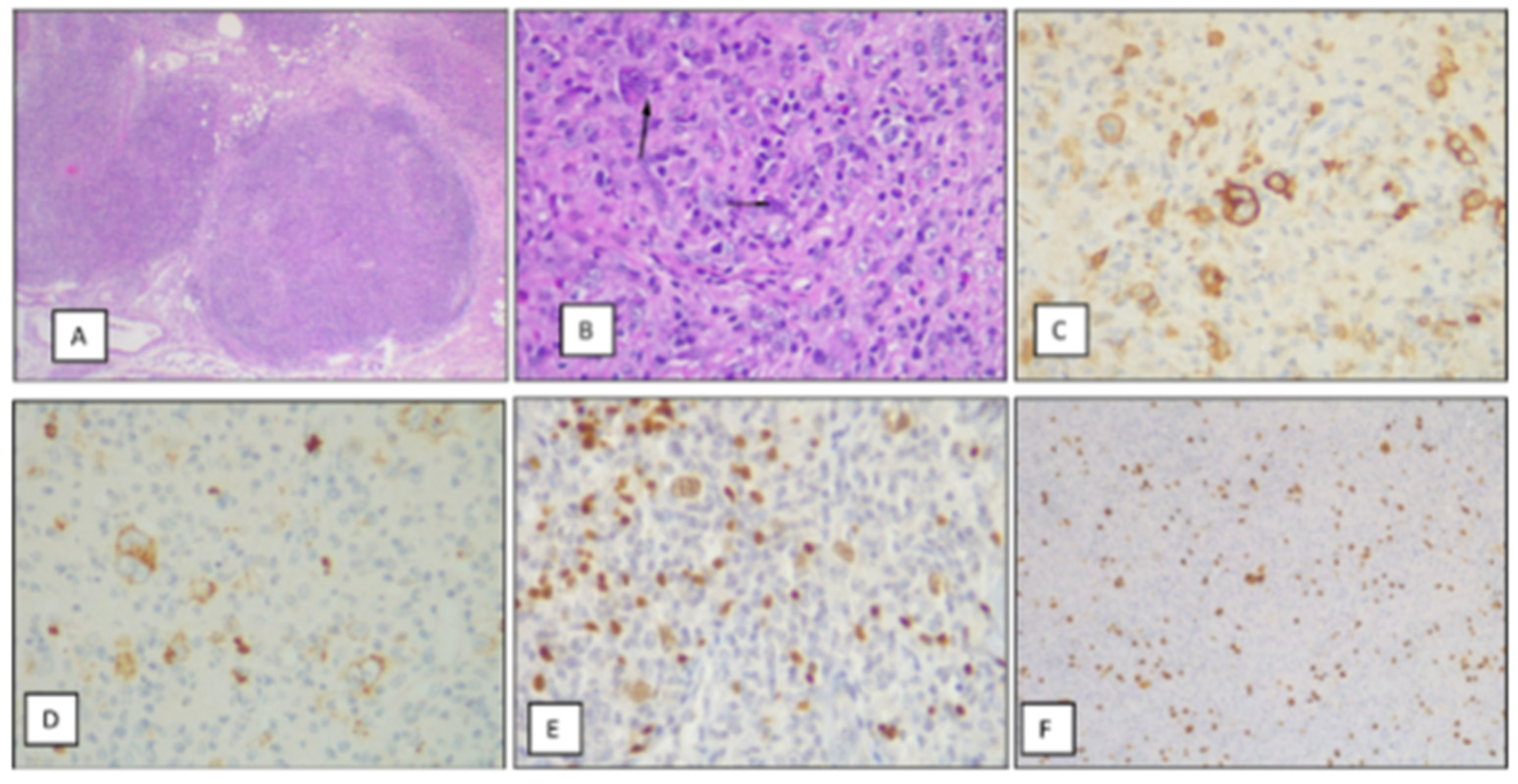
An excisional biopsy of a right anterior cervical lymph node was performed, and the histological examination revealed disruption of the nodal architecture due to a heterogeneous cellular proliferation. This included large cells with vesicular nuclei, often binucleated, prominent purplish nucleoli, and evident cytoplasm, which was sometimes eosinophilic and occasionally retracted. The proliferation was accompanied by lymphocytes, histiocytes, and plasma cells. The neoplastic cells were arranged in a scattered pattern. The overall proliferation exhibited a nodular pattern, with complete septa separating the nodules, and areas of very low cellularity were prominent. These areas showed hyaline collagen deposition and abundant vascularization. The neoplastic cells were positive for CD30, CD15, PAX5 (weak), and EBER, and negative for CD20. Based on these findings, a diagnosis of classical nodular sclerosis Hodgkin lymphoma was made. (See Figure 1).
Differential diagnoses included non-Hodgkin lymphoma and infectious lymphadenopathy; however, these were excluded based on histopathological findings and immunophenotypic characterization.
Staging studies were performed. A computed axial tomography (CAT) scan of the chest, abdomen, and pelvis revealed a large, multilobulated mediastinal mass located in the anterosuperior mediastinum, with heterogeneous enhancement, hypodensity, and apparent invasion of the superior vena cava, with anterior displacement. The mass affected both pulmonary hila, with compression of the segmental bronchus of the left lower lobe, where increased density and an air bronchogram were observed, without volume loss, suggestive of pneumonia. The volume measured 424.7 cm3. A right pleural effusion was present. Right axillary lymphadenopathy was observed, with the largest node measuring 20 mm in short axis. In the upper abdominal sections, heterogeneous splenomegaly with multiple hypodense areas was observed. Heterogeneous hepatomegaly with focal lesions and retroperitoneal lymph nodes were also noted.
The patient was stratified as stage IVB high-risk and was treated according to the LH-GALOP 2017 protocol [5].
Initially, the patient received one cycle of ABVD (Adriamycin, Bleomycin, Vinblastine, and Dacarbazine), followed by one cycle of ESHAP (Etoposide, Methylprednisolone, Cisplatin, and Cytarabine). An interim positron emission tomography with fluorodeoxyglucose (FDG PET/CT) was performed, revealing a Deauville score of 3. As a result, the patient was switched to chemotherapy according to the F protocol arm (three cycles of ABVD and three cycles of ESHAP). Post-chemotherapy PET/CT showed a Deauville score of 2, and complete remission was achieved. According to the protocol guidelines, no radiotherapy or further therapy was administered.
The patient developed anemia requiring a blood transfusion after the first ABVD cycle (Common Terminology Criteria for Adverse Events [CTCAE] Grade 3 hematologic toxicity). He also experienced catheter-related Candida parapsilosis colonization following the third ABVD cycle. Antifungal treatment was administered based on the pathogen and its sensitivity for 14 days, and the implanted catheter was removed (CTCAE Grade 3 hematologic toxicity). Additionally, the patient developed febrile neutropenia with oral gingivostomatitis after the sixth ESHAP cycle (CTCAE Grade 3 hematologic/infectious toxicity). Empiric acyclovir was initiated based on the clinical suspicion of herpes simplex virus infection in the setting of profound immunosuppression, resulting in prompt clinical improvement. The patient remained in complete remission after 27 months of follow-up, with no evidence of sequelae.

3. Discussion

Patients with DS have a significantly higher risk of developing neoplasms compared to the general population, with acute leukemia representing the most common association [1,2]. In these patients, the incidence of solid tumors is 50% lower than in individuals without DS [6]. Solid tumors secrete pro-angiogenic factors [7]. Endostatin, an endogenous inhibitor of angiogenesis, is significantly elevated in patients with DS, suggesting that it may play a protective role against the development of solid tumors in this population [7].
HL is an uncommon condition in patients with DS, and only isolated cases have been documented [3,4]. The first case was reported in 1970 [8]. It remains unclear whether the development of HL is related to the syndrome itself or to the associated immunodeficiency [9,10].
Multiple immunological abnormalities have been described in patients with DS, affecting both humoral and cellular immunity. These include a reduction in CD4+ B and T lymphocytes, an increase in CD8+ T lymphocytes, and an expansion of NK cells. These abnormalities may be associated with an increased predisposition to neoplastic diseases [9,10].
From 1971 to 2025, seven pediatric cases have been reported in the literature (see Table 1) [3,4,8,11,12,13].
Despite the limited number of patients, the median age was 10 years (range, 5–13 years). All patients were diagnosed with classical HL, of nodular sclerosis or mixed cellularity variants, and presented with B symptoms at diagnosis. Regarding clinical events, three patients experienced disease relapse: two cases resulted in death, while the remaining case achieved a second complete remission. One patient did not receive treatment due to family refusal and died. It is worth noting that one patient developed severe toxicity to the initial chemotherapy regimen, requiring a second-line treatment and subsequently achieving a second complete remission [3,4,8,11,12,13].
Sandoval et al. suggested that patients with DS and HL tend to be younger than the typical age observed in HL cases [4]. In addition, fever has been reported as the predominant B symptom in these patients. However, none of these findings reached statistical significance [4].
A critical aspect to consider in this group of patients is the treatment-related toxicity [1,3]. Currently, high overall survival rates have been achieved in patients with HL. In this context, it becomes essential to individualize treatment intensity based on factors such as initial stage, risk, and patient response. This has led to significant efforts to avoid radiotherapy in patients who show an adequate response at the end of treatment, as indicated by a PET/CT [5].
The protocol used for the treatment of this patient aims to reduce long-term toxicity. Thus, radiotherapy was not administered even in advanced disease, as complete remission was achieved at the end of chemotherapy. The cumulative doses of the ABVD and ESHAP combination are relatively low, with no exposure to high alkylating agents or excessive accumulation of topoisomerase II inhibitors (doxorubicin, etoposide) [5].
This concern is further heightened in patients with DS, as they represent a group with increased sensitivity to chemotherapy [1]. The literature reports defects in deoxyribonucleic acid repair mechanisms, which explains the chromosomal fragility observed in these patients [1]. Specifically, the development of second neoplasms, both solid and hematopoietic, associated with chemotherapy and/or radiotherapy, does not show a plateau in the age of onset in this group of patients [4].
In the previously reported cases, none of the patients developed second neoplasms. However, the follow-up time has been short. Another clinically relevant point is that patients with DS may present associated conditions that can influence treatment, such as congenital heart diseases [3,4,8,11,12,13].
The main limitation of this review is the very small number of reported cases, which precludes any firm conclusions regarding treatment-related toxicity or optimal treatment intensity. In addition, the published cases are highly heterogeneous with respect to patient characteristics, disease extent, treatment strategies, and follow-up, limiting meaningful comparisons across reports. Although treatment-related toxicities have been described, ranging up to CTCAE grade 4, the available evidence is insufficient to establish the true incidence or severity of adverse events across different therapeutic approaches [3,4,8,11,12,13].
In these cases, the use of reduced-dose chemotherapy regimens would not be justified. However, there are few reports in the literature. In the present case, the events were classified as CTCAE grade 3 without any further complications.

4. Conclusions

Patients with DS have an increased risk of developing neoplasms, although HL is not a common condition in this population. A critical aspect to consider is the treatment-related toxicity in this group of patients. Given the high survival rates of patients with HL, current strategies focus on individualizing treatment intensity based on initial disease characteristics and patient response. This consideration becomes even more important in patients with DS.

Author Contributions

Conceptualization, L.B.Q. and M.J.S.; investigation, L.B.Q. and E.A.; data curation, L.B.Q. and E.A.; writing—original draft preparation, L.B.Q.; writing—review and editing, L.G.M., H.Z., M.S.F., P.Z. and E.A.; supervision, P.Z., M.S.F. and E.A. All authors have read and agreed to the published version of the manuscript.

Funding

This research received no external funding.

Institutional Review Board Statement

Ethical review and approval were waived for this study because it is a retrospective case report describing a single patient and does not constitute human subjects research according to institutional policies.

Informed Consent Statement

Written informed consent for publication of this case report, including histopathological images, was obtained from the patient’s parents/legal guardians.

Data Availability Statement

Data are available from the corresponding author upon reasonable request. Data are not publicly available due to privacy and ethical restrictions.

Conflicts of Interest

The authors declare no conflicts of interest.

Abbreviations

DSDown syndrome
HLHodgkin lymphoma
CATComputed axial tomography
EBEREpstein–Barr virus RNA
FDG PET/CTPositron emission tomography with fluorodeoxyglucose
CTCAECommon Terminology Criteria for Adverse Events

References

  1. Baruchel, A.; Bourquin, J.-P.; Crispino, J.; Cuartero, S.; Hasle, H.; Hitzler, J.; Klusmann, J.-H.; Izraeli, S.; Lane, A.A.; Malinge, S. Down syndrome and leukemia: From basic mechanisms to clinical advances. Haematologica 2023, 108, 2570–2581. [Google Scholar] [CrossRef] [Scilit]
  2. Hasle, H.; Clemmensen, I.H.; Mikkelsen, M. Risks of leukaemia and solid tumours in individuals with Down’s syndrome. Lancet 2000, 355, 165–169. [Google Scholar] [CrossRef] [Scilit] [PubMed]
  3. Kusumakumary, P.; Jyothirmayi, R.; Chellam, V.G.; Krishnan, N.M. Hodgkin’s disease in association with Down syndrome: A case report. Pediatr. Hematol. Oncol. 1996, 13, 469–471. [Google Scholar] [CrossRef] [Scilit] [PubMed]
  4. Sandoval, C.; Hudson, M.M.; Ozkaynak, M.F.; Tugal, O.; Jayabose, S. Down syndrome and Hodgkin disease in childhood. Med. Pediatr. Oncol. 2000, 34, 304–305. [Google Scholar] [CrossRef] [Scilit]
  5. Alfaro, E.; Schelotto, M.; Zamaro, H.; Sánchez La Rosa, C.; Guitter, M.; Romero, C.; Galluzzo, L.; Pinto, N.; Peruzzo, L.; Pages, C.; et al. Radiotherapy may be omitted in children and adolescents with classical Hodgkin lymphoma in complete remission after frontline risk and response-adapted chemotherapy: A multicenter clinical trial (LH-GALOP 2017). Leuk. Lymphoma 2026, 67, 1760–1767. [Google Scholar] [CrossRef] [Scilit] [PubMed]
  6. Satgé, D.; Vekemans, M. Down syndrome patients are less likely to develop some (but not all) malignant solid tumours. Clin. Genet. 2011, 79, 289–290, Reply on Pussegoda, K. Clin. Genet. 2011, 79, 291–292. [Google Scholar] [CrossRef] [Scilit] [PubMed]
  7. Zorick, T.S.; Mustacchi, Z.; Bando, S.Y.; Zatz, M.; Moreira-Filho, C.A.; Olsen, B.; Passos-Bueno, M.R. High serum endostatin levels in Down syndrome: Implications for improved treatment and prevention of solid tumours. Eur. J. Hum. Genet. 2001, 9, 811–814. [Google Scholar] [CrossRef] [Scilit] [PubMed]
  8. McCormick, D.P.; Meyer, W.J.; Nesbit, M.E. Coexistence of Hodgkin’s disease and Down’s syndrome. Am. J. Dis. Child. 1971, 122, 71–73. [Google Scholar] [PubMed]
  9. Satgé, D.; Seidel, M.G. The pattern of malignancies in Down syndrome and its potential context with the immune system. Front. Immunol. 2018, 9, 3058. [Google Scholar] [CrossRef] [Scilit] [PubMed]
  10. Fargnoli, M.C.; Peris, K.; Frascione, P.; Barbati, R.; Anemona, L.; Uccini, S.; Francesconi, F.; Chimenti, S. Psoriasis, Kaposi’s sarcoma and Hodgkin’s disease in a patient with Down’s syndrome. Dermatology 2004, 209, 158–159. [Google Scholar] [CrossRef] [Scilit] [PubMed]
  11. Gurjal, A.; Rao, S.; Gladstone, B.; Nair, C.N.; Pai, S.K.; Kurkure, P.A.; Advani, S.H. Down’s syndrome with Hodgkin’s disease. Indian Pediatr. 1993, 30, 684–687. [Google Scholar] [PubMed]
  12. Eckrich, M.J.; Domm, J.; Ho, R.; Whitlock, J.A.; Frangoul, H. Autologous stem cell transplant in a patient with Down syndrome and relapsed Hodgkin lymphoma. Pediatr. Blood Cancer 2009, 53, 1327–1328. [Google Scholar] [CrossRef] [Scilit] [PubMed]
  13. Vonasek, J.; Edslev, P.W.; d’Amore, F.; Hasle, H. Brentuximab vedotin monotherapy is an effective treatment in a frail pediatric patient with Down syndrome and classical Hodgkin lymphoma. Pediatr. Blood Cancer 2020, 67, e28082. [Google Scholar] [CrossRef] [Scilit] [PubMed]
Figure 1. Histopathological and immunohistochemical findings of classical Hodgkin lymphoma in a patient with Down syndrome. (A,B) Lymph node with replacement by a proliferation forming nodules. Two different cell populations are observed: a smaller population consists of large cells with lobulated nuclei, prominent nucleoli, and a purplish coloration (arrows). These cells are accompanied by abundant inflammatory cells, including lymphocytes and eosinophils. (C) Immunohistochemical staining with CD30 antibody shows strong positivity in the cytoplasmic membrane and paranuclear region. (D) Immunohistochemical staining with CD15 antibody reveals weak granular positivity in the cell membrane and cytoplasm, with accentuation in the paranuclear region. (E) Intense nuclear positivity for PAX5 is observed in accompanying B lymphocytes, contrasting with the weak positivity observed in the large cells (Reed–Sternberg and Hodgkin mononuclear cells). (F) In situ hybridization for Epstein–Barr virus shows positivity in most neoplastic cells and some accompanying cells.
Lymphatics 04 00042 g001
Table 1. Published pediatric cases of Hodgkin lymphoma in patients with Down syndrome: clinical characteristics, treatment, outcomes, and treatment-related toxicities.
Authors/Year of PublicationStudy TypeAge/SexHistology Stage (Ann Arbor)TreatmentNumber of CyclesRTStatus/LFUToxicity
Kusumakumaru et al., 1996 [3].Case report11 years/FC/NSIVBFamily refusal of treatment--Died due to PD-
Sandoval et al., 2000 [4].Case series5 years/FC/NSIIBCOPP/ABVD6Yes
Dose: 2100 cGy
CR + 7 monthsNo data
10 years/MC/NSIVBCOPPNo dataNRRelapse (died due to PD)CTCAE Grade 3 hematologic toxicity
CTCAE Grade 4 infectious toxicity
McCormick et al., 1971 [8].Case report8 years/MC/NSIIBNo dataNo dataYes
NR
Relapse (died due to PD)No data
Gurjal et al., 1993 [11].Case report6 years/M C/NSIIBCOPP6Yes
Dose: NR
CR + 2 monthsNo data
Eckrich et al., 2009 [12].Case report11 years/MC/NSIIIBCOPP/ABV6NRRelapse (received treatment with high-dose chemotherapy and ASCT). CR + 4 monthsCTCAE Grade 3 infectious toxicity
Vonasek et al., 2020 [13].Letter to the editor13 years/FC/NSIVBOEPA2
(Dose reduction in second cycle because of toxicity)
NoCR + 2 yearsCTCAE Grade 4 infectious toxicity
CTCAE Grade 4 cardiac toxicity
CTCAE Grade 4 gastrointestinal toxicity
BV12
F: female; M: male; NS: nodular sclerosis; RT: radiotherapy; LFU: last follow-up; CR: complete remission; PD: progressive disease; NR: not reported; BV: brentuximab vedotin; ASCT: autologous stem cell transplant; COPP: cyclophosphamide, doxorubicin, prednisolone, procarbazine, vincristine; ABV: doxorubicin, bleomycin, vinblastine; OEPA: vincristine, etoposide, prednisolone, doxorubicin. Toxicities were graded according to the Common Terminology Criteria for Adverse Events (CTCAE), when reported.
Disclaimer/Publisher’s Note: The statements, opinions and data contained in all publications are solely those of the individual author(s) and contributor(s) and not of MDPI and/or the editor(s). MDPI and/or the editor(s) disclaim responsibility for any injury to people or property resulting from any ideas, methods, instructions or products referred to in the content.