


Abstract
Lymph node-to-vein anastomosis (LNVA) is an emerging physiologic treatment for fluid-predominant lymphedema that combines the efficacy of lymphatic bypass with reduced technical complexity. Despite its advantages, LNVA is limited by challenges in identifying suitable lymph nodes and recipient veins. This study evaluated whether three-dimensional stereolithography (SLA) could improve surgical planning, intraoperative navigation, and efficiency in robotic LNVA. A retrospective comparative study was conducted of 29 patients who underwent robotic inguinal LNVA between November 2024 and September 2025. Thirteen procedures were performed using standard robotic LNVA (control group), and sixteen were performed with the addition of SLA-assisted planning and navigation (study group). Patient-specific SLA models were created from contrast-enhanced CT data, segmented into lymph nodes, veins, arteries, and bony landmarks, and printed at 1:1 scale for incision planning and real-time intraoperative reference. Outcome measures included operative time, time to identification of target structures (TITS), surgeon-perceived operative difficulty (SPOD), and early patient-reported outcomes. Mean operative time was similar between groups (171 vs. 161 min), but TITS was significantly shorter with SLA (36 vs. 27 min; p = 0.021). Double LNVA was achieved in 69% of SLA cases compared with 8% of controls, without prolonging operative duration. SPOD was significantly lower in the SLA group (p < 0.001). All anastomoses were patent intraoperatively, and all patients reported symptom relief at one month. Model fabrication required approximately eight hours and averaged $270 per case. Stereolithography enhances robotic LNVA by providing a tangible three-dimensional roadmap that improves intraoperative orientation, reduces identification time, and enables multiple anastomoses without added operative burden. With modest cost and rapid production, SLA makes LNVA more precise, reproducible, and scalable—facilitating wider adoption and serving as a foundation for future outcome-based research.
1. Introduction
Lymph node-to-vein anastomosis (LNVA) is an emerging treatment for fluid-predominant lymphedema, reflecting two decades of advancements in lymphatic surgery. Techniques have progressed from vascularized lymph node transfer (VLNT) [1,2,3] to vascularized lymph vessel transfer (VLVT) [4,5,6] and, recently, to lymphaticovenular anastomosis (LVA) [7,8,9,10,11,12,13], each reducing procedural morbidity while preserving efficacy [6].
LVA, a bypass technique, avoids donor-site injury and promotes rapid recovery. However, its success requires advanced supermicrosurgical skills, which remain uncommon due to the steep learning curve and limited opportunities for high-volume training. Consequently, many centers continue to rely on more invasive transfer procedures, which are technically more accessible [7,8].
LNVA combines LVA’s physiologic benefits with reduced technical complexity [14,15]. Unlike LVA, which anastomoses small lymphatic vessels to veins, LNVA uses larger lymph nodes, which are easier to manipulate surgically and drain their associated lymphatic network, or lymphosome. This enhances drainage capacity and makes LNVA more accessible, reversing the trend of increasing technical demands [16].
In our practice, LNVA is the preferred approach for fluid-predominant lymphedema due to its superior efficiency and outcomes. However, challenges in preoperative node selection and intraoperative navigation remain. To address these, we integrated three-dimensional stereolithography (SLA) for enhanced surgical planning and execution. This study evaluates SLA’s impact on robotic LNVA, comparing standard and SLA-assisted procedures to assess improvements in planning accuracy, intraoperative efficiency, technical execution, and early postoperative outcomes.
2. Results
A total of 29 patients underwent robot-assisted inguinal LNVA for lower extremity lymphedema between November 2024 and September 2025, including 13 performed prior to the adoption of stereolithography (control group) and 16 performed with stereolithography-assisted planning and navigation (study group). All patients had at least one month of postoperative follow-up.
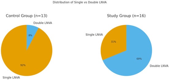
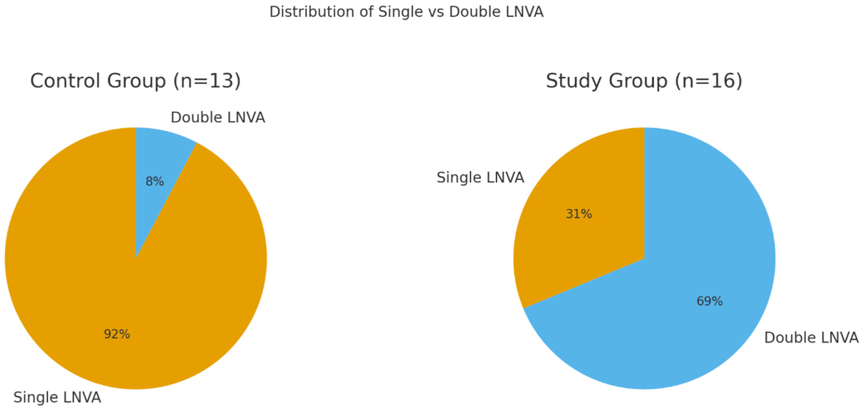
Double LNVA was uncommon in the control group, performed in only 1 case (8%), but frequent in the study group, performed in 11 of 16 cases (69%) (Figure 1). This distribution was considered in interpreting operative time and TITS, since double LNVA requires performance of two anastomoses, and identification and preparation of two nodes and two veins.
Figure 1.
Distribution of single versus double lymph node-to-vein anastomosis (LNVA) in control and study groups. Double LNVA was rare in the control group (1 of 13 cases, 8%) but common in the study group using SLA (11 of 16 cases, 69%), reflecting the improved efficiency and predictability of stereolithography-assisted planning.
2.1. Operative Time
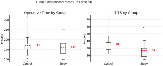
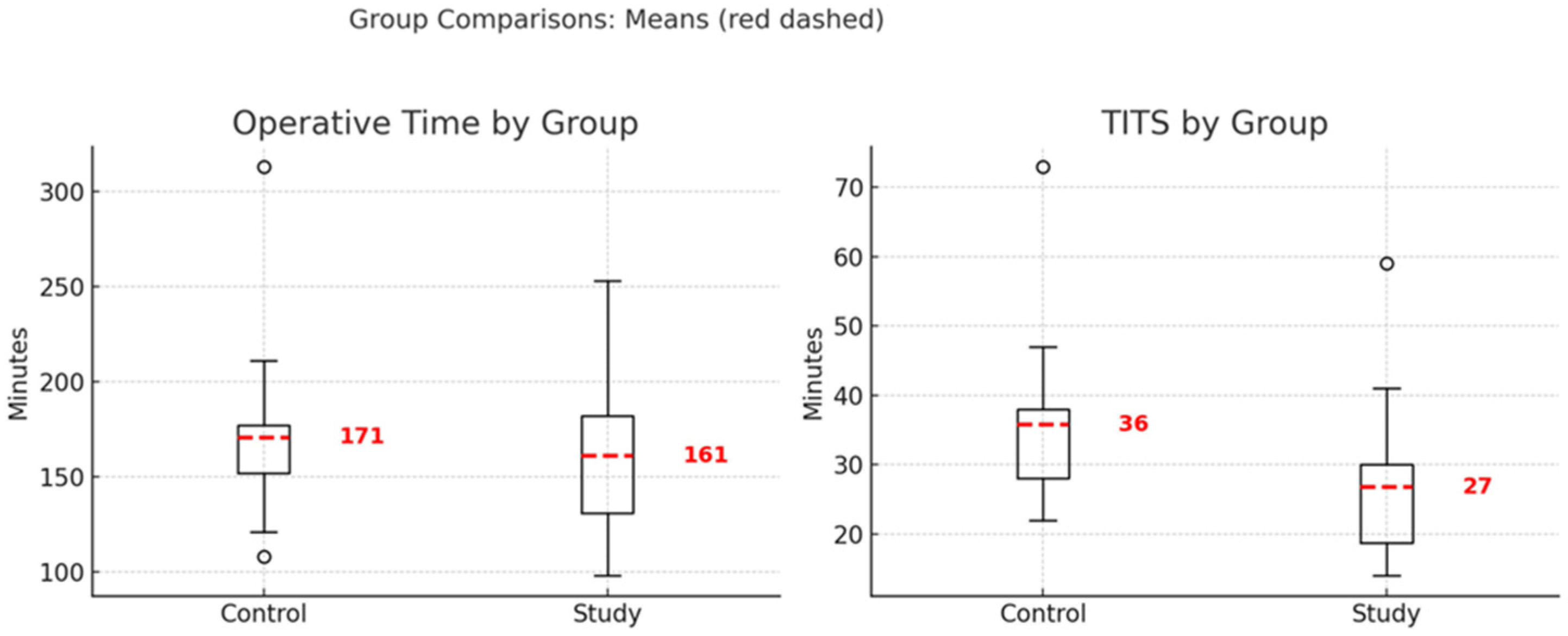
The mean operative time was 171 min (median 157, range 108–313) in the control group and 161 min (median 163, range 98–253) in the study group (Figure 2). This difference was not statistically significant (t-test p = 0.57, Mann–Whitney p = 0.79). Operative times were more tightly clustered in the study group (range 155 min) than in the control group (range 205 min), indicating greater consistency. Notably, the study group achieved shorter mean operative time despite the majority of its cases involving double LNVA, compared with only a single double case in the control group.
Figure 2.
Comparison of operative time and time to identification of target structures (TITS) between control and the study group using SLA. Mean operative time was slightly shorter in the study group (161 vs. 171 min), while mean TITS was significantly reduced (27 vs. 36 min).
2.2. Time to Identification of Target Structures (TITS)
TITS was also shorter in the study group: mean 36 min (median 32, range 22–73) in the control group versus mean 27 min (median 24, range 14–59) in the study group. This reduction was statistically significant by Mann–Whitney testing (p = 0.021), with a concordant trend on the t-test (p = 0.062). The shorter TITS in the study group is particularly notable given the substantially greater dissection required to identify two nodes and two veins in many cases, compared with only one double case in the control group.
2.3. Single vs. Double LNVA in the Study Group
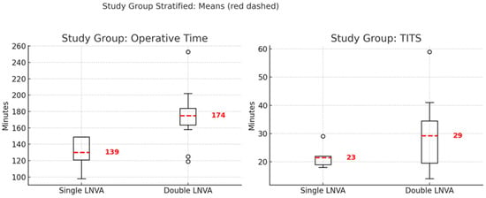
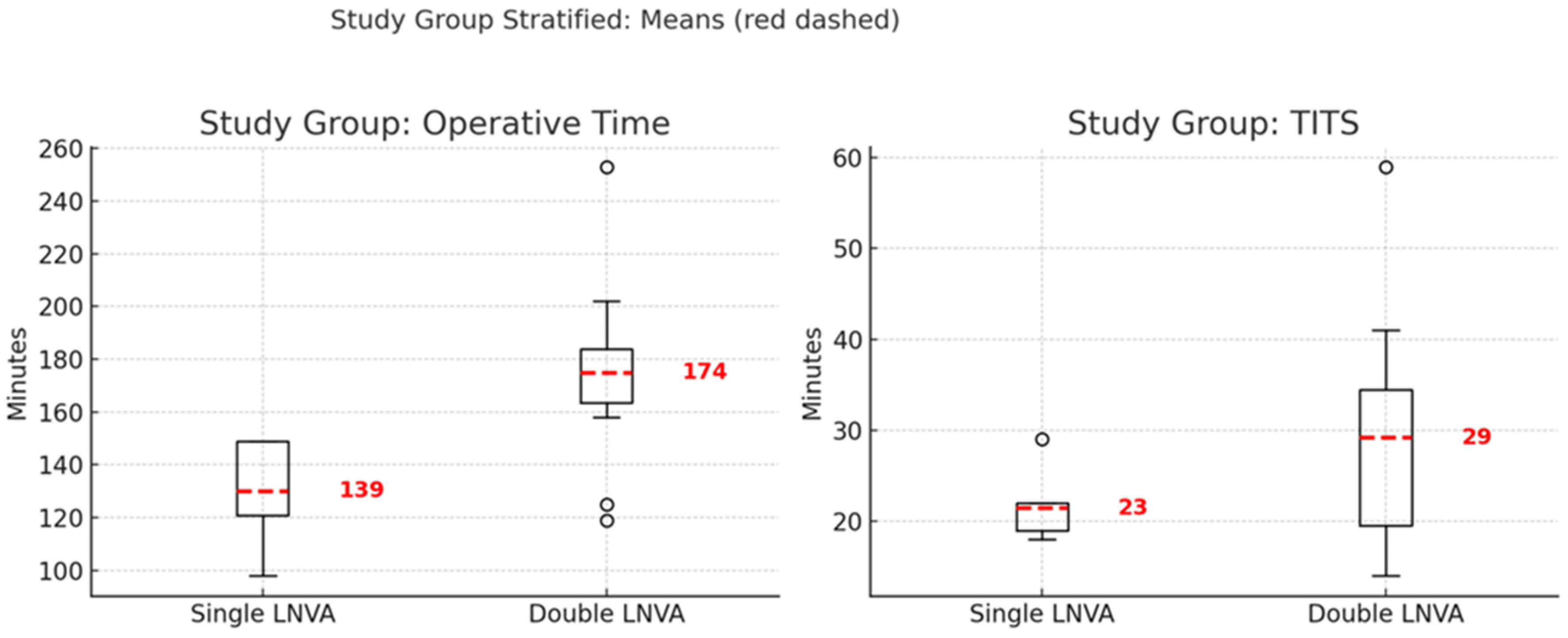
Within the study group, operative time for single versus double LNVA was 139 vs. 174 min (median 141 vs. 174; range 98–182 vs. 119–253; Welch t-test p = 0.053, Mann–Whitney p = 0.082), and TITS was 23 vs. 29 min (median 21 vs. 28; range 18–33 vs. 14–59; Welch t-test p = 0.294, Mann–Whitney p = 0.744). In contrast, the single control-group double LNVA required prolonged operative time (313 min) and TITS (73 min) (Figure 3).
Figure 3.
Comparison of operative time and time to identification of target structures (TITS) within the stereolithography (SLA) group, stratified by single and double lymph node-to-vein anastomosis (LNVA). Mean operative time was 139 min for single and 174 min for double LNVA, while TITS was 23 and 29 min, respectively. These findings indicate that SLA facilitated multiple anastomoses without proportionally increasing operative complexity or time.
2.4. Per-Anastomosis Analysis
When normalized per anastomosis, study-group double LNVA required less time per anastomosis than both single LNVA in the control group (operative time 158.8 vs. 87.5 min; TITS 32.7 vs. 14.6 min) and single LNVA in the study group (operative time 130.0 vs. 87.5 min; TITS 21.4 vs. 14.6 min). Thus, stereolithography facilitated completion of more anastomoses with greater per-anastomosis efficiency.
2.5. Surgeon-Perceived Operative Difficulty (SPOD)
SPOD was significantly lower in the study group compared with controls: mean 2.3 (median 2, range 1–3) versus 1.3 (median 1, range 1–3) (t-test p = 0.0002; Mann–Whitney p = 0.0005).
2.6. Surgical Success and Patient-Reported Outcomes
All procedures were technically successful (100% intraoperative patency confirmed by dye washout and/or ICG lymphangiography [17]). At one month, patient-reported outcomes (PROOP) were uniformly favorable: all patients reported symptom relief, subjective improvement in overall well-being, and satisfaction with surgery.
2.7. Complications
No postoperative complications occurred in either group during the one-month follow-up period.
3. Discussion
Lymph node-to-vein anastomosis (LNVA) is a novel physiologic approach for fluid-predominant lymphedema. Like lymphaticovenular anastomosis (LVA) [18], it creates a direct lymphatic–venous bypass without donor site morbidity. Unlike LVA, LNVA is “pseudo-supermicrosurgical,” involving structures at the boundary between supermicrosurgery and microsurgery (~0.8 mm), which makes the procedure technically more accessible [19,20]. Physiologically, LNVA also offers broader drainage: whereas LVA diverts a single lymphatic channel, LNVA leverages an entire lymph node and its lymphosome, potentially providing greater efficacy [19,21].
Despite these advantages, LNVA has distinct technical hurdles. The most critical is identifying a functionally preserved, qualitatively superior lymph node preoperatively and then localizing it intraoperatively with a suitable recipient vein. Earlier approaches such as MRI, radionuclide lymphoscintigraphy, and gamma probe guidance often produced only a “field of beeps,” failing to distinguish optimal nodes and leading to prolonged dissections [22]. Reliable node/vein selection and efficient intraoperative localization therefore are LNVA’s Achilles’ heel.
We previously refined the approach by combining high- and ultra-high-frequency ultrasound for node selection with a radar reflector (SAVI SCOUT®, Merit Medical, South Jordan, UT, USA) for intraoperative targeting. This strategy improved node identification but left two challenges unresolved: the time-consuming search for a suitable vein and the inability to consistently identify more than one node. As a result, most procedures yielded a single anastomosis, and double LNVA remained rare and incidental.
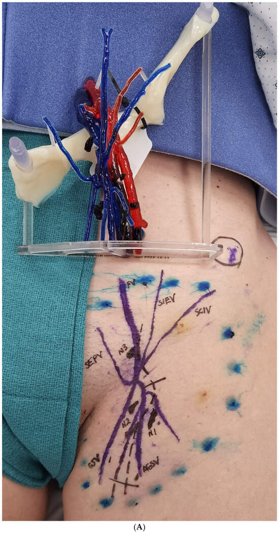
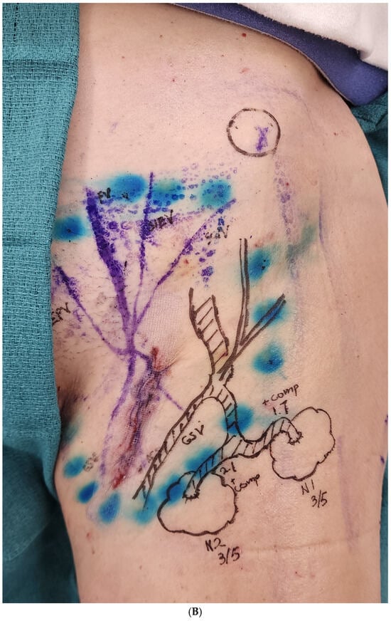
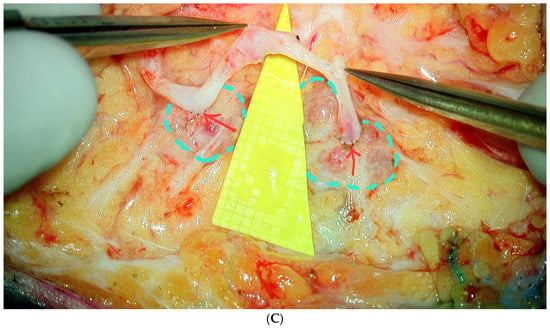
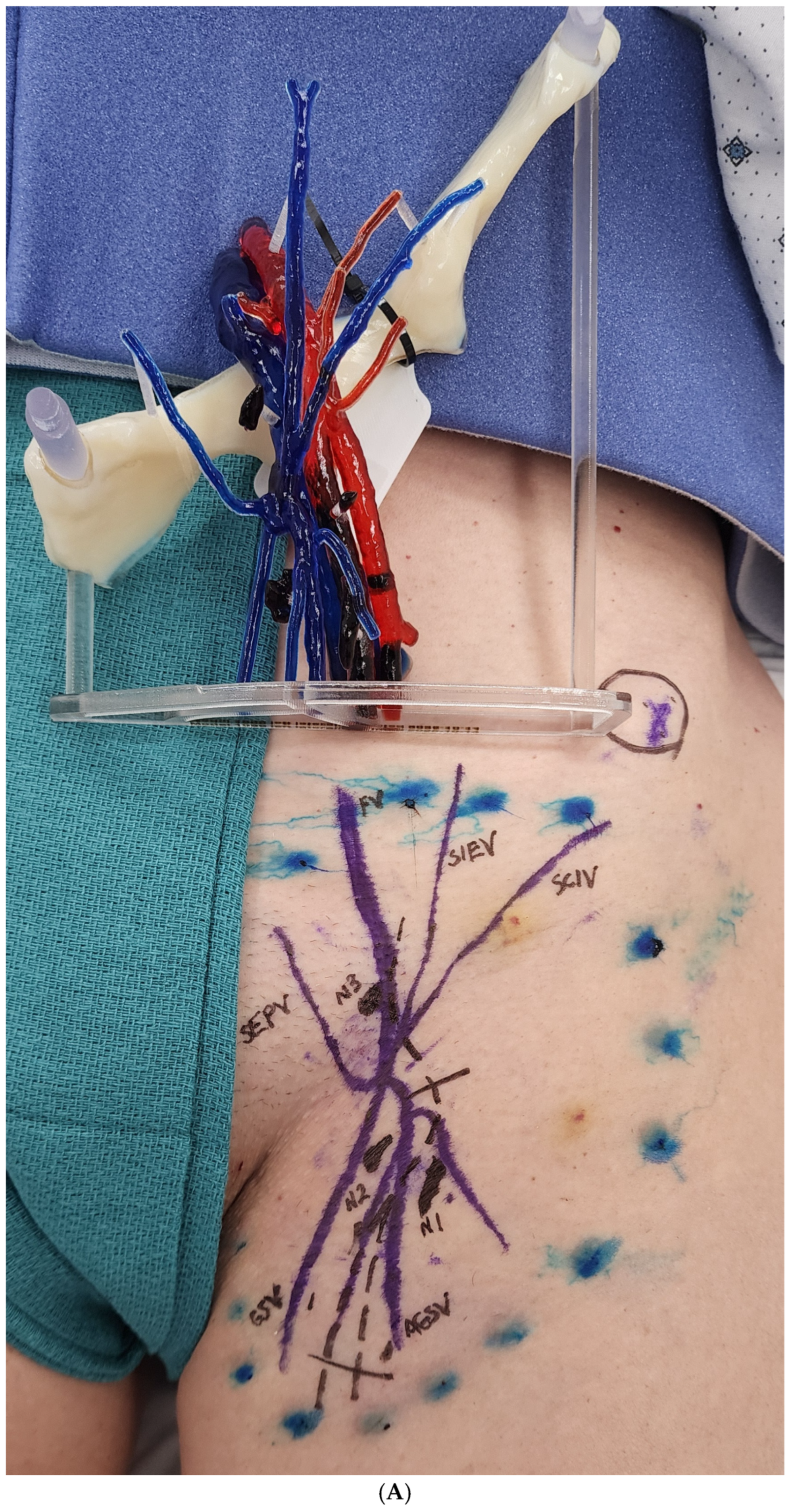
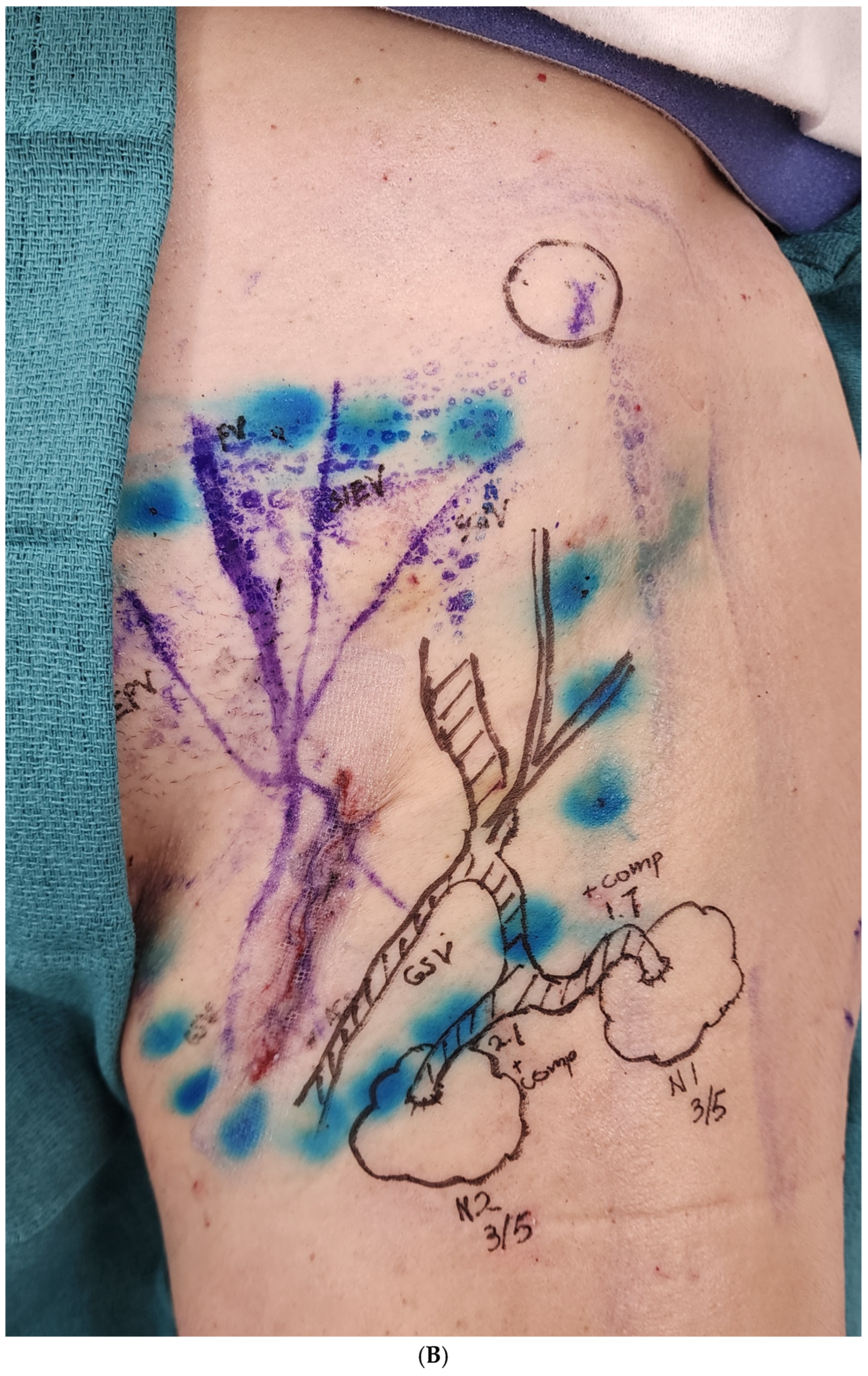
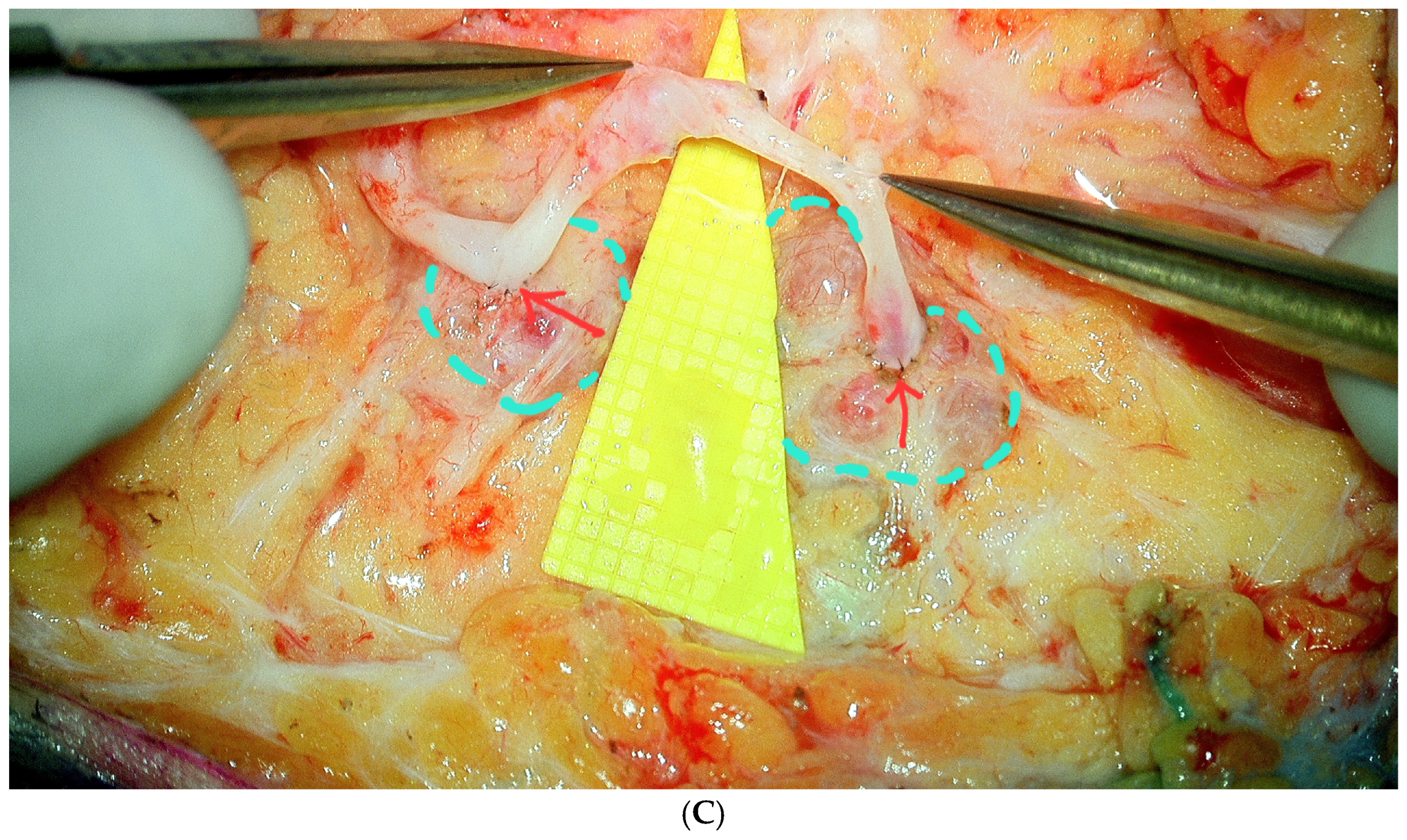
The introduction of three-dimensional stereolithography (SLA) markedly altered this workflow. SLA produced tangible anatomical models that guided incision planning, facilitated predictable identification of both nodes and veins, and standardized surgical execution [23,24]. This translated into shorter time to identification of target structures (TITS), narrower variability across cases, and more consistent operative performance. Importantly, SLA expanded LNVA beyond its original single-anastomosis concept. With spatial relationships defined, additional nodes and veins could be deliberately and quickly targeted, enabling double LNVA in two-thirds of SLA cases (Figure 4A–C). Unlike the one control-group double LNVA, which required prolonged operative time (313 min) and TITS (73 min), SLA-enabled double LNVA did not significantly prolong either measure, despite doubling the amount of operative load.
Figure 4.
(A–C) Three-dimensional stereolithography (SLA) provided surgeons with a clear three-dimensional understanding of nodal and vascular anatomy, allowing efficient identification of all relevant structures. Once the first node was identified, adjacent anatomy was rapidly recognized, enabling precise planning and execution of double lymph node-to-vein anastomoses (LNVA). In (A,B), two nodes (N1 and N2) were anastomosed using a bifurcated accessory great saphenous vein. (C) demonstrates another case of double LNVA achieved with a single vein, combining one end-to-node and one side-to-node anastomosis. (A) Patient-specific three-dimensional stereolithographic (SLA) model positioned over the patient’s groin to project deep femoral/inguinal vascular and nodal anatomy onto the skin surface. The 1:1 model reproduces the femoral artery and vein with branch vessels (red = arterial, blue = venous) and target lymph nodes, enabling precise external marking of nodes (N1, N2, N3) and recipient veins (e.g., SIEV, SCIV, SEPV, GSV, AGSV) for planned double LNVA and trocar/incision placement. (B) Intraoperative skin marking after transfer of the three-dimensional stereolithographic (SLA) model projections. Vascular pathways (including the great saphenous vein and superficial branches) and targeted lymph nodes (N1, N2) are outlined on the skin, with adjacent dye-marked lymphatic basins visible. This surface roadmap confirms the relationship between the planned recipient vein and both nodes, enabling execution of double LNVA (end-to-node and side-to-node) through a limited approach while avoiding non-target structures. (C) Intraoperative view demonstrating successful double lymph node-to-vein anastomoses (LNVA) within the mapped field. Two target lymph nodes (dashed circles) are exposed adjacent to the planned venous recipient, and a single bifurcated vein segment is mobilized to allow sequential anastomosis to each node (red arrows). Use of the preplanned SLA-based surface roadmap enabled rapid identification of both nodes and safe creation of two LNVA sites through a limited dissection corridor.
Together, these findings show that SLA simplifies intraoperative navigation, improves reproducibility, and facilitates multiple anastomoses without additional operative burden. By reducing variability, SLA not only enhances consistency for experienced surgeons but may also accelerate skill acquisition for trainees and expand LNVA beyond specialized centers. By providing surgeons with a reproducible ‘roadmap,’ SLA may also shorten the learning curve and support training in physiologic lymphedema surgery.
The production of each stereolithographic LNVA model required approximately eight hours of total effort, including segmentation, digital design, and post-processing. Material costs averaged $270 per model, ranging from about $130 with the smaller printer setup to $290 with the full polyjet system. In the context of a complex robotic supermicrosurgical procedure, this expense is modest—well justified by the improved efficiency, precision, and ease of intraoperative navigation the model provides. Lower-cost alternatives using single-material fused-filament or basic stereolithography printers can further reduce cost while still delivering clinically useful anatomic visualization.
This study was not designed to evaluate long-term outcomes. The focus was technical—demonstrating SLA’s effect on efficiency, reproducibility, and feasibility of LNVA. Questions of durability and comparative efficacy against other physiologic techniques such as LVA or vascularized lymph node transfer remain important but require dedicated investigation.
Limitations include the retrospective design, modest cohort size, and short-term follow-up, reflecting the early adoption phase of SLA. In addition, all cases were performed at a single high-volume supermicrosurgical center, which may introduce selection bias. While these factors restrict conclusions regarding long-term outcomes or generalizability, they do not alter the central finding: stereolithography streamlines LNVA, enhances reproducibility, and enables multiple anastomoses without significantly prolonging operative metrics.
4. Materials and Methods
4.1. Study Design and Patient Selection
This was a retrospective comparative study conducted under approval from the Cleveland Clinic Institutional Review Board (IRB) and in accordance with the Declaration of Helsinki. All diagnoses were confirmed with indocyanine green lymphography [25,26,27]. The fluid-predominance nature was established by a combination of: (1) patient-reported reversibility of swelling, (2) physical examination confirming absence of fibrotic lipodystrophy, and (3) bioimpedance spectroscopy demonstrating body composition patterns consistent with fluid-predominant disease [28]. Consecutive patients who underwent robot-assisted lymph node-to-vein anastomosis (LNVA) between October 2024 and August 2025 were included and stratified into two cohorts: those treated prior to the adoption of three-dimensional (3D) stereolithography (control group) and those treated with stereolithography-assisted preoperative planning and intraoperative navigation (study group). Only patients with at least one month of postoperative follow-up were included. There was no recruitment gap between cohorts, and apart from the use of stereolithography in the study group, all surgical workflows including imaging, patient preparation, and robotic execution, were identical. All procedures were performed by the corresponding author.
4.2. Control Group: Standard Robot-Assisted LNVA
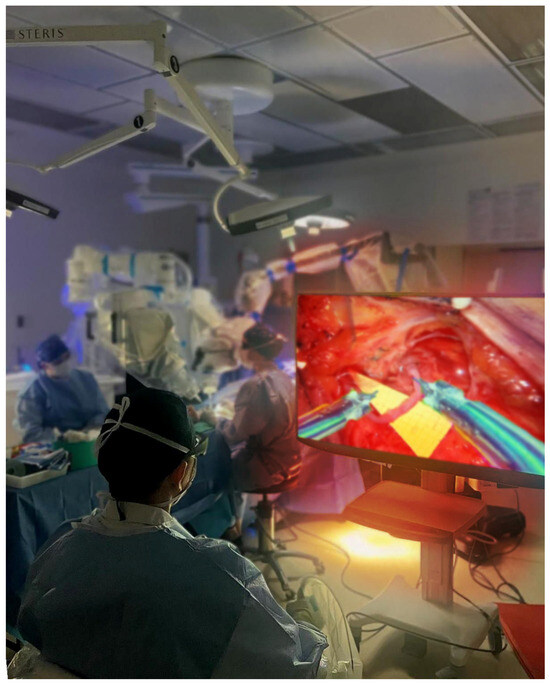
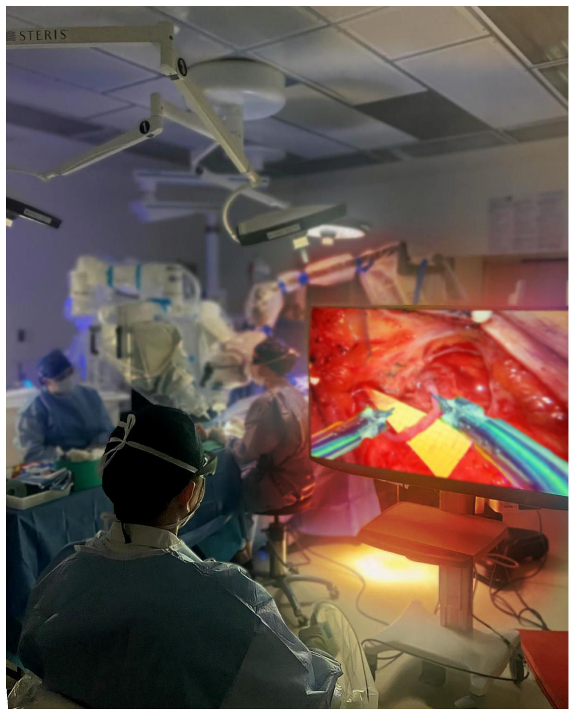
Procedures followed our previously published robotic LNVA workflow [19]. Briefly, all patients underwent preoperative high-frequency and ultra-high-frequency duplex ultrasound to identify and select functioning lymph nodes and appropriate recipient veins [6,7]. The optimal lymph node was marked with a radar reflector (SAVI SCOUT®, Merit Medical, South Jordan, UT, USA) placed under ultrasound guidance. At surgery, incision was strategically placed directly over the targeted lymph node and adjacent vein. Following meticulous preservation of afferent/efferent lymphatic channels and hilar vasculature, a small anterior capsulotomy was created in the lymph node, and an end-to-node or side-to-node LNVA was performed using 10-0 or 11-0 nylon sutures with 90- or 130-micron needles under robotic assistance (Figure 5). (Symani® Surgical System, MMI, Jacksonville, FL, USA).
Figure 5.
Intraoperative view of the robotic lymphatic surgery setup. The primary surgeon is seated at the robotic console (posterior view), observing the operative field on a high-definition monitor displaying the lymph node-to-vein anastomosis (LNVA). The robotic patient cart and assistant team are positioned at the bedside in the background within the operating room.
Anastomotic patency was confirmed intraoperatively by direct washout or indocyanine green (ICG) transit. A second LNVA was performed when feasible, specifically when (1) both a second suitable lymph node and an appropriate recipient vein were encountered in the dissection field, and (2) preparation of these structures could be completed without causing additional lymphatic injuries. This criterion applied equally to both the control and study groups.
4.3. Study Group: Standard Robot-Assisted LNVA with 3D Stereolithography (SLA)
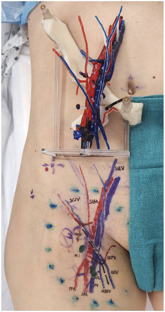
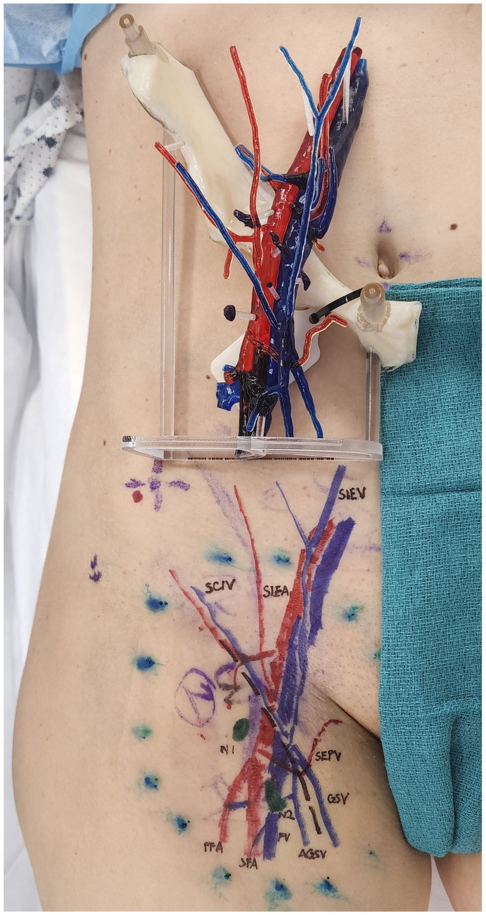
The study group underwent the same standard robotic LNVA workflow described above, with the addition of 3D stereolithography (SLA) for preoperative planning and intraoperative navigation. Patients underwent contrast-enhanced multi-detector computed tomography (MDCT) (320 detector rows, 0.25 mm slice thickness), with both arterial and venous phases. Data were exported in DICOM format and imported into dedicated segmentation software, version 28.0 (Materialise Mimics, Leuven, Belgium). Using thresholding based on Hounsfield units, lymph nodes, arteries, veins, and adjacent anatomic landmarks including bones and skin were segmented. Segmented structures were converted into stereolithographic (STL) files and printed as 1:1 scale resin models. The primary model incorporated lymph nodes and major vessels, while a transparent skin layer was printed separately and assembled for realistic spatial reference (Figure 6).
Figure 6.
Patient-specific three-dimensional stereolithographic (SLA) model and overlying skin template used for preoperative incision planning in robotic lymph node-to-vein anastomosis (LNVA). The 1:1 model reproduces the patient’s arterial (red), venous (blue), and nodal (green) anatomy segmented from contrast-enhanced CT. A transparent/acrylic skin plate permits direct surface projection of the deep vascular and nodal targets onto the patient, facilitating accurate incision placement, trocar positioning, and targeted lymph node dissection.
The model was brought into the operating room for preoperative incision planning. Intraoperatively, the model served as a visual reference outside the sterile field to maintain spatial orientation, analogous to image-guided navigation. Surgeons used the model in conjunction with the SAVI SCOUT® probe to guide skin incision, optimize dissection trajectory, and confirm anatomic orientation.
4.4. Outcome Measures
The following outcomes were recorded and compared between groups:
- Time to identification of target structures (TITS)—measured from skin incision to exposure of both targeted node and recipient vein.
- Operative time—total skin-to-skin time.
- Surgical success—intraoperative patency confirmed by washout sign and/or ICG transit.
- Surgeon-perceived operative difficulty (SPOD)—rated on a three-point scale (1 = low, 2 = intermediate, 3 = high).
- Patient-reported outcome at one postoperative month (PROOP)—assessed with two questions: (1) Are your lymphedema symptoms relieved? (2) Are you happy with your surgery?
- Complications—all postoperative complications occurring within the first month after surgery were tracked.
5. Conclusions
Three-dimensional stereolithography improves LNVA by reducing variability, shortening identification time, and enabling multiple anastomoses without significantly increasing operative duration. By making LNVA more reproducible and scalable, SLA supports broader clinical adoption and provides a foundation for future outcome-focused studies.
Author Contributions
Conceptualization, W.F.C. and Y.M.; methodology, W.F.C.; software, R.K. and Y.A.; validation, R.K., Y.A., W.F.C., Y.M. and E.T.C.; formal analysis, Y.M.; investigation, Y.M.; resources, D.C.F.C. and E.T.C.; data curation, Y.M., E.T.C., D.C.F.C. and E.K.; writing—original draft preparation, W.F.C. and Y.M.; writing—review and editing, Y.M.; visualization, R.K. and Y.A.; supervision, W.F.C.; project administration, Y.M.; funding acquisition, not applicable. All authors have read and agreed to the published version of the manuscript.
Funding
This research received no external funding.
Institutional Review Board Statement
The study was conducted in accordance with the Declaration of Helsinki, and approved by the Institutional Review Board of the Integrated Surgical Institute, (Code: 16-306, Date: 31 March 2026), for studies involving humans.
Informed Consent Statement
Informed consent was obtained from all subjects involved in the study.
Data Availability Statement
The original contributions presented in this study are included in the article. Further inquiries can be directed to the corresponding author.
Conflicts of Interest
The authors declare no conflicts of interest.
Abbreviations
The following abbreviations are used in this manuscript:
| 3D | Three-dimensional |
| CT | Computed tomography |
| DICOM | Digital Imaging and Communications in Medicine |
| ICG | Indocyanine green |
| IRB | Institutional Review Board |
| LNVA | Lymph node-to-vein anastomosis |
| LVA | Lymphaticovenular anastomosis |
| MDCT | Multi-detector computed tomography |
| MMI | Medical Microinstruments (manufacturer of the Symani® system) |
| MRI | Magnetic resonance imaging |
| PROOP | Patient-reported outcome at one postoperative month |
| SAVI SCOUT | Radar-reflector localization system for intraoperative targeting |
| SLA | (Three-dimensional) stereolithography |
| SPOD | Surgeon-perceived operative difficulty |
| STL | Stereolithography file format |
| TITS | Time to identification of target structures |
| VLNT | Vascularized lymph node transfer |
| VLVT | Vascularized lymph vessel transfer |
| N1 | First targeted lymph node |
| N2 | Second targeted lymph node |
| GSV | Great saphenous vein |
| AGSV | Accessory great saphenous vein |
| SIEV | Superficial inferior epigastric vein |
| SCIV | Superficial circumflex iliac vein |
| SEPV | Superficial external pudendal vein |
References
- Becker, C.; Hidden, G. Transfer of free lymphatic flaps. Microsurgery and anatomical study. J. Mal. Vasc. 1988, 13, 119–122. [Google Scholar]
- Chen, H.C.; O’Brien, B.M.; Rogers, I.W.; Pribaz, J.J.; Eaton, C. Lymph node transfer for the treatment of obstructive lymphoedema in the canine model. Br. J. Plast. Surg. 1990, 43, 578–586. [Google Scholar] [CrossRef] [Scilit] [PubMed]
- O’Brien, B.M.; Hickey, M.J.; Hurley, J.V.; Dvir, E.; Khazanchi, R.K.; Chris Pederson, W.; Pribaz, J.J. Microsurgical transfer of the greater omentum in the treatment of canine obstructive lymphoedema. Br. J. Plast. Surg. 1990, 43, 440–446. [Google Scholar] [CrossRef] [Scilit]
- Chen, W.F.; McNurlen, M.; Ding, J.; Bowen, M. Vascularized Lymph Vessel Transfer for Extremity Lymphedema—Is Transfer of Lymph Node Still Necessary? Int. Microsurg. J. 2019, 3, 1. [Google Scholar] [CrossRef] [Scilit]
- Narushima, M.; Mihara, M.; Yamamoto, T.; Hara, H.; Ohshima, A.; Kikuchi, K.; Todokoro, K.; Seki, Y.; Iida, T.; Nakagawa, M.; et al. Lymphadiposal Flaps and Lymphaticovenular Anastomoses for Severe Leg Edema: Functional Reconstruction for Lymph Drainage System. J. Reconstr. Microsurg. 2015, 32, 050–055. [Google Scholar] [CrossRef] [Scilit] [PubMed]
- Orfahli, L.M.; Fahradyan, V.; Chen, W.F. Vascularized lymph vessel transplant (VLVT): Our experience and lymphedema treatment algorithm. Ann. Breast Surg. 2022, 6, 8. [Google Scholar] [CrossRef] [Scilit]
- Pandey, S.K.; Fahradyan, V.; Orfahli, L.M.; Chen, W.F. Supermicrosurgical lymphaticovenular anastomosis vs. vascularized lymph vessel transplant—Technical optimization and when to perform which. Plast. Aesthetic Res. 2022, 8, 47. [Google Scholar] [CrossRef] [Scilit]
- Chen, W.F.; Tuncer, F.B.; Zeng, W. Advanced Technical Pearls for Successful Supermicrosurgical Lymphaticovenular Anastomosis. Ann. Plast. Surg. 2021, 86, S165–S172. [Google Scholar] [CrossRef] [Scilit]
- Yamada, Y. The Studies on Lymphatic Venous Anastomosis in Lvmphedema. Nagoya J. Med. Sci. 1969, 32, 1–21. [Google Scholar]
- Gilbert, A.; O’Brien, B.M.; Vorrath, J.W.; Sykes, P.J. Lymphaticovenous anastomosis by microvascular technique. Br. J. Plast. Surg. 1976, 29, 355–360. [Google Scholar] [CrossRef] [Scilit]
- O’brien, B.M.; Sykes, P.J.; Threlfall, G.N.; Browning, F.S.C. Microlymphaticovenous anastomoses for obstructive lymphedema. Plast. Reconstr. Surg. 1977, 60, 197–211. [Google Scholar] [CrossRef] [Scilit]
- Koshima, I.; Nanba, Y.; Tsutsui, T.; Takahashi, Y.; Itoh, S. Long-term follow-up after lymphaticovenular anastomosis for lymphedema in the leg. J. Reconstr. Microsurg. 2003, 19, 209–215. [Google Scholar] [CrossRef] [Scilit]
- Koshima, I.; Nanba, Y.; Tsutsui, T.; Takahashi, Y.; Itoh, S.; Fujitsu, M. Minimal invasive lymphaticovenular anastomosis under local anesthesia for leg lymphedema: Is it effective for stage III and IV? Ann. Plast. Surg. 2004, 53, 261–266. [Google Scholar] [CrossRef] [Scilit] [PubMed]
- Bailey, E.A.; Pandey, S.K.; Chen, W.F. Advances in Surgical Lymphedema Management: The Emergence and Refinement of Lymph Node-to-Vein Anastomosis (LNVA). Curr. Surg. Rep. 2024, 12, 83–88. [Google Scholar] [CrossRef] [Scilit]
- Pak, C.S.; Suh, H.P.; Kwon, J.G.; Cho, M.J.; Hong, J.P. Lymph Node to Vein Anastomosis (LNVA) for lower extremity lymphedema. J. Plast. Reconstr. Aesthetic Surg. 2021, 74, 2059–2067. [Google Scholar] [CrossRef] [Scilit]
- Gousopoulos, E.; Grünherz, L.; Giovanoli, P.; Lindenblatt, N. Robotic-assisted microsurgery for lymphedema treatment. Plast. Aesthetic Res. 2023, 10, 7. [Google Scholar] [CrossRef] [Scilit]
- Zhao, H.; Yamamoto, T.; Hara, H.; Ding, J.; Chen, W. Indocyanine Green Lymphographic Evidence of Surgical Efficacy Following Microsurgical and Supermicrosurgical Lymphedema Reconstructions. J. Reconstr. Microsurg. 2016, 32, 688–698. [Google Scholar] [CrossRef] [Scilit]
- Chen, W.F. How to Get Started Performing Supermicrosurgical Lymphaticovenular Anastomosis to Treat Lymphedema. Ann. Plast. Surg. 2018, 81, S15–S20. [Google Scholar] [CrossRef] [Scilit]
- Chen, W.F.; Cheong, D.C.; Clemente, E.T.; Salman, M. Robot-Assisted Lymph Node-to-Vein Anastomosis: Lessons from the First 22 Cases at a High-Volume Lymphatic Supermicrosurgery Center. Curr. Oncol. 2025, 32, 377. [Google Scholar] [CrossRef] [Scilit] [PubMed]
- Chen, W.F. Supermicrosurgery for the Treatment of Lymphedema; Cleveland Clinic, 2020. Available online: https://consultqd.clevelandclinic.org/supermicrosurgery-for-the-treatment-of-lymphedema (accessed on 1 November 2025).
- Suami, H.; Scaglioni, M.F. Anatomy of the Lymphatic System and the Lymphosome Concept with Reference to Lymphedema. Semin. Plast. Surg. 2018, 32, 5–11. [Google Scholar] [CrossRef] [Scilit] [PubMed]
- Kukreja-Pandey, S.; Bailey, E.A.; Chen, W.F. D118. Inguinal Lymph Node to Vein Anastomosis—“The Cleveland Clinic Experience and Technical Refinements”. Plast. Reconstr. Surg. Glob. Open 2024, 12, 123–124. [Google Scholar] [CrossRef] [Scilit]
- Segaran, N.; Saini, G.; Mayer, J.L.; Naidu, S.; Patel, I.; Alzubaidi, S.; Oklu, R. Application of 3D Printing in Preoperative Planning. J. Clin. Med. 2021, 10, 917. [Google Scholar] [CrossRef] [Scilit]
- Shauly, O.; Ash, M.; Buclez, J.; Al-Nowaylati, A.-R. Future Applications for 3-dimensional Printed Virtual Surgical Planning in Microsurgery. Plast. Reconstr. Surg. Glob. Open 2025, 13, e7220. [Google Scholar] [CrossRef] [Scilit] [PubMed]
- Mihara, M.; Hara, H.; Araki, J.; Kikuchi, K.; Narushima, M.; Yamamoto, T.; Iida, T.; Yoshimatsu, H.; Murai, N.; Mitsui, K. Indocyanine Green (ICG) Lymphography Is Superior to Lymphoscintigraphy for Diagnostic Imaging of Early Lymphedema of the Upper Limbs. PLoS ONE 2012, 7, e38182–2. [Google Scholar] [CrossRef] [Scilit]
- Chen, W.F.; Pandey, S.K. Distal-to-proximal sequential ICG injection technique (DOPSIT) for lymphatic vessels mapping. Plast. Aesthetic Res. 2023, 10, 5. [Google Scholar] [CrossRef] [Scilit]
- Figueroa, B.A.; Lammers, J.D.; Al-Malak, M.; Pandey, S.; Chen, W.F. Lymphoscintigraphy versus Indocyanine Green Lymphography—Which Should Be the Gold Standard for Lymphedema Imaging? Lymphatics 2023, 1, 25–33. [Google Scholar] [CrossRef] [Scilit]
- Qin, E.S.; Bowen, M.J.; James, S.L.; Chen, W.F. Multi-segment bioimpedance can assess patients with bilateral lymphedema. J. Plast. Reconstr. Aesthetic Surg. 2020, 73, 328–336. [Google Scholar] [CrossRef] [Scilit] [PubMed]
Disclaimer/Publisher’s Note: The statements, opinions and data contained in all publications are solely those of the individual author(s) and contributor(s) and not of MDPI and/or the editor(s). MDPI and/or the editor(s) disclaim responsibility for any injury to people or property resulting from any ideas, methods, instructions or products referred to in the content. |
© 2026 by the authors. Licensee MDPI, Basel, Switzerland. This article is an open access article distributed under the terms and conditions of the Creative Commons Attribution (CC BY) license.