


Abstract
Secondary lymphedema is a chronic, progressive condition characterized by limb swelling that arises after disruption of lymphatic channels from trauma or iatrogenic injury. In patients with complex soft tissue defects, reconstruction intended to restore the limb may inadvertently disrupt lymphatic drainage, highlighting the need for lymphatic-conscious surgical approaches. We present a retrospective case series of three patients with lower extremity soft tissue injuries who underwent flap-based reconstruction at our institution between 2021 and 2024. Reconstructive techniques, intraoperative decision-making, and postoperative lymphatic outcomes were analyzed over a follow-up period of 11 to 38 months. In the first two cases, disruption of established or new lymphatics during flap inset or repeated flap re-elevations may have contributed to the development of lymphedema. By contrast, indocyanine green (ICG) lymphography-guided flap planning and inset in the third case preserved lymphatic drainage pathways which could potentially lead to the development of lymphedema. Post-traumatic flap-based extremity reconstruction requires careful attention to native and transferred lymphatics to decrease the risk of secondary lymphedema. These cases draw attention to the concepts of preserving lymphatics, maintaining lymphatic axiality, and using ICG lymphography when planning flap-based reconstruction.
1. Introduction
Lymphedema is a chronic, progressive condition marked by limb swelling, chronic pain, and significant functional impairment. Most lymphedema cases are acquired and result from the disruption of the lymphatic system, most commonly after cancer-related surgery [1]. Post-traumatic lymphedema (PTL) is a type of secondary lymphedema characterized by chronic swelling that arises after musculoskeletal or soft tissue injury from traumatic or iatrogenic insult following trauma [2,3]. It is a relatively understudied condition with an incidence estimated broadly between 20% and 50% [4].
Although the pathophysiology of PTL has not been fully elucidated, several mechanisms have been proposed. PTL often arises after disruption of the superficial lymphatics network and is exacerbated by a protracted inflammatory response triggered by soft tissue and/or bone injury [5,6]. Such an injury can impair lymphangiogenesis which relies on surrounding healthy tissue for effective regeneration [6]. Tissue loss or scar formation during wound healing may create a physical barrier to lymphatic reconstitution by increasing the distance between disrupted lymphatic channels [5]. Even in the absence of an open injury, significant tissue damage, and dermal backflow on imaging, closed fractures can cause persistent edema through a protracted inflammatory response characterized by increased lymphatic flow from lymphatic vessel dilation [7].
Transient physiologic swelling due to acute inflammation is expected for up to 3 months after injury, and the initial lymphatic response to injury is hyperdynamic drainage of the extremity [7]. As with post-cancer lymphedema, however, prolonged high working pressures eventually result in lymphatic vessel hypertrophy and ultimately leakage and sclerosis [4]. If left undiagnosed and untreated, patients may suffer chronic and progressive sequelae, including trophic skin changes, delayed wound healing, and recurrent cellulitis, which significantly lower their functional and psychosocial wellbeing [4].
Unfortunately, reconstructive surgery techniques that restore form and function after trauma may also inadvertently cause iatrogenic lymphatic damage and worsen the risk of lymphedema. The use of skin grafts, the approach to flap inset and the need for repeated flap elevations may each contribute to the development of lymphedema if not considered carefully, making it difficult to gauge the relative contribution of any single factor. Given the importance of maintaining lymphatic integrity in the treatment and prevention of PTL, reconstructive surgeons should prioritize surgical decision-making and planning that respects the lymphatic pathways that restore lymphatic flow to prevent additional postoperative impairment. Understanding this balance is crucial when operating on patients with traumatic injuries who are at an increased risk for lymphatic compromise.
In this case series, we highlight unsuccessful and successful management of lymphatic drainage after lower extremity soft tissue reconstruction. Given that secondary lymphedema after traumatic or iatrogenic injury is difficult to treat, the aim of this study is to emphasize prevention: “first, do no harm”.
2. Detailed Case Description
The three patients selected for this case series had a history of lower extremity soft tissue injury either from infection and/or trauma. All patients received surgical reconstruction by plastic surgeons at our institution between the years of 2021 to 2024. They were followed postoperatively by the senior author (P.S.B.), who evaluated them for lower extremity lymphedema. Patients were diagnosed with post-traumatic lymphedema if they exhibited persistent limb swelling at least six months following their most recent injury or surgical intervention, along with objective evidence of lymphatic dysfunction on imaging. Indocyanine green (ICG) lymphography was performed by injecting 0.2 mL of 2.5 mg/mL ICG into the 1st and 4th web spaces of the foot and lateral heel after cleansing the injection sites and anesthetizing with 1% lidocaine. Patients with lymphoscintigraphy demonstrating dermal backflow or indocyanine green (ICG) lymphography showing dermal backflow, stardust, or splash patterns were diagnosed with PTL [6]. Limb circumference measurements were obtained, and volumes were calculated using the truncated cone formula [8]. Patients were followed for 11 to 38 months after reconstructive surgery.
2.1. Case 1
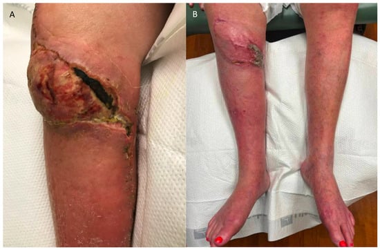
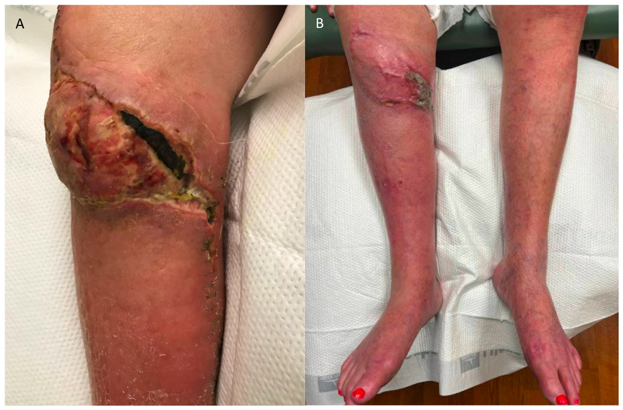
Patient 1 was a 62-year-old woman with body mass index (BMI) 33, and a history of rheumatoid arthritis and right total knee arthroplasty complicated by a postoperative soft-tissue infection requiring debridement and coverage of the distal knee. She underwent reconstruction with a pedicled right medial gastrocnemius flap and split-thickness skin graft (Figure 1A,B). During flap elevation, the proximal and distal wound edges were raised as fasciocutaneous flaps to create a pathway to accommodate the rotated gastrocnemius muscle. A split-thickness skin graft was then placed over the entire length of the rotated muscle flap and interposed between the elevated skin flaps. Care was taken to preserve the saphenous vein and the sural nerve, which were skeletonized during flap dissection. However, an attempt to visualize and preserve the surrounding lymphatic channels was not made.
Figure 1.
(A) Patient 1 postoperative photo taken 3 weeks after right pedicled gastrocnemius flap and split-thickness skin graft demonstrating wound edge necrosis and partial dehiscence of the pedicled gastrocnemius flap. The patient’s immediate postoperative course was complicated by cellulitis. (B) Postoperative photo at 3 months showing healing of right pedicled gastrocnemius flap.
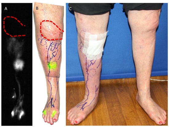
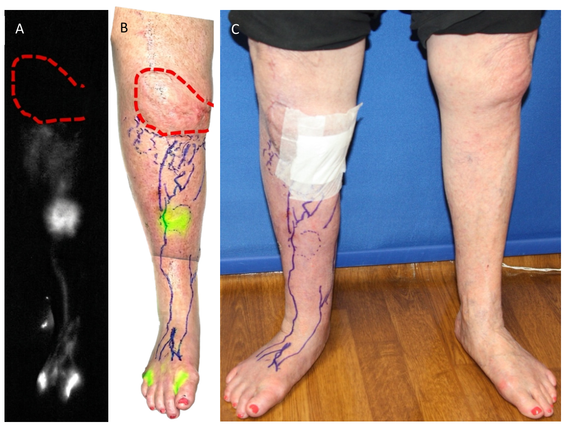
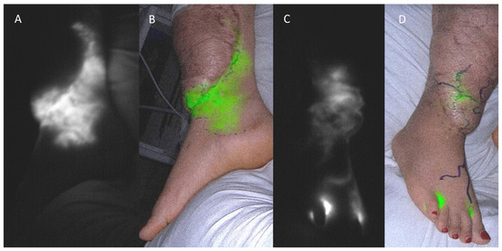
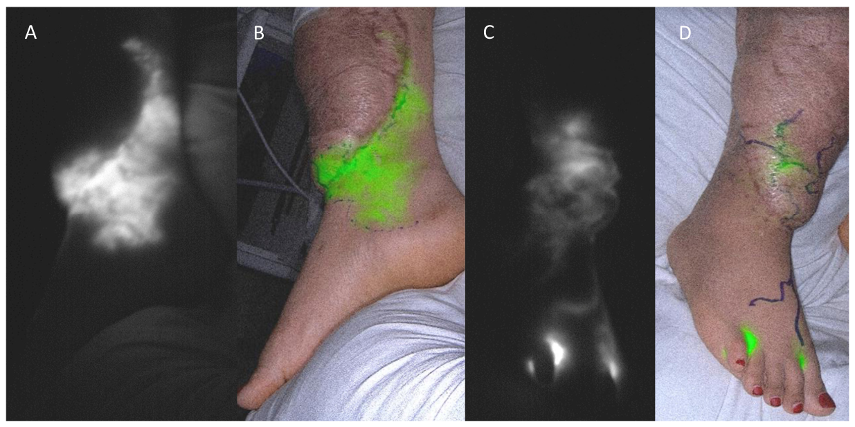
The patient returned four months later with persistent right lower extremity pain and swelling. Lower extremity venous duplex ultrasound showed patent veins without evidence of venous reflux or thrombus. Lymphoscintigraphy revealed dermal backflow in the proximal lower leg. Indocyanine green (ICG) lymphography demonstrated four lymphatic channels which terminated in patches of dermal backflow immediately distal to the skin-grafted gastrocnemius flap (Figure 2A–C). No channels traversed the skin-grafted area of the muscle flap, and no reconstitution of drainage was observed proximal to the flap. Based on these findings and her clinical presentation, she was diagnosed with International Society of Lymphology (ISL) stage I. Excess limb volume compared with the normal contralateral limb was 14%. Given her persistent symptoms, she was deemed a surgical candidate and underwent lymphovenous bypass (LVB) at two sites. Despite preoperative compliance, she did not adhere to a compression regimen postoperatively and symptoms persisted. Lymphedema Life Impact Scale (LLIS) scores ranged from 37 to 57, reflecting severe symptom burden. She was lost to follow-up approximately two months after LVB.
Figure 2.
(A) Patient 1 indocyanine green (ICG) lymphography of the right lower extremity demonstrating dermal backflow distal to the skin-grafted gastrocnemius flap (outlined in red dotted line) in fluorescent mode. (B) ICG lymphography of the right lower extremity in overlay mode. The green signal immediately distal to the flap highlights an area of dermal backflow. (C) Patient 1 demonstrating erythema and swelling in the right lower extremity compared to the left lower extremity when standing.
2.2. Case 2
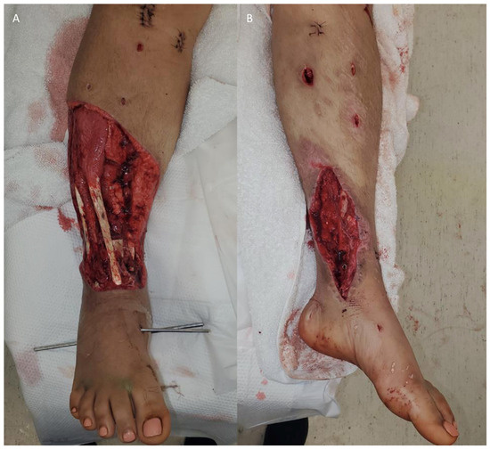
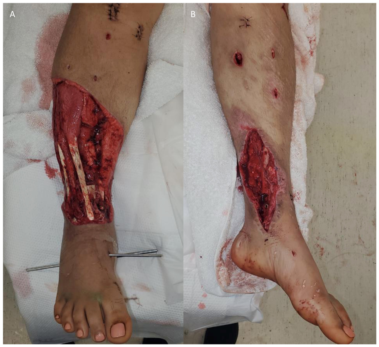
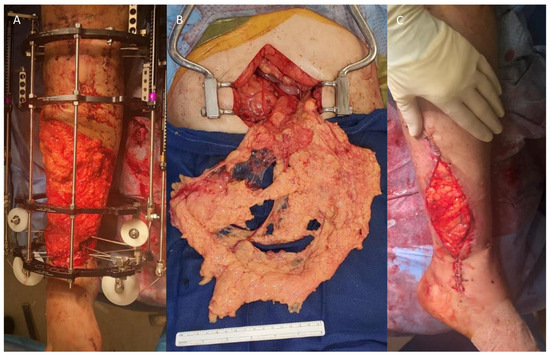
Patient 2 was a 31-year-old woman (BMI 30) who sustained a right open, comminuted tibia fracture with a near circumferential degloving over the defect and a left extra-articular distal tibia fracture after a motor vehicle collision. She initially underwent bilateral open reduction and internal fixations in Central America. Her postoperative course was complicated by bilateral lower-extremity hardware infections requiring overlying soft tissue debridement, hardware removal, placement of antibiotic beads, and external fixation at our institution. The resulting defect was a near-circumferential soft tissue wound, 24 × 28 cm, over a 12 cm segmental right tibial defect (Figure 3A,B). The posterior tibial vessels were patent, but anterior tibial and peroneal vessels were obstructed within the wound.
Figure 3.
(A) Patient 2 intraoperative photo of open, comminuted right tibial fracture with large tibial defect. (B) Left lower extremity wound with infected hardware visible at the wound base.
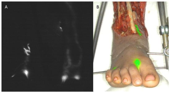
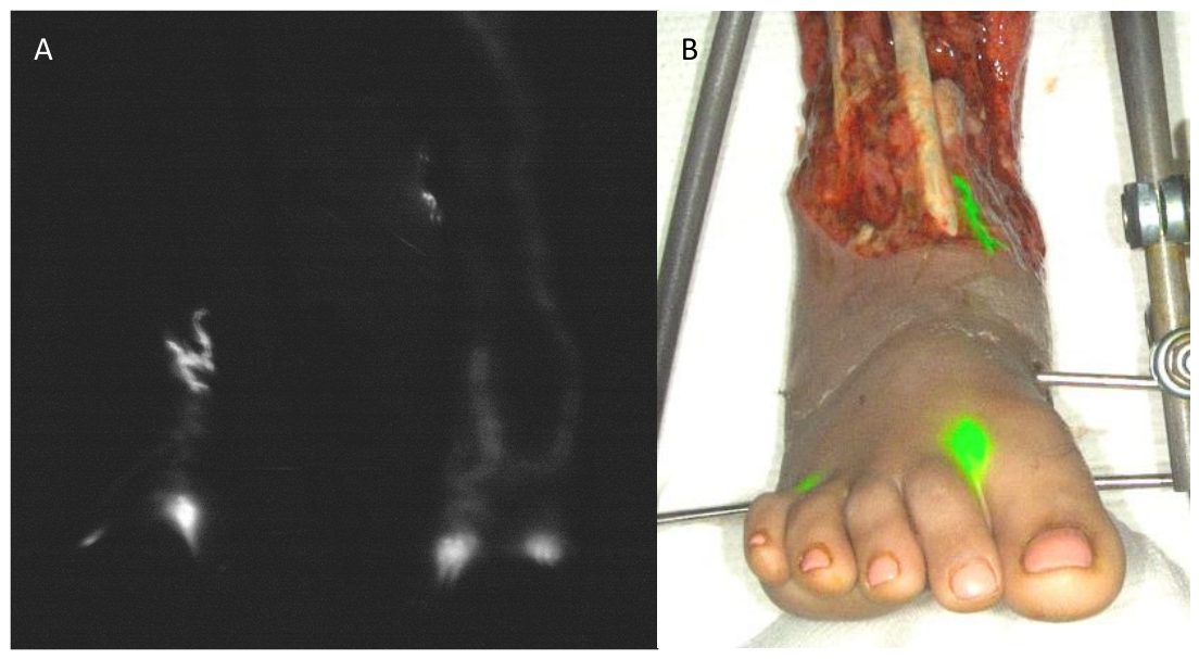
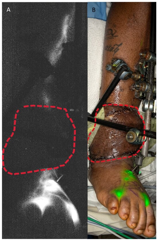
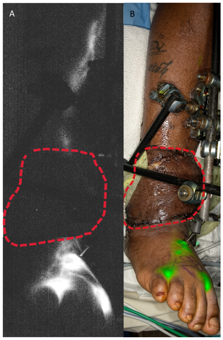
ICG lymphography demonstrated normal lymphatic drainage in the left lower extremity. In the right lower extremity, there was leakage of lymphatic fluid into the open wound at the dorsal ankle, and no reconstitution of ICG flow proximal to the soft tissue defect (Figure 4).
Figure 4.
Patient 2 indocyanine green (ICG) lymphography in (A) fluorescent mode and (B) overlay mode demonstrating lymphatic leakage (in green) into the dorsal ankle wound of the right lower extremity.
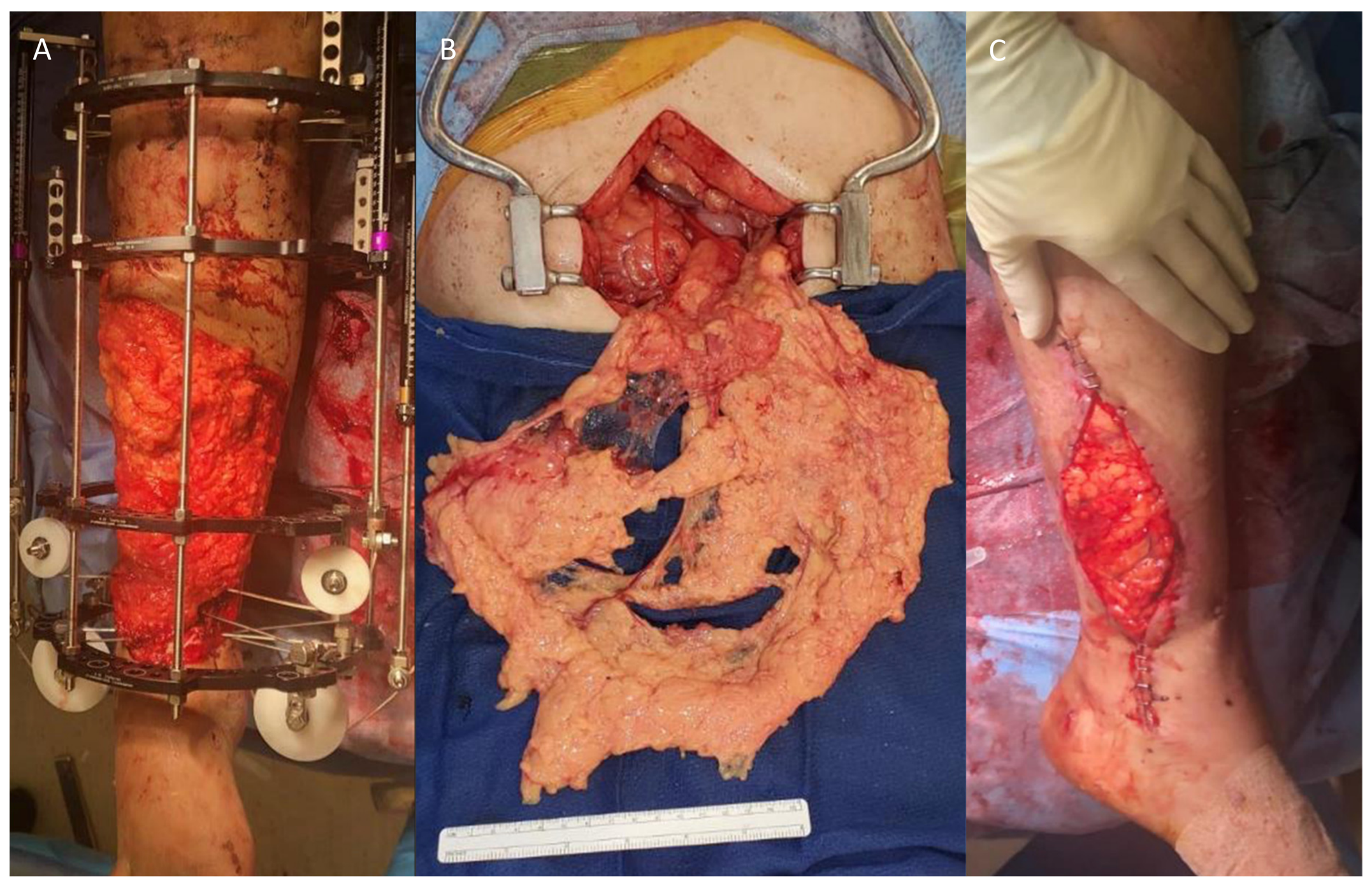
Orthopedic surgery performed a proximal osteotomy and applied a cable-guided transport mechanism for the right tibial defect. The bilateral lower extremity soft tissue defects were reconstructed with split omental free flaps and split-thickness skin grafts (Figure 5A–C).
Figure 5.
(A) Patient 2 right lower extremity in external fixator with a split omental free flap prior to split-thickness skin grafting. (B) In situ omental flap prior to free tissue transfer. (C) Split omental free flap to left lower extremity prior to split-thickness skin grafting.
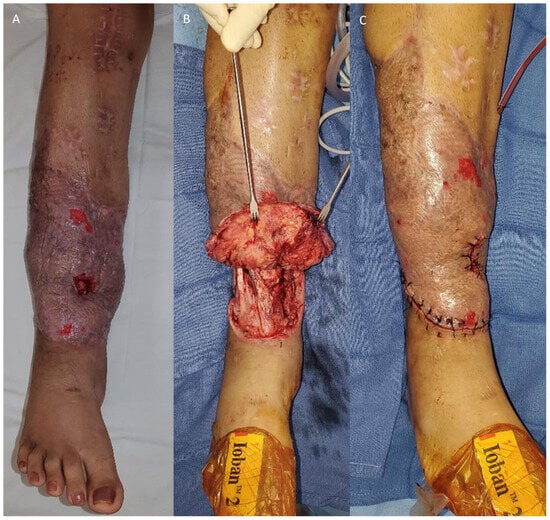
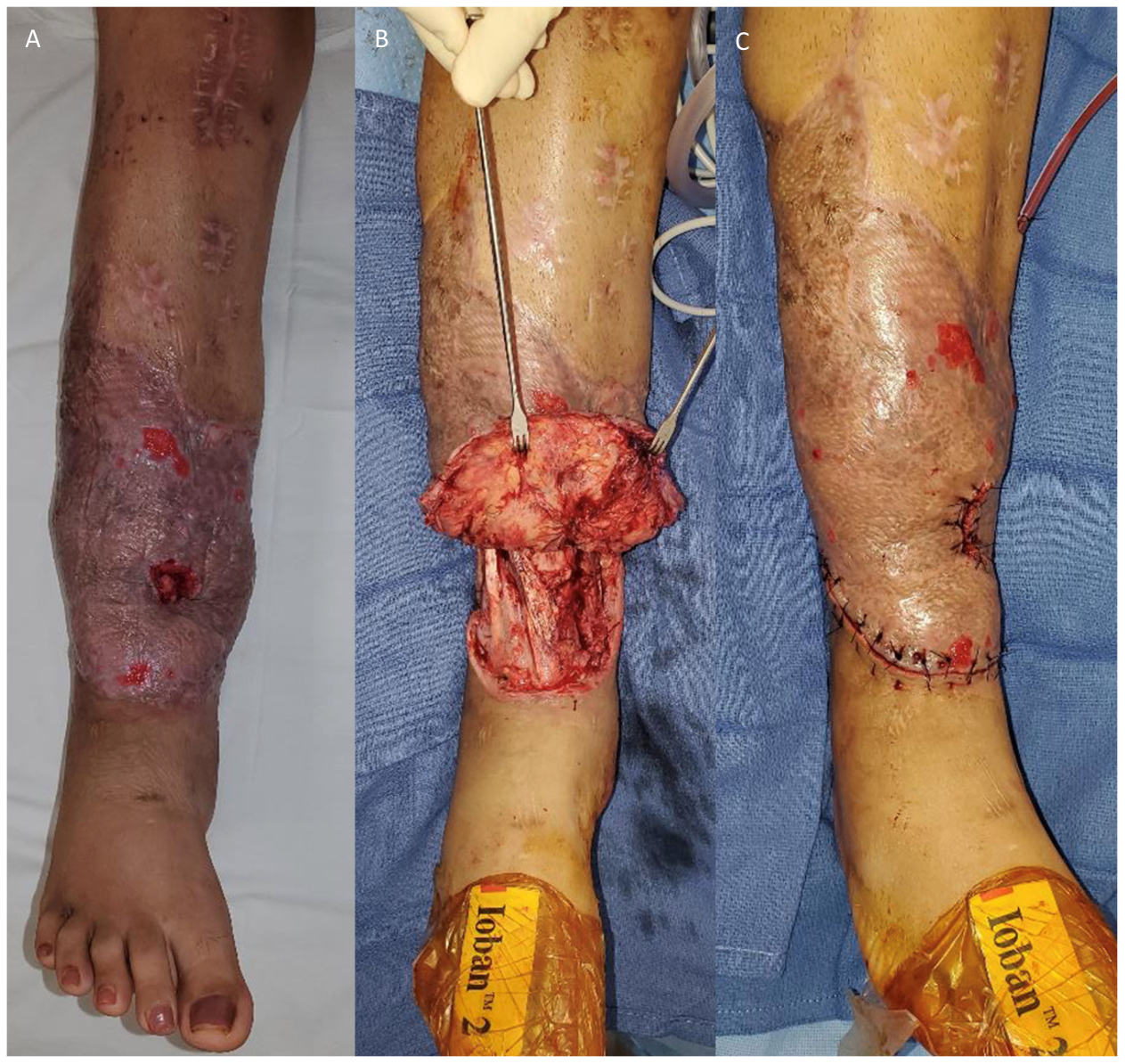
The right leg omental flap comprised roughly 85% of the omental tissue and was anastomosed to the anterior tibial vessels in the proximal leg. 111 days after cable guided transport and flap coverage, the patient underwent right lower extremity external fixator and cable removal, and right tibia medullary nailing. The right lower extremity flap was elevated by incising the entire distal edge of the flap, extending proximally along both the medial and lateral borders of the flap (Figure 6A–C).
Figure 6.
(A) Patient 2 postoperative photo 3 months after split omental free tissue transfer to right lower extremity. (B) Flap elevation for cable removal. (C) Split omental free flap inset and drain placement.
Despite satisfactory healing of the free flap, the patient developed a deep infection with extended-spectrum beta-lactamase E. coli in the bone transport docking site beneath the flap, 3 months after removal of the cable distraction device. This necessitated free flap elevation twice more for osseous debridement, hardware removal, and internal fixation over the span of five months.
After the last flap elevation, the patient experienced increased swelling of the flap and right foot, which was initially attributed to a resolving infection and recent surgery. However, in-clinic ICG lymphography revealed abnormal pooling of superficial lymphatic fluid in the distal portion of the right lower extremity omental flap that was not traveling through the flap to drain into the proximal lower leg (Figure 7A–D). Despite daily use of compression garments, the flap remained increasingly swollen by the end of day. Bioimpedance analysis demonstrated an unaffected-to-affected limb ratio of 1.14 at 5 kHz, consistent with increased fluid buildup. She subsequently underwent right foot LVB, which improved swelling in the right calf but not in the right foot or omental flap. Although she participated in both preoperative and postoperative physical therapy, her LLIS score remained elevated at 44, reflecting ongoing symptom burden.
Figure 7.
(A,B) Patient 2 medial view of right lower extremity ICG lymphography demonstrating superficial lymphatic pooling (green signal) at the border of the flap and native tissue. (C,D) Anterolateral view of right lower extremity ICG lymphography showing dermal backflow (green signal) over the distal portion of the omental flap.
2.3. Case 3
Patient 3 was a 33-year-old woman (BMI 32) who was involved in a high-speed motor vehicle collision sustaining a complex open right ankle fracture requiring internal fixation. Her postoperative course was complicated by soft tissue necrosis and hardware infection requiring hardware removal, external fixation, and serial debridement. The resulting composite soft tissue defects measured 13 × 30 cm altogether and involved nearly the circumference of the ankle, with preservation of an anterior skin bridge just proximal to the dorsal ankle, and fixed dead space in the Kager triangle between the Achilles tendon and the ankle joint.
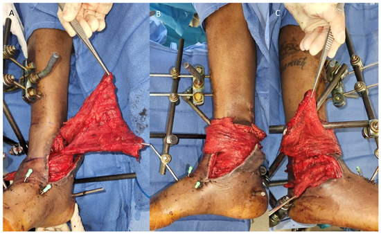
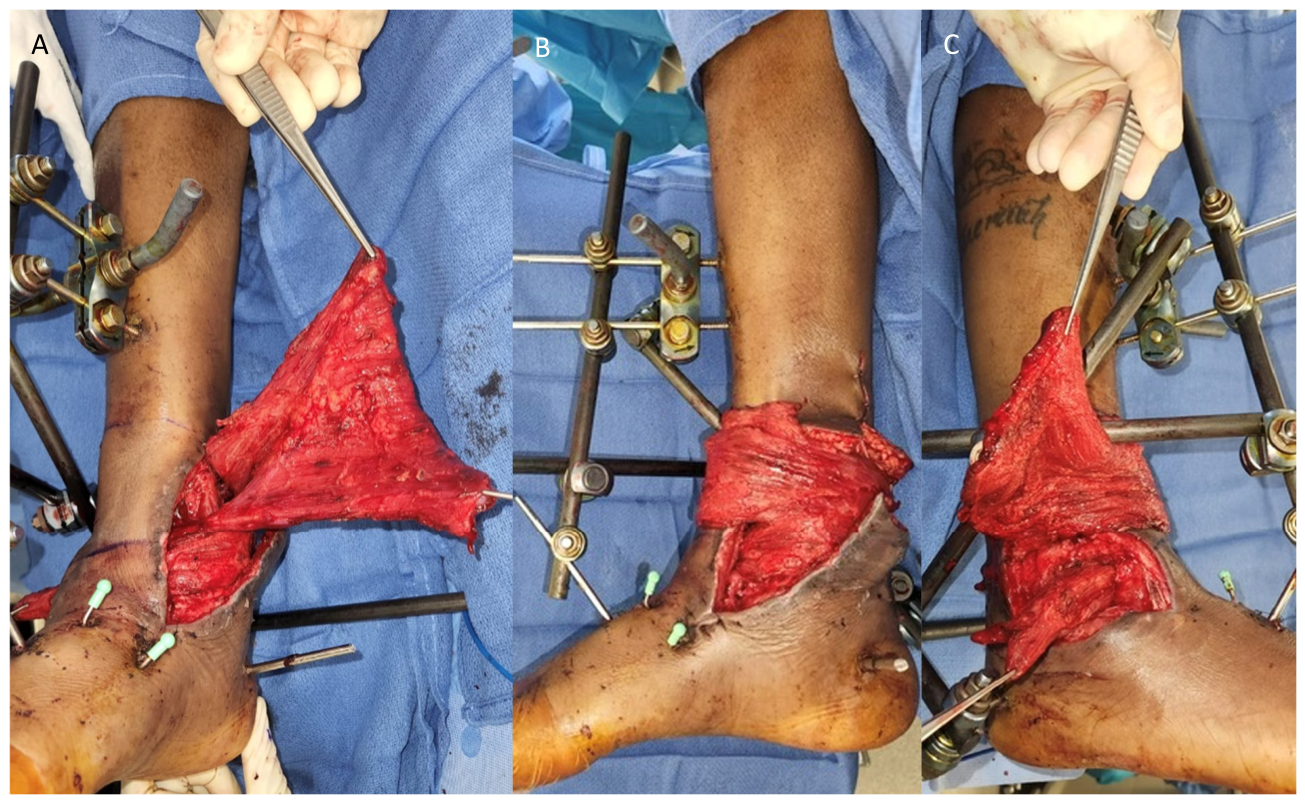
Definitive wound coverage was performed using a free left latissimus dorsi muscle flap with split-thickness skin graft. Given that the primary lymphatic drainage of the foot is over the dorsal ankle, the anterior skin bridge was preserved and not divided for the near-circumferential inset of the flap. Instead, it was carefully de-epithelialized to allow the free flap to wrap superficially around this area to reach the proximal component of the lateral defect. The flap was split along the muscle fibers between the transverse and descending pedicle branches. This allowed flap coverage both over the anterior skin bridge to the lateral ankle wound, as well as under the skin bridge where there was an existing area of undermining to fill the distal and deeper portion of the lateral ankle defect. The insertion of the latissimus muscle was wrapped circumferentially around the exposed Achilles tendon (Figure 8A–C).
Figure 8.
Patient 3 (A) anteromedial, (B) medial, and (C) lateral views of latissimus dorsi free flap inset to cover patient 3’s ankle wound. Note, the dorsal ankle skin bridge was left intact, de-epithelialized and covered by the muscle flap.
All exposed muscle flap was covered with split-thickness skin grafts. 11 months after flap coverage, the patient experienced avascular necrosis of the right talus with subsequent collapse, necessitating flap elevation and re-advancement to allow for placement of a cadaveric allograft. ICG imaging revealed intact lymphatic channels emerging proximally from under the muscle flap, along the dorsal ankle skin bridge that was previously preserved and de-epithelialized (Figure 9A,B).
Figure 9.
Patient 3 indocyanine green lymphography, in (A) fluorescent mode and (B) overlay mode, performed 11 months after free tissue transfer displaying intact lymphatic channels emerging from under the muscle flap along the dorsal ankle skin bridge that was previous preserved, de-epithelialized and buried under the muscle flap.
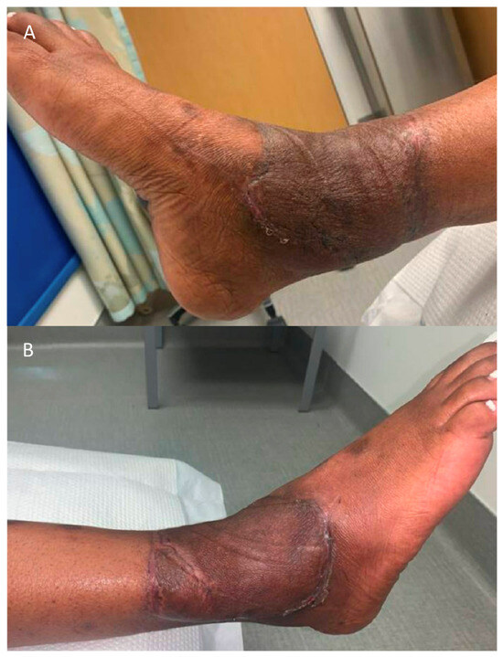
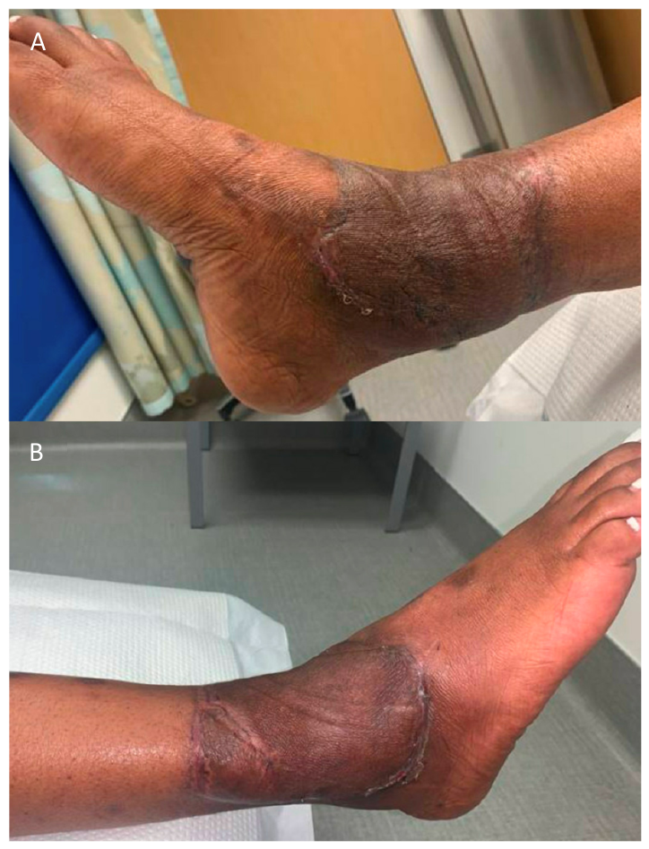
Postoperative swelling was completely resolved 23 months after flap coverage (Figure 10A,B).
Figure 10.
Patient 3 (A) medial and (B) lateral views of right ankle latissimus dorsi free flap and split-thickness skin graft 23 months after flap coverage.
3. Discussion
Microsurgical reconstruction provides effective surgical options for lymphedema, including lymphovenous bypass, vascularized lymph node transfer (VLNT), and vascularized lymph vessel transfer, but a surgical cure for lymphedema is rare. For patients who present with traumatic injuries, the risk of lymphedema may be decreased by considering the location, orientation, and viability of the lymphatics at the time of free flap reconstruction. The three cases in this series demonstrate the impact of reconstructive decision-making on the development of lymphedema after soft tissue injury.
Inadequate preoperative planning as evidenced by incisions that transect dominant lymphatic pathways, lymphatic discontinuity within skin graft zones, infections and repeated flap re-elevations for revision, may impose a cumulative mechanical and inflammatory burden on the lymphatic network. These disruptions may impair both the structural continuity and physiologic function of superficial lymphatic vessels. Repeated re-elevations, debridements, and infectious complications may promote scar formation, fibrosis, and misalignment of regenerating lymphatic channels at flap borders and within a flap. Therefore, the potential contribution of repeated mechanical disruption to the lymphatic system in the development of PTL cannot be overlooked. Given this risk, surgeons should consider using ICG lymphography to evaluate lymphatic drainage in preoperative planning for reconstruction and for flap re-elevation. ICG lymphography has demonstrated superiority over other imaging modalities [9] for fine resolution and in the setting of trauma.
In the first case, we suspect that the choice of incision for muscle flap inset may have disrupted a critical lymphatic pathway of the proximal medial calf by permanently separating the local subdermal lymphatics. Muscle flaps have few to no large lymphatic channels capable of reestablishing connections with native lymphatics and restoring lymphatic drainage. A study using ICG to map lymphatic drainage after muscle flap reconstruction consistently found obstruction just distal to the muscle flap [3]. The capacity of skin grafts to support lymphatic regeneration remains less clear, with studies reporting variable outcomes. Meier et al. demonstrated regeneration of lymphatic networks following meshed skin grafting on deep dermal and full-thickness burn injuries, although the depth of debridement was not described [10]. By contrast, van Zanten et al. observed scar formation and absence of restored lymphatic flow after flap coverage in patients with Gustilo-Anderson grade IIIB fractures [3]. Given the retrospective nature of this case, it is difficult to determine whether different reconstructive choices would have changed the lymphedema outcome.
Our third patient’s right lower extremity wound was treated with a free latissimus dorsi muscle flap, which does not contain lymphatic channels that can reinosculate with surrounding subdermal lymphatics [11]. Despite this potential limitation to lymphatic flow, flap inset was designed to avoid the disruption of native critical lymphatic areas which was evident in the flap inset for the second patient. The preservation of lymphatic anatomy was clearly demonstrated by fluorescent lymphography 11 months after muscle flap coverage.
The second patient’s wound was covered with a lymph node-containing free omental flap, which may have initially facilitated lymphatic drainage. However, repeated disruption of lymphatic connections within the flap and at its border with native skin during flap re-elevation and debridement may have contributed to the development of lymphedema. Osteomyelitis directly beneath the flap could also explain the worsening of the flap edema in the acute period. After treatment of the infection, however, the patient developed swelling distally in the foot, which is less readily explained by a localized infection alone. Similarly, the repeated surgeries to treat infection did not involve any of the remaining areas of native tissue and therefore would not be expected to impact any residual native lymphatic function. Another alternative explanation is that it was inevitable for lymphatic disruption from the initial injury to eventually manifest as clinical swelling. While this possibility must be considered, the flap swelling and foot swelling only began 11 months after injury, and simultaneously, despite the clear interruption of all drainage pathways from the foot at the time of the initial injury. Although lymphography was not performed during the asymptomatic period to demonstrate it, this does suggest the likelihood of some degree of lymphatic reconstitution following the initial reconstruction.
Alternative reconstructive options for this patient include the use of lymph vessel-containing flaps. In 2018, Yamamoto et al. introduced the concept of lymph axiality in which superficial lymphatic collectors are mapped using ICG lymphography and aligned so that lymph flows linearly between the donor and recipient sites [12]. Alignment of lymphatic vessel stumps at the flap and recipient site of less than 2 cm predicted postoperative lymph flow restoration [12]. Fasciocutaneous flap reconstruction resulted in a higher rate of lymphatic reconstitution and a lower rate of lymphedema when ICG lymphography was used intra-operatively to plan flap inset [13]. Sedbon et al. similarly reported a pilot study using ICG lymphography for fasciocutaneous flap inset, in which no patients whose flap was inset respecting lymph axiality developed lymphedema [14]. In our patient’s case, no single lymphatic vessel-containing fasciocutaneous flap would have had the necessary area to cover the wound, which measured 24 × 28 cm.
When lymph vessel or lymph node-containing flaps are effective, newly established lymphatic channels form across the borders of the flap and native skin. These channels are vulnerable during subsequent flap revisions or re-elevations, as surgical dissection may disrupt newly reinosculated lymphatic pathways. We have previously demonstrated preservation of reestablished lymphatic connections between native and flap skin during flap elevation and revision, using ICG to map and avoid disruption to these channels [15]. The same technique could have been applied patient two during revision in an effort to preserve any new channels.
Another potential option for the second patient might have been immediate, prophylactic LVB. Pereira et al. described the use of LVB to treat post-traumatic lymphedema [16], however it has not been described in the immediate post-traumatic setting. The immediate post-traumatic wound environment often includes an under-recognized zone of injury given a high-energy mechanism. Near-circumferential wounds also result in significant disruption of the superficial venous network, further limiting suitable venous outflow targets. In the case of patient two, venous drainage was compromised, with only the posterior tibial vessels still patent at the ankle.
Although correlation does not imply causality in this small series, the cases can serve to highlight several practical considerations for lymphatic-conscious reconstruction following lower extremity trauma. Lymphatic pathways should be considered early in the reconstructive timeline. When possible, ICG lymphography should be performed prior to definitive reconstruction to identify preserved channels and guide operative planning. Flap inset should be designed to avoid disruption of native lymphatic pathways. Reconstructive options should prioritize fasciocutaneous or lymph-node containing flaps that can reconstitute subdermal lymphatic networks when lymphatic flow must be reestablished across the injured segment. When lymphatic vessel-containing flaps are used, attention should be given to maintaining lymphatic axiality to facilitate reconstitution. And, when re-elevating lymphatic tissue-containing flaps, fluorescent lymphography should be used to avoid disrupting newly formed lymphatic connections between flap and native tissue.
4. Conclusions
The outcomes of these cases highlight the importance of considering lymphatic drainage during reconstruction of lower extremity soft tissue injuries. In addition to achieving adequate wound coverage, reconstructive planning should preserve or restore lymphatic drainage by considering both native and transferred lymphatics. We can “first do no harm” by preserving native lymphatic channels when possible, selecting appropriate flaps with appropriate lymphatic axiality, and using ICG lymphography to guide flap inset and re-elevation.
Future studies should attempt to establish causal links between reconstructive choices for extremity trauma and the subsequent risk of PTL. This can be accomplished using larger, prospective cohorts, using protocolized ICG lymphography to follow the status of lymphatic drainage at all stages in the sequence of reconstruction regardless of the presence or absence of swelling, and using a standardized battery of objective and validated patient-reported outcomes at regular intervals.
Author Contributions
Conceptualization, S.W., J.M. and P.S.B.; investigation, S.W., J.M. and P.S.B.; writing—original draft preparation, S.W., J.M. and P.S.B.; writing—review and editing, S.W., J.M. and P.S.B. All authors have read and agreed to the published version of the manuscript.
Funding
This research received no external funding.
Institutional Review Board Statement
This study was conducted in accordance with the Declaration of Helsinki and approved by the Institutional Review Board of Cedars-Sinai Medical Center (STUDY00001811, date of approval 12 December 2021); the requirement for informed consent was waived due to the retrospective nature of the study.
Informed Consent Statement
Patient consent was waived due to the retrospective nature of the study and the use of de-identified data.
Data Availability Statement
The raw data supporting the conclusions of this article will be made available by the authors on request.
Conflicts of Interest
The authors declare no conflicts of interest.
Abbreviations
The following abbreviations are used in this manuscript:
| PTL | Post-traumatic lymphedema |
| BMI | Body mass index |
| ICG | Indocyanine green |
| LVB | Lymphovenous bypass |
| E. coli | Escherichia coli |
| VLNT | Vascularized lymph node transfer |
References
- Grada, A.A.; Phillips, T.J. Lymphedema: Pathophysiology and clinical manifestations. J. Am. Acad. Dermatol. 2017, 77, 1009–1020. [Google Scholar] [CrossRef] [Scilit] [PubMed]
- Szczesny, G.; Olszewski, W.L. The Pathomechanism of Posttraumatic Edema of Lower Limbs: I. The Effect of Extravasated Blood, Bone Marrow Cells, and Bacterial Colonization on Tissues, Lymphatics, and Lymph Nodes. J. Trauma Acute Care Surg. 2002, 52, 315. [Google Scholar] [CrossRef] [Scilit] [PubMed]
- van Zanten, M.C.; Mistry, R.M.; Suami, H.; Campbell-Lloyd, A.; Finkemeyer, J.P.; Piller, N.B.; Caplash, Y. The Lymphatic Response to Injury with Soft-Tissue Reconstruction in High-Energy Open Tibial Fractures of the Lower Extremity. Plast. Reconstr. Surg. 2017, 139, 483. [Google Scholar] [CrossRef] [Scilit] [PubMed]
- Minasian, R.A.; Samaha, Y.; Brazio, P.S. Post-traumatic lymphedema: Review of the literature and surgical treatment options. Plast. Aesthetic Res. 2022, 9, 18. [Google Scholar] [CrossRef] [Scilit]
- Pereira, N.; Cambara, Á.; Kufeke, M.; Roa, R. Prevention and Treatment of Posttraumatic Lymphedema by Soft Tissue Reconstruction with Lymphatic Vessels Free Flap: An Observational Study. Ann. Plast. Surg. 2021, 86, 434. [Google Scholar] [CrossRef] [Scilit] [PubMed]
- Thng, C.B.; Sun, J.M. Current Insights into Post-Traumatic Lymphedema. Trauma Care 2025, 5, 24. [Google Scholar] [CrossRef] [Scilit]
- Szczesny, G.; Olszewski, W.L. The Pathomechanism of Posttraumatic Edema of the Lower Limbs: II—Changes in the Lymphatic System. J. Trauma Acute Care Surg. 2003, 55, 350. [Google Scholar] [CrossRef] [Scilit] [PubMed]
- Brorson, H.; Höijer, P. Standardised measurements used to order compression garments can be used to calculate arm volumes to evaluate lymphoedema treatment. J. Plast. Surg. Hand Surg. 2012, 46, 410–415. [Google Scholar] [CrossRef] [Scilit] [PubMed]
- Mihara, M.; Hara, H.; Araki, J.; Kikuchi, K.; Narushima, M.; Yamamoto, T.; Iida, T.; Yoshimatsu, H.; Murai, N.; Mitsui, K.; et al. Indocyanine Green (ICG) Lymphography Is Superior to Lymphoscintigraphy for Diagnostic Imaging of Early Lymphedema of the Upper Limbs. PLoS ONE 2012, 7, e38182. [Google Scholar] [CrossRef] [Scilit] [PubMed]
- Meier, T.O.; Guggenheim, M.; Vetter, S.T.; Husmann, M.; Haile, S.R.; Amann-Vesti, B.R. Microvascular regeneration in meshed skin transplants after severe burns. Burns 2011, 37, 1010–1014. [Google Scholar] [CrossRef] [Scilit] [PubMed]
- Dahl, V.A.; Tadisina, K.K.; Xu, K.Y. Limb Salvage and Treatment of Posttraumatic Lymphedema in a Mangled Lower Extremity. Plast. Reconstr. Surg. Glob. Open 2024, 12, e6209. [Google Scholar] [CrossRef] [Scilit] [PubMed]
- Yamamoto, T.; Iida, T.; Yoshimatsu, H.; Fuse, Y.; Hayashi, A.; Yamamoto, N. Lymph Flow Restoration after Tissue Replantation and Transfer: Importance of Lymph Axiality and Possibility of Lymph Flow Reconstruction without Lymph Node Transfer or Lymphatic Anastomosis. Plast. Reconstr. Surg. 2018, 142, 796. [Google Scholar] [CrossRef] [Scilit] [PubMed]
- Yamamoto, T.; Yamamoto, N.; Kageyama, T.; Sakai, H.; Fuse, Y.; Tsukuura, R. Lymph-interpositional-flap transfer (LIFT) based on lymph-axiality concept: Simultaneous soft tissue and lymphatic reconstruction without lymph node transfer or lymphatic anastomosis. J. Plast. Reconstr. Aesthetic Surg. 2021, 74, 2604–2612. [Google Scholar] [CrossRef] [Scilit] [PubMed]
- Sedbon, T.; Azuelos, A.; Bosc, R.; D’andrea, F.; Pensato, R.; Maruccia, M.; Meningaud, J.P.; Hersant, B.; La Padula, S. Spontaneous Lymph Flow Restoration in Free Flaps: A Pilot Study. J. Clin. Med. 2023, 12, 229. [Google Scholar] [CrossRef] [Scilit] [PubMed]
- Almadani, H.; Rhyu, H.; Cantor, A.; Brazio, P.S. Fasciocutaneous Vascularized Lymph Node Transfer for Head and Neck Lymphedema: A Case Report. Microsurgery 2025, 45, e70098. [Google Scholar] [CrossRef] [Scilit] [PubMed]
- Pereira, N.; Oñate, V.; Roa, R. A Comprehensive Approach to Posttraumatic Lymphedema Surgical Treatment. Arch. Plast. Surg. 2023, 50, 422–431, PubMed PMID: 37564707. [Google Scholar] [CrossRef] [Scilit]
Disclaimer/Publisher’s Note: The statements, opinions and data contained in all publications are solely those of the individual author(s) and contributor(s) and not of MDPI and/or the editor(s). MDPI and/or the editor(s) disclaim responsibility for any injury to people or property resulting from any ideas, methods, instructions or products referred to in the content. |
© 2026 by the authors. Licensee MDPI, Basel, Switzerland. This article is an open access article distributed under the terms and conditions of the Creative Commons Attribution (CC BY) license.