1. Introduction
Multiple myeloma (MM) is frequently interpreted as the consequence of abnormal proliferation of a unique clone; however, intraclonal heterogeneity has increasingly been recognized as a result of genetic mutations acquired by plasma cells over time [
1]. The branching evolution model proposes the existence of various populations of malignant subclones, with different overall frequencies that can change over time [
2,
3]. Such changes may affect the monoclonal protein (MP) produced by plasma cells, making it a potential surrogate marker of clonal heterogeneity [
4]. Intraclonal heterogeneity has been associated with disease progression, resistance to therapy, and poor survival outcomes [
5]. However, other factors may affect the amount of MP detected, both clinical, such as the time of diagnosis (earlier vs. later), but also technical factors, namely inconsistent reporting on protein characterization, thereby precluding solid conclusions regarding its clinical impact to date [
6]. In this context, particularly in patients with intact immunoglobulin MM, disease assessment relies predominantly on the monoclonal protein quantified by electrophoretic techniques. At the same time, serum free light chains (sFLC) are often interpreted in relation to it, assuming a parallel behavior between these biomarkers, which may not necessarily reflect their underlying biology.
Immunoglobulins are produced by plasma cells, the terminally differentiated stage of B-lymphocytes involved in the humoral immune response. These cells synthesize immunoglobulin molecules composed of two heavy chains and two light chains, encoded by genes located on different chromosomes (chromosome 14 for heavy chains and chromosomes 2 and 22 for κ and λ light chains, respectively), allowing independent genetic regulation of heavy and light chain production.
In healthy individuals, heavy chains (HC) and light chains (LC) are asynchronously synthesized with a 10–40% excess of LC over HC to ensure perfect Immunoglobulin (Ig) assembly and avoid toxic HC aggregates [
7]. The LC excess is secreted to the serum as sFLC. Accordingly, the interpretation of sFLC excess requires contextualization relative to intact immunoglobulin production, as physiological LC excess is an inherent feature of Ig synthesis. Clonal plasma cells can produce any of the five HC isotypes [
8]. Occasionally, MM cells stop secreting any HC, and only the LC is produced [
9]. In fact, sFLC concentrations can vary from normal values to those several orders of magnitude greater.
While individual concentrations of monoclonal sFLC and monoclonal intact Ig might depend on several factors, the sFLC-to-MP ratio is likely to depend mainly on the mutations affecting the respective genes. In fact, silencing of HC genes, but not LC genes, is seen in light chain escape relapses and is associated with more negative outcomes [
10]. This prognostic association may reflect a competitive advantage of these clonal cells due to less energy spent in the production of the HC. Additionally, the proportion of pathological FLC to the complete monoclonal HC can also vary tremendously [
10]. In healthy individuals, the sFLC-to-MP ratio typically ranges between 10% and 40%; values greater than this threshold may indicate higher genetic variability affecting HC and LC secretion [
7].
If the secreted protein represents gene translation, intraclonal heterogeneity at diagnosis may be inferred from MP and sFLC values. Exhaustive genomic studies to characterize MM patients are not ideal because of their complex, dynamic genetic profile (over the course of the disease, genes suffer several uncontrolled alterations, including many nonsense mutations) [
11]. In MM, laboratory biomarkers play a central role in diagnosis and disease monitoring. Serum biomarkers are particularly valuable for patient follow-up, as they allow more frequent and less invasive assessment of disease status. In MM, the biological complexity of the disease suggests that a more integrated interpretation of laboratory biomarkers may provide additional insight beyond conventional approaches.
In this context, the present study is an exploratory approach to assess the relationship between serum free light chains and monoclonal protein in intact immunoglobulin multiple myeloma, and its possible association with clinical outcome.
2. Results
The molar excess of sFLC varied between 0.7% and 5910.0%, with a median value of 30.3%. The median percentage of sFLC excess was not significantly different when comparing values of samples from IgG patients with values from IgA patients (p = 0.1982). Medians were 45.1% for IgGκ, 53.2% for IgGλ, 19.8% for IgAκ, and 26.9% for IgAλ.
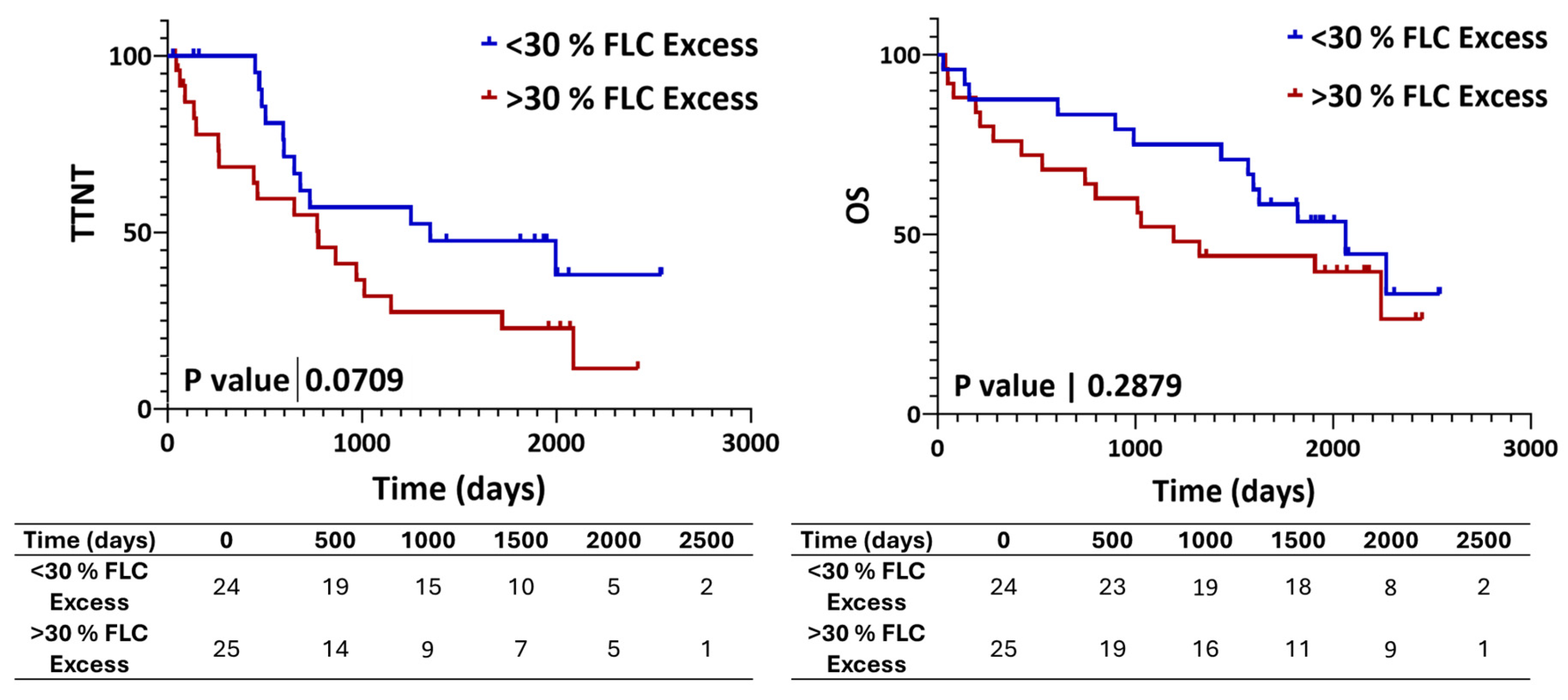
Patients were then divided into 2 groups using the median molar excess value (30%), which was used as an exploratory cutoff to distinguish different degrees of deviation from the expected physiological relationship between monoclonal protein and free light chain production within this abnormal cohort. Furthermore, this cutoff allowed balanced group sizes. The group with <30% excess (n = 24) demonstrated a longer median TTNT (1351 days vs. 775 days for the ≥30% excess group) and, although not reaching statistical significance (
p = 0.0709), a similar pattern was observed for OS, as illustrated in
Figure 1.
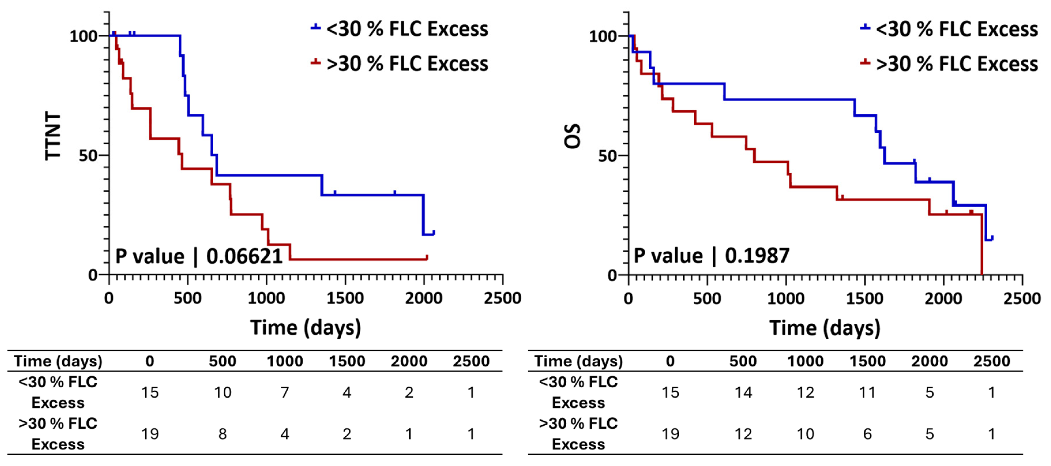
Patients were also divided by the therapeutic approach, i.e., non-transplanted vs. transplanted. Analyzing the sub-group of patients that did not receive autologous stem-cell transplantation (ASCT), the difference between groups approached statistical significance for TTNT (
p = 0.0662), with median values of 463 days and 667 days for the ≥30% excess and <30% excess groups, respectively (
Figure 2). No statistically significant differences were observed for OS (
p = 0.1987), although median OS values differed (799 vs. 1625 days for the ≥30% and <30% excess groups, respectively).
3. Discussion
It is generally accepted that changes in MP and/or sFLC values reflect proportional changes in tumor burden, i.e., a 50% decrease in the concentrations of the respective proteins is expected to correlate with a comparable reduction in tumor burden. However, differences in MP values may also arise from variable secretion rates of tumor cells, giving rise to oligosecretory or hypersecretory phenotypes.
The interpretation of varying levels of monoclonal sFLC in relation to MP concentrations at diagnosis is less well established. Reports addressing the genetic alterations involving the Ig Light chain loci [
12] remain limited, and the biological mechanisms underlying imbalances in free light chain (FLC) production are not fully understood. Aberrant rearrangements affecting genes encoding Ig HC may explain the absence of HC production in LC MM [
13] and the variability in MP production rates across patients.
Very high sFLC concentrations might also be explained by the presence of distinct MM subclones, with some producing intact Ig and LC, while others produce LC only [
14]. The absence of HC production is likely to impose lower energetic demands, potentially conferring a competitive advantage to these plasma cell populations.
In this context, the findings from our study suggest that different magnitudes of sFLC production excess relative to intact immunoglobulin concentration may be associated with the coexistence of distinct MM subclones with different secretion profiles, consistent with the concept of intraclonal heterogeneity. The presence of subclonal architecture is well documented, even in the precursor stages of the disease, and it continues to evolve throughout its clinical course [
15]. During treatment, MM plasma cells are subjected to selective pressures that may favor the emergence of resistant or more aggressive subpopulations, reinforcing the need for biomarkers capable of dynamically capturing such changes.
Intraclonal diversity has been associated with treatment resistance [
4]. In our cohort, patients with ≥30% sFLC-to-MP ratio at diagnosis showed a trend towards less favorable outcomes compared with those with lower ratios. However, the analysis was limited to univariate methods, and the independent prognostic value of the sFLC-to-MP ratio could not be established. In addition, time to next treatment (TTNT) does not exclusively reflect disease progression, as it may also be influenced by variability in treatment initiation across patients, which was not specifically addressed in this study. Importantly, subclonal populations may not be uniformly active over time and may be influenced by changes in the tumor microenvironment. In this context, the presence of sFLC excess at diagnosis may reflect an underlying biological potential for subsequent clonal selection or expansion [
15].
Subclones may remain transiently suppressed by therapy or by competing clones; however, shifts in microenvironmental conditions or additional genomic events may promote selective growth advantages. Accordingly, in a subset of patients, phenomena such as FLC escape driven by intraclonal heterogeneity may already be biologically primed at diagnosis.
4. Materials and Methods
This analysis originated from a laboratory-based evaluation of serum biomarkers in multiple myeloma, during which variability in the relationship between serum free light chains and monoclonal protein was observed. The cohort for this retrospective study included MM patients with monoclonal intact Ig MM and abnormal sFLC ratio diagnosed between 2016 and 2017 at the Portuguese Oncology Institute of Porto (IPO Porto). Of the 56 eligible patients, 7 were excluded due to a lack of follow-up information or because there was a decision not to treat the patient, resulting in a final cohort of 49 patients. The study population included 23 females and 26 males, with a median age of 73 years (range: 36–85 years). Of these, 35 had IgG MM (27 IgGκ, 8 IgGλ) and 14 had IgA MM (12 IgAκ and 2 IgAλ). Patients received heterogeneous treatment approaches, reflecting real-world clinical practice. Clinical data were limited to variables relevant for outcome assessment, including transplant status and treatment exposure.
Serum free light chains (sFLC) were quantified using the Freelite assay (The Binding Site, Birmingham, UK), and MP levels were determined by capillary electrophoresis using the Minicap system (Sebia, Lisses, France).
The excess of sFLC relative to monoclonal protein was estimated by converting the measured concentrations of both parameters into molar concentration, taking into account the different molecular weights of the corresponding intact immunoglobulin isotype and free light chains.
Survival curves were generated using GraphPad Prism version 8.2.1, employing the Kaplan–Meier method and Log-rank (Mantel–Cox) test. Time to next treatment (TTNT) and overall survival (OS) were used as endpoints for survival analysis. A statistical significance level of p < 0.05 was considered significant.
5. Conclusions
The findings of this retrospective study should be interpreted in light of its inherent methodological constraints. Importantly, the molar relationship between serum free light chains and monoclonal protein, as explored here, represents a relatively under-investigated parameter in intact immunoglobulin MM, reflecting the intrinsic biology of heavy and light chain synthesis and secretion.
Our results raise the possibility that marked sFLC excess relative to monoclonal protein may reflect underlying intraclonal heterogeneity, a biological feature with recognized clinical implications. In this exploratory context, although not intended to establish definitive clinical associations, these observations highlight a potentially informative and readily accessible analytical perspective that warrants further evaluation in larger studies.
Future integration of patients exhibiting pronounced sFLC excess into genomic or functional analyses may help clarify the biological and clinical significance of this phenomenon.
Author Contributions
Conceptualization, A.M.P. and S.C.; methodology, A.M.P. and S.C.; formal analysis, A.M.P. and S.C.; investigation, A.M.P., C.A. and S.C.; resources, A.M.P. and S.C.; data curation, A.M.P.; writing—original draft preparation, A.M.P.; writing—review and editing, S.C., J.P.B., J.M.M. and G.M.; visualization, A.M.P.; project administration, A.M.P. All authors have read and agreed to the published version of the manuscript.
Funding
This research received no external funding.
Institutional Review Board Statement
The study was conducted in accordance with the Declaration of Helsinki and approved by the Ethics Committee of Portuguese Oncology Institute of Porto (approval reference 236/021; date of approval: 14 October 2021).
Informed Consent Statement
Broad informed consent for the research use of retrospective clinical data was obtained from all participants, in accordance with institutional policies and ethical guidelines.
Data Availability Statement
The datasets generated and/or analyzed during the current study are not publicly available due to institutional and data protection regulations, but may be made available from the corresponding author upon reasonable request and with permission of the Portuguese Oncology Institute of Porto.
Conflicts of Interest
The authors declare no conflicts of interest.
Abbreviations
The following abbreviations are used in this manuscript:
| ASCT | Autologous Stem Cell Transplantation |
| FLC | Free Light Chains |
| HC | Heavy Chain |
| Ig | Immunoglobulin |
| LC | Light Chain |
| MGUS | Monoclonal Gammopathy of Undetermined Significance |
| MM | Multiple Myeloma |
| MP | Monoclonal Protein |
| OS | Overall Survival |
| SMM | Smoldering Multiple Myeloma |
| sFLC | Serum Free Light Chains |
| TTNT | Time to Next Treatment |
References
- Morgan, G.J.; Walker, B.A.; Davies, F.E. The genetic architecture of multiple myeloma. Nat. Rev. Cancer 2012, 12, 335–348. [Google Scholar] [CrossRef] [Scilit] [PubMed]
- Keats, J.J.; Chesi, M.; Egan, J.B.; Garbitt, V.M.; Palmer, S.E.; Braggio, E.; Van Wier, S.; Blackburn, P.R.; Baker, A.S.; Dispenzieri, A.; et al. Clonal competition with alternating dominance in multiple myeloma. Blood 2012, 120, 1067–1076. [Google Scholar] [CrossRef] [Scilit] [PubMed]
- Corre, J.; Munshi, N.; Avet-Loiseau, H. Genetics of multiple myeloma: Another heterogeneity level? Blood 2015, 125, 1870–1876. [Google Scholar] [CrossRef] [Scilit] [PubMed]
- Brioli, A.; Melchor, L.; Cavo, M.; Morgan, G.J. The impact of intra-clonal heterogeneity on the treatment of multiple myeloma. Br. J. Haematol. 2014, 165, 441–454. [Google Scholar] [CrossRef] [Scilit] [PubMed]
- Bolli, N.; Avet-Loiseau, H.; Wedge, D.C.; Van Loo, P.; Alexandrov, L.B.; Martincorena, I.; Dawson, K.J.; Iorio, F.; Nik-Zainal, S.; Bignell, G.R.; et al. Heterogeneity of genomic evolution and mutational profiles in multiple myeloma. Nat. Commun. 2014, 5, 2997. [Google Scholar] [CrossRef] [Scilit] [PubMed]
- Baughn, L.B.; Jessen, E.; Sharma, N.; Tang, H.; Smadbeck, J.B.; Long, M.D.; Pearce, K.; Smith, M.; Dasari, S.; Sachs, Z.; et al. Mass Cytometry reveals unique phenotypic patterns associated with subclonal diversity and outcomes in multiple myeloma. Blood Cancer J. 2023, 13, 84. [Google Scholar] [CrossRef] [Scilit] [PubMed]
- Nakano, T.; Matsui, M.; Inoue, I.; Awata, T.; Katayama, S.; Murakoshi, T. Free immunoglobulin light chain: Its biology and implications in diseases. Clin. Chim. Acta 2011, 412, 843–849. [Google Scholar] [CrossRef] [Scilit] [PubMed]
- Rajkumar, S.V. Plasma Cell Disorders. In Goldman’s Cecil Medicine; Elsevier: Amsterdam, The Netherlands, 2012; pp. 1233–1243. [Google Scholar]
- McPherson, R.A.; Massey, H.D. Laboratory Evaluation of Immunoglobulin Function and Humoral Immunity. In Henry’s Clinical Diagnosis and Management by Laboratory Methods, 22nd ed.; Elsevier: Amsterdam, The Netherlands, 2011; pp. 899–913. [Google Scholar]
- Brioli, A.; Giles, H.; Pawlyn, C.; Campbell, J.P.; Kaiser, M.F.; Melchor, L.; Jackson, G.H.; Gregory, W.M.; Owen, R.G.; Child, J.A.; et al. Serum free immunoglobulin light chain evaluation as a marker of impact from intraclonal heterogeneity on myeloma outcome. Blood 2014, 123, 3414–3419. [Google Scholar] [CrossRef] [Scilit] [PubMed]
- Liu, Y.; Beyer, A.; Aebersold, R. On the Dependency of Cellular Protein Levels on mRNA Abundance. Cell 2016, 165, 535–550. [Google Scholar] [CrossRef] [Scilit] [PubMed]
- Clarke, S.E.; Fuller, K.A.; Erber, W.N. Chromosomal defects in multiple myeloma. Blood Rev. 2024, 64, 101168. [Google Scholar] [CrossRef] [Scilit] [PubMed]
- Szczepański, T.; van ’t Veer, M.B.; Wolvers-Tettero, I.L.M.; Langerak, A.W.; van Dongen, J.J. Molecular features responsible for the absence of immunoglobulin heavy chain protein synthesis in an IgH− subgroup of multiple myeloma. Blood 2000, 96, 1087–1093. [Google Scholar] [CrossRef] [Scilit] [PubMed]
- Ayliffe, M.J.; Davies, F.E.; De Castro, D.; Morgan, G.J. Demonstration of changes in plasma cell subsets in multiple myeloma. Haematologica 2007, 92, 1135–1138. [Google Scholar] [CrossRef] [Scilit] [PubMed]
- Dutta, A.K.; Fink, J.L.; Grady, J.P.; Morgan, G.J.; Mullighan, C.G.; To, L.B.; Hewett, D.R.; Zannettino, A.C. Subclonal evolution in disease progression from MGUS/SMM to multiple myeloma is characterised by clonal stability. Leukemia 2019, 33, 457–468. [Google Scholar] [CrossRef] [Scilit] [PubMed]
| Disclaimer/Publisher’s Note: The statements, opinions and data contained in all publications are solely those of the individual author(s) and contributor(s) and not of MDPI and/or the editor(s). MDPI and/or the editor(s) disclaim responsibility for any injury to people or property resulting from any ideas, methods, instructions or products referred to in the content. |