


Abstract
Breast cancer is among the most common cancers globally. While several advancements have been made to improve breast cancer management, there is still a need to find innovative ways to deliver drug therapy to improve efficacy and side effects associated with treatment. Among these methods, local and targeted drug delivery is particularly promising. The current review highlights localized polymer-based methods to effectively deliver breast cancer therapy, with a specific focus on the strengths and limitations of these systems. Injectable and surgically implanted scaffolds, microneedles, topical patches, liquid and semisolid topical drug carriers are amongst some of the delivery systems discussed. Highlighted in the discussion is how physiological changes that occur during breast cancer should be considered and utilized when developing drug formulations, by specifically exploiting the tumor microenvironment. Remaining gaps and future areas for drug delivery research are highlighted including personalized medicine and insights into novel drug delivery systems like nanomedicines and three-dimensional drug printing.
1. Introduction
Breast cancer is a global issue that occurs in every country of the world. In 2022, it was estimated that 2.3 million women were diagnosed with breast cancer and 670,000 died from it [1]. Besides skin cancer, breast cancer is the most common cancer [2]. Due to the higher exposure of estrogen and progesterone, being born female is the biggest risk factor for breast cancer. Though, breast cancer still affects men, with 0.5–1% of breast cancers occurring in men. In the United States, the overall lifetime risk of a woman developing breast cancer is one in eight [2]. While the rate of deaths from breast cancer is decreasing, there is still a need for innovative prevention, screening, and treatment options to eliminate breast cancer [1,2].
The female breast is comprised mainly of adipose tissue which contains lobes, lobules, and mammary ducts [3]. In the lobules, there are glands that produce milk which is then carried by the mammary ducts to the nipple. Most breast cancers originate from the epithelial cells lining the mammary ducts or lobules and are histologically classified as carcinomas. Carcinomas can be further divided into carcinoma in situ and invasive cancer. In addition to the pathology of breast cancer, the hormone receptor and human epidermal growth factor receptor 2 (HER2) status are both significant for prognosis and treatment strategies [4]. Hormone receptors are nuclear transcription factors that promote cell growth by activating signal transduction pathways. They include estrogen receptors (ERs) and progesterone receptors (PRs). The HER2 gene is located on chromosome 17q21 and encodes a transmembrane tyrosine kinase growth factor receptor responsible for normal cell growth. Overexpression of the HER2 gene is associated with increased tumor aggressiveness and mortality [5]. The type of breast cancer is important for understanding the management and can be utilized when developing new treatments.
Unlike some cancers, often there are few or no symptoms at all in the early stages of breast cancer. Examples of symptoms that people may experience include a breast lump or thickening, changes in the size or appearance of the breast, and changes in the nipple appearance or areola [1]. In addition to being female, other risk factors for breast cancer include increasing age, family history, obesity, age of menarche, age of first pregnancy, and tobacco use [6]. Additionally, 5 to 10% of breast cancers can be characterized as having a genetic mutation, with Breast Cancer gene (BRCA) 1 and 2 being the most common [7]. Broadly, management of breast cancer consists of surgical treatment, radiation, chemotherapy, hormonal therapy, immunotherapy, and other targeted drug therapy [8].
A recent and rapidly developing focus for cancer treatment is the use of polymeric drug delivery systems. Polymeric drug delivery is broadly described as a formulation that introduces a biologically active substance into the body, but is unique in that the rate, time, and site of drug delivery in the body can be modified [9]. The polymeric excipients used for the synthesis of these drug carriers can be systematically chosen to optimize or create a desired effect, including increased drug efficacy or a reduction in side effects. Additionally, they can be used to increase drug solubility or bioavailability of the drug at the desired site of action. Some examples of polymeric drug delivery systems include nanoparticles, liposomes, dendrimers, hydrogels, and implants [10]. These innovative systems offer a potential solution for the localized delivery of therapeutic molecules to a specific area of the body, for example the breast tissue in the case of breast cancer [11].
The focus of this review will be on how recent advancements in polymeric drug delivery are being utilized to improve breast cancer management. Specifically, there are advantages and limitations of innovative localized polymer-based drug therapies compared to conventional management strategies in both the neoadjuvant and adjuvant chemotherapy setting. Through a detailed literature search, a comprehensive analysis will be conducted to identify recent advancements in localized polymeric drug delivery systems that have been targeted to improve breast cancer treatment outcomes. This up-to-date review will serve as a summary of the literature in this area and will provide emphasis on applications for future research and investigations. Compared to previously reported breast cancer drug delivery reviews and literature, this manuscript emphasizes the role of localized polymeric drug delivery strategies, how they target previous gaps in breast cancer treatment, and provides recommendations for next steps. To identify the literature, Google Scholar, PubMed, and the Cochrane Library were searched up to February 2026. Research performed in the last 10 years was prioritized and only English-language studies were included. Keywords in the search strategy included “cancer,” “breast cancer,” “cancer of the breast,” “mammary cancer,” “drug delivery,” “medication,” “dosage forms,” “pharmaceutical formulations”, “polymeric drug delivery”, “small molecule drugs for breast cancer”, biologic drugs for breast cancer”, and “drug technologies.” Search results were screened by publication title by the reviewers, and articles with titles and abstracts that included relevant keywords were further screened for inclusion, using the full text, based on relevancy to the review topic. Formulation development studies and preclinical trials were considered the best evidence, and review articles were used to supplement evidence and identify other primary literature. The literature included was managed using reference management software.
2. Barriers to Historic Breast Cancer Treatment
2.1. Surgical Removal
Surgical removal of cancerous tissue is a common therapy option used for breast cancer treatment. It can be used both for curative intent and to improve quality of life. There are various types of surgeries depending on the stage and goals of treatments [8]. For example, a lumpectomy is done to remove just the tumor and surrounding tissue and is considered for early-stage breast cancers [12]. Mastectomy is another surgical option for breast cancer and generally involves the removal of all of the breast tissue, although there are different types [12]. This surgery may be chosen for larger tumors, advanced disease, or recurrence. While surgery is a first line therapy option for breast cancer, there are limitations. In patients of poor health who are frail, the elderly, or individuals with multiple comorbidities, surgery may not be an option due to the associated risks. There are also complications of surgery which include bleeding, infections, nerve pain, and mental health implications [13]. It is possible that undetected microscopic disease remains following surgery, which is why surgery is often concurrent with other treatments like chemotherapy and radiation therapy [14]. In one retrospective cohort study, breast conserving surgery (BCS) alone was compared with BCS plus adjuvant radiation therapy. It was found that the hazard ratio was higher for patients who did not receive radiation therapy for recurrence or death [14].
2.2. Radiation Therapy
Radiation therapy is another non-drug treatment option for breast cancer. Broadly, this therapy uses high-frequency radiation that causes damage to cell deoxyribonucleic acid (DNA), which results in cell death and tumor shrinkage [15]. It can be provided as an external (external beam radiotherapy (EBRT)) or internal source (brachytherapy) therapy [16]. Radiation therapy is often used as an adjuvant therapy, following BCS, to reduce the risk of local recurrence [17]. It is also used in instances where the tumor is unresectable or in instances of metastatic disease. It is associated with side effects like fatigue, skin changes, swelling, and tenderness [18]. Radiation itself can also be associated with secondary malignancies, with an absolute risk between 0.2% and 1% per year [18]. For breast cancer, lung cancer, leukemia, and the cancer of the opposite breast are associated secondary malignancies.
2.3. Chemotherapy
Chemotherapy is a group of drugs used to kill or slow the growth of cancerous cells. It can be given before surgery (neoadjuvant) or after surgery (adjuvant) and is used to shrink the tumor prior to surgery or kill an of the remaining cancers cells, respectively [19]. In the case of metastatic disease, it may be used to prolong survival and improve quality of life [19]. There are many different drugs used for breast cancer management, and these include anthracyclines (doxorubicin, epirubicin), taxanes (paclitaxel, docetaxel), 5-fluorouracil, cyclophosphamide, capecitabine, and gemcitabine [20]. Most chemotherapy agents used for breast cancer treatment must be given intravenously and administered frequently, often every 2 to 3 weeks. Traditional chemotherapy is limited by the extensive side effect profile, due to its lack of specificity to cancer cells. For example, in mice it was found that after 24 h, less than 0.5% of the total dose administered intravenously was available at the target tumor site (Figure 1) [21]. Additionally, chemotherapy agents often show higher efficacy at higher doses, thus increasing the risk of adverse reactions [10]. Most chemotherapeutic agents have low aqueous solubility, limiting intravenous administration. Accordingly, the drugs are often modified, and these modifications can increase side effects like hypersensitivity reactions [22]. The side effects depend on the specific agent, but generally include heart damage, nausea and vomiting, hypertension, nerve damage, fatigue, hair loss, and increased risk of secondary cancer like leukemias [23]. Chemotherapy can also impact ovarian function and fertility, with 33% to 76% of women 50 years and older experiencing persistent post-treatment amenorrhea after systemic treatment for breast cancer [24]. Resistance to chemotherapy, the failure of a drug to produce a beneficial response, is another major concern for chemotherapy [25]. Other limitations of traditional chemotherapy include cost on the health care system associated with clinic visits to administer the medication and monitor the patient [26]. Futhermore, there is a burden on the patient to be able to get a facility where the medication can be administered, and the time required [27].
Figure 1.
Examples of issues associated with systemic breast cancer treatment options.
2.4. Hormone Therapy
In patients who have hormone-positive breast cancer, hormone therapy, or endocrine therapy, can be used to slow or stop the growth of the breast tumor by stopping the body’s ability to produce the hormone or by blocking the receptors [28]. Examples include tamoxifen and aromatase inhibitors like anastrozole, letrozole, or exemestane. Hormone therapy is frequently used in breast cancer management as an adjuvant therapy and is often continued for 5 to 10 years [29]. Endocrine therapy has been used to decrease mortality and prevent reoccurrence of breast cancer. For example, adjuvant tamoxifen decreased mortality at 15 years by a third in women with early-stage estrogen receptor positive breast cancer [30]. The majority of the endocrine therapy used for breast cancer are oral agents, that thus have the advantage of ease of administration. Examples of side effects of endocrine therapy include osteoporosis, menopausal symptoms, blood clots, stroke, nausea, bone pain, depression, and uterine cancer [31]. Additionally, given these agents are prescribed for several years and must be given daily, there is a risk of non-adherence [32]. For instance, in one systematic review that looks at adherence to adjuvant hormonal therapy in breast cancer survivors, adherence ranged from 41 to 72% [32].
2.5. Immunotherapy
Immunotherapy is a growing area for cancer research and generally refers to therapy that is used to enhance a person’s own immune system to fight cancer. One type of immunotherapy used in breast cancer is an immune check point inhibitor, which works by blocking checkpoint proteins, which normally would prevent the immune system, particularly T-cells, from attacking cancer cells [33]. An example of an immune checkpoint inhibitor used in breast cancer is pembrolizumab. Trastuzumab, a monoclonal antibody, is another example of immunotherapy that can be used to treat HER2-positive breast cancer [34]. One concern for immunotherapy is resistance, which can include the patient not responding to the primary dose or some patients relapsing or progressing after a period of treatment, referred to as primary and acquired resistance, respectively [35]. A major limitation of immunotherapy is cost, with most costing more than CAD 100,000 per patient [36]. Additionally, due to the mechanism of action, a possible side effect of immunotherapy is immune related adverse effects (IrAEs), which can occur at any time during and after treatment discontinuation [37]. Possible examples of IrAEs include colitis, hepatitis, pneumonitis, and acute kidney injuries.
3. Changes in Physiological Parameters in Breast Cancer
3.1. General Changes in Cancer That Support Tumor Growth and Progression
It is well recognized that there are “hallmark” characteristics to describe changes that allow cancer to occur, and as more research has been done, this list has evolved [38]. First, modifications occur in cancer cells that allow for chronic proliferation, by evading the normal cell growth and division cycle [38]. For example, cancer cells can acquire the ability to sustain proliferative signals by producing growth factor ligands themselves or by sending signals to surrounding cells to supply growth factors. Epidermal growth factor receptor (EGFR) inhibitors are one class of cancer therapy that target this mechanism of cancer growth [39].
Another change that occurs in cancer cells is the ability to evade growth suppressors. In a normal cell, there is a signal that prevents the cell from dividing, mainly by means of tumor suppressor genes. In cancer cells however, this signalling process is disturbed and cell proliferation continues [40]. Examples of these genes are RB (retinoblastoma-associated) and tumor protein 53(TP53). RB normally decides whether a cell should proceed with its normal growth cycle, so a cancer cell with an RB defect is missing this gatekeeper [40].
Cancer cells also can resist cell death. In healthy cells, programed cell death, referred to as apoptosis, is a barrier to cancer development [41]. Extracellular or intercellular signals can trigger apoptosis, if there is a defect with a cell. There are several ways cancer cells are able to escape apoptosis, the most commonly mentioned is the loss of the TP53 tumor suppressor function [41,42].
Like healthy cells, cancer cells must have vascular access to supply nutrients and oxygen and evacuate waste. Thus, another key factor for cancer development is inducing angiogenesis [40]. During tumor progression, there is often continuous sprouting of new vessels to sustain neoplastic growths. Examples of angiogenic regulators are vascular endothelial growth factor-A (VEGF-A) and thrombospondin-1 (TSP-1) [40].
Another characteristic of cancer cells is enabling replicative importability. Normally, cells are limited to a finite number of cell divisions, set by telomerase [43]. Yet, some cancer cells are able to divide without limit. One way is by upregulating expression of telomerase such that they are able to maintain telomeric DNA at lengths sufficient to avoid triggering senescence or apoptosis [43]. Telomerase inhibitors are a class of cancer drugs that target this mechanism [44].
Metastasis is a multistep process that occurs in cancer. Broadly, this involves local invasion, then transfer of cancer cells via nearby blood and lymphatic vessels, and escape of cancer cells into and the parenchyma of tissue [45]. Once cancer cells distribute to distant tissue, they can form small nodules of cancer cells that can grow into larger tumors.
3.2. Examples of Specific Physiologic Characteristics and Drug Targets in Breast Cancer
Broadly, the tumor microenvironment (TME) refers to the surrounding cells, tissues, and blood vessels that encompass a tumor. In breast cancer, the TME can be categorized as cellular, soluble, and physical components [46,47]. The cellular department can be further divided into local, regional, and metastatic compartments, which refers to the tumor cells, interaction between the tumor cells and an adjacent cell, and interaction with the vascular/lymph cells, respectively. Some examples of the cellular compartment are lymphocytes, plasma cells, dendritic cells, fibroblasts, adipocytes, lymph nodes, and blood [46]. The soluble factors include interleukins, tumor necrosis factor, and vascular endothelial growth factor. Oxygen and pH levels are examples of physical factors. The TME influences the malignant progression and clinical outcome to chemotherapy [48]. For example, in breast cancer, the blood vessels are lined by defective endothelial cells, leading to leaky vessels and reduced blood flow [49]. This decrease in blood flow can lead to impaired chemotherapy delivery.
The TME can also be targeted for drug therapy (Table 1). PD-1/PD-L1 (programmed cell death protein-1/programmed death-ligand 1) inhibitors are one class for drugs that directly act on the TME in breast cancer [46]. PD-1 is expressed on immune cells, like B-cells, T-cells, and macrophages, and when engaged it inhibits T-cell activation and contributes to immune resistance in the TME. Thus, these drugs work by restoring the response of cytotoxic T-cells to cancer cells [50]. Additionally, cancer-associated fibroblasts (CAFs) are an important therapeutic target within the tumor microenvironment. They are key stromal components of the tumor microenvironment and are among the most abundant (about 80%) non-malignant cells in many solid tumors particularly in desmoplastic cancers, such as pancreatic and breast carcinomas, and they significantly influence tissue structure. CAFs actively contribute to tumor progression by driving extracellular matrix remodelling, promoting angiogenesis, and enhancing tumor cell proliferation, invasion, and migration [51]. Beyond these effects, they contribute to the development of an immunosuppressive microenvironment by altering immune cell recruitment and function, enabling tumor immune evasion and therapeutic resistance. Owing to their diverse and critical roles in cancer biology, CAFs have emerged as a promising target for therapeutic intervention, with strategies focused on inhibiting their activation, disrupting their interactions with tumor cells, or reprogramming them toward a tumor-suppressive phenotype [51].
Table 1.
Characteristics of the TME in breast cancer and impact on drug delivery strategies.
Other properties of the TME that can be used to optimize drug targeting include pH, high glutathione levels, and overexpression of enzymes [52]. In general, the pH of the TME is more acidic, so drug delivery systems can be developed to have drug release at more acidic pHs than the physiological environment. Additionally, high levels of glutathione found in the TME can also be utilized for targeted drug release, by cleaving disulfide bonds on drug carriers.
The immune system also influences breast cancer, with breast cancer generally be classified as moderately immunogenic given its ability to impact and elicit immune responses [53]. In breast cancer, there are diverse populations of immune cells present in the stroma. Immunoediting, the process describing the interaction between the host immune system and tumor cells, involves recognition of cancer cells as foreign and triggering an immune response [53]. However, some of the tumor cell variants can escape the immune response and remain dormant while the immune cells prevent further tumor growth. Eventually, the tumor cells can escape the immune recognition and continue to grow and proliferate through various mechanisms.
Hormone receptor and HER2-positive breast cancer are other unique targets for some breast cancers. The estrogen receptor is involved in proliferation and invasion of cancer in estrogen receptor-positive breast cancer [54]. When estrogen binds to the estrogen receptor it leads to activation of downstream gene expression. Similarly, HER2 plays a role in promoting cell growth and survival in HER2-positive breast cancers [54]. Other examples of targeting pathways in breast cancer are phosphoinositide 3-kinase/mammalian target of rapamycin (PI3K/mTOR), cyclin-dependent kinases (CDK) 4/6, histone deacetylases, and poly (ADP-ribose) polymerases (PARPs) [54].
4. Examples of Innovation in Localized and Targeted Polymeric Drug Delivery for Breast Cancer
Polymeric drug delivery is an evolving field of technology that uses polymeric biomaterials to encapsulate and facilitate effective and safe drug delivery to different body sites for the management of several diseases [55]. Localized drug delivery refers to transfer of a drug to a specific target and is used to optimize drug availability at the site of action while minimalizing side effects due to off-target drug effects. Local drug delivery can be achieved by methods such as implants or injections. Targeted drug delivery is an approach that selectively directs active drug molecules to a specific tissue, organ, cell type, or molecular target (e.g., the tumor site) while minimizing exposure to healthy tissues [56]. The key goal is to increase drug concentration at the desired site of action, improve therapeutic efficacy, and reduce systemic toxicity and adverse effects. A typical localized and targeted drug therapy is achieved by utilizing polymeric drug delivery systems. These systems are constructed using biocompatible polymers, whose properties can be exploited for drug delivery [55]. The rate, time, and place of drug release can all be tailored using this method of drug delivery. Examples of natural polymers used include arginine derivatives, chitosan derivatives, cyclodextrin derivatives, and polysaccharides [9]. Conversely, examples of synthetic polymers used include Poly (N-isopropyl acrylamide)s and Poly (ethylenimine)s [9]. Biodegradable polymers are often used, given that they break down naturally in the intentioned physiological environment and negate the need to remove any devices or material.
In recent years, there has been a surplus of research expanding on the use of locally delivered or targeted polymeric drug delivery systems for breast cancer therapy. Summarized are locally delivered polymeric drug delivery systems for breast cancer treatment. Additionally included are targeted polymeric formulations given systemically but designed such that the drug effects are targeted to breast cancer cells, thus decreasing unwanted side effects (Figure 2).
Figure 2.
Examples of Localized Polymeric Drug Delivery with Applications for Breast Cancer Treatment.
4.1. Scaffolds
Scaffolds are polymeric injections or implants. Polymer implants are classified as two- or three-dimensional forms made of one or more polymers [57] while polymeric injectables are colloidal solutions that can form a hydrogel, which is typically able to form specific structure after being injected into the respective tissue [58]. Polymer-based hydrogels are made of cross-linked polymeric materials. One of the unique properties associated with hydrogels is their ability to adsorb and retain large amounts of fluids while maintaining their three-dimensional structure [59]. This swelling property can be utilized to control drug release properties. Hydrogels closely mimic biologic tissues and thus have the advantage of minimal side effects from the drug delivery system [60]. The properties of hydrogels can also be modified in such a way that they can respond to external stimuli like pH, temperature, or light. Hydrogels can be classified using various methods which include mesh size, swelling ratio, and microstructure [61]. Hydrogels can be used for local drug delivery by being injected into the tumor site.
Compared to implants, the injectable drug carriers minimize the incision size. Polymer-based implants have been frequently investigated for use in local cancer drug delivery, majorly because of their ability to bypass first pass metabolism. In some instances, the scaffold is injected intra-tumorally to increase concentration of the drug within the target tissue and avoid side effects associated with off-target pharmacological actions [58]. They can also be used for systemic effects to eliminate the needs for repeated doses by providing a sustained release.
In 2020, Yang et al. developed an implantable multi-drug delivery system with time-programmed release based using three-dimensional (3D) printing intended for breast cancer [62,63]. This scaffold was made from poly-lactic-co-glycolic acid (PLGA) and contained the two drugs, 5-fluorouracil and Dactolisib (i.e., NVP-BEZ235). The implantable scaffold was found to release the drug over a long period of time, with a slow-release stage lasting up to two weeks, suggesting the possibility of a one-time implantation. The formulation also demonstrated prolonged drug levels at the tumor site, with reduced doses needed, and less exposure to healthy tissue. A limitation of the formulation is the need to surgically implant it (Table 2).
In another study, researchers developed biodegradable PLGA implants containing doxorubicin-loaded chitosan nanoparticles [64]. This drug delivery system was subcutaneously implanted into mice with murine breast cancer cell line breast tumors. The implant demonstrated sustained release and tumor-specific drug delivery. When efficacy was measured against multiple injections of doxorubicin at the same amounts/doses, the implants had comparable inhibition of tumor growth.
Scaffolds also have potential use for prevention of tumor recurrence following mastectomy. In one study, researchers prepared 3D-printed scaffold made from caffeic acid grafted to chitosan, sodium alginate, nanoclay, and carbon dots–curcumin nanoparticles [65]. In mice models, the scaffold was found to have good long-term antibacterial effects, wound healing potential, and inhibited postoperative residual cancer cells.
In the setting of triple negative breast cancers, one research group investigated the use of localized drug release from an implantable microporous poly-di-methyl-siloxane (PDMS) for the targeted delivery of Luteinizing Hormone-Releasing Hormone conjugated paclitaxel and Luteinizing Hormone-Releasing Hormone conjugated prodigiosin [66]. Using vitro studies, a significant reduction in the percentage of cell growth in a time-dependent manner was found. In mice models, following partial surgical removal of the tumor and implantation, there was no tumor regrowth six weeks post-treatment implant. At the same time, no adverse effects of the drugs on the liver, kidneys, or lungs were detected.
Similarly, 3D-printed poly (lactic-co-glycolic acid), gelatin, and chitosan scaffold loaded with 5-fluorouracil and doxorubicin hydrochloride have also been prepared [67]. The scaffold had a sponge-like structure and for in situ implantation, drug release was influenced by tumor pH (slightly acidic environment), leading to sustained drug release. This formulation offers another potential option for reducing tumor growth and recurrence, while reducing drug toxicity. The scaffold also had good blood absorption, promoting rapid wound healing.
Electrospun drug delivery systems are another avenue for local drug delivery to the tumor site. Electrospun fibers have several favourable drug delivery characteristics including the ability to manipulate drug release due to their highly porous property, high surface-to-volume ratio, and the ability to encapsulate more than one drug directly into the fibers [68]. Ding and colleagues developed docetaxel-loaded poly-D,L-lactide (PDLLA) fibers for preventing local breast cancer recurrence [68]. In mice models, the locoregional recurrence after primary tumor resection decreased with subcutaneous administration of the formulation docetaxel loaded PDLLA fibers (16.7%) compared with the blank PDLLA nanofibers (88.9%), systemic (75.0%), or locally (77.8%) administered docetaxel and the control group (100%).
Due to the side effects associated with long-term adjuvant chemotherapy postoperatively, one strategy is to release multiple drugs in a time-programmed manner. Researchers utilized this strategy by developing an implantable time-programmed drug release hierarchical-structure ultra-fine fiber device [69]. The electrospun hierarchical-structured fibers had a hydrophilic internal chamber containing polyethylene glycol (PEG) and doxorubicin and the hydrophobic fiber matrix contained poly (d,l-lactic acid) (PLA) and disulfiram. When the drug delivery system was implanted into mice models with resected breast tumors, a rapid release of doxorubicin occurred due to the water absorption of PEG. This allows for rapid residual tumor cell elimination. At the same time, the disulfiram is released in a sustained manner, preventing spread via surrounding tissues and blood vessels and subsequent tumor metastasis.
Hydrogel scaffolds are another option for preventing local breast cancer relapses. Lei and colleagues developed paclitaxel containing hydrogel made of copolymeric poly (ethyleneglycol)-poly (ε-caprolactone)-poly (ethylene glycol) [70]. At room temperature the formulation is free flowing, but once injected it forms a gel within seconds at body temperature and the drug is released in a sustained fashion as it biodegrades. In mice models, the hydrogel containing paclitaxel significantly inhibited tumor recurrence after primary tumor removal compared to systemic and locally administered paclitaxel alone.
Temperature and pH of the breast cancer tumor environment can also be exploited for drug delivery systems. Fathi et al. (2019) investigated a pH and thermo-responsive hydrogel containing doxorubicin for intratumoral injection [71]. The hydrogel was made up of a blend of poly (N-isopropylacrylamide-co-itaconic acid) (PNIAAm-co-IA) and chitosan (CS). The researchers found that doxorubicin had accelerated release in acidic conditions at 37 °C compared to neutral pH and the temperature of 40 °C.
OncoGel is a paclitaxel containing hydrogel formulation of poly (lactide-co-glycolide) and poly (ethylene glycol) tri-block copolymer (PLGA-PEG-PLGA) [72]. It is a thermosensitive controlled release formulation that is free flowing at temperatures between 2 and 15 °C and water insoluble at body temperature. It is administered via intralesional injection or placement into the tumor cavity. One requirement for use is that the tumor must be accessible for the drug to be injected. The needle used for injection is selected based on the type of lesion it is injected into. In mice with human breast carcinoma xenografts, less than 0.1% of radioactively labelled paclitaxel was measured in the other organs or blood following intertumoral injection of OncoGel [73].
Nanofibers are another method of delivering breast cancer drug therapy. One research group developed pH-responsive polymeric nanofibers encapsulated with an active targeting micellar system that was loaded with paclitaxel [74]. Cholic acid conjugated to poly (bis (carboxyphenoxy) phosphazene) (PCPP) polymeric micelles were used to encapsulate the paclitaxel. The micelles were then encapsulated into the core–shell psyllium husk mucilage nanofibers using electrospinning. The resulting nanofibers were intended for injection subcutaneously near the solid tumor region. In vitro drug release demonstrated sustained drug release over 180 h and were effectively internalized into the skin. Compared to paclitaxel alone, the drug-loaded nanofibers exhibited higher cytotoxicity for 8 days.
Another way researchers were able to formulate 5-Fluorouracil to be delivered locally was by developing a 3D bio-printed two-layer implantable scaffold [75]. The first layer was made of the polymers poly-ε-caprolactone (PCL) and chitosan (CS) and was loaded with 5-Fluorouracil and was designed to release the drug within 4 weeks. The second layer was made from PCL and contained gold nanoparticles; the purpose of the second layer was a radiation enhancer. In vitro testing revealed that 32% of the drug was released after one month with an initial burst release of 17.22%.
Table 2.
Emerging innovations in localized polymeric scaffolds targeting breast cancer.
4.2. Liposomes
Liposomes are closed lipids vesicles comprised of phospholipids bilayers. They are optimal for both drug delivery of hydrophilic and hydrophobic drugs due to their hydrophobic lipid bilayer and internal aqueous core [76]. However, liposomes alone are easily subject to degradation from acidic pH, enzymes, and the immune system in the biological environment [68]. Drug delivery by liposomes can be improved by modification with polymers, referred to as liposome complexes [77]. Examples of such structural changes include polymer-coated liposomes and polymer-grafted liposomes [76]. One example of liposome complexes that can be used to target drug delivery to tumors is by using a polymer sensitive to the same pH as the tumor matrix (pH 6.5). At this pH, the surface properties of the liposome polymer complex changes, and the drug is released [78].
Doxorubicin is a potent chemotherapy agent used for breast cancer, with high toxicity, including irreversible cardiomyopathy and liver damage. To reduce toxicity of doxorubicin, researchers prepared PEGylated liposomes, labelled with Aptamers (Apt)-A6 that can recognize and bind HER-2 receptors on HER-2-positive breast cancer cells, using thin film hydration methods [79]. The liposomes were found to range in size from 100 to 200 nm. The best formulation had an entrapment efficiency of 93% and had increased uptake into breast cancer cells. Additionally, they were more effective at killing cancer cells than free doxorubicin (Table 3).
As mentioned, the tumor microenvironment can be exploited for targeted drug delivery in breast cancer. Specifically, there are notable characteristics of lower extracellular pH and increased angiogenesis capacity of the breast cancer TME. In 2021, Jain et al. utilized these characteristics by developing VEGF antibody functionalized PEGylated sensitive liposomes containing docetaxel [80]. The liposomes were 206 nm in diameter and had an entrapment efficiency of 62%. Compared to free docetaxel, the liposomal formulation had a sustained drug release profile and prolonged blood circulation time. The formulation design promoted increased cellular uptake and targeting into breast cancer cells.
Researchers have also attempted to increase the skin penetration of topically applied tamoxifen by encapsulating it in a liposome-PEG-PEI (poly (ethylene imine)) complex carrier. The formulation was effective as increasing toxicity in breast cancer cells and administration by the transdermal route inhibited 86% of tumor growth in mice models [81].
4.3. Micelles
Micelles can be categorized as self-aggregated colloids derived from amphiphilic molecules [82]. Before a specific concentration is reached, it is referred to as the critical micellar concentration (CMC), and the polymers exist in solution as single molecules. On the other hand, once the CMC is reached, the polymers self-assemble into an external hydrophilic shell and lipophilic core [83]. They are often spherical in shape but can also be rod-like or disk-like [83]. The drug can be released from the micelle either by diffusion or micelle disassembly [84]. Micelles have several unique advantages compared to other conventional drug formulations and these include their small size, easy preparation, and good solubilization properties. Due to their hydrophilic nature, they can also have prolonged blood circulation time [85]. The size of the micelle is an important characteristic for drug delivery, with micelles having sizes less than 30 nm being able to reach poorly permeable tumors [86]. A difficulty associated with micelles is the ability to study their interaction with the biological environment.
One research group prepared micelle drug delivery system for the setting of triple negative breast cancer. The formulation was conjugate methotrexate polyethylene glycol modified chitosan 2,3-dimethylmaleic anhydride (DMMA) polymeric micelles loaded with doxorubicin [87]. Polyethylene glycol (PEG) was used to shield protein adsorption, and the methotrexate can be used to provide targeted drug delivery to local breast cancer tissue by binding to the folic acid receptor overexpressed on the surface of tumor cells. In mice models with breast tumors, the tumor volume was significantly inhibited after 9 days with the micellar treatment compared to the free drug group (Table 3).
Peng et al. formulated Herceptin-conjugated, paclitaxel-loaded, polycaprolactone (PCL)-PEG worm-like nanocrystal micelles [88]. The formulation remained stable in circulation in the TME and rapidly targeted and entered HER2-overexpressing tumor cells while sparing the normal tissue. In mice models, the micelle formulation resulted in an increased tumor size reduction and prolonged survival compared to the free drug alone.
4.4. Dendrimers
Dendrimers consist of hyperbranched, three-dimensional polymers that are made of a central core, branches, and terminal functional groups attached to the branches [89]. Because of their many internal cavities, high customization, and good biocompatibility, they are useful for localized drug delivery [90]. Drugs can either be loaded into dendrimers by non-covalent or covalent interactions [89]. In non-covalent interactions the drug is wrapped inside the dendrimer and shielded from the body’s metabolism process while covalent interactions involve the formation of a covalent bond between the drug and dendrimer which must be cleaved before the bound drug is released.
Human epidermal growth factor receptor 2 (HER2)-positive breast cancer cells show a higher expression of the HER-2 gene, compared to normal adult tissue, making it an ideal target to localize breast cancer therapy. Researchers utilized this fact to deliver targeted therapy of trastuzumab and neratinib loaded in maleimide-Poly (ethylene) glycol-N-hydroxy succinimide-based dendrimers [91]. They found the dendrimers loaded with trastuzumab and neratinib were more selective and had higher antiproliferation activity compared to neratinib alone or neratinib-only loaded dendrimer (Table 3).
4.5. Nanoparticles
Researchers investigated Peptide 1(GEII peptide)-modified polylactic-co-glycolic acid (PLGA)/D-α-Tocopheryl polyethylene glycol 1000 succinate (TPGS) nanoparticles used to deliver salinomycin, a chemotherapy agent with poor aqueous solubility, to breast cancer cells [92]. GEII is a peptide that has high EGFR affinity; EGFR is commonly overexpressed on the surface of breast cancer cells making it a valuable target. PLGA and TPGS modifications allow the NP to be smaller and have improved drug encapsulation efficiency. The NPs were prepared using nanoprecipitation methods and they were found to have improved therapeutic efficacy, including a marked reduction in tumor volume in mice models at 30 days (Table 3).
Another way local delivery to breast cancer cells has been achieved with nanoparticles is targeting CD-44 receptors in cancer cells. Ghosh and coworkers developed mesoporous silica nanoparticles conjugated with hyaluronic acid loaded with the natural antioxidant curcumin [93]. The hyaluronic acid was used to bind with the CD-44 receptors on the breast cancer cells. In mice models, the nanohybrid formulation decreased tumor volume more than free curcumin due to the increased bioavailability and higher uptake into the tumor tissue.
Magnetic nanoparticles present another means of providing targeted drug delivery to local breast cancer tissue in the setting of a magnetic field. Zou and colleagues developed doxorubicin-loaded chitosan-coated mesoporous iron oxide-based magnetic nanoparticles [94]. This formulation demonstrated a concentration-dependent cell proliferation inhibitory action against breast cancer cells. Overall, the formulation had a high potential for breast cancer targeting under an alternating current magnetic field.
Table 3.
Emerging innovations in localized polymeric liposomes, micelles, dendrimers, and nanocarriers targeting breast cancer.
4.6. Transdermal Patch
Transdermal drug delivery systems include microneedles, adhesive patches, creams, and gels [95]. They offer a steady and controlled rate of absorption into local tissue and the blood stream. Adhesive transdermal patches are generally either reservoir- or matrix-based in design. For reservoir-type patches the rate of drug release is controlled by the rate-limiting membrane where in matrix-based patches, the rate of drug release is dependent on the polymer matrix the drug is dispersed in [96].
Microneedles are a minimally invasive topical drug delivery system that relies on microsized needles penetrating the skin to deliver drug molecules through the dermal layers into systemic circulation. The needles are generally of a height less than 1000 μm [76] and because of the small size, the microneedles can rapidly penetrate the epidermis and dermis layers of the skin with negligible pain. There are different kinds of microneedles including biodegradable, hollow, drug-coated, and solid; and the type of microneedle influences the drug release behaviour that occurs [97]. Key advantages of microneedles compared to other drug delivery systems include bypassing first pass metabolism, minimized systemic side effects, and localized drug delivery. Compared to regular needles, they are convenient, cause less fear, and may be easily self-administered [98]. Topical semisolids like creams and gels are another method for delivering drugs locally, they are advantageous due to their ease of use and patient comfortability, however they are limited by dosing inaccuracy, lack of adhesion, and effective skin penetration [99].
Transdermal drug delivery is an innovative way of transporting chemotherapy agents with the benefit of being non-invasive, bypassing first-pass metabolism, reducing gastric discomfort, and minimizing systemic toxicity [100]. However, not all drugs are ideal for this delivery route; for example, doxorubicin is hydrophilic with a high molecular weight making effective skin penetration challenging. Abu-Huwaij et al. addressed this issue by creating a transdermal patch with carbopol 971 (CP) and polyvinyl alcohol (PVA) loaded with starch-coated iron oxide nanoparticles conjugated to doxorubicin [100]. The patch maintained consistent thickness, weight, and 97% doxorubicin loading. Compared to free doxorubicin patches, the NP patch displayed a fivefold increase in drug penetration into systemic circulation (Table 4).
Table 4.
Emerging innovations in localized polymeric transdermal patches targeting breast cancer.
Resveratrol is a phytoestrogen that may have antiproliferative effects in breast cancer, but it is limited by its low bioavailability and short half-life. To overcome these challenges, Gadag et al. created resveratrol transdermal patches using drug release controlling polymers, namely hydroxypropyl methylcellulose (HPMC) E15LV, HPMC-K4M, and polyvinyl pyrrolidone (PVP) K30, along with glycerol and capryol 90 as penetration enhancer and plasticizer [101]. When applied to the breast of female rats with breast cancer, the patch significantly reduced the tumor volume with lower systemic concentrations, compared to the oral formulation.
Another avenue for preparing transdermal patches for breast cancer drug delivery is multilayer films. One research group developed multilayer polymer films made of alginate and chitosan, loaded with tamoxifen, which could then potentially be employed as a patch or injected [102]. The film was effective at releasing tamoxifen in a sustained release manner and the release rate could be manipulated by the number of deposited bilayers.
Furthermore, Rehman et al., investigated the use of electrospinning to formulate a patch made of PCL and PVA nanofibers, loaded with paclitaxel [103]. The optimized fiber has an average diameter of 547 nm and 85% entrapment. The paclitaxel was released from the formulation in a sustained manner over 17 days.
4.7. Microneedles
Bhatnagar and colleagues developed polyvinyl pyrrolidone (PVP) and a polyvinyl alcohol (PVA) composite of dissolvable microneedles containing two anticancer drugs, namely doxorubicin and docetaxel, using a micromolding technique [104]. The microneedles were successfully loaded with 533 ± 65 µg and 227 ± 23 µg of doxorubicin and docetaxel, respectively. In mouse models, the microneedles dissolved within one hour of insertion. Compared to intertumoral injections, the doxorubicin and docetaxel microneedles demonstrated greater survival after 16 days with reduced toxicity. One challenge of this formulation is the ability to load the required dose of the drug without compromising the microneedle formulation and strength, although the authors hypothesized that a microdosing strategy or increasing patch size could be used to resolve this problem (Table 5).
Table 5.
Emerging innovations in localized polymeric microneedles targeting breast cancer.
In another study, researchers investigated gemcitabine-loaded polyethylene glycol diacrylate (PEGDA) microneedles for localized drug delivery in inflammatory breast cancer [105]. Given the toxicities associated with gemcitabine given parenteral, along with its short half-life of 15 to 20 min, microneedle formulation offers a unique way to improve gemcitabine drug delivery. The microneedles were made using 3D printing and had 100% drug release after the first hour post insertion in porcine skin.
To minimize side effects associated with ribociclib, investigators created a solvent-free hyaluronic acid (HA), polyvinyl alcohol (PVA), and polyvinyl pyrrolidone (PVP) microneedles containing ultra-deformable transfersomes loaded with ribociclib for hormone receptor-positive and HER2-negative breast cancer [106]. In vitro and in vivo studies showed the drug delivery system provided drug concentration within the therapeutic window for up to 48 h, which could lead to reduced dosing frequency and lower doses. The formulation also produced an 8.3-fold reduction of ribociclib concentration in vital organs, decreasing unwanted toxicities.
Proteolysis-targeting chimeras (PROTACs) is an upcoming technique to degrade disease-related proteins, like the estrogen receptor in breast cancer. To deliver PROTACs locally to diseased tissue, Cheng and co-investigators, developed PROTAC, specifically ERα-degrading PROTAC ERD308, and Palbociclib-loaded microneedle patches for estrogen receptor breast cancer [107]. The PROTAC was encapsulated in pH-sensitive micelles made up of monoarylated poly (ethylene glycol) (MPEG)-poly (β-amino ester), which allowed for sustained drug release. In the ER-positive breast cancer model, the micelle displayed rapid demicellization in the mildly acidic tumor environment. The formulation led to 4 days of sustained drug release with deep tumor penetration and more restricted drug distribution than intravenous drug delivery, with 87% of the drug to the tumor. In mice tumor models, the formulation led to 80% of tumor reduction. Another advantage of the quick demicellization of this formulation is that it reduces the risk of nanomaterial-induced endothelial leakiness (NanoEL), a consequence associated with the microneedles reaching and disrupting the blood vessels in the dermis.
Gene therapy is an upcoming breast cancer treatment approach that involves the introduction of genetic material into cancer cells to alter gene expression and cause cell death and slowed cell growth. Zhang and researchers created a soluble microneedle formulation made of oligo-hyaluronic acid [108]. The microneedles contained nanoparticles with CRISPR/Cas9 gene editing plasmids to block F-Box protein 44 (FBXO44), delivered alongside hydrophobic photosensitizers. Inhibition of FBXO44 leads to double stranded DNA breakage in cancer cells and thus may be a useful target for triple negative breast cancer. The addition of the photodynamic therapy was used to promote lysosomal escape of the nanocarriers from the delivery system. The researchers found that compared to conventional pure hyaluronic acid-based microneedles, the microneedles had the ability to be trimmed and bent without damaging the tips, thus better fitting various tumor shapes for better penetration.
4.8. Topical Liquid and Semi Solid Drug Products
Due to tamoxifen’s poor bioavailability and poor aqueous solubility, it is favourable to find alternative routes of delivery outside of the oral route. To deliver tamoxifen topically, researchers formulated phospholipid-based mixed micelles loaded with tamoxifen delivered in a semisolid formulation made of various amounts of soya lecithin, polysorbate 90, Tween 80, sodium/chloride/dextrose, and water [109]. In in vitro studies, the topical formulation offered controlled release of the drug and had increased cytotoxicity compared to the naïve drug alone. The formulation also enhanced bioavailability in epidermis and dermis skin layers by fivefold and threefold, respectively. For histological studies, the formulation maintained the integrity of the skin. Specifically, compared to normal saline, conventional formulation, and the mixed micelles, there were no marked changes in the animal skin (Table 6).
Table 6.
Emerging innovations in localized polymeric topical liquid and semi solid drug products targeting breast cancer.
To deliver tetrahydrocurcumin transdermally, the active metabolite of curcumin, which is an antioxidant with anticancer properties, one research group prepared nanostructured lipid carriers coated in chitosan [110]. The chitosan was used to improve skin deposition and permeability. Compared to the unencapsulated tetrahydrocurcumin, the chitosan-coated tetrahydrocurcumin nanostructured lipid carriers had enhanced in vitro skin permeation, cell uptake, and cytotoxicity in breast cancer cells.
To overcome the challenges of daily dosing and side effects of the orally delivered raloxifene, Vora et al. investigated a weekly administered transdermal delivery system [111]. Specifically, they developed a reservoir-based polymeric gel loaded with raloxifene. Two polymers were used to prepare the gels: Eudragit®S100 and colloidal silicon dioxide. Both polymers were able to deliver target concentrations of the drug over 7 days during in vitro tests.
Musa et al., developed a liquid crystalline gel formulation loaded with the breast cancer agent exemestane, by the phase separation coacervation method, using glyceryl monooleate, Tween 80, and Pluronic® F127 [112]. The formulation had an encapsulation efficiency of 85–92% and vesicle size of 119.9–466.2 nm. Even with low exemestane concentrations (12.5 and 25 μg/mL), the formulation was able to induce cancer cell death in in vitro effectiveness studies. The formulation led to sustained drug release and did not result in any visible adverse reactions in rat models.
5. Possible Next Steps and Recommendations
As evident by the numerous examples of innovative localized drug delivery formulations being investigated for breast cancer, breast cancer drug delivery has made a lot of progress. Although several novel localized polymeric drug delivery systems have been developed, none have advanced beyond the research phase or been integrated into breast cancer treatment guidelines. This accentuates the need for continued research and efforts to overcome the existing barriers. One issue with localized breast cancer therapies is the ability to target metastatic sites, along with the ability to accurately target the breast cancer tissue [113]. One solution to this would be administering low-dose systemic chemotherapy in combination with the local therapy or the local therapy may be designed to deliver sufficient concentrations to metastasis, especially if it is sustained release. To help improve precision to tumoral drug delivery, imaging modalities could be an option [113]. Another issue facing localized breast cancer therapies, specifically topical products like patches or creams, is the concern of cytotoxicity. For drug delivery systems that are self-administered, like patches or creams, there is the risk of contamination with non-target tissue, other persons, or improper disposal. A key solution to this would be the need for proper education and counselling on use and disposal of the medication. One area of breast cancer drug delivery that should be further explored is drug therapy release from breast implants. Given that mammaplasty is commonly performed during breast cancer management, breast implants represent a unique opportunity for localized drug delivery. Drug-eluting implants could provide sustained and targeted release of therapeutic agents at the site of disease or reconstruction, potentially reducing the need for additional procedures, hospital visits, and systemic drug exposure. This strategy warrants further investigation as a novel approach for adjuvant breast cancer therapy. Studies have been done to investigate implants that contain drug to prevent post-operative complications, but it could be considered for applications in adjuvant chemotherapy (Figure 3) [114]. Another restriction to the current literature is the utilization of mice models and the differences that exist between mice models and human breast cancer. For example, differences exist between mammary gland development, tumor origin, and hormone receptor status. Utilization of alternative models including larger animal models would be beneficial [115]. More broadly, there are also general barriers to the production of localized polymeric drug delivery systems, including large scale production of them. The lab process of making the technologies is not often easily scaled to larger industrial settings, along with cost of materials and equipment. Additionally, all studies identified during the literature search were pre-clinical, highlighting the barrier of advancing to clinical phases, despite some having impressive findings, due to issues like regulatory approval, funding, and ethical barriers. Specifically, there remain significant barriers to translating pre-clinical studies in animal and in vitro models into patient-centered clinical trial outcomes, such as overall survival [116,117]. Developing more innovative preclinical models that more closely replicate human disease while incorporating clinically relevant endpoints may improve the inclusion of promising developmental therapies into clinical trials. The clinical development and implementation of these agents are further challenged by the complexity and time commitment of clinical trials, and ethical considerations related to their safety and efficacy. A more harmonized approach to research and development of oncology drugs, with pooled resources and global collaboration, may assist in overcoming some of the many barriers to advancements in oncology drug development. Lastly, personalized medicine is an attractive approach for delivery of breast cancer treatment. Specifically utilizing genetic testing and acknowledging the diversity of breast cancer to deliver patient specific treatment regimens that exploit localized therapies to limit side effects [118].
Figure 3.
Example of sustained drug release from drug loaded breast implant.
6. Concluding Remarks
Breast cancer is a prevalent illness that many die from each year despite the availability of various drug therapies and surgical regimens. As such, there is a need for continuous innovative treatment strategy development for breast cancer management. Localized drug delivery using polymer formulations is one approach for delivering the drug while minimizing drug exposure and systemic side effects. Utilizing polymers in these formulations offers a unique way to use their attractive and varying properties including biodegradability and sustained release mechanisms. Various formulations, like hydrogels, patches, and injections are being developed and the unique breast cancer tumor microenvironment can be exploited to further increase successful and safe drug therapy. Future research should build on these innovations with the aim of reducing side effects, increasing drug efficacy, and improving health outcomes in this population.
Author Contributions
E.A.K.: Writing—original draft, writing—review and editing, investigation, methodology, data collection, formal analysis, visualization; O.A.A.: Conceptualization, methodology, investigation, visualization, writing—review and editing, formal analysis, funding acquisition, project administration, supervision. All authors have read and agreed to the published version of the manuscript.
Funding
This work was funded through the Research Establishment Grant awarded by the Faculty of Health and College of Pharmacy, Dalhousie University, Canada (Grant ID: R34000).
Institutional Review Board Statement
Not applicable.
Informed Consent Statement
Not applicable.
Data Availability Statement
No new data were created or analyzed in this study. Data sharing is not applicable to this article.
Conflicts of Interest
There are no conflicts to declare.
Abbreviations
| 3D | Three-dimensional |
| Aptamers | Apt |
| BCS | Breast-conserving surgery |
| BRCA | Breast cancer gene |
| CAF | Cancer-associated fibroblasts |
| CDK | Cyclin-dependent kinases |
| CMC | Critical micellar concentration |
| CP | Carbopol 971 |
| CS | Chitosan |
| DMMA | Dimethylmaleic anhydride |
| DNA | Deoxyribonucleic acid |
| EBRT | External beam radiotherapy |
| EGFR | Epidermal growth factor receptor |
| ER | Estrogen receptors |
| FBXO44 | F-Box protein 44 |
| HA | Hyaluronic acid |
| HER2 | Human epidermal growth factor receptor 2 |
| HPMC | Hydroxypropyl methylcellulose |
| IrAEs | Immune-related adverse effects |
| MPEG | Monoarylated poly (ethylene glycol) |
| mTOR | Mammalian target of rapamycin |
| NanoEL | Nanomaterial-induced endothelial leakiness |
| PARPs | Poly (ADP-ribose) polymerases |
| PCL | Poly-ε-Caprolactone |
| PCPP | Poly (bis (carboxyphenoxy) phosphazene) |
| PD-1 | Programmed cell death protein-1 |
| PD-L1 | Programmed death-ligand 1 |
| PDLLA | Poly-D,L-lactide |
| PDMS | Poly-di-methyl-siloxane |
| PEG | Polyethylene glycol |
| PEGDA | Polyethylene glycol diacrylate |
| PEI | Poly (ethylene imine) |
| PI3K | Phosphoinositide 3-kinase |
| PLA | Poly (d,l-lactic acid) |
| PLGA | Poly-lactic-co-glycolic acid |
| PNIAAm-co-IA | Poly (N-isopropylacrylamide-co-itaconic acid) |
| PR | Progesterone receptors |
| PROTACs | Proteolysis-targeting chimeras |
| PVA | Polyvinyl alcohol |
| PVP | Polyvinyl pyrrolidone |
| RB | Retinoblastoma-associated |
| TME | Tumor microenvironment |
| TP53 | Tumor protein 53 |
| TPGS | D- α-Tocopheryl polyethylene glycol 1000 succinate |
| TSP-1 | Thrombospondin-1 |
| VEGF-A | Vascular endothelial growth factor-A |
References
- World Health Organization. Breast Cancer. 2024. Available online: https://www.who.int/news-room/fact-sheets/detail/breast-cancer (accessed on 30 January 2026).
- American Cancer Society. Key Statistics for Breast Cancer. 2024. Available online: https://www.cancer.org/cancer/types/breast-cancer/about/how-common-is-breast-cancer.html (accessed on 30 December 2025).
- Smolarz, B.; Nowak, A.; Romanowicz, H. Breast Cancer—Epidemiology, Classification, Pathogenesis and Treatment (Review of Literature). Cancers 2022, 14, 2569. [Google Scholar] [CrossRef] [Scilit] [PubMed]
- Tommasi, C.; Airò., G.; Pratticò, F.; Testi, I.; Corianò, M.; Pellegrino, B.; Denaro, N.; Demurtas, L.; Dessì, M.; Murgia, S.; et al. Hormone Receptor-Positive/HER2-Positive Breast Cancer: Hormone Therapy and Anti-HER2 Treatment: An Update on Treatment Strategies. J. Clin. Med. 2024, 13, 1873. [Google Scholar] [CrossRef] [Scilit] [PubMed]
- Loibl, S.; Gianni, L. HER2-positive breast cancer. Lancet 2017, 389, 2415–2429. [Google Scholar] [CrossRef] [Scilit] [PubMed]
- Centers for Disease Control and Prevention. Breast Cancer Risk Factors. 2024. Available online: https://www.cdc.gov/breast-cancer/risk-factors/index.html (accessed on 30 March 2026).
- Menon, G.; Alkabban, F.M.; Ferguson, T. Breast Cancer. In StatPearls; StatPearls Publishing: Treasure Island, FL, USA, 2024. Available online: https://www.ncbi.nlm.nih.gov/books/NBK482286/ (accessed on 10 May 2026).
- Wang, J.; Wu, S.G. Breast Cancer: An Overview of Current Therapeutic Strategies, Challenge, and Perspectives. Breast Cancer 2023, 15, 721–730. [Google Scholar] [CrossRef] [Scilit] [PubMed]
- Sung, Y.K.; Kim, S.W. Recent advances in polymeric drug delivery systems. Biomater. Res. 2020, 24, 12. [Google Scholar] [CrossRef] [Scilit] [PubMed]
- Wolinsky, J.B.; Colson, Y.L.; Grinstaff, M.W. Local Drug Delivery Strategies for Cancer Treatment: Gels, Nanoparticles, Polymeric Films, Rods, and Wafers. J. Control Release 2012, 159, 14–26. [Google Scholar] [CrossRef] [Scilit] [PubMed]
- Ding, L.; Agrawal, P.; Singh, S.K.; Chhonker, Y.S.; Sun, J.; Murry, D.J. Polymer-Based Drug Delivery Systems for Cancer Therapeutics. Polymers 2024, 16, 843. [Google Scholar] [CrossRef] [Scilit] [PubMed]
- Golara, A.; Kozłowski, M.; Lubikowski, J.; Cymbaluk-Ploska, A. Types of Breast Cancer Surgery and Breast Reconstruction. Cancers 2024, 18, 3212. [Google Scholar]
- de Boniface, J.; Szulkin, R.; Johansson, A.L. Medical and surgical postoperative complications after breast conservation versus mastectomy in older women with breast cancer: Swedish population-based register study of 34 139 women. Br. J. Surg. 2023, 110, 344–352. [Google Scholar] [CrossRef] [Scilit] [PubMed]
- Guidolin, K.; Lock, M.; Vogt, K.; McClure, J.A.; Winick-Ng, J.; Vinden, C.; Brackstone, M. Recurrence and mortality after breast-conserving surgery without radiation. Curr. Oncol. 2019, 26, 380–388. [Google Scholar] [CrossRef] [Scilit] [PubMed]
- Chaput, G.; Regnier, L. Radiotherapy. Can. Fam. Physician 2021, 67, 753–757. [Google Scholar] [CrossRef] [Scilit] [PubMed]
- Mehta, S.; Suhag, V.; Semwal, M.; Sharma, N. Radiotherapy: Basic Concepts and Recent Advances. Med. J. Armed Forces India 2011, 66, 158–162. [Google Scholar]
- Sanjeev, J.; Khan, F.; Pant, I.; Shukla, A. Role of Radiotherapy in Early Breast Cancer: An Overview. Int. J. Health Sci. 2007, 1, 259–264. [Google Scholar]
- Majeed, H.; Gupta, V. Adverse Effects of Radiation Therapy; StatPearls Publishing: Treasure Island, FL, USA, 2023. Available online: https://www.ncbi.nlm.nih.gov/books/NBK563259/ (accessed on 24 February 2026).
- Moo, T.A.; Sanford, R.; Dang, C.; Morrow, M. Overview of Breast Cancer Therapy. PET Clin. 2018, 13, 339–354. [Google Scholar] [CrossRef] [Scilit] [PubMed]
- Anampa, J.; Makower, D.; Sparano, J.A. Progress in adjuvant chemotherapy for breast cancer: An overview. BMC Med. 2015, 13, 195. [Google Scholar] [CrossRef] [Scilit] [PubMed]
- Spareeboom, A.; Tellingen, O.V.; Nooigen, W.J.; Beijnen, J.H. Tissue distribution, metabolism and excretion of paclitaxel in mice. Anticancer Drugs 1996, 7, 78–86. [Google Scholar] [CrossRef] [Scilit] [PubMed]
- Gelderblom, H.; Verweij, J.; Nooter, K.; Sparreboom, A. Cremophor EL: The drawbacks and advantages of vehicle selection for drug formulation. Eur. J. Cancer 2001, 37, 1590–1598. [Google Scholar] [CrossRef] [Scilit] [PubMed]
- Di Nardo, P.; Lisanti, C.; Garutti, M.; Buriolla, S.; Alberti, M.; Mazzeo, R.; Puglisi, F. Chemotherapy in patients with early breast cancer: Clinical overview and management of long-term side effects. Expert Opin. Drug Saf. 2022, 21, 1341–1355. [Google Scholar] [CrossRef] [Scilit] [PubMed]
- Langeh, U.; Kumar, V.; Ahuja, P.; Singh, C.; Singh, A. An update on breast cancer chemotherapy-associated toxicity and their management approaches. Health Sci. Rev. 2023, 9, 100119. [Google Scholar] [CrossRef] [Scilit]
- Lainetti, P.d.F.; Leis-Filho, A.F.; Laufer-Amorim, R.; Battazza, A.; Fonseca-Alves, C.E. Mechanisms of Resistance to Chemotherapy in Breast Cancer and Possible Targets in Drug Delivery Systems. Pharmaceutics 2020, 12, 1193. [Google Scholar] [CrossRef] [Scilit] [PubMed]
- Garaszczuk, R.; Yong, J.H.E.; Sun, Z.; de Oliveira, C. The Economic Burden of Cancer in Canada from a Societal Perspective. Curr. Oncol. 2022, 29, 2735–2748. [Google Scholar] [CrossRef] [Scilit] [PubMed]
- Carrera, P.M.; Kantarjian, H.M.; Blinder, V.S. The Financial Burden and Distress of Patients with Cancer: Understanding and Stepping-Up Action on the Financial Toxicity of Cancer Treatment. CA Cancer J. Clin. 2018, 68, 153–165. [Google Scholar] [CrossRef] [Scilit] [PubMed]
- Schettini, F.; Buono, G.; Cardalesi, C.; Desideri, I.; De Placido, S.; Mastro, L.D. Hormone Receptor/Human Epidermal Growth Factor Receptor 2-positive breast cancer: Where we are now and where we are going. Cancer Treat. Rev. 2016, 46, 20–26. [Google Scholar] [CrossRef] [Scilit] [PubMed]
- American Cancer Society. Hormone Therapy for Breast Cancer. 2024. Available online: https://www.cancer.org/cancer/types/breast-cancer/treatment/hormone-therapy-for-breast-cancer.html (accessed on 15 December 2025).
- Early Breast Cancer Trialists’ Collaborative Group (EBCTCG). Aromatase inhibitors versus tamoxifen in premenopausal women with oestrogen receptor-positive early-stage breast cancer treated with ovarian suppression: A patient-level meta-analysis of 7030 women from four randomised trials. Lancet Oncol. 2022, 23, 382–392. [Google Scholar] [CrossRef] [Scilit] [PubMed]
- Garreau, J.R.; Delamelena, T.; Walts, D.; Karamlou, K.; Johnson, N. Side effects of aromatase inhibitors versus tamoxifen: The patients’ perspective. Am. J. Surg. 2006, 192, 496–498. [Google Scholar] [CrossRef] [Scilit] [PubMed]
- Mruphy, C.C.; Bartholomew, L.K.; Carpentier, M.Y.; Bluethmann, S.M.; Vernon, S.W. Adherence to Adjuvant Hormonal Therapy among Breast Cancer Survivors in Clinical Practice: A Systematic Review. Breast Cancer Res. Treat. 2012, 134, 459–478. [Google Scholar] [CrossRef] [Scilit] [PubMed]
- Tokumaru, Y.; Joyce, D.; Takabe, K. Current status and limitations of immunotherapy for breast cancer. Surgery 2019, 167, 628–630. [Google Scholar] [PubMed]
- Greenblatt, K.; Khaddour, K. Trastuzumab. In StatPearls; StatPearls Publishing: Treasure Island, FL, USA, 2024. Available online: https://www.ncbi.nlm.nih.gov/books/NBK532246/ (accessed on 9 April 2026).
- Sharma, P.; Hu-Lieskovan, S.; Wargo, J.A.; Ribas, A. Primary, Adaptive, and Acquired Resistance to Cancer Immunotherapy. Cell 2017, 168, 707–723. [Google Scholar] [CrossRef] [Scilit] [PubMed]
- Schaft, N.; Dörrie, J.; Schuler, G.; Schuler-Thurner, B.; Sallam, H.; Klein, S.; Eisenberg, G.; Frankenburg, S.; Lotem, M.; Khatib, A. The future of affordable cancer immunotherapy. Front. Immunol. 2023, 14, 1248867. [Google Scholar] [CrossRef] [Scilit] [PubMed]
- Choi, J.; Lee, S.Y. Clinical Characteristics and Treatment of Immune-Related Adverse Events of Immune Checkpoint Inhibitors. Immune Netw. 2020, 20, 9. [Google Scholar] [CrossRef] [Scilit] [PubMed]
- Hanahan, D.; Weinberg, R.A. The hallmarks of cancer. Cell 2000, 100, 57–70. [Google Scholar] [CrossRef] [Scilit] [PubMed]
- Masuda, H.; Zhang, D.; Bartholomeusz, C.; Doihara, H.; Hortobagyi, G.N.; Ueno, N.T. Role of Epidermal Growth Factor Receptor in Breast Cancer. Breast Cancer Res. Treat. 2012, 136, 331–345. [Google Scholar] [CrossRef] [Scilit] [PubMed]
- Hanahan, D.; Weinberg, R.A. Hallmarks of cancer: The next generation. Cell 2011, 144, 646–674. [Google Scholar] [CrossRef] [Scilit] [PubMed]
- Sharma, A.; Boise, L.H.; Shanmugam, M. Cancer Metabolism and the Evasion of Apoptotic Cell Death. Cancers 2019, 11, 1144. [Google Scholar] [CrossRef] [Scilit] [PubMed]
- Aubrey, B.J.; Strasser, A.; Kelly, G.L. Tumor-Suppressor Functions of the TP53 Pathway. Cold Spring Harb. Perspect. Med. 2016, 6, a026062. [Google Scholar] [CrossRef] [Scilit] [PubMed]
- Bailey, S.M. Editorial: Hallmark of cancer: Replicative immortality. Front Oncol. 2023, 13, 1204094. [Google Scholar] [CrossRef] [Scilit] [PubMed]
- Ali, J.H.; Walter, M. Combining old and new concepts in targeting telomerase for cancer therapy: Transient, immediate, complete and combinatory attack (TICCA). Cancer Cell Int. 2023, 23, 197. [Google Scholar] [CrossRef] [Scilit] [PubMed]
- Welch, D.R.; Hurst, D.R. Defining the Hallmarks of Metastasis. Cancer Res. 2019, 79, 3011–3027. [Google Scholar] [CrossRef] [Scilit] [PubMed]
- Li, J.J.; Tsang, J.Y.; Tse, G.M. Tumor Microenvironment in Breast Cancer—Updates on Therapeutic Implications and Pathologic Assessment. Cancers 2021, 13, 4233. [Google Scholar] [CrossRef] [Scilit] [PubMed]
- Deepak, K.G.K.; Vempati, R.; Nagaraju, G.P.; Dasari, V.R.; Rao, D.N.; Malla, R.R. Tumor microenvironment: Challenges and opportunities in targeting metastasis of triple negative breast cancer. Pharmacol. Res. 2020, 153, 104683. [Google Scholar] [CrossRef] [Scilit] [PubMed]
- Dias, A.S.; Almeida, C.R.; Helguero, L.A.; Duarte, I.F. Metabolic crosstalk in the breast cancer microenvironment. Eur. J. Cancer 2019, 121, 154–171. [Google Scholar] [CrossRef] [Scilit] [PubMed]
- Hashizume, H.; Baluk, P.; Morikawa, S.; McLean, J.W.; Thurston, G.; Roberge, S.; Jain, R.K.; McDonald, D.M. Openings between Defective Endothelial Cells Explain Tumor Vessel Leakiness. Am. J. Pathol. 2000, 156, 1363–1380. [Google Scholar] [CrossRef] [Scilit] [PubMed]
- Vaddepally, R.K.; Kharel, P.; Pandey, R.; Garje, R.; Chandra, A.B. Review of Indications of FDA-Approved Immune Checkpoint Inhibitors per NCCN Guidelines with the Level of Evidence. Cancers 2020, 12, 738. [Google Scholar] [CrossRef] [Scilit] [PubMed]
- Lan, X.; Li, W.; Zhao, K.; Wang, J.; Zhao, H. Revisiting the role of cancer-associated fibroblasts in tumor microenvironment. Front. Immunol. 2025, 16, 1582532. [Google Scholar] [CrossRef] [Scilit] [PubMed]
- Zhou, W.; Jia, Y.; Liu, Y.; Chen, Y.; Zhao, P. Tumor Microenvironment-Based Stimuli-Responsive Nanoparticles for Controlled Release of Drugs in Cancer Therapy. Pharmaceutics 2022, 14, 2346. [Google Scholar] [CrossRef] [Scilit] [PubMed]
- Henriques, B.; Mendes, F.; Martins, D. Immunotherapy in Breast Cancer: When, How, and What Challenges? Biomedicines 2021, 9, 1687. [Google Scholar] [CrossRef] [Scilit] [PubMed]
- Mohamed, A.; Krajewski, K.; Cakar, B.; Ma, C.X. Targeted Therapy for Breast Cancer. Am. J. Pathol. 2013, 183, 1096–1112. [Google Scholar] [CrossRef] [Scilit] [PubMed]
- Bramhe, P.; Waghmare, S.; Rarokar, N.; Sabale, P.; Khedekar, P.; Sabale, V.; Potey, L. Polymer blends innovation: Advancement in novel drug delivery. Int. J. Polym. Mater. Polym. Biomater. 2024, 74, 957–974. [Google Scholar] [CrossRef] [Scilit]
- Woodring, R.N.; Gurysh, E.G.; Bachelder, E.M.; Ainslie, K.M. Drug Delivery Systems for Localized Cancer Combination Therapy. ACS Appl. Bio Mater. 2024, 6, 934–950. [Google Scholar]
- Calori, I.R.; Braga, G.; De Jesus, P.D.C.C.; Bi, H.; Tedesco, A.C. Polymer scaffolds as drug delivery systems. Eur. Polym. J. 2020, 129, 109621. [Google Scholar] [CrossRef] [Scilit]
- Joshi, B.; Kaur, J.; Lahooti, B.; Varahachalam, S.P.; Jayant, R.D.; Joshi, A. 9-Drug-releasing nano-bioimplants: From basics to current progress. In Engineered Nanostructures for Therapeutics and Biomedical Applications; Kaushik, A.K., Kumar, S., Chaudhary, G.R., Eds.; Woodhead Publishing Series in Biomaterials; Woodhead Publishing: Delhi, India, 2023; pp. 273–295. Available online: https://www.sciencedirect.com/science/article/pii/B9780128212400000068 (accessed on 30 November 2025).
- Thang, N.H.; Chien, T.B.; Cuong, D.X. Polymer-Based Hydrogels Applied in Drug Delivery: An Overview. Gels 2023, 9, 523. [Google Scholar] [CrossRef] [Scilit] [PubMed]
- Li, Y.; Yang, D.; Wu, Z.; Gao, F.L.; Gao, X.Z.; Zhao, H.Y.; Li, X.; Yu, Z.Z. Self-adhesive, self-healing, biocompatible and conductive polyacrylamide nanocomposite hydrogels for reliable strain and pressure sensors. Nano Energy 2023, 109, 108324. [Google Scholar] [CrossRef] [Scilit]
- Liu, B.; Chen, K. Advances in Hydrogel-Based Drug Delivery Systems. Gels 2024, 10, 262. [Google Scholar] [CrossRef] [Scilit] [PubMed]
- Yang, Y.; Qiao, X.; Huang, R.; Chen, H.; Shi, X.; Wang, J.; Tan, W.; Tan, Z. E-jet 3D printed drug delivery implants to inhibit growth and metastasis of orthotopic breast cancer. Biomaterials 2020, 230, 119618. [Google Scholar] [CrossRef] [Scilit] [PubMed]
- Seetharam, A.A.; Choudhry, H.; Bakhrebah, M.A.; Abdulaal, W.H.; Gupta, M.S.; Rizvi, S.M.D.; Alam, Q.; Gowda, D.V.; Moin, A. Microneedles Drug Delivery Systems for Treatment of Cancer: A Recent Update. Pharmaceutics 2020, 12, 1101. [Google Scholar] [CrossRef] [Scilit] [PubMed]
- Kefayat, A.; Vaezifar, S. Biodegradable PLGA implants containing doxorubicin-loaded chitosan nanoparticles for treatment of breast tumor-bearing mice. Int. J. Biol. Macromol. 2019, 136, 48–56. [Google Scholar] [CrossRef] [Scilit] [PubMed]
- Su, Y.; Liu, Y.; Hu, X.; Lu, Y.; Zhang, J.; Jin, W.; Liu, W.; Shu, Y.; Cheng, Y.Y.; Li, W.; et al. Caffeic acid-grafted chitosan/sodium alginate/nanoclay-based multifunctional 3D-printed hybrid scaffolds for local drug release therapy after breast cancer surgery. Carbohydr. Polym. 2024, 324, 121441. [Google Scholar] [CrossRef] [Scilit] [PubMed]
- Eluu, S.C.; Obayemi, J.D.; Salifu, A.A.; Yiporo, D.; Oko, A.O.; Aina, T.; Oparah, J.C.; Ezeala, C.C.; Etinosa, P.O.; Ugwu, C.M.; et al. In-vivo studies of targeted and localized cancer drug release from microporous poly-di-methyl-siloxane (PDMS) devices for the treatment of triple negative breast cancer. Sci. Rep. 2024, 14, 31. [Google Scholar] [CrossRef] [Scilit] [PubMed]
- Shi, X.; Cheng, Y.; Wang, J.; Chen, H.; Wang, X.; Li, X.; Tan, W.; Tan, Z. 3D printed intelligent scaffold prevents recurrence and distal metastasis of breast cancer. Theranostics 2020, 10, 10652–10664. [Google Scholar] [CrossRef] [Scilit] [PubMed]
- Ding, Q.; Li, Z.; Yang, Y.; Guo, G.; Luo, F.; Chen, Z.; Yang, Y.; Qian, Z.; Shi, S. Preparation and therapeutic application of docetaxel-loaded poly(d,l-lactide) nanofibers in preventing breast cancer recurrence. Drug Deliv. 2016, 23, 2677–2685. [Google Scholar] [CrossRef] [Scilit] [PubMed]
- Li, X.; Xu, F.; He, Y.; Li, Y.; Hou, J.; Yang, G.; Zhou, S. A Hierarchical Structured Ultrafine Fiber Device for Preventing Postoperative Recurrence and Metastasis of Breast Cancer. Adv. Funct. Mater. 2020, 30, 2004851. [Google Scholar] [CrossRef] [Scilit]
- Lei, N.; Gong, C.; Qian, Z.; Luo, F.; Wang, C.; Wang, H.; Wei, Y. Therapeutic application of injectable thermosensitive hydrogel in preventing local breast cancer recurrence and improving incision wound healing in a mouse model. Nanoscale 2012, 4, 5686–5693. [Google Scholar] [CrossRef] [Scilit] [PubMed]
- Fathi, M.; Alami-Milani, M.; Geranmayeh, M.H.; Barar, J.; Erfan-Niya, H.; Omidi, Y. Dual thermo-and pH-sensitive injectable hydrogels of chitosan/(poly(N-isopropylacrylamide-co-itaconic acid)) for doxorubicin delivery in breast cancer. Int. J. Biol. Macromol. 2019, 128, 957–964. [Google Scholar] [CrossRef] [Scilit] [PubMed]
- Elstad, N.L.; Fowers, K.D. OncoGel (ReGel/paclitaxel)—Clinical applications for a novel paclitaxel delivery system. Adv. Drug Deliv. Rev. 2009, 61, 785–794. [Google Scholar] [CrossRef] [Scilit] [PubMed]
- Zentner, G.M.; Rathi, R.; Shih, C.; McRea, J.C.; Seo, M.H.; Oh, H.; Rhee, B.G.; Mestecky, J.; Moldoveanu, Z.; Morgan, M.; et al. Biodegradable block copolymers for delivery of proteins and water-insoluble drugs. J. Control. Release 2001, 72, 203–215. [Google Scholar] [CrossRef] [Scilit] [PubMed]
- Mehnath, S.; Chitra, K.; Karthikeyan, K.; Jeyaraj, M. Localized delivery of active targeting micelles from nanofibers patch for effective breast cancer therapy. Int. J. Pharm. 2020, 584, 119412. [Google Scholar] [CrossRef] [Scilit] [PubMed]
- Di Luca, M.; Hoskins, C.; Corduas, F.; Onchuru, R.; Oluwasanmi, A.; Mariotti, D.; Conti, B.; Lamprou, D.A. 3D printed biodegradable multifunctional implants for effective breast cancer treatment. Int. J. Pharm. 2022, 629, 122363. [Google Scholar] [CrossRef] [Scilit] [PubMed]
- Cao, Y.; Dong, X.; Chen, X. Polymer-Modified Liposomes for Drug Delivery: From Fundamentals to Applications. Pharmaceutics 2022, 14, 778. [Google Scholar] [CrossRef] [Scilit] [PubMed]
- Sriwidodo; Umar, A.; Wathoni, N.; Zothantluanga, J.H.; Das, S.; Luckanagul, J.A. Liposome-polymer complex for drug delivery system and vaccine stabilization. Heliyon 2022, 8, e08934. [Google Scholar] [CrossRef] [Scilit] [PubMed]
- Ravazzolo, E.; Salmaso, S.; Mastrotto, F.; Bersani, S.; Gallon, E.; Caliceti, P. PH-responsive lipid core micelles for tumour targeting. Eur. J. Pharm. Biopharm. 2013, 83, 346–357. [Google Scholar] [CrossRef] [Scilit] [PubMed]
- Chowdhury, N.; Chaudhry, S.; Hall, N.; Olverson, G.; Zhang, Q.J.; Mandal, T.; Dash, S.; Kundu, A. Targeted Delivery of Doxorubicin Liposomes for Her-2+ Breast Cancer Treatment. AAPS PharmSciTech 2020, 21, 202. [Google Scholar] [CrossRef] [Scilit] [PubMed]
- Jain, S.; Deore, S.V.; Ghadi, R.; Chaudhari, D.; Kuche, K.; Katiyar, S.S. Tumor microenvironment responsive VEGF-antibody functionalized pH sensitive liposomes of docetaxel for augmented breast cancer therapy. Mater. Sci. Eng. C 2021, 121, 111832. [Google Scholar] [CrossRef] [Scilit] [PubMed]
- Lin, Y.L.; Chen, C.H.; Wu, H.Y.; Tsai, N.M.; Jian, T.Y.; Chang, Y.C.; Lin, C.H.; Wu, C.H.; Hsu, F.T.; Leung, T.K.; et al. Inhibition of breast cancer with transdermal tamoxifen-encapsulated lipoplex. J. Nanobiotechnology 2016, 14, 11. [Google Scholar] [CrossRef] [Scilit] [PubMed]
- Ghezzi, M.; Pescina, S.; Padula, C.; Santi, P.; Del Favero, E.; Cantù, L.; Nicoli, S. Polymeric micelles in drug delivery: An insight of the techniques for their characterization and assessment in biorelevant conditions. J. Control. Release 2021, 332, 312–336. [Google Scholar] [CrossRef] [Scilit] [PubMed]
- Owen, S.C.; Chan, D.P.Y.; Shoichet, M.S. Polymeric micelle stability. Nano Today 2012, 7, 53–65. [Google Scholar] [CrossRef] [Scilit]
- Negut, I.; Bita, B. Polymeric Micellar Systems—A Special Emphasis on “Smart” Drug Delivery. Pharmaceutics 2023, 15, 976. [Google Scholar] [CrossRef] [Scilit] [PubMed]
- Yousefpour Marzbali, M.; Yari Khosroushahi, A. Polymeric micelles as mighty nanocarriers for cancer gene therapy: A review. Cancer Chemother. Pharmacol. 2017, 79, 637–649. [Google Scholar] [CrossRef] [Scilit] [PubMed]
- Cabral, H.; Matsumoto, Y.; Mizuno, K.; Chen, Q.; Murakami, M.; Kimura, M.; Terada, Y.; Kano, M.R.; Miyazono, K.; Uesaka, M.J.N.N.; et al. Accumulation of sub-100 nm polymeric micelles in poorly permeable tumours depends on size. Nat. Nanotechnol. 2011, 6, 815–823. [Google Scholar] [CrossRef] [Scilit] [PubMed]
- Cao, Z.; Liu, R.; Li, Y.; Luo, X.; Hua, Z.; Wang, X.; Xue, Z.; Zhang, Z.; Lu, C.; Lu, A.; et al. MTX-PEG-modified CG/DMMA polymeric micelles for targeted delivery of doxorubicin to induce synergistic autophagic death against triple-negative breast cancer. Breast Cancer Res. BCR 2023, 25, 3. [Google Scholar] [CrossRef] [Scilit] [PubMed]
- Peng, J.; Chen, J.; Xie, F.; Bao, W.; Xu, H.; Wang, H.; Xu, Y.; Du, Z. Herceptin-conjugated paclitaxel loaded PCL-PEG worm-like nanocrystal micelles for the combinatorial treatment of HER2-positive breast cancer. Biomaterials 2019, 222, 119420. [Google Scholar] [CrossRef] [Scilit] [PubMed]
- An, H.; Deng, X.; Wang, F.; Xu, P.; Wang, N. Dendrimers as Nanocarriers for the Delivery of Drugs Obtained from Natural Products. Polymers 2023, 15, 2292. [Google Scholar] [CrossRef] [Scilit] [PubMed]
- Wang, J.; Li, B.; Qiu, L.; Qiao, X.; Yang, H. Dendrimer-based drug delivery systems: History, challenges, and latest developments. J. Biol. Eng. 2022, 16, 18. [Google Scholar] [CrossRef] [Scilit] [PubMed]
- Aleanizy, F.S.; Alqahtani, F.Y.; Seto, S.; Al Khalil, N.; Aleshaiwi, L.; Alghamdi, M.; Alquadeib, B.; Alkahtani, H.; Aldarwesh, A.; Alqahtani, Q.H.; et al. Trastuzumab Targeted Neratinib Loaded Poly-Amidoamine Dendrimer Nanocapsules for Breast Cancer Therapy. Int. J. Nanomed. 2020, 15, 5433–5443. [Google Scholar] [CrossRef] [Scilit] [PubMed]
- Li, K.; Pang, L.; Pan, X.; Fan, S.; Wang, X.; Wang, Q.; Dai, P.; Gao, W.; Gao, J. GE11 Modified PLGA/TPGS Nanoparticles Targeting Delivery of Salinomycin to Breast Cancer Cells. Technol. Cancer Res. Treat. 2021, 20, 15330338211004954. [Google Scholar] [CrossRef] [Scilit] [PubMed]
- Ghosh, S.; Dutta, S.; Sarkar, A.; Kundu, M.; Sil, P.C. Targeted delivery of curcumin in breast cancer cells via hyaluronic acid modified mesoporous silica nanoparticle to enhance anticancer efficiency. Colloids Surf. B Biointerfaces 2021, 197, 111404. [Google Scholar] [CrossRef] [Scilit] [PubMed]
- Zou, Y.; Liu, P.; Liu, C.H.; Zhi, X.T. Doxorubicin-loaded mesoporous magnetic nanoparticles to induce apoptosis in breast cancer cells. BioMed Pharmacother. 2015, 69, 355–360. [Google Scholar] [CrossRef] [Scilit] [PubMed]
- Karthikeyan, E.; Sivaneswari, S. Advancements in Transdermal Drug Delivery Systems: Enhancing Medicine with Pain-Free and Controlled Drug Release. Intell. Pharm. 2025, 3, 277–295. [Google Scholar] [CrossRef] [Scilit]
- Karve, T.; Dandekar, A.; Agrahari, V.; Melissa Peet, M.; Banga, A.K.; Doncel, G.F. Long-acting transdermal drug delivery formulations: Current developments and innovative pharmaceutical approaches. Adv. Drug Deliv. Rev. 2024, 210, 115326. [Google Scholar] [CrossRef] [Scilit] [PubMed]
- Fan, M.; Zheng, J.; Huang, Y.; Lu, H.; Lu, M. Transdermal therapeutic systems in breast cancer therapy. J. Drug Deliv. Sci. Technol. 2023, 90, 105139. [Google Scholar] [CrossRef] [Scilit]
- Aldawood, F.K.; Andar, A.; Desai, S. A Comprehensive Review of Microneedles: Types, Materials, Processes, Characterizations and Applications. Polymers 2021, 13, 2815. [Google Scholar] [CrossRef] [Scilit] [PubMed]
- Slavkova, M.; Tzankov, B.; Popova, T.; Voycheva, C. Gel Formulations for Topical Treatment of Skin Cancer: A Review. Gels 2023, 9, 352. [Google Scholar] [CrossRef] [Scilit] [PubMed]
- Abu-Huwaij, R.; Abed, M.; Hamed, R. Innovative transdermal doxorubicin patches prepared using greenly synthesized iron oxide nanoparticles for breast cancer treatment. Mater. Technol. 2024, 39, 2330278. [Google Scholar] [CrossRef] [Scilit]
- Gadag, S.; Narayan, R.; Nayak, Y.; Garg, S.; Nayak, U.Y. Design, development and evaluation of Resveratrol transdermal patches for breast cancer therapy. Int. J. Pharm. 2023, 632, 122558. [Google Scholar] [CrossRef] [Scilit] [PubMed]
- Criado-Gonzalez, M.; Fernandez-Gutierrez, M.; San Roman, J.; Mijangos, C.; Hernández, R. Local and controlled release of tamoxifen from multi (layer-by-layer) alginate/chitosan complex systems. Carbohydr. Polym. 2019, 206, 428–434. [Google Scholar] [CrossRef] [Scilit] [PubMed]
- Rehman, R.; Rafiq, M.; Shafi, H.; Rather, A.H.; Khan, R.S.; Raza, S.N.; Rather, S.U.; Majeed, S.; Khan, N.A.; Sheikh, F.A. Designing sustained release from nanofiber patch for paclitaxel as prospective localized nanotherapeutic delivery in breast cancer. Int. J. Pharm. 2025, 671, 125158. [Google Scholar] [CrossRef] [Scilit] [PubMed]
- Dissolvable Microneedle Patch Containing Doxorubicin and Docetaxel Is Effective in 4T1 Xenografted Breast Cancer Mouse Model—ScienceDirect [Internet]. Available online: https://www-sciencedirect-com.ezproxy.library.dal.ca/science/article/pii/S0378517318309323 (accessed on 8 February 2026).
- Alafnan, A.; Seetharam, A.A.; Hussain, T.; Gupta, M.S.; Rizvi, S.M.D.; Moin, A.; Alamri, A.; Unnisa, A.; Awadelkareem, A.M.; Elkhalifa, A.O.; et al. Development and Characterization of PEGDA Microneedles for Localized Drug Delivery of Gemcitabine to Treat Inflammatory Breast Cancer. Materials 2022, 15, 7693. [Google Scholar] [CrossRef] [Scilit] [PubMed]
- Sharma, M.; Mittapelly, N.; Banala, V.T.; Urandur, S.; Gautam, S.; Marwaha, D.; Rai, N.; Singh, N.; Gupta, A.; Mitra, K.; et al. Amalgamated Microneedle Array Bearing Ribociclib-Loaded Transfersomes Eradicates Breast Cancer via CD44 Targeting. Biomacromolecules 2022, 23, 661–675. [Google Scholar] [CrossRef] [Scilit] [PubMed]
- Cheng, X.; Hu, S.; Cheng, K. Microneedle Patch Delivery of PROTACs for Anti-Cancer Therapy. ACS Nano 2023, 17, 11855–11868. [Google Scholar] [CrossRef] [Scilit] [PubMed]
- Wang, T.; Chen, G.; Zhang, S.; Li, D.; Wei, G.; Zhao, X.; Liu, Y.; Ding, D.; Zhang, X. Steerable Microneedles Enabling Deep Delivery of Photosensitizers and CRISPR/Cas9 Systems for Effective Combination Cancer Therapy. Nano Lett. 2023, 23, 7990–7999. [Google Scholar] [CrossRef] [Scilit] [PubMed]
- Kumar, P.; Kumar, R.; Singh, B.; Malik, R.; Sharma, G.; Chitkara, D.; Katare, O.; Raza, K. Biocompatible Phospholipid-Based Mixed Micelles for Tamoxifen Delivery: Promising Evidences from In-Vitro Anticancer Activity and Dermatokinetic Studies. AAPS PharmSciTech 2016, 18, 2037. [Google Scholar] [CrossRef] [Scilit] [PubMed]
- Truong, T.H.; Alcantara, K.P.; Bulatao, B.P.I.; Sorasitthiyanukarn, F.N.; Muangnoi, C.; Nalinratana, N.; Vajragupta, O.; Rojsitthisak, P.; Rojsitthisak, P. Chitosan-coated nanostructured lipid carriers for transdermal delivery of tetrahydrocurcumin for breast cancer therapy. Carbohydr. Polym. 2022, 288, 119401. [Google Scholar] [CrossRef] [Scilit] [PubMed]
- Vora, D.; Dandekar, A.; Bhattaccharjee, S.; Singh, O.N.; Agrahari, V.; Peet, M.M.; Doncel, G.F.; Banga, A.K. Formulation Development for Transdermal Delivery of Raloxifene, a Chemoprophylactic Agent against Breast Cancer. Pharmaceutics 2022, 14, 680. [Google Scholar] [CrossRef] [Scilit] [PubMed]
- Musa, M.N.; David, S.R.; Zulkipli, I.N.; Mahadi, A.H.; Chakravarthi, S.; Rajabalaya, R. Development and evaluation of exemestane-loaded lyotropic liquid crystalline gel formulations. BioImpacts 2017, 7, 227–239. [Google Scholar] [CrossRef] [Scilit] [PubMed]
- De Souza, R.; Zahedi, P.; Allen, C.J.; Piquette-Miller, M. Polymeric drug delivery systems for localized cancer chemotherapy. Drug Deliv. 2010, 17, 365–375. [Google Scholar] [CrossRef] [Scilit] [PubMed]
- Escobar, K.; Carrera, I.; Naveas, N.; Pulido, R.; Manso, M.; de Oliveira Guarnieri, J.P.; Lancellotti, M.; Cotta, M.A.; Corrales-Ureña, Y.R.; Rischka, K.; et al. Functionalization of breast implants by cyclodextrin in-situ polymerization: A local drug delivery system for augmentation mammaplasty. Front Bioeng. Biotechnol. 2023, 11, 1254299. [Google Scholar] [CrossRef] [Scilit] [PubMed]
- Zeng, L.; Li, W.; Chen, C.S. Breast cancer animal models and applications. Zool. Res. 2020, 41, 477–494. [Google Scholar] [CrossRef] [Scilit] [PubMed]
- Moon, H.; Zang, D.Y.; Ryu, M.H.; Seo, Y.; Oh, B.; Hwang, S.; Farrand, L. FDA regulatory considerations for oncology drug development. Pharmacol. Res. Perspect. 2024, 12, e1254. [Google Scholar] [CrossRef] [Scilit] [PubMed]
- Musyuni, P.; Mangla, B.; Javed, S.; Kumar, P.; Ahsan, W. Regulatory challenges and opportunities in cell-based therapies: Overcoming barriers to advancement and patient care. Regen. Med. 2025, 20, 609–624. [Google Scholar] [CrossRef] [Scilit] [PubMed]
- Singh, D.; Dhiman, V.K.; Pandey, M.; Dhiman, V.K.; Sharma, A.; Pandey, H.; Verma, S.K.; Pandey, R. Personalized medicine: An alternative for cancer treatment. Cancer Treat. Res. Commun. 2024, 42, 100860. [Google Scholar] [CrossRef] [Scilit] [PubMed]
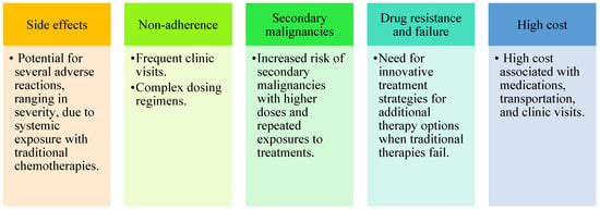
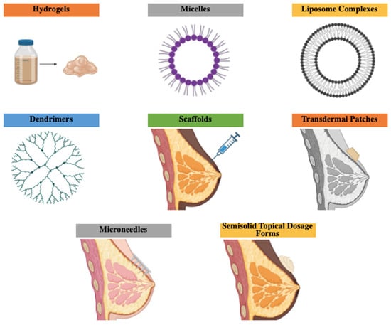
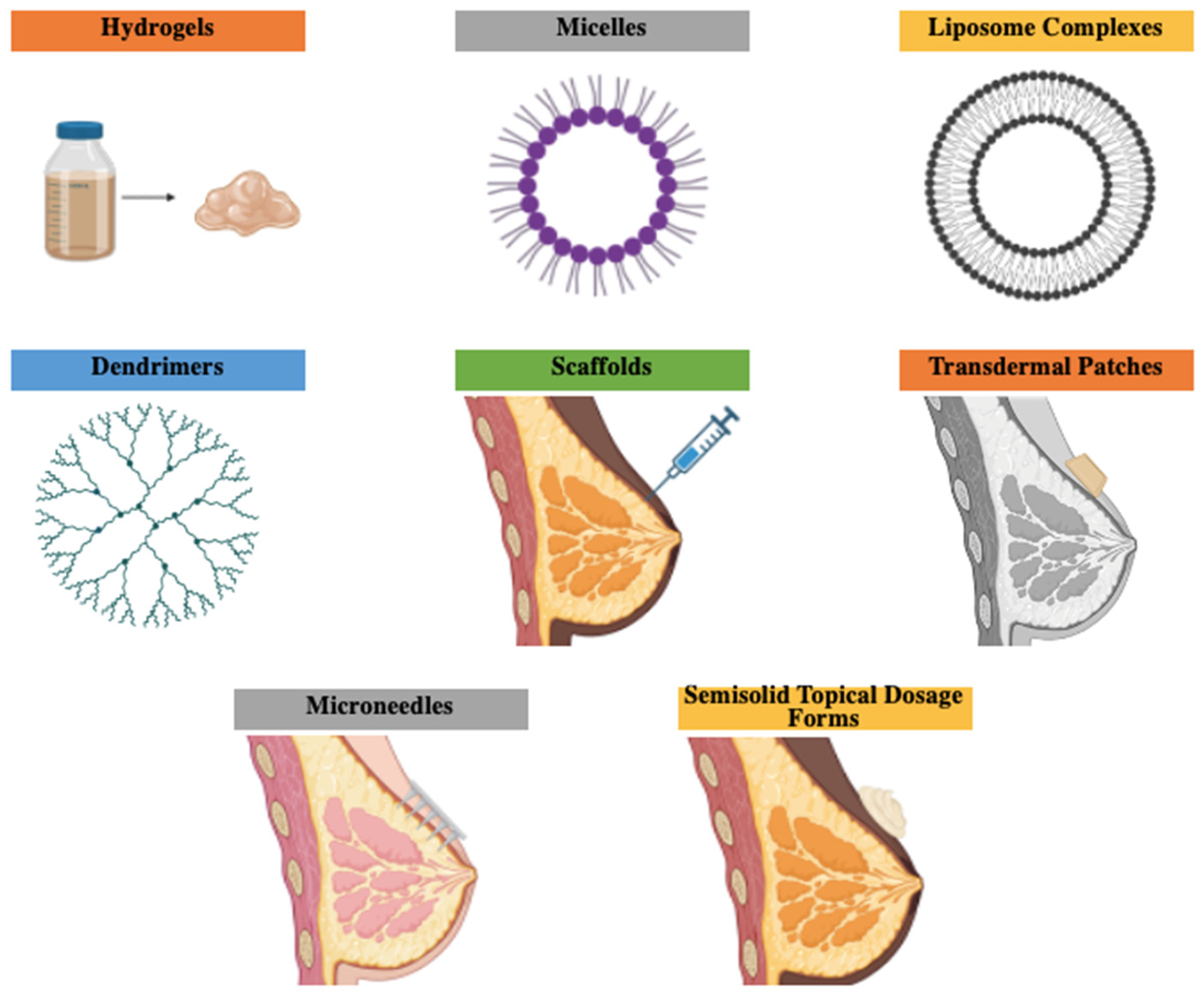
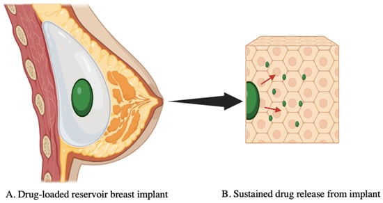
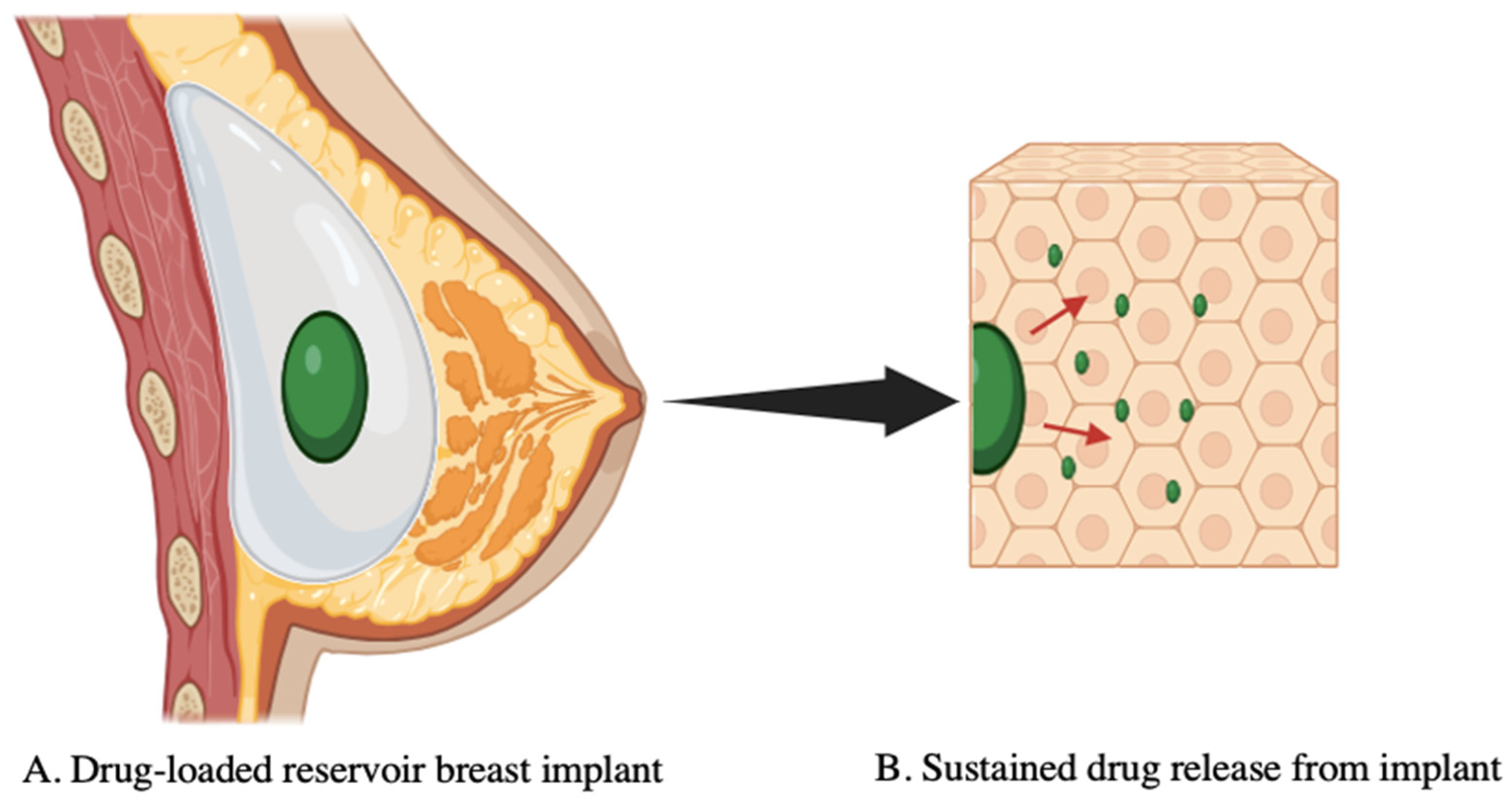

Disclaimer/Publisher’s Note: The statements, opinions and data contained in all publications are solely those of the individual author(s) and contributor(s) and not of MDPI and/or the editor(s). MDPI and/or the editor(s) disclaim responsibility for any injury to people or property resulting from any ideas, methods, instructions or products referred to in the content. |
© 2026 by the authors. Licensee MDPI, Basel, Switzerland. This article is an open access article distributed under the terms and conditions of the Creative Commons Attribution (CC BY) license.