


Abstract
The therapeutic potential of pharmacologically targeting kinases involved in regulating the DNA damage response (DDR) has been investigated for over two decades. Inhibitors of ATM, ATR, CHK1, CHK2 and WEE1 have been developed with the aim of subverting cell cycle checkpoint function in cancer cells, promoting cell death. The DNA repair pathway non-homologous end-joining can also be targeted through DNA-PK inhibition. However, despite extensive preclinical and clinical studies, none of the many candidate inhibitors have yet made it through to clinical approval. Emerging evidence for tumour biomarkers associated with enhanced sensitivity to DDR kinase inhibition may provide a way through this impasse. Clinical testing in appropriately stratified cohorts is now becoming increasingly common, with some promising results. Building on results obtained with small-molecule inhibitors, targeted protein degradation (TPD) utilising proteolysis-targeting chimaeras (PROTACs) or molecular glues for degradation of DDR kinases is a rapidly developing strategy. This review discusses the current ATM, ATR, DNA-PK, CHK1, CHK2 and WEE1 inhibitors that show the most promise as monotherapies and combination treatments in solid tumours, as well as the potential benefits of using TPD technology over small-molecule inhibitors. Established and emerging biomarkers that can be applied to patient selection are also discussed.
Keywords:
ATM; ATR; checkpoint kinases; CHK1; CHK2; DNA damage response; PROTACs; small-molecule inhibitors; WEE1 1. Introduction to the DNA Damage Response
The DNA damage response (DDR) is a cellular mechanism vital for maintaining genomic integrity. If unrepaired, damage to DNA, whether occurring via endogenous or exogenous sources, can lead to mutations that affect biological fitness, including the promotion of carcinogenesis. Although in some circumstances lower fidelity repair mechanisms, such as non-homologous end-joining (NHEJ) or translesion synthesis—which can themselves lead to mutations—are employed, on the whole, the DDR protects genome stability through the activation of signalling cascades that coordinate a complex network of biochemical processes, leading to outcomes such as cell cycle arrest, DNA repair pathway activation, senescence or apoptosis. The repair pathways employed and the differing cellular outcomes are dependent on the nature and extent of the damage, as well as the cell type and tissue context, and the cell cycle stage at which the damage occurs.
Pharmacological targeting of the DDR is a compelling approach for the treatment of cancer for two reasons. Firstly, most untargeted cancer treatments involve the induction of DNA damage. Co-administration of an inhibitor of the DDR (DDRi) can therefore serve to potentiate the effects of these treatments in fast-dividing cancer cells. Secondly, genome instability, driven by the loss of pathways for genome maintenance, is one of the hallmarks of cancer. The resulting loss of functional redundancy creates an over-reliance on remaining pathways, which, if disrupted with a DDRi, can cause cell death via synthetic lethality.
The strength of this approach is exemplified by the successful development of the numerous poly(ADP-ribose) polymerase (PARP) inhibitors that have been licenced for clinical use (reviewed in [1]). However, pharmacological targeting of other components of the DDR is currently under development. In this review, we will focus on the various kinases whose concerted activity serves to co-ordinate DNA repair and cell cycle regulatory components of the DDR, and discuss the recent progress that has been made towards their clinical targeting (Figure 1).
Figure 1.
Cell cycle checkpoint control in the DNA damage response. Apical kinases ATM and ATR are activated in response to DNA damage and subsequently phosphorylate numerous protein targets, of which only a few related to cell cycle control are shown here for clarity. ATM is activated by the presence of DNA double-strand breaks (DSBs) via the Mre11–Rad50–Nbs1 complex (see main text for details). Active ATM phosphorylates the effector kinase CHK2, which in turn can phosphorylate downstream targets such as CDC25 phosphatases and p53. Phosphorylation of CDC25 phosphatases inactivates them, resulting in the loss of their ability to relieve inhibitory phosphorylation of cyclin dependent kinases (CDKs), and cell cycle arrest occurs. Phosphorylation of the transcription factor p53 results in its stabilisation, leading to transcriptional activation of numerous downstream targets involved in apoptosis, DNA repair, and in the case of the CDK inhibitor protein p21, cell cycle arrest. p53 can be phosphorylated by both ATM and CHK2. DNA-PK, consisting of the catalytic subunit DNA-PKCS and the DSB sensors Ku70/80, is also activated by DSBs and plays a minor backup role to checkpoint signalling, but its major function is as a participant in the non-homologous end-joining pathway for DSB repair. ATR is activated by stalled replication forks through interaction with the single-stranded DNA-binding protein RPA via ATR-interacting protein (ATRIP, see main text for details). It too is able to phosphorylate p53, but this is thought to be less important than its phosphorylation of the effector kinase CHK1, and is indicated with a dashed arrow. Targets of ATR include CDC25 phosphatases, similar to ATM, and the checkpoint kinase Wee1, which is able to inactivate CDK1 via inhibitory phosphorylation causing cell cycle arrest. Created in Biorender, https://BioRender.com/m58r118 (accessed on 1 June 2026).
ATM (Ataxia–Telangiectasia Mutated), ATR (Ataxia–Telangiectasia and Rad3-related) and DNA-PK (DNA-dependent protein kinase) are members of the phosphatidylinositol 3-kinase like kinase (PIKK) family of serine/threonine kinases. They share many substrates in common, and each plays an apical role in signalling cascades that orchestrate the repair of DNA damage, ensuring proper cell cycle progression and preventing genomic instability [2]. ATM kinase is primarily activated in response to double-strand breaks (DSBs). Upon recruitment to DSB sites by the Mre11–Rad50–Nbs1 (MRN) complex, dimeric ATM undergoes autophosphorylation and dimer dissociation, becoming an active monomer that phosphorylates various downstream targets [3]. These include the histone variant H2AX, the tumour suppressor protein p53 and the downstream effector kinase, checkpoint kinase 2 (CHK2). This leads to cell cycle arrest, allowing time for DNA repair, and if the damage is irreparable, triggers apoptosis or senescence to prevent the propagation of damaged DNA.
ATR kinase responds primarily to replication stress occurring through nucleotide depletion or the replication fork encountering a DNA lesion, both of which can lead to stalling of replication forks. Single-stranded DNA (ssDNA) accumulating at the stalled fork is initially bound by replication protein A (RPA), which attracts the ATR-interacting protein (ATRIP), which in turn recruits ATR [4]. ATR is then activated by either ETAA1 [5,6,7,8] or TOPBP1 [9] and the Rad9–Rad1–Hus1 (9-1-1) complex [10,11], depending on the nature of the lesion. Activated ATR phosphorylates various substrates, including the effector kinase checkpoint kinase 1 (CHK1), halting cell cycle progression and coordinating DNA repair [12]. ATR also plays a crucial role in suppressing origin firing and preventing DNA degradation at new and ongoing replication forks [13].
Because RPA-coated ssDNA is produced as a normal intermediate during replication, ATR and CHK1 signalling is activated during every S phase even in unperturbed cells, and monitors fork progression and origin firing [14,15]. However, the amount of CHK1 activation varies depending on whether a checkpoint response is required due to the presence of DNA damage [16]. Therefore, the greater the amount of ssDNA present in a perturbed fork, the more CHK1 activation enabling a stronger checkpoint response [17].
The DNA-PK holoenzyme consists of the PIKK catalytic subunit DNA-PKCS and the DSB-sensing heterodimer Ku70/80. Like ATM, it acts in response to DSBs, and shares some substrates in common, including p53 [18,19] and CHK2 [20], but it is much less important for checkpoint regulation [2]. Instead, its most significant role in the DDR is as the master regulator of NHEJ, serving to coordinate end synapsis and the assembly and activation of the repair machinery, and playing both scaffolding and enzymatic roles in these processes. In this way it plays a direct and essential role in the resolution of DSBs. This also includes the joining of DSBs produced during V(D)J recombination as part of adaptive immunity [21]. There is significant crosstalk between DNA-PK and ATM, affecting repair pathway choice. DNA-PK phosphorylates ATM, inhibiting its activity, and abrogation of DNA-PK function results in ATM hyperactivation and an amplified p53 response [22,23]. Conversely, phosphorylation of DNA-PK by ATM promotes NHEJ [24].
WEE1 kinase is another key player in the DDR, functioning primarily to activate the G2/M checkpoint. WEE1 phosphorylates and inhibits cyclin-dependent kinases (CDKs), particularly CDK1 and CDK2, preventing the cell from entering mitosis (M phase) prematurely [25,26]. This inhibition allows time for DNA repair before cell division proceeds. Inhibitory phosphorylation of CDK1 and CDK2 is reversed by CDC25 phosphatases, which in turn are downregulated through phosphorylation by CHK1 [27,28], highlighting the interconnected nature of DDR signalling pathways.
Loss of checkpoint function, whether through somatic mutation or pharmacological inhibition, leads to the accumulation of unrepaired DNA damage that is carried through to mitosis, causing genomic instability or death through mitotic catastrophe. While loss of checkpoint function through somatic mutation helps to drive cancer progression, the potential for checkpoint inhibition to cause cancer cell death, particularly where backup regulatory pathways are missing, provides the rationale for the pharmacological targeting of these kinases in cancer treatments.
2. The Potential of Targeting the DDR via Kinase Inhibition
Many small-molecule inhibitors (SMIs) have been developed targeting important proteins involved in the cellular DDR, namely ATM, ATR, DNA-PK, CHK1, CHK2 and WEE1. Here, we highlight some of the key findings related to the SMIs and the most recent clinical trial evidence for their use in the treatment of cancer.
2.1. ATM Inhibition
Given that ATM has a major role in mediating cell cycle checkpoints and occupies an apical position in DDR pathways, it presents an attractive drug target [29,30,31]. Studies have shown that ATM inhibition greatly sensitises cells to genotoxic agents that cause DSBs, such as ionising radiation and topoisomerase inhibitors [32,33]. The first selective ATM inhibitor (ATMi), KU-55933, was reported by Hickson et al. in 2004 and demonstrated the ability to suppress ionising radiation-dependent p53 phosphorylation and to radiosensitise U2OS and HeLa cancer cell lines [32]. Unfortunately, while KU-55933 and subsequent early compounds, KU-59403 and KU-60019, showed high selectivity towards ATM compared to the closely related kinases mTOR, ATR, PI3K, and DNA-dependent protein kinase (DNA-PK), they also exhibited low oral bioavailability, low aqueous solubility and only moderate cellular potency. Therefore, translating these compounds from bench to clinic did not prove feasible [34,35].
These early ATMis have therefore been superseded by more selective and potent compounds, such as AZD0156 [36] (Table 1). With the advantage of good oral bioavailability, AZD0156 was also shown to have excellent general kinome selectivity, a sub-nanomolar cellular IC50 (0.57 nM), and to potentiate the effects of established chemotherapeutics such as the topoisomerase inhibitor irinotecan and olaparib, a PARP inhibitor [36]. AZD0156 significantly enhanced tumour regression by olaparib in mouse colorectal cancer [36] and triple-negative breast cancer [37] xenograft models. A Phase 1 clinical trial of AZD0156 (NCT02588105) to test safety and efficacy in advanced solid tumours either as a monotherapy or in combination completed in 2019. However, dose-limiting haematological toxicity led to suspension of its further development.
Table 1.
Current status of ATM inhibitors and ongoing trials. Information obtained from ClinicalTrials.gov (1 June 2026).
More promising, particularly for brain cancers, is AZD1390, an ATMi of the same chemical class as AZD0156 but, unlike AZD0156, it is not a substrate for endothelial efflux transporters and can therefore readily cross the blood–brain barrier [39,40]. Accordingly, AZD1390 was shown to promote tumour regression in a range of preclinical xenograft and orthotopic tumour models in mouse brains in combination with either ionising radiation or temozolomide [39,41]. A subsequent study demonstrated that AZD1390’s radiosensitising effects were limited to TP53-defective patient-derived xenografts (PDXs), with TP53 wild-type glioblastoma models exhibiting no enhancement of radioresponse [42]. At the time of writing, there are five ongoing trials of AZD1390 in combination with radiotherapy for the treatment of gliomas and other brain cancers (Table 1).
M3541 and M4076 (lartesertib) are members of a class of 1,3-dihydro-imidazo[4,5-c]quinolin-2-one ATMis that reportedly show similar selectivity and potency to AZD1390 [43,44]. M3541 was designed to be orally administered and showed good selectivity against a panel of 292 protein kinases [43]. In cellular assays, M3541 inhibited radiation-dependent phosphorylation of ATM and its downstream targets, attenuated DSB repair and decreased clonogenic survival [44]. Additionally, it showed synergistic effects in combination with ionising radiation in 79 different tumour cell lines, independent of p53 status and tumour origin, and excellent potentiation of radiotherapy in mouse xenograft models [44]. However, a Phase I trial of M3541 in combination with radiotherapy that began in 2017 was terminated early and further clinical development was halted due to unfavourable pharmacokinetic properties indicative of poor absorption at higher doses [45].
Related compound M4076 (lartesertib) shows similar activity to M3541, but with increased solubility and potency. Like M3541, it can enhance radiotherapy sensitivity in both cellular (head and neck, colon, lung, gastric, breast, and melanoma) and xenograft (head and neck and lung) models of advanced solid tumours. Pairwise testing in cellular models of lartesertib in combination with a panel of 79 chemotherapeutics demonstrated synergistic effects with PARP and topoisomerase inhibitors. These results were further validated in tumour xenografts [44]. A Phase 1 monotherapy trial of lartesertib in patients with advanced solid tumours (NCT04882917) was completed in 2023, and it remains under active clinical development.
More recently developed ATM-targeting therapies that offer promise include XRD-0394, a dual inhibitor of ATM and DNA-PK that, according to an early report, can enhance the effects of ionising radiation and PARP and topoisomerase inhibitors [38], and WSD0628, which shows a promising ability to cross the blood–brain barrier [46].
Accumulating evidence suggests that the efficacy of ATM inhibitors is contingent upon the distinct molecular and genetic profiles of specific tumour types. For example, AZD0156 shows notably effective radiosensitisation of high-grade glioblastomas that have lost function of ATRX, a histone chaperone whose gene is mutated in 30% of paediatric glioblastoma cases. The authors also demonstrated that ATRX regulates CHK1 expression, which they propose leads to a reliance on ATM for survival in ATRX mutants [47]. Mutations in ATRX are a frequent hallmark of the alternative lengthening of the telomeres (ALT) pathway that provides a telomerase-independent mechanism for telomere maintenance [48]. Constitutive activation of ATM was observed in an ALT-dependent subset of neuroblastoma patient-derived cell lines and xenografts; treatment of these with AZD0156 was able to overcome chemotherapy resistance irrespective of ATRX status [49]. This suggests that tumour ALT status may be an appropriate biomarker for stratification for ATMi treatment, but at present there are no clinical trials testing ATMis specifically in ALT+ tumours.
There is some evidence to suggest that ATM inhibitors may see particular utility in enhancing the effects of particle beam therapy. Particle radiotherapies such as proton beam therapy or carbon ion beam therapy offer significant advantages over conventional (photonic) radiotherapy, which employs X-rays. In particular, they precisely target energy deposition to a small volume (the Bragg peak), resulting in better targeting and a high linear energy transfer (LET) causing complex, clustered DNA damage [50]. A 2021 report [49] demonstrated that AZD0156 enhances the relative biological effectiveness (RBE) of proton Bragg peak irradiation in both cell culture and xenograft models of triple-negative breast cancer (TNBC). The authors propose that the clustered damage produced by proton Bragg peak irradiation results in increased reliance on homologous recombination (HR) for repair and survival. Inhibition of ATM re-routes cells into using error-prone and toxic NHEJ for repair, recapitulating similar effects they observe in homologous recombination defective cells. Interestingly, equivalent enhancement of RBE was not seen with ATR inhibition (via AZD6738) [49].
It is becoming increasingly evident that the cancer therapeutic effects of ATM inhibition may lie beyond the simple potentiation of DNA damage in tumour cells. Inhibition of ATM in non-cancer components of the tumour microenvironment can result in immunopotentiation. For example, it has been shown that ATM inhibition in cancer associated myofibroblasts can overcome immunotherapy resistance by suppressing myofibroblast differentiation, which in turn enables cytotoxic T cell infiltration [51]. Additionally, two recent studies have shown that ATM inhibition can potentiate a radiation-induced type 1 interferon response in mouse models of pancreatic cancer [52,53]. These studies suggest that ATMi can exert immunomodulatory effects via both the innate and adaptive pathways and could be used in combination with immune checkpoint inhibitors to reprogramme the tumour microenvironment. A trial of AZD1390 in combination with radiotherapy and the anti-PD-L1 monoclonal antibody therapy durvalumab for treatment of soft tissue sarcoma (NCT05116254) has recently been completed, but results have not yet been reported.
2.2. ATR Inhibition
The ATR/CHK1 pathway is essential for cell survival under replication stress [12], therefore targeting this pathway therapeutically, especially in combination with other treatments, should be advantageous in cancer treatment. Furthermore, p53-deficient tumours, which lack a functional ATM-CHK2-dependent checkpoint, are thought to be particularly susceptible to targeting of the ATR/CHK1 axis [54]. Pharmacological targeting of ATR has been studied for 25 years, since the discovery of its inhibition by the radiosensitiser caffeine, albeit with a weak IC50 (mM range) and low selectivity against other PIKKs [55]. The years since have seen significant progress in ATRi development and eleven are currently in clinical trials, more than for any of the other kinases discussed in this review (Table 2).
Table 2.
ATR inhibitors—current status and ongoing trials. Bold and italic trial identifiers indicate trials in tumours with ATM defects. Data obtained from ClinicalTrials.gov (1 June 2026).
A first-in-class potent ATRi, berzosertib (VX-970, VE-822, M6620) was first reported in 2012 [56]. Based on the lead compound VE-821, which showed good selectivity for ATR but poor pharmacokinetics, berzosertib was able to enhance tumour-selective killing by gemcitabine-radiation combination treatment in a pancreatic ductal adenocarcinoma xenograft model [56]. Subsequent preclinical studies showed berzosertib-induced sensitisation to platinum chemotherapeutics in models of lung [57], oesophageal [58] and colorectal cancer [59]. A Phase 1 trial of berzosertib that tested its monotherapy activity—the first such trial published for an ATRi—reported one patient with advanced colorectal cancer who showed an exceptional response. Subsequent analysis showed that their tumour had multiple disrupted DDR genes and proteins, including loss of expression of ATM and ARID1A; the latter being involved in DNA repair processes via its role in chromatin remodelling [60].
Berzosertib has also been tested for its ability to potentiate radiotherapy. An early preclinical study showed that it could increase the radiosensitivity of TNBC cell lines (BT-549, HCC1806 and MDA-MB-231), but not of a non-cancerous human breast epithelial cell line (MCF10A). It was also demonstrated that PDX models were radiosensitised by the ATRi irrespective of HR-proficiency [61]. Berzosertib also promotes radiation-induced cytotoxicity in non-small cell lung cancer (NSCLC) cell lines via increased mitotic cell death associated with suppression of the G2/M checkpoint and elevated levels of micronuclei. These effects translated into radiosensitisation of intracranial NSCLC patient-derived xenografts (PDXs) [62]. This work is now being moved forward in ongoing clinical trials of berzosertib in combination with radiotherapy for patients with brain metastasis from NSCLC (NCT02589522) and chemotherapy-resistant TNBC tumours (NCT04052555). The results of a clinical trial of berzosertib in combination with radiotherapy (NCT03641547) were published in late 2023, the first ATRi to achieve this milestone, with encouraging results that suggest preclinical models may translate into future therapeutic success [63].
Although reasonably selective for ATR, berzosertib also inhibits ATM with a reported IC50 of 85 nM, which may promote off-target effects [64]. Ceralasertib (AZD6738) has been reported to be a weaker ATRi than berzosertib, with an IC50 of 134 nM versus the former compound’s 5 nM, but it has improved selectivity, with an IC50 34-fold lower for ATR than for its next tightest-binding PIKK target, mTOR [64]. Ceralasertib was unable to inhibit colony growth of any of 21 head and neck squamous cell carcinoma (HNSCC) cell lines when used alone, however, when used in combination with cisplatin it caused varying levels of sensitisation; the combination also inhibited tumour growth in two HNSCC PDX models [65]. Further evidence of ceralasertib’s potential in combination therapy was provided in reports of its synergy with radioimmunotherapy (radiotherapy and anti-PD-L1) against hepatocellular carcinoma mice xenografts [66] and with both photon and proton radiation in various cancer cell lines and a TNBC xenograft model [67]. Ceralasertib was also able to overcome PARPi resistance in PDX models of olaparib-resistant, BRCA1/2 mutant ovarian cancer [68].
However, ceralasertib has been less successful in clinical trials than in preclinical models. A Phase 2 trial of ceralasertib in patients with unresectable or metastatic melanoma resistant to anti-PDL1 therapy showed good tolerability but only a low response rate [69]. A similarly low response rate was also observed in another Phase 2 trial (NCT02937818) of ceralasertib in combination with olaparib for treatment of platinum-refractory small cell lung cancer (SCLC) [70].
Another ATRi, elimusertib (BAY1895344), was reported as being effective as both a single agent and in combination therapy [71]. Elimusertib was suggested to have more potent in vivo anti-tumour efficacy as a monotherapy compared to ceralasertib and berzosertib in GRANTA-519 tumours in mice. Furthermore, in combination with various PARP inhibitors, elimusertib demonstrated synergistic effects in BRCA1-deficient MDA-MB-436 breast cancer cells [71]. Elimusertib was also able to synergise with temozolomide in an in vitro model of glioblastoma, however this did not translate into success in PDX models due to poor pharmacokinetic properties, including high clearance [72].
In various PDX models, elimusertib exhibited potent antitumour activity as a monotherapy, specifically in models harbouring alterations in DDR proteins, such as ATM and BRCA1/2 [73]. Evidence from in vitro assays and clinical trials suggest that elimusertib is likely to be particularly efficacious against DDR-altered cancers [74]. However, a trial of elimusertib in combination with the PARP inhibitor, niraparib was terminated early due to a lack of anticipated benefit compared to standard therapies (NCT04267939). Bayer have since halted clinical investigation of elimusertib due to issues with both tolerability and efficacy, although five clinical trials remain in progress.
Gartisertib (M4344, VX-803) has potency similar to that of elimusertib and was shown to be particularly effective in cancer cells with replication stress or neuroendocrine gene expression signatures [75]. However, due to liver toxicity observed in a Phase 1 trial, further development of gartisertib has been suspended [76].
Other ATR inhibitors in development include tuvusertib (M1774) and camonsertib (RP-3500). Camonsertib is a highly potent ATRi that has a reported IC50 of 0.33 nM in whole cell activity assays and similar selectivity properties to elimusertib [64]. It has reasonably good tolerability, with the main adverse effect being on-target anaemia, manageable through dosage optimisation [77]. A Phase 1 trial of camonsertib as a monotherapy (TRESR) tested for predictive biomarkers for clinical benefit rate from a panel of genes that had previously been identified by a CRISPR-Cas9 chemogenomic screen, albeit in a heavily pre-treated population of patients. Nevertheless, it raised some intriguing points by identifying several biallelic loss-of-function gene alterations that were associated with either RECIST 1.1. or tumour marker response and/or clinical benefit. These included ATM, BRCA1 and BRCA2 but also non-HR-canonical genes including SETD2 and RNASEH2B [78]. The observation of anti-tumour activity in BRCA1 null ovarian cancers that had failed PARP inhibitor treatment provides further evidence for the exciting possibility that ATRi may be able to overcome resistance to PARPi [78].
Additionally, an exceptional and durable response was observed in a patient in the trial, whose metastatic melanoma had biallelic loss of ATRX and was ALT+ [79]. This is consistent with an earlier report that ALT+ cells are hypersensitive to ATR inhibitors. The underlying mechanism is based on the accumulation of RPA at telomeres in the absence of ATRX, which promotes ALT-associated HR via ATR activation [80].
A novel ATRi, tuvusertib, is structurally related to gartisertib (M4344, VX-803), which it superseded in clinical development. Although early data suggested that gartisertib had some promising characteristics, including a potency similar to elimusertib [75], further development was suspended following high levels of liver toxicity in a Phase 1 trial [76]. Tuvusertib, in contrast, is less potent in single agent proliferation assays than either elimusertib or gartisertib, but is more potent that berzosertib and ceralasertib. A recent report on a Phase 1 trial (NCT04170153) of tuvusertib applied as a monotherapy in an unstratified population of patients with a range of solid tumours indicated that loss-of-function mutations in TP53, ARID1A, ATRX and DAXX were predictors of response, where loss of function of the latter gene is also associated with the ALT pathway [81]. A Phase 2 trial of tuvusertib in combination with the anti-PDL1 immune checkpoint inhibitor, avelumab, for treatment of ARID1A-defective endometrial cancer is ongoing (NCT06518564).
ATRis have also been tested for efficacy in combination with other antineoplastic agents. Non-cytotoxic doses of gartisertib and tuvusertib have both been shown to synergise with topoisomerase I inhibitors and other DNA damaging agents in cellular assays, including patient derived organoids, and xenografts [75,82,83]. However, a Phase 1 trial of berzosertib in combination with topotecan found no improvement in progression-free survival [84].
Progress has been made in identifying predictive biomarkers for the successful combination of ATRis with other cancer treatments. For example, treatment of selected cancer cell lines and patient derived organoids with tuvusertib was shown to overcome the resistance to a wide range of DNA damaging agents that is associated with loss of the interferon-inducible antiviral restriction factor Schlafen 11 (SLFN11). SLFN11 is recruited to and blocks stalled replication forks, sensitising cancer cells to DNA-damaging agents, with SLFN11 loss leading to resistance. The study’s authors propose that SLFN11 status could be used for patient stratification in future trials [82]. A CRISPR-based chemogenomic approach was used to identify several biomarkers associated with hypersensitivity to the combination of ATR and PARP inhibitors [85], with the aim of overcoming issues of dose-limiting haematological toxicity previously identified in clinical trials of this combination [86]. The genes identified included RAD51 paralogs, RAD51B and RAD51D, RNase H2 components, RNASEH2A, RNASEH2B, and RNASEH2C, and ATM.
As suggested by some of the preclinical and clinical studies discussed above, loss of ATM function may be a biomarker of ATRi sensitivity. Accordingly, pharmacological inhibition of ATM function has been shown to enhance the cytotoxicity of both tuvusertib [87] and ceralasertib [88]. Conversely, CRISPR/Cas9 genome-wide screening of ATRi-resistant soft tissue sarcoma cell lines identified ATM pathway genes as drivers of resistance [88].
This crosstalk between ATR and ATM is potentially therapeutically exploitable through patient stratification approaches. A number of clinical trials of ATRis on tumours harbouring ATM defects have been carried out with this goal in mind (Trial identifiers highlighted in bold italics in Table 2). Results have been mixed. A Phase 1b trial of elimusertib in advanced solid tumours found that while some patient cohorts, particularly those with gynaecological cancers, saw good response, no association between ATM status and progression free survival was observed. This suggests that ATM deficiency is not prognostic of sensitivity to elimusertib itself, and additional determinants are required for efficacy [89].
A recently developed ATRi, alnodesertib (ART0380), was shown to have selective activity against ATM-defective cancer cells in a study that also included the most extensive analysis to date of ATM variants in cancer mapped to their phenotypic outcomes [90]. A Phase 1/2a trial of alnodesertib in combination with irinotecan for the treatment of patients with cancers that lack immunohistochemically determined ATM expression is ongoing (NCT04657068), with early results leading to it being granted FDA Fast Track designation in September 2025 [91].
Since 2020, the clinical development of ATRis has seen many initially promising candidates abandoned. Most recently, a late 2025 update on the POP-ART study of tuvusertib in combination with a topoisomerase I inhibitor stated that its industrial development has been halted. Nevertheless, a number of ATR inhibitors remain in the pipeline (Table 2), and the promising results obtained with alnodesertib suggest that clinical approval of an ATRi for cancer treatment is edging closer to being a reality.
2.3. DNA-PK Inhibition
When considering DNA-PK inhibition, it is important to note that, in contrast to ATM and ATR, DNA-PK is a direct participant in DNA repair, and the effects of its inhibition lie mostly in the attenuation of NHEJ, rather than through checkpoint disruption. Aberrant expression of DNA-PKCS is a feature of many different types of cancer, where upregulation of NHEJ can help to drive both radio- and chemoresistance, resulting in poor prognosis [92]. DNA-PK is therefore an attractive target for improving therapeutic response and patient survival in such tumours. However, while the first DNA-PK inhibitor was reported more than 30 years ago, the earlier generation of DNA-PK inhibitors had substantial issues with selectivity and/or undesirable pharmacokinetic properties, such as poor solubility [93]. The route towards clinically relevant DNA-PK inhibitors has therefore been slow, with most progress having taken place over the last decade, resulting in several inhibitors having reached clinical trials (Table 3).
Table 3.
Current status and ongoing trials of DNA-PK inhibitors. Information obtained from ClinicalTrials.gov (30 June 2026).
Wortmannin, a highly potent pan-PIKK inhibitor, established DNA-PK as a tractable target whose inhibition results in potentiation of the cell-killing effects of ionising radiation through the inhibition of DSB repair [94,95]. However, lack of selectivity and high toxicity made wortmannin unsuitable for further clinical development. Subsequent attempts to produce more selective DNA-PK inhibitors led to the discovery of a series of derivatives of the PI3K inhibitor LY294002, as summarised in a review by Matsumoto [96]. These include highly selective, nanomolar inhibitors such as NU7026 [97] and NU7441 [98]. Insight gained from such inhibitors include the important observation that DNA-PK therapeutic targeting is synthetically lethal with ATM functional deficiency [99]. However, despite promising results in cellular and preclinical models, poor pharmacokinetic properties have limited their use largely to mechanistic studies of DNA-PK function. Later generation DNA-PK inhibitors have aimed to address these shortcomings with the goal of developing potent, orally available, selective inhibitors with improved half-lives [93].
The most clinically advanced of these inhibitors, peposertib (also known as M3814 and nedisertib) is a highly selective DNA-PK inhibitor, with a sub-nanomolar IC50 and over 1000-fold selectivity over ATM and ATR [100]. Evidence suggests that its therapeutic potential lies in the potentiation of other cancer treatments. In preclinical testing, it showed minimal effects as a monotherapy but could enhance the cytotoxic effects of ionising radiation [101] and chemotherapeutics, including topoisomerase II inhibitors [102] and the DSB-inducing antibody-drug conjugate, Mylotarg [103]. Similarly, in a report of a Phase 1 single agent trial (NCT 02316197), the first for any orally available DNA-PK inhibitor, peposertib was demonstrated to be reasonably well-tolerated but offered little clinical benefit in an unstratified population of patients with advanced solid tumours [104]. However, and consistent with preclinical studies, a Phase 1 study (NCT02516813) of peposertib in combination with palliative radiotherapy with and without cisplatin for the treatment of head and neck cancer found it to be a potent radiosensitiser, albeit with dose-limiting toxicity [105]. Similarly potent radiosensitisation was observed in a Phase 1b trial of peposertib in combination with capecitabine-based chemoradiation but was again associated with dose-limiting toxicity [106]. Several ongoing trials aim to identify predictive biomarkers to improve peposertib’s efficacy. For example, NCT05687136, which investigates the combination of peposertib with tuvusertib, will examine biomarkers of activation of the DDR (e.g., ƔH2AX) and of replication stress, alongside tumour ATM status.
AZD7648 is a potent, highly selective DNA-PK inhibitor that is able to potentiate cancer treatments such as doxorubicin, ionising radiation and olaparib in cell culture and xenograft models [107]. To date, it has been subject to just one clinical trial (NCT03907969), now completed, in which it was tested as a monotherapy or in combination with pegylated liposomal doxorubicin for the treatment of advanced malignancies. Only limited efficacy was seen in both mono- and combination therapies and the latter was associated with an unexpectedly high toxicity profile [108]. Consequently, the trial was terminated early, and no further trials are listed in ClinicalTrials.gov.
Several of the DNA-PK inhibitors under clinical development and listed in Table 3 act as dual or triple inhibitors, having similar IC50s for non-DNA- targets. In contrast to highly non-specific PIKK inhibitors like Wortmannin, the polypharmacology of such drugs is regarded as advantageous, resulting in enhanced cytotoxicity. One such drug is the dual DNA-PK/ATM inhibitor XRD-0394, discussed in Section 2.1, which aims to exploit the functional crosstalk between DNA-PK and ATM [38]. Additionally, bosmolisib (BR101801) is a DNA-PK inhibitor that also targets PI3KƔ and PI3Kδ, and has been demonstrated to synergise with ionising radiation in cancer cell lines and xenograft models [109]. It has been trialled for efficacy as a single agent in haematological malignancies, although results are as yet unpublished.
A further example is CC-115, a potent inhibitor of DNA-PK that has a similar IC50 for mTOR (mechanistic target of rapamycin) [110]. In cellular assays, CC-115 was demonstrated to inhibit both NHEJ and mTOR signalling and to have both growth inhibitory and pro-apoptotic effects in a wide range of cancer cell lines, with enhanced effects in ATM defective lines [111]. Results from a Phase 1 trial in patients with advanced malignancies demonstrated reasonable tolerability and some evidence of efficacy in a subset of patients [112]. A more recent report from a Phase 1b trial that tested CC-115 in combination with androgen receptor inhibition demonstrated a positive trend. However, it was established that under the recommended Phase 2 dose conditions, inhibition of DNA-PK was minimal [113]. Any pharmacological benefit therefore most probably derives from mTOR inhibition, illustrating the challenges of interpreting polypharmacology results. Similar challenges may also apply to the extensively trialled PI3K/mTOR inhibitor, samotolisib (LY3023414) [114,115,116], which is also reported to inhibit DNA-PK [117].
Accumulating evidence suggests that the anti-tumour effects of DNA-PK inhibition may partly lie in immunomodulatory effects, with inhibitors having been shown to promote a hot tumour phenotype. For example, a number of studies have linked inhibition of DNA-PK with stimulation of a Type I interferon response [118,119]. Furthermore, inhibition of DNA-PK was recently shown to increase expression of tumour-associated antigens and neoantigens and promote T cell activation [120]. These emerging details may lead to increased interest in DNA-PK inhibition as a therapeutic strategy.
2.4. CHK1 and/or CHK2 Inhibition
The Ser/Thr protein kinase CHK1 is phosphorylated at Ser-317 and Ser-345 by ATR, leading to its activation and initiation of the DDR and checkpoint signalling cascade of the ATR-CHK1-dependent pathway (Figure 1) [121]. The therapeutic potential of CHK1 inhibition has been explored since 1996 when the first defined CHK1 inhibitor, UCN-01, was shown to induce cell death in Chinese hamster ovary cell lines and enhance radiosensitivity in p53-deficient lymphoma and colon carcinoma cells [122,123,124]. Although CHK1 inhibition has shown some promise as a monotherapy, synergistic inhibitory effects have also been seen when using CHK1 inhibitors in combination with genotoxic agents. For example, UCN-01 in combination with gemcitabine was demonstrated to result in cancer cell death in TNBC by overriding the G1/S phase checkpoint [125]. Similar to the ATRi ceralasertib, the CHK1i MK8775 was also able to restore PARPi sensitivity to olaparib-resistant BRCA1/2 mutant xenografts, suggesting the ATR/CHK1 axis is an important mediator of PARPi sensitivity [68].
Clinical trials of various CHK1 inhibitors in combination with gemcitabine or other antimetabolites have been completed or are currently ongoing (Table 4). However, further development of several of these inhibitors, i.e., AZD7762, MK8766, rabusertib, was suspended due to issues with toxicity and efficacy [126,127,128], suggesting that CHK1 may potentially be flawed as a target due to on-target toxicity and insufficient therapeutic benefit. Nevertheless, several CHK1 inhibitors remain under development.
Table 4.
CHK1/CHK2 inhibitors: current status and ongoing trials. Information obtained from ClinicalTrials.gov (1 June 2026).
CHK2 is a Ser/Thr protein kinase, phosphorylated by ATM at Thr68 upon sensing of DSBs [129] (Figure 1). This kinase has been less investigated compared to the ATR-CHK1 axis and fewer inhibitors have been generated against this target. However, selective CHK2 inhibitors such as BML-277 [130] and CCT241533 [131] have been developed, with some evidence for effectiveness of CHK2 inhibition in overcoming oxaliplatin-resistance [132]. At present, the only CHK2-selective inhibitor to have reached clinical development is lasmotinib (PHI-101) [133]. However, it is also an FLT3 (FMS-like tyrosine kinase 3) inhibitor and has also been investigated for treatment of acute myeloid leukaemia (AML) on that basis.
Targeting both the ATM-CHK2 axis and the ATR-CHK1 axis could prove to be more effective than targeting either checkpoint kinase alone; therefore, dual-targeting inhibitors have been explored despite concerns about tolerability associated with the loss of both checkpoints in healthy cells. For example, AZD7762, a dual CHK1/CHK2 inhibitor, caused cardiotoxicity when it was used in a clinical trial, resulting in early termination of the study [127]. Fortunately, another dual CHK1/CHK2 inhibitor, prexasertib (also known as ACR-368 or LY2606368), has shown promise as a potential treatment option for some types of cancer, with several trials completed or ongoing [134] (Table 4).
Biomarker-driven use of CHK1/2 inhibitors may provide the key to assuring future clinical benefit. A pilot clinical study suggested that prexasertib may be useful for cyclin E (CCNE1) over-expressing high-grade serous ovarian cancer (HGSC) [135]. More recently, a more comprehensive Phase 2 study (NCT03414047) reported that a subset of HGSC patients showed durable response on treatment with prexasertib [136]. However, this was independent of HR status or a range of putative genomic biomarkers including CCNE1 expression, suggesting that further work to establish predictive biomarkers was required. A subsequent Phase 2 study identified that upregulation of genes involved in high-fidelity replication fork initiation and fork progression (POLA1, POLE and GINS3) was associated with prexasertib resistance [137]. Preclinical data suggested that prexasertib may be of therapeutic benefit in PARPi-resistant BRCA mutant HGSC; however, a follow-up Phase 2 trial (NCT02203513) showed clinical benefit was restricted to a subset of patients whose tumours overexpressed CCNE1 and the replication stress associated RecQ helicase, BLM [138].
Collectively, prexasertib clinical trial data support a more stratified approach to the use of CHK1i that takes into account the expression of genes associated with replication stress, fork stabilisation and high-fidelity replication. In 2023, the FDA granted Fast Track approval for prexasertib monotherapy treatment of patients with platinum-resistant ovarian or endometrial cancers stratified on the basis of proteomic profiling of biomarkers using the proprietary OncoSignature platform (NCT05548296). A recent study in cellular and xenograft models has demonstrated that tumour cells harbouring oncogene-carrying extrachromosomal DNA (ecDNA), often associated with poor prognosis, are particularly sensitive to CHK1 inhibition. This was shown to be due to hyperactive transcription on ecDNA causing transcription-replication conflict that in turn leads to elevated replication stress [139]. This discovery is currently being followed up in a Phase 1 trial with the CHK1 inhibitor BBI-355 (NCT05827614).
2.5. WEE1 Inhibition
WEE1, a serine/threonine kinase, phosphorylates CDK1 at Tyr15 to inhibit the formation of the mitotic promoting factor (MPF or Cyclin B-CDK1 complex) and to arrest the cell cycle at the G2/M checkpoint when DNA damage is detected. Conceptually, inhibition of WEE1 has been proposed to result in synthetic lethality in TP53-deficient cancers through abrogation of the G2/M checkpoint, which, alongside their inherently disrupted G1/S checkpoint, leads to mitotic catastrophe [140].
The orally bioavailable WEE1 inhibitor adavosertib (MK1775, AZD1775) was first reported in 2009 and shown to potentiate the anti-tumour effects of gemcitabine and platinum drugs in xenograft models. Consistent with the model proposed above, the authors reported that adavosertib-treated cells bypass the G2/M DNA damage checkpoint, leading to unscheduled mitosis, and further demonstrated that p53-defective tumour cells were more sensitive to adavosertib than p53-competent matched pairs [141]. Similar p53-dependent effects were also seen for radiosensitisation by adavosertib in a subsequent study [142].
Notably, WEE1 inhibition causes defects in homologous recombination [143], providing a rationale for combination treatment with PARP inhibitors. The effect of adavosertib in combination with olaparib was studied in two TP53 wild-type gastric adenocarcinoma cell lines, MKN45 and AGS. Increasing doses of adavosertib resulted in decreased viability and enhanced apoptosis through increased mitotic DNA damage [144]. This study, which also showed that the combination led to inhibited tumour proliferation of xenograft models, suggests synthetic lethality can arise through co-inhibition of WEE1 and PARP. Similarly, it was shown that TNBC cell lines exhibit a decreased capacity to repair DNA DSBs when adavosertib is used as a single agent or in combination with other DDR inhibitors, such as olaparib and ceralasertib, although one unexpected observation from this study was that the p53 status of the cell lines tested did not correlate with their sensitivity to adavosertib. Nevertheless, synergistic effects were observed for the combination of olaparib and adavosertib [145]. Adavosertib not only appears to enhance the effect of olaparib but has also been reported to overcome PARP inhibitor resistance in CAPAN1 (pancreatic cancer) and SUM149 (breast cancer) models that have HR-reactivating secondary BRCA1/2 mutations [146].
Adavosertib was the first WEE1 inhibitor to reach clinical trials [147] and the majority of clinical trials have focused on its use as a combination treatment with concurrent chemotherapy, rather than as a monotherapy (Table 5). For example, an open-label Phase 1 clinical trial of adavosertib assessed it for use in borderline-resectable or unresectable stage III/IVB HNSCC suitable for definitive chemoradiation [148]. This study demonstrated that a combination treatment of adavosertib, docetaxel and cisplatin was tolerable while still being able to reduce tumour growth. The first trial to study definitive-dose radiotherapy with WEE1 inhibition and cisplatin reported that the standard dose of cisplatin was too toxic when administered in combination with adavosertib, concluding that lower doses of cisplatin were more beneficial [149]. However, as noted in the study itself, a major limitation of this trial was the unknown p53-status of the tumours, given that WEE1 inhibition may be expected to have a greater impact on p53-deficient tumours for the reasons discussed above.
Table 5.
Current status and ongoing trials of WEE1 inhibitors. Information obtained from ClinicalTrials.gov (1 June 2026).
Studies with adavosertib have suggested that overexpression of CCNE1 may be a predictive biomarker of sensitivity to WEE1 inhibition [150]. Overexpression of cyclin E is a frequent occurrence in many types of cancer and is associated with poor prognosis [151]. Over-active cyclin E/CDK2 promotes replication stress and aberrant mitotic entry and progression [152], leading to reliance on WEE1 function for survival [153]. A clinical trial of single agent adavosertib treatment of patients selected based on tumour CCNE1 amplification demonstrated promising efficacy, particularly against epithelial ovarian cancer. However, it is worth noting that concurrent TP53 tumour mutations were seen in 90% of the patients enrolled in the study, and it may therefore be the case that optimal synthetic lethality is only seen with concurrent CCNE1 amplification and TP53 dysfunction [154]. Further evidence for CCNE1 amplification as a biomarker of adavosertib response has also been observed in clinical trials for uterine serous cancer [155].
Although adavosertib has shown excellent cytotoxic potency in various cell lines and xenograft models, its translation into clinical use has proven problematic, with myelosuppression—including thrombocytopenia and neutropenia—as a serious side effect. This ultimately led to AstraZeneca suspending further clinical development of the drug in 2022. It has been proposed that one key issue with adavosertib is its lack of selectivity, as it is also a potent inhibitor of the mitotic regulator polo-like kinase 1 (PLK1) [156]. However, subsequent reports suggest that PLK1 inhibition by adavosertib may be negligible at therapeutic concentrations [157,158], and a study of PLK1-sparing WEE1 inhibitors showed that they were still able to cause thrombocytopenia in an in vitro assay [159].
These data suggest that it is the inhibition of WEE1 itself that may underpin the severe side effects of adavosertib, suggesting that there may be some fundamental issues with WEE1 as a therapeutic target. Insight into how WEE1i toxicity arises has been provided by a recent study that shows that both adavosertib and another WEE1 inhibitor, Debio 0123 are able to induce activation of the integrated stress response (ISR), potentially via direct binding and paradoxical activation of GCN2 kinase [160]. This raises the possibility that checkpoint perturbation without ISR-mediated toxic effects could be achieved through WEE1i dose manipulation or combination with other agents.
Several other selective WEE1 inhibitors, including azenosertib (Zn-c3), SC0191 and IMP7068, are currently in ongoing trials (Table 5), and other promising candidates are in preclinical development (see, e.g., [161,162]). There are yet to be any peer-reviewed publications on IMP7068, but early reports suggest it has significantly improved selectivity over PLK1 compared to adavosertib and is currently in a Phase 1 trial as a monotherapy (NCT04768868) [163].
First reported in 2021 and most clinically advanced of these alternative WEE1is, azenosertib has good oral bioavailability and a similar IC50 to adavosertib (75 nM and 78 nM, respectively, in a lung cancer xenograft model) but greatly improved kinase selectivity [164]. Azenosertib is currently in multiple ongoing clinical trials as both a monotherapy and in combination with traditional chemotherapeutics and targeted therapies. Early results are promising. A dose-escalation Phase 1 trial demonstrated that azenosertib has single agent anti-tumour effects in heavily pretreated patients with various types of cancer [165]. Preclinical and clinical testing of azenosertib in the treatment of ovarian cancer and uterine serous carcinomas corroborated the previous observation from adavosertib that CCNE1-amplified tumours show enhanced sensitivity to WEE1 inhibition [166].
As these trials reach maturity, it will be interesting to see if the improved selectivity of next generation WEE1is such as azenosertib and IMP7068 will translate into greater efficacy and/or fewer side effects. A further priority of WEE1i research should be the identification and validation of biomarkers of sensitivity to enable stratification.
3. Development of Kinase Degraders as Alternatives to Classic Inhibitors
In recent years, the concept of targeted protein degradation (TPD) has seen increasing application in the development of novel cancer agents [167]. The majority of TPD agents are proteolysis-targeting chimeras (PROTACs), bifunctional molecules that conjugate an E3 ubiquitin ligase-targeting domain to a protein of interest (POI)-binding ligand via a linker [168]. This brings the target protein into close proximity to the E3 ubiquitin ligase, leading to polyubiquitination and subsequent degradation by the ubiquitin proteasome system, generating a chemical knockdown phenotype (Figure 2) [169].
Figure 2.
Hijacking the ubiquitin–proteasome system with a heterobifunctional molecule. Proteolysis targeting chimaeras (PROTACs) contain two binding domains that recruit a protein of interest, the warhead, and an E3 ligase. When the PROTAC is bound to its targets, it forms a ternary complex that allows the protein of interest to be polyubiquitylated and targeted for degradation by the 26S proteasome [170].
PROTACs (and other TPD agents) offer several advantages over SMIs. Firstly, by acting catalytically, they can be used at substoichiometric concentrations, reducing the potential for off-target effects. Off-target effects can also be reduced through enhanced selectivity for POIs versus the parent SMI, which can occur due to increased serendipitous protein-drug interactions. PROTACs also do not have the limitation of needing to bind to an active or allosteric site to have their desired effect, opening up the possibility of targeting proteins previously regarded as ‘undruggable’ and enabling protein-protein interactions to be disrupted, in addition to enzymatic activities.
In the past decades, many PROTACs have been developed to target a wide range of proteins related to cancer. In mid-2026, vepdegestrant became the first PROTAC to achieve FDA approval. The drug, an oestrogen receptor (ER) degrader, has been approved for the treatment of ER positive, human epidermal growth factor receptor 2 (HER2)-negative, ESR1-mutated advanced or metastatic breast cancer. The first PROTAC molecule developed to target the DDR was a PARP1-targeting PROTAC that was shown to induce apoptotic cleavage of PARP1, rather than true degradation [171]. However, several reports of truly PARP1-degrading PROTACs have followed since [172,173,174,175,176], in addition to a series of dual-targeting, trifunctional PROTACs which simultaneously target PARP and the epidermal growth factor receptor (EGFR) for degradation [177].
The first reported PROTAC to target a DNA damage response kinase was ZNL-02-96, a PROTAC based on an adavosertib warhead conjugated to pomalidomide, a recruiter of the E3 ligase, cereblon (CRBN) (Table 6). The compound was shown to be a potent WEE1 degrader while sparing PLK1 and was effective in downregulating WEE1 signalling and inducing cell cycle changes at doses 10-fold lower than adavosertib [178]. Aublette et al. reported the synthesis of a range of WEE1 degraders also based on adavosertib, with the best performing two, MA055 and MA071, recruiting von Hippel–Lindau (VHL) and CRBN, respectively. They demonstrated that the linker length between the adavosertib warhead and the E3 ligase recruiting moiety was critical in determining how effective the PROTAC was in promoting WEE1 degradation [179]. Similar linker effects were observed by Zhu et al. who also synthesised a range of WEE1 PROTACs. Their best performing compound in cell proliferation and degradation assays, 42a, is similar in structure to ZNL-02-096 with only one additional methylene group in the linker distinguishing it from the earlier compound [180].
Table 6.
Current PROTACs targeting DNA damage response kinases. The ligand for the E3 ligase is shown in blue and the ligand for the protein of interest is shown in red.
A recent report of a PROTAC (Abd100, Ramotac-1) comprising a VE-821-based warhead conjugated to lenalidomide as a CRBN recruiter delivered the first proof-of concept of pharmacological degradation of ATR as a cancer treatment strategy [181]. Although only limited biological characterisation has been carried out so far, it has been demonstrated that Abd100 could potentiate replication stress-induced cytotoxicity in primary leukaemic cells [182]. Cytotoxicity was not seen in equivalent CRBN knockout cells providing evidence that its effects are due to ATR degradation and not inhibition [182].
As work progresses on Abd100 and any future ATR targeting PROTACs it will be interesting to see how their efficacy and tolerability compare to ATR inhibitors. Evidence suggests that their effects are likely to be quite different. Expression of kinase-dead ATR has shown that ATR’s enzymatic activity is required for its exchange at damage sites and downstream activation of CHK1. Inhibition of this process via expression of a kinase-dead mutant, and presumably also through drug-mediated inhibition, leads to the accumulation of RPA on ssDNA, blocking repair and checkpoint activation and having a dominant negative effect [183].
A 2020 screen of 91 PROTAC compounds constructed from a diverse range of parental kinase inhibitors identified two, derived from TAE684 and AT7519 respectively, which were able to act as CHK1 degraders, demonstrating proof of concept. However, both compounds also induced degradation of many other kinases and so are not practicable for development as therapeutic agents [184]. More recently, MA203, a rabusertib-derived CRBN PROTAC has been developed to selectively target activated CHK1 for degradation and exhibits cytotoxic effects as a single agent and in combination with replication stress-inducing hydroxyurea [185].
The development of WEE1 and ATR targeting PROTACs has involved the well-established PROTAC design concept of conjugating a drug targeting the POI to an E3 ligase recruiting moiety via a linker. However, the first reported ATM-targeting PROTAC was discovered by a more serendipitous process. Meisoindigo is a drug derived from traditional Chinese medicine that has long been used in the treatment of CML in China, but its cellular target was unknown [186]. In a recent study a VHL-type PROTAC-derivative of meisoindigo was synthesised and identified as an ATM degrader by proteomic analysis. Promisingly, it can sensitise cells and xenografts to ceralasertib [187]. Whether an ATM degrader will offer therapeutic advantages over ATM inhibitors is currently unexplored but there is some evidence to suggest that it might. Mouse models have shown that expression of a kinase-dead ATM protein in ATM knockouts causes increased genomic instability, resulting in embryonic lethality, in comparison to the knockouts themselves, which are viable [188,189,190,191]. This observation suggests that ATM degraders could offer improved tolerability over ATM inhibitors.
Molecular glues function in a similar way to PROTACs by targeting a protein of interest for degradation, but they do so by remodelling the interaction surfaces of proteins, generating neosubstrates that enable novel interactions to occur. Typically smaller in size than PROTACs, they can offer pharmacokinetic advantages over their larger bivalent counterparts [192]. A recent proteomic screen of a combinatorial library of potential molecular glues led to the discovery of several monovalent WEE1 degraders, with one demonstrating half-maximal degradation of WEE1 at sub-nanomolar concentrations [193]. BMS-986463, another WEE1-targeting molecular glue developed by Bristol Myers Squibb is currently in an escalating dose Phase 1 trial in patients with advanced solid tumours (NCT06476808).
The development of degraders targeting DNA damage response kinases is still in its early days, with only one candidate compound in clinical testing to date. However, this is becoming an increasingly active area of research, and so it can be expected that the next few years will see more compounds developed and moving into clinical testing.
4. Conclusions and Future Prospects
After more than two decades of development, the goal of treating cancer through the therapeutic targeting of DDR kinases has still not been achieved, and issues of tolerability and efficacy remain key considerations. However, whether as monotherapies or in combination with other agents, recently emerging clinical trial evidence suggests that, for a subset of patients, checkpoint kinase inhibitors do offer some clinical benefit. The granting of FDA Fast Track status to the ATRi alnodesertib and the CHK1i prexasertib for cancers with specific molecular profiles represents significant milestones. As work on identifying biomarkers continues to progress, and more effective patient stratification becomes a reality, it is likely that some DDR kinase inhibitors will join PARP inhibitors in the arsenal of anti-cancer drugs that target the DDR. However, although these inhibitors show great promise, acquired resistance remains a concern. Therefore, gaining a further understanding of resistance pathways, and how to evade them, will be crucial for the field to progress.
The development of novel inhibitors of DDR kinases remains a very active area of research, and the coming years are likely to see more entering clinical trials. Newly emerging therapeutic strategies may help to overcome some of the apparent on-target toxicity associated with DDR kinase inhibition. In this respect, the recent development of several PROTACs and molecular glues targeting DDR kinases represents exciting progress. A recent proof-of-concept report of a multi-payload antibody–drug conjugate that links Topoisomerase I, PARP and ATR inhibitors to a tumour-targeting antibody suggests an additional means by which efficacy can be maximised while sparing normal tissue [194]. Although the preclinical research into these compounds shows promise, there is a need for more research in cell lines and patient-relevant models such as xenografts and tumoroids.
Author Contributions
Conceptualisation, L.A.B.-B., M.S.G., J.L.P. and S.L.A.; writing—original draft preparation, L.A.B.-B., A.M.R. and S.L.A.; writing—review and editing, L.A.B.-B., A.M.R., A.B.F., J.L.P., M.S.G. and S.L.A.; supervision, A.B.F., J.L.P., M.S.G. and S.L.A.; funding acquisition, M.S.G., J.L.P. and S.L.A. All authors have read and agreed to the published version of the manuscript.
Funding
This work was partly funded through a PhD studentship for L.A.B.-B. obtained through the North West Cancer Research Doctoral Training Programme, grant number JXR30229.
Institutional Review Board Statement
Not Applicable.
Informed Consent Statement
Not Applicable.
Data Availability Statement
No new data were created or analysed in this study. Data sharing is not applicable to this article.
Conflicts of Interest
The authors declare that they have no conflicts of interest.
Abbreviations
The following abbreviations are used in this manuscript.
| ALT | Alternative lengthening of telomeres |
| AML | Acute myeloid leukaemia |
| ATM | Ataxia–Telangiectasia mutated |
| ATR | Ataxia–Telangiectasia and Rad3-related |
| ATRIP | ATR-interacting protein |
| CHK1 | Checkpoint kinase 1 |
| CHK2 | Checkpoint kinase 2 |
| CRBN | Cereblon |
| DDR | DNA damage response |
| DNA-PK | DNA-dependent protein kinase |
| DSB | Double-strand break |
| ecDNA | Extrachromosomal DNA |
| EGFR | Epidermal growth factor receptor |
| ER | Oestrogen receptor |
| FLT3 | FMS-like tyrosine kinase 3 |
| HER2 | Human epidermal growth factor receptor 2 |
| HGSC | High-grade serous ovarian cancer |
| HNSCC | Head and neck squamous cell carcinoma |
| HR | Homologous recombination |
| ISR | Integrated stress response |
| LET | Linear energy transfer |
| MPF | Mitotic promoting factor |
| MRN | Mre11–Rad50–Nbs1 |
| NHEJ | Non-homologous end joining |
| NSCLC | Non-small-cell lung cancer |
| PARP | Poly(ADP-ribose) polymerase |
| PDX | Patient-derived xenograft |
| PIKK | Phodphatidylinositol-3-kinase like kinase |
| PLK1 | Polo-like kinase 1 |
| POI | Protein of interest |
| PROTAC | Proteolysis targeting chimaera |
| RBE | Relative biological effectiveness |
| RPA | Replication protein A |
| SCLC | Small-cell lung cancer |
| SMI | Small-molecule inhibitor |
| TNBC | Triple-negative breast cancer |
| TPD | Targeted protein degradation |
| VHL | Von Hippel–Lindau |
References
- Xiang, J.; Li, J.; Guo, Z.; Wu, J.; Ma, D.; Xu, H.; Shang, T.; Pu, P.; Cong, L.; Zhou, R.; et al. Clinical application of PARP inhibitors and emerging strategies to overcome resistance: A pan-cancer perspective. Biomark. Res. 2026, 14, 43. [Google Scholar] [CrossRef] [Scilit] [PubMed]
- Blackford, A.N.; Jackson, S.P. ATM, ATR, and DNA-PK: The Trinity at the Heart of the DNA Damage Response. Mol. Cell 2017, 66, 801–817. [Google Scholar] [CrossRef] [Scilit] [PubMed]
- Lee, J.H.; Paull, T.T. Cellular functions of the protein kinase ATM and their relevance to human disease. Nat. Rev. Mol. Cell Biol. 2021, 22, 796–814. [Google Scholar] [CrossRef] [Scilit] [PubMed]
- Zou, L.; Elledge, S.J. Sensing DNA damage through ATRIP recognition of RPA-ssDNA complexes. Science 2003, 300, 1542–1548. [Google Scholar] [CrossRef] [Scilit] [PubMed]
- Bass, T.E.; Luzwick, J.W.; Kavanaugh, G.; Carroll, C.; Dungrawala, H.; Glick, G.G.; Feldkamp, M.D.; Putney, R.; Chazin, W.J.; Cortez, D. ETAA1 acts at stalled replication forks to maintain genome integrity. Nat. Cell Biol. 2016, 18, 1185–1195. [Google Scholar] [CrossRef] [Scilit] [PubMed]
- Feng, S.; Zhao, Y.; Xu, Y.; Ning, S.; Huo, W.; Hou, M.; Gao, G.; Ji, J.; Guo, R.; Xu, D. Ewing Tumor-associated Antigen 1 Interacts with Replication Protein A to Promote Restart of Stalled Replication Forks. J. Biol. Chem. 2016, 291, 21956–21962. [Google Scholar] [CrossRef] [Scilit] [PubMed]
- Haahr, P.; Hoffmann, S.; Tollenaere, M.A.; Ho, T.; Toledo, L.I.; Mann, M.; Bekker-Jensen, S.; Raschle, M.; Mailand, N. Activation of the ATR kinase by the RPA-binding protein ETAA1. Nat. Cell Biol. 2016, 18, 1196–1207. [Google Scholar] [CrossRef] [Scilit] [PubMed]
- Lee, Y.C.; Zhou, Q.; Chen, J.; Yuan, J. RPA-Binding Protein ETAA1 Is an ATR Activator Involved in DNA Replication Stress Response. Curr. Biol. 2016, 26, 3257–3268. [Google Scholar] [CrossRef] [Scilit] [PubMed]
- Kumagai, A.; Lee, J.; Yoo, H.Y.; Dunphy, W.G. TopBP1 activates the ATR-ATRIP complex. Cell 2006, 124, 943–955. [Google Scholar] [CrossRef] [Scilit] [PubMed]
- Delacroix, S.; Wagner, J.M.; Kobayashi, M.; Yamamoto, K.; Karnitz, L.M. The Rad9-Hus1-Rad1 (9-1-1) clamp activates checkpoint signaling via TopBP1. Genes Dev. 2007, 21, 1472–1477. [Google Scholar] [CrossRef] [Scilit] [PubMed]
- Lee, J.; Kumagai, A.; Dunphy, W.G. The Rad9-Hus1-Rad1 checkpoint clamp regulates interaction of TopBP1 with ATR. J. Biol. Chem. 2007, 282, 28036–28044. [Google Scholar] [CrossRef] [Scilit] [PubMed]
- Saldivar, J.C.; Cortez, D.; Cimprich, K.A. The essential kinase ATR: Ensuring faithful duplication of a challenging genome. Nat. Rev. Mol. Cell Biol. 2017, 18, 622–636. [Google Scholar] [CrossRef] [Scilit] [PubMed]
- Leung, W.; Simoneau, A.; Saxena, S.; Jackson, J.; Patel, P.S.; Limbu, M.; Vindigni, A.; Zou, L. ATR protects ongoing and newly assembled DNA replication forks through distinct mechanisms. Cell Rep. 2023, 42, 112792. [Google Scholar] [CrossRef] [Scilit] [PubMed]
- Buisson, R.; Boisvert, J.L.; Benes, C.H.; Zou, L. Distinct but Concerted Roles of ATR, DNA-PK, and Chk1 in Countering Replication Stress during S Phase. Mol. Cell 2015, 59, 1011–1024. [Google Scholar] [CrossRef] [Scilit] [PubMed]
- Moiseeva, T.N.; Yin, Y.; Calderon, M.J.; Qian, C.; Schamus-Haynes, S.; Sugitani, N.; Osmanbeyoglu, H.U.; Rothenberg, E.; Watkins, S.C.; Bakkenist, C.J. An ATR and CHK1 kinase signaling mechanism that limits origin firing during unperturbed DNA replication. Proc. Natl. Acad. Sci. USA 2019, 116, 13374–13383. [Google Scholar] [CrossRef] [Scilit] [PubMed]
- Iyer, D.R.; Rhind, N. The Intra-S Checkpoint Responses to DNA Damage. Genes 2017, 8, 74. [Google Scholar] [CrossRef] [Scilit] [PubMed]
- Byun, T.S.; Pacek, M.; Yee, M.C.; Walter, J.C.; Cimprich, K.A. Functional uncoupling of MCM helicase and DNA polymerase activities activates the ATR-dependent checkpoint. Genes Dev. 2005, 19, 1040–1052. [Google Scholar] [CrossRef] [Scilit] [PubMed]
- Lees-Miller, S.P.; Sakaguchi, K.; Ullrich, S.J.; Appella, E.; Anderson, C.W. Human DNA-activated protein kinase phosphorylates serines 15 and 37 in the amino-terminal transactivation domain of human p53. Mol. Cell. Biol. 1992, 12, 5041–5049. [Google Scholar] [CrossRef] [Scilit] [PubMed]
- Shieh, S.Y.; Ikeda, M.; Taya, Y.; Prives, C. DNA damage-induced phosphorylation of p53 alleviates inhibition by MDM2. Cell 1997, 91, 325–334. [Google Scholar] [CrossRef] [Scilit] [PubMed]
- Li, J.; Stern, D.F. Regulation of CHK2 by DNA-dependent protein kinase. J. Biol. Chem. 2005, 280, 12041–12050. [Google Scholar] [CrossRef] [Scilit] [PubMed]
- Chen, S.; Lees-Miller, J.P.; He, Y.; Lees-Miller, S.P. Structural insights into the role of DNA-PK as a master regulator in NHEJ. Genome Instab. Dis. 2021, 2, 195–210. [Google Scholar] [CrossRef] [Scilit] [PubMed]
- Zhou, Y.; Lee, J.H.; Jiang, W.; Crowe, J.L.; Zha, S.; Paull, T.T. Regulation of the DNA Damage Response by DNA-PKcs Inhibitory Phosphorylation of ATM. Mol. Cell 2017, 65, 91–104. [Google Scholar] [CrossRef] [Scilit] [PubMed]
- Finzel, A.; Grybowski, A.; Strasen, J.; Cristiano, E.; Loewer, A. Hyperactivation of ATM upon DNA-PKcs inhibition modulates p53 dynamics and cell fate in response to DNA damage. Mol. Biol. Cell 2016, 27, 2360–2367. [Google Scholar] [CrossRef] [Scilit] [PubMed]
- Lu, H.; Zhang, Q.; Laverty, D.J.; Puncheon, A.C.; Augustine, M.M.; Williams, G.J.; Nagel, Z.D.; Chen, B.P.C.; Davis, A.J. ATM phosphorylates the FATC domain of DNA-PKcs at threonine 4102 to promote non-homologous end joining. Nucleic Acids Res. 2023, 51, 6770–6783. [Google Scholar] [CrossRef] [Scilit] [PubMed]
- Gu, Y.; Rosenblatt, J.; Morgan, D.O. Cell cycle regulation of CDK2 activity by phosphorylation of Thr160 and Tyr15. EMBO J. 1992, 11, 3995–4005. [Google Scholar] [CrossRef] [Scilit] [PubMed]
- Heald, R.; McLoughlin, M.; McKeon, F. Human wee1 maintains mitotic timing by protecting the nucleus from cytoplasmically activated Cdc2 kinase. Cell 1993, 74, 463–474. [Google Scholar] [CrossRef] [Scilit] [PubMed]
- Peng, C.Y.; Graves, P.R.; Thoma, R.S.; Wu, Z.; Shaw, A.S.; Piwnica-Worms, H. Mitotic and G2 checkpoint control: Regulation of 14-3-3 protein binding by phosphorylation of Cdc25C on serine-216. Science 1997, 277, 1501–1505. [Google Scholar] [CrossRef] [Scilit] [PubMed]
- Sanchez, Y.; Wong, C.; Thoma, R.S.; Richman, R.; Wu, Z.; Piwnica-Worms, H.; Elledge, S.J. Conservation of the Chk1 checkpoint pathway in mammals: Linkage of DNA damage to Cdk regulation through Cdc25. Science 1997, 277, 1497–1501. [Google Scholar] [CrossRef] [Scilit] [PubMed]
- Tang, S.; Li, Z.; Yang, L.; Shen, L.; Wang, Y. A potential new role of ATM inhibitor in radiotherapy: Suppressing ionizing Radiation-Activated EGFR. Int. J. Radiat. Biol. 2020, 96, 461–468. [Google Scholar] [CrossRef] [Scilit] [PubMed]
- Golding, S.E.; Rosenberg, E.; Khalil, A.; McEwen, A.; Holmes, M.; Neill, S.; Povirk, L.F.; Valerie, K. Double strand break repair by homologous recombination is regulated by cell cycle-independent signaling via ATM in human glioma cells. J. Biol. Chem. 2004, 279, 15402–15410. [Google Scholar] [CrossRef] [Scilit] [PubMed]
- Bensimon, A.; Aebersold, R.; Shiloh, Y. Beyond ATM: The protein kinase landscape of the DNA damage response. FEBS Lett. 2011, 585, 1625–1639. [Google Scholar] [CrossRef] [Scilit] [PubMed]
- Hickson, I.; Zhao, Y.; Richardson, C.J.; Green, S.J.; Martin, N.M.; Orr, A.I.; Reaper, P.M.; Jackson, S.P.; Curtin, N.J.; Smith, G.C. Identification and characterization of a novel and specific inhibitor of the ataxia-telangiectasia mutated kinase ATM. Cancer Res. 2004, 64, 9152–9159. [Google Scholar] [CrossRef] [Scilit] [PubMed]
- García, M.E.G.; Kirsch, D.G.; Reitman, Z.J. Targeting the ATM Kinase to Enhance the Efficacy of Radiotherapy and Outcomes for Cancer Patients. Semin. Radiat. Oncol. 2022, 32, 3–14. [Google Scholar] [CrossRef] [Scilit] [PubMed]
- Batey, M.A.; Zhao, Y.; Kyle, S.; Richardson, C.; Slade, A.; Martin, N.M.; Lau, A.; Newell, D.R.; Curtin, N.J. Preclinical evaluation of a novel ATM inhibitor, KU59403, in vitro and in vivo in p53 functional and dysfunctional models of human cancer. Mol. Cancer Ther. 2013, 12, 959–967. [Google Scholar] [CrossRef] [Scilit] [PubMed]
- Hickson, I.; Pike, K.G.; Durant, S.T. Targeting ATM for Cancer Therapy: Prospects for Drugging ATM. In Targeting the DNA Damage Response for Anti-Cancer Therapy; Pollard, J., Curtin, N., Eds.; Springer International Publishing: Cham, Switzerland, 2018; pp. 185–208. [Google Scholar]
- Pike, K.G.; Barlaam, B.; Cadogan, E.; Campbell, A.; Chen, Y.; Colclough, N.; Davies, N.L.; de-Almeida, C.; Degorce, S.L.; Didelot, M.; et al. The Identification of Potent, Selective, and Orally Available Inhibitors of Ataxia Telangiectasia Mutated (ATM) Kinase: The Discovery of AZD0156 (8-{6-[3-(Dimethylamino)propoxy]pyridin-3-yl}-3-methyl-1-(tetrahydro-2H-pyran-4-yl)-1,3-dihydro-2H-imidazo[4,5-c]quinolin-2-one). J. Med. Chem. 2018, 61, 3823–3841. [Google Scholar] [CrossRef] [Scilit] [PubMed]
- Riches, L.C.; Trinidad, A.G.; Hughes, G.; Jones, G.N.; Hughes, A.M.; Thomason, A.G.; Gavine, P.; Cui, A.; Ling, S.; Stott, J.; et al. Pharmacology of the ATM Inhibitor AZD0156: Potentiation of Irradiation and Olaparib Responses Preclinically. Mol. Cancer Ther. 2020, 19, 13–25. [Google Scholar] [CrossRef] [Scilit] [PubMed]
- Gilmer, T.M.; Lai, C.H.; Guo, K.; Deland, K.; Ashcraft, K.A.; Stewart, A.E.; Wang, Y.; Fu, J.; Wood, K.C.; Kirsch, D.G.; et al. A Novel Dual ATM/DNA-PK Inhibitor, XRD-0394, Potently Radiosensitizes and Potentiates PARP and Topoisomerase I Inhibitors. Mol. Cancer Ther. 2024, 23, 751–765. [Google Scholar] [CrossRef] [Scilit] [PubMed]
- Durant, S.T.; Zheng, L.; Wang, Y.; Chen, K.; Zhang, L.; Zhang, T.; Yang, Z.; Riches, L.; Trinidad, A.G.; Fok, J.H.L.; et al. The brain-penetrant clinical ATM inhibitor AZD1390 radiosensitizes and improves survival of preclinical brain tumor models. Sci. Adv. 2018, 4, eaat1719. [Google Scholar] [CrossRef] [Scilit] [PubMed]
- Jucaite, A.; Stenkrona, P.; Cselenyi, Z.; De Vita, S.; Buil-Bruna, N.; Varnas, K.; Savage, A.; Varrone, A.; Johnstrom, P.; Schou, M.; et al. Brain exposure of the ATM inhibitor AZD1390 in humans-a positron emission tomography study. Neuro-Oncology 2021, 23, 687–696. [Google Scholar] [CrossRef] [Scilit] [PubMed]
- Tew, B.Y.; Kalfa, A.J.; Yang, Z.; Hurth, K.M.; Simon, T.; Abnoosian, E.; Durant, S.T.; Hamerlik, P.; Salhia, B. ATM-Inhibitor AZD1390 Is a Radiosensitizer for Breast Cancer CNS Metastasis. Clin. Cancer Res. 2023, 29, 4492–4503. [Google Scholar] [CrossRef] [Scilit] [PubMed]
- Chen, J.; Laverty, D.J.; Talele, S.; Bale, A.; Carlson, B.L.; Porath, K.A.; Bakken, K.K.; Burgenske, D.M.; Decker, P.A.; Vaubel, R.A.; et al. Aberrant ATM signaling and homology-directed DNA repair as a vulnerability of p53-mutant GBM to AZD1390-mediated radiosensitization. Sci. Transl. Med. 2024, 16, eadj5962. [Google Scholar] [CrossRef] [Scilit] [PubMed]
- Fuchss, T.; Mederski, W.W.; Zenke, F.T.; Dahmen, H.; Zimmermann, A.; Blaukat, A. Highly potent and selective ATM kinase inhibitor M3541: A clinical candidate drug with strong antitumor activity in combination with radiotherapy. Cancer Res. 2018, 78, 329. [Google Scholar] [CrossRef] [Scilit]
- Zimmermann, A.; Zenke, F.T.; Chiu, L.Y.; Dahmen, H.; Pehl, U.; Fuchss, T.; Grombacher, T.; Blume, B.; Vassilev, L.T.; Blaukat, A. A New Class of Selective ATM Inhibitors as Combination Partners of DNA Double-Strand Break Inducing Cancer Therapies. Mol. Cancer Ther. 2022, 21, 859–870. [Google Scholar] [CrossRef] [Scilit] [PubMed]
- Waqar, S.N.; Robinson, C.; Olszanski, A.J.; Spira, A.; Hackmaster, M.; Lucas, L.; Sponton, L.; Jin, H.; Hering, U.; Cronier, D.; et al. Phase I trial of ATM inhibitor M3541 in combination with palliative radiotherapy in patients with solid tumors. Investig. New Drugs 2022, 40, 596–605. [Google Scholar] [CrossRef] [Scilit]
- Rathi, S.; Oh, J.H.; Zhang, W.; Mladek, A.C.; Garcia, D.A.; Xue, Z.; Burgenske, D.M.; Zhang, W.; Le, J.; Zhong, W.; et al. Preclinical Systemic Pharmacokinetics, Dose Proportionality, and Central Nervous System Distribution of the ATM Inhibitor WSD0628, a Novel Radiosensitizer for the Treatment of Brain Tumors. J. Pharmacol. Exp. Ther. 2024, 390, 260–275. [Google Scholar] [CrossRef] [Scilit] [PubMed]
- Qin, T.; Mullan, B.; Ravindran, R.; Messinger, D.; Siada, R.; Cummings, J.R.; Harris, M.; Muruganand, A.; Pyaram, K.; Miklja, Z.; et al. ATRX loss in glioma results in dysregulation of cell-cycle phase transition and ATM inhibitor radio-sensitization. Cell Rep. 2022, 38, 110216. [Google Scholar] [CrossRef] [Scilit] [PubMed]
- Lovejoy, C.A.; Li, W.; Reisenweber, S.; Thongthip, S.; Bruno, J.; de Lange, T.; De, S.; Petrini, J.H.; Sung, P.A.; Jasin, M.; et al. Loss of ATRX, genome instability, and an altered DNA damage response are hallmarks of the alternative lengthening of telomeres pathway. PLoS Genet. 2012, 8, e1002772. [Google Scholar] [CrossRef] [Scilit] [PubMed]
- Zhou, Q.; Howard, M.E.; Tu, X.; Zhu, Q.; Denbeigh, J.M.; Remmes, N.B.; Herman, M.G.; Beltran, C.J.; Yuan, J.; Greipp, P.T.; et al. Inhibition of ATM Induces Hypersensitivity to Proton Irradiation by Upregulating Toxic End Joining. Cancer Res. 2021, 81, 3333–3346. [Google Scholar] [CrossRef] [Scilit] [PubMed]
- Melia, E.; Parsons, J.L. DNA damage and repair dependencies of ionising radiation modalities. Biosci. Rep. 2023, 43, BSR20222586. [Google Scholar] [CrossRef] [Scilit] [PubMed]
- Mellone, M.; Piotrowska, K.; Venturi, G.; James, L.; Bzura, A.; Lopez, M.A.; James, S.; Wang, C.; Ellis, M.J.; Hanley, C.J.; et al. ATM Regulates Differentiation of Myofibroblastic Cancer-Associated Fibroblasts and Can Be Targeted to Overcome Immunotherapy Resistance. Cancer Res. 2022, 82, 4571–4585. [Google Scholar] [CrossRef] [Scilit] [PubMed]
- Jin, W.J.; Zangl, L.M.; Hyun, M.; Massoud, E.; Schroeder, K.; Alexandridis, R.A.; Morris, Z.S. ATM inhibition augments type I interferon response and antitumor T-cell immunity when combined with radiation therapy in murine tumor models. J. Immunother. Cancer 2023, 11, e168824. [Google Scholar] [CrossRef] [Scilit]
- Zhang, Q.; Jiang, L.; Wang, W.; Huber, A.K.; Valvo, V.M.; Jungles, K.M.; Holcomb, E.A.; Pearson, A.N.; The, S.; Wang, Z.; et al. Potentiating the radiation-induced type I interferon antitumoral immune response by ATM inhibition in pancreatic cancer. JCI Insight 2024, 9, e168824. [Google Scholar] [CrossRef] [Scilit] [PubMed]
- Toledo, L.I.; Murga, M.; Fernandez-Capetillo, O. Targeting ATR and Chk1 kinases for cancer treatment: A new model for new (and old) drugs. Mol. Oncol. 2011, 5, 368–373. [Google Scholar] [CrossRef] [Scilit] [PubMed]
- Sarkaria, J.N.; Busby, E.C.; Tibbetts, R.S.; Roos, P.; Taya, Y.; Karnitz, L.M.; Abraham, R.T. Inhibition of ATM and ATR kinase activities by the radiosensitizing agent, caffeine. Cancer Res. 1999, 59, 4375–4382. [Google Scholar] [PubMed]
- Fokas, E.; Prevo, R.; Pollard, J.R.; Reaper, P.M.; Charlton, P.A.; Cornelissen, B.; Vallis, K.A.; Hammond, E.M.; Olcina, M.M.; Gillies McKenna, W.; et al. Targeting ATR in vivo using the novel inhibitor VE-822 results in selective sensitization of pancreatic tumors to radiation. Cell Death Dis. 2012, 3, e441. [Google Scholar] [CrossRef] [Scilit] [PubMed]
- Hall, A.B.; Newsome, D.; Wang, Y.; Boucher, D.M.; Eustace, B.; Gu, Y.; Hare, B.; Johnson, M.A.; Milton, S.; Murphy, C.E.; et al. Potentiation of tumor responses to DNA damaging therapy by the selective ATR inhibitor VX-970. Oncotarget 2014, 5, 5674–5685. [Google Scholar] [CrossRef] [Scilit] [PubMed]
- Leszczynska, K.B.; Dobrynin, G.; Leslie, R.E.; Ient, J.; Boumelha, A.J.; Senra, J.M.; Hawkins, M.A.; Maughan, T.; Mukherjee, S.; Hammond, E.M. Preclinical testing of an Atr inhibitor demonstrates improved response to standard therapies for esophageal cancer. Radiother. Oncol. 2016, 121, 232–238. [Google Scholar] [CrossRef] [Scilit] [PubMed]
- Combes, E.; Andrade, A.F.; Tosi, D.; Michaud, H.A.; Coquel, F.; Garambois, V.; Desigaud, D.; Jarlier, M.; Coquelle, A.; Pasero, P.; et al. Inhibition of Ataxia-Telangiectasia Mutated and RAD3-Related (ATR) Overcomes Oxaliplatin Resistance and Promotes Antitumor Immunity in Colorectal Cancer. Cancer Res. 2019, 79, 2933–2946. [Google Scholar] [PubMed]
- Yap, T.A.; O’Carrigan, B.; Penney, M.S.; Lim, J.S.; Brown, J.S.; de Miguel Luken, M.J.; Tunariu, N.; Perez-Lopez, R.; Rodrigues, D.N.; Riisnaes, R.; et al. Phase I Trial of First-in-Class ATR Inhibitor M6620 (VX-970) as Monotherapy or in Combination With Carboplatin in Patients With Advanced Solid Tumors. J. Clin. Oncol. 2020, 38, 3195–3204. [Google Scholar] [CrossRef] [Scilit] [PubMed]
- Tu, X.; Kahila, M.M.; Zhou, Q.; Yu, J.; Kalari, K.R.; Wang, L.; Harmsen, W.S.; Yuan, J.; Boughey, J.C.; Goetz, M.P.; et al. ATR Inhibition Is a Promising Radiosensitizing Strategy for Triple-Negative Breast Cancer. Mol. Cancer Ther. 2018, 17, 2462–2472. [Google Scholar] [CrossRef] [Scilit] [PubMed]
- Baschnagel, A.M.; Elnaggar, J.H.; VanBeek, H.J.; Kromke, A.C.; Skiba, J.H.; Kaushik, S.; Abel, L.; Clark, P.A.; Longhurst, C.A.; Nickel, K.P.; et al. ATR Inhibitor M6620 (VX-970) Enhances the Effect of Radiation in Non-Small Cell Lung Cancer Brain Metastasis Patient-Derived Xenografts. Mol. Cancer Ther. 2021, 20, 2129–2139. [Google Scholar] [CrossRef] [Scilit] [PubMed]
- Javed, S.R.; Lord, S.; El Badri, S.; Harman, R.; Holmes, J.; Kamzi, F.; Maughan, T.; McIntosh, D.; Mukherjee, S.; Ooms, A.; et al. CHARIOT: A phase I study of berzosertib with chemoradiotherapy in oesophageal and other solid cancers using time to event continual reassessment method. Br. J. Cancer 2024, 130, 467–475. [Google Scholar] [PubMed]
- Black, W.C.; Abdoli, A.; An, X.; Auger, A.; Beaulieu, P.; Bernatchez, M.; Caron, C.; Chefson, A.; Crane, S.; Diallo, M.; et al. Discovery of the Potent and Selective ATR Inhibitor Camonsertib (RP-3500). J. Med. Chem. 2024, 67, 2349–2368. [Google Scholar] [CrossRef] [Scilit] [PubMed]
- Leonard, B.C.; Lee, E.D.; Bhola, N.E.; Li, H.; Sogaard, K.K.; Bakkenist, C.J.; Grandis, J.R.; Johnson, D.E. ATR inhibition sensitizes HPV. Oral Oncol. 2019, 95, 35–42. [Google Scholar] [CrossRef] [Scilit] [PubMed]
- Sheng, H.; Huang, Y.; Xiao, Y.; Zhu, Z.; Shen, M.; Zhou, P.; Guo, Z.; Wang, J.; Wang, H.; Dai, W.; et al. ATR inhibitor AZD6738 enhances the antitumor activity of radiotherapy and immune checkpoint inhibitors by potentiating the tumor immune microenvironment in hepatocellular carcinoma. J. Immunother. Cancer 2020, 8, e000340. [Google Scholar] [CrossRef] [Scilit] [PubMed]
- Bright, S.J.; Manandhar, M.; Flint, D.B.; Kolachina, R.; Ben Kacem, M.; Martinus, D.K.; Turner, B.X.; Qureshi, I.; McFadden, C.H.; Marinello, P.C.; et al. ATR inhibition radiosensitizes cells through augmented DNA damage and G2 cell cycle arrest abrogation. JCI Insight 2024, 9, e179599. [Google Scholar] [CrossRef] [Scilit] [PubMed]
- Biegala, L.; Statkiewicz, M.; Gajek, A.; Szymczak-Pajor, I.; Rusetska, N.; Sliwinska, A.; Marczak, A.; Mikula, M.; Rogalska, A. Molecular mechanisms restoring olaparib efficacy through ATR/CHK1 pathway inhibition in olaparib-resistant BRCA1/2(MUT) ovarian cancer models. Biochim. Biophys. Acta Mol. Basis Dis. 2025, 1871, 167574. [Google Scholar] [PubMed]
- Schlaak, M.; Cimminiello, C.; Pigozzo, J.; Lee, J.; Grabbe, S.; Rutkowski, P.; Mackiewicz, J.; Tsai, K.K.; Ascierto, P.A.; Mandala, M.; et al. MONETTE: A Randomized Phase II Study of Ceralasertib plus Durvalumab or Ceralasertib Monotherapy in Patients with Advanced Melanoma Resistant to PD-(L)1 Inhibition. Clin. Cancer Res. 2026, 32, 2921–2933. [Google Scholar] [CrossRef] [Scilit] [PubMed]
- Reinmuth, N.; Juan-Vidal, O.; Kowalski, D.; Bryl, M.; Kryzhanivska, A.; Vicente, D.; Horvath, Z.; Galffy, G.; Csanky, E.; Papai Szekely, Z.; et al. Novel Combinations of Immunotherapies or DNA Damage Repair Inhibitors in Platinum-Refractory Extensive-Stage Small Cell Lung Cancer: The Phase II BALTIC Study. Clin. Cancer Res. 2024, 30, 4055–4067. [Google Scholar] [CrossRef] [Scilit] [PubMed]
- Wengner, A.M.; Siemeister, G.; Lücking, U.; Lefranc, J.; Wortmann, L.; Lienau, P.; Bader, B.; Bömer, U.; Moosmayer, D.; Eberspächer, U.; et al. The Novel ATR Inhibitor BAY 1895344 Is Efficacious as Monotherapy and Combined with DNA Damage-Inducing or Repair-Compromising Therapies in Preclinical Cancer Models. Mol. Cancer Ther. 2020, 19, 26–38. [Google Scholar] [CrossRef] [Scilit] [PubMed]
- Rathi, S.; Mladek, A.C.; Oh, J.H.; Dragojevic, S.; Burgenske, D.M.; Zhang, W.; Talele, S.; Zhang, W.; Bakken, K.K.; Carlson, B.L.; et al. Factors Influencing the Central Nervous System (CNS) Distribution of the Ataxia Telangiectasia Mutated and Rad3-Related Inhibitor Elimusertib (BAY1895344): Implications for the Treatment of CNS Tumors. J. Pharmacol. Exp. Ther. 2024, 391, 346–360. [Google Scholar] [CrossRef] [Scilit] [PubMed]
- Pico, C.X.C.; Li, D.; Lanier, C.D.; Evans, K.; Raso, M.G.; DiPeri, T.; Rizvi, Y.; Ha, M.J.; Chen, H.; Zhao, M.; et al. Anti-tumor activity of ATR inhibitor BAY 1895344 in patient-derived xenograft (PDX) models with DNA damage response (DDR) pathway alterations. Mol. Cancer Ther. 2021, 20, P058. [Google Scholar] [CrossRef] [Scilit]
- Lücking, U.; Wortmann, L.; Wengner, A.M.; Lefranc, J.; Lienau, P.; Briem, H.; Siemeister, G.; Bömer, U.; Denner, K.; Schäfer, M.; et al. Damage Incorporated: Discovery of the Potent, Highly Selective, Orally Available ATR Inhibitor BAY 1895344 with Favorable Pharmacokinetic Properties and Promising Efficacy in Monotherapy and in Combination Treatments in Preclinical Tumor Models. J. Med. Chem. 2020, 63, 7293–7325. [Google Scholar] [CrossRef] [Scilit] [PubMed]
- Jo, U.; Senatorov, I.S.; Zimmermann, A.; Saha, L.K.; Murai, Y.; Kim, S.H.; Rajapakse, V.N.; Elloumi, F.; Takahashi, N.; Schultz, C.W.; et al. Novel and Highly Potent ATR Inhibitor M4344 Kills Cancer Cells With Replication Stress, and Enhances the Chemotherapeutic Activity of Widely Used DNA Damaging Agents. Mol. Cancer Ther. 2021, 20, 1431–1441. [Google Scholar] [CrossRef] [Scilit] [PubMed]
- Burris, H.A.; Berlin, J.; Arkenau, T.; Cote, G.M.; Lolkema, M.P.; Ferrer-Playan, J.; Kalapur, A.; Bolleddula, J.; Locatelli, G.; Goddemeier, T.; et al. A phase I study of ATR inhibitor gartisertib (M4344) as a single agent and in combination with carboplatin in patients with advanced solid tumours. Br. J. Cancer 2024, 130, 1131–1140. [Google Scholar] [CrossRef] [Scilit] [PubMed]
- Fontana, E.; Rosen, E.; Lee, E.K.; Hojgaard, M.; Mettu, N.B.; Lheureux, S.; Carneiro, B.A.; Cote, G.M.; Carter, L.; Plummer, R.; et al. Ataxia telangiectasia and Rad3-related (ATR) inhibitor camonsertib dose optimization in patients with biomarker-selected advanced solid tumors (TRESR study). J. Natl. Cancer Inst. 2024, 116, 1439–1449. [Google Scholar] [CrossRef] [Scilit] [PubMed]
- Yap, T.A.; Fontana, E.; Lee, E.K.; Spigel, D.R.; Hojgaard, M.; Lheureux, S.; Mettu, N.B.; Carneiro, B.A.; Carter, L.; Plummer, R.; et al. Camonsertib in DNA damage response-deficient advanced solid tumors: Phase 1 trial results. Nat. Med. 2023, 29, 1400–1411. [Google Scholar] [CrossRef] [Scilit] [PubMed]
- Ngoi, N.Y.L.; Silverman, I.M.; Johnson, A.; Meng, C.; Schonhoft, J.D.; Zimmermann, M.; Ulanet, D.; Kim, H.; Torrado, C.; Salguero, C.; et al. Exceptional response to the ATR inhibitor, camonsertib, in a patient with ALT+ metastatic melanoma. npj Precis. Oncol. 2025, 9, 227. [Google Scholar] [CrossRef] [Scilit] [PubMed]
- Flynn, R.L.; Cox, K.E.; Jeitany, M.; Wakimoto, H.; Bryll, A.R.; Ganem, N.J.; Bersani, F.; Pineda, J.R.; Suva, M.L.; Benes, C.H.; et al. Alternative lengthening of telomeres renders cancer cells hypersensitive to ATR inhibitors. Science 2015, 347, 273–277. [Google Scholar] [CrossRef] [Scilit] [PubMed]
- Yap, T.A.; Tolcher, A.W.; Plummer, R.; Mukker, J.K.; Enderlin, M.; Hicking, C.; Grombacher, T.; Locatelli, G.; Szucs, Z.; Gounaris, I.; et al. First-in-Human Study of the Ataxia Telangiectasia and Rad3-Related (ATR) Inhibitor Tuvusertib (M1774) as Monotherapy in Patients with Solid Tumors. Clin. Cancer Res. 2024, 30, 2057–2067. [Google Scholar] [CrossRef] [Scilit] [PubMed]
- Jo, U.; Arakawa, Y.; Zimmermann, A.; Taniyama, D.; Mizunuma, M.; Jenkins, L.M.; Maity, T.; Kumar, S.; Zenke, F.T.; Takebe, N.; et al. The Novel ATR Inhibitor M1774 Induces Replication Protein Overexpression and Broad Synergy with DNA-targeted Anticancer Drugs. Mol. Cancer Ther. 2024, 23, 911–923. [Google Scholar] [CrossRef] [Scilit] [PubMed]
- Seidel, P.; Rubarth, A.; Zodel, K.; Peighambari, A.; Neumann, F.; Federkiel, Y.; Huang, H.; Hoefflin, R.; Adlesic, M.; Witt, C.; et al. ATR represents a therapeutic vulnerability in clear cell renal cell carcinoma. JCI Insight 2022, 7, e156087. [Google Scholar] [CrossRef] [Scilit] [PubMed]
- Takahashi, N.; Hao, Z.; Villaruz, L.C.; Zhang, J.; Ruiz, J.; Petty, W.J.; Mamdani, H.; Riess, J.W.; Nieva, J.; Pachecho, J.M.; et al. Berzosertib Plus Topotecan vs Topotecan Alone in Patients With Relapsed Small Cell Lung Cancer: A Randomized Clinical Trial. JAMA Oncol. 2023, 9, 1669–1677. [Google Scholar] [CrossRef] [Scilit] [PubMed]
- Zimmermann, M.; Bernier, C.; Kaiser, B.; Fournier, S.; Li, L.; Desjardins, J.; Skeldon, A.; Rimkunas, V.; Veloso, A.; Young, J.T.F.; et al. Guiding ATR and PARP inhibitor combinationswith chemogenomic screens. Cell Rep. 2022, 40, 111081. [Google Scholar] [CrossRef] [Scilit] [PubMed]
- Yap, T.A.; Krebs, M.G.; Postel-Vinay, S.; Bang, Y.J.; El-Khoueiry, A.; Abida, W.; Harrington, K.; Sundar, R.; Carter, L.; Castanon-Alvarez, E.; et al. 1LBA-Phase I modular study of AZD6738, a novel oral, potent and selective ataxia telangiectasia Rad3-related (ATR) inhibitor in combination (combo) with carboplatin, olaparib or durvalumab in patients (pts) with advanced cancers. Eur. J. Cancer 2016, 69, S2. [Google Scholar] [CrossRef] [Scilit]
- Turchick, A.; Zimmermann, A.; Chiu, L.Y.; Dahmen, H.; Elenbaas, B.; Zenke, F.T.; Blaukat, A.; Vassilev, L.T. Selective Inhibition of ATM-dependent Double-strand Break Repair and Checkpoint Control Synergistically Enhances the Efficacy of ATR Inhibitors. Mol. Cancer Ther. 2023, 22, 859–872. [Google Scholar] [CrossRef] [Scilit] [PubMed]
- Spalato-Ceruso, M.; Laroche-Clary, A.; Perret, R.; Valverde, Y.; Chaire, V.; Derieppe, M.A.; Velasco, V.; Bourdon, A.; Italiano, A. Genome-wide CRISPR/Cas9 library screening identified ATM signaling network genes as critical drivers for resistance to ATR inhibition in soft-tissue sarcomas: Synthetic lethality and therapeutic implications. Exp. Hematol. Oncol. 2023, 12, 51. [Google Scholar] [CrossRef] [Scilit] [PubMed]
- Yap, T.A.; Tan, D.S.P.; Stathis, A.; Shapiro, G.I.; Iwasa, S.; Joerger, M.; Zhang, J.; Plummer, R.; Sawyer, M.B.; Tan, D.S.W.; et al. Phase Ib Basket Expansion Trial and Alternative-Schedule Dose-Escalation Study of ATR Inhibitor Elimusertib in Advanced Solid Tumors with DNA Damage Response Defects. Cancer Discov. 2025, 15, 2019–2035. [Google Scholar] [CrossRef] [Scilit] [PubMed]
- Pilie, P.G.; Giuliani, V.; Wang, W.L.; McGrail, D.J.; Bristow, C.A.; Ngoi, N.Y.L.; Kyewalabye, K.; Wani, K.M.; Le, H.; Campbell, E.; et al. Ataxia-Telangiectasia Mutated Loss-of-Function Displays Variant and Tissue-Specific Differences across Tumor Types. Clin. Cancer Res. 2024, 30, 2121–2139. [Google Scholar] [PubMed]
- Artios. Artios Receives U.S. FDA Fast Track Designation for Alnodesertib in ATM-Negative Metastatic Colorectal Cancer (mCRC). Available online: https://www.artios.com/press-release/artios-receives-u-s-fda-fast-track-designation-for-alnodesertib-in-atm-negative-metastatic-colorectal-cancer-mcrc/ (accessed on 1 June 2026).
- Medova, M.; Medo, M.; Hovhannisyan, L.; Munoz-Maldonado, C.; Aebersold, D.M.; Zimmer, Y. DNA-PK in human malignant disorders: Mechanisms and implications for pharmacological interventions. Pharmacol. Ther. 2020, 215, 107617. [Google Scholar] [CrossRef] [Scilit] [PubMed]
- Watson, E.; Hutchinson, J.R.R.; Harnor, S.J.; Gaughan, L.; Cano, C. Targeting DNA-PK: Medicinal chemistry insights into small-molecule inhibitor discovery and optimisation. RSC Med. Chem. 2026. [Google Scholar] [CrossRef] [Scilit] [PubMed]
- Chernikova, S.B.; Wells, R.L.; Elkind, M.M. Wortmannin sensitizes mammalian cells to radiation by inhibiting the DNA-dependent protein kinase-mediated rejoining of double-strand breaks. Radiat. Res. 1999, 151, 159–166. [Google Scholar] [CrossRef] [Scilit] [PubMed]
- Hashimoto, M.; Rao, S.; Tokuno, O.; Yamamoto, K.; Takata, M.; Takeda, S.; Utsumi, H. DNA-PK: The major target for wortmannin-mediated radiosensitization by the inhibition of DSB repair via NHEJ pathway. J. Radiat. Res. 2003, 44, 151–159. [Google Scholar] [CrossRef] [Scilit] [PubMed]
- Matsumoto, Y. Development and Evolution of DNA-Dependent Protein Kinase Inhibitors toward Cancer Therapy. Int. J. Mol. Sci. 2022, 23, 4264. [Google Scholar] [CrossRef] [Scilit] [PubMed]
- Willmore, E.; de Caux, S.; Sunter, N.J.; Tilby, M.J.; Jackson, G.H.; Austin, C.A.; Durkacz, B.W. A novel DNA-dependent protein kinase inhibitor, NU7026, potentiates the cytotoxicity of topoisomerase II poisons used in the treatment of leukemia. Blood 2004, 103, 4659–4665. [Google Scholar] [CrossRef] [Scilit] [PubMed]
- Leahy, J.J.; Golding, B.T.; Griffin, R.J.; Hardcastle, I.R.; Richardson, C.; Rigoreau, L.; Smith, G.C. Identification of a highly potent and selective DNA-dependent protein kinase (DNA-PK) inhibitor (NU7441) by screening of chromenone libraries. Bioorg. Med. Chem. Lett. 2004, 14, 6083–6087. [Google Scholar] [CrossRef] [Scilit] [PubMed]
- Riabinska, A.; Daheim, M.; Herter-Sprie, G.S.; Winkler, J.; Fritz, C.; Hallek, M.; Thomas, R.K.; Kreuzer, K.A.; Frenzel, L.P.; Monfared, P.; et al. Therapeutic targeting of a robust non-oncogene addiction to PRKDC in ATM-defective tumors. Sci. Transl. Med. 2013, 5, 189ra78. [Google Scholar] [CrossRef] [Scilit] [PubMed]
- Sun, Q.; Guo, Y.; Liu, X.; Czauderna, F.; Carr, M.I.; Zenke, F.T.; Blaukat, A.; Vassilev, L.T. Therapeutic Implications of p53 Status on Cancer Cell Fate Following Exposure to Ionizing Radiation and the DNA-PK Inhibitor M3814. Mol. Cancer Res. 2019, 17, 2457–2468. [Google Scholar] [CrossRef] [Scilit] [PubMed]
- Zenke, F.T.; Zimmermann, A.; Sirrenberg, C.; Dahmen, H.; Kirkin, V.; Pehl, U.; Grombacher, T.; Wilm, C.; Fuchss, T.; Amendt, C.; et al. Pharmacologic Inhibitor of DNA-PK, M3814, Potentiates Radiotherapy and Regresses Human Tumors in Mouse Models. Mol. Cancer Ther. 2020, 19, 1091–1101. [Google Scholar] [CrossRef] [Scilit] [PubMed]
- Wise, H.C.; Iyer, G.V.; Moore, K.; Temkin, S.M.; Gordon, S.; Aghajanian, C.; Grisham, R.N. Activity of M3814, an Oral DNA-PK Inhibitor, In Combination with Topoisomerase II Inhibitors in Ovarian Cancer Models. Sci. Rep. 2019, 9, 18882. [Google Scholar] [CrossRef] [Scilit] [PubMed]
- Carr, M.I.; Zimmermann, A.; Chiu, L.Y.; Zenke, F.T.; Blaukat, A.; Vassilev, L.T. DNA-PK Inhibitor, M3814, as a New Combination Partner of Mylotarg in the Treatment of Acute Myeloid Leukemia. Front. Oncol. 2020, 10, 127. [Google Scholar] [CrossRef] [Scilit] [PubMed]
- van Bussel, M.T.J.; Awada, A.; de Jonge, M.J.A.; Mau-Sorensen, M.; Nielsen, D.; Schoffski, P.; Verheul, H.M.W.; Sarholz, B.; Berghoff, K.; El Bawab, S.; et al. A first-in-man phase 1 study of the DNA-dependent protein kinase inhibitor peposertib (formerly M3814) in patients with advanced solid tumours. Br. J. Cancer 2021, 124, 728–735. [Google Scholar] [PubMed]
- Samuels, M.; Falkenius, J.; Bar-Ad, V.; Dunst, J.; van Triest, B.; Yachnin, J.; Rodriguez-Gutierrez, A.; Kuipers, M.; You, X.; Sarholz, B.; et al. A Phase 1 Study of the DNA-PK Inhibitor Peposertib in Combination With Radiation Therapy With or Without Cisplatin in Patients With Advanced Head and Neck Tumors. Int. J. Radiat. Oncol. Biol. Phys. 2024, 118, 743–756. [Google Scholar] [CrossRef] [Scilit] [PubMed]
- Romesser, P.B.; Capdevila, J.; Garcia-Carbonero, R.; Philip, T.; Fernandez Martos, C.; Tuli, R.; Rodriguez-Gutierrez, A.; Kuipers, M.; Becker, A.; Coenen-Stass, A.; et al. A Phase Ib Study of the DNA-PK Inhibitor Peposertib Combined with Neoadjuvant Chemoradiation in Patients with Locally Advanced Rectal Cancer. Clin. Cancer Res. 2024, 30, 695–702. [Google Scholar] [PubMed]
- Fok, J.H.L.; Ramos-Montoya, A.; Vazquez-Chantada, M.; Wijnhoven, P.W.G.; Follia, V.; James, N.; Farrington, P.M.; Karmokar, A.; Willis, S.E.; Cairns, J.; et al. AZD7648 is a potent and selective DNA-PK inhibitor that enhances radiation, chemotherapy and olaparib activity. Nat. Commun. 2019, 10, 5065. [Google Scholar] [CrossRef] [Scilit] [PubMed]
- Yap, T.A.; LoRusso, P.; Miller, R.E.; Kristeleit, R.; Paulovich, A.G.; McMorn, S.; Oplustil O’Connor, L.; Lombardi, B.; Marco-Casanova, P.; Gangl, E.T.; et al. The DNA-PK inhibitor AZD7648 alone or combined with pegylated liposomal doxorubicin in patients with advanced cancer: Results of a first-in-human Phase I/IIa study. Br. J. Cancer 2025, 133, 168–177. [Google Scholar] [CrossRef] [Scilit] [PubMed]
- Yoon, Y.N.; Lee, E.; Kwon, Y.J.; Gim, J.A.; Kim, T.J.; Kim, J.S. PI3Kdelta/gamma inhibitor BR101801 extrinsically potentiates effector CD8+ T cell-dependent antitumor immunity and abscopal effect after local irradiation. J. Immunother. Cancer 2022, 10, e003762. [Google Scholar] [CrossRef] [Scilit] [PubMed]
- Mortensen, D.S.; Perrin-Ninkovic, S.M.; Shevlin, G.; Elsner, J.; Zhao, J.; Whitefield, B.; Tehrani, L.; Sapienza, J.; Riggs, J.R.; Parnes, J.S.; et al. Optimization of a Series of Triazole Containing Mammalian Target of Rapamycin (mTOR) Kinase Inhibitors and the Discovery of CC-115. J. Med. Chem. 2015, 58, 5599–5608. [Google Scholar] [CrossRef] [Scilit] [PubMed]
- Tsuji, T.; Sapinoso, L.M.; Tran, T.; Gaffney, B.; Wong, L.; Sankar, S.; Raymon, H.K.; Mortensen, D.S.; Xu, S. CC-115, a dual inhibitor of mTOR kinase and DNA-PK, blocks DNA damage repair pathways and selectively inhibits ATM-deficient cell growth in vitro. Oncotarget 2017, 8, 74688–74702. [Google Scholar] [CrossRef] [Scilit] [PubMed]
- Munster, P.; Mita, M.; Mahipal, A.; Nemunaitis, J.; Massard, C.; Mikkelsen, T.; Cruz, C.; Paz-Ares, L.; Hidalgo, M.; Rathkopf, D.; et al. First-In-Human Phase I Study Of A Dual mTOR Kinase And DNA-PK Inhibitor (CC-115) In Advanced Malignancy. Cancer Manag. Res. 2019, 11, 10463–10476. [Google Scholar] [CrossRef] [Scilit] [PubMed]
- Zhao, J.L.; Antonarakis, E.S.; Cheng, H.H.; George, D.J.; Aggarwal, R.; Riedel, E.; Sumiyoshi, T.; Schonhoft, J.D.; Anderson, A.; Mao, N.; et al. Phase 1b study of enzalutamide plus CC-115, a dual mTORC1/2 and DNA-PK inhibitor, in men with metastatic castration-resistant prostate cancer (mCRPC). Br. J. Cancer 2024, 130, 53–62. [Google Scholar] [PubMed]
- Bendell, J.C.; Varghese, A.M.; Hyman, D.M.; Bauer, T.M.; Pant, S.; Callies, S.; Lin, J.; Martinez, R.; Wickremsinhe, E.; Fink, A.; et al. A First-in-Human Phase 1 Study of LY3023414, an Oral PI3K/mTOR Dual Inhibitor, in Patients with Advanced Cancer. Clin. Cancer Res. 2018, 24, 3253–3262. [Google Scholar] [CrossRef] [Scilit] [PubMed]
- Kondo, S.; Tajimi, M.; Funai, T.; Inoue, K.; Asou, H.; Ranka, V.K.; Wacheck, V.; Doi, T. Phase 1 dose-escalation study of a novel oral PI3K/mTOR dual inhibitor, LY3023414, in patients with cancer. Investig. New Drugs 2020, 38, 1836–1845. [Google Scholar] [CrossRef] [Scilit]
- Rubinstein, M.M.; Hyman, D.M.; Caird, I.; Won, H.; Soldan, K.; Seier, K.; Iasonos, A.; Tew, W.P.; O’Cearbhaill, R.E.; Grisham, R.N.; et al. Phase 2 study of LY3023414 in patients with advanced endometrial cancer harboring activating mutations in the PI3K pathway. Cancer 2020, 126, 1274–1282. [Google Scholar] [PubMed]
- Smith, M.C.; Mader, M.M.; Cook, J.A.; Iversen, P.; Ajamie, R.; Perkins, E.; Bloem, L.; Yip, Y.Y.; Barda, D.A.; Waid, P.P.; et al. Characterization of LY3023414, a Novel PI3K/mTOR Dual Inhibitor Eliciting Transient Target Modulation to Impede Tumor Growth. Mol. Cancer Ther. 2016, 15, 2344–2356. [Google Scholar] [CrossRef] [Scilit] [PubMed]
- Nakamura, K.; Karmokar, A.; Farrington, P.M.; James, N.H.; Ramos-Montoya, A.; Bickerton, S.J.; Hughes, G.D.; Illidge, T.M.; Cadogan, E.B.; Davies, B.R.; et al. Inhibition of DNA-PK with AZD7648 Sensitizes Tumor Cells to Radiotherapy and Induces Type I IFN-Dependent Durable Tumor Control. Clin. Cancer Res. 2021, 27, 4353–4366. [Google Scholar] [CrossRef] [Scilit] [PubMed]
- Wang, W.; McMillan, M.T.; Zhao, X.; Wang, Z.; Jiang, L.; Karnak, D.; Lima, F.; Parsels, J.D.; Parsels, L.A.; Lawrence, T.S.; et al. DNA-PK Inhibition and Radiation Promote Antitumoral Immunity through RNA Polymerase III in Pancreatic Cancer. Mol. Cancer Res. 2022, 20, 1137–1150. [Google Scholar] [CrossRef] [Scilit] [PubMed]
- Nielsen, A.J.; Albert, G.K.; Sanchez, A.; Chen, J.; Liu, J.; Davalos, A.S.; Geng, D.; Bradeen, X.; Hintzsche, J.D.; Robinson, W.; et al. DNA-PK inhibition enhances neoantigen diversity and increases T cell responses to immunoresistant tumors. J. Clin. Investig. 2024, 134, e180278. [Google Scholar] [CrossRef] [Scilit] [PubMed]
- Walworth, N.C.; Bernards, R. rad-dependent response of the chk1-encoded protein kinase at the DNA damage checkpoint. Science 1996, 271, 353–356. [Google Scholar] [CrossRef] [Scilit] [PubMed]
- Bunch, R.T.; Eastman, A. Enhancement of cisplatin-induced cytotoxicity by 7-hydroxystaurosporine (UCN-01), a new G2-checkpoint inhibitor. Clin. Cancer Res. 1996, 2, 791–797. [Google Scholar] [PubMed]
- Wang, Q.; Fan, S.; Eastman, A.; Worland, P.J.; Sausville, E.A.; O’Connor, P.M. UCN-01: A potent abrogator of G2 checkpoint function in cancer cells with disrupted p53. J. Natl. Cancer Inst. 1996, 88, 956–965. [Google Scholar] [CrossRef] [Scilit] [PubMed]
- Graves, P.R.; Yu, L.; Schwarz, J.K.; Gales, J.; Sausville, E.A.; O’Connor, P.M.; Piwnica-Worms, H. The Chk1 protein kinase and the Cdc25C regulatory pathways are targets of the anticancer agent UCN-01. J. Biol. Chem. 2000, 275, 5600–5605. [Google Scholar] [CrossRef] [Scilit] [PubMed]
- Bennett, C.N.; Tomlinson, C.C.; Michalowski, A.M.; Chu, I.M.; Luger, D.; Mittereder, L.R.; Aprelikova, O.; Shou, J.; Piwinica-Worms, H.; Caplen, N.J.; et al. Cross-species genomic and functional analyses identify a combination therapy using a CHK1 inhibitor and a ribonucleotide reductase inhibitor to treat triple-negative breast cancer. Breast Cancer Res. 2012, 14, R109. [Google Scholar] [CrossRef] [Scilit] [PubMed]
- Laquente, B.; Lopez-Martin, J.; Richards, D.; Illerhaus, G.; Chang, D.Z.; Kim, G.; Stella, P.; Richel, D.; Szcylik, C.; Cascinu, S.; et al. A phase II study to evaluate LY2603618 in combination with gemcitabine in pancreatic cancer patients. BMC Cancer 2017, 17, 137. [Google Scholar] [CrossRef] [Scilit] [PubMed]
- Sausville, E.; Lorusso, P.; Carducci, M.; Carter, J.; Quinn, M.F.; Malburg, L.; Azad, N.; Cosgrove, D.; Knight, R.; Barker, P.; et al. Phase I dose-escalation study of AZD7762, a checkpoint kinase inhibitor, in combination with gemcitabine in US patients with advanced solid tumors. Cancer Chemother. Pharmacol. 2014, 73, 539–549. [Google Scholar] [CrossRef] [Scilit] [PubMed]
- Webster, J.A.; Tibes, R.; Morris, L.; Blackford, A.L.; Litzow, M.; Patnaik, M.; Rosner, G.L.; Gojo, I.; Kinders, R.; Wang, L.; et al. Randomized phase II trial of cytosine arabinoside with and without the CHK1 inhibitor MK-8776 in relapsed and refractory acute myeloid leukemia. Leuk. Res. 2017, 61, 108–116. [Google Scholar] [CrossRef] [Scilit] [PubMed]
- Matsuoka, S.; Rotman, G.; Ogawa, A.; Shiloh, Y.; Tamai, K.; Elledge, S.J. Ataxia telangiectasia-mutated phosphorylates Chk2 in vivo and in vitro. Proc. Natl. Acad. Sci. USA 2000, 97, 10389–10394. [Google Scholar] [CrossRef] [Scilit] [PubMed]
- Arienti, K.L.; Brunmark, A.; Axe, F.U.; McClure, K.; Lee, A.; Blevitt, J.; Neff, D.K.; Huang, L.; Crawford, S.; Pandit, C.R.; et al. Checkpoint kinase inhibitors: SAR and radioprotective properties of a series of 2-arylbenzimidazoles. J. Med. Chem. 2005, 48, 1873–1885. [Google Scholar] [PubMed]
- Anderson, V.E.; Walton, M.I.; Eve, P.D.; Boxall, K.J.; Antoni, L.; Caldwell, J.J.; Aherne, W.; Pearl, L.H.; Oliver, A.W.; Collins, I.; et al. CCT241533 is a potent and selective inhibitor of CHK2 that potentiates the cytotoxicity of PARP inhibitors. Cancer Res. 2011, 71, 463–472. [Google Scholar] [CrossRef] [Scilit] [PubMed]
- Hsieh, C.C.; Hsu, S.H.; Lin, C.Y.; Liaw, H.J.; Li, T.W.; Jiang, K.Y.; Chiang, N.J.; Chen, S.H.; Lin, B.W.; Chen, P.C.; et al. CHK2 activation contributes to the development of oxaliplatin resistance in colorectal cancer. Br. J. Cancer 2022, 127, 1615–1628. [Google Scholar] [CrossRef] [Scilit] [PubMed]
- Park, S.J.; Chang, S.J.; Suh, D.H.; Kong, T.W.; Song, H.; Kim, T.H.; Kim, J.W.; Kim, H.S.; Lee, S.J. A phase IA dose-escalation study of PHI-101, a new checkpoint kinase 2 inhibitor, for platinum-resistant recurrent ovarian cancer. BMC Cancer 2022, 22, 28. [Google Scholar] [CrossRef] [Scilit] [PubMed]
- Angius, G.; Tomao, S.; Stati, V.; Vici, P.; Bianco, V.; Tomao, F. Prexasertib, a checkpoint kinase inhibitor: From preclinical data to clinical development. Cancer Chemother. Pharmacol. 2020, 85, 9–20. [Google Scholar] [PubMed]
- Lee, J.M.; Nair, J.; Zimmer, A.; Lipkowitz, S.; Annunziata, C.M.; Merino, M.J.; Swisher, E.M.; Harrell, M.I.; Trepel, J.B.; Lee, M.J.; et al. Prexasertib, a cell cycle checkpoint kinase 1 and 2 inhibitor, in BRCA wild-type recurrent high-grade serous ovarian cancer: A first-in-class proof-of-concept phase 2 study. Lancet Oncol. 2018, 19, 207–215. [Google Scholar] [CrossRef] [Scilit] [PubMed]
- Konstantinopoulos, P.A.; Lee, J.M.; Gao, B.; Miller, R.; Lee, J.Y.; Colombo, N.; Vergote, I.; Credille, K.M.; Young, S.R.; McNeely, S.; et al. A Phase 2 study of prexasertib (LY2606368) in platinum resistant or refractory recurrent ovarian cancer. Gynecol. Oncol. 2022, 167, 213–225. [Google Scholar] [CrossRef] [Scilit] [PubMed]
- Giudice, E.; Huang, T.T.; Nair, J.R.; Zurcher, G.; McCoy, A.; Nousome, D.; Radke, M.R.; Swisher, E.M.; Lipkowitz, S.; Ibanez, K.; et al. The CHK1 inhibitor prexasertib in BRCA wild-type platinum-resistant recurrent high-grade serous ovarian carcinoma: A phase 2 trial. Nat. Commun. 2024, 15, 2805. [Google Scholar] [CrossRef] [Scilit] [PubMed]
- Gupta, N.; Huang, T.T.; Nair, J.R.; An, D.; Zurcher, G.; Lampert, E.J.; McCoy, A.; Cimino-Mathews, A.; Swisher, E.M.; Radke, M.R.; et al. BLM overexpression as a predictive biomarker for CHK1 inhibitor response in PARP inhibitor-resistant BRCA-mutant ovarian cancer. Sci. Transl. Med. 2023, 15, eadd7872. [Google Scholar] [CrossRef] [Scilit] [PubMed]
- Tang, J.; Weiser, N.E.; Wang, G.; Chowdhry, S.; Curtis, E.J.; Zhao, Y.; Wong, I.T.; Marinov, G.K.; Li, R.; Hanoian, P.; et al. Enhancing transcription-replication conflict targets ecDNA-positive cancers. Nature 2024, 635, 210–218. [Google Scholar] [CrossRef] [Scilit] [PubMed]
- Wang, Y.; Decker, S.J.; Sebolt-Leopold, J. Knockdown of Chk1, Wee1 and Myt1 by RNA interference abrogates G2 checkpoint and induces apoptosis. Cancer Biol. Ther. 2004, 3, 305–313. [Google Scholar] [CrossRef] [Scilit] [PubMed]
- Hirai, H.; Iwasawa, Y.; Okada, M.; Arai, T.; Nishibata, T.; Kobayashi, M.; Kimura, T.; Kaneko, N.; Ohtani, J.; Yamanaka, K.; et al. Small-molecule inhibition of Wee1 kinase by MK-1775 selectively sensitizes p53-deficient tumor cells to DNA-damaging agents. Mol. Cancer Ther. 2009, 8, 2992–3000. [Google Scholar] [CrossRef] [Scilit] [PubMed]
- Bridges, K.A.; Hirai, H.; Buser, C.A.; Brooks, C.; Liu, H.; Buchholz, T.A.; Molkentine, J.M.; Mason, K.A.; Meyn, R.E. MK-1775, a Novel Wee1 Kinase Inhibitor, Radiosensitizes p53-Defective Human Tumor Cells. Clin. Cancer Res. 2011, 17, 5638–5648. [Google Scholar] [CrossRef] [Scilit] [PubMed]
- Krajewska, M.; Heijink, A.M.; Bisselink, Y.J.; Seinstra, R.I.; Sillje, H.H.; de Vries, E.G.; van Vugt, M.A. Forced activation of Cdk1 via wee1 inhibition impairs homologous recombination. Oncogene 2013, 32, 3001–3008. [Google Scholar] [PubMed]
- Lin, X.; Chen, D.; Zhang, C.; Zhang, X.; Li, Z.; Dong, B.; Gao, J.; Shen, L. Augmented antitumor activity by olaparib plus AZD1775 in gastric cancer through disrupting DNA damage repair pathways and DNA damage checkpoint. J. Exp. Clin. Cancer Res. 2018, 37, 129. [Google Scholar] [CrossRef] [Scilit] [PubMed]
- Ha, D.H.; Min, A.; Kim, S.; Jang, H.; Kim, S.H.; Kim, H.J.; Ryu, H.S.; Ku, J.L.; Lee, K.H.; Im, S.A. Antitumor effect of a WEE1 inhibitor and potentiation of olaparib sensitivity by DNA damage response modulation in triple-negative breast cancer. Sci. Rep. 2020, 10, 9930. [Google Scholar] [CrossRef] [Scilit] [PubMed]
- Dréan, A.; Williamson, C.T.; Brough, R.; Brandsma, I.; Menon, M.; Konde, A.; Garcia-Murillas, I.; Pemberton, H.N.; Frankum, J.; Rafiq, R.; et al. Modeling Therapy Resistance in. Mol. Cancer Ther. 2017, 16, 2022–2034. [Google Scholar] [CrossRef] [Scilit] [PubMed]
- Kong, A.; Mehanna, H. WEE1 Inhibitor: Clinical Development. Curr. Oncol. Rep. 2021, 23, 107. [Google Scholar] [CrossRef] [Scilit] [PubMed]
- Mendez, E.; Rodriguez, C.P.; Kao, M.C.; Raju, S.; Diab, A.; Harbison, R.A.; Konnick, E.Q.; Mugundu, G.M.; Santana-Davila, R.; Martins, R.; et al. A Phase I Clinical Trial of AZD1775 in Combination with Neoadjuvant Weekly Docetaxel and Cisplatin before Definitive Therapy in Head and Neck Squamous Cell Carcinoma. Clin. Cancer Res. 2018, 24, 2740–2748. [Google Scholar] [CrossRef] [Scilit] [PubMed]
- Chera, B.S.; Sheth, S.H.; Patel, S.A.; Goldin, D.; Douglas, K.E.; Green, R.L.; Shen, C.J.; Gupta, G.P.; Moore, D.T.; Grilley Olson, J.E.; et al. Phase 1 trial of adavosertib (AZD1775) in combination with concurrent radiation and cisplatin for intermediate-risk and high-risk head and neck squamous cell carcinoma. Cancer 2021, 127, 4447–4454. [Google Scholar] [CrossRef] [Scilit] [PubMed]
- Chen, X.; Low, K.H.; Alexander, A.; Jiang, Y.; Karakas, C.; Hess, K.R.; Carey, J.P.W.; Bui, T.N.; Vijayaraghavan, S.; Evans, K.W.; et al. Cyclin E Overexpression Sensitizes Triple-Negative Breast Cancer to Wee1 Kinase Inhibition. Clin. Cancer Res. 2018, 24, 6594–6610. [Google Scholar] [CrossRef] [Scilit] [PubMed]
- Pang, W.; Li, Y.; Guo, W.; Shen, H. Cyclin E: A potential treatment target to reverse cancer chemoresistance by regulating the cell cycle. Am. J. Transl. Res. 2020, 12, 5170–5187. [Google Scholar] [PubMed]
- Bagheri-Yarmand, R.; Nanos-Webb, A.; Biernacka, A.; Bui, T.; Keyomarsi, K. Cyclin E deregulation impairs mitotic progression through premature activation of Cdc25C. Cancer Res. 2010, 70, 5085–5095. [Google Scholar] [CrossRef] [Scilit] [PubMed]
- Aarts, M.; Sharpe, R.; Garcia-Murillas, I.; Gevensleben, H.; Hurd, M.S.; Shumway, S.D.; Toniatti, C.; Ashworth, A.; Turner, N.C. Forced mitotic entry of S-phase cells as a therapeutic strategy induced by inhibition of WEE1. Cancer Discov. 2012, 2, 524–539. [Google Scholar] [CrossRef] [Scilit] [PubMed]
- Fu, S.; Yao, S.; Yuan, Y.; Previs, R.A.; Elias, A.D.; Carvajal, R.D.; George, T.J.; Yuan, Y.; Yu, L.; Westin, S.N.; et al. Multicenter Phase II Trial of the WEE1 Inhibitor Adavosertib in Refractory Solid Tumors Harboring CCNE1 Amplification. J. Clin. Oncol. 2023, 41, 1725–1734. [Google Scholar] [CrossRef] [Scilit] [PubMed]
- Liu, J.F.; Colombo, N.; Oza, A.M.; Frenel, J.S.; Corr, B.R.; Rubinstein, M.M.; Nevadunsky, N.S.; Lheureux, S.; Gaba, L.; Gonzalez Cortijo, L.; et al. ADAGIO: A Phase IIb, Open-Label, Single-Arm, Multicenter Study Assessing the Efficacy and Safety of Adavosertib (AZD1775) as Treatment for Recurrent or Persistent Uterine Serous Carcinoma. J. Clin. Oncol. 2025, 43, 2897–2907. [Google Scholar] [CrossRef] [Scilit] [PubMed]
- Wright, G.; Golubeva, V.; Rix, L.L.R.; Bemdt, N.; Luo, Y.; Ward, G.A.; Gray, J.E.; Schonbrunn, E.; Lawrence, H.R.; Monteiro, A.N.A.; et al. Dual Targeting of WEE1 and PLK1 by AZD1775 Elicits Single Agent Cellular Anticancer Activity. ACS Chem. Biol. 2017, 12, 1883–1892. [Google Scholar] [CrossRef] [Scilit] [PubMed]
- Serpico, A.F.; D’Alterio, G.; Vetrei, C.; Della Monica, R.; Nardella, L.; Visconti, R.; Grieco, D. Wee1 Rather Than Plk1 Is Inhibited by AZD1775 at Therapeutically Relevant Concentrations. Cancers 2019, 11, 819. [Google Scholar] [CrossRef] [Scilit] [PubMed]
- Yang, X.; Smith, J.L.; Beck, M.T.; Wilkinson, J.M.; Michaud, A.; Vasta, J.D.; Robers, M.B.; Willson, T.M. Development of Cell Permeable NanoBRET Probes for the Measurement of PLK1 Target Engagement in Live Cells. Molecules 2023, 28, 2950. [Google Scholar] [CrossRef] [Scilit] [PubMed]
- Guler, S.; DiPoto, M.C.; Crespo, A.; Caldwell, R.; Doerfel, B.; Grossmann, N.; Ho, K.; Huck, B.; Jones, C.C.; Lan, R.; et al. Selective Wee1 Inhibitors Led to Antitumor Activity In Vitro and Correlated with Myelosuppression. ACS Med. Chem. Lett. 2023, 14, 566–576. [Google Scholar] [CrossRef] [Scilit] [PubMed]
- Tjeerdsma, R.B.; Ng, T.F.; Roorda, M.; Bianchi, D.; Yang, S.; Bonnet, C.; VanInsberghe, M.; Everts, M.; Bakker, F.J.; de Boer, H.R.; et al. WEE1 inhibitors trigger GCN2-mediated activation of the integrated stress response. Nat. Commun. 2025, 16, 11598. [Google Scholar] [CrossRef] [Scilit] [PubMed]
- Syphers, J.L.; Wright, J.A.; Liu, S.; Gee, Y.S.; Gao, F.; Mudududdla, R.; Che, D.Q.; Chang, A.; Sloan, E.K.; Narasimhan, V.; et al. Discovery of WEE1 Kinase Inhibitors with Potent Activity against Patient-Derived, Metastatic Colorectal Cancer Organoids. J. Med. Chem. 2025, 68, 8065–8090. [Google Scholar] [CrossRef] [Scilit] [PubMed]
- Wang, Y.; Xu, C.; Jiang, Y.; Tu, Z.; Yan, J.; Guo, L.; Dong, C.; Liu, J.; Yang, X.; Wang, Z.; et al. Advanced Design, Synthesis, and Evaluation of Highly Selective Wee1 Inhibitors: Enhancing Pharmacokinetics and Antitumor Efficacy. J. Med. Chem. 2024, 67, 9927–9949. [Google Scholar] [CrossRef] [Scilit] [PubMed]
- Cai, S.X.; Ma, N.; Wang, X.; Jiang, Y.; Zhang, H.; Guo, M.; Zhou, R.; Tian, Y.E. Abstract 3091: Discovery and development of a potent and highly selective WEE1 inhibitor IMP7068. Cancer Res. 2023, 83, 3091. [Google Scholar] [CrossRef] [Scilit]
- Huang, P.Q.; Boren, B.C.; Hegde, S.G.; Liu, H.; Unni, A.K.; Abraham, S.; Hopkins, C.D.; Paliwal, S.; Samatar, A.A.; Li, J.; et al. Discovery of ZN-c3, a Highly Potent and Selective Wee1 Inhibitor Undergoing Evaluation in Clinical Trials for the Treatment of Cancer. J. Med. Chem. 2021, 64, 13004–13024. [Google Scholar] [CrossRef] [Scilit] [PubMed]
- Ma, J.; Liu, W.; Li, J.; Kim, D.; Kim, S.; Levy, A.; Cai, Z.; Bunker, K.D.; Recio-Boiles, A.; Segar, J.M.; et al. Azenosertib Is a Potent and Selective WEE1 Kinase Inhibitor with Broad Antitumor Activity Across a Range of Solid Tumors. Mol. Cancer Ther. 2025, 24, 1171–1185. [Google Scholar] [CrossRef] [Scilit] [PubMed]
- Kim, D.; Chung, H.; Liu, W.; Jeong, K.; Ozmen, T.Y.; Ozmen, F.; Rames, M.J.; Kim, S.; Guo, X.; Jameson, N.; et al. Cyclin E1/CDK2 activation defines a key vulnerability to WEE1 kinase inhibition in gynecological cancers. npj Precis. Oncol. 2025, 9, 3. [Google Scholar] [CrossRef] [Scilit] [PubMed]
- Brodermann, M.H.; Henderson, E.K.; Sellar, R.S. The emerging role of targeted protein degradation to treat and study cancer. J. Pathol. 2024, 263, 403–417. [Google Scholar] [CrossRef] [Scilit] [PubMed]
- Sun, X.; Gao, H.; Yang, Y.; He, M.; Wu, Y.; Song, Y.; Tong, Y.; Rao, Y. PROTACs: Great opportunities for academia and industry. Signal Transduct. Target. Ther. 2019, 4, 64. [Google Scholar] [CrossRef] [Scilit] [PubMed]
- Gao, H.; Sun, X.; Rao, Y. PROTAC Technology: Opportunities and Challenges. ACS Med. Chem. Lett. 2020, 11, 237–240. [Google Scholar] [CrossRef] [Scilit] [PubMed]
- Buckley-Benbow, L.A. Abrogating the G2/M Checkpoint with PROTACs to Enhance DNA Damaging Therapies. Doctoral Thesis, Lancaster University, Lancaster, UK, 2024. [Google Scholar]
- Zhao, Q.; Lan, T.; Su, S.; Rao, Y. Induction of apoptosis in MDA-MB-231 breast cancer cells by a PARP1-targeting PROTAC small molecule. Chem. Commun. 2019, 55, 369–372. [Google Scholar] [CrossRef] [Scilit]
- Cao, C.; Yang, J.; Chen, Y.; Zhou, P.; Wang, Y.; Du, W.; Zhao, L.; Chen, Y. Discovery of SK-575 as a Highly Potent and Efficacious Proteolysis-Targeting Chimera Degrader of PARP1 for Treating Cancers. J. Med. Chem. 2020, 63, 11012–11033. [Google Scholar] [CrossRef] [Scilit] [PubMed]
- Wang, S.; Han, L.; Han, J.; Li, P.; Ding, Q.; Zhang, Q.J.; Liu, Z.P.; Chen, C.; Yu, Y. Uncoupling of PARP1 trapping and inhibition using selective PARP1 degradation. Nat. Chem. Biol. 2019, 15, 1223–1231. [Google Scholar] [CrossRef] [Scilit] [PubMed]
- Zhang, Z.; Chang, X.; Zhang, C.; Zeng, S.; Liang, M.; Ma, Z.; Wang, Z.; Huang, W.; Shen, Z. Identification of probe-quality degraders for Poly(ADP-ribose) polymerase-1 (PARP-1). J. Enzym. Inhib. Med. Chem. 2020, 35, 1606–1615. [Google Scholar] [CrossRef] [Scilit]
- Wu, Y.; Wu, M.; Zheng, X.; Yu, H.; Mao, X.; Jin, Y.; Wang, Y.; Pang, A.; Zhang, J.; Zeng, S.; et al. Discovery of a potent and selective PARP1 degrader promoting cell cycle arrest via intercepting CDC25C-CDK1 axis for treating triple-negative breast cancer. Bioorg. Chem. 2024, 142, 106952. [Google Scholar] [CrossRef] [Scilit] [PubMed]
- Lin, S.; Tu, G.; Yu, Z.; Jiang, Q.; Zhang, L.; Liu, J.; Liu, Q.; Huang, X.; Xu, J.; Lin, Y.; et al. Discovery of CN0 as a novel proteolysis-targeting chimera (PROTAC) degrader of PARP1 that can activate the cGAS/STING immunity pathway combined with daunorubicin. Bioorg. Med. Chem. 2022, 70, 116912. [Google Scholar] [CrossRef] [Scilit] [PubMed]
- Zheng, M.; Huo, J.; Gu, X.; Wang, Y.; Wu, C.; Zhang, Q.; Wang, W.; Liu, Y.; Liu, Y.; Zhou, X.; et al. Rational Design and Synthesis of Novel Dual PROTACs for Simultaneous Degradation of EGFR and PARP. J. Med. Chem. 2021, 64, 7839–7852. [Google Scholar] [CrossRef] [Scilit] [PubMed]
- Li, Z.; Pinch, B.J.; Olson, C.M.; Donovan, K.A.; Nowak, R.P.; Mills, C.E.; Scott, D.A.; Doctor, Z.M.; Eleuteri, N.A.; Chung, M.; et al. Development and Characterization of a Wee1 Kinase Degrader. Cell Chem. Biol. 2020, 27, 57–65.e9. [Google Scholar] [CrossRef] [Scilit] [PubMed]
- Aublette, M.C.; Harrison, T.A.; Thorpe, E.J.; Gadd, M.S. Selective Wee1 degradation by PROTAC degraders recruiting VHL and CRBN E3 ubiquitin ligases. Bioorg. Med. Chem. Lett. 2022, 64, 128636. [Google Scholar] [CrossRef] [Scilit] [PubMed]
- Zhu, S.; Liu, J.; Xiao, D.; Wang, P.; Ma, J.; Hu, X.; Fu, J.; Zhou, Y.; Li, J.; Lu, W. Design, synthesis, and biological evaluation of Wee1 kinase degraders. Eur. J. Med. Chem. 2022, 243, 114786. [Google Scholar] [CrossRef] [Scilit] [PubMed]
- Alfayomy, A.M.; Ashry, R.; Kansy, A.G.; Sarnow, A.C.; Erdmann, F.; Schmidt, M.; Kramer, O.H.; Sippl, W. Design, synthesis, and biological characterization of proteolysis targeting chimera (PROTACs) for the ataxia telangiectasia and RAD3-related (ATR) kinase. Eur. J. Med. Chem. 2024, 267, 116167. [Google Scholar] [CrossRef] [Scilit] [PubMed]
- Kansy, A.G.; Ashry, R.; Mustafa, A.M.; Alfayomy, A.M.; Radsak, M.P.; Zeyn, Y.; Bros, M.; Sippl, W.; Kramer, O.H. Pharmacological degradation of ATR induces antiproliferative DNA replication stress in leukemic cells. Mol. Oncol. 2024, 18, 1958–1965. [Google Scholar] [CrossRef] [Scilit] [PubMed]
- Menolfi, D.; Jiang, W.; Lee, B.J.; Moiseeva, T.; Shao, Z.; Estes, V.; Frattini, M.G.; Bakkenist, C.J.; Zha, S. Kinase-dead ATR differs from ATR loss by limiting the dynamic exchange of ATR and RPA. Nat. Commun. 2018, 9, 5351. [Google Scholar] [CrossRef] [Scilit] [PubMed]
- Donovan, K.A.; Ferguson, F.M.; Bushman, J.W.; Eleuteri, N.A.; Bhunia, D.; Ryu, S.; Tan, L.; Shi, K.; Yue, H.; Liu, X.; et al. Mapping the Degradable Kinome Provides a Resource for Expedited Degrader Development. Cell 2020, 183, 1714–1731.e10. [Google Scholar] [CrossRef] [Scilit] [PubMed]
- Ashry, R.; Abdelsalam, M.; Hausen, J.; Hieber, C.; Zeyn, Y.; Sarnow, A.C.; Schmidt, M.; Najafi, S.; Oehme, I.; Bros, M.; et al. Identification of a Proteolysis-Targeting-Chimera that Addresses Activated Checkpoint Kinase-1 Reveals its Non-Catalytic Functions in Tumor Cells. Angew. Chem. Int. Ed. Engl. 2025, 64, e202514788. [Google Scholar] [CrossRef] [Scilit] [PubMed]
- Xiao, Z.; Qian, L.; Liu, B.; Hao, Y. Meisoindigo for the treatment of chronic myelogenous leukaemia. Br. J. Haematol. 2000, 111, 711–712. [Google Scholar] [CrossRef] [Scilit] [PubMed]
- Liu, T.T.; Wang, Q.; Zhou, Y.; Ye, B.; Liu, T.; Yan, L.; Fan, J.; Xu, J.; Zhou, Y.; Xia, Z.; et al. Discovery of a Meisoindigo-Derived PROTAC as the ATM Degrader: Revolutionizing Colorectal Cancer Therapy via Synthetic Lethality with ATR Inhibitors. J. Med. Chem. 2024, 67, 7620–7634. [Google Scholar] [CrossRef] [Scilit] [PubMed]
- Yamamoto, K.; Wang, Y.; Jiang, W.; Liu, X.; Dubois, R.L.; Lin, C.S.; Ludwig, T.; Bakkenist, C.J.; Zha, S. Kinase-dead ATM protein causes genomic instability and early embryonic lethality in mice. J. Cell Biol. 2012, 198, 305–313. [Google Scholar] [CrossRef] [Scilit] [PubMed]
- Barlow, C.; Hirotsune, S.; Paylor, R.; Liyanage, M.; Eckhaus, M.; Collins, F.; Shiloh, Y.; Crawley, J.N.; Ried, T.; Tagle, D.; et al. Atm-deficient mice: A paradigm of ataxia telangiectasia. Cell 1996, 86, 159–171. [Google Scholar] [CrossRef] [Scilit] [PubMed]
- Elson, A.; Wang, Y.; Daugherty, C.J.; Morton, C.C.; Zhou, F.; Campos-Torres, J.; Leder, P. Pleiotropic defects in ataxia-telangiectasia protein-deficient mice. Proc. Natl. Acad. Sci. USA 1996, 93, 13084–13089. [Google Scholar] [CrossRef] [Scilit] [PubMed]
- Daniel, J.A.; Pellegrini, M.; Lee, B.S.; Guo, Z.; Filsuf, D.; Belkina, N.V.; You, Z.; Paull, T.T.; Sleckman, B.P.; Feigenbaum, L.; et al. Loss of ATM kinase activity leads to embryonic lethality in mice. J. Cell Biol. 2012, 198, 295–304. [Google Scholar] [CrossRef] [Scilit] [PubMed]
- Kozicka, Z. Clues for glues: From serendipity to nature’s blueprints in degrader discovery. Essays Biochem. 2025, 69, 379–391. [Google Scholar] [CrossRef] [Scilit] [PubMed]
- Razumkov, H.; Jiang, Z.; Baek, K.; You, I.; Geng, Q.; Donovan, K.A.; Tang, M.T.; Metivier, R.J.; Mageed, N.; Seo, P.; et al. Discovery of CRBN-Dependent WEE1 Molecular Glue Degraders from a Multicomponent Combinatorial Library. J. Am. Chem. Soc. 2024, 146, 31433–31443. [Google Scholar] [CrossRef] [Scilit] [PubMed]
- Sun, W.; Weng, W.; Shi, J.; Ma, B.; DeMarco, K.D.; Gui, F.; Jin, R.; Ruscetti, M.; Jia, L.; Hu, W.; et al. SPARC: A Multipayload ADC Architecture for Programmable Drug Combinations. Bioconjug. Chem. 2025, 36, 2158–2171. [Google Scholar] [CrossRef] [Scilit] [PubMed]
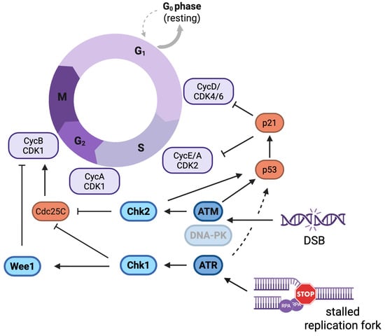
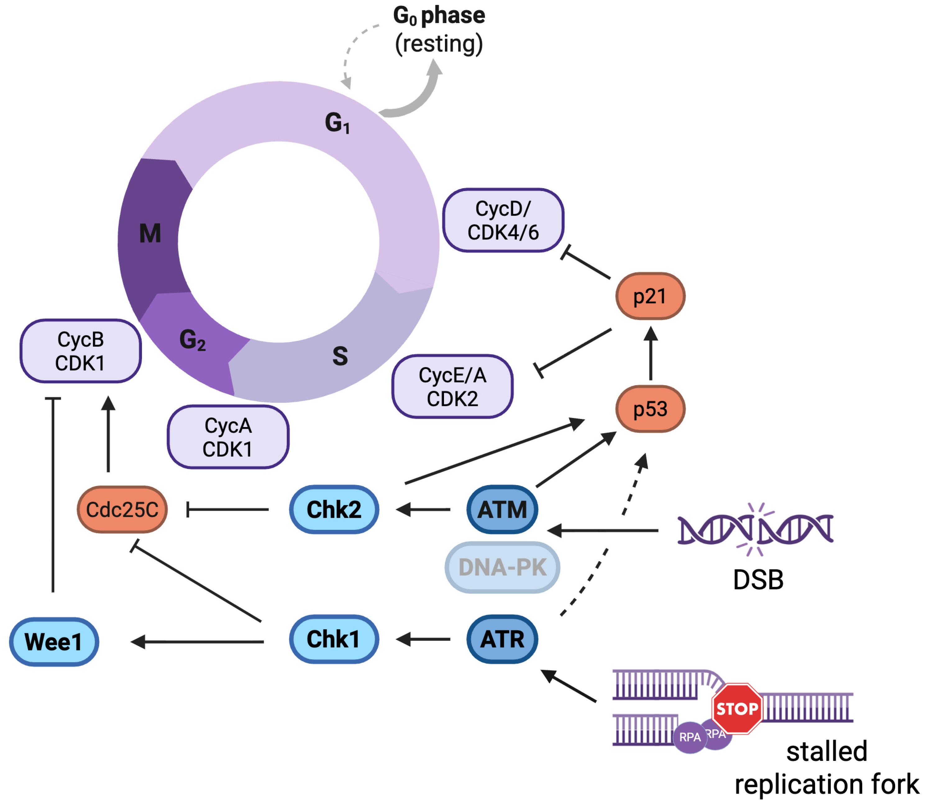
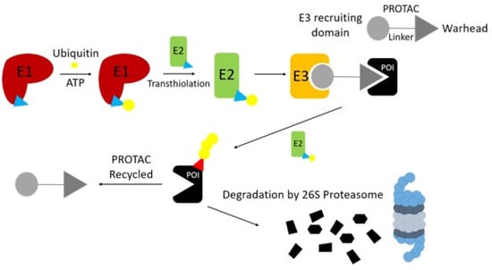
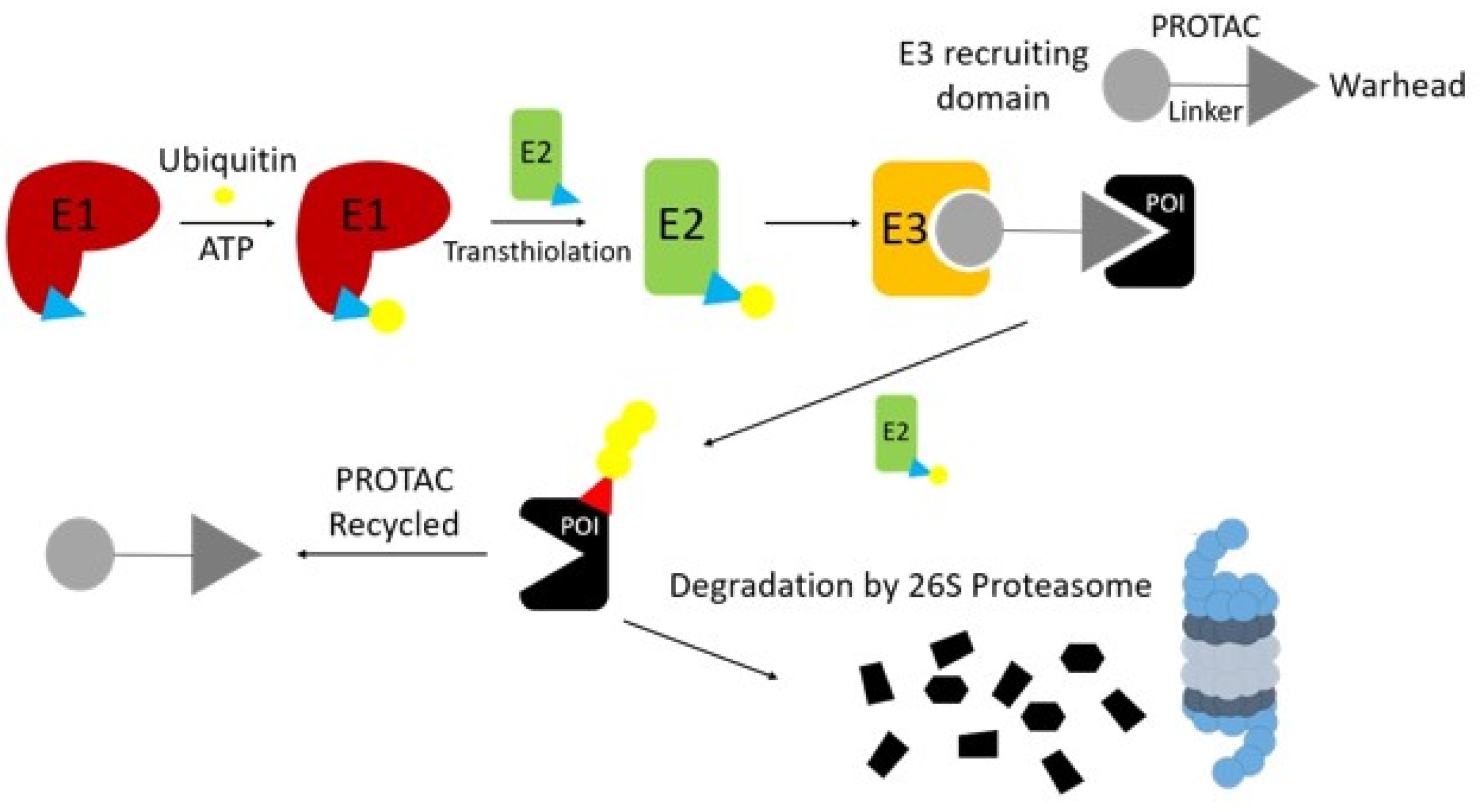

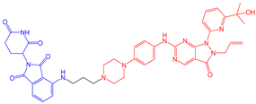
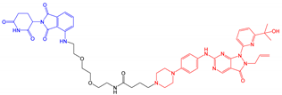
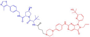
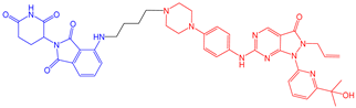
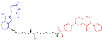
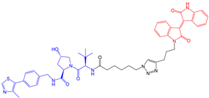
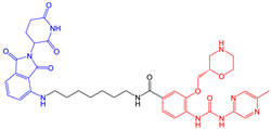
Disclaimer/Publisher’s Note: The statements, opinions and data contained in all publications are solely those of the individual author(s) and contributor(s) and not of MDPI and/or the editor(s). MDPI and/or the editor(s) disclaim responsibility for any injury to people or property resulting from any ideas, methods, instructions or products referred to in the content. |
© 2026 by the authors. Licensee MDPI, Basel, Switzerland. This article is an open access article distributed under the terms and conditions of the Creative Commons Attribution (CC BY) license.