


Abstract
Lipoprotein(a) [Lp(a)] has emerged as a major genetically determined cardiovascular risk factor that extends beyond the traditional low-density lipoprotein cholesterol (LDL-C)-centred model of atherosclerotic disease. Despite optimal LDL-C lowering, a substantial proportion of patients continue to experience cardiovascular events, highlighting the clinical relevance of residual cardiovascular risk. This review summarizes current evidence regarding the epidemiology, genetics, pathophysiology and therapeutic implications of Lp(a) in cardiovascular disease. Epidemiological, genetic, and Mendelian randomization studies consistently demonstrate an independent and likely causal association between elevated Lp(a) and atherosclerotic cardiovascular disease, ischemic stroke, calcific aortic valve stenosis, heart failure, and recurrent cardiovascular events, even in patients with well-controlled LDL-C levels. Lp(a) promotes atherosclerosis through proatherogenic, proinflammatory, and prothrombotic mechanisms, largely mediated by oxidized phospholipids and the structural homology of apolipoprotein(a) with plasminogen. Current guidelines increasingly recognize Lp(a) as a risk-enhancing factor capable of refining cardiovascular risk stratification beyond traditional algorithms, thus recommending measuring Lp(a) at least once in a lifetime in all adults. Accurate measurement and standardization of Lp(a) remain essential in clinical practice due to apo(a) isoform size variability; reporting in molar concentrations (nmol/L) is preferred as it better reflects particle number being less affected by isoform size variations and improves the reliability of cardiovascular risk stratification. Collectively, these findings support the integration of Lp(a) into precision-based cardiovascular prevention strategies and suggest a paradigm shift from an exclusively LDL-centric approach toward genetically informed risk assessment and treatment.
1. Introduction
Nowadays, cardiovascular diseases are the leading cause of mortality in the Western world. The high prevalence of coronary events cannot be explained only by traditional risk factors. Residual cardiovascular risk is defined as the probability of major cardiovascular events onset nevertheless treating patients according to recommended standard of care. It is represented by non-modifiable factors (age, sex, ethnicity and genetic predisposition) and modifiable factors (obesity, diabetes, smoking, hypertension, HDL-cholesterol and triglycerides levels). Dyslipidaemia, with high LDL-C levels, is the main factor influencing the outbreak of cardiovascular diseases. However, despite statin treatment, which is the leading treatment in reducing LDL-C levels, other lipid profile abnormalities are responsible for residual cardiovascular risk [1]. Indeed, beyond LDL-C particles, other apolipoprotein B-containing lipoproteins, such as Lp(a), have a key role in the development of atherosclerosis [2]. For example, as demonstrated by the Jupiter study, the association between basal levels of Lp(a) and the first cardiovascular event is independent of LDL-C levels. Indeed, Lp(a) is associated with an increased risk of aortic stenosis, myocardial infarction, heart failure, ischemic stroke, and cardiovascular death [3]. By integrating traditional and emerging cardiovascular risk factors, the purpose of this review is to outline the Lp(a) role in cardiovascular risk definition.
2. Structure and Biology of Lp(a)
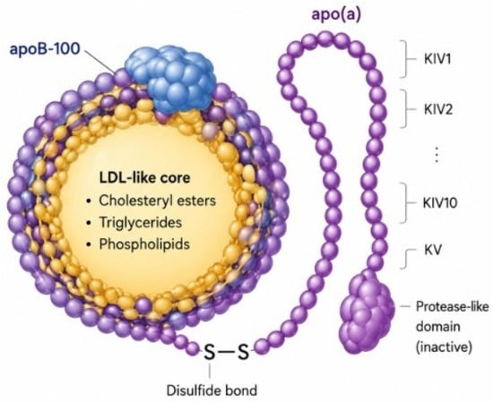
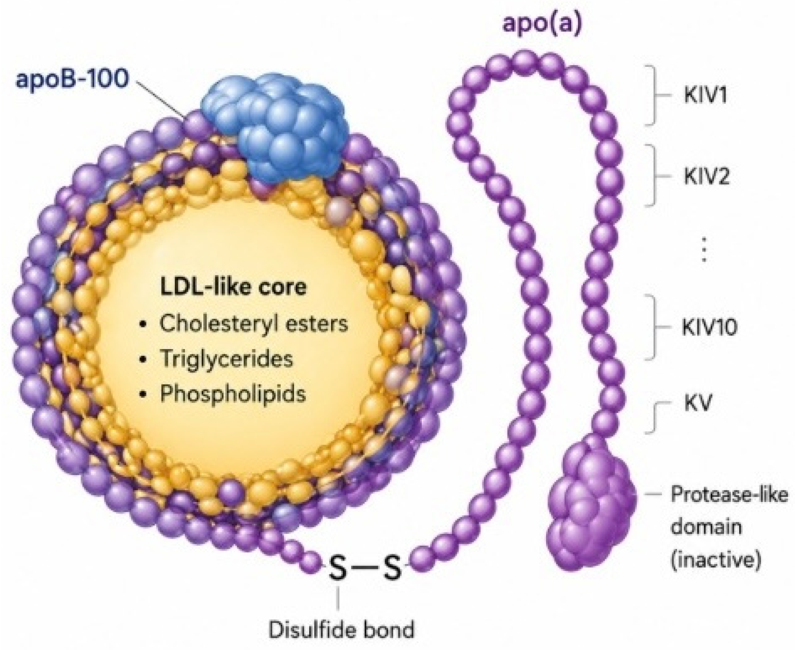
Lp(a) is an LDL-like particle characterized by the covalent binding of apolipoprotein(a) [apo(a)] to apolipoprotein B-100 (apoB-100) via a disulfide bond (Figure 1) [4]. Its lipid core contains cholesterol esters, triglycerides, and phospholipids, similar to LDL, while apo(a) confers distinct structural and functional properties [4,5]. A key feature distinguishing Lp(a) from LDL is its role as a preferential carrier of oxidized phospholipids (OxPL), which are major contributors to its pathogenic potential [6,7]. As illustrated in Figure 1, the unique structural organization of Lp(a), consisting of an LDL-like particle covalently linked to apo(a) and enriched in oxidized phospholipids (OxPLs), provides the molecular basis for its pro-atherogenic, pro-inflammatory, pro-thrombotic, and pro-calcific properties.
Figure 1.
Structural organization of lipoprotein(a). Lp(a) consists of an LDL-like particle containing apolipoprotein B-100 (apoB-100) covalently linked to apolipoprotein(a) [apo(a)] through a disulfide bond. Apo(a) is characterized by multiple kringle IV (KIV1–10) domains, a single kringle V (KV) domain, and an inactive protease-like domain. The LDL-like core contains cholesterol esters, triglycerides, and phospholipids, whereas apo(a) confers unique structural and functional properties that distinguish Lp(a) from LDL. This structural organization enables Lp(a) to preferentially carry oxidized phospholipids (OxPLs) and other bioactive molecules that contribute to its pro-atherogenic, pro-inflammatory, pro-thrombotic, and pro-calcific properties.
Apo(a) is a highly polymorphic glycoprotein structurally homologous to plasminogen, characterized by multiple kringle IV repeats (types 1–10), a single kringle V domain, and an inactive protease-like domain [4,8]. This structural homology underlies the ability of Lp(a) to interfere with fibrinolysis. Apo(a) competes with plasminogen for binding sites on fibrin and endothelial surfaces, thereby reducing plasmin generation and impairing clot dissolution [8,9]. This antifibrinolytic effect represents a central mechanism underlying the pro-thrombotic properties of Lp(a) [9,10]. Beyond impairing fibrinolysis, this structural similarity may further enhance thrombus persistence at sites of vascular injury, thereby amplifying cardiovascular risk.
A defining feature of Lp(a) is the marked size heterogeneity of apo(a), determined by the variable number of kringle IV type 2 repeats encoded by the LPA gene [4,11]. This genetic polymorphism gives rise to multiple apo(a) isoforms with distinct molecular weights. An inverse relationship exists between apo(a) isoform size and circulating Lp(a) concentrations, with smaller isoforms generally associated with higher plasma levels and increased cardiovascular risk [11,12]. As a genetically determined trait, Lp(a) levels remain relatively stable throughout life and are minimally influenced by environmental or lifestyle factors [5,12].
This extensive isoform heterogeneity also represents a major analytical challenge, as it may influence assay performance and complicate the standardization of Lp(a) measurement across laboratories.
Lp(a) contributes to atherogenesis through multiple complementary pathways. Its LDL-like component promotes cholesterol deposition within the arterial wall, while associated OxPL induce endothelial dysfunction, oxidative stress, and foam cell formation [6,7,13]. In addition, Lp(a) is retained within the subendothelial space through interactions with extracellular matrix components [7,13,14].
Beyond its atherogenic role, Lp(a) exerts pro-thrombotic effects by inhibiting fibrinolysis via apo(a), increasing the expression of plasminogen activator inhibitor-1 (PAI-1), and enhancing platelet activation [9,10,15]. These mechanisms establish a pathophysiological link between lipid accumulation and thrombosis, thereby promoting plaque progression and instability [13,15].
Beyond atherosclerosis, increasing evidence supports a causal role for Lp(a) in the development of calcific aortic valve stenosis (CAVS) [15,16]. OxPL carried by Lp(a) accumulate within the aortic valve, where they trigger inflammatory signaling and promote the osteogenic differentiation of valvular interstitial cells (VICs) [14,16]. In addition, autotaxin (ATX), an enzyme preferentially associated with Lp(a), converts lysophosphatidylcholine into lysophosphatidic acid (LPA), further amplifying inflammatory and pro-calcific pathways [14]. These mechanisms ultimately promote extracellular matrix remodeling, calcium deposition, and progressive valve calcification [14,16].
Whether selective Lp(a)-lowering therapies can delay the onset or progression of CAVS remains under investigation and is currently being evaluated in ongoing randomized clinical trials.
Lp(a) is a potent mediator of vascular inflammation. OxPL carried by Lp(a) activate endothelial cells and upregulate adhesion molecules such as vascular cell adhesion molecule-1 (VCAM-1) and intercellular adhesion molecule-1 (ICAM-1), facilitating monocyte recruitment [6,7,17]. Following transmigration into the intima, monocytes differentiate into macrophages and internalize Lp(a), contributing to foam cell formation [13,17]. In parallel, Lp(a) stimulates the release of pro-inflammatory cytokines, including interleukin-6 (IL-6), thereby amplifying local inflammatory responses [17,18]. This pro-inflammatory milieu plays a critical role in plaque development, progression, and rupture [13,18].
The inflammatory effects of Lp(a) extend beyond the arterial wall to the aortic valve [15]. OxPL-rich Lp(a) particles activate valvular endothelial and interstitial cells, promoting the release of inflammatory mediators and osteogenic factors such as bone morphogenetic protein-2 (BMP-2) and runt-related transcription factor-2 (RUNX2) [14,16]. Through ATX-mediated generation of lysophosphatidic acid, Lp(a) further enhances VIC mineralization and valve calcification [14,19]. Collectively, these findings identify inflammation as the key biological process linking elevated Lp(a) to both atherosclerotic cardiovascular disease and calcific aortic valve stenosis [15,16].
3. Genetics of Lp(a): A Biomarker “Stamped at Birth”
Lp(a) is an LDL-like particle whose blood concentration is under strict genetic control. Its levels are largely set by variants in the LPA gene and show striking variation between individuals and ethnic groups, with relatively modest influence from environment or lifestyle [20].
Lp(a) blood concentration is largely controlled by one single gene, the LPA gene, whose kringle IV-2 copy-number variation dictates apo(a) isoform size and is the main determinant of plasma Lp(a) levels, explaining a large share of inter-individual variance [21,22,23,24]. Heritability estimates reach ~90%, making Lp(a) one of the most heritable quantitative traits [21,23]. Additionally, LPA single-nucleotide polymorphisms (SNPs) and genes outside the LPA locus, including the APOE, APOH, and CEPT gene regions, further modulate concentrations and contribute to Lp(a) variability across different populations [24,25].
Lp(a) levels are highly right-skewed; the inter-individual Lp(a) concentrations extend for about three orders of magnitude (<0.1 mg/dL to >300 mg/dL) [23]. Marked ethnic differences are consistent: Black populations have the highest levels, followed by South Asians, Whites, Hispanics, and East Asians, with substantial within-group variability [25,26]. Recent evidence from a multi-ethnic cohort has demonstrated that plasma Lp(a) concentrations and their association with coronary artery disease (CAD) risk differ substantially across racial and ethnic groups. These findings indicate that the use of a single universal Lp(a) threshold may result in inaccurate cardiovascular risk classification in populations with distinct baseline Lp(a) distributions. Consequently, race- and ethnicity-specific Lp(a) cutoff values may provide a more accurate assessment of CAD risk and improve the clinical utility of Lp(a)-based screening and risk stratification strategies, highlighting the importance of developing and validating population-specific thresholds [27].
Recent evidence from a multi-ethnic cohort emphasizes the importance of race/ethnicity as a key variable in assigning Lp(a) cutoff values for CAD risk assessment and the need to develop the most clinically useful Lp(a) cutoff values in individual race/ethnicity groups [27].
Evidence from the Young Finns Study supports the notion that Lp(a) concentrations are highly stable across the life course, as reflected by a 25-year rank-order tracking correlation coefficient of r = 0.85 [28].
Nevertheless, a recent work shows clinically relevant intra-individual variation (≥20–50% of the Lp(a) baseline values) in a sizeable minority [29]. Furthermore, Lp(a) levels are reported to be influenced by kidney/liver disease, inflammation, and other factors [25]. Because Lp(a) levels are genetically set, according to the 2019 European Society of Cardiology (ESC) and the European Atherosclerosis Society (EAS) Guidelines for the management of dyslipidaemias, Lp(a) measurement should be considered at least once in each adult person’s lifetime (Class IIa, Level C), to identify individuals with very high inherited Lp(a) levels [30]. New data, however, suggest repeat testing may be useful in those near risk thresholds or with changing clinical status, challenging a strict “once only” strategy [29,31,32]. Furthermore, high heritability and autosomal-dominant transmission underpin emerging recommendations for routine family screening when markedly elevated Lp(a) is found [33]. In particular, according to the 2026 ACC/AHA Guideline on the Management of Dyslipidaemias, cascade Lp(a) testing has potential value for enhanced screening in families with high Lp(a), especially if there is a personal or family history of premature ASCVD [20].
4. Epidemiological Evidence and Cardiovascular Risk
Experimental and imaging studies suggest that Lp(a)-associated OxPLs promote endothelial dysfunction, monocyte recruitment, transendothelial migration of inflammatory cells, smooth muscle cell proliferation, and macrophage-derived foam cell formation, thereby accelerating plaque development and progression. In addition, elevated Lp(a) levels have been associated with enhanced necrotic core formation and progression of coronary artery calcification, further contributing to plaque instability and adverse cardiovascular outcomes [3].
A substantial body of epidemiological evidence supports a likely causal contribution of Lp(a) to cardiovascular disease across various populations and clinical contexts. Large prospective datasets, such as the UK Biobank, demonstrate a continuous and approximately linear association between Lp(a) levels and atherosclerotic cardiovascular disease (ASCVD), with risk increasing even at modest increments supporting a dose–response relationship without a clear lower threshold [34].
Elevated Lp(a) promotes arterial wall inflammation and atherosclerotic plaque formation within the carotid and cerebral vasculature, particularly in large-artery disease. Overall, available mechanistic data suggest that Lp(a)-related stroke risk is predominantly driven by accelerated atherosclerosis rather than venous or cardioembolic thrombotic mechanisms [3].
This graded association is further corroborated by more recent electronic health record–based risk models, which show a progressively greater increase in risk at higher concentrations, thus helping to define clinically meaningful thresholds; notably, a particularly strong association has been described for ischemic stroke [35].
Consistent findings from longitudinal real-world cohorts indicate that individuals with the highest Lp(a) levels have a markedly increased incidence of major adverse cardiovascular events (MACE), reinforcing its role as a predictor of coronary artery disease and myocardial infarction in both primary and secondary prevention settings [36]. Importantly, the effect of Lp(a) appears to be independent of, and additive to, traditional lipid parameters. Recent evidence shows that elevated Lp(a) levels are associated with increased cardiovascular risk even in individuals with well-controlled LDL-C, highlighting its contribution to residual cardiovascular risk beyond LDL lowering [37].
The association between Lp(a) and cardiovascular outcomes persists independently of systemic inflammation. Studies incorporating inflammatory markers such as C-reactive protein (CRP) demonstrate that elevated Lp(a) confers excess cardiovascular risk regardless of inflammatory status, suggesting distinct and non-overlapping pathogenic pathways [38]. More recent data continue to reinforce the robustness of these findings, with contemporary analyses confirming Lp(a) as an independent predictor of ASCVD events even after adjustment for established risk factors, with a persistent increase in risk per incremental rise in concentration [39].
The pathogenic role of Lp(a) in calcific aortic valve stenosis (CAVS) is increasingly supported by the fact that oxidized phospholipids (OxPLs), predominantly transported by Lp(a), accumulate within the aortic valve leaflets where they activate inflammatory pathways involving NF-κB and IL-6, thereby creating a pro-calcific microenvironment that promotes VICs osteogenic differentiation through increased expression of BMP2, RUNX2 and alkaline phosphatase [40]. This process results in progressive microcalcification and macrocalcification of the aortic valve. Additional mediators transported by Lp(a), including autotaxin, catalyzes the generation of lysophosphatic acid (LPA), which further amplifies inflammatory signaling and mineralization through LPAR1-dependent pathways, accelerating leaflet fibrosis and calcium deposition [41]. Current epidemiological and genetic evidence suggest that high Lp(a) concentrations may contribute to an earlier need for aortic valve replacement [3]. Although currently available lipid-lowering therapies have not shown to halt CAVS progression, ongoing phase 3 outcome trials evaluating Pelacarsen and Olpasiran may clarify whether selective Lp(a) reduction can modify the natural history of valve calcification and delay the need for aortic valve replacement [42].
Elevated Lp(a) concentrations have also been associated with an increased risk of heart failure, particularly at very high levels. This association is likely mediated, at least in part, by the contribution of Lp(a) to coronary artery disease and calcific aortic valve stenosis, which promote ventricular dysfunction and adverse cardiac remodelling. In addition, Lp(a)-associated OxPLs may contribute directly to myocardial inflammation and remodelling, although mechanistic evidence remains less established compared with ASCVD and aortic valve disease [3].
The association between Lp(a) and type 2 diabetes mellitus appears paradoxical, as several epidemiological studies have reported an increased risk of incident diabetes among individuals with very low Lp(a) concentrations. The biological mechanisms underlying this inverse relationship remain poorly understood. Current hypotheses include potential effects related to apo(a) isoform size, hepatic lipoprotein metabolism, insulin resistance pathways, or genetically determined alterations in Lp(a) synthesis and clearance. However, the causal nature of this association remains uncertain [3] (Table 1).
Table 1.
Association between Lp(a) levels and cardiometabolic outcomes.
5. Lp(a) and Residual Risk: Gaps in Traditional Model
Traditional cardiovascular risk scores such as SCORE2 are widely used to estimate the 10-year probability of a first atherosclerotic cardiovascular disease (ASCVD) event and to guide preventive strategies in clinical practice. SCORE2 integrates conventional cardiovascular risk factors, including age, sex, smoking status, blood pressure, and lipid parameters, particularly LDL-C [43]. Although these tools significantly improved population-based cardiovascular risk prediction, several limitations have emerged, particularly in individuals who develop cardiovascular disease despite apparently optimal control of traditional risk factors.
One of the main limitations of traditional risk scores is their inability to fully account for residual cardiovascular risk related to non-traditional and genetically determined factors. In this context, Lp(a) has gained increasing attention as an independent and causal risk factor for ASCVD and calcific aortic valve disease [3]. Elevated Lp(a) may confer substantial cardiovascular risk even in individuals classified as low or intermediate risk according to conventional algorithms.
Recent European Society of Cardiology guidelines acknowledged these shortcomings and introduced the concept of risk modifiers to refine cardiovascular risk assessment [44]. These modifiers include factors not routinely captured by SCORE2 but may significantly influence individual cardiovascular risk, such as family history of premature ASCVD, coronary artery calcium burden, chronic inflammatory conditions, and elevated Lp(a) levels. Similarly, contemporary American guidelines recognize elevated Lp(a) as a “risk-enhancing factor” that may refine clinical risk assessment beyond traditional risk estimation models, although it is not directly incorporated into standard scores such as PREVENT [20].
The addition of Lp(a) as a risk modifier is particularly relevant because elevated Lp(a) exerts proatherogenic, proinflammatory, and prothrombotic effects independently of LDL-C levels [45]. Reliance on traditional lipid assessment alone may therefore fail to identify individuals with substantial hidden cardiovascular risk—a limitation that has become increasingly important in the contemporary era of widespread statin use, where improved management of conventional risk factors has paradoxically made residual risk pathways more clinically visible.
A substantial proportion of patients who experience cardiovascular events present with LDL-C levels within the recommended target range. Epidemiological and clinical data consistently demonstrate that optimal LDL-C control, achieved through statin therapy or combination lipid-lowering regimens, does not eliminate cardiovascular risk entirely—a phenomenon commonly referred to as residual cardiovascular risk [46].
Lp(a) has emerged as a major contributor to this residual risk for a mechanistically distinct reason: unlike LDL-C, Lp(a) concentrations are not meaningfully reduced by standard lipid-lowering therapies, including statins and ezetimibe. Indeed, statins may paradoxically increase Lp(a) levels modestly in some patients [47]. Consequently, individuals who achieve excellent LDL-C control under guideline-directed therapy may nonetheless carry significantly elevated Lp(a) levels, perpetuating atherogenic, thrombogenic, and inflammatory processes within the arterial wall through pathways entirely independent of LDL-C metabolism.
The prothrombotic properties of Lp(a) are largely attributable to the structural homology of apolipoprotein(a) with plasminogen, which contributes to impaired fibrinolysis and enhanced thrombus formation. These mechanisms, combined with the promotion of oxidative stress, macrophage foam cell formation, and endothelial dysfunction, explain why LDL-C reduction alone may be insufficient in patients with elevated Lp(a)—and why such patients continue to experience events despite apparently adequate lipid management. The integration of Lp(a) into cardiovascular risk assessment offers meaningful improvements in stratification beyond what is achievable with traditional risk models alone. Lp(a) contributes independently to cardiovascular risk prediction even after adjustment for all major traditional risk factors. Mendelian randomization studies have provided robust evidence of a causal relationship between elevated Lp(a) and increased risk of myocardial infarction, ischemic stroke, and aortic valve stenosis, lending biological plausibility to its use as a risk stratification tool rather than merely an epidemiological association [48].
The clinical utility of Lp(a) in risk reclassification is most evident in two distinct populations: individuals classified as low or intermediate risk by conventional algorithms in whom elevated Lp(a) unmasks a substantially higher true risk, and patients with a strong family history of premature ASCVD but without markedly elevated LDL-C, in whom Lp(a) measurement frequently reveals the underlying genetic driver of familial cardiovascular risk. Several studies have demonstrated that the addition of Lp(a) to conventional risk scores reclassifies a significant proportion of patients into higher risk categories, reported in prospective cohort data [49].
Current European guidelines suggest that Lp(a) levels above 50 mg/dL (approximately 125 nmol/L) confer an additional cardiovascular risk burden sufficient to upwardly reclassify risk in borderline cases, potentially shifting therapeutic decisions—such as the initiation of statin therapy in primary prevention or the intensification of LDL-C lowering targets. This threshold-based approach provides a practical framework for incorporating Lp(a) into everyday clinical practice without requiring complex probabilistic modelling.
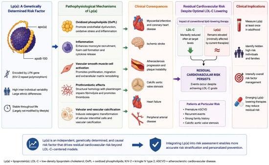
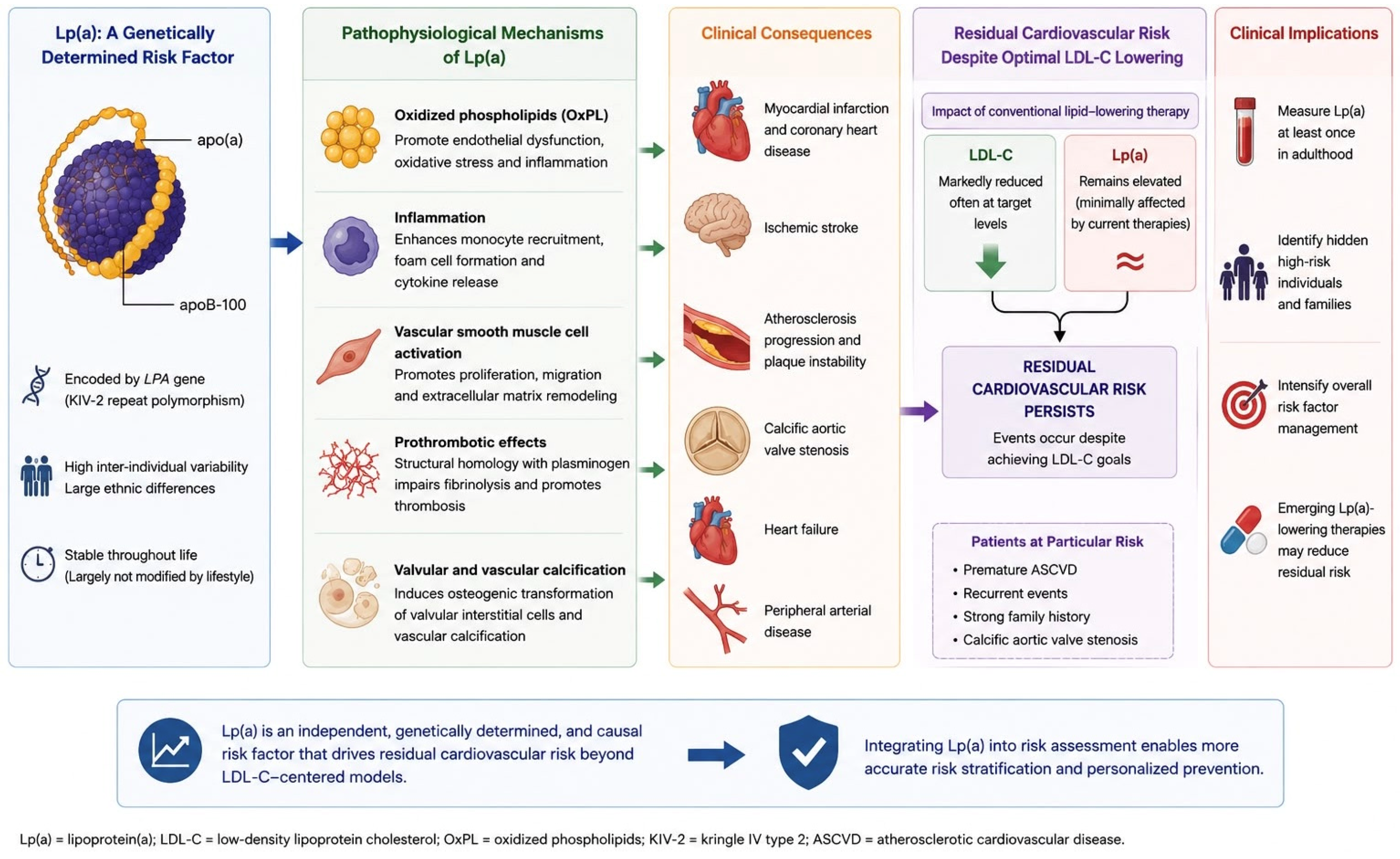
As discussed above, a substantial proportion of cardiovascular events occur in individuals without markedly elevated LDL-C or other major traditional risk factors [50]. In these cases, elevated Lp(a) may represent the overlooked determinant contributing to disease progression and vascular damage, constituting a form of hidden risk not captured by conventional screening (Figure 2).
Figure 2.
Elevated Lp(a) contributes to persistent residual cardiovascular risk through proatherogenic, proinflammatory, prothrombotic, and procalcific pathways despite optimal LDL-C lowering.
This hidden risk is particularly important in individuals with a strong family history of premature cardiovascular disease [51]. Because Lp(a) concentrations are predominantly genetically determined and remain relatively stable throughout life, a single lifetime measurement is often sufficient to detect an inherited predisposition to atherosclerotic disease, although repeat testing may be considered in selected clinical circumstances, such as values near decision thresholds.
The identification of hidden high-risk individuals further has implications for family screening. Since elevated Lp(a) is inherited in an autosomal codominant manner, relatives of affected patients may also carry significantly elevated levels. Early recognition within families allows preventive interventions to begin before the development of overt cardiovascular disease, potentially reducing long-term morbidity and mortality [52].
The recognition of elevated Lp(a) has important implications for both primary and secondary cardiovascular prevention. In primary prevention, the identification of high Lp(a) levels may justify earlier and more intensive intervention in individuals who would otherwise be considered at moderate or borderline cardiovascular risk. Although lifestyle modifications have limited direct effects on Lp(a) concentrations, optimization of all modifiable risk factors becomes particularly important in these patients [53]. Strict control of LDL-C, blood pressure, smoking cessation, healthy diet, regular physical activity, and diabetes management may help mitigate the excess cardiovascular risk associated with elevated Lp(a). Moreover, the presence of high Lp(a) may lower the threshold for initiating pharmacological lipid-lowering therapy and prompt clinicians to adopt more aggressive LDL-C reduction targets to compensate for the additional risk burden.
In secondary prevention, the clinical relevance of Lp(a) becomes even more pronounced. Patients with established cardiovascular disease and elevated Lp(a) are at increased risk of recurrent myocardial infarction, stroke, and progressive atherosclerotic disease despite standard therapy [54].
In this context, Lp(a) serves as a marker of persistent residual risk that operates independently of achieved LDL-C levels, and its measurement in patients with established ASCVD may provide important prognostic information beyond conventional lipid assessment. Importantly, evidence that reducing Lp(a) may translate into clinical benefit already exists: long-term lipoprotein apheresis in patients with isolated Lp(a) elevation and progressive cardiovascular disease has been shown to markedly reduce annual cardiovascular event rates, supporting the concept that Lp(a)-driven risk is not only identifiable but also modifiable [55].
The presence of elevated Lp(a) in this population may therefore justify intensified follow-up, more aggressive LDL-C lowering targets, and comprehensive management of all concomitant modifiable risk factors, with the goal of attenuating the excess vascular risk that standard therapies alone cannot fully address.
6. Measurement and Standardization
Measuring Lp(a) in clinical practice is not as straightforward as it might seem. Although it is increasingly recognized as an important cardiovascular risk factor, several analytical issues still limit its routine use [4,56].
One of the main challenges lies in how Lp(a) is quantified. Current laboratory methods rely on immunoassays, which can report results either as mass concentration (mg/dL) or as molar concentration (nmol/L). However, these two units are not directly interchangeable. This is because Lp(a) particles vary significantly in size due to differences in apo(a) isoforms, meaning that the same mass does not necessarily correspond to the same number of particles [56,57].
This variability is largely driven by the kringle IV type 2 (KIV-2) repeats in the LPA gene, which determine apo(a) size. As a result, assays that measure Lp(a) in mg/dL may underestimate levels in individuals with larger isoforms and overestimate them in those with smaller ones, potentially leading to misclassification of cardiovascular risk [56,57].
For this reason, there is growing consensus that measurements expressed in nmol/L are more reliable, as they better reflect the actual number of Lp(a) particles and are less influenced by isoform size [4,56]. Nevertheless, not all assays are fully isoform-independent, and complete standardization across laboratories is still lacking. The main analytical challenges and their clinical implications are summarized in Table 2.
Table 2.
Main analytical issues in lipoprotein(a) measurement and their clinical implications.
Efforts to improve harmonization, particularly by the International Federation of Clinical Chemistry and Laboratory Medicine (IFCC), have made progress, but variability between assays remains an important limitation [57].
From a clinical standpoint, current guidelines—including those from the ESC and the EAS—recommend measuring Lp(a) at least once in a lifetime in all adults [4,30,43]. This recommendation is based on the strong genetic determination of Lp(a) levels, which tend to remain stable over time and are minimally influenced by environmental or lifestyle factors [56]. Therefore, routine serial monitoring is generally not recommended in clinical practice. However, repeat measurement may be considered in selected clinical situations, such as when the initial measurement is suspected to be affected by analytical variability, when confirmation of an unexpected result may influence cardiovascular risk assessment and patient management, or when results obtained using different assays require confirmation [4,56].
In terms of interpretation, Lp(a) levels above 50 mg/dL (approximately 125 nmol/L) are generally considered associated with increased cardiovascular risk [4,30,43]. However, the relationship between Lp(a) and cardiovascular risk is continuous rather than defined by a strict threshold, and values should always be interpreted within the broader clinical context [4,56].
7. Therapeutic Strategies: Current Status and Future Directions
Traditional lipid-lowering therapies have demonstrated limited efficacy in reducing Lp(a) concentrations [58]. Statins remain the cornerstone of LDL-C lowering and cardiovascular risk reduction; however, their effect on Lp(a) is neutral or may be slightly increasing, highlighting the biological independence between LDL-C and Lp(a) metabolism [59]. Ezetimibe exerts negligible effects on circulating Lp(a) levels.
PCSK9 inhibitors, including evolocumab and alirocumab, reduce Lp(a) by approximately 20–30% and have demonstrated robust cardiovascular benefit in large outcome trials such as FOURIER and ODYSSEY outcomes [60,61]. Importantly, the magnitude of Lp(a) reduction appears modest relative to the observed cardiovascular benefit, suggesting that LDL-C lowering remains the principal driver of outcome improvement, while Lp(a) reduction may contribute as a secondary mechanism.
Lipoprotein apheresis remains the most effective available intervention for lowering Lp(a), achieving acute reductions of 60–70%, but its use is restricted to highly selected patients due to procedural burden, cost, and limited accessibility [58]. Lifestyle interventions, although essential for global cardiovascular risk management, have minimal impact on Lp(a) concentrations, underscoring its predominantly genetically determined nature.
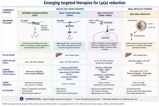
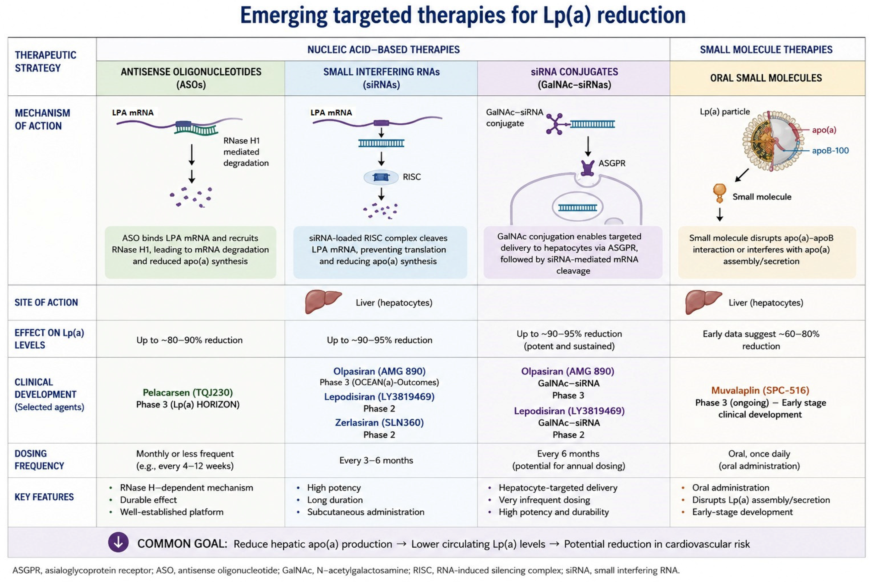
Recent advances in molecular therapeutics have enabled direct targeting of hepatic apolipoprotein(a) synthesis, representing a mechanistic shift from conventional lipid modulation to gene-silencing strategies [62]. These approaches primarily include antisense oligonucleotides (ASOs) and small interfering RNA (siRNA) technologies (Figure 3).
Figure 3.
Mechanisms of action of emerging lipoprotein(a)-lowering therapies, including antisense oligonucleotides (ASOs), small interfering RNA (siRNA), and small-molecule inhibitors. ASOs and siRNAs reduce hepatic apolipoprotein(a) synthesis by targeting LPA mRNA, whereas small-molecule inhibitors disrupt the interaction between apolipoprotein(a) and apolipoprotein B, thereby preventing lipoprotein(a) assembly. The arrow in the lower section of the figure indicates the reduction of apo(a) synthesis.
Pelacarsen, an antisense oligonucleotide targeting apo(a) mRNA, has demonstrated dose-dependent reductions in Lp(a) exceeding 80% in phase 2 clinical studies [63]. Similarly, siRNA-based agents such as olpasiran have shown sustained and profound reductions in circulating Lp(a), often approaching 90% or greater, with infrequent dosing regimens [64]. These therapies represent a high level of specificity and durability in Lp(a) suppression.
An additional emerging class includes oral small-molecule inhibitors, such as muvalaplin, which disrupt the non-covalent interaction between apo(a) and apoB. These agents are currently in early-stage clinical development and represent a potentially scalable oral strategy for Lp(a) reduction; however, long-term efficacy and outcome data remain unavailable.
Collectively, these therapies establish a new therapeutic paradigm by directly suppressing Lp(a) biosynthesis or assembly at its source, rather than modulating downstream lipid pathways. Despite strong genetic, epidemiological, and mechanistic evidence supporting a causal role of Lp(a) in atherosclerotic cardiovascular disease, several clinically fundamental questions remain unresolved: the optimal threshold for intervention, the magnitude of Lp(a) reduction required for clinical benefit, the role of therapy in primary prevention, and the identification of patient subgroups most likely to benefit [65].
Current evidence from mendelian randomization studies supports a log-linear relationship between absolute Lp(a) reduction and cardiovascular risk, suggesting that substantial lowering may be required to achieve clinically meaningful event reduction [66]. Overall, the level of evidence supporting Lp(a) as a causal cardiovascular risk factor is high; however, the level of evidence supporting clinical benefit from pharmacological lowering remains low, reflecting the absence of completed outcome trials.
Several phase 3 cardiovascular outcome trials are currently ongoing. The Lp(a) HORIZON trial (pelacarsen) and the OCEAN(a)-Outcomes trial (olpasiran) are designed to determine whether selective Lp(a) lowering reduces major adverse cardiovascular events [63,64]. Additional studies, including ACCLAIM-Lp(a) and trials evaluating emerging oral agents, will further refine the clinical role of this pathway.
At present, therefore, Lp(a)-lowering therapies can be classified as high biological plausibility interventions with low-to-emerging clinical outcome evidence.
The emergence of potent and selective Lp(a)-lowering therapies may redefine cardiovascular prevention, transitioning from a predominantly LDL-centric model toward a genetically informed, precision-based framework [67]. While LDL-C lowering remains the foundation of risk reduction, Lp(a) represents an independent, genetically determined determinant of residual cardiovascular risk, particularly relevant in premature atherosclerotic cardiovascular disease, recurrent events despite optimal therapy, calcific aortic valve stenosis, and unexplained familial aggregation of cardiovascular events [68].
This evolving framework supports earlier identification of genetically mediated risk and selective therapeutic intervention in high-risk phenotypes. Current consensus statements suggest that Lp(a) measurement should be considered at least once in each adult’s lifetime for cardiovascular risk assessment, given its genetic determination and lifelong stability, reflecting its stability as a genetically determined biomarker rather than a modifiable lipid parameter requiring serial monitoring [69].
If ongoing outcome trials confirm that selective Lp(a) lowering reduces cardiovascular events, future prevention strategies are likely to integrate LDL-C and Lp(a) as complementary and mechanistically distinct therapeutic targets, enabling a shift toward precision cardiovascular medicine grounded in causal biology.
Ultimately, Lp(a) may represent a prototype of genetically guided cardiovascular prevention, in which risk stratification and therapeutic intervention converge within a unified causal framework.
8. Conclusions
Lipoprotein(a) represents one of the most important genetically determined contributors to residual cardiovascular risk beyond traditional LDL-C centred paradigms. Increasing evidence supports its causal involvement in atherosclerotic cardiovascular disease, ischemic stroke, calcific aortic valve stenosis, and recurrent cardiovascular events despite optimal conventional therapy. The unique pathophysiological profile of Lp(a), characterized by combined proatherogenic, proinflammatory, and prothrombotic properties, distinguishes it from other lipid fractions and explains its independent contribution to cardiovascular disease progression.
Because Lp(a) levels are largely established at birth, remain relatively stable throughout life, and are minimally influenced by lifestyle interventions, its measurement provides a powerful tool for the early identification of individuals with hidden inherited cardiovascular risk. In this context, incorporating Lp(a) into cardiovascular risk assessment, measuring its levels at least once during adulthood, represents a valuable strategy for improving patient reclassification and enabling earlier preventive interventions, particularly among individuals with premature ASCVD, strong familial predisposition, or persistent residual risk despite controlled LDL-C levels.
While currently available lipid-lowering therapies have limited impact on Lp(a), emerging RNA-based therapies capable of selectively and profoundly reducing Lp(a) concentrations may soon transform clinical practice. If ongoing cardiovascular outcome trials confirm that targeted Lp(a) lowering reduces major adverse cardiovascular events, future prevention strategies are likely to evolve toward a precision-based and genetically informed model of cardiovascular risk estimation and treatment, integrating both LDL-C and Lp(a) as complementary therapeutic targets.
Ultimately, Lp(a) may become a cornerstone biomarker of precision cardiovascular medicine, bridging genetic predisposition, individualized risk stratification, and mechanism-driven therapeutic intervention.
Author Contributions
Conceptualization F.N., G.C. and A.S.; methodology F.S.L., P.G., F.N., G.C. and A.S.; resources A.S., E.L., J.B., C.S., M.G., B.A., G.T. and M.M.; data curation A.S., E.L., J.B., C.S., M.G., B.A., G.T. and M.M.; writing—original draft preparation A.S., E.L., J.B., C.S., M.G., B.A., G.T. and M.M.; writing—review and editing P.G., G.C. and F.N.; supervision F.S.L., F.N. and G.C. All authors have read and agreed to the published version of the manuscript.
Funding
This research received no external funding.
Institutional Review Board Statement
Not applicable.
Informed Consent Statement
Not applicable.
Data Availability Statement
The data from this manuscript are derived from publicly available published clinical trials and study results.
Conflicts of Interest
The authors declare no conflicts of interest.
References
- Sucato, V.; Comparato, F.; Ortello, A.; Galassi, A.R.; Novo, G. Residual Cardiovascular Risk: Role of Remnants Cholesterol, Monocyte/HDL Ratio and Lipoprotein Ratios on Personalized Cardiovascular Prevention. J. Pers. Med. 2024, 14, 460. [Google Scholar] [CrossRef] [Scilit] [PubMed]
- Ferrari, R.; Catapano, A.L. Residual Cardiovascular Risk. Eur. Heart J. Suppl. 2016, 18, C1. [Google Scholar] [CrossRef] [Scilit] [PubMed]
- Kronenberg, F.; More, S.; Stroes, E.S.G.; Ference, B.A.; Arsenault, B.J.; Berglund, L.; Dweck, M.R.; Koschinsky, M.; Lambert, G.; Mach, F.; et al. Lipoprotein(a) in atherosclerotic cardiovascular disease and aortic stenosis: A European Atherosclerosis Society consensus statement. Eur. Heart J. 2022, 43, 3925–3946. [Google Scholar] [CrossRef] [Scilit] [PubMed]
- Tsimikas, S. A Test in Context: Lipoprotein(a). J. Am. Coll. Cardiol. 2017, 69, 692–711. [Google Scholar] [CrossRef] [Scilit] [PubMed]
- Kronenberg, F. Lipoprotein(a); Springer: Cham, Switzerland, 2021; pp. 201–232. [Google Scholar] [CrossRef] [Scilit] [PubMed]
- Tsimikas, S.; Brilakis, E.S.; Miller, E.R.; McConnell, J.P.; Lennon, R.J.; Kornman, K.S.; Wiztum, J.L.; Berger, P.B. Oxidized Phospholipids, Lp(a) Lipoprotein, and Coronary Artery Disease. N. Engl. J. Med. 2005, 353, 46–57. [Google Scholar] [CrossRef] [Scilit] [PubMed]
- Bergmark, C.; Dewan, A.; Orsoni, A.; Merki, E.; Miller, E.R.; Shin, M.J.; Binder, C.J.; Hörkkö, S.; Krauss, R.M.; Chapman, M.J.; et al. A novel function of lipoprotein [a] as a preferential carrier of oxidized phospholipids in human plasma. J. Lipid Res. 2008, 49, 2230–2239. [Google Scholar] [CrossRef] [Scilit] [PubMed]
- McLean, J.W.; Tomlinson, J.E.; Kuang, W.J.; Eaton, D.L.; Chen, E.Y.; Fless, G.M.; Scanu, A.M.; Lawn, R.M. CDNA sequence of human apolipoprotein(a) is homologous to plasminogen. Nature 1987, 330, 132–137. [Google Scholar] [CrossRef] [Scilit] [PubMed]
- Hajjar, K.A.; Gavishi, D.; Breslow, J.L.; Nachman, R.L. Lipoprotein(a) modulation of endothelial cell surface fibrinolysis and its potential role in atherosclerosis. Nature 1989, 339, 303–305. [Google Scholar] [CrossRef] [Scilit] [PubMed]
- Boffa, M.B.; Koschinsky, M.L. Lipoprotein (a): Truly a direct prothrombotic factor in cardiovascular disease? J. Lipid Res. 2016, 57, 745–757. [Google Scholar] [CrossRef] [Scilit] [PubMed]
- Kraft, H.G.; Lingenhel, A.; Pang, R.W.; Delport, R.; Trommsdorff, M.; Vermaak, H.; Janus, E.D.; Ultermann, G. Frequency Distributions of Apolipoprotein(a) Kringle IV Repeat Alleles and Their Effects on Lipoprotein(a) Levels in Caucasian, Asian, and African Populations: The Distribution of Null Alleles Is Non-Random. Eur. J. Hum. Genet. 1996, 4, 74–87. [Google Scholar] [CrossRef] [Scilit] [PubMed]
- Clarke, R.; Peden, J.F.; Hopewell, J.C.; Kyriakou, T.; Goel, A.; Heath, S.C.; Parish, S.; Barlera, S.; Franzosi, M.G.; Rust, S.; et al. Genetic Variants Associated with Lp(a) Lipoprotein Level and Coronary Disease. N. Engl. J. Med. 2009, 361, 2518–2528. [Google Scholar] [CrossRef] [Scilit] [PubMed]
- Libby, P. Inflammation in atherosclerosis. Nature 2002, 420, 868–874. [Google Scholar] [CrossRef] [Scilit] [PubMed]
- Bouchareb, R.; Mahmut, A.; Nsaibia, M.J.; Boulanger, M.C.; Dahou, A.; Lépine, J.L.; Laflamme, M.H.; Hadji, F.; Couture, C.; Trahan, S.; et al. Autotaxin Derived From Lipoprotein(a) and Valve Interstitial Cells Promotes Inflammation and Mineralization of the Aortic Valve. Circulation 2015, 132, 677–690. [Google Scholar] [CrossRef] [Scilit] [PubMed]
- Kamstrup, P.R.; Tybjærg-Hansen, A.; Nordestgaard, B.G. Elevated Lipoprotein(a) and Risk of Aortic Valve Stenosis in the General Population. J. Am. Coll. Cardiol. 2014, 63, 470–477. [Google Scholar] [CrossRef] [Scilit] [PubMed]
- Capoulade, R.; Chan, K.L.; Yeang, C.; Mathieu, P.; Bossé, Y.; Dumesnil, J.G.; Tam, J.W.; Teo, K.K.; Mahmut, A.; Yang, X.; et al. Oxidized Phospholipids, Lipoprotein(a), and Progression of Calcific Aortic Valve Stenosis. J. Am. Coll. Cardiol. 2015, 66, 1236–1246. [Google Scholar] [CrossRef] [Scilit] [PubMed]
- Hansson, G.K. Inflammation, Atherosclerosis, and Coronary Artery Disease. N. Engl. J. Med. 2005, 352, 1685–1695. [Google Scholar] [CrossRef] [Scilit] [PubMed]
- Ridker, P.M. Inflammation, C-Reactive Protein, and Cardiovascular Disease. Circ. Res. 2014, 114, 594–595. [Google Scholar] [CrossRef] [Scilit] [PubMed]
- Zheng, K.H.; Tsimikas, S.; Pawade, T.; Kroon, J.; Jenkins, W.S.A.; Doris, M.K.; White, A.C.; Timmers, N.K.L.M.; Hjortnaes, J.; Rogers, M.A.; et al. Lipoprotein(a) and Oxidized Phospholipids Promote Valve Calcification in Patients with Aortic Stenosis. J. Am. Coll. Cardiol. 2019, 73, 2150–2162. [Google Scholar] [CrossRef] [Scilit] [PubMed]
- Blumenthal, R.S.; Morris, P.B.; Gaudino, M.; Johnson, H.M.; Anderson, T.S.; Bittner, V.A.; Blankstein, R.; Brewer, C.; Cho, L.; de Ferranti, S.D.; et al. 2026 ACC/AHA/AACVPR/ABC/ACPM/ADA/AGS/APhA/ASPC/NLA/PCNA Guideline on the Management of Dyslipidemia. JACC 2026, 87, 2624–2757. [Google Scholar] [CrossRef] [Scilit] [PubMed]
- Kamstrup, P.R. Lipoprotein(a): The common, likely causal, yet elusive risk factor for cardiovascular disease. J. Lipid Res. 2017, 58, 1731–1732. [Google Scholar] [CrossRef] [Scilit] [PubMed]
- Dumitrescu, L.; Glenn, K.; Brown-Gentry, K.; Stephard, C.; Wong, M.; Rieder, M.J.; Smith, J.D.; Nickerson, D.A.; Crawford, D.C. Variation in LPA Is Associated with Lp(a) Levels in Three Populations from the Third National Health and Nutrition Examination Survey. PLoS ONE 2011, 6, e16604. [Google Scholar] [CrossRef] [Scilit] [PubMed]
- Coassin, S.; Kronenberg, F. Lipoprotein(a) beyond the kringle IV repeat polymorphism: The complexity of genetic variation in the LPA gene. Atherosclerosis 2022, 349, 17–35. [Google Scholar] [CrossRef] [Scilit] [PubMed]
- Cesaro, A.; Scherillo, G.; De Michele, G.; Acerbo, V.; Signore, G.; Panico, D.; Porcelli, G.; Scialla, F.; Raucci, G.; Rotolo, F.P.; et al. Circulating culprit or therapeutic bullseye: Lipoprotein(a) in cardiovascular risk assessment and novel therapeutic prospects. J. Cardiovasc. Aging 2024, 4, 10. [Google Scholar] [CrossRef] [Scilit]
- Enkhmaa, B.; Anuurad, E.; Berglund, L. Lipoprotein (a): Impact by ethnicity and environmental and medical conditions. J. Lipid Res. 2016, 57, 1111–1125. [Google Scholar] [CrossRef] [Scilit] [PubMed]
- Mehta, A.; Jain, V.; Saeed, A.; Saseen, J.J.; Gulati, M.; Ballantyne, C.M.; Virani, S.S. Lipoprotein(a) and ethnicities. Atherosclerosis 2022, 349, 42–52. [Google Scholar] [CrossRef] [Scilit] [PubMed]
- Guan, W.; Cao, J.; Steffen, B.T.; Post, W.S.; Stein, J.H.; Tattersall, M.C.; Kaufman, J.D.; McConnell, J.P.; Hoefner, D.M.; Warnick, R.; et al. Race Is a Key Variable in Assigning Lipoprotein(a) Cutoff Values for Coronary Heart Disease Risk Assessment. Arterioscler. Thromb. Vasc. Biol. 2015, 35, 996–1001. [Google Scholar] [CrossRef] [Scilit] [PubMed]
- Raitakari, O.; Kivelä, A.; Pahkala, K.; Rovio, S.; Mykkänen, J.; Olli, A.A.; Loo, B.M.; Lyytikäinen, L.P.; Lehtimäki, T.; Kähönen, M.; et al. Long-term tracking and population characteristics of lipoprotein (a) in the Cardiovascular Risk in Young Finns Study. Atherosclerosis 2022, 356, 18–27. [Google Scholar] [CrossRef] [Scilit] [PubMed]
- Burzyńska, M.; Jankowski, P.; Banach, M.; Chudzik, M. Is a Single Lipoprotein(a) Measurement Once in a Lifetime Sufficient? The Results from the STAR-Lp(a) Study. Med. Sci. 2025, 13, 320. [Google Scholar] [CrossRef] [Scilit] [PubMed]
- Mach, F.; Baigent, C.; Catapano, A.L.; Koskinas, K.C.; Casula, M.; Badimon, L.; Chapman, M.J.; De Backer, G.G.; Delgado, V.; Ference, B.A.; et al. 2019 ESC/EAS Guidelines for the management of dyslipidaemias: Lipid modification to reduce cardiovascular risk the task force for the management of dyslipidaemias of the European Society of Cardiology (ESC) and European Atherosclerosis Society (EAS). Eur. Heart J. 2020, 41, 111–188. [Google Scholar] [CrossRef] [Scilit] [PubMed]
- Deshotels, M.R.; Sun, C.; Nambi, V.; Virani, S.S.; Matsushita, K.; Yu, B.; Ballantyne, C.M.; Hoogeveen, R.C. Temporal Trends in Lipoprotein(a) Concentrations: The Atherosclerosis Risk in Communities Study. J. Am. Heart Assoc. 2022, 11, e026762. [Google Scholar] [CrossRef] [Scilit] [PubMed]
- Harb, T.; Ziogos, E.; Blumenthal, R.S.; Gerstenblith, G.; Leucker, T.M. Intra-individual variability in lipoprotein(a): The value of a repeat measure for reclassifying individuals at intermediate risk. Eur. Heart J. Open 2024, 4, oeae064. [Google Scholar] [CrossRef] [Scilit] [PubMed]
- Reyes-Soffer, G.; Yeang, C.; Michos, E.D.; Boatwright, W.; Ballantyne, C.M. High lipoprotein(a): Actionable strategies for risk assessment and mitigation. Am. J. Prev. Cardiol. 2024, 18, 100651. [Google Scholar] [CrossRef] [Scilit] [PubMed]
- Patel, A.P.; Wang, M.; Pirruccello, J.P.; Ellinor, P.T.; Ng, K.; Kathiresan, S.; Khera, A.V. Lp(a) (Lipoprotein[a]) Concentrations and Incident Atherosclerotic Cardiovascular Disease. Arterioscler. Thromb. Vasc. Biol. 2021, 41, 465–474. [Google Scholar] [CrossRef] [Scilit] [PubMed]
- Fan, W.; Wu, C.; Wong, N.D. Lipoprotein(a) Atherosclerotic Cardiovascular Disease Risk Score Development and Prediction in Primary Prevention From Real-World Data. Circ. Genom. Precis. Med. 2025, 18, e004631. [Google Scholar] [CrossRef] [Scilit] [PubMed]
- Berman, A.N.; Biery, D.W.; Besser, S.A.; Singh, A.; Shiyovich, A.; Weber, B.N.; Huck, D.M.; Divakaran, S.; Hainer, J.; Kaur, G.; et al. Lipoprotein(a) and Major Adverse Cardiovascular Events in Patients With or Without Baseline Atherosclerotic Cardiovascular Disease. J. Am. Coll. Cardiol. 2024, 83, 873–886. [Google Scholar] [CrossRef] [Scilit] [PubMed]
- Bhatia, H.S.; Wandel, S.; Willeit, P.; Lesogor, A.; Bailey, K.; Ridker, P.M.; Nestel, P.; Simes, J.; Tonkin, A.; Schwartz, G.G.; et al. Independence of Lipoprotein(a) and Low-Density Lipoprotein Cholesterol–Mediated Cardiovascular Risk: A Participant-Level Meta-Analysis. Circulation 2025, 151, 312–321. [Google Scholar] [CrossRef] [Scilit] [PubMed]
- Small, A.M.; Pournamdari, A.; Melloni, G.E.M.; Scirica, B.M.; Bhatt, D.L.; Raz, I.; Braunwald, E.; Giugliano, R.P.; Sabatine, M.S.; Peloso, G.M.; et al. Lipoprotein(a), C-Reactive Protein, and Cardiovascular Risk in Primary and Secondary Prevention Populations. JAMA Cardiol. 2024, 9, 385–391. [Google Scholar] [CrossRef] [Scilit] [PubMed]
- Ward, A.; Kron, B.; Lozama, A.; Sandhu, A.; Khandelwal, A.; Rodriguez, F.; Dash, R.; Nallamshetty, S. Elevated Lipoprotein(a) Independently Increases Risk for Short-Term Atherosclerotic Cardiovascular Events in Machine Learning Predictive Models. JACC Adv. 2025, 4, 102253. [Google Scholar] [CrossRef] [Scilit] [PubMed]
- Bhatia, H.S.; Dweck, M.R.; Craig, N.; Capoulade, R.; Pibarot, P.; Trainor, P.J.; Whelton, S.P.; Rikhi, R.; Lidani, K.C.F.; Post, W.S.; et al. Oxidized Phospholipids and Calcific Aortic Valvular Disease. J. Am. Coll. Cardiol. 2024, 84, 2430–2441. [Google Scholar] [CrossRef] [Scilit] [PubMed]
- Yoon, D.; Choi, B.; Kim, J.E.; Kim, E.K.; Chung, S.H.; Min, H.J.; Sung, Y.; Chang, E.J.; Song, J.K. Autotaxin inhibition attenuates the aortic valve calcification by suppressing inflammation-driven fibro-calcific remodeling of valvular interstitial cells. BMC Med. 2024, 22, 122. [Google Scholar] [CrossRef] [Scilit] [PubMed]
- Tsimikas, S. Potential Causality and Emerging Medical Therapies for Lipoprotein(a) and Its Associated Oxidized Phospholipids in Calcific Aortic Valve Stenosis. Circ. Res. 2019, 124, 405–415. [Google Scholar] [CrossRef] [Scilit] [PubMed]
- Visseren, F.L.J.; Mach, F.; Smulders, Y.M.; Carballo, D.; Koskinas, K.C.; Bäck, M.; Benetos, A.; Biffi, A.; Boavida, J.M.; Capodanno, D.; et al. 2021 ESC Guidelines on cardiovascular disease prevention in clinical practice. Eur. Heart J. 2021, 42, 3227–3337. [Google Scholar] [CrossRef] [Scilit] [PubMed]
- Mach, F.; Koskinas, K.C.; van Lennep, J.R.; Tokgözoğlu, L.; Badimon, L.; Baigent, C.; Benn, M.; Binder, C.J.; Catapano, A.L.; De Backer, G.G.; et al. 2025 Focused Update of the 2019 ESC/EAS Guidelines for the management of dyslipidaemias. Eur. Heart J. 2025, 46, 4359–4378. [Google Scholar] [CrossRef] [Scilit] [PubMed]
- Volgman, A.S.; Koschinsky, M.L.; Mehta, A.; Rosenson, R.S. Genetics and Pathophysiological Mechanisms of Lipoprotein(a)-Associated Cardiovascular Risk. J. Am. Heart Assoc. 2024, 13, e033654. [Google Scholar] [CrossRef] [Scilit] [PubMed]
- Vijayaraghavan, K.; Baum, S.; Desai, N.R.; Voyce, S.J. Intermediate and long-term residual cardiovascular risk in patients with established cardiovascular disease treated with statins. Front. Cardiovasc. Med. 2024, 10, 1308173. [Google Scholar] [CrossRef] [Scilit] [PubMed]
- Tsimikas, S.; Gordts, P.L.S.M.; Nora, C.; Yeang, C.; Witztum, J.L. Statin therapy increases lipoprotein(a) levels. Eur. Heart J. 2020, 41, 2275–2284. [Google Scholar] [CrossRef] [Scilit] [PubMed]
- Burgess, S.; Ference, B.A.; Staley, J.R.; Freitag, D.F.; Mason, M.A.; Nielsen, S.F.; Willeit, P.; Young, R.; Surendran, P.; Karthikeyan, S.; et al. Association of LPA Variants With Risk of Coronary Disease and the Implications for Lipoprotein(a)-Lowering Therapies. JAMA Cardiol. 2018, 3, 619–627. [Google Scholar] [CrossRef] [Scilit] [PubMed]
- Willeit, P.; Kiechl, S.; Kronenberg, F.; Wiztum, J.L.; Santer, P.; Mayr, M.; Xu, Q.; Mayr, A.; Willeit, J.; Tsimikas, S. Discrimination and Net Reclassification of Cardiovascular Risk With Lipoprotein(a). J. Am. Coll. Cardiol. 2014, 64, 851–860. [Google Scholar] [CrossRef] [Scilit] [PubMed]
- Sachdeva, A.; Cannon, C.P.; Deedwania, P.C.; LaBresh, K.A.; Smith, S.C., Jr.; Dai, D.; Hernandez, A.; Fonarow, G.C. Lipid levels in patients hospitalized with coronary artery disease: An analysis of 136,905 hospitalizations in Get With The Guidelines. Am. Heart J. 2009, 157, 111–117.e2. [Google Scholar] [CrossRef] [Scilit] [PubMed]
- Fan, Y.; Fan, W.; Hu, X.; Tsai, M.Y.; Hoogeveen, R.C.; Budoff, M.J.; Wong, N.D. Lipoprotein(a), family history, and incidence of premature ASCVD events in a pooled US cohort. Am. J. Prev. Cardiol. 2025, 24, 101319. [Google Scholar] [CrossRef] [Scilit] [PubMed]
- Annink, M.E.; Janssen, E.S.; Reeskamp, L.F. Effectiveness of cascade screening for elevated lipoprotein(a), an underdiagnosed family disorder. Curr. Opin. Lipidol. 2024, 35, 290–296. [Google Scholar] [CrossRef] [Scilit] [PubMed]
- Parcha, V.; Bittner, V.A. Lipoprotein (a) in primary cardiovascular disease prevention is actionable today. Am. Heart J. Plus Cardiol. Res. Pract. 2025, 57, 100581. [Google Scholar] [CrossRef] [Scilit] [PubMed]
- MacDougall, D.E.; Tybjærg-Hansen, A.; Knowles, J.W.; Stern, T.P.; Hartsuff, B.K.; McGowan, M.P.; Baum, S.J.; Wilemon, K.A.; Nordestgaard, B.G. Lipoprotein(a) and recurrent atherosclerotic cardiovascular events: The US Family Heart Database. Eur. Heart J. 2025, 46, 4762–4775. [Google Scholar] [CrossRef] [Scilit] [PubMed]
- Gianos, E.; Barton Duell, P.; Toth, P.P.; Moriarty, P.M.; Thompson, G.R.; Brinton, E.A.; Hudgins, L.C.; Nametka, M.; Byrne, K.H.; Raghuveer, G.; et al. Lipoprotein Apheresis: Utility, Outcomes, and Implementation in Clinical Practice: A Scientific Statement From the American Heart Association. Arterioscler. Thromb. Vasc. Biol. 2024, 44, e304–e321. [Google Scholar] [CrossRef] [Scilit] [PubMed]
- Kronenberg, F. Human Genetics and the Causal Role of Lipoprotein(a) for Various Diseases. Cardiovasc. Drugs Ther. 2016, 30, 87–100. [Google Scholar] [CrossRef] [Scilit] [PubMed]
- Marcovina, S.M.; Albers, J.J. Lipoprotein (a) measurements for clinical application. J. Lipid Res. 2016, 57, 526–537. [Google Scholar] [CrossRef] [Scilit] [PubMed]
- Sahebkar, A.; Simental-Mendía, L.E.; Watts, G.F.; Serban, M.-C.; Banach, M. Comparison of the effects of fibrates versus statins on plasma lipoprotein(a) concentrations: A systematic review and meta-analysis of head-to-head randomized controlled trials. BMC Med. 2017, 15, 22. [Google Scholar] [CrossRef] [Scilit] [PubMed]
- Sahebkar, A.; Simental-Mendía, L.E.; Pirro, M.; Banach, M.; Watts, G.F.; Sirtori, C.; Al-Rasadi, K.; Atkin, S.L. Impact of ezetimibe on plasma lipoprotein(a) concentrations as monotherapy or in combination with statins: A systematic review and meta-analysis of randomized controlled trials. Sci. Rep. 2018, 8, 17887. [Google Scholar] [CrossRef] [Scilit] [PubMed]
- Sabatine, M.S.; Giugliano, R.P.; Keech, A.C.; Honarpour, N.; Wiviott, S.D.; Murphy, S.A.; Kuder, J.F.; Wang, H.; Liu, T.; Wasserman, S.M.; et al. Evolocumab and Clinical Outcomes in Patients with Cardiovascular Disease. N. Engl. J. Med. 2017, 376, 1713–1722. [Google Scholar] [CrossRef] [Scilit] [PubMed]
- Schwartz, G.G.; Steg, P.G.; Szarek, M.; Bhatt, D.L.; Bittner, V.A.; Diaz, R.; Edelberg, J.M.; Goodman, S.G.; Hanotin, C.; Harrington, R.A.; et al. Alirocumab and Cardiovascular Outcomes after Acute Coronary Syndrome. N. Engl. J. Med. 2018, 379, 2097–2107. [Google Scholar] [CrossRef] [Scilit] [PubMed]
- Katzmann, J.L.; Packard, C.J.; Chapman, M.J.; Katzmann, I.; Laufs, U. Targeting RNA With Antisense Oligonucleotides and Small Interfering RNA in Dyslipidemias. J. Am. Coll. Cardiol. 2020, 76, 563–579. [Google Scholar] [CrossRef] [Scilit] [PubMed]
- Tsimikas, S.; Karwatowska-Prokopczuk, E.; Gouni-Berthold, I.; Tardif, J.C.; Baum, S.J.; Steinhagen-Thiessen, E.; Shapiro, M.D.; Stroes, E.S.; Moriarty, P.M.; Nordestgaard, B.G.; et al. Lipoprotein(a) Reduction in Persons with Cardiovascular Disease. N. Engl. J. Med. 2020, 382, 244–255. [Google Scholar] [CrossRef] [Scilit] [PubMed]
- O’Donoghue, M.L.; Rosenson, R.S.; Gencer, B.; López, A.G.; Lepor, N.E.; Baum, S.J.; Stout, E.; Gaudet, D.; Knusel, B.; Kuder, J.F.; et al. Small Interfering RNA to Reduce Lipoprotein(a) in Cardiovascular Disease. N. Engl. J. Med. 2022, 387, 1855–1864. [Google Scholar] [CrossRef] [Scilit] [PubMed]
- Obi, M.F.; Sharma, M.; Ahmad, S.; Daim, S.U.R.; Kanu, N.T.; Diaz, L. Lipoprotein(a)’s Role in Atherosclerosis and Aortic Stenosis: A Contemporary Literature Review. Cureus 2024, 16, e62984. [Google Scholar] [CrossRef] [Scilit] [PubMed]
- Alebna, P.L.; Han, C.Y.; Ambrosio, M.; Kong, G.; Cyrus, J.W.; Harley, K.; Kang, L.; Small, A.M.; Chevli, P.; Bhatia, H.; et al. Association of Lipoprotein(a) With Major Adverse Cardiovascular Events Across hs-CRP. JACC Adv. 2024, 3, 101409. [Google Scholar] [CrossRef] [Scilit] [PubMed]
- Nordestgaard, B.G.; Chapman, M.J.; Ray, K.; Borén, J.; Andreotti, F.; Watts, G.F.; Ginsberg, H.; Amarenco, P.; Catapano, A.; Descamps, O.S.; et al. Lipoprotein(a) as a cardiovascular risk factor: Current status. Eur. Heart J. 2010, 31, 2844–2853. [Google Scholar] [CrossRef] [Scilit] [PubMed]
- Reyes-Soffer, G.; Ginsberg, H.N.; Berglund, L.; Barton Duell, P.; Heffron, S.P.; Kamstrup, P.R.; Lloyd-Jones, D.M.; Marcovina, S.M.; Yeang, C.; Koschinsky, M.L.; et al. Lipoprotein(a): A Genetically Determined, Causal, and Prevalent Risk Factor for Atherosclerotic Cardiovascular Disease: A Scientific Statement From the American Heart Association. Arterioscler. Thromb. Vasc. Biol. 2022, 42, E48–E60. [Google Scholar] [CrossRef] [Scilit] [PubMed]
- Kronenberg, F.; Mora, S.; Stroes, E.S.G.; Ference, B.A.; Arsenault, B.J.; Berglund, L.; Dweck, M.R.; Koschinsky, M.L.; Lambert, G.; Mach, F.; et al. Frequent questions and responses on the 2022 lipoprotein(a) consensus statement of the European Atherosclerosis Society. Atherosclerosis 2023, 374, 107–120. [Google Scholar] [CrossRef] [Scilit] [PubMed]

Disclaimer/Publisher’s Note: The statements, opinions and data contained in all publications are solely those of the individual author(s) and contributor(s) and not of MDPI and/or the editor(s). MDPI and/or the editor(s) disclaim responsibility for any injury to people or property resulting from any ideas, methods, instructions or products referred to in the content. |
© 2026 by the authors. Licensee MDPI, Basel, Switzerland. This article is an open access article distributed under the terms and conditions of the Creative Commons Attribution (CC BY) license.