


Abstract
Glucocorticoids are stress hormones released into circulation that play central roles in the immediate and slower adaptive responses of the brain and body to environmental and internal stimuli, whether perceived or imagined. Receptors for glucocorticoids translocate from the cytosol to the nucleus and mitochondria to regulate the expression of numerous genes involved in neuroplasticity, metabolism, inflammation, immunity, and the cytoskeleton. Rapid effects of glucocorticoids are mediated by distinct receptor-associated mechanisms to facilitate or suppress neurotransmitter release. Feedforward and feedback mechanisms in neurotransmission thus depend on glucocorticoid receptor localization and functional states across synapses. Post-translational modifications of glucocorticoid receptors generate specific docking sites for signalling effectors which vary between active and inactive neurons. Diversity of glucocorticoid reactivity produces heterogeneous cellular responses that integrate prior experience. The activity-dependent trans-synaptic neurotrophic messenger BDNF emerges as a key modulator of glucocorticoid receptor signalling diversity. These mechanisms have been investigated with deletion mutants in a variety of preclinical models, pointing not only at possible causes of human diseases but also at potential strategies for mitigating them. This review specifically focuses on neurotrophic glucocorticoid signalling as a coincidence detection mechanism between BDNF and glucocorticoids, extending beyond classical genomic and non-genomic frameworks.
1. Introduction
Glucocorticoid (cortisol in humans and corticosterone in rodents; hereafter referred to as CORT) steroid hormones released from the adrenal gland act primarily through nuclear receptors, firmly characterized molecularly, as well as through putative membrane-associated receptors to adjust cell homeostasis from internal and external demands. While glucocorticoid signalling has been extensively studied in immunological contexts, where it plays major roles in inflammation and immune regulation, the present review focuses on its functions in the central nervous system. These receptors engage both rapid non-genomic (seconds-to-minutes) and slower genomic (hours-to-days) mechanisms across diverse cell types influencing development, growth, metabolism, immunity and behaviour [1,2]. The low-affinity glucocorticoid receptor (GR) is widespread in the body, while the high-affinity mineralocorticoid receptor (MR) displays more restricted cell type-specific expression, and is often co-expressed with GR [3]. Both MR and GR are well established soluble and diffusible transcription factors trafficking between subcellular comportments, unlike their membrane-bound counterparts that transduce rapid non-genomic activities locally (e.g., cytoskeletal remodelling, kinase activities, receptor trafficking) [4]. Structurally, the GR is composed of an intrinsically disordered N-terminal transactivation domain (AF-1), a central DNA-binding domain (DBD), and a C-terminal ligand-binding domain (LBD/AF-2), whose coordinated interactions determine transcriptional outcomes. The existence of membrane-associated GR and MR remains debated, although rapid glucocorticoid effects are well established [5,6,7,8]. Rapid CORT effects have been demonstrated in neuroendocrine cells [9] as well as in multiple brain regions including the hypothalamus, amygdala [10], hippocampus [4], and cortex [5]. These effects concentrate at the synapse (inhibitory and excitatory) in the amygdala [11], hippocampus [12], cortex [13], and hypothalamus [14], where the GR and MR are enriched as cytoplasmic and putative membrane-bound receptors [8,15]. Molecular mechanisms of rapid signalling involve G-proteins, membrane receptors, kinases and phosphatases, ion exchange, vesicular and cytoskeletal remodelling [14]. For example, brain-derived neurotrophic factor (BDNF) acting through its receptor TrkB plays a central role in activity-dependent synaptic plasticity. However, the interaction between BDNF/TrkB signalling and CORT receptor signalling remains relatively underappreciated. The activity-dependent release of BDNF and the systemic diffusion of CORT converge to position the GR as a coincidence detector integrating hormonal and synaptic signals.
Rapid regulation of neurotransmission by CORT involves the feedforward pathway via low levels of CORT exclusively binding to the MR for stimulating neurotransmitter release [2,4]. Then, the feedback pathway when CORT levels are higher via the membrane-bound glucocorticoid receptors (mGRs) is used for inhibiting neurotransmitter release with retrograde endocannabinoid signalling (eCB-CB1) [9,14]. Within minutes, a variety of kinases phosphorylate the post-synaptic GR at multiple sites. The GR thus traffics to several subcellular compartments (mitochondria, nucleus) in hyperphosphorylated form sustained over long periods of time by the detachment of the phosphatase PP5 upon CORT-binding [3,16]. The GR phosphorylation code determines distinct combinations of the CORT-dependent sites and CORT-independent ones, with functional consequences [17,18,19]. Signalling between the BDNF receptor TrkB and GR is driven by phosphorylation cascades involving the residues TyrY705 and Tyr706 in the tyrosine kinase domain, an effect that is lacking in cells expressing the truncated TrkB.T1 isoform devoid of the tyrosine kinase domain, like in astrocytes [20]. However, the signalling cascade remains incompletely understood given that all of TrkB phosphorylation-sensitive docking sites for downstream effectors (Tyr816 for the PLCγ/Ca2+ pathway, Ser478 for the tiam/RAC1/Actin pathway, and Tyr515 for the Shc/Grb2/Sos/Ras pathway [20]) are dispensable for eliciting GR phosphorylation. The modes of release of BDNF as an activity-dependent trans-synaptic messenger, and CORT as a brain-penetrant diffusible adrenal hormone, make the GR a coincidence detector of glucocorticoids and neurotransmission (Figure 1).
Figure 1.
Receptors for coincidence detection of CORT and neurotransmission. The glucocorticoid receptor (GR) diffuses between compartments and membranes (mGR) as a phosphorylated isoform (p-GR), whose stability depends on the detachment of the protein phosphate-5 (PP5) upon CORT binding. The kinases initiating this pathway are the TrkB via tyrosine phosphorylations transformed into serine/threonine phosphorylations by MAPK. Together, CORT and BDNF change p-GR as a marker of coincidence detection. Coincidence is directly controlled by neurotransmitter release from the pre-synapse by CORT via the feedforward and the retrograde pathways. Adapted from [21].
In the absence of neurotransmission, the importance of the feedforward, feedback and coincident pathways is limited. But when neural networks are activated by behavioural experience, these pathways shape the diversity of CORT responses [17]. Diversity is generated by the CORT-dependent and independent phospho-sensitive docking sites for downstream effectors. The BDNF-dependent sites are detected with the activity marker c-Fos in neurons of the cortex in response to learning, whereas CORT-dependent phosphorylation of the GR is widespread to most cell types in the brain [13]. In the hippocampus, the feedforward pathway facilitates presynaptic neurotransmitter release by a hypothetical membrane-associated MR [8,22,23]. In the hypothalamus, the GR feedback pathway uses the e-CB1 system to suppress presynaptic glutamate release and the nitric oxide–guanylate cyclase system to regulate GABA release [7]. Here, I describe how the neurotrophic glucocorticoid pathway integrates with the more established feedforward and feedback pathways, forming a tripartite response to CORT made of accelerators and brakes that depend on timing with neurotransmission. In particular, I emphasize the coincidence detection mechanism between BDNF and CORT, linking molecular events, structural remodelling, and signalling pathways to synaptic plasticity, behaviour, and diseases.
Too much or too little coincidence detection could cause maladaptive plasticity underlying chronic diseases related to stress in sex-, molecular-, cellular-, and behavioural-specific manners. This review presents the coincidence pathway as an opportunity to stratify individuals to the maladaptive consequences of chronic stress with objective markers (resilience versus vulnerability), to engineer new solutions to promote efficacy (responders versus non-responders), and to dissociate the beneficial from the detrimental effects of glucocorticoid therapies. Novel therapeutic frameworks for the management of the neuropathology of stress and its related disorders will emerge from a better characterization of the neurotrophic glucocorticoid signalling pathway.
2. Neurotrophic GR Signalling: From Structure to Action Bias
Neurotrophic glucocorticoid signalling is defined here as the activity-dependent modulation of glucocorticoid receptor function by neurotrophic factors, particularly BDNF, through phosphorylation-dependent mechanisms that bias downstream signalling outcomes. BDNF-dependent sites are distinguishable from CORT-dependent sites (Figure 2). Molecular dynamics of helix-12 in the ligand-binding domain (AF-2) predict docking of either corepressors or coactivators. Screening for ligands with agonistic or antagonistic effects have been based on helix-12 conformational changes [24,25]. Mutations of helix-12 often disrupt GR signalling and can even cause glucocorticoid resistance. That is because ligand-driven molecular dynamics in helix-12 transmit structural changes from AF-2 to the transactivation domain (AF-1), and beyond to the docking effectors [26]. Structural flexibility can explain how ligands with distinct scaffolds and pharmacophores could produce partial agonism or full agonism with specific responses. Ligands promoting the antagonist AF-2 conformation produced anti-inflammatory effects devoid of GR transactivation. Others ligands stabilizing the AF-2 antagonist conformation produced responses lacking transrepression at GR target genes [27]. However, such dissociative ligands have shown limited success in the clinic, likely because physiological responses require coordinated activation and repression of gene networks [28,29]. Moreover, GR ligands can act as partial agonists and/or antagonists depending on tissue context, reflecting allosteric modulation of receptor conformation and interaction networks [30]. Further characterization indicated that GR compounds can propagate various allosteric states to converging or diverging downstream protein interaction networks. For example, selective ligands can direct binding of the GR to either the Nuclear-Receptor-Coactivator-2 (NRC-2) or the Peroxisome-Proliferative-Activated-Receptor-Gamma-Coactivator-1 (PPARγC-1) via subtle but distinct conformational changes [26]. Mutational analyses revealed that the AF-2 bound to the Heat-Shock-Protein-90 (HSP-90) and 14-3-3 can adopt alternative high/low affinity states in the AF-1 domain upon CORT binding [31,32,33,34]. Such cooperativity between the AF-2 and AF-1 modulates docking of downstream effectors like the Tata-Box-Binding Protein (TBP) and the Tumour-Susceptibility-101 (TSC-101), which tightens the avidity of the DNA-binding domain (DBD) for GRE and nGRE (Negative Glucocorticoid Responsive Element) palindromic responsive elements [35]. Therefore, structural cooperativity explains the diversity of responses to orthosteric ligands typically stabilizing one conformation, while allosteric ligands can promote multiple conformations as agonistic or antagonistic depending on the context [36].
Figure 2.
Phosphorylation-sensitive GR signalling rewrites the transcriptome. (A). CORT binding (1) causes molecular dynamics (2) transmitted from AF-2 to DBD for binding to DNA (3) and from AF-2 to AF-1 for recruiting the transcriptional machinery (4). Cooperativity between AF-1 and DBD based on cellular experience marked by phosphorylation refines transcription regulation of target genes (5). BDNF co-release with CORT primes GR modulation of target genes (5). Class 1 of target genes is exclusively induced upon pairing of BDNF and CORT stimulation, class 2 gene induction is facilitated by BDNF, and class 3 gene repression is potentiated by BDNF. (B). Phosphorylation of the AF-1 domain and hinge region (H) at multiple CORT-dependent and BDNF-dependent sites are conserved interspecies. (C). The phosphorylation code signals different structural, signalling, synaptic and behavioural outcomes according to the coincidence of CORT and BDNF releases and deletion of BDNF-dependent sites. The synaptic and behavioural consequences of mutations in CORT-dependent sites only and AF-1 truncation (Scissor) by Caspase-1 cleavage remain to be determined.
Changing modulation from positive to negative allostery does not necessarily require that CORT binds to its pharmacophore differently [36]. It is the cooperativity between AF-2 and AF-1 that sets the response (Figure 2). Structures prone to cooperativity between AF-2 and AF-1 cost energy that varies between the agonistic and antagonistic states [33]. The energetic cost required to fold the AF-1 can be reduced by CORT-independent mechanisms via post-translational modifications [37]. The AF-1 is the most intrinsically disordered domain of the GR and also the most phosphorylated, given its impact on structural folding (Figure 2). The phosphoryl moiety creates steric and electrostatic interreference that can modify intra-molecular interactions between distal and proximal residues, like between Ser211 and Arg214. Folding of the AF-1 is more pronounced when three CORT-dependent sites are phosphorylated simultaneously (Ser203, Ser211, Ser226) rather than independently, causing greater loss-of-function in the triple mutant than in single mutants [38]. Indeed, triple phosphorylation at Ser203, Ser226, and Ser211 stabilizes the hydrogen bond between Ser211 and Trp213 that folds a docking site for direct binding with coregulators [39].
Alternative translational start codons generate GR isoforms that differ in the length of the AF-1 domain, associated with distinct phosphorylation forms, transcriptional activities, DNA-binding affinities [34], transcriptomes and tissue expressions [40,41]. Moreover, a caspase-1 cleavage site in the AF-1 domain has been associated with truncated hypophosphorylated forms of GR and blunted transcriptional response to glucocorticoid therapy in patients with acute lymphoblastic leukemia [42]. Often, phosphorylation sites next to a proline residue can adopt cis or trans conformations that can dissociate direct protein–protein interactors. For instance, phosphorylation of Thr524 and Ser617 switches cis/trans conformation of Pro526 in the AF-2 domain that allows the carbonyl oxygen of Leu525 to form a hydrogen bond with amines of Lys120 and Asn173, and folds a docking site for the scaffold protein 14-3-3, a negative regulator of GR transcriptional activity [32]. Double mutants lacking phosphorylation at Thr524 and Ser617 in the AF-2 impairs binding of the AF-1 to 14-3-3 but incompletely as it also depends on phosphosite Ser134 within the AF-1 [43,44]. The phosphorylation of Ser134 next to a proline residue is not at all dependent on CORT binding and comes hand-in-hand with the phosphorylation of Ser267, also next to a proline residue, albeit p-Ser267 can also be enhanced by CORT inhibition of phosphatases [18]. Previously, GR phosphorylation at Ser134 could be triggered by hydrogen peroxide, hypoxia, glucose deprivation, osmotic shock, and UV light [43,45], which all activate stress kinases (e.g., AMP-activated protein kinase, protein kinase A, AKT, ERK1/2, P38 and JNK). CORT-independent phosphorylation of Ser134 and Ser267 does not force the GR into the nucleus, does not interfere with GR ubiquitination and degradation, does not change CORT binding to AF-2, and does not interfere with DNA binding [16,18,46]. It affords permissive effects specifically detected in neurons (inhibitory and excitatory) contrary to CORT-dependent phosphorylation of the GR that are widespread to the glia, neurons, fibroblasts, endothelium and pericytes.
The stoichiometry of GR phosphorylation reflects a balance between kinase and phosphatase activities [3]. In neurons, the unliganded GR binds to the phosphatase PP5 to reduce constitutive phosphorylation, which detaches from the GR chaperone complex upon CORT binding to the AF-2, thereby prolonging the half-life of p-GR [18]. PP1 is also deactivated from CORT binding but it does not bind directly to the GR [18] such that PP1 deactivation lifts a brake on GR kinases (e.g., AKT, CDK, ERK, P38, JNK). Therefore, dephosphorylation is more than an off-switch, participating in structural cooperativity between domains with consequences in nuclear trafficking and binding to DNA. The GR-target gene DUSP1 is an inducible MAPK-phosphatase that feedbacks on GR-kinases, with consequences on cytoskeletal dynamics and neuronal connectivity [47]. Accumulation of p-GR in the nucleus in the presence of the nuclear export blocker leptomycin-B suggests that no constitutive-active GR-phosphatases reside in this organelle [16]. A dynamic equilibrium between phosphorylation and dephosphorylation events shape alternative conformations favourable for docking downstream effectors [1,48,49] organizing the specific actions in tissues, cells [50] and subcellular compartments [18,51]. In the nucleus, GR signalling involves both monomeric and dimeric mechanisms, depending on the genomic context and interacting partners (distinct palindromic DNA sequences to drive transcriptional activation or repression [52,53,54]). Phosphorylation of the GR at Ser267 and Ser134 creates docking sites for specific nuclear interactors regulating the transcription of genes responding to the coincidence of BDNF and CORT exposure. For example, AP-1 and NFκB act as cofactors of the GR triggering transrepression of target genes contrary to CREB1, SRC1-3 and CBP that promote transactivation at GR target genes [3,55,56]. Among them, Dusp1 and Nr4a1 regulate synaptic density, mitochondrial function, and behavioural adaptation to stress upon coincidence detection of CORT and BDNF co-release [57,58].
3. Neurotrophic GR Signalling in Neuroplasticity
CORT secretion is rhythmic, modulated by internal factors (e.g., biological clock, inflammation, infection) and external cues (e.g., novelty, stress, exercise). Peak CORT levels mobilize cellular energy stores, profile gene expression, and change the electrical and morphological properties of cells. Nadir CORT level as timed with an individual’s inactive period of the day serves as a necessary OFF switch [47]. During the active period of the day, experience facilitates the pairing between neuroactivity and peak CORT levels that lifts the GR from blocking protein synthesis (Figure 3A). When peak CORT levels are prolonged by chronic stress, the temporal coupling between CORT and BDNF signalling is disrupted, impairing mTOR-dependent protein synthesis and synaptic plasticity (Figure 3B). Substitution of Ser134 and Ser267 by alanine residues in transgenic mice prevented CORT-independent GR phosphorylation, mTOR signalling, and the expression of downstream protein products. For example, the AMPA receptor subunit GluA1 was less mobilized at the surface of synaptoneurosomes from the motor cortex of mice lacking BDNF-sensitive phosphorylation sites than in wildtype controls after motor skill learning. Two-photon uncaging of AMPA (or glutamate) confirmed the lesser mobilization of synaptic GluA1 that prevented synaptic growth and the induction of long-term potentiation (LTP) in mice lacking BDNF-sensitive phosphorylation sites compared to wildtype controls [13,59]. The difficulty to induce in vivo LTP in mice lacking BDNF-sensitive phosphorylation sites corresponded with an excessive GABAergic tone exposed upon application of the GABA-A receptor antagonist bicuculine that coincided with poorer retention of procedural learning [13].
Figure 3.
The GR blocks the mTOR-translation pathway required for long term potentiation (LTP). (A) Acute stress mediates BDNF-dependent GR phosphorylation that disengages its inhibitory effect on the mTOR pathway for mobilizing GluA1/2 required for long-term neuroplasticity. (B) Chronic stress suppresses BDNF-dependent effects on the mTOR pathway that can no longer keep up with the mobilization of GluA1 causing long-term depression (LTD). GABAergic tone is stronger in mice lacking BDNF-dependent GR phosphorylation sites than in wildtype controls because the excitatory field potentials were exaggerated by GABA-A receptor blockades with bicuculline. Adapted from [60].
At nadir CORT level, excitation transmitted through dendritic spines is balanced by flanking inhibitory synapses. A novel experience, like a 20 min rotarod session, frees up—by disinhibition—the excitatory synapses of the motor cortex to undergo glutamatergic plasticity at their inputs, eventually leading to new dendritic spine growth and maintenance. Spine enlargement induced by learning is blocked by the GR antagonist RU486 and by the mutation of BDNF-dependent GR phosphosites Ser134 and Ser267, both of which are required for mobilizing GluA1 at the synapses [13]. Local dendritic synthesis of GluA1 increases the abundance of AMPA receptors at synapses and strengthens the new synapses associated with learning [61]. This raises the threshold for the later induction of LTP, which consolidates experience-related information and prevents other context-unrelated information from being stored in these synapses [22]. Acquired experience through the repetition of the same rotarod session also disinhibits the excitatory synapses to undergo glutamatergic plasticity but without structural remodelling in the motor cortex.
On acute stress, the pairing of peak CORT levels with BDNF release in the active neuronal networks promotes dendritic spine growth by disinhibition. In contrast, chronic stress disrupts the oscillating levels of CORT peaks and troughs that are necessary to maintain activity-dependent BDNF release. Pathways no longer coincident impair BDNF-dependent GR phosphorylation, causing more inhibitory tone flanking of excitatory synapses, and less glutamatergic plasticity, eventually leading to poor survival of newly formed dendritic spines and pre-existing ones formed before stress exposure (Figure 4A). These observations are consistent with excessive GABAergic tone, reduced dendritic spine formation, exaggerated spine elimination, and impaired maintenance of learning-induced synaptic changes in the cortex of mice lacking Ser134 and Ser267 in the GR (knockin mutants GRKI) [59]. Remarkably, all structural dysregulations caused by knockin deletion of Ser134 and Ser267 are cued to learning, and did not manifest at rest contrary to the excessive inhibitory tone [13,59]. This is congruent with chronic stress changing the cellular composition (number of paravalbumin, somatostatin interneurons) and molecular profiles of inhibitory and excitatory synapse components in rodent brains [62,63,64]. Inhibitory control of excitatory drive by interneurons, including the parvalbumin and somatostatin subclass, is critical to determine the direction and magnitude of responses to CORT in hypothalamic, amygdalar, hippocampal and cortical neuronal networks [65]. This effect is specifically impaired in somatostatin neurons of BDNF-Met66 mice in the context of fear extinction [66]. Synapse elimination induced by chronic stress is observed in cortical glutamatergic neurons and parvalbumin interneurons where BDNF and CORT release become unpaired. Restoring coincidence detection with chemogenetic tools, enriched housing conditions, or the antidepressant drug ketamine promotes resilience to stress [18,65,67,68]. The mechanism of action may rely on activity-dependent co-clustering dynamics of inhibitory synapses and dendritic spines in the cortex [69,70]. Synaptic clustering cued by learning is altered by chronic stress and mitochondrial function [71]. By changing the expression of mitochondrial proteins derived from GR-target genes (Nr4a1), CORT and BDNF co-stimulation facilitate functional mitochondrial coupling (more ATP production, less ROS accumulation), whereas unpaired coincidence detection uncoupled mitochondrial function with synaptic clustering [58,72,73]. Restoration of NR4A1 expression with a conditional inducible genetic system corrected mitochondrial function with synapse number [58], notably by normalizing the expression of the mitochondrial uncouplers’ UCPs [57]. Other mechanisms may involve the retrograde trans-synaptic cannabinoid and opioid signalling to suppress the release of neurotransmitters at hypothalamic glutamatergic and GABAergic synapses in a time period compatible with the remodeling of synaptic contacts [74,75].
Figure 4.
A coincidence detector shapes dendritic spine dynamics. (A) Acute but severe stress elevates CORT and BDNF release in an activity-dependent manner. Coincident pathways phosphorylate the GR, causing a disinhibition that frees up the excitatory glutamatergic synapses to undergo plasticity at their inputs, eventually leading to new dendritic spine growth and maintenance. Chronic stress prolongs peak CORT levels interfering with activity-dependent BDNF release. Pathways no longer coincident disrupt BDNF-dependent GR phosphorylation, causing the attrition of hypoactive dendritic spines. (B) Less activity-dependent secretion of BDNF and less phosphorylation of Ser134 and Ser267 in the GR are reported in mice carrying the BDNFmet66 variant compared to the BDNFval66 variant. Double transgenic mice who are carriers of BDNFmet66, GRKI (Ala134/Ala267) experience reduced spine formation and increased spine elimination in the motor cortex after rotarod training without additive effects compared to GRKI mono-transgenic mice. Therefore, the GRKI allele acts downstream of the BDNFval66 allele. See [13] for details.
The Val66Met polymorphism of BDNF impairs its secretion via the regulated pathway, and causes learning disabilities in rodents and humans [76,77]. Functional redundancy with mice lacking Ser134 and Ser267 prompted us to generate double mutants carrying both the BDNF polymorphism and GR phospho-deficiency [13]. Double mutants with BDNF-Met66 and GR-Ala134/Ala267 showed no additional effect compared to the single mutants (BDNF-met66 and GR-Ser134/Ser267 or BDNF-Val66 and GR-Ala134/Ala267) exhibiting impaired memory retention, and exaggerated elimination of learning-induced new dendritic spines. As expected, dendritic spine remodelling and motor skills were unaffected in the mice that were not trained (Figure 4B). Remarkably, the BDNF-Met66 genotype predicts fear extinction learning deficits in mice prior exposed to chronic CORT injections [66]. Therefore, the neurotrophic glucocorticoid pathway can be conceptualized as BDNF operating through the GR as a signalling platform beyond the dogmatic assumption that BDNF only acts downstream of GR signalling. The BDNF→GR pathway has largely been overlooked because it is rapid and non-genomic compared to the dogmatic GR→BDNF pathway that is slow and genomic.
4. Neurotrophic GR Signalling and Behaviour
The neurotrophic glucocorticoid pathway is essential to learn inhibitory avoidance, contextual fear, coping with stress and appetite control [47,78,79,80,81]. Consistently, carriers of the BDNFMet66 allele are more susceptible to develop depressive-like behaviours when exposed to mild stressors that are not pathogenic in BDNFVal66 carriers in mice [82,83,84,85] and in humans [86]. Chronic stress suppresses the neurotrophic glucocorticoid signalling in the corticolimbic pathway, which processes emotional experiences, and bolsters it in the mesolimbic system that manages reward pathways. For example, the expression of BDNF and/or TrkB is diminished in the corticolimbic system whereas BDNF is upregulated in the mesolimbic system long after cessation of stress in animal models and in postmortem human brains [87,88,89,90,91]. A reduction in BDNF/TrkB signalling in the corticolimbic areas of the brain decreases synapse number and neuroplasticity associated with a regressing neuronal network, while an elevation of BDNF/TrkB signalling in the mesolimbic areas boosts synaptic growth and neuroplasticity associated with an expanding neuronal network [91,92,93,94]. Increasing BDNF-GR signalling by chronic stress is also a strong predictor of susceptibility versus resiliency to the neuropathology of stress. Indeed, only susceptible animals had increased BDNF levels in the nucleus accumbens—NAcc—albeit both the susceptible and resilient animals possessed increased BDNF signalling in the ventral tegmental area (VTA) [95]. BDNF and CORT modulate the excitability of dopaminergic VTA neurons in opposite fashion according to the duration of stress exposure [96]. Consequently, TrkB antagonists have been injected in the mesolimbic areas and TrkB agonists have been used in corticolimbic areas to reverse the consequences of chronic stress and depressive-like behaviours [89,97] (Figure 5). Such regional and contextual diversity also suggests that systemic administration of brain-penetrant TrkB agonists may have counterproductive effects such as heightened anxiety, behavioural sensitization to social stress, and drug abuse [98,99,100]. The reported anxiolytic and antidepressant activities of systemic administration with TrkB antagonists is also counterintuitive given that TrkB is required to elicit antidepressant and anxiolytic effects in rodents [60,101,102,103]. One possible mechanism is that the expression of TrkB.FL (with kinase activity) and TrkB.T1 (truncated, lacking the kinase domain) differs according to the coincidence of TrkB inhibition and CORT elevations by pharmacology or by stress [104].
Figure 5.
Brain regions differentially affected by chronic stress and its manipulation to promote resilience. Where TrkB signalling decreases GR function decreases, and vice versa. In the chronically stressed brain, levels of BDNF-TrkB decrease in brain areas of the corticolimbic pathway (grey) where neurons become atrophic with regressed connectivity, but increase in areas of the mesolimbic pathway (purple) where neurons become hypertrophic with new synaptic growth. In the grey regions, injection of TrkB agonists or GR analogues ameliorates neuronal plasticity and stress-induced depressive phenotypes. In the purple regions, injection of GR or TrkB antagonists ameliorate cellular, physiological, and behavioural coping to stress.
CORT modulates the neuronal representation of behavioural experience in a context-dependent manner [74]. Such engram networks made of excitatory and inhibitory neurons increase size in the dentate gyrus of the hippocampus during the acquisition of contextual fear, and CORT injection post-conditioning facilitates this response [105]. Consistently, injection of the GR antagonist RU486 post-conditioning or deletion of Ser134 and Ser267 in the GR impair the retrieval of emotional memories [81]. However, CORT injection at the time of hippocampal engram reactivation decreases its excitability, thereby reducing memory recall [106]. It is speculated that CORT injection post-conditioning promotes the consolidation of emotional memories at the cost of accuracy by regressing functional projections between the hippocampus and the amygdala, and by expanding functional connectivity between cortical areas and the amygdala using norepinephrine as a conditioning factor [107,108]. Engram capture post-conditioning indicated that amnesia in mice corresponded with lesser neuronal engagement in the sensory cortex and higher neuronal engagement in the hippocampus [81]. Dendritic spine imaging post-conditioning indicated that amnesia corresponded with lesser connectivity in the sensory cortex and higher connectivity in the hippocampus in mice, in agreement with the functional connectivity mapping of two distinct networks temporally dissociated for cued and uncued recognition memory in humans [81,109]. Further chemogenetic experiments in mice with designer receptors exclusively activated by designer drug (DREADD) expression in pyramidal neurons of the sensory cortex and/or hippocampus determined that downscaling of a hyperactive hippocampus is both necessary and sufficient to improve long-term memory whereas upscaling of a hypoactive sensory cortex is sufficient but not necessary for remembering [81]. Indeed, chronic gain of excitation bilaterally throughout the hippocampus can cause severe amnesia [110], and multi-unit recordings established that augmenting the activity in the sensory cortex could moderate hippocampal activity [111]. It is therefore speculated that cortical networks regulate attentional states and stabilize behavioural representations in the hippocampus [112].
Acute stress and CORT exposure at resting state before attention-demanding tasks reduced the activity in the default mode network that is necessary for updating prior beliefs [113]. On the contrary, peak CORT levels correspond with increased functional coupling in the sensory salience network that is necessary to improve performance in subsequent attention tasks [114]. Chronic stress (or CORT injections) shifts behavioural representation in the brain from cognitive and goal-directed toward being more habit-directed, often compulsive, a maladaptive phenomenon that can even become pathogenic in individuals susceptible to stress-related disorders due to a loss of self-control when making emotional choices under duress. This shift can be reverted by antidepressant treatment on the condition that CORT circadian oscillating secretions are restored [115,116].
5. Neurotrophic GR Signalling in Diseases: Cause or Solution?
The major disturbances of GR signalling that could go awry in diseases are the availability of the ligand (natural or synthetic), the expression of the receptor and its effectors, and timing of signalling with the internal and external factors. A major difficulty in targeting the GR with minimal unwanted effects is to respect the diversity of responses across context dynamics, cells and tissues [17].
5.1. Stress-Related Neuropathology
Disruption of endogenous BDNF activity potentiates sensitivity to stress and related disorders [117]. Such common factors associated with stress vulnerability are genetic (by common factors (BDNF-Met66 variant)), inflammatory (tPA that cleaves pro-BDNF into Mature BDNF [118]), pharmacological (antidepressants, addictions), experiential (exercise, stress, enrichment) and possibly due to pre-existing conditions (neurodegeneration, psychiatric). Experiments in mice indicate that such factors compound to disrupt the physiological interaction between CORT and BDNF systems [119]. The unpairing between BDNF-TrkB and CORT-GR pathways contributes to the neuropathology of stress because GR signalling is no longer diversified by its conditioning factor that reflects ongoing behavioural experience [47,120]. When CORT signalling in active neurons and synapses is no longer different than in inactive neurons and synapses, a normally well-tolerated stressor can become pathogenic because CORT reactivity is inappropriate. Chronic deviation of CORT reactivity from its homeostatic set-point can be maladaptive for neuronal plasticity [121,122]. It happens upon prolonged exposure to threats—real or perceived—foods, and drugs—rewarding or punishing [120]. Chronic stress enhances drug reward and promotes drug-seeking behaviour by shifting the plasticity set point within the mesocorticolimbic network, leading to maladaptive behavioural representations that underlie stress-related neuropathology [51]. In particular, a lack of motivation and behavioural inflexibility result when TrkB signalling regresses and GR reactivity decreases in the prefrontal cortex. On the contrary, sensitization to appetitive or aversive conditioning result when TrkB signalling bolsters and GR reactivity increases in the mesolimbic areas [51]. Divergence in other brain regions like the amygdala and hippocampus has also been previously described [123].
5.2. Metabolic and Circadian Influences
A circadian phase-shift in the expression of BDNF [124], GR [125] and peak levels of CORT [126] resulted when mice were fed a high fat and sugar diet. With this regimen given ad libitum, the engram size of recognition memory shrunk in the sensory cortex and grew in the hippocampus of mice that showed deficits in the retrieval of recognition memory and contextual fear conditioning. This corresponded with a decrease in GR phosphorylation at BDNF-dependent sites in the sensory cortex and an increase in the hippocampus [81]. A 14 h fasting during the inactive period of the day was sufficient to restore the neurotrophic glucocorticoid signalling, the connectivity and neuronal representations of behaviours in opposite directions in the sensory cortex, and in the hippocampus that are necessary for remembering emotional memories [81]. This effect is not merely due to lesser consumption of food and quantity of calories ingested but rather the result of improved circadian regulation of CORT reactivity, and other hormones as well [81,127]. Mice lacking Ser134 and Ser267 in the GR presented with memory deficits and neuroplasticity dysfunctions by default that failed to respond to the high fat and sugar diet ad libitum, and when combined with the 14 h fasting [81].
5.3. GR Resistance in Inflammatory Diseases
Resistance to GR ligands—natural or synthetic—can be congenital or acquired, which presents as GR haploinsufficiency or loss-of-function mutations, abnormally elevated expression of GR-β dominant-negative isoform, CORT secretion defects, and altered dynamics of MAPK signalling [128]. Inhibitors of caspase-1 ameliorate glucocorticoid therapy in patients with acute lymphoblastic leukemia by preventing GR resistance due to GR truncation and hypophosphorylation in the AF-1 [42]. Inhibitors of P38 kinase can also restore CORT sensitivity in the airways and smooth muscle cells of patients with asthma and chronic obstructive pulmonary dysfunctions by limiting CORT-dependent GR phosphorylation when CORT levels are elevated abnormally due to chronic inflammation [129]. Alternatively, suppressing MAPK activity to reduce CORT-dependent GR phosphorylation when CORT levels are abnormally chronically elevated and accelerate the resolution of inflammation is attainable with activators of the phosphatase DUSP1 in inflammatory bone disorders, atherosclerosis, pulmonary disease [130], major depression, and Alzheimer’s disease (AD) [131]. Consistently, suppression of Ser211 phosphorylation with a dissociative GR ligand previously promoted anti-inflammatory responses in mice presenting with chronic inflammation and abnormally elevated or dysregulated CORT levels [132]. Finally, inhibitors of PP5 or its genetic invalidation that increase GR phosphorylation also ameliorate adipogenesis and glucose tolerance induced by a high fat diet in animals [133,134]. Therefore, structural cavities involved in allosteric modulation by phospho-sensitive mechanisms provide a framework for designing better glucocorticoid therapies with dissociative effects [135].
5.4. Alzheimer’s Disease
The risk of early onset dementia could be due to an abnormal stress response because a systemic reduction in CORT reactivity is often reported in patients with Alzheimer’s disease [136]. Chronic CORT administration increases TAU phosphorylation, amyloidosis, neuronal atrophy, synaptic loss and cognitive impairment in animal models [131,137,138,139,140]. In contrast, acute CORT administration does not promote amyloidosis and behavioural impairments [141]. The effects of CORT must accumulate with pathogenic factors of AD to accelerate dementia progression [142]. For instance, a decrease in GR phosphorylation at BDNF-dependent sites in the cortex and hippocampus of patients who died of AD is consistent with lesser levels of BDNF, TrkB and p-TrkB, and proportionate to their latest cognitive impairments [59,143]. The established pathogenic mutations in the amyloid precursor protein and presinilyn-1 (APP/PS1) also revealed the insufficiency of neurotrophic glucocorticoid signalling in the early stage of neuropathology in a mouse model that was associated with early unexpected death, and deficits of LTP, learning-induced synapse consolidation, and memory performance without altering the expected course of amyloid-β build up and cerebrovascular pathology [59]. The problem is that the neurotrophic glucocorticoid pathway is dampened when GR phosphorylation at CORT-dependent sites is upregulated in the brains of patients with AD and APP/PS1 mice, and the more so the greater the cognitive deficits [59]. Too much CORT-dependent GR phosphorylation is associated with inflammation and with the acceleration of AD progression [144]. It is consistent with both microglia and astrocytes mobilizing in the peri-plaque domain to phagocytose dystrophic dendrites and synapses in the brains of the APP/PS1 mice and patients with AD [145,146]. A solution would be to dissociate the inflammatory CORT-dependent GR phosphosites from the neurotrophic BDNF-dependent GR phosphosites [147]. Allosteric ligands or inverse agonists could provide better clinical relief than broad-spectrum antagonists [36,44,148]. Indeed, clinical trials with Mifepristone, a non-selective full antagonist of GR, reported cognitive improvement in AD but the side effects hindered interest [149]. Reverse translation in animal models confirmed that full antagonists of the GR reduced AD neuropathology and amnesia, but also generated unwanted effects targeting peripheral organs [150]. Sub-chronic blockage of the GR with Mifepristone in the early stage of amyloidosis in mice minimized the deficits in synaptic plasticity and memory [151]. Alternatively, Mifepristone systemic administration for 3 days to block GR transiently in adults was sufficient to reduce the progression of cognitive impairment and neuropathological features of AD in mouse models of early life stress [152]. Perseverance with this strategy is unfortunately limited to its clinical possible application given that 95% of AD cases are sporadic and difficult to predict in the early stages. Other strategies used GR modulators that can act as inverse agonists and partial antagonists in distinct tissues like CORT113176 and CORT108297 to correct behavioural deficits hand-in-hand with lowering CORT levels chronically induced by intracerebrovascular injection of amyloid-β oligomers [148]. Interest was limited because the model of AD is acute and does not recapitulate the neuropathology of AD. The ability of antidepressant drugs to boost the neurotrophic glucocorticoid pathway [18,68] and decrease amyloid-β concentrations in humans and animal models indicates that a significant proportion of dementia cases might be preventable [153,154,155,156,157]. Together, the recent findings indicate that targeting the neurotrophic glucocorticoid pathway is very credible for mitigating the neuropathology of stress.
6. Sex Dimorphism
Emerging evidence indicates that CORT and BDNF signalling exhibit sex-dependent differences, likely shaped by interactions with sex hormones such as estrogens and androgens [158]. In particular, females exhibit a higher activation of CORT release in response to stress and lower negative feedback of the hypothalamo–pituitary–adrenal axis (HPA) [159]. In parallel, females have higher activation of the amygdala in response to negative emotions, consistent with sex dimorphism in structure, connectivity, cell composition, and transcriptional profile. Several stress-related genes such as Nr3c1 and activity-dependent genes such as Bdnf, cFos, Nr4a1 and Dusp1 have been found to be regulated by acute stress in opposite directions in several brain regions of male and female rodents [160,161]. These differences may contribute to sex-specific vulnerability to stress-related disorders, although their precise impact on neurotrophic glucocorticoid signalling remains to be fully elucidated.
7. Conclusions
Neurotrophic glucocorticoid signalling is a conditional and highly dynamic process characterized by rapid onset, transient activation, and the potential for sustained effects across multiple subcellular compartments. It arises from coordinated signalling between TrkB and GR, mediated by cascades of tyrosine, serine, and threonine phosphorylation events involving MAP kinases. These processes are tightly regulated by opposing phosphatases, including the dual-specificity phosphatase DUSP1 and the serine/threonine phosphatases PP5 and PP1.
In this framework, BDNF promotes kinase activation, whereas CORT modulates phosphatase activity by inhibiting PP5 and PP1 while inducing DUSP1, thereby establishing a coincidence detection mechanism that diversifies GR signalling outputs. This neurotrophic glucocorticoid pathway complements established feedforward and feedback mechanisms, enabling the GR to adopt diverse conformational and functional states across cellular compartments—from synaptic membranes to the cytoplasm, nucleus, and mitochondria—depending on ligand levels, effector availability, and temporal context.
Dysregulation of this pathway, whether through excessive or insufficient activation, may lead to maladaptive plasticity underlying chronic stress-related disorders. To investigate this, we generated deletion mutants that selectively disrupt components of this pathway in preclinical mouse models of depression, obesity, Alzheimer’s disease, tauopathy, and stress. These studies suggest that neurotrophic glucocorticoid signalling may serve both as a biomarker to stratify individuals based on resilience versus vulnerability to chronic stress, and as a framework to dissociate beneficial from adverse effects of glucocorticoid therapies (i.e., responders versus non-responders).
A more comprehensive understanding of this pathway is likely to inform the development of novel therapeutic strategies for stress-related neuropathology. In particular, next-generation glucocorticoids may exploit allosteric modulation not only within the ligand-binding domain but also within the intrinsically disordered transactivation domain. Such approaches could enable the design of dissociative ligands that selectively enhance GR function in networks where it is diminished (e.g., executive circuits) while attenuating its activity where it is excessive (e.g., reward circuits), thereby improving therapeutic precision.
Funding
This research received no external funding.
Institutional Review Board Statement
Not applicable.
Informed Consent Statement
Not applicable.
Data Availability Statement
No new data were created or analyzed in this study. Data sharing is not applicable to this article.
Conflicts of Interest
The author declares no conflicts of interest.
References
- Garabedian, M.J.; Harris, C.A.; Jeanneteau, F. Glucocorticoid receptor action in metabolic and neuronal function. F1000Research 2017, 6, 1208. [Google Scholar] [CrossRef] [Scilit]
- Joels, M.; Sarabdjitsingh, R.A.; Karst, H. Unraveling the time domains of corticosteroid hormone influences on brain activity: Rapid, slow, and chronic modes. Pharmacol. Rev. 2012, 64, 901–938. [Google Scholar] [CrossRef] [Scilit] [PubMed]
- Arango-Lievano, M.; Lambert, W.M.; Jeanneteau, F. Molecular Biology of Glucocorticoid Signaling. Adv. Exp. Med. Biol. 2015, 872, 33–57. [Google Scholar] [CrossRef] [Scilit] [PubMed]
- Groeneweg, F.L.; Karst, H.; de Kloet, E.R.; Joels, M. Rapid non-genomic effects of corticosteroids and their role in the central stress response. J. Endocrinol. 2011, 209, 153–167. [Google Scholar] [CrossRef] [Scilit] [PubMed]
- Liston, C.; Cichon, J.M.; Jeanneteau, F.; Jia, Z.; Chao, M.V.; Gan, W.-B. Circadian glucocorticoid oscillations promote learning-dependent synapse formation and maintenance. Nat. Neurosci. 2013, 16, 698–705. [Google Scholar] [CrossRef] [Scilit] [PubMed]
- Hu, W.; Zhang, M.; Czéh, B.; Flügge, G.; Zhang, W. Stress Impairs GABAergic Network Function in the Hippocampus by Activating Nongenomic Glucocorticoid Receptors and Affecting the Integrity of the Parvalbumin-Expressing Neuronal Network. Neuropsychopharmacology 2010, 35, 1693–1707. [Google Scholar] [CrossRef] [Scilit] [PubMed]
- Di, S.; Maxson, M.M.; Franco, A.; Tasker, J.G. Glucocorticoids regulate glutamate and GABA synapse-specific retrograde transmission via divergent nongenomic signaling pathways. J. Neurosci. 2009, 29, 393–401. [Google Scholar] [CrossRef] [Scilit] [PubMed]
- Karst, H.; Den Boon, F.S.; Vervoort, N.; Adrian, M.; Kapitein, L.C.; Joëls, M. Non-genomic steroid signaling through the mineralocorticoid receptor: Involvement of a membrane-associated receptor? Mol. Cell. Endocrinol. 2022, 541, 111501. [Google Scholar] [CrossRef] [Scilit] [PubMed]
- Harris, C.; Weiss, G.L.; Di, S.; Tasker, J.G. Cell signaling dependence of rapid glucocorticoid-induced endocannabinoid synthesis in hypothalamic neuroendocrine cells. Neurobiol. Stress 2019, 10, 100158. [Google Scholar] [CrossRef] [Scilit] [PubMed]
- Di, S.; Itoga, C.A.; Fisher, M.O.; Solomonow, J.; Roltsch, E.A.; Gilpin, N.W.; Tasker, J.G. Acute Stress Suppresses Synaptic Inhibition and Increases Anxiety via Endocannabinoid Release in the Basolateral Amygdala. J. Neurosci. 2016, 36, 8461–8470. [Google Scholar] [CrossRef] [Scilit] [PubMed]
- Johnson, L.R.; Farb, C.; Morrison, J.H.; McEwen, B.S.; LeDoux, J.E. Localization of glucocorticoid receptors at postsynaptic membranes in the lateral amygdala. Neuroscience 2005, 136, 289–299. [Google Scholar] [CrossRef] [Scilit] [PubMed]
- Yoshiya, M.; Komatsuzaki, Y.; Hojo, Y.; Ikeda, M.; Mukai, H.; Hatanaka, Y.; Murakami, G.; Kawata, M.; Kimoto, T.; Kawato, S. Corticosterone rapidly increases thorns of CA3 neurons via synaptic/extranuclear glucocorticoid receptor in rat hippocampus. Front. Neural Circuits 2013, 7, 52647. [Google Scholar] [CrossRef] [Scilit] [PubMed]
- Arango-Lievano, M.; Borie, A.M.; Dromard, Y.; Murat, M.; Desarmenien, M.G.; Garabedian, M.J.; Jeanneteau, F. Persistence of learning-induced synapses depends on neurotrophic priming of glucocorticoid receptors. Proc. Natl. Acad. Sci. USA 2019, 116, 13097–13106. [Google Scholar] [CrossRef] [Scilit] [PubMed]
- Harrison, L.M.; Tasker, J.G. Multiplexed membrane signaling by glucocorticoids. Curr. Opin. Endocr. Metab. Res. 2022, 26, 100390. [Google Scholar] [CrossRef] [Scilit] [PubMed]
- Russo, M.F.; Ah Loy, S.R.; Battle, A.R.; Johnson, L.R. Membrane Associated Synaptic Mineralocorticoid and Glucocorticoid Receptors Are Rapid Regulators of Dendritic Spines. Front. Cell. Neurosci. 2016, 10, 161. [Google Scholar] [CrossRef] [Scilit] [PubMed]
- Arango-Lievano, M.; Jeanneteau, F. Timing and crosstalk of glucocorticoid signaling with cytokines, neurotransmitters and growth factors. Pharmacol. Res. 2016, 113, 1–17. [Google Scholar] [CrossRef] [Scilit] [PubMed]
- Huzard, D.; Rappeneau, V.; Meijer, O.C.; Touma, C.; Arango-Lievano, M.; Garabedian, M.J.; Jeanneteau, F. Experience and activity-dependent control of glucocorticoid receptors during the stress response in large-scale brain networks. Stress 2021, 24, 130–153. [Google Scholar] [CrossRef] [Scilit] [PubMed]
- Arango-Lievano, M.; Lambert, W.M.; Bath, K.G.; Garabedian, M.J.; Chao, M.V.; Jeanneteau, F. Neurotrophic-priming of glucocorticoid receptor signaling is essential for neuronal plasticity to stress and antidepressant treatment. Proc. Natl. Acad. Sci. USA 2015, 112, 15737–15742. [Google Scholar] [CrossRef] [Scilit] [PubMed]
- Thacker, J.S.; Mielke, J.G. The combined effects of corticosterone and brain-derived neurotrophic factor on plasticity-related receptor phosphorylation and expression at the synaptic surface in male Sprague-Dawley rats. Horm. Behav. 2022, 145, 105233. [Google Scholar] [CrossRef] [Scilit] [PubMed]
- Jeanneteau, F.; Arango-Lievano, M.; Chao, M.V. Neurotrophins and synaptogenesis. In Comprehensive Developmental Neuroscience: Cellular Migration and Formation of Neuronal Connections; Kolodkin, A., Ed.; Elsevier: Amsterdam, The Netherlands, 2019; pp. 639–659. [Google Scholar]
- McEwen, B.S. Preserving Neuroplasticity: Role of Glucocorticoids and Neurotrophins via Phosphorylation. Proc. Natl. Acad. Sci. USA 2015, 112, 15544–15545. [Google Scholar] [CrossRef] [Scilit] [PubMed]
- Sarabdjitsingh, R.A.; Jezequel, J.; Pasricha, N.; Mikasova, L.; Kerkhofs, A.; Karst, H.; Groc, L.; Joels, M. Ultradian corticosterone pulses balance glutamatergic transmission and synaptic plasticity. Proc. Natl. Acad. Sci. USA 2014, 111, 14265–14270. [Google Scholar] [CrossRef] [Scilit] [PubMed]
- Sarabdjitsingh, R.A.; Pasricha, N.; Smeets, J.A.; Kerkhofs, A.; Mikasova, L.; Karst, H.; Groc, L.; Joels, M. Hippocampal Fast Glutamatergic Transmission Is Transiently Regulated by Corticosterone Pulsatility. PLoS ONE 2016, 11, e0145858. [Google Scholar] [CrossRef] [Scilit] [PubMed]
- Hu, X.; Du, S.; Tunca, C.; Braden, T.; Long, K.R.; Lee, J.; Webb, E.G.; Dietz, J.D.; Hummert, S.; Rouw, S.; et al. The Antagonists But Not Partial Agonists of Glucocorticoid Receptor Ligands Show Substantial Side Effect Dissociation. Endocrinology 2011, 152, 3123–3134. [Google Scholar] [CrossRef] [Scilit] [PubMed]
- Kroon, J.; Viho, E.M.G.; Gentenaar, M.; Koorneef, L.L.; Van Kooten, C.; Rensen, P.C.N.; Kooijman, S.; Hunt, H.; Meijer, O.C. The development of novel glucocorticoid receptor antagonists: From rational chemical design to therapeutic efficacy in metabolic disease models. Pharmacol. Res. 2021, 168, 105588. [Google Scholar] [CrossRef] [Scilit] [PubMed]
- Köhler, C.; Carlström, G.; Gunnarsson, A.; Weininger, U.; Tångefjord, S.; Ullah, V.; Lepistö, M.; Karlsson, U.; Papavoine, T.; Edman, K.; et al. Dynamic allosteric communication pathway directing differential activation of the glucocorticoid receptor. Sci. Adv. 2020, 6, eabb5277. [Google Scholar] [CrossRef] [Scilit] [PubMed]
- Hu, X.; Pang, J.; Zhang, J.; Shen, C.; Chai, X.; Wang, E.; Chen, H.; Wang, X.; Duan, M.; Fu, W.; et al. Discovery of Novel GR Ligands toward Druggable GR Antagonist Conformations Identified by MD Simulations and Markov State Model Analysis. Adv. Sci. 2022, 9, 2102435. [Google Scholar] [CrossRef] [Scilit] [PubMed]
- De Bosscher, K.; Beck, I.M.; Ratman, D.; Berghe, W.V.; Libert, C. Activation of the Glucocorticoid Receptor in Acute Inflammation: The SEDIGRAM Concept. Trends Pharmacol. Sci. 2016, 37, 4–16. [Google Scholar] [CrossRef] [Scilit] [PubMed]
- Vandevyver, S.; Dejager, L.; Tuckermann, J.; Libert, C. New Insights into the Anti-inflammatory Mechanisms of Glucocorticoids: An Emerging Role for Glucocorticoid-Receptor-Mediated Transactivation. Endocrinology 2013, 154, 993–1007. [Google Scholar] [CrossRef] [Scilit] [PubMed]
- De Bosscher, K.; Desmet, S.J.; Clarisse, D.; Estébanez-Perpiña, E.; Brunsveld, L. Nuclear receptor crosstalk—defining the mechanisms for therapeutic innovation. Nat. Rev. Endocrinol. 2020, 16, 363–377. [Google Scholar] [CrossRef] [Scilit] [PubMed]
- Zalachoras, I.; Houtman, R.; Atucha, E.; Devos, R.; Tijssen, A.M.I.; Hu, P.; Lockey, P.M.; Datson, N.A.; Belanoff, J.K.; Lucassen, P.J.; et al. Differential targeting of brain stress circuits with a selective glucocorticoid receptor modulator. Proc. Natl. Acad. Sci. USA 2013, 110, 7910–7915. [Google Scholar] [CrossRef] [Scilit] [PubMed]
- Ricketson, D.; Hostick, U.; Fang, L.; Yamamoto, K.R.; Darimont, B.D. A Conformational Switch in the Ligand-binding Domain Regulates the Dependence of the Glucocorticoid Receptor on Hsp90. J. Mol. Biol. 2007, 368, 729–741. [Google Scholar] [CrossRef] [Scilit] [PubMed]
- Munier, C.C.; De Maria, L.; Edman, K.; Gunnarsson, A.; Longo, M.; MacKintosh, C.; Patel, S.; Snijder, A.; Wissler, L.; Brunsveld, L.; et al. Glucocorticoid receptor Thr524 phosphorylation by MINK1 induces interactions with 14-3-3 protein regulators. J. Biol. Chem. 2021, 296, 100551. [Google Scholar] [CrossRef] [Scilit] [PubMed]
- Motlagh, H.N.; Hilser, V.J. Agonism/antagonism switching in allosteric ensembles. Proc. Natl. Acad. Sci. USA 2012, 109, 4134–4139. [Google Scholar] [CrossRef] [Scilit] [PubMed]
- Li, J.; White, J.T.; Saavedra, H.; Wrabl, J.O.; Motlagh, H.N.; Liu, K.; Sowers, J.; Schroer, T.A.; Thompson, E.B.; Hilser, V.J. Genetically tunable frustration controls allostery in an intrinsically disordered transcription factor. eLife 2017, 6, e30688. [Google Scholar] [CrossRef] [Scilit] [PubMed]
- White, J.T.; Rives, J.; Tharp, M.E.; Wrabl, J.O.; Thompson, E.B.; Hilser, V.J. Tumor Susceptibility Gene 101 Regulates the Glucocorticoid Receptor through Disorder-Mediated Allostery. Biochemistry 2021, 60, 1647–1657. [Google Scholar] [CrossRef] [Scilit] [PubMed]
- Viho, E.M.G.; Buurstede, J.C.; Mahfouz, A.; Koorneef, L.L.; van Weert, L.; Houtman, R.; Hunt, H.J.; Kroon, J.; Meijer, O.C. Corticosteroid Action in the Brain: The Potential of Selective Receptor Modulation. Neuroendocrinology 2019, 109, 266–276. [Google Scholar] [CrossRef] [Scilit] [PubMed]
- Liberman, A.C.; Antunica-Noguerol, M.; Arzt, E. Modulation of the Glucocorticoid Receptor Activity by Post-Translational Modifications. Nucl. Recept. Res. 2014, 1, 1–15. [Google Scholar] [CrossRef] [Scilit]
- Khan, S.H.; McLaughlin, W.A.; Kumar, R. Site-specific phosphorylation regulates the structure and function of an intrinsically disordered domain of the glucocorticoid receptor. Sci. Rep. 2017, 7, 15440. [Google Scholar] [CrossRef] [Scilit] [PubMed]
- Kumar, R.; Thompson, E. Role of Phosphorylation in the Modulation of the Glucocorticoid Receptor’s Intrinsically Disordered Domain. Biomolecules 2019, 9, 95. [Google Scholar] [CrossRef] [Scilit] [PubMed]
- Cao, Y.; Bender, I.K.; Konstantinidis, A.K.; Shin, S.C.; Jewell, C.M.; Cidlowski, J.A.; Schleimer, R.P.; Lu, N.Z. Glucocorticoid receptor translational isoforms underlie maturational stage-specific glucocorticoid sensitivities of dendritic cells in mice and humans. Blood 2013, 121, 1553–1562. [Google Scholar] [CrossRef] [Scilit] [PubMed]
- Oakley, R.H.; Ramamoorthy, S.; Foley, J.F.; Busada, J.T.; Lu, N.Z.; Cidlowski, J.A. Glucocorticoid receptor isoform-specific regulation of development, circadian rhythm, and inflammation in mice. FASEB J. 2018, 32, 5258–5271. [Google Scholar] [CrossRef] [Scilit] [PubMed]
- Paugh, S.W.; Bonten, E.J.; Savic, D.; Ramsey, L.B.; Thierfelder, W.E.; Gurung, P.; Malireddi, R.K.; Actis, M.; Mayasundari, A.; Min, J.; et al. NALP3 inflammasome upregulation and CASP1 cleavage of the glucocorticoid receptor cause glucocorticoid resistance in leukemia cells. Nat. Genet. 2015, 47, 607–614. [Google Scholar] [CrossRef] [Scilit] [PubMed]
- Galliher-Beckley, A.J.; Williams, J.G.; Cidlowski, J.A. Ligand-independent phosphorylation of the glucocorticoid receptor integrates cellular stress pathways with nuclear receptor signaling. Mol. Cell. Biol. 2011, 31, 4663–4675. [Google Scholar] [CrossRef] [Scilit] [PubMed]
- Jeanneteau, F.; Meijer, O.C.; Moisan, M. Structural basis of glucocorticoid receptor signaling bias. J. Neuroendocrinol. 2023, 35, e13203. [Google Scholar] [CrossRef] [Scilit] [PubMed]
- Regan Anderson, T.M.; Ma, S.H.; Raj, G.V.; Cidlowski, J.A.; Helle, T.M.; Knutson, T.P.; Krutilina, R.I.; Seagroves, T.N.; Lange, C.A. Breast Tumor Kinase (Brk/PTK6) Is Induced by HIF, Glucocorticoid Receptor, and PELP1-Mediated Stress Signaling in Triple-Negative Breast Cancer. Cancer Res. 2016, 76, 1653–1663. [Google Scholar] [CrossRef] [Scilit] [PubMed]
- Lambert, W.M.; Xu, C.F.; Neubert, T.A.; Chao, M.V.; Garabedian, M.J.; Jeanneteau, F.D. Brain-derived neurotrophic factor signaling rewrites the glucocorticoid transcriptome via glucocorticoid receptor phosphorylation. Mol. Cell. Biol. 2013, 33, 3700–3714. [Google Scholar] [CrossRef] [Scilit] [PubMed]
- Jeanneteau, F.; Chao, M.V. Are BDNF and glucocorticoid activities calibrated? Neuroscience 2013, 239, 173–195. [Google Scholar] [CrossRef] [Scilit] [PubMed]
- De Vink, P.J.; Koops, A.A.; D’Arrigo, G.; Cruciani, G.; Spyrakis, F.; Brunsveld, L. Cooperativity as quantification and optimization paradigm for nuclear receptor modulators. Chem. Sci. 2022, 13, 2744–2752. [Google Scholar] [CrossRef] [Scilit] [PubMed]
- Keenan, C.R.; Lew, M.J.; Stewart, A.G. Biased signalling from the glucocorticoid receptor: Renewed opportunity for tailoring glucocorticoid activity. Biochem. Pharmacol. 2016, 112, 6–12. [Google Scholar] [CrossRef] [Scilit] [PubMed]
- Hilser, V.J.; Thompson, E.B. Structural Dynamics, Intrinsic Disorder, and Allostery in Nuclear Receptors as Transcription Factors. J. Biol. Chem. 2011, 286, 39675–39682. [Google Scholar] [CrossRef] [Scilit] [PubMed]
- Jeanneteau, F.; Borie, A.; Chao, M.V.; Garabedian, M.J. Bridging the Gap between Brain-Derived Neurotrophic Factor and Glucocorticoid Effects on Brain Networks. Neuroendocrinology 2019, 109, 277–284. [Google Scholar] [CrossRef] [Scilit] [PubMed]
- Hudson, W.H.; Youn, C.; Ortlund, E.A. The structural basis of direct glucocorticoid-mediated transrepression. Nat. Struct. Mol. Biol. 2013, 20, 53–58. [Google Scholar] [CrossRef] [Scilit] [PubMed]
- Johnson, T.A.; Paakinaho, V.; Kim, S.; Hager, G.L.; Presman, D.M. Genome-wide binding potential and regulatory activity of the glucocorticoid receptor’s monomeric and dimeric forms. Nat. Commun. 2021, 12, 1987. [Google Scholar] [CrossRef] [Scilit] [PubMed]
- Lim, H.-W.; Uhlenhaut, N.H.; Rauch, A.; Weiner, J.; Hübner, S.; Hübner, N.; Won, K.-J.; Lazar, M.A.; Tuckermann, J.; Steger, D.J. Genomic redistribution of GR monomers and dimers mediates transcriptional response to exogenous glucocorticoid In Vivo. Genome Res. 2015, 25, 836–844. [Google Scholar] [CrossRef] [Scilit] [PubMed]
- Hudson, W.H.; Vera, I.M.S.D.; Nwachukwu, J.C.; Weikum, E.R.; Herbst, A.G.; Yang, Q.; Bain, D.L.; Nettles, K.W.; Kojetin, D.J.; Ortlund, E.A. Cryptic glucocorticoid receptor-binding sites pervade genomic NF-κB response elements. Nat. Commun. 2018, 9, 1337. [Google Scholar] [CrossRef] [Scilit] [PubMed]
- Newton, R.; Holden, N.S. Separating Transrepression and Transactivation: A Distressing Divorce for the Glucocorticoid Receptor? Mol. Pharmacol. 2007, 72, 799–809. [Google Scholar] [CrossRef] [Scilit] [PubMed]
- Jeanneteau, F.; Barrere, C.; Vos, M.; De Vries, C.J.; Rouillard, C.; Levesque, D.; Dromard, Y.; Moisan, M.P.; Duric, V.; Franklin, T.C.; et al. The stress-induced transcription factor NR4A1 adjusts mitochondrial function and synapse number in prefrontal cortex. J. Neurosci. 2018, 38, 1335–1350. [Google Scholar] [CrossRef] [Scilit] [PubMed]
- Dromard, Y.; Arango-Lievano, M.; Fontanaud, P.; Tricaud, N.; Jeanneteau, F. Dual imaging of dendritic spines and mitochondria in vivo reveals hotspots of plasticity and metabolic adaptation to stress. Neurobiol. Stress 2021, 15, 100402. [Google Scholar] [CrossRef] [Scilit] [PubMed]
- Dromard, Y.; Arango-Lievano, M.; Borie, A.; Dedin, M.; Fontanaud, P.; Torrent, J.; Garabedian, M.J.; Ginsberg, S.D.; Jeanneteau, F. Loss of glucocorticoid receptor phosphorylation contributes to cognitive and neurocentric damages of the amyloid-β pathway. Acta Neuropathol. Commun. 2022, 10, 91. [Google Scholar] [CrossRef] [Scilit] [PubMed]
- Chater, T.E.; Goda, Y. The role of AMPA receptors in postsynaptic mechanisms of synaptic plasticity. Front. Cell Neurosci. 2014, 8, 401. [Google Scholar] [CrossRef] [Scilit] [PubMed]
- Shepard, R.; Coutellier, L. Changes in the Prefrontal Glutamatergic and Parvalbumin Systems of Mice Exposed to Unpredictable Chronic Stress. Mol. Neurobiol. 2018, 55, 2591–2602. [Google Scholar] [CrossRef] [Scilit] [PubMed]
- Shepard, R.; Page, C.E.; Coutellier, L. Sensitivity of the prefrontal GABAergic system to chronic stress in male and female mice: Relevance for sex differences in stress-related disorders. Neuroscience 2016, 332, 1–12. [Google Scholar] [CrossRef] [Scilit] [PubMed]
- Puralewski, R.; Vasilakis, G.; Seney, M.L. Sex-related factors influence expression of mood-related genes in the basolateral amygdala differentially depending on age and stress exposure. Biol. Sex Differ. 2016, 7, 50. [Google Scholar] [CrossRef] [Scilit] [PubMed]
- Chen, C.C.; Lu, J.; Yang, R.; Ding, J.B.; Zuo, Y. Selective activation of parvalbumin interneurons prevents stress-induced synapse loss and perceptual defects. Mol. Psychiatry 2018, 23, 1614–1625. [Google Scholar] [CrossRef] [Scilit] [PubMed]
- Raju, S.; Notaras, M.; Grech, A.M.; Schroeder, A.; Van Den Buuse, M.; Hill, R.A. BDNF Val66Met genotype and adolescent glucocorticoid treatment induce sex-specific disruptions to fear extinction and amygdala GABAergic interneuron expression in mice. Horm. Behav. 2022, 144, 105231. [Google Scholar] [CrossRef] [Scilit] [PubMed]
- Ng, L.H.L.; Huang, Y.; Han, L.; Chang, R.C.; Chan, Y.S.; Lai, C.S.W. Ketamine and selective activation of parvalbumin interneurons inhibit stress-induced dendritic spine elimination. Transl. Psychiatry 2018, 8, 272. [Google Scholar] [CrossRef] [Scilit] [PubMed]
- Wang, Z.; Hu, X.; Wang, Z.; Chen, J.; Wang, L.; Li, C.; Deng, J.; Yue, K.; Wang, L.; Kong, Y.; et al. Ketamine alleviates PTSD-like effect and improves hippocampal synaptic plasticity via regulation of GSK-3β/GR signaling of rats. J. Psychiatr. Res. 2024, 178, 259–269. [Google Scholar] [CrossRef] [Scilit] [PubMed]
- Boivin, J.R.; Nedivi, E. Functional implications of inhibitory synapse placement on signal processing in pyramidal neuron dendrites. Curr. Opin. Neurobiol. 2018, 51, 16–22. [Google Scholar] [CrossRef] [Scilit] [PubMed]
- Villa, K.L.; Berry, K.P.; Subramanian, J.; Cha, J.W.; Oh, W.C.; Kwon, H.-B.; Kubota, Y.; So, P.T.C.; Nedivi, E. Inhibitory Synapses Are Repeatedly Assembled and Removed at Persistent Sites In Vivo. Neuron 2016, 89, 756–769. [Google Scholar] [CrossRef] [Scilit] [PubMed]
- Ciubuc-Batcu, M.T.; Stapelberg, N.J.C.; Headrick, J.P.; Renshaw, G.M.C. A mitochondrial nexus in major depressive disorder: Integration with the psycho-immune-neuroendocrine network. Biochim. Biophys. Acta BBA Mol. Basis Dis. 2024, 1870, 166920. [Google Scholar] [CrossRef] [Scilit] [PubMed]
- Krasner, H.; Ong, C.V.; Hewitt, P.; Vida, T.A. From Stress to Synapse: The Neuronal Atrophy Pathway to Mood Dysregulation. Int. J. Mol. Sci. 2025, 26, 3219. [Google Scholar] [CrossRef] [Scilit] [PubMed]
- Gong, Q.; Yan, X.-J.; Lei, F.; Wang, M.-L.; He, L.-L.; Luo, Y.-Y.; Gao, H.-W.; Feng, Y.-L.; Yang, S.-L.; Li, J.; et al. Proteomic profiling of the neurons in mice with depressive-like behavior induced by corticosterone and the regulation of berberine: Pivotal sites of oxidative phosphorylation. Mol. Brain 2019, 12, 118. [Google Scholar] [CrossRef] [Scilit] [PubMed]
- Jeanneteau, F.; Coutellier, L. The glucocorticoid footprint on the memory engram. Curr. Opin. Endocr. Metab. Res. 2022, 25, 100378. [Google Scholar] [CrossRef] [Scilit] [PubMed]
- dos-Santos, R.C.; Sweeten, B.L.W.; Stelly, C.E.; Tasker, J.G. The Neuroendocrine Impact of Acute Stress on Synaptic Plasticity. Endocrinology 2023, 164, bqad149. [Google Scholar] [CrossRef] [Scilit] [PubMed]
- Fritsch, B.; Reis, J.; Martinowich, K.; Schambra, H.M.; Ji, Y.; Cohen, L.G.; Lu, B. Direct current stimulation promotes BDNF-dependent synaptic plasticity: Potential implications for motor learning. Neuron 2010, 66, 198–204. [Google Scholar] [CrossRef] [Scilit] [PubMed]
- McHughen, S.A.; Rodriguez, P.F.; Kleim, J.A.; Kleim, E.D.; Marchal Crespo, L.; Procaccio, V.; Cramer, S.C. BDNF val66met polymorphism influences motor system function in the human brain. Cereb. Cortex 2010, 20, 1254–1262. [Google Scholar] [CrossRef] [Scilit] [PubMed]
- Chen, D.Y.; Bambah-Mukku, D.; Pollonini, G.; Alberini, C.M. Glucocorticoid receptors recruit the CaMKIIalpha-BDNF-CREB pathways to mediate memory consolidation. Nat. Neurosci. 2012, 15, 1707–1714. [Google Scholar] [CrossRef] [Scilit] [PubMed]
- Gourley, S.L.; Swanson, A.M.; Jacobs, A.M.; Howell, J.L.; Mo, M.; Dileone, R.J.; Koleske, A.J.; Taylor, J.R. Action control is mediated by prefrontal BDNF and glucocorticoid receptor binding. Proc. Natl. Acad. Sci. USA 2012, 109, 20714–20719. [Google Scholar] [CrossRef] [Scilit] [PubMed]
- Revest, J.M.; Le Roux, A.; Roullot-Lacarriere, V.; Kaouane, N.; Vallee, M.; Kasanetz, F.; Rouge-Pont, F.; Tronche, F.; Desmedt, A.; Piazza, P.V. BDNF-TrkB signaling through Erk1/2 phosphorylation mediates the enhancement of fear memory induced by glucocorticoids. Mol. Psychiatry 2013, 19, 1001–1009. [Google Scholar] [CrossRef] [Scilit] [PubMed]
- Chakraborty, P.; Dromard, Y.; André, E.M.; Dedin, M.; Arango-Lievano, M.; Raner, A.; Besnard, A.; Silva, T.S.; Helbling, J.-C.; Ferreira, G.; et al. Meal scheduling corrects obesogenic diet induced-uncoupling of cortico-hippocampal activities supporting memory. eBioMedicine 2025, 117, 105783. [Google Scholar] [CrossRef] [Scilit] [PubMed]
- Chen, Z.Y.; Jing, D.; Bath, K.G.; Ieraci, A.; Khan, T.; Siao, C.J.; Herrera, D.G.; Toth, M.; Yang, C.; McEwen, B.S.; et al. Genetic variant BDNF (Val66Met) polymorphism alters anxiety-related behavior. Science 2006, 314, 140–143. [Google Scholar] [CrossRef] [Scilit] [PubMed]
- Notaras, M.; Du, X.; Gogos, J.; van den Buuse, M.; Hill, R.A. The BDNF Val66Met polymorphism regulates glucocorticoid-induced corticohippocampal remodeling and behavioral despair. Transl. Psychiatry 2017, 7, e1233. [Google Scholar] [CrossRef] [Scilit] [PubMed]
- Soliman, F.; Glatt, C.E.; Bath, K.G.; Levita, L.; Jones, R.M.; Pattwell, S.S.; Jing, D.; Tottenham, N.; Amso, D.; Somerville, L.H.; et al. A genetic variant BDNF polymorphism alters extinction learning in both mouse and human. Science 2010, 327, 863–866. [Google Scholar] [CrossRef] [Scilit] [PubMed]
- Yu, H.; Wang, D.D.; Wang, Y.; Liu, T.; Lee, F.S.; Chen, Z.Y. Variant brain-derived neurotrophic factor Val66Met polymorphism alters vulnerability to stress and response to antidepressants. J. Neurosci. 2012, 32, 4092–4101. [Google Scholar] [CrossRef] [Scilit] [PubMed]
- de Assis, G.G.; Gasanov, E.V. BDNF and Cortisol integrative system—Plasticity vs. degeneration: Implications of the Val66Met polymorphism. Front. Neuroendocr. 2019, 55, 100784. [Google Scholar] [CrossRef] [Scilit] [PubMed]
- Lakshminarasimhan, H.; Chattarji, S. Stress leads to contrasting effects on the levels of brain derived neurotrophic factor in the hippocampus and amygdala. PLoS ONE 2012, 7, e30481. [Google Scholar] [CrossRef] [Scilit] [PubMed]
- Barfield, E.T.; Gerber, K.J.; Zimmermann, K.S.; Ressler, K.J.; Parsons, R.G.; Gourley, S.L. Regulation of actions and habits by ventral hippocampal trkB and adolescent corticosteroid exposure. PLoS Biol. 2017, 15, e2003000. [Google Scholar] [CrossRef] [Scilit] [PubMed]
- Barfield, E.T.; Gourley, S.L. Adolescent Corticosterone and TrkB Pharmaco-Manipulations Sex-Dependently Impact Instrumental Reversal Learning Later in Life. Front. Behav. Neurosci. 2017, 11, 237. [Google Scholar] [CrossRef] [Scilit] [PubMed]
- Autry, A.E.; Monteggia, L.M. Brain-derived neurotrophic factor and neuropsychiatric disorders. Pharmacol. Rev. 2012, 64, 238–258. [Google Scholar] [CrossRef] [Scilit] [PubMed]
- Blugeot, A.; Rivat, C.; Bouvier, E.; Molet, J.; Mouchard, A.; Zeau, B.; Bernard, C.; Benoliel, J.J.; Becker, C. Vulnerability to depression: From brain neuroplasticity to identification of biomarkers. J. Neurosci. 2011, 31, 12889–12899. [Google Scholar] [CrossRef] [Scilit] [PubMed]
- Choi, D.C.; Gourley, S.L.; Ressler, K.J. Prelimbic BDNF and TrkB signaling regulates consolidation of both appetitive and aversive emotional learning. Transl. Psychiatry 2012, 2, e205. [Google Scholar] [CrossRef] [Scilit] [PubMed]
- Chou, D.; Huang, C.C.; Hsu, K.S. Brain-derived neurotrophic factor in the amygdala mediates susceptibility to fear conditioning. Exp. Neurol. 2014, 255, 19–29. [Google Scholar] [CrossRef] [Scilit] [PubMed]
- Banasr, M.; Dwyer, J.M.; Duman, R.S. Cell atrophy and loss in depression: Reversal by antidepressant treatment. Curr. Opin. Cell Biol. 2011, 23, 730–737. [Google Scholar] [CrossRef] [Scilit] [PubMed]
- Der-Avakian, A.; Mazei-Robison, M.S.; Kesby, J.P.; Nestler, E.J.; Markou, A. Enduring deficits in brain reward function after chronic social defeat in rats: Susceptibility, resilience, and antidepressant response. Biol. Psychiatry 2014, 76, 542–549. [Google Scholar] [CrossRef] [Scilit] [PubMed]
- Douma, E.H.; de Kloet, E.R. Stress-induced plasticity and functioning of ventral tegmental dopamine neurons. Neurosci. Biobehav. Rev. 2019, 108, 48–77. [Google Scholar] [CrossRef] [Scilit] [PubMed]
- Pitts, E.G.; Li, D.C.; Gourley, S.L. Bidirectional coordination of actions and habits by TrkB in mice. Sci. Rep. 2018, 8, 4495. [Google Scholar] [CrossRef] [Scilit] [PubMed]
- Wook Koo, J.; Labonte, B.; Engmann, O.; Calipari, E.S.; Juarez, B.; Lorsch, Z.; Walsh, J.J.; Friedman, A.K.; Yorgason, J.T.; Han, M.H.; et al. Essential Role of Mesolimbic Brain-Derived Neurotrophic Factor in Chronic Social Stress-Induced Depressive Behaviors. Biol. Psychiatry 2016, 80, 469–478. [Google Scholar] [CrossRef] [Scilit] [PubMed]
- Lobo, M.K.; Covington, H.E., 3rd; Chaudhury, D.; Friedman, A.K.; Sun, H.; Damez-Werno, D.; Dietz, D.M.; Zaman, S.; Koo, J.W.; Kennedy, P.J.; et al. Cell type-specific loss of BDNF signaling mimics optogenetic control of cocaine reward. Science 2010, 330, 385–390. [Google Scholar] [CrossRef] [Scilit] [PubMed]
- Graham, D.L.; Krishnan, V.; Larson, E.B.; Graham, A.; Edwards, S.; Bachtell, R.K.; Simmons, D.; Gent, L.M.; Berton, O.; Bolanos, C.A.; et al. Tropomyosin-related kinase B in the mesolimbic dopamine system: Region-specific effects on cocaine reward. Biol. Psychiatry 2009, 65, 696–701. [Google Scholar] [CrossRef] [Scilit] [PubMed]
- Cazorla, M.; Premont, J.; Mann, A.; Girard, N.; Kellendonk, C.; Rognan, D. Identification of a low-molecular weight TrkB antagonist with anxiolytic and antidepressant activity in mice. J. Clin. Investig. 2011, 121, 1846–1857. [Google Scholar] [CrossRef] [Scilit] [PubMed]
- Castren, E.; Antila, H. Neuronal plasticity and neurotrophic factors in drug responses. Mol. Psychiatry 2017, 22, 1085–1095. [Google Scholar] [CrossRef] [Scilit] [PubMed]
- Lee, M.S.; Kim, Y.H.; Park, W.S.; Park, O.K.; Kwon, S.H.; Hong, K.S.; Rhim, H.; Shim, I.; Morita, K.; Wong, D.L.; et al. Temporal variability of glucocorticoid receptor activity is functionally important for the therapeutic action of fluoxetine in the hippocampus. Mol. Psychiatry 2016, 21, 252–260. [Google Scholar] [CrossRef] [Scilit] [PubMed]
- Duman, R.S.; Aghajanian, G.K.; Sanacora, G.; Krystal, J.H. Synaptic plasticity and depression: New insights from stress and rapid-acting antidepressants. Nat. Med. 2016, 22, 238–249. [Google Scholar] [CrossRef] [Scilit] [PubMed]
- Azogu, I.; Liang, J.; Plamondon, H. Sex-specific differences in corticosterone secretion, behavioral phenotypes and expression of TrkB.T1 and TrkB.FL receptor isoforms: Impact of systemic TrkB inhibition and combinatory stress exposure in adolescence. Prog. Neuro-Psychopharmacol. Biol. Psychiatry 2018, 86, 10–23. [Google Scholar] [CrossRef] [Scilit] [PubMed]
- Cunningham, T.J.; Mattingly, S.M.; Tlatenchi, A.; Wirth, M.M.; Alger, S.E.; Kensinger, E.A.; Payne, J.D. Higher post-encoding cortisol benefits the selective consolidation of emotional aspects of memory. Neurobiol. Learn. Mem. 2021, 180, 107411. [Google Scholar] [CrossRef] [Scilit] [PubMed]
- Lesuis, S.L.; Brosens, N.; Immerzeel, N.; van der Loo, R.J.; Mitrić, M.; Bielefeld, P.; Fitzsimons, C.P.; Lucassen, P.J.; Kushner, S.A.; van den Oever, M.C.; et al. Glucocorticoids Promote Fear Generalization by Increasing the Size of a Dentate Gyrus Engram Cell Population. Biol. Psychiatry 2021, 90, 494–504. [Google Scholar] [CrossRef] [Scilit] [PubMed]
- Bahtiyar, S.; Gulmez Karaca, K.; Henckens, M.J.A.G.; Roozendaal, B. Norepinephrine and glucocorticoid effects on the brain mechanisms underlying memory accuracy and generalization. Mol. Cell. Neurosci. 2020, 108, 103537. [Google Scholar] [CrossRef] [Scilit] [PubMed]
- Atucha, E.; Vukojevic, V.; Fornari, R.V.; Ronzoni, G.; Demougin, P.; Peter, F.; Atsak, P.; Coolen, M.W.; Papassotiropoulos, A.; McGaugh, J.L.; et al. Noradrenergic activation of the basolateral amygdala maintains hippocampus-dependent accuracy of remote memory. Proc. Natl. Acad. Sci. USA 2017, 114, 9176–9181. [Google Scholar] [CrossRef] [Scilit] [PubMed]
- Burianová, H.; Ciaramelli, E.; Grady, C.L.; Moscovitch, M. Top-down and bottom-up attention-to-memory: Mapping functional connectivity in two distinct networks that underlie cued and uncued recognition memory. NeuroImage 2012, 63, 1343–1352. [Google Scholar] [CrossRef] [Scilit] [PubMed]
- Krueger, J.N.; Wilmot, J.H.; Teratani-Ota, Y.; Puhger, K.R.; Nemes, S.E.; Crestani, A.P.; Lafreniere, M.M.; Wiltgen, B.J. Amnesia for context fear is caused by widespread disruption of hippocampal activity. Neurobiol. Learn. Mem. 2020, 175, 107295. [Google Scholar] [CrossRef] [Scilit] [PubMed]
- Bellistri, E.; Aguilar, J.; Brotons-Mas, J.R.; Foffani, G.; de la Prida, L.M. Basic properties of somatosensory-evoked responses in the dorsal hippocampus of the rat. J. Physiol. 2013, 591, 2667–2686. [Google Scholar] [CrossRef] [Scilit] [PubMed]
- Aly, M.; Turk-Browne, N.B. Attention Stabilizes Representations in the Human Hippocampus. Cereb. Cortex 2015, 26, bhv041. [Google Scholar] [CrossRef] [Scilit] [PubMed]
- Zhang, W.; Hashemi, M.M.; Kaldewaij, R.; Koch, S.B.J.; Beckmann, C.; Klumpers, F.; Roelofs, K. Acute stress alters the ‘default’ brain processing. NeuroImage 2019, 189, 870–877. [Google Scholar] [CrossRef] [Scilit] [PubMed]
- Shields, G.S.; Rivers, A.M.; Ramey, M.M.; Trainor, B.C.; Yonelinas, A.P. Mild acute stress improves response speed without impairing accuracy or interference control in two selective attention tasks: Implications for theories of stress and cognition. Psychoneuroendocrinology 2019, 108, 78–86. [Google Scholar] [CrossRef] [Scilit] [PubMed]
- Hermans, E.J.; Henckens, M.J.A.G.; Joëls, M.; Fernández, G. Dynamic adaptation of large-scale brain networks in response to acute stressors. Trends Neurosci. 2014, 37, 304–314. [Google Scholar] [CrossRef] [Scilit] [PubMed]
- Schwabe, L.; Hermans, E.J.; Joëls, M.; Roozendaal, B. Mechanisms of memory under stress. Neuron 2022, 110, 1450–1467. [Google Scholar] [CrossRef] [Scilit] [PubMed]
- Notaras, M.; Van Den Buuse, M. Neurobiology of BDNF in fear memory, sensitivity to stress, and stress-related disorders. Mol. Psychiatry 2020, 25, 2251–2274. [Google Scholar] [CrossRef] [Scilit] [PubMed]
- Arango-Lievano, M.; Anastasia, A.; Jeanneteau, F. ProBDNF Biology and Emerging Roles in the CNS. In Brain-Derived Neurotrophic Factor (BDNF): Therapeutic Approaches, Role in Neuronal Development and Effects on Cognitive Health; Nova Biomedical: Waltham, MA, USA, 2015. [Google Scholar]
- Grech, A.M.; Ratnayake, U.; Hannan, A.J.; Van Den Buuse, M.; Hill, R.A. Sex-Dependent Effects of Environmental Enrichment on Spatial Memory and Brain-Derived Neurotrophic Factor (BDNF) Signaling in a Developmental “Two-Hit” Mouse Model Combining BDNF Haploinsufficiency and Chronic Glucocorticoid Stimulation. Front. Behav. Neurosci. 2018, 12, 227. [Google Scholar] [CrossRef] [Scilit] [PubMed]
- Numakawa, T.; Kajihara, R. Involvement of brain-derived neurotrophic factor signaling in the pathogenesis of stress-related brain diseases. Front. Mol. Neurosci. 2023, 16, 1247422. [Google Scholar] [CrossRef] [Scilit] [PubMed]
- Picard, M.; Juster, R.P.; McEwen, B.S. Mitochondrial allostatic load puts the “gluc” back in glucocorticoids. Nat. Rev. Endocrinol. 2014, 10, 303–310. [Google Scholar] [CrossRef] [Scilit] [PubMed]
- Myers, B.; McKlveen, J.M.; Herman, J.P. Glucocorticoid actions on synapses, circuits, and behavior: Implications for the energetics of stress. Front. Neuroendocr. 2014, 35, 180–196. [Google Scholar] [CrossRef] [Scilit] [PubMed]
- Wei, Q.; Kumar, V.; Moore, S.; Li, F.; Murphy, G.G.; Watson, S.J.; Akil, H. High emotional reactivity is associated with activation of a molecularly distinct hippocampal-amygdala circuit modulated by the glucocorticoid receptor. Neurobiol. Stress 2023, 27, 100581. [Google Scholar] [CrossRef] [Scilit] [PubMed]
- Genzer, Y.; Dadon, M.; Burg, C.; Chapnik, N.; Froy, O. Effect of dietary fat and the circadian clock on the expression of brain-derived neurotrophic factor (BDNF). Mol. Cell. Endocrinol. 2016, 430, 49–55. [Google Scholar] [CrossRef] [Scilit] [PubMed]
- Quagliarini, F.; Mir, A.A.; Balazs, K.; Wierer, M.; Dyar, K.A.; Jouffe, C.; Makris, K.; Hawe, J.; Heinig, M.; Filipp, F.V.; et al. Cistromic Reprogramming of the Diurnal Glucocorticoid Hormone Response by High-Fat Diet. Mol. Cell 2019, 76, 531–545.e5. [Google Scholar] [CrossRef] [Scilit] [PubMed]
- Appiakannan, H.S.; Kestyus, D.R.; Weber, E.T. Effects of high fat diet and chronic circadian challenge on glucocorticoid regulation in C57BL/6J mice. Physiol. Behav. 2019, 204, 100–105. [Google Scholar] [CrossRef] [Scilit] [PubMed]
- Helbling, J.-C.; Ginieis, R.; Mortessagne, P.; Ruiz-Gayo, M.; Bakoyiannis, I.; Ducourneau, E.-G.; Ciocca, D.; Bouleté, I.-M.; Favereaux, A.; Ces, A.; et al. Time-restricted feeding prevents memory impairments induced by obesogenic diet consumption, via hippocampal thyroid hormone signaling. Mol. Metab. 2024, 90, 102061. [Google Scholar] [CrossRef] [Scilit] [PubMed]
- Sevilla, L.M.; Jiménez-Panizo, A.; Alegre-Martí, A.; Estébanez-Perpiñá, E.; Caelles, C.; Pérez, P. Glucocorticoid Resistance: Interference between the Glucocorticoid Receptor and the MAPK Signalling Pathways. Int. J. Mol. Sci. 2021, 22, 10049. [Google Scholar] [CrossRef] [Scilit] [PubMed]
- Bouazza, B.; Debba-Pavard, M.; Amrani, Y.; Isaacs, L.; O’Connell, D.; Ahamed, S.; Formella, D.; Tliba, O. Basal p38 mitogen-activated protein kinase regulates unliganded glucocorticoid receptor function in airway smooth muscle cells. Am. J. Respir. Cell Mol. Biol. 2014, 50, 301–315. [Google Scholar] [CrossRef] [Scilit] [PubMed]
- Hoppstädter, J.; Ammit, A.J. Role of Dual-Specificity Phosphatase 1 in Glucocorticoid-Driven Anti-inflammatory Responses. Front. Immunol. 2019, 10, 1446. [Google Scholar] [CrossRef] [Scilit] [PubMed]
- Arango-Lievano, M.; Peguet, C.; Catteau, M.; Parmentier, M.L.; Wu, S.; Chao, M.V.; Ginsberg, S.D.; Jeanneteau, F. Deletion of Neurotrophin Signaling through the Glucocorticoid Receptor Pathway Causes Tau Neuropathology. Sci. Rep. 2016, 6, 37231. [Google Scholar] [CrossRef] [Scilit] [PubMed]
- De Bosscher, K.; Berghe, W.V.; Beck, I.M.E.; Van Molle, W.; Hennuyer, N.; Hapgood, J.; Libert, C.; Staels, B.; Louw, A.; Haegeman, G. A fully dissociated compound of plant origin for inflammatory gene repression. Proc. Natl. Acad. Sci. USA 2005, 102, 15827–15832. [Google Scholar] [CrossRef] [Scilit] [PubMed]
- Jacob, W.; Rosenzweig, D.; Vázquez-Martin, C.; Duce, S.L.; Cohen, P.T.W. Decreased adipogenesis and adipose tissue in mice with inactivated protein phosphatase 5. Biochem. J. 2015, 466, 163–176. [Google Scholar] [CrossRef] [Scilit] [PubMed]
- Grankvist, N.; Amable, L.; Honkanen, R.E.; Sjöholm, Å.; Ortsäter, H. Serine/threonine protein phosphatase 5 regulates glucose homeostasis in vivo and apoptosis signalling in mouse pancreatic islets and clonal MIN6 cells. Diabetologia 2012, 55, 2005–2015. [Google Scholar] [CrossRef] [Scilit] [PubMed]
- Meijer, O.C.; Koorneef, L.L.; Kroon, J. Glucocorticoid receptor modulators. Ann. D’endocrinol. 2018, 79, 107–111. [Google Scholar] [CrossRef] [Scilit] [PubMed]
- Arenaza-Urquijo, E.M.; Przybelski, S.A.; Machulda, M.M.; Knopman, D.S.; Lowe, V.J.; Mielke, M.M.; Reddy, A.L.; Geda, Y.E.; Jack, C.R.; Petersen, R.C.; et al. Better stress coping associated with lower tau in amyloid-positive cognitively unimpaired older adults. Neurology 2020, 94, e1571–e1579. [Google Scholar] [CrossRef] [Scilit] [PubMed]
- Elahi, M.; Motoi, Y.; Shimonaka, S.; Ishida, Y.; Hioki, H.; Takanashi, M.; Ishiguro, K.; Imai, Y.; Hattori, N. High-fat diet–induced activation of SGK1 promotes Alzheimer’s disease–associated tau pathology. Hum. Mol. Genet. 2021, 30, 1693–1710. [Google Scholar] [CrossRef] [Scilit] [PubMed]
- Du, F.; Yu, Q.; Swerdlow, R.H.; Waites, C.L. Glucocorticoid-driven mitochondrial damage stimulates Tau pathology. Brain 2023, 146, 4378–4394. [Google Scholar] [CrossRef] [Scilit] [PubMed]
- Dey, A.; Hao, S.; Wosiski-Kuhn, M.; Stranahan, A.M. Glucocorticoid-mediated activation of GSK3β promotes tau phosphorylation and impairs memory in type 2 diabetes. Neurobiol. Aging 2017, 57, 75–83. [Google Scholar] [CrossRef] [Scilit] [PubMed]
- Yi, J.H.; Brown, C.; Whitehead, G.; Piers, T.; Lee, Y.S.; Perez, C.M.; Regan, P.; Whitcomb, D.J.; Cho, K. Glucocorticoids activate a synapse weakening pathway culminating in tau phosphorylation in the hippocampus. Pharmacol. Res. 2017, 121, 42–51. [Google Scholar] [CrossRef] [Scilit] [PubMed]
- Hou, Y.; Luo, S.; Zhang, Y.; Jia, Y.; Li, H.; Xiao, C.; Bao, H.; Du, J. Contrasting effects of acute and long-term corticosterone treatment on amyloid-β, beta-secretase 1 expression, and nuclear factor kappa B nuclear translocation. J. Integr. Neurosci. 2019, 18, 393. [Google Scholar] [CrossRef] [Scilit] [PubMed]
- Numakawa, T.; Kajihara, R. An Interaction between Brain-Derived Neurotrophic Factor and Stress-Related Glucocorticoids in the Pathophysiology of Alzheimer’s Disease. Int. J. Mol. Sci. 2024, 25, 1596. [Google Scholar] [CrossRef] [Scilit] [PubMed]
- Ginsberg, S.D.; Malek-Ahmadi, M.H.; Alldred, M.J.; Che, S.; Elarova, I.; Chen, Y.; Jeanneteau, F.; Kranz, T.M.; Chao, M.V.; Counts, S.E.; et al. Selective decline of neurotrophin and neurotrophin receptor genes within CA1 pyramidal neurons and hippocampus proper: Correlation with cognitive performance and neuropathology in mild cognitive impairment and Alzheimer’s disease. Hippocampus 2019, 29, 422–439. [Google Scholar] [CrossRef] [Scilit] [PubMed]
- Graff, J.; Rei, D.; Guan, J.S.; Wang, W.Y.; Seo, J.; Hennig, K.M.; Nieland, T.J.; Fass, D.M.; Kao, P.F.; Kahn, M.; et al. An epigenetic blockade of cognitive functions in the neurodegenerating brain. Nature 2012, 483, 222–226. [Google Scholar] [CrossRef] [Scilit] [PubMed]
- Hemonnot-Girard, A.-L.; Meersseman, C.; Pastore, M.; Garcia, V.; Linck, N.; Rey, C.; Chebbi, A.; Jeanneteau, F.; Ginsberg, S.D.; Lachuer, J.; et al. Comparative analysis of transcriptome remodeling in plaque-associated and plaque-distant microglia during amyloid-β pathology progression in mice. J. Neuroinflamm. 2022, 19, 234. [Google Scholar] [CrossRef] [Scilit] [PubMed]
- Huang, Y.; Happonen, K.E.; Burrola, P.G.; O’Connor, C.; Hah, N.; Huang, L.; Nimmerjahn, A.; Lemke, G. Microglia use TAM receptors to detect and engulf amyloid β plaques. Nat. Immunol. 2021, 22, 586–594. [Google Scholar] [CrossRef] [Scilit] [PubMed]
- Jeanneteau, F. Stress and the risk of Alzheimer dementia: Can deconstructed engrams be rebuilt? J. Neuroendocrinol. 2023, 35, e13235. [Google Scholar] [CrossRef] [Scilit] [PubMed]
- Pineau, F.; Canet, G.; Desrumaux, C.; Hunt, H.; Chevallier, N.; Ollivier, M.; Belanoff, J.K.; Givalois, L. New selective glucocorticoid receptor modulators reverse amyloid-beta peptide-induced hippocampus toxicity. Neurobiol. Aging 2016, 45, 109–122. [Google Scholar] [CrossRef] [Scilit] [PubMed]
- Pomara, N.; Doraiswamy, P.M.; Tun, H.; Ferris, S. Mifepristone (RU 486) for Alzheimer’s disease. Neurology 2002, 58, 1436. [Google Scholar] [CrossRef] [Scilit] [PubMed]
- Baglietto-Vargas, D.; Medeiros, R.; Martinez-Coria, H.; LaFerla, F.M.; Green, K.N. Mifepristone alters amyloid precursor protein processing to preclude amyloid beta and also reduces tau pathology. Biol. Psychiatry 2013, 74, 357–366. [Google Scholar] [CrossRef] [Scilit] [PubMed]
- Lanté, F.; Chafai, M.; Raymond, E.F.; Salgueiro Pereira, A.R.; Mouska, X.; Kootar, S.; Barik, J.; Bethus, I.; Marie, H. Subchronic Glucocorticoid Receptor Inhibition Rescues Early Episodic Memory and Synaptic Plasticity Deficits in a Mouse Model of Alzheimer’s Disease. Neuropsychopharmacology 2015, 40, 1772–1781. [Google Scholar] [CrossRef] [Scilit] [PubMed]
- Lesuis, S.L.; Weggen, S.; Baches, S.; Lucassen, P.J.; Krugers, H.J. Targeting glucocorticoid receptors prevents the effects of early life stress on amyloid pathology and cognitive performance in APP/PS1 mice. Transl. Psychiatry 2018, 8, 53. [Google Scholar] [CrossRef] [Scilit] [PubMed]
- Mokhber, N.; Abdollahian, E.; Soltanifar, A.; Samadi, R.; Saghebi, A.; Haghighi, M.; Azarpazhooh, A. Comparison of Sertraline, Venlafaxine and Desipramine Effects on Depression, Cognition and the Daily Living Activities in Alzheimer Patients. Pharmacopsychiatry 2014, 47, 131–140. [Google Scholar] [CrossRef] [Scilit] [PubMed]
- Wang, Y.-J.; Gong, W.-G.; Ren, Q.-G.; Zhang, Z.-J. Escitalopram Alleviates Alzheimer’s Disease-Type Tau Pathologies in the Aged P301L Tau Transgenic Mice. J. Alzheimer’s Dis. 2020, 77, 807–819. [Google Scholar] [CrossRef] [Scilit] [PubMed]
- Zhou, C.; Chao, F.; Zhang, Y.; Jiang, L.; Zhang, L.; Fan, J.; Wu, Y.; Dou, X.; Tang, Y. Fluoxetine delays the cognitive function decline and synaptic changes in a transgenic mouse model of early Alzheimer’s disease. J. Comp. Neurol. 2019, 527, 1378–1387. [Google Scholar] [CrossRef] [Scilit] [PubMed]
- Yang, X.; Zu, H.; Zhao, Y.; Yao, K. Agomelatine Prevents Amyloid Plaque Deposition, Tau Phosphorylation, and Neuroinflammation in APP/PS1 Mice. Front. Aging Neurosci. 2022, 13, 766410. [Google Scholar] [CrossRef] [Scilit] [PubMed]
- Sheline, Y.I.; West, T.; Yarasheski, K.; Swarm, R.; Jasielec, M.S.; Fisher, J.R.; Ficker, W.D.; Yan, P.; Xiong, C.; Frederiksen, C.; et al. An Antidepressant Decreases CSF Aβ Production in Healthy Individuals and in Transgenic AD Mice. Sci. Transl. Med. 2014, 6, 236re4. [Google Scholar] [CrossRef] [Scilit] [PubMed]
- Kim, S.; Gacek, S.A.; Mocchi, M.M.; Redei, E.E. Sex-Specific Behavioral Response to Early Adolescent Stress in the Genetically More Stress-Reactive Wistar Kyoto More Immobile, and Its Nearly Isogenic Wistar Kyoto Less Immobile Control Strain. Front. Behav. Neurosci. 2021, 15, 779036. [Google Scholar] [CrossRef] [Scilit] [PubMed]
- Bangasser, D.A.; Valentino, R.J. Sex differences in stress-related psychiatric disorders: Neurobiological perspectives. Front. Neuroendocrinol. 2014, 35, 303–319. [Google Scholar] [CrossRef] [Scilit] [PubMed]
- Brivio, E.; Lopez, J.P.; Chen, A. Sex differences: Transcriptional signatures of stress exposure in male and female brains. Genes Brain Behav. 2020, 19, e12643. [Google Scholar] [CrossRef] [Scilit] [PubMed]
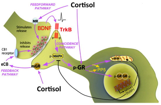
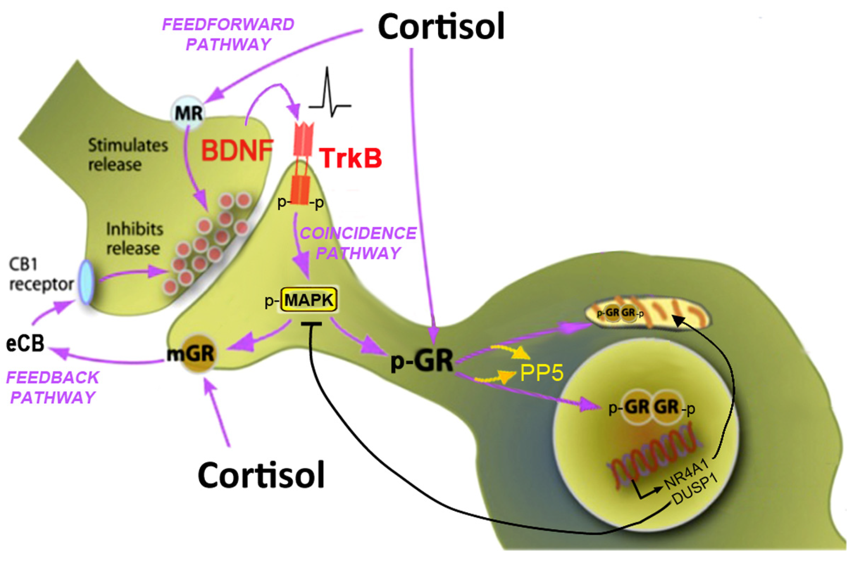
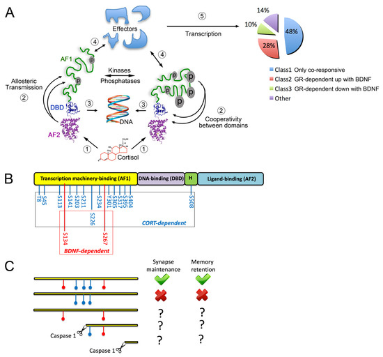
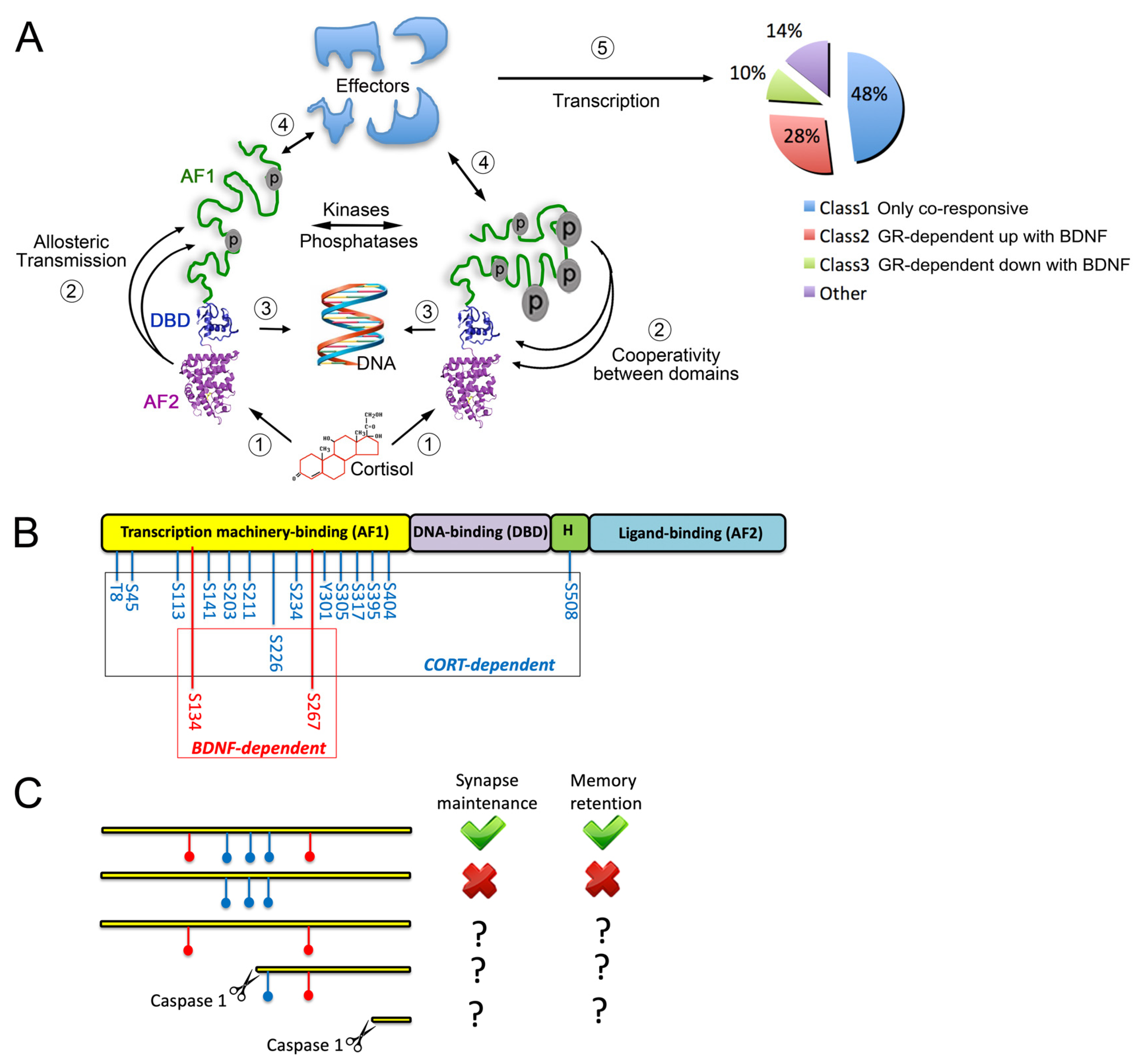
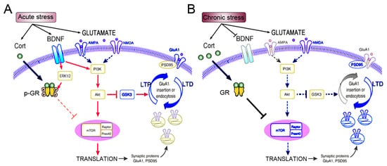
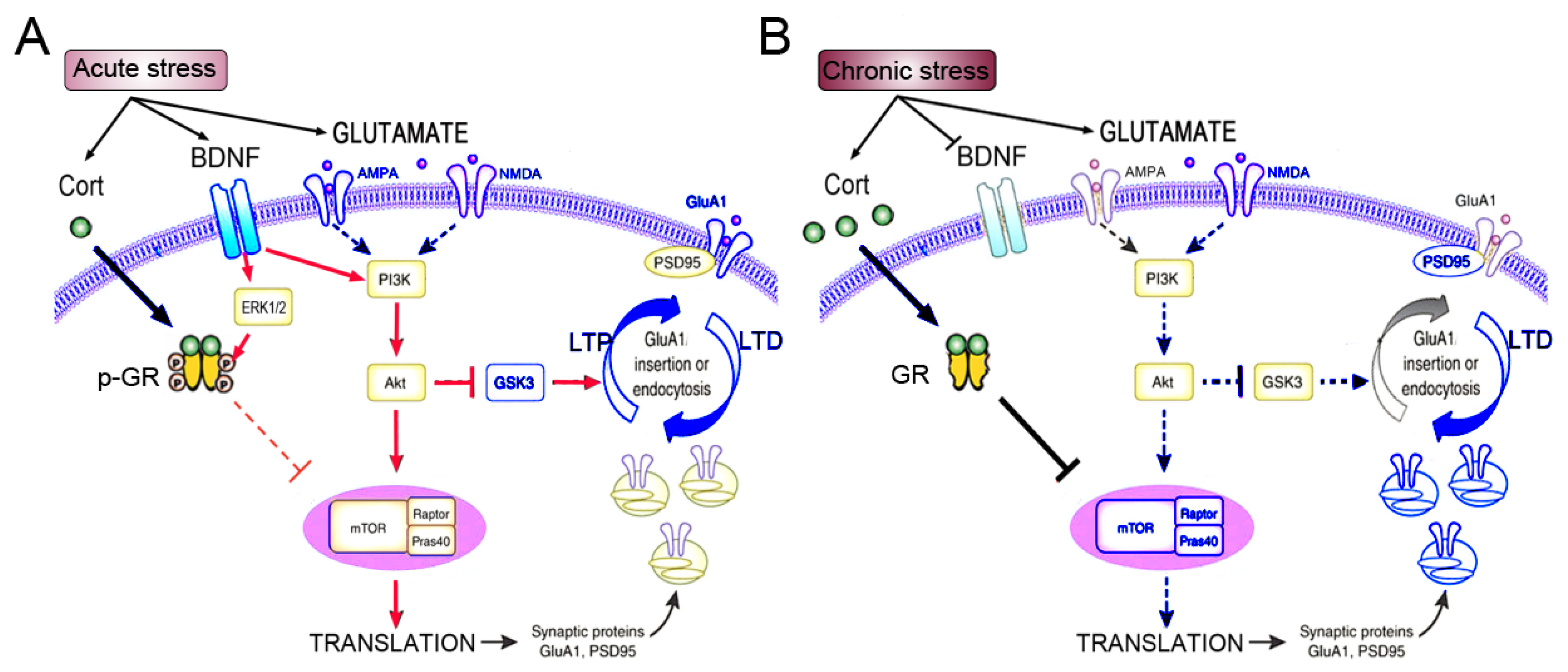
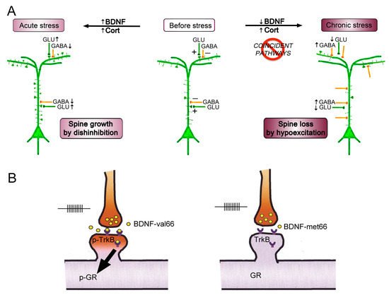
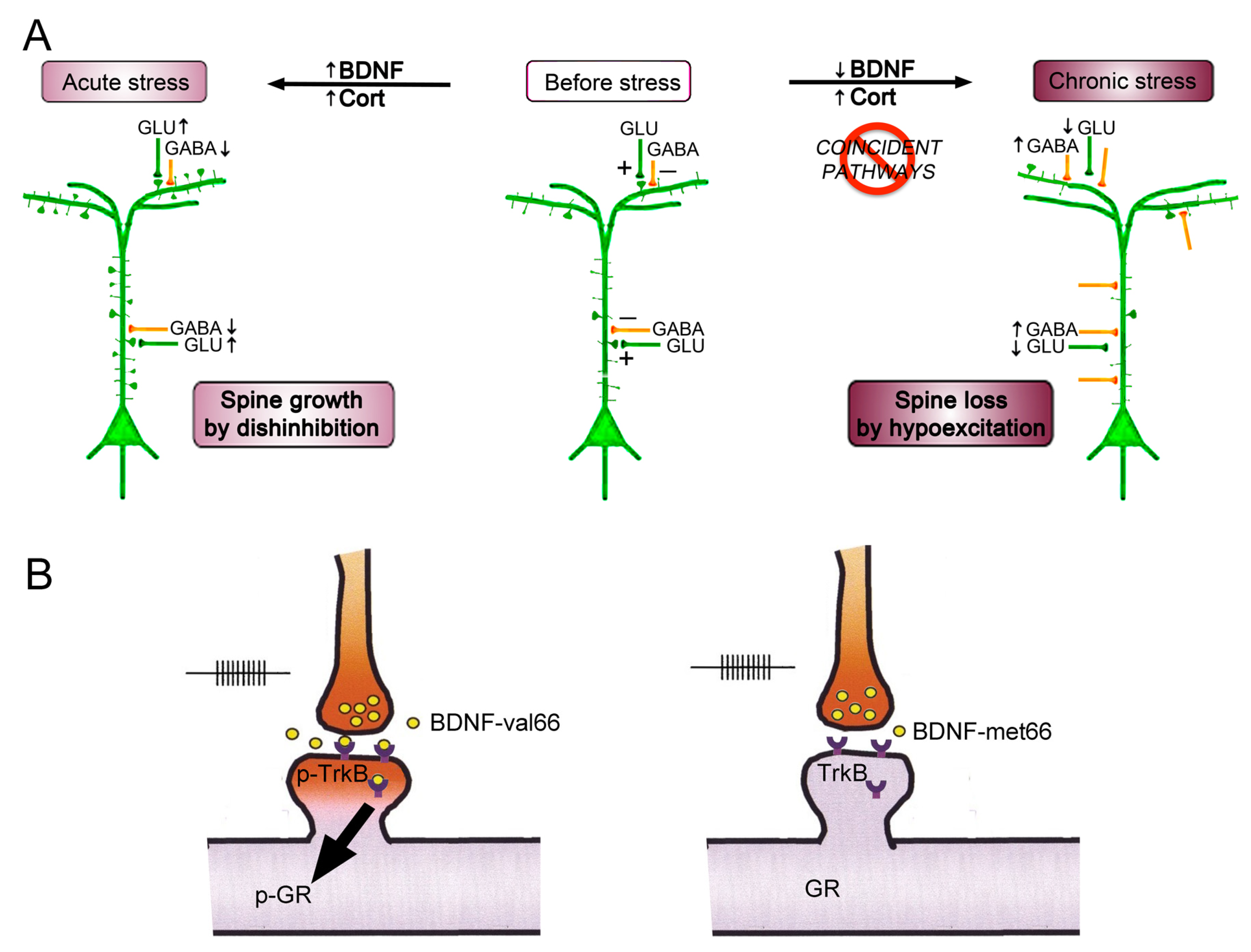
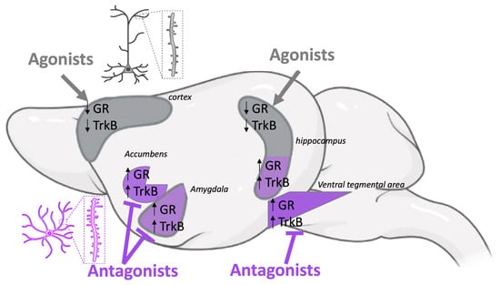
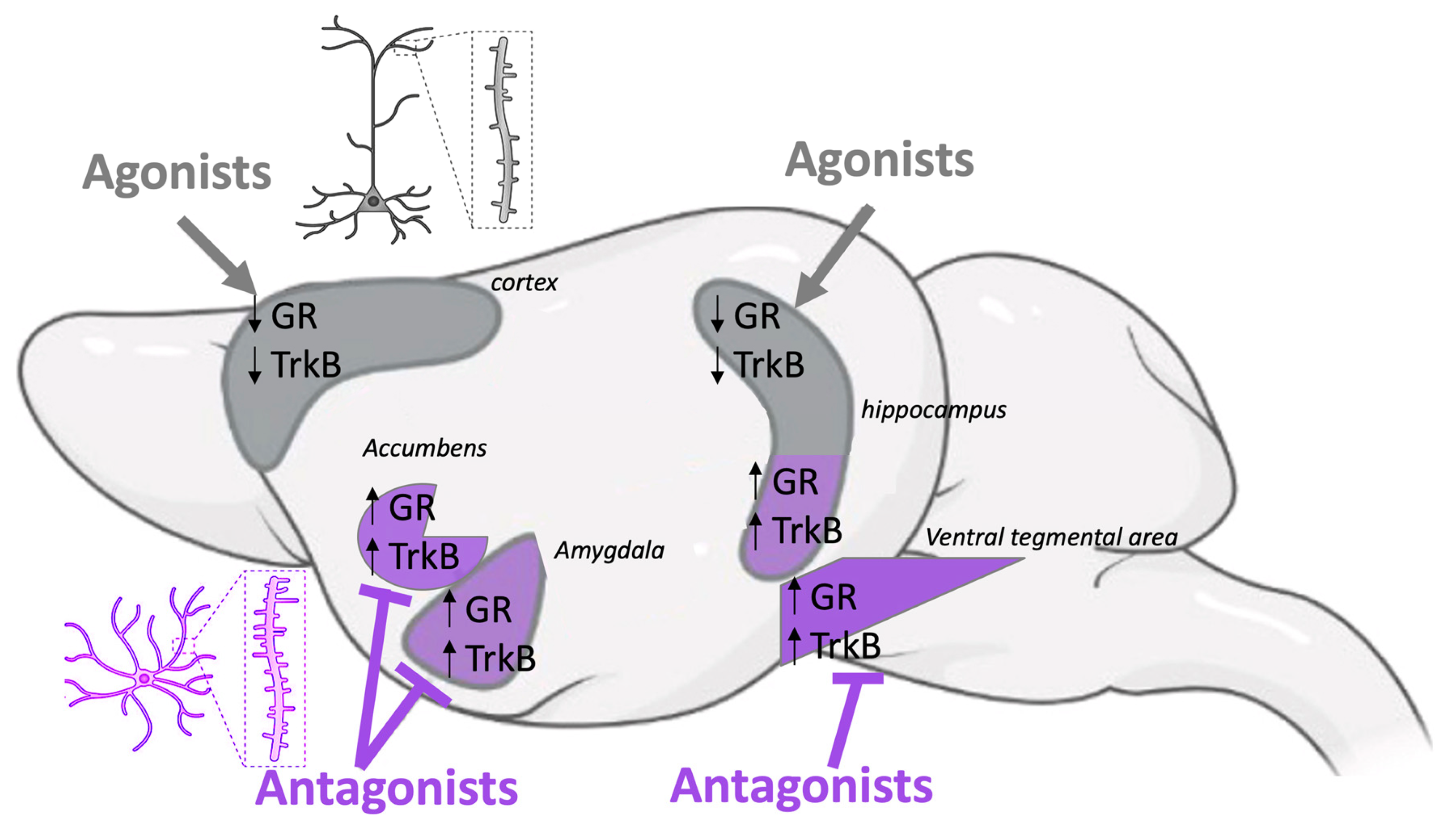
Disclaimer/Publisher’s Note: The statements, opinions and data contained in all publications are solely those of the individual author(s) and contributor(s) and not of MDPI and/or the editor(s). MDPI and/or the editor(s) disclaim responsibility for any injury to people or property resulting from any ideas, methods, instructions or products referred to in the content. |
© 2026 by the author. Licensee MDPI, Basel, Switzerland. This article is an open access article distributed under the terms and conditions of the Creative Commons Attribution (CC BY) license.