


Abstract
Objectives: The aim of this study was to investigate radiography students’ perceptions of the usefulness of hands-on 3D-printed models for supporting their understanding of key image quality concepts in magnetic resonance imaging (MRI). Students’ views on the perceived contribution of these models to their learning experience, confidence, and independent learning were also evaluated. Methods: This questionnaire-based study included 79 students. Three 3D-printed MRI model sets were designed and fabricated for use in hands-on teaching sessions. Following the sessions, participants completed a seven-item questionnaire divided into two sections. The first section assessed students’ perceptions of the teaching value of each model set, whereas the second section assessed students’ perceived learning benefits. Likert-scale responses were summarized descriptively, and the internal consistency of the seven-item questionnaire was assessed using Cronbach’s alpha. Results: In terms of the perceived teaching value of the models, the highest proportion of “strongly agree” responses were observed for pixel–voxel dimensionality and voxel geometry model sets, with 58 (73.4%) and 55 (69.6%), respectively. Regarding students’ perceived learning benefits, students strongly agreed that the 3D-printed MRI models supported their confidence, independent learning, and learning experience in understanding MRI image-quality concepts, with 49 (62%), 47 (59.5%), and 46 (58.2%), respectively. The seven-item questionnaire demonstrated good internal consistency (Cronbach’s α = 0.896, 95% CI [0.835, 0.957]). Conclusion: Radiography students expressed strong favorable views regarding the hands-on use of 3D-printed models and perceived them as helpful tools for visualizing core MRI image quality concepts.
1. Introduction
The theoretical and practical aspects of magnetic resonance imaging (MRI) are considered challenging and require a strong grasp of the underlying concepts, constituting difficulties for both radiographers and radiography students [1,2]. Radiography students may face challenges in applying classroom-based MRI knowledge in real clinical situations, particularly in busy clinical departments such as MRI units, where high service demands can make guided practice more difficult [3]. An objective structured clinical examination (OSCE)-based study revealed that most MR radiographers could not answer more than half of the questions correctly [4].
As MRI is inherently complex and traditional teaching alone may not provide sufficient hands-on experience, additional teaching approaches are required to support a clearer understanding. Previous studies have examined several technology-enhanced approaches to MRI education, including computer-based simulation, virtual reality (VR), and mobile learning applications [1,5,6]. A study by Treceño-Fernández et al. (2021) showed that MRI computer-based simulation improved applied learning outcomes [1], Cheewakul et al. (2026) reported higher immediate post-test scores and high satisfaction with a VR module [5], and Alsharif et al. (2018) found that a mobile learning app significantly improved MR radiographers’ knowledge of image quality [6]. Such tools could potentially enhance MRI-related learning and should be considered useful adjuncts to traditional teaching.
Three-dimensional (3D) printing has emerged as a promising tool in health profession education and has been applied across medicine [7], dentistry [8] and nursing [9]. According to Xie et al. (2025), the use of 3D-printing technology in medical education is associated with improved student understanding and enhanced practical skills [10]. Studies in the medical field that investigated the usefulness of using 3D-printed models compared with traditional methods reported better academic performance in students with the use of 3D-printed models [11]. Despite growing interest in 3D-printed models within radiography, existing research has primarily concentrated on their use in fabricating anatomical structures, phantoms and test objects for performance assessment purposes [12,13,14,15]. Studies investigating 3D-printed models in MRI education are limited and largely focused on phantom-based applications [16,17].
In this study, three 3D-printed MRI model sets were designed and fabricated for hands-on teaching sessions, and students’ perceptions of their usefulness in supporting understanding key MRI image quality concepts were collected. Each model set represented a fundamental image quality concept that students may find difficult to visualize mentally using the traditional teaching methods. The study also evaluated students’ views on the model sets’ perceived contribution to their learning experience, confidence, and independent learning.
2. Methods
2.1. Participants
Ethical approval was obtained prior to student recruitment and informed consent was obtained from all participants before inclusion. A total of 79 third- and fourth-year radiography students at the College of Applied Medical Sciences, Taibah University, Saudi Arabia, were prospectively enrolled on a voluntary basis. This was an exploratory, single-group, post-session perception study. The study did not include a baseline assessment, pre- and post-testing, a control or comparison group, or delayed retention assessments.
2.2. Three-Dimensional-Printed Models
The designs for the three 3D-printed MRI model sets were designed using several software programs, including Adobe Illustrator for iPad, version 3.0.10 (Adobe Inc., San Jose, CA, USA), Tinkercad (Autodesk, San Francisco, CA, USA; web-based application) and Bambu Studio (version 2.2.160; Bambu Lab, Shenzhen, China). The models were then fabricated using a Bambu Lab P1S printer (Bambu Lab, Shenzhen, China) with a build volume of 256 × 256 × 256 mm3 and a combo option for multicolor printing. This enabled the models to be printed in multiple colors using polylactic acid (PLA) bioplastic filament. The PLA filament diameter was 1.75 mm and the nozzle diameter was 0.4 mm.
Vector representations of mid-axial T2-weighted brain MR images were manually created in Adobe Illustrator for iPad, version 3.0.10 (Adobe Inc., San Jose, CA, USA) and exported as a Scalable Vector Graphics (SVG) file. The SVG file for each model was separated into four layers representing cerebrospinal fluid (CSF) (white), gray matter (light gray), white matter (dark gray), and background (black). The separated layers were imported into Tinkercad (Autodesk, San Francisco, CA, USA; web-based application) for 3D model construction and conversion to Standard Triangle Language (STL) format, with further manual adjustments applied during modeling. The resulting STL file was then color-assigned and sliced in Bambu Studio prior to fabrication.
The 3D-printed MRI model sets were designed to illustrate the following MRI image quality concepts: the differences between two-dimensional (2D) and 3D images in terms of slice gap and the effect of thin versus thick slices on spatial resolution, the difference between isotropic and anisotropic voxels, and the distinction between pixels and voxels. Table 1 summarizes the technical specifications of the 3D-printed model sets, including their dimensions, print time, and weight. The following sections provide a detailed description of the fabricated 3D-printed model sets.
Table 1.
Technical specifications of the 3D-printed model sets.
2.3. Two-Dimensional–Three-Dimensional Slice Continuity Model Set
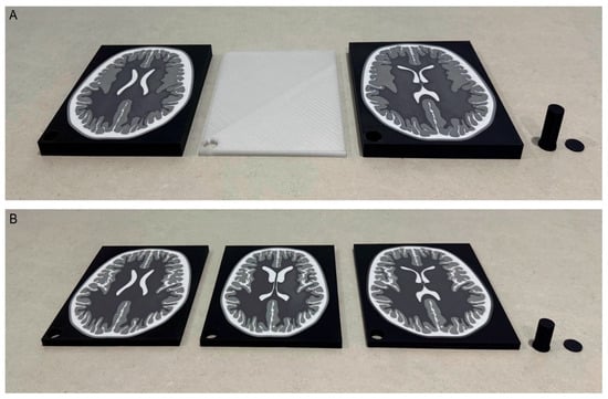
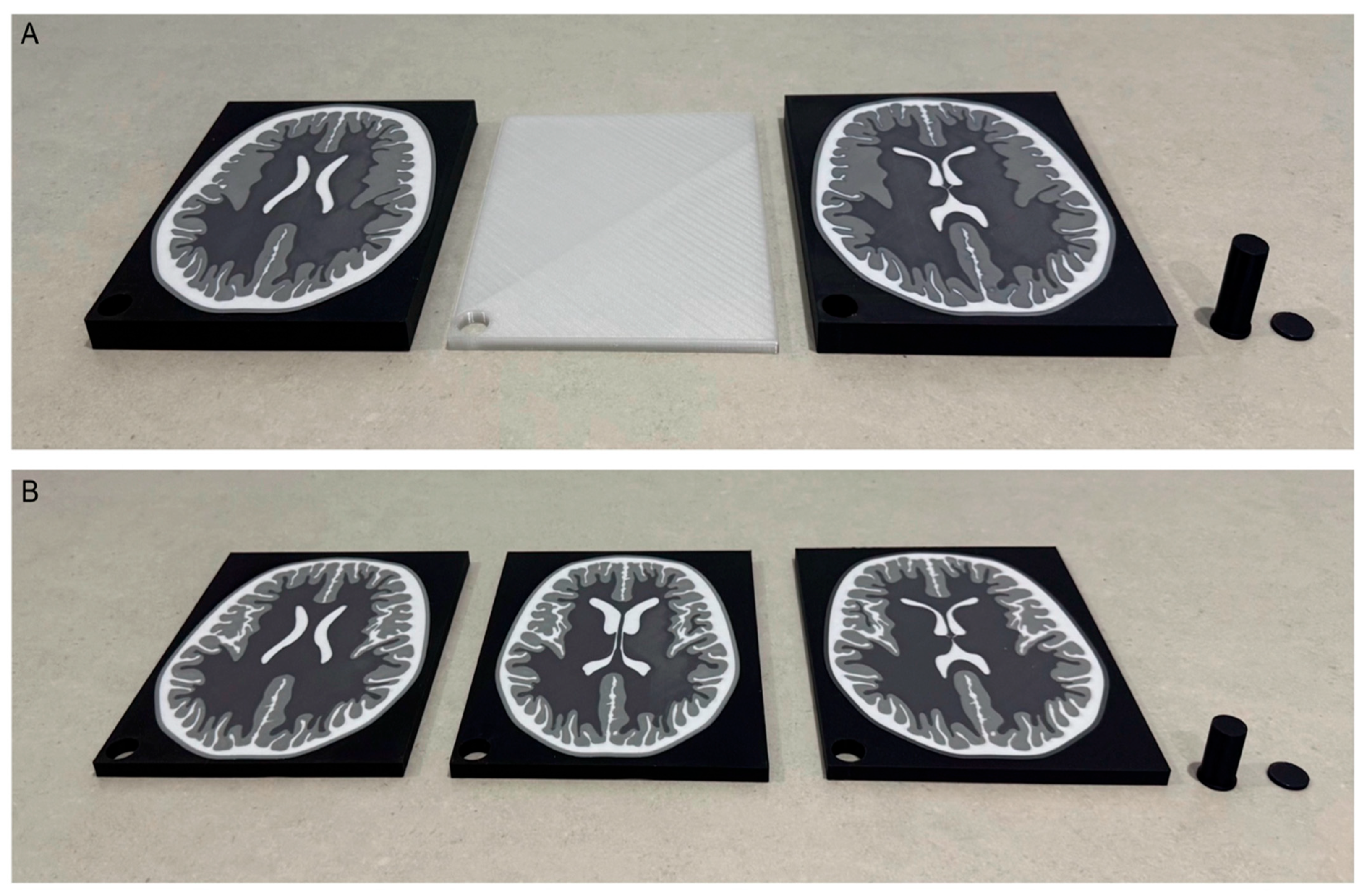
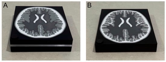
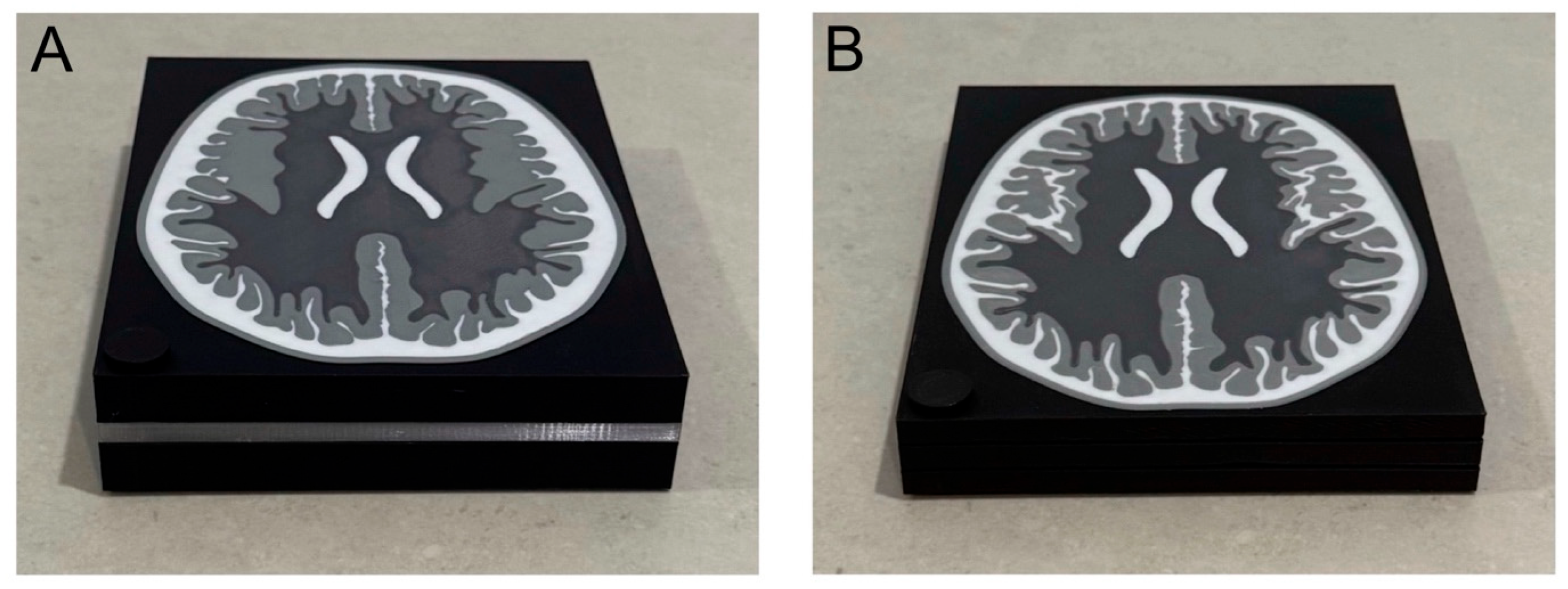
This model set consisted of two imaging configurations: the 2D slice gap model represented a 2D MRI image configuration with two thick slices (16 mm), and a single slice-gap object was fabricated from clear filament to illustrate slice gaps between neighboring 2D slices (Figure 1). The 3D contiguous-slice model simulated 3D MRI images configuration with three thin slices (8 mm) to demonstrate slice continuity (Figure 1). The simulated MRI images used for both model sets were digitally created to depict brain images at different anatomical levels and were mounted on holders to maintain stacking (Figure 2).
Figure 1.
The 3D-printed objects of the 2D slice gap model are shown separately, including two mid-axial T2-weighted MRI brain images at different anatomical levels, a slice gap, and an MRI slice holder (A). (B) The individual 3D-printed objects of the 3D contiguous-slice model, including three brain images at different anatomical levels and an MRI slice holder.
Figure 2.
Final configuration of the 2D slice gap model following assembly of all components (A) and the 3D contiguous-slice model (B). These models were fabricated to illustrate slice gap and slice continuity concepts and to demonstrate standard (A) and high spatial resolution (B) configurations.
This model set was also designed to demonstrate standard and high spatial resolution configurations. Spatial resolution and partial volume averaging (PVA) effects were illustrated using different slice thickness values. The 2D slice gap model with thick slices demonstrated standard spatial resolution. In contrast, objects with thin 3D slices exhibited reduced PVA effects and higher spatial resolution, thereby improving visualization of small anatomical structures. The total print times and weights for the 2D slice gap model and 3D contiguous-slice model were 14:03 h and 524.47 g and 12:43 h and 324.68 g, respectively.
2.4. Voxel Geometry Model Set
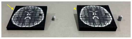
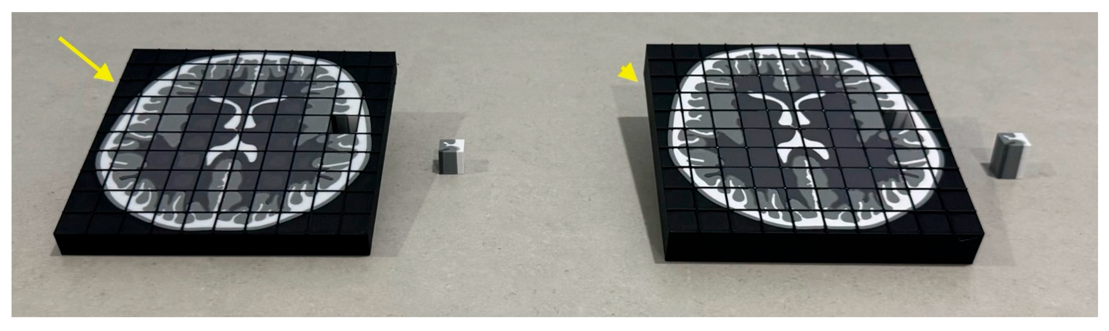
This model set comprised two fabricated models to demonstrate the difference between isotropic and anisotropic voxel geometry (Figure 3). Both models were constructed with a planar surface and z-axis depth to represent volumetric structure. Square grids were applied on the surface of the 3D-printed axial MRI brain images to represent 3D voxel arrangements. In the isotropic model, the axial MRI brain object was designed with overall dimensions of 150 × 150 × 15 mm, comprising 10 voxels in each row and column and resulting in a voxel size of 15 × 15 × 15 mm, thereby reflecting uniform voxel geometry in all directions.
Figure 3.
The 3D-printed voxel geometry model set illustrating isotropic and anisotropic voxel geometry. The isotropic model has equal dimensions along the x-, y-, and z-axes (arrow), whereas the anisotropic model has unequal dimensions, with greater depth along the z-axis (arrowhead).
In contrast, in the anisotropic voxel model, the axial MRI brain object retained the same in-plane dimensions but had a greater z-axis depth, resulting in dimensions of 150 × 150 × 22 mm. Accordingly, each voxel size measured 15 × 15 × 22 mm, representing nonuniform voxel geometry. Both models were additionally colored in the z-direction to demonstrate the depth continuity of anatomical structures in the third dimension. In this model set, a single cubic element in each model was designed to be removable and replaceable within the image plane, with colors reflecting the underlying anatomical structures. However, the removable single cubic elements were fabricated slightly smaller to facilitate ease of removal (Figure 3).
2.5. Pixel–Voxel Dimensionality Model Set
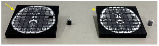
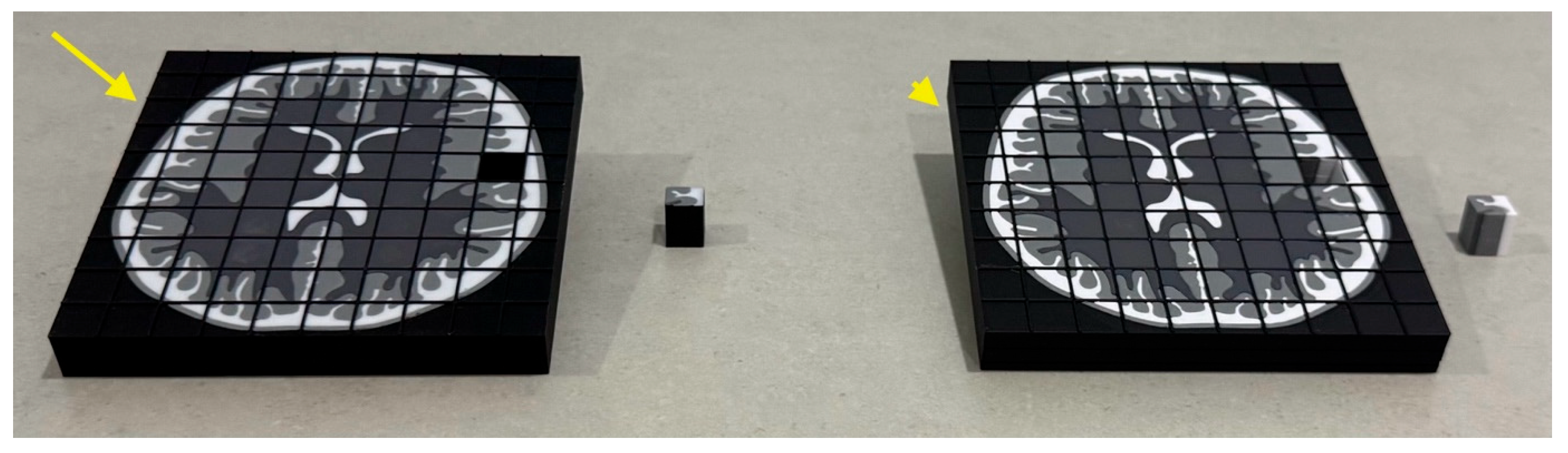
This model set included two fabricated models designed to demonstrate the difference between 2D pixels and 3D voxels (Figure 4). Square grids were applied on the surface of the 3D-printed axial MRI brain images (150 × 150 × 18 mm) to illustrate 2D pixel and 3D voxel arrangements. Similar to the voxel geometry model set, this model set consisted of 10 voxels/pixels in each row and column, resulting in a size of 15 × 15 × 18 mm. In the voxel model, a single cubic voxel object was designed to be extractable from the image plane, with colors corresponding to the underlying anatomical structures in the third dimension. In contrast, the z direction was kept entirely black to indicate the lack of depth extension for the pixel model (Figure 4).
Figure 4.
The 3D-printed pixel–voxel dimensionality model set illustrating the distinction between a two-dimensional pixel and a three-dimensional voxel. The pixel model represents anatomical information within a single plane, without z-axis depth (arrow), whereas the voxel model represents anatomical information extending through the third dimension, demonstrating depth along the z-axis (arrowhead).
2.6. Questionnaire
The questionnaire was divided into two sections and consisted of seven statements rated on a 5-point Likert scale (1 = strongly disagree, 2 = disagree, 3 = neutral, 4 = agree, and 5 = strongly agree). The first section assessed students’ perceptions of the teaching value of the model sets for illustrating specific MRI image quality concepts, including slice gap, slice thickness, and spatial resolution; isotropic and anisotropic imaging; and pixel–voxel differences. In the second section, the statements evaluated students’ views on whether the models supported their learning experience, confidence, and independent learning in understanding MRI image quality concepts. The questionnaire items were informed by previous perception-based studies evaluating 3D-printed educational models in medical education [18,19] and were designed to reflect the MRI image quality concepts addressed in the hands-on MRI teaching sessions. Cronbach’s alpha was used to assess the internal consistency of the seven-item questionnaire and was not interpreted as evidence of questionnaire validity. The questionnaire was developed and administered using Google Forms. A pilot test was conducted with five students to ensure clarity, readability, and completion time.
2.7. Statistical Analysis
All statistical analyses were performed using JASP version 0.96 (JASP Team, University of Amsterdam, Netherlands). Descriptive statistics were calculated for each questionnaire item, and internal consistency of the seven-item questionnaire was assessed using Cronbach’s alpha.
3. Results
A total of 79 radiography students (n = 79; 34 males and 45 females) were prospectively enrolled in this study. Participants rated their level of agreement with the questionnaire statements after attending the hands-on teaching session using the 3D-printed model sets to illustrate key MRI image quality concepts. The values for most items were concentrated in the strongly agree and agree categories, as presented in Table 2. Only a small proportion of students provided negative and neutral responses across the questionnaire statements. Concerning the teaching value of the model sets, the highest proportion of “strongly agree” responses was observed for the model sets demonstrating the difference between pixels and voxels and the distinction between isotropic and anisotropic voxels, with 58 (73.4%) and 55 (69.6%), respectively. Although “strongly agree” was the most frequent response category, “agree” responses ranged from 12 (15.2%) to 20 (25.3%), while “neutral” responses were less common, ranging from 5 (6.3%) to 7 (8.9%).
Table 2.
Radiography students’ evaluations following the hands-on teaching session using 3D-printed models to illustrate MRI image quality concepts.
With respect to student-reported learning support, the highest proportions of “strongly agree” responses were observed for the statements that the 3D-printed brain MRI models supported students’ confidence, independent learning, and learning experience in understanding MRI image quality concepts, with 49 (62%), 47 (59.5%), and 46 (58.2%), respectively. The highest proportions of neutral responses were observed for the statements that the 3D-printed models assisted students’ independent learning and supported their learning experience in understanding MRI image quality concepts, with fourteen (17.7%) and nine (11.4%) neutral responses, respectively. The seven-item questionnaire demonstrated good internal consistency, with Cronbach’s α = 0.896, SE = 0.031, 95% CI [0.835, 0.957].
4. Discussion
This study involved the development of three sets of 3D-printed MRI models for use in hands-on teaching sessions. It aimed to evaluate radiography students’ perceptions of the models in supporting their understanding of key MRI image quality concepts. Each set of models translated a theoretical foundational concept in MRI that is often difficult for students to visualize into a tangible 3D-printed representation. The study also evaluated students’ views on whether the model sets supported their learning experience, confidence, and independent learning.
Research on the use of 3D-printed models in MRI as teaching tools remains very limited, with the existing literature predominantly being focused on phantom-based applications. Yusuff et al. (2025) developed a cost-effective 3D-printed MRI phantom as an educational tool to demonstrate system performance and image quality parameters [16], whereas Habeeb et al. (2024) primarily focused on MRI phantoms replicating anatomical structures and tissue contrast of the head [17]. In contrast, the present study moved beyond the use of phantoms, which require MRI scanners, by utilizing 3D-printed models that do not rely on specialized equipment such as scanners and that allow for flexible use in classroom settings to support the teaching of key MRI image quality concepts.
The clinical performance of 3D MRI sequences has been reported to be superior to that of conventional 2D sequences, owing to the absence of interslice gaps and the use of thinner slices, which may enhance spatial resolution and improve the detection of subtle lesions [20,21]. When voxel dimensions are equal in all three directions (x, y, and z), isotropic imaging provides several advantages, including improved multiplanar reconstruction [22], reduced PVA artifact [22], and enhanced visualization of subtle anatomical structures [23]. Concepts such as slice gaps, slice continuity, and the influence of the z-direction on spatial resolution and PVA can be challenging for radiography students to understand. A study by Kada (2020) found that 46% of final-year radiography students were unable to explain the relationship between slice thickness and PVA artifact [24]. In the current study, students reported positive views of the pixel–voxel dimensionality model set. This pattern of responses may indicate that students perceived the tangible 3D representations of theoretical MRI image quality concepts as supporting their understanding of these concepts.
Previous literature indicated that students viewed the integration of 3D-printed models in anatomy [25], nursing [26], and dentistry [27] as a valuable teaching approach. The present findings demonstrated positive evaluations among radiography students toward the use of 3D-printed models for illustrating MRI image quality concepts. Students also demonstrated positive views regarding the contribution of 3D-printed models to improving their learning experience, confidence, and ability to learn independently. These findings may encourage radiography educators across a range of educational contexts to incorporate this promising technology into their teaching practices.
Although the initial setup required substantial investment in equipment and system preparation, the ongoing printing cost was economically reasonable [18], with all fabricated models together consuming only 1896.54 g of material. In this study, the print time was reported individually for each 3D-printed model to reflect the actual printing time; however, this required more time than printing multiple objects together in a single session. Printing multiple objects simultaneously may, however, be both economically and practically feasible. The filament colors used in this study were selected to resemble the colors of brain tissue in MRI T2 images, and this approach may have contributed to improving students’ learning experience, as suggested by the previous literature [28].
This study has several limitations. First, the study population was drawn from only one academic institution, which may restrict the wider applicability of the findings. Second, although participants completed the evaluation anonymously and no identifying information was collected, social desirability bias cannot be excluded. Third, the study focused on the design and fabrication of 3D-printed models as visual teaching aids for MRI image quality concepts. Future work could develop additional 3D-printed models for other imaging modalities and evaluate students’ perceptions of their usefulness as visual learning aids. However, existing studies using 3D-printed models in MRI education often require access to MRI scanners, which may reduce the practicality of using such models in classroom settings. Fourth, neither expert review nor validation of the seven-item questionnaire used in this study was conducted. However, the questionnaire items were informed by previous perception-based studies assessing 3D-printed models in medical education. Finally, this study focused on students’ perceptions of the benefits of hands-on use of the 3D-printed model sets, rather than evaluating their impact on academic performance. Therefore, this study did not include baseline assessment, pre- and post-testing, comparison groups, follow-up, and delayed assessments, which would be required to investigate the academic impact of the 3D-printed models. The study also did not include comparisons with traditional teaching methods or alternative educational tools, such as virtual reality. However, such perception-based evaluations may serve as an initial investigation and provide valuable findings about the usability and acceptability of these models in radiography education.
In conclusion, radiography students reported favorable evaluations of the hands-on use of 3D-printed MRI models and perceived them as useful tools for visualizing fundamental MRI image quality concepts. The pixel–voxel dimensionality model set received the highest number of strongly agree responses across other model sets, which may reflect the difficulty students often face in mentally visualizing this concept using conventional teaching methods alone. Questionnaire responses further indicated that students perceived the models as supporting their confidence, independent learning, and overall learning experience.
Funding
No funding was received for this study.
Institutional Review Board Statement
This study was approved by Taibah University Research Ethics Committee (approval number: TU-AMS-26-03-024; approval date: 30 March 2026).
Informed Consent Statement
Informed consent was obtained from all participants before participation. The privacy rights of human subjects were observed throughout the study.
Data Availability Statement
Data related to this study are available from the author upon reasonable request.
Acknowledgments
The author gratefully acknowledges the radiography students at Taibah University for their participation in completing the questionnaire.
Conflicts of Interest
The author declares no conflicts of interest.
Abbreviations
| MRI | Magnetic resonance imaging |
| OSCE | Objective structured clinical examination |
| VR | Virtual reality |
| 3D | Three-dimensional |
| PLA | Polylactic acid |
| SVG | Scalable vector graphics |
| CSF | Cerebrospinal fluid |
| STL | Standard triangle language |
| 2D | Two-dimensional |
| PVA | Partial volume averaging |
References
- Treceño-Fernández, D.; Calabia-del-Campo, J.; Matute-Teresa, F.; Bote-Lorenzo, M.L.; Gómez-Sánchez, E.; de Luis-García, R.; Alberola-López, C. Magnetic Resonance Simulation in Education: Quantitative Evaluation of an Actual Classroom Experience. Sensors 2021, 21, 6011. [Google Scholar] [CrossRef] [Scilit] [PubMed]
- Zanardo, M.; Doniselli, F.M.; Monti, C.B.; Cornacchione, P.; Durante, S.; Sconfienza, L.M.; Sardanelli, F. MRI physics and technical issues: Where do Italian radiographers search for information? J. Med. Imaging Radiat. Sci. 2021, 52, 104–111. [Google Scholar] [CrossRef] [Scilit]
- Gallagher, S. Graduate radiographers’ experience of learning MRI practice: A pilot study using constructivist grounded theory methodology. J. Med. Imaging Radiat. Sci. 2023, 54, 51–57. [Google Scholar] [CrossRef] [Scilit] [PubMed]
- Westbrook, C.; Talbot, J. What do MRI radiographers really know? Eur. J. Radiogr. 2009, 1, 52–60. [Google Scholar] [CrossRef] [Scilit]
- Cheewakul, J.; Marshall, S.; Chaichulee, S. Development, validation, and comparative evaluation of an immersive virtual reality for MRI patient preparation training in radiologic technology education. Comput. Educ. X Real. 2026, 8, 100134. [Google Scholar] [CrossRef] [Scilit]
- Alsharif, W.; Davis, M.; Rainford, L.; Cradock, A.; McGee, A. Validation of the educational effectiveness of a mobile learning app to improve knowledge about MR image quality optimisation and artefact reduction. Insights Imaging 2018, 9, 721–730. [Google Scholar] [CrossRef] [Scilit] [PubMed]
- Salazar, D.; Thompson, M.; Rosen, A.; Zuniga, J. Using 3D Printing to Improve Student Education of Complex Anatomy: A Systematic Review and Meta-analysis. Med. Sci. Educ. 2022, 32, 1209–1218. [Google Scholar] [CrossRef] [Scilit] [PubMed]
- Fayyaz, Y.; Ali, M.; Ullah, R.; Shaikh, M.S. Applications of 3D-printed teeth in dental education: A narrative review. J. Taibah Univ. Med. Sci. 2024, 19, 816–822. [Google Scholar] [CrossRef] [Scilit] [PubMed]
- Tan, H.; Huang, E.; Deng, X.; Ouyang, S. Application of 3D printing technology combined with PBL teaching model in teaching clinical nursing in congenital heart surgery: A case-control study. Medicine 2021, 100, e25918. [Google Scholar] [CrossRef] [Scilit] [PubMed]
- Xie, G.; Wang, T.; Fu, H.; Liu, D.; Deng, L.; Zheng, X.; Li, L.; Liao, J. The role of three-dimensional printing models in medical education: A systematic review and meta-analysis of randomized controlled trials. BMC Med. Educ. 2025, 25, 826. [Google Scholar] [CrossRef] [Scilit] [PubMed]
- Chen, S.; Pan, Z.; Wu, Y.; Gu, Z.; Li, M.; Liang, Z.; Zhu, H.; Yao, Y.; Shui, W.; Shen, Z.; et al. The role of three-dimensional printed models of skull in anatomy education: A randomized controlled trail. Sci. Rep. 2017, 7, 575. [Google Scholar] [CrossRef] [Scilit] [PubMed]
- Anwari, V.; Lai, A.; Ursani, A.; Rego, K.; Karasfi, B.; Sajja, S.; Paul, N. 3D printed CT-based abdominal structure mannequin for enabling research. 3D Print. Med. 2020, 6, 3. [Google Scholar] [CrossRef] [Scilit] [PubMed]
- Noonoo, J.B.; Sosu, E.; Hasford, F. Three-dimensional image quality test phantom for planar X-ray imaging. S. Afr. J. Sci. 2023, 119, 14269. [Google Scholar] [CrossRef] [Scilit]
- Ogden, K.M.; Morabito, K.E.; Depew, P.K. 3D printed testing aids for radiographic quality control. J. Appl. Clin. Med. Phys. 2019, 20, 127–134. [Google Scholar] [CrossRef] [Scilit] [PubMed]
- Dukov, N.; Valkova, V.M.; Yordanova, M.; Tsapaki, V.; Bliznakova, K. Design and Use of a Custom Phantom for Regular Tests of Radiography Apparatus: A Feasibility Study. J. Imaging 2024, 10, 258. [Google Scholar] [CrossRef] [Scilit] [PubMed]
- Yusuff, H.; Zorn, P.E.; Giraudeau, C.; Wach, B.; Choquet, P.; Chatelin, S.; Dillenseger, J.P. Development of a cost-effective 3D-printed MRI phantom for enhanced teaching of system performance and image quality concepts. Magn. Reson. Mater. Phys. Biol. Med. 2025, 38, 561–574. [Google Scholar]
- Habeeb, Y.; Zorn, P.E.; Blindauer, F.; Kharouf, N.; Sémeril, D.; Bierry, G.; Kremer, S.; Dillenseger, J.P. High-fidelity anatomical phantoms for MRI practical training. Phys. Med. 2024, 127, 104832. [Google Scholar] [CrossRef] [Scilit] [PubMed]
- Tripodi, N.; Kelly, K.; Husaric, M.; Wospil, R.; Fleischmann, M.; Johnston, S.; Harkin, K. The Impact of Three-Dimensional Printed Anatomical Models on First-Year Student Engagement in a Block Mode Delivery. Anat. Sci. Educ. 2020, 13, 769–777. [Google Scholar] [CrossRef] [Scilit] [PubMed]
- Ward, S.; Maresse, S.; Sun, Z. Three-dimensional-printed models: A novel approach to ultrasound education of the placental cord insertion site. Appl. Sci. 2025, 15, 8221. [Google Scholar] [CrossRef] [Scilit]
- Pinnizzotto, M.; Ragusi, M.; Maino, C.; Allegranza, P.; Talei Franzesi, C.; Pellegatta, S.; Gandola, D.; Turati, M.; Corso, R.; Ippolito, D. Optimizing Knee MRI: Diagnostic Performance of a 3D PDW SPAIR-Based Short Protocol. Appl. Sci. 2025, 15, 8870. [Google Scholar] [CrossRef] [Scilit]
- Abdulaal, O.M.; Rainford, L.; MacMahon, P.; Kavanagh, E.; Galligan, M.; Cashman, J.; McGee, A. 3T MRI of the knee with optimised isotropic 3D sequences: Accurate delineation of intra-articular pathology without prolonged acquisition times. Eur. Radiol. 2017, 27, 4563–4570. [Google Scholar] [CrossRef] [Scilit] [PubMed]
- Li, C.Q.; Chen, W.; Rosenberg, J.K.; Beatty, P.J.; Kijowski, R.; Hargreaves, B.A.; Busse, R.F.; Gold, G.E. Optimizing isotropic three-dimensional fast spin-echo methods for imaging the knee. J. Magn. Reson. Imaging 2014, 39, 1417–1425. [Google Scholar] [PubMed]
- Wen, J.; Desai, N.S.; Jeffery, D.; Aygun, N.; Blitz, A. High-Resolution Isotropic Three-Dimensional MR Imaging of the Extraforaminal Segments of the Cranial Nerves. Magn. Reson. Imaging Clin. N. Am. 2018, 26, 101–119. [Google Scholar] [CrossRef] [Scilit] [PubMed]
- Kada, S. Knowledge of CT exposure parameters among Norwegian student radiographers. BMC Med. Educ. 2020, 20, 302. [Google Scholar] [CrossRef] [Scilit] [PubMed]
- Láinez Ramos-Bossini, A.J.; López Cornejo, D.; Redruello Guerrero, P.; Ruiz Santiago, F. The Educational Impact of Radiology in Anatomy Teaching: A Field Study Using Cross-Sectional Imaging and 3D Printing for the Study of the Spine. Acad. Radiol. 2024, 31, 329–337. [Google Scholar] [CrossRef] [Scilit] [PubMed]
- Hammoud, S.; Browne, F.; Lenane, P.; Manning, S.; Gallagher, S.; Joyce, L.; Igoe, A.; Vallence, D.; Mcauliffe, E. Evaluating the use of 3D skin models as simulation-based educational tools among nursing students: A quasi-experimental study. Nurse Educ. Today 2025, 146, 106519. [Google Scholar] [CrossRef] [Scilit] [PubMed]
- Hanafi, A.; Donnermeyer, D.; Schäfer, E.; Bürklein, S. Perception of a modular 3D print model in undergraduate endodontic education. Int. Endod. J. 2020, 53, 1007–1016. [Google Scholar] [CrossRef] [Scilit] [PubMed]
- Yuen, J. What Is the Role of 3D Printing in Undergraduate Anatomy Education? A Scoping Review of Current Literature and Recommendations. Med. Sci. Educ. 2020, 30, 1321–1329. [Google Scholar] [CrossRef] [Scilit] [PubMed]

Disclaimer/Publisher’s Note: The statements, opinions and data contained in all publications are solely those of the individual author(s) and contributor(s) and not of MDPI and/or the editor(s). MDPI and/or the editor(s) disclaim responsibility for any injury to people or property resulting from any ideas, methods, instructions or products referred to in the content. |
© 2026 by the author. Published by MDPI on behalf of the Academic Society for International Medical Education. Licensee MDPI, Basel, Switzerland. This article is an open access article distributed under the terms and conditions of the Creative Commons Attribution (CC BY) license.