


Abstract
The thyroid ima artery (TIA) is an uncommon arterial variation, with reported prevalence varying substantially among anatomical studies, which primarily supplies the isthmus and inferior portion of the thyroid gland. Its variable origin and midline location in the neck make it susceptible to injury during surgical procedures like tracheostomies. During a routine cadaveric dissection, a TIA was observed in an 83-year-old white South African male, associated with an absent left inferior thyroid artery. The artery originated from the brachiocephalic trunk, measured 5.93 mm in diameter at its origin, was 18.43 mm in length, and bifurcated into two terminal branches. This case contributes to the limited anatomical literature describing the TIA in African populations and reinforces the importance of recognising this variation during anterior neck surgery. A thorough knowledge of its prevalence, location, and course can help clinicians prevent surgical complications and haemorrhage during anterior neck procedures.
1. Introduction
The thyroid ima artery (TIA) (also known as the arteria thyroidea ima) was first documented in 1772 by the German anatomist Johann E. Neubauer, leading to its historical name, Neubauer’s thyroid artery [1,2,3]. The TIA is a small vessel (averaging 3–5 mm in diameter) located in the neck’s midline and has a highly variable prevalence [4,5]. It may present as a single trunk or with multiple bifurcations and terminal arborisations, and its origin, diameter, arterial supply, and termination all show variation in the literature [2,6,7,8]. When present, the TIA generally ascends along the anterior aspect of the trachea to supply the inferior aspect of the thyroid gland and/or isthmus, thereby providing an additional, unpaired source of arterial supply [3,9,10].
The thyroid gland develops between the fourth and seventh gestational weeks. During this period, the gland forms from the endoderm on the floor of the stomodeum and descends into the neck [11]. Simultaneously, branches of the aortic arch develop to supply it [6,11]. This early vascularisation exists as a dense, rich network which, in the final week of thyroid development, gives rise to the paired superior and inferior thyroid arteries, while the remaining vessels typically regress [11]. The persistence of an embryonic arterial channel has been proposed as one explanation for the presence of the TIA, although its precise developmental basis remains unclear [11]. This embryological origin has been linked to the descent of the thyroid gland and variable arterial differentiation during the foetal period [3,5]. Early researchers like Faller and Schaerer [6] believed the TIA was a remnant of the thymic artery.
The reported prevalence of the TIA varies considerably among anatomical studies, reflecting differences in study design, detection methods, and target populations. Early anatomical observations illustrated this variability, ranging from Adachi’s finding [12] of 0.4% (1 of 271) to higher frequencies reported in classical reference works [13]. Contemporary evidence demonstrates a similar pattern: a meta-analysis by Yurasakpong et al. [8] identified an overall prevalence of 3.8%, noting higher detection rates in foetuses and European populations, while a recent systematic review by Bartziotas et al. [9] observed prevalence estimates between 0.1% and 5.71%, with cadaveric investigations consistently yielding higher detection rates than imaging-based studies. Beyond acting as an accessory source of arterial supply to the thyroid isthmus and inferior pole [3,10], the TIA is frequently associated with unilateral or bilateral absence of the inferior thyroid artery [1,3,9,13,14]. This recurrent co-occurrence highlights the TIA’s role as an alternative pathway when conventional vessels are absent, though whether this reflects a true developmental compensatory mechanism remains unestablished from current anatomical data. Furthermore, persistent TIA channels have been implicated in meeting increased vascular demands under specific physiological or pathological conditions [2], as well as serving as feeding vessels for parathyroid adenomas [15].
The origin of the TIA is also highly variable. The brachiocephalic trunk is the most frequently reported origin. However, the artery may also arise from the aortic arch, common carotid artery, subclavian artery, internal thoracic artery, thyrocervical trunk or other vessels [8,9]. Recent case reports continue to document unusual origins, including an origin directly from the aortic arch with an uncommon aortic arch branching pattern [16]. This variation in origin is clinically relevant because the TIA may arise from major vessels within the root of the neck or superior mediastinum and subsequently cross the operative field of anterior neck procedures.
The significant increase in tracheostomies (5–15%) performed during the COVID-19 pandemic has underscored the clinical importance of understanding the TIA prevalence and course [17,18,19]. Due to reported cases of haemorrhage linked to the TIA during percutaneous dilatational tracheostomy [20,21], surgical teams must recognise this vascular variant to safely navigate and execute interventions across the midline of the neck and suprasternal notch.
Most published anatomical descriptions and prevalence studies of the TIA originate from European and Asian populations, whereas reports from African populations remain comparatively scarce. Although studies from African populations have documented variation in the origins of the superior and inferior thyroid arteries, direct information concerning the prevalence and anatomical characteristics of the TIA in African populations remains limited. Population-specific anatomical data is relevant and may contribute to surgical education, anatomical reference data and safer surgical practice.
2. Case Report
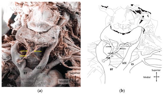
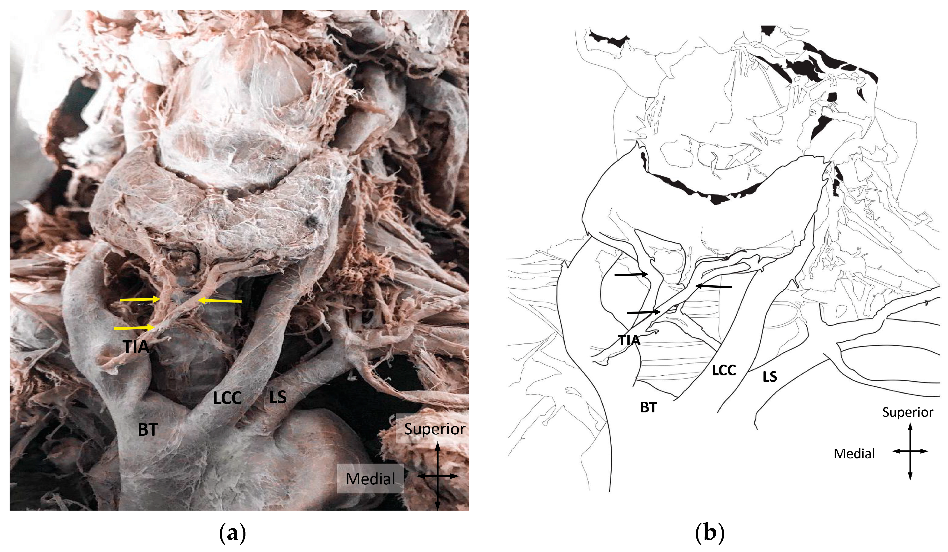
During routine dissection of the root of the neck, a thyroid ima artery (TIA) was noted in an 83-year-old formalin-embalmed white South African male cadaver. The TIA originated from the brachiocephalic trunk, ascended anteromedially in the midline of the neck and divided into two main branches (Figure 1a,b). The vessel exhibited a proximal diameter of 5.93 mm where it originated from the brachiocephalic trunk, with a total length of 18.43 mm to its bifurcation. Measurements were obtained using a digital Vernier calliper (accuracy ±0.01 mm). Following bifurcation, the two branches coursed superiorly within the anterior neck and were traced to their terminal distribution. The branches supplied the isthmus and both inferior poles of the thyroid gland.
Figure 1.
Demonstration of the origin, course and bifurcation of the TIA in (a) cadaveric dissection and (b) line diagram. LCC: left common carotid artery; BT: brachiocephalic trunk; LS: left subclavian artery; TIA: thyroid ima artery. The arrows indicate the TIA bifurcating into its left and right branches.
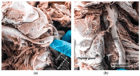
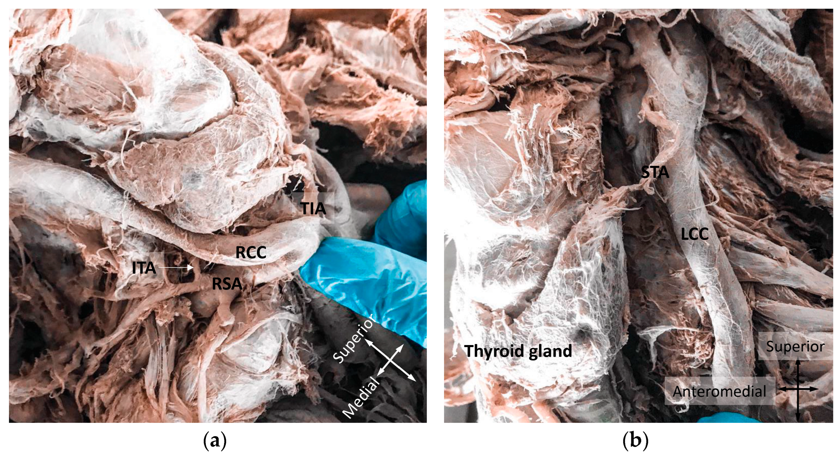
The arterial supply of the thyroid gland was further investigated by dissecting the origin and course of the superior and inferior thyroid arteries. Bilateral superior thyroid arteries were identified, each arising directly from the respective external carotid artery as described in standard anatomical textbooks [22,23]. Vascular asymmetry was noted, with the right inferior thyroid artery originating from the right thyrocervical trunk, in contrast to an absent left inferior thyroid artery (Figure 2a,b).
Figure 2.
(a) Right and (b) left views of the arterial supply to the thyroid gland. ITA: inferior thyroid artery; RSA: right subclavian artery; RCC: right common carotid artery; TIA: thyroid ima artery; STA: superior thyroid artery; LCC: left common carotid artery.
3. Discussion
The TIA is an uncommon anatomical variation in the arterial supply of the thyroid gland, with prevalence estimates varying considerably among anatomical studies according to study population and methodology [3,7]. A systematic review and meta-analysis by Yurasakpong et al. [8] estimated an overall prevalence of 3.8% among 4335 subjects, with substantial heterogeneity between studies. The prevalence was higher in European populations (5.3%) than in Asian (4.0%), American (2.2%) and African (1.6%) populations. In addition, this study reported higher prevalence in cadaveric studies (4.3%) than in imaging-based studies (3.3%), suggesting that differences in detection methods may contribute to the variation between reported estimates. More recently, Bartziotas et al. [9] reviewed 20 studies published between 2010 and 2025 and reported prevalence estimates ranging from 0.1% to 5.71%, with consistently higher detection rates in cadaveric studies than in imaging studies. The occurrence of a TIA in the present South African cadaver therefore contributes to the relatively limited anatomical evidence from African populations.
The origin of the TIA is also highly variable. Yurasakpong et al. [8] reported that the brachiocephalic trunk was the most common origin, accounting for approximately 74% of cases, followed by the right common carotid artery (9.6%) and aortic arch (7.7%), with less frequent origins from other arteries. The present TIA originated from the brachiocephalic trunk, consistent with the most frequently reported origin in the literature and recent cadaveric reports.
Chin et al. [3] reported that the TIA is typically a small vessel measuring approximately 3–5 mm in diameter. In this case, the TIA exhibited a proximal diameter of 5.93 mm at its brachiocephalic origin, exceeding standard literature ranges, and measured 18.43 mm in length, similar to the 17.8 mm documented by Lovasova et al. [24]. The relatively large calibre observed in the present case may be related to the extensive distribution of the artery, as its branches supplied both the inferior thyroid poles and the isthmus. Recent cadaveric reports demonstrate considerable branching variability; for example, Totlis et al. [25] described a TIA arising from the brachiocephalic trunk that divided into multiple anterior and posterior branches supplying the thyroid gland, parathyroid glands and infrahyoid muscles. The bifurcation observed in the present case represents another pattern within the recognised range of TIA branching morphology.
It has been suggested that the TIA provides an alternative vascular route when the inferior thyroid artery is absent [1]. Early observations by Faller and Schaerer [6] revealed that the rate of aplasia among TIA presentations ranged between 0.20 and 5.9%. In a post-mortem study, the inferior thyroid artery was absent in 4.3% of cases on the right and 9.5% on the left in females, and in 5.9% of cases on both the right and left in males [2]. The coexistence of the TIA with absence or deficiency of an inferior thyroid artery is particularly relevant to the present case: the left inferior thyroid artery was absent, while the left branch of the TIA supplied the inferior aspect of the left thyroid lobe. This anatomical relationship aligns with previous reports describing the TIA alongside unilateral or bilateral absence of the inferior thyroid arteries [9,14,26]. In the present case, the TIA provides an alternative arterial supply to a region where the conventional left inferior thyroid artery was absent. Although this may represent a compensatory arrangement, the developmental relationship between persistence of the TIA and absence of the inferior thyroid artery cannot be definitively established from a single specimen.
African data on thyroid arterial variation remain sparse. Magoma et al. [27] investigated the origins of the thyroid arteries in 50 Kenyan cadavers and found variant origins in over 25% of superior thyroid arteries and 11% of inferior thyroid arteries, though they did not report on TIA prevalence. Similarly, Tsegay et al. [28] examined 16 cadavers from North Ethiopia, noting variations in the superior thyroid artery while the inferior thyroid artery consistently arose from the thyrocervical trunk, with no TIA identified. These studies highlight both the presence of thyroid arterial variation in African populations and the paucity of TIA-specific data. Consequently, the 1.6% African subgroup prevalence reported by Yurasakpong et al. [8] should not be interpreted as representative of the continent as a whole.
Recent non-African studies further underscore the broad clinical and morphological variability of this vessel. A 2024 cadaveric study of 84 body donors identified a TIA in 14.3% of specimens, substantially higher than the pooled ranges reported by Yurasakpong et al. [8] and Bartziotas et al. [9]. Additionally, Zaborowski et al. [16] described a TIA arising directly from the aortic arch in an 82-year-old male cadaver, in association with an unusual five-branch aortic arch. Such structural variation demonstrates that the TIA differs not only in prevalence, but also in its arterial origin, course, and relationship with surrounding structures.
The clinical significance of the TIA relates directly to its midline course along the anterior aspect of the trachea. Injury to the vessel during tracheostomy, cricothyrotomy, thyroidectomy or other anterior neck procedures can cause fatal haemorrhage [15,21]. This risk was underscored during the COVID-19 pandemic due to a marked increase in surgical and percutaneous tracheostomies [17,18,19,29]. In the present case, the TIA’s midline position, paired bifurcation and wide distribution across the isthmus and both inferior poles demonstrate that a single variant vessel may supply an extensive area of thyroid tissue. Operating clinicians must maintain awareness of potential midline vascular variations to prevent intraoperative haemorrhage and ensure safe surgical planning in the lower anterior neck. While direct cadaveric dissection cannot yield population-level prevalence data, documenting such detailed morphometrics expands the anatomical reference base for clinical practice and surgical education.
4. Conclusions
Although the TIA is uncommon, failure to recognise its presence during anterior neck procedures, such as tracheostomy, thyroidectomy and emergency airway management, can lead to severe intraoperative haemorrhage. This case contributes valuable morphometric data to the limited anatomical literature on thyroid vascular variations in South African populations. Documenting such rare variants remains essential for enhancing surgical education and clinical safety. Larger-scale epidemiological and cadaveric studies in Southern Africa are warranted to clarify regional prevalence and further inform safe operative practice in the lower anterior neck.
Author Contributions
Both authors contributed to the dissection, measurement and material preparation. The first draft was written by K.S., while the final version was written by S.v.d.W. All authors have read and agreed to the published version of the manuscript.
Funding
This research received no external funding.
Institutional Review Board Statement
The study utilised an embalmed human cadaver from the Department of Anatomy and Histology. The cadaver was obtained through either family donation or as an unclaimed body in accordance with the South African National Health Act (Act No. 61 of 2003). No personally identifiable information was collected or accessed. The study was conducted in accordance with the ethical principles outlined in the Declaration of Helsinki (2024), ensuring the confidentiality, integrity, and ethical handling of all anatomical material. Ethical approval has been granted by the Sefako Makgatho University Research Committee (SMUREC/M/110/2021:PG; 1 July 2021).
Informed Consent Statement
The prosection was performed on an embalmed adult cadaver from the Department of Anatomy’s cadaveric collection. The specimen was obtained either through voluntary body donation by the family or as an unclaimed body, in accordance with the provisions of the South African National Health Act (Act No. 61 of 2003). No identifiable personal information was accessed, collected, or recorded during the study. The use and handling of the cadaveric material complied with applicable ethical requirements and the principles outlined in the Declaration of Helsinki (2024).
Data Availability Statement
The original contributions presented in this study are included in the article. Further inquiries can be directed to the corresponding author.
Acknowledgments
The authors sincerely thank those who donated their bodies to science so that anatomical research and teaching could be performed. Results from such research can potentially increase scientific knowledge and can improve patient care. Therefore, these donors and their families deserve our highest respect. The authors would also like to thank Marinda Pretorius for her assistance in creating the line diagram and for editing the figures. Thank you for your input and assistance in elevating the visual aspect of the paper.
Conflicts of Interest
The authors declare no conflicts of interest.
Abbreviations
The following abbreviations are used in this manuscript:
| TIA | Thyroid ima artery |
References
- Pratt, G.W. The thyroidea ima artery. J. Anat. Physiol. 1916, 50, 239–242. [Google Scholar] [PubMed]
- Rimi, K.R.; Ara, S.; Hossain, M.; Shefyetullah, K.M.; Naushaba, H.; Bose, B.K. Postmortem study of thyroid arteries in Bangladeshi people. Bangladesh J. Anat. 2009, 7, 26–33. [Google Scholar] [CrossRef] [Scilit]
- Chin, J.; Zhou, Y.; Wan, P.J.; Lomiguen, C.M. The prevalence of thyroid ima artery and its clinical significance. Int. J. Otorhinolaryngol. Head Neck Surg. 2019, 5, 845–849. [Google Scholar] [CrossRef] [Scilit]
- Banna, F.A.M.H.; Sultana, Z. Observation of arteria thyroidea ima in Bangladeshi people: A postmortem study. J. Enam Med. Coll. 2018, 8, 25–28. [Google Scholar] [CrossRef] [Scilit][Green Version]
- Vasović, L.; Arsić, S.; Vlajković, S.; Zdravković, D. Morphological aspect of the thyroid ima artery in human fetuses. Arch. Ital. Anat. Embriol. 2004, 109, 189–197. [Google Scholar]
- Faller, A.; Schaerer, O. Über die Variabilität der Arteriae thyreoideae. Acta Anat. 1947, 4, 119–122. [Google Scholar] [CrossRef] [Scilit]
- Koenigsberg, R.A.; Pereira, L.; Nair, B.; McCormick, D.; Schwartzman, R. Unusual vertebral artery origins: Examples and related pathology. Catheter. Cardiovasc. Interv. 2003, 59, 244–250. [Google Scholar] [CrossRef] [Scilit] [PubMed]
- Yurasakpong, L.; Nantasenamat, C.; Janta, S.; Eiamratchanee, P.; Coey, J.; Chaiyamoon, A.; Kruepunga, N.; Senarai, T.; Langer, M.F.; Meemon, K.; et al. The decreasing prevalence of the thyroid ima artery: A systematic review and machine learning-assisted meta-analysis. Ann. Anat. 2022, 239, 151803. [Google Scholar] [CrossRef] [Scilit] [PubMed]
- Bartziotas, D.; Mpartziotas, I.; Chrysikos, D.; Shehade, A.; Troupis, T. Prevalence, Surgical Anatomy, and Clinical Significance of the Thyroidea Ima Artery in Thyroidectomy: A Systematic Review. Cureus 2026, 2, e110102. [Google Scholar] [CrossRef] [Scilit] [PubMed]
- Wolpert, S.M. The thyroidea ima artery: An unusual collateral vessel. Radiology 1969, 92, 333–334. [Google Scholar] [CrossRef] [Scilit] [PubMed]
- Bąk, K.; Fuksa, K.; Lipski, M.; Wajda, J.; Pasternak, A.; Patra, A.; Walocha, J.A. Thyroid ima artery (the artery of Neubauer)—How much do we know? Folia Morphol. 2025, 84, 263–266. [Google Scholar] [CrossRef] [Scilit] [PubMed]
- Adachi, B. Das Arteriensystem der Japaner; Tokyo Kenkyusha Press: Kyoto, Japan, 1928; pp. 210–296. [Google Scholar]
- Lippert, H.; Pabst, R. Arterial Variations in Man, Classification and Frequency; J.F. Bergmann-Verlag: Munich, Germany, 1985. [Google Scholar]
- Yilmaz, E.; Çelik, H.H.; Durgun, B.; Atasever, A.; Ilgi, S. Arteria thyroidea ima arising from the brachiocephalic trunk with bilateral absence of inferior thyroid arteries: A case report. Surg. Radiol. Anat. 1993, 15, 197–199. [Google Scholar] [CrossRef] [Scilit] [PubMed]
- Krudy, A.G.; Doppman, J.L.; Brennan, M.F. The significance of the thyroidea ima artery in arteriographic localization of parathyroid adenomas. Radiology 1980, 136, 45–51. [Google Scholar] [CrossRef] [Scilit] [PubMed]
- Zaborowski, G.; Wysoczański, J.; Karaś, R.; Lepich, T.; Bajor, G. Rare variant of aortic arch branching—A case report. Folia Morphol. 2025, 84, 1097–1102. [Google Scholar] [CrossRef] [Scilit] [PubMed]
- Guo, Y.R.; Cao, Q.D.; Hong, Z.S.; Tan, Y.Y.; Chen, S.D.; Jin, H.J.; Tan, K.S.; Wang, D.Y.; Yan, Y. The origin, transmission and clinical therapies on coronavirus disease 2019 (COVID-19) outbreak—An update on the status. Mil. Med. Res. 2020, 7, 11. [Google Scholar] [CrossRef] [Scilit] [PubMed]
- Möhlenkamp, S.; Thiele, H. Ventilation of COVID-19 patients in intensive care units: Beatmung von COVID-19-Patienten auf Intensivstationen. Herz 2020, 45, 329–331. [Google Scholar] [CrossRef] [Scilit] [PubMed]
- Wu, Z.; McGoogan, J.M. Characteristics of and important lessons from the coronavirus disease 2019 (COVID-19) outbreak in China: Summary of a report of 72,314 cases from the Chinese Center for Disease Control and Prevention. JAMA 2020, 323, 1239–1242. [Google Scholar] [CrossRef] [Scilit] [PubMed]
- Kamparoudi, P.; Paliouras, D.; Gogakos, A.S.; Rallis, T.; Schizas, N.C.; Lazopoulos, A.; Chatzinikolaou, F.; Sarafis, P.; Serchan, P.; Katsikogiannis, N.; et al. Percutaneous tracheostomy—Beware of the thyroidea ima artery. Ann. Transl. Med. 2016, 4, 449. [Google Scholar] [CrossRef] [Scilit] [PubMed]
- Gwilym, S.; Cooney, A. Acute fatal haemorrhage during percutaneous dilatational tracheostomy. Br. J. Anaesth. 2004, 92, 298. [Google Scholar] [CrossRef] [Scilit] [PubMed]
- Standring, S. Gray’s Anatomy, 41st ed.; Elsevier: Boston, MA, USA, 2015. [Google Scholar]
- Moore, K.L.; Dalley, A.F.; Agur, A. Clinically Oriented Anatomy, 8th ed.; Lippincott Williams & Wilkins: Baltimore, MD, USA, 2017. [Google Scholar]
- Lovasova, K.; Kachlik, D.; Santa, M.; Kluchova, D. Unilateral occurrence of five different thyroid arteries—A need of terminological systematization: A case report. Surg. Radiol. Anat. 2017, 39, 925–929. [Google Scholar] [CrossRef] [Scilit] [PubMed]
- Totlis, T.; Natsis, K.; Achlatis, V.; Pettas, T.; Piagkou, M. Thyroidea ima artery multiple branching pattern over the trachea. Surg. Radiol. Anat. 2023, 45, 813–817. [Google Scholar] [CrossRef] [Scilit] [PubMed]
- Yohannan, D.G.; Rajan, R.; Chandran, A.B.; Krishnapillai, R. An unusual origin and course of the thyroidea ima artery, with absence of inferior thyroid artery bilaterally. Surg. Radiol. Anat. 2019, 41, 235–237. [Google Scholar] [CrossRef] [Scilit] [PubMed]
- Magoma, G.; Saidi, H.; Kaisha, W.O. Origin of thyroid arteries in a Kenyan population. Ann. Afr. Surg. 2012, 9, 50–54. Available online: https://www.annalsofafricansurgery.com/origin-of-thyroid-arteries-in-a-ken (accessed on 17 August 2026).
- Tsegay, A.T.; Berhe, T.; Amdeslase, F.; Hayelom, H. Variations on the arterial supply of thyroid gland and its clinical significance in selected universities of North Ethiopia. Int. J. Anat. Res. 2019, 7, 6830–6834. [Google Scholar] [CrossRef] [Scilit]
- McGrath, B.A.; Brenner, M.J.; Warrilow, S.J.; Pandian, V.; Arora, A.; Cameron, T.S.; Añon, J.M.; Martínez, G.H.; Truog, R.D.; Block, S.D.; et al. Tracheostomy in the COVID-19 era: Global and multidisciplinary guidance. Lancet Respir. Med. 2020, 8, 717–725. [Google Scholar] [CrossRef] [Scilit] [PubMed]
Disclaimer/Publisher’s Note: The statements, opinions and data contained in all publications are solely those of the individual author(s) and contributor(s) and not of MDPI and/or the editor(s). MDPI and/or the editor(s) disclaim responsibility for any injury to people or property resulting from any ideas, methods, instructions or products referred to in the content. |
© 2026 by the authors. Licensee MDPI, Basel, Switzerland. This article is an open access article distributed under the terms and conditions of the Creative Commons Attribution (CC BY) license.