


Abstract
Morphological disciplines, namely Human Anatomy, Histology, and Embryology, have traditionally provided the foundational knowledge for medical education, offering spatial, cellular, and temporal coordinates of the human body. However, reducing these disciplines to static and purely descriptive learning undermines their deeper purpose: interpreting morphology as the dynamic outcome of biological processes. This review emphasizes three interrelated pillars of morphological sciences—cell differentiation, tissue homeostasis, and organ remodeling—as essential frameworks for understanding both normal physiology and disease pathogenesis. Cell differentiation establishes functional identity, tissue homeostasis ensures structural stability, and organ remodeling enables adaptation to both physiological and pathological stimuli. Dysregulation of these programs underlies a wide range of conditions, from degenerative diseases and chronic inflammation to neoplasms. Integrating classical morphological knowledge with modern approaches—including stem cell biology, organoids, tissue engineering, and computational modeling—enables predictive and regenerative strategies in personalized medicine. Furthermore, recent advances in artificial intelligence applied to histopathology have enhanced our capacity to detect early deviations from homeostasis and guide targeted interventions. By combining spatial, cellular, and molecular perspectives, the morphological sciences can provide clinicians with tools to interpret disease as the result of altered biological programs, anticipate pathology, and design precise therapeutic strategies. This integrated approach highlights the renewed centrality of morphology in contemporary medicine, bridging foundational knowledge with predictive, regenerative, and personalized healthcare.
1. Introduction: From Descriptive Morphology to the Dynamic Biology of Tissues
For decades, the teaching of Human Anatomy, Histology, and Embryology has constituted the foundation of medical education, providing future physicians with the spatial, cellular, and temporal coordinates necessary to understand the structure of the human body. However, reducing these disciplines to purely descriptive—static and memorization-based—knowledge betrays their deeper purpose, which is to interpret morphology as the dynamic expression of underlying biological processes.
By the late 19th century, scholars such as Rudolf Virchow [1,2] had recognized that disease could not be understood solely at the organ level but had to be traced back to ‘cellular pathology,’ based on alterations of the structural and functional units of tissues. From this perspective, morphology is not a mere inventory of forms, but the visible outcome of cellular programs regulated across both space and time.
Consistently, Embryology has shown that the anatomical complexity of the adult organism arises from highly coordinated processes of cell differentiation, migration, selective proliferation, and programmed cell death, driven by molecular signals and mechanical interactions. Histology, in turn, allows us to understand how these programs are maintained—or altered—during postnatal life, through mechanisms of tissue renewal, adaptation, and repair.
Within this framework, three key concepts emerge as common interpretative pillars across the morphological sciences: cell differentiation, tissue homeostasis, and organ remodeling. They represent not only the biological foundations of tissue structure, but also the essential lens for understanding the origin and progression of disease. While several review articles have addressed specific aspects of tissue biology, such as stem cell regulation, tissue engineering, or the application of artificial intelligence in pathology, these topics are often discussed in a fragmented manner, without a unifying conceptual framework. Organoids, for instance, have emerged as advanced three-dimensional models capable of recapitulating tissue architecture and function, bridging basic biology with clinical translation and enabling disease modeling, drug screening, and personalized therapeutic approaches [3,4,5]. Similarly, recent developments in computational pathology and artificial intelligence have led to the emergence of multimodal and foundation models capable of integrating histological, molecular, and clinical data, significantly enhancing diagnostic and predictive capabilities [6,7]. Moreover, the convergence of artificial intelligence with organoid-based systems is opening new perspectives for dynamic modeling of biological processes, enabling real-time analysis of tissue behavior and improved prediction of therapeutic responses [8].
In contrast, the present review proposes an integrated interpretative model based on three core biological programs—cell differentiation, tissue homeostasis, and organ remodeling—considered as a continuous and interconnected system spanning from development to disease. This perspective aims to bridge classical morphological disciplines with contemporary advances in computational and regenerative medicine, providing a coherent framework that not only describes tissue structure but also interprets its dynamic behavior across physiological and pathological conditions. In this sense, the originality of our approach lies in the integration of traditionally separated domains into a single conceptual continuum, offering both educational and translational value.
Cell differentiation defines the morphological and functional identity of cells within a tissue, tissue homeostasis ensures their stability over time, and organ remodeling allows adaptation to physiological and pathological stimuli. These processes, traditionally addressed in a fragmented manner within medical curricula, constitute a biological continuum that spans the study of Anatomy, Histology, and Embryology, extending toward Physiology, Pathophysiology, and other related disciplines.
A paradigmatic example of this integrated vision is the historic classification of tissues into labile, stable, and permanent types, proposed by Giulio Bizzozero [9], who anticipated concepts now central to stem cell biology and regenerative medicine. This classification, originating from histological observations, still holds significant explanatory value today for understanding the varying responses of tissues to damage, inflammation, and aging.
Contemporary literature has further reinforced this framework, showing that disruptions in differentiation and tissue homeostasis programs represent a crucial event in the development of numerous pathologies, ranging from degenerative diseases to neoplasms, and including chronic inflammatory disorders [10,11].
Considering this evidence, the morphological sciences should not be regarded merely as preparatory ‘basic’ disciplines in a reductive sense. Instead, they must be recognized as central interpretative tools for understanding Medicine. Therefore, studying Anatomy, Histology, and Embryology enables future physicians to acquire not only a descriptive language but also a true biological lens through which to interpret disease.
This narrative review is based on a selective analysis of the current literature, with references chosen for their relevance to the proposed conceptual framework. Priority was given to recent and high-impact studies in the fields of morphological sciences, stem cell biology, tissue engineering, and computational pathology, without aiming to provide a systematic or exhaustive review.
To facilitate the understanding of the key processes that govern tissue structure and function, which will be discussed in more detail in the following sections, Table 1 summarizes essential aspects of cell differentiation, tissue homeostasis, and organ remodeling. This overview highlights their definitions, principal cellular players, representative physiological examples, and potential pathological consequences, providing a concise framework for interpreting dynamic morphological processes.
Table 1.
Key morphological processes in health and disease 1.
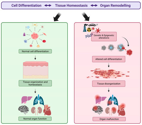
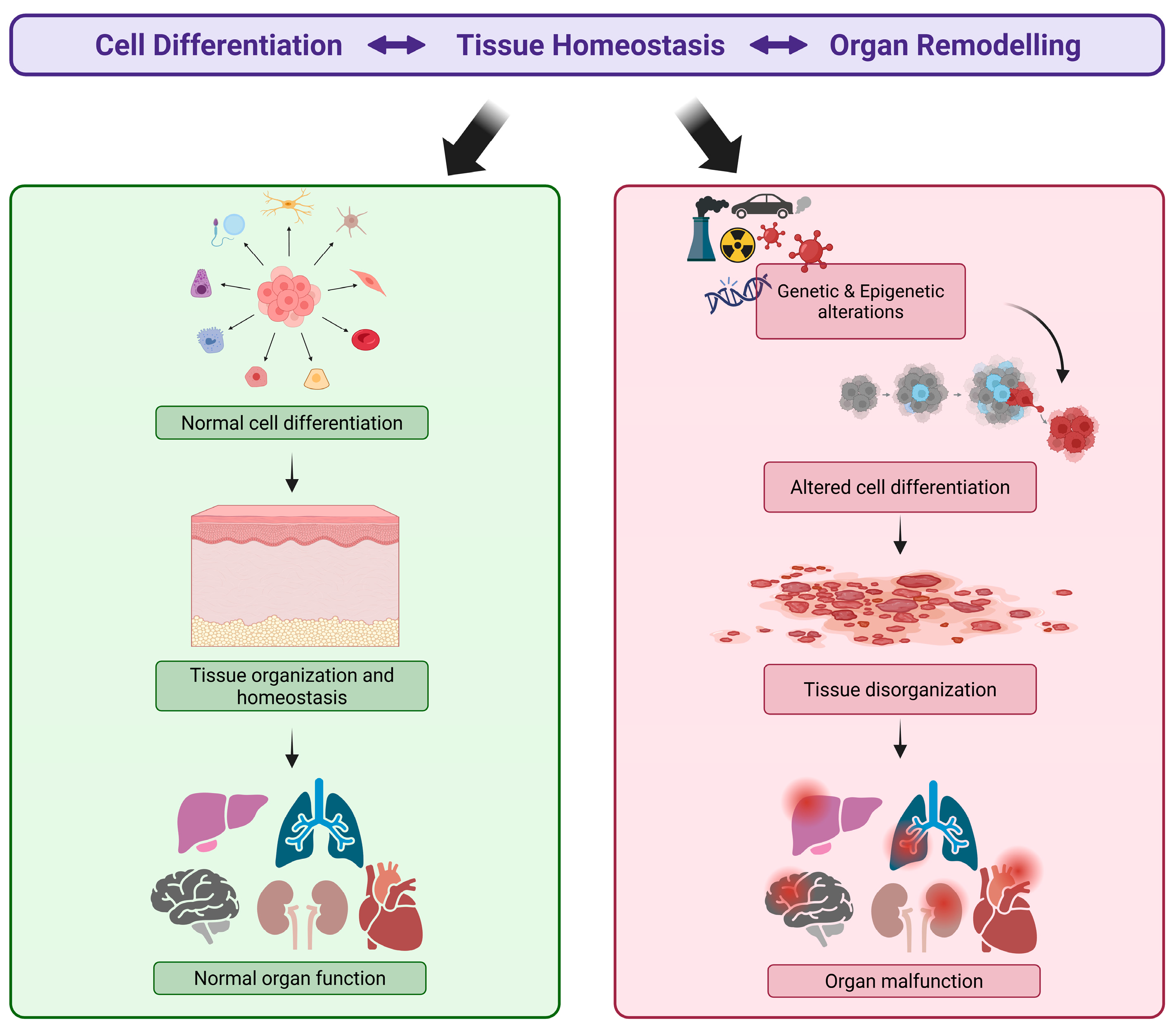
At the same time, in Figure 1 we illustrate the key concept that alterations in cellular differentiation, driven by genetic or epigenetic causes, lead to tissue disorganization, which in turn results in organ dysfunction and, ultimately, disease of the organism. Understanding this etiopathogenetic pathway helps both clinicians and biomedical researchers to identify the origins of disease and to intervene through integrated strategies, including lifestyle modifications, pharmacological treatments, or surgical approaches. From a clinical perspective, this framework reflects the progression from early molecular alterations to overt disease, supporting the interpretation of pathological findings as part of a continuum rather than isolated events. This approach may facilitate earlier diagnosis and more precise identification of therapeutic targets. These and related concepts will be further discussed in the following sections.
Figure 1.
Conceptual framework linking cell differentiation to disease: an etiopathogenetic continuum.
Schematic representation of the relationship between cell differentiation, tissue homeostasis, and organ remodeling under physiological and pathological conditions. Under normal circumstances, proper cell differentiation ensures tissue organization and homeostasis, ultimately sustaining normal organ function. Genetic and epigenetic alterations—including molecular stress and other environmental factors—can disrupt cell differentiation, leading to tissue disorganization. This process progressively results in organ malfunction and, ultimately, disease, outlining a continuous etiopathogenetic pathway from molecular alterations to organism-level pathology. Created in BioRender. Bucchieri, F. (2026) https://BioRender.com/1x7xceh (accessed on 15 May 2026).
2. Cell Differentiation: From Totipotency to Functional Specificity
Cell differentiation represents one of the fundamental biological processes that transform a single totipotent cell, present in the zygote, into a wide array of mature cells with highly specialized functions. Historically, the concept was outlined in the early embryological studies of the 19th century. Pioneers such as Wilhelm Roux and Hans Spemann demonstrated that organ formation depends on spatial and temporal interactions among differentiating cells, anticipating modern notions of induction and morphogenesis [12,13].
During embryonic development, differentiation is orchestrated by a complex network of transcription factors, morphogen gradients, cell–cell and cell–extracellular matrix interactions, which guide the transition from undifferentiated cells to cells with specific functional identities [14,15,16]. This process not only establishes tissue morphology but also the intrinsic functional specialization of cells within each organ system.
In adult tissues, differentiation remains essential due to the presence of undifferentiated or progenitor cell populations in most tissues [17]. These cells sustain physiological turnover in highly regenerative tissues such as intestinal epithelium, epidermis, endometrium, and contribute to repair processes following injury [18,19]. The maintenance of proper differentiation programs is therefore crucial for preserving tissue identity over time. Alterations in differentiation primarily affect cellular identity and lineage commitment, leading to pathological conditions such as metaplasia, dysplasia, and neoplasia, in which cells lose their appropriate differentiation state or acquire aberrant phenotypes [20,21].
In this context, differentiation should be interpreted as the biological process that defines what a cell is and what it is able to do within a tissue. For example, dysplasia of the bronchial epithelium reflects an incomplete or abnormal differentiation process often associated with chronic exposure to toxic agents [22]. Similarly, intestinal metaplasia of the gastric mucosa represents a shift in cell identity driven by environmental stress, highlighting the plasticity—but also the vulnerability—of epithelial progenitor cells [23].
More recent concepts in stem cell biology and regenerative medicine have further reinforced the centrality of differentiation. Alterations in cell fate and niche signaling can lead to degenerative diseases, chronic inflammatory conditions, and tumorigenesis [10,24].
Finally, the analysis of cell differentiation is fundamental for modern therapeutic strategies. From a translational perspective, understanding differentiation programs provides the foundation for modern therapeutic strategies. The use of pluripotent stem cells, organoid models, and regenerative approaches is directly based on the ability to control and direct cellular identity [25,26,27]. Groundbreaking studies on organoid systems have demonstrated their ability to recapitulate key aspects of tissue development and differentiation, providing robust experimental platforms for studying human biology and disease [28]. In parallel, artificial intelligence (AI) applied to histopathology can detect subtle alterations in differentiation patterns, enabling early identification of disease states [7,29,30].
3. Tissue Homeostasis: Maintaining Biological Balance
Tissue homeostasis refers to the ability of tissue to maintain its structural integrity, cellular composition, and functionality capacity over time through a dynamic balance of proliferation, differentiation, and programmed cell death. This concept was historically anticipated by Giulio Bizzozero’s classification of tissues into labile, stable, and permanent types [9], reflecting different regenerative capacities across tissues.
From a biological perspective, homeostasis is maintained through the integration of multiple regulatory signals, including molecular, biomechanical, vascular and neural inputs, which contribute to the regulation of the cellular microenvironment (niche). In this context, tissue homeostasis should be understood as a condition of dynamic equilibrium rather than static stability, in which continuous cellular renewal is precisely regulated to maintain tissue function without altering its overall structure.
Innervation plays a particularly relevant role in this regulatory network. During embryonic development, the formation of neuronal connections with target tissues occurs through complex axonal growth trajectories and neurotropic signals accompanying organogenesis. Adequate innervation is necessary for the growth and maturation of various organs, and its absence or alteration can lead to abnormal development or disorganized functions [31,32,33,34].
In adult tissues, innervation continues to influence tissue regeneration and response following injury. For example, sensory and sympathetic nerve fibers in the bone marrow niche regulate mesenchymal stem cells (MSCs) and the hematopoietic population, affecting their ability to self-renew and differentiate [35]. Loss of innervation leads to a reduction in stem cell numbers and an impaired capacity to maintain tissue balance [36].
Neural modulation of homeostasis can also occur indirectly, through influence on the microenvironment, for example, via the release of neuropeptides, neurotrophic factors such as NGF (Nerve growth factor) and BDNF (Brain-derived neurotrophic factor), and neurotransmitters that interact with immune, stromal, and progenitor cells [34,37,38]. These signals not only promote cell survival, but can also direct the differentiation fate of cells in contexts of repair and regeneration [31]. The concept of stem cell-driven tissue homeostasis has been extensively characterized in multiple tissues, highlighting the role of specialized niches in regulating cellular turnover and regeneration [39].
When homeostatic mechanisms are disrupted, issues may undergo quantitative or functional alterations rather than immediate structural reorganization. These include degeneration and atrophy, resulting from insufficient cellular replacement [40]; abnormal hypertrophy and hyperplasia due to excessive proliferation in response to chronic stimuli or aberrant signals, as observed in endometrial hyperplasia [41]; and early stages of tumorigenesis, associated with deregulated proliferation and differentiation [42].
Thus, tissue homeostasis represents the biological condition that preserves tissue integrity over time, ensuring that cellular turnover and environmental responses remain within physiological limits.
4. Organ Remodeling: Adaptation, Repair, and Degeneration
Organ remodeling represents the macroscopic and microscopic expression of tissue adaptation to prolonged physiological and pathological stimuli. Rather than an exceptional event, it is a continuous process that accompanies the entire lifespan, reflecting the ability of tissues to modify their architecture, cellular composition, and extracellular matrix organization in response to functional, metabolic, mechanical, and inflammatory changes [43].
Under physiological conditions, remodeling allows functional adaptation of organs. Examples include cardiac hypertrophy in athletes [44], uterine changes during pregnancy [45], bone adaptations in response to mechanical load [46], and structural changes in skeletal muscle with aging [47]. In these contexts, remodeling is an orderly, partially reversible, and finely regulated process aimed at preserving organ homeostasis and function.
From a mechanistic perspective, remodeling involves coordinated interactions among differentiated cells, progenitor cells, immune cells, and extracellular matrix components [48]. These components respond to mechanical, metabolic, and inflammatory signals, integrating local and systemic responses to reshape tissue structure [49].
In this context, inflammation plays a central role in remodeling processes. Physiologically, it contributes to the removal of damaged tissue, recruitment of reparative cells, and activation of regeneration programs [50]. However, when inflammation becomes excessive, persistent, or self-perpetuating—thus losing its beneficial aspects—it shifts from a maladaptive response, thereby justifying targeted pharmacological intervention [51].
In contrast to homeostasis, which maintains structural stability, remodeling reflects a shift in tissue organization in response to sustained stimuli. This distinction is particularly relevant in pathological conditions, where remodeling may initially serve an adaptive function but progressively lead to structural and functional impairment.
In acute injury, remodeling can result in effective regeneration, especially in tissues with high plasticity. However, chronic stimuli such as persistent inflammation, hypoxia, oxidative stress, or mechanical overload may lead to maladaptive remodeling characterized by excessive extracellular matrix deposition, vascular alterations, and progressive loss of functional parenchyma [52,53,54].
These processes culminate in pathological outcomes such as fibrosis scarring, and organ dysfunction, as observed in conditions including pulmonary fibrosis, post-infarction cardiac remodeling, liver cirrhosis, and chronic kidney disease [55,56,57,58].
Therefore, organ remodeling should be interpreted as the temporal evolution of tissue responses beyond homeostatic control, representing the transition from adaptive changes to irreversible structural damage when regulatory mechanisms fail.
5. Disease as a Biological Program Error: An Evolutionary Perspective on Complexity
Disease cannot be understood solely as a macroscopic alteration of organs and tissues, but it often represents the outcome of errors in the biological programs that regulate cellular differentiation, tissue homeostasis, and remodeling. Historically, Rudolf Virchow had already made it clear by the late 19th century that pathology should be traced back to the dysfunction of cellular units rather than merely macroscopic organ changes, thus laying the foundation for the concept of “cellular pathology” [59].
When cellular differentiation processes are incomplete or aberrant, phenomena such as metaplasia, dysplasia, or, in extreme cases, neoplasia can occur, in which immature cells proliferate outside physiological control. Metaplasia may represent an adaptation to chronic stimuli—for example, in the gastric or bronchial mucosa—as it is an adaptive process in which one differentiated cell type is replaced by another, often in response to chronic stress, inflammation, or environmental insults (e.g., acid reflux or cigarette smoke) [15]. Conversely, dysplasia refers to progressively more severe alterations in the differentiation pathway, potentially serving as a precursor to neoplasia, in which differentiation is completely arrested, and cells proliferate without acquiring specialized functions [60].
Similarly, disruption of tissue homeostasis can lead to atrophy or chronic degeneration, and maladaptive remodeling may culminate in fibrosis, dysfunctional scarring, and organ failure [61]. Morphological analysis—both macroscopic and microscopic—allows reconstruction of the “biological history” of these processes, revealing how an initial adaptive response can progress to irreversible degeneration.
An even broader perspective emerges when considering the evolutionary trajectory of life on Earth. Over billions of years, life has evolved from extremely simple forms to increasingly complex organisms, characterized by multiple levels of structural organization, integrated multicellular functions, and intricate regulatory networks. This biological complexity encompasses not only anatomical form but also the web of interactions among genes, proteins, and cellular processes, which underlie the physiology of advanced organisms [62,63,64].
From a molecular perspective, more complex organisms do not necessarily possess a greater number of protein-coding genes, but they exhibit an increase in protein–protein interactions, alternative splicing variants, proteins with multifunctional domains, and intricate regulatory networks. These features expand the possibilities for adaptive responses but also create points of vulnerability where errors can arise. This phenomenon of “evolutionary complexity” has been described as an accumulation of molecular information, including so-called unnecessary complexity, which can act as an “evolutionary baggage” influencing both normal function and susceptibility to disease [65].
From this perspective, a general principle can be hypothesized: as the biological complexity of an organism increases, so do the chances of malfunctions in the underlying biological programs that regulate it. More complex organisms—such as mammals, and especially humans—possess more elaborate regulatory networks compared to simpler life forms. This richness of interactions and levels of control entails a greater number of “critical nodes”, points at which an error can translate into pathological dysfunction. In this sense, a complex organism is inherently more susceptible to a wider variety of diseases, as it has a greater number of regulatory systems that can be disrupted by genetic, environmental, or molecular factors.
From a clinical and educational standpoint, this evolutionary perspective has decisive implications. Understanding which biological program has been altered allows us to distinguish between reversible and irreversible processes, guiding pharmacological therapies, regenerative strategies, or surgical interventions more precisely. In other words, disease is not merely a phenomenon to be treated, but a process to be interpreted biologically, with profound implications for prevention, early diagnosis, and personalized medicine.
To provide a clear overview of how errors in biological programs relate to disease and how these processes are influenced by organismal complexity, Table 2 summarizes key relationships between cellular differentiation, tissue homeostasis, and organ remodeling, alongside their potential pathological outcomes and examples. This table illustrates how increasing biological complexity introduces new regulatory nodes and potential vulnerabilities, highlighting the evolutionary basis for disease susceptibility and the importance of interpreting pathology in the context of dynamic cellular programs.
Table 2.
Relationships between biological programs, tissue dynamics, and disease in the context of evolutionary complexity 1.
6. Morphological Sciences and Medicine: An Integrated Perspective
Morphological sciences—Human Anatomy on one hand, and Histology and Embryology on the other—should not be considered isolated disciplines. Rather, they are essential tools for an integrated understanding of human pathology. Human Anatomy provides the spatial map of the body, identifying the location, size, shape, and relationships among organs and tissues. This knowledge is fundamental for the clinical approach, physical examination, and interpretation of diagnostic imaging, from conventional radiology to computed tomography and magnetic resonance imaging [66].
Histology and Embryology, on the other hand, provide a more microscopic and molecular perspective. They allow us to understand cellular and tissue composition, the structure of the extracellular matrix, the organization of cellular compartments, and the presence of early alterations caused by stress, inflammation, or disease [16]. Embryology, integrated with Organogenesis, provides the framework for the origin and differentiation of tissues, enabling the interpretation of congenital malformations and structural anomalies considering fundamental biological principles such as induction, morphogenesis, and cellular plasticity [67].
A practical example of the integration between morphology and medicine is the understanding of metaplasia, dysplasia, and neoplasia. A physician trained in interpreting differentiation programs can view phenomena such as nodular prostatic hyperplasia, intestinal metaplasia of the gastric mucosa, or dysplasia of the bronchial epithelium not merely as histological alterations, but as expressions of errors or adaptations in cellular programs, with significant prognostic and therapeutic implications [21].
The integrated approach becomes particularly relevant in the era of predictive and regenerative medicine. Insights into tissue biology and differentiation programs have led to the development of models using pluripotent stem cells and organoids, as well as tissue engineering techniques aimed at personalized therapies, including the creation of the so-called “digital twins”—all strategies grounded in a deep understanding of cellular structure and function [68,69,70,71]. Moreover, the application of AI in morphological disciplines relies on the ability to recognize cellular and tissue patterns associated not only with normal differentiation but also with homeostatic dysfunctions, enabling more accurate clinical predictions and targeted therapeutic interventions [72].
In summary, medical training cannot overlook the integrated knowledge of the morphological sciences. Anatomy primarily indicates the location, shape, and relationships of organs and anatomical regions, laying the foundation for understanding clinical examination. Histology helps reveal their structure and the basis of cellular alterations. Embryology explains the origin and developmental sequence of cells and tissues, culminating in organogenesis, which completes the study of intrauterine formation of the human organism. Only by combining these perspectives is it possible to accurately interpret the biological events underlying disease, anticipate the most severe clinical manifestations, and design the most effective therapeutic interventions.
7. Towards Predictive and Regenerative Medicine: Clinical and Pharmacological Implications
Understanding fundamental morphological processes—cellular differentiation, tissue homeostasis, and organ remodeling—forms the basis for modern therapeutic strategies and the advancement toward predictive, regenerative, and personalized medicine. Knowledge of differentiation programs and cellular plasticity has enabled the development of induced pluripotent stem cells directed toward specific cell lineages, paving the way for regenerative therapies and personalized preclinical models useful for drug testing, toxicity prediction, and the study of disease mechanisms [73,74]. In parallel, the creation of organoids—three-dimensional structures derived from stem cells that recapitulate the architecture and function of native tissues and organs—allows in vitro modeling of both physiological and pathological conditions and enables testing of therapeutic responses in settings that faithfully reproduce the behavior of real tissues [75]. Tissue engineering, by combining cells, biomaterials, and biochemical signals, promotes the regeneration of damaged or degenerated tissues, with applications ranging from partial repair to the replacement of compromised organs, leveraging knowledge of how the extracellular matrix, differentiation signals, and cell–cell interactions guide tissue formation and function [25,76].
In the era of digital medicine, the concept of predictive medicine is based on the ability to anticipate disease progression before it becomes clinically evident. In this context, the so-called patients’ digital twins—personalized computational models integrating clinical, genetic, radiological, and histological data—allow simulation of disease progression and prediction of therapeutic responses, testing alternative clinical scenarios in silico and guiding targeted treatment decisions. Recent literature further supports the integration of organoid technologies and artificial intelligence as key drivers of predictive and regenerative medicine [71]. The application of AI in morphological disciplines is based on the ability to recognize cellular and tissue patterns associated not only with physiological conditions but also with homeostatic dysfunctions that precede disease. Deep learning algorithms analyze digital histological images quantitatively, extract features invisible to the human eye and integrate this information with clinical and molecular data to generate more accurate diagnostic and prognostic predictions, transforming digital pathology from a qualitative discipline into an advanced quantitative tool [7]. Recent large-scale studies have demonstrated the clinical applicability of artificial intelligence in pathology, showing high diagnostic accuracy and potential for integration into routine workflows [77].
Understanding altered cellular programs and biological pathways has profound pharmacological implications. For instance, distinguishing between beneficial, physiological inflammatory processes—necessary to activate post-injury regenerative programs—and pathological, persistent inflammation is crucial for targeted therapy, helping to avoid unnecessary or counterproductive anti-inflammatory treatments [78]. The integration of morphological and biological data into predictive models enables patient stratification based on biological patterns and expected responses, optimizing drug selection, reducing adverse effects, and improving clinical outcomes. This strategy lies at the core of precision medicine, which goes beyond treating the macroscopic symptoms of disease and aims to correct the errors in cellular programs underlying pathological processes [79,80]. Without appropriate and thorough knowledge of the morphological sciences, all of this would not be possible or, at best, would be left to chance rather than guided by a scientific method.
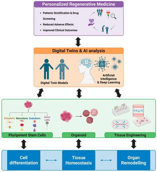
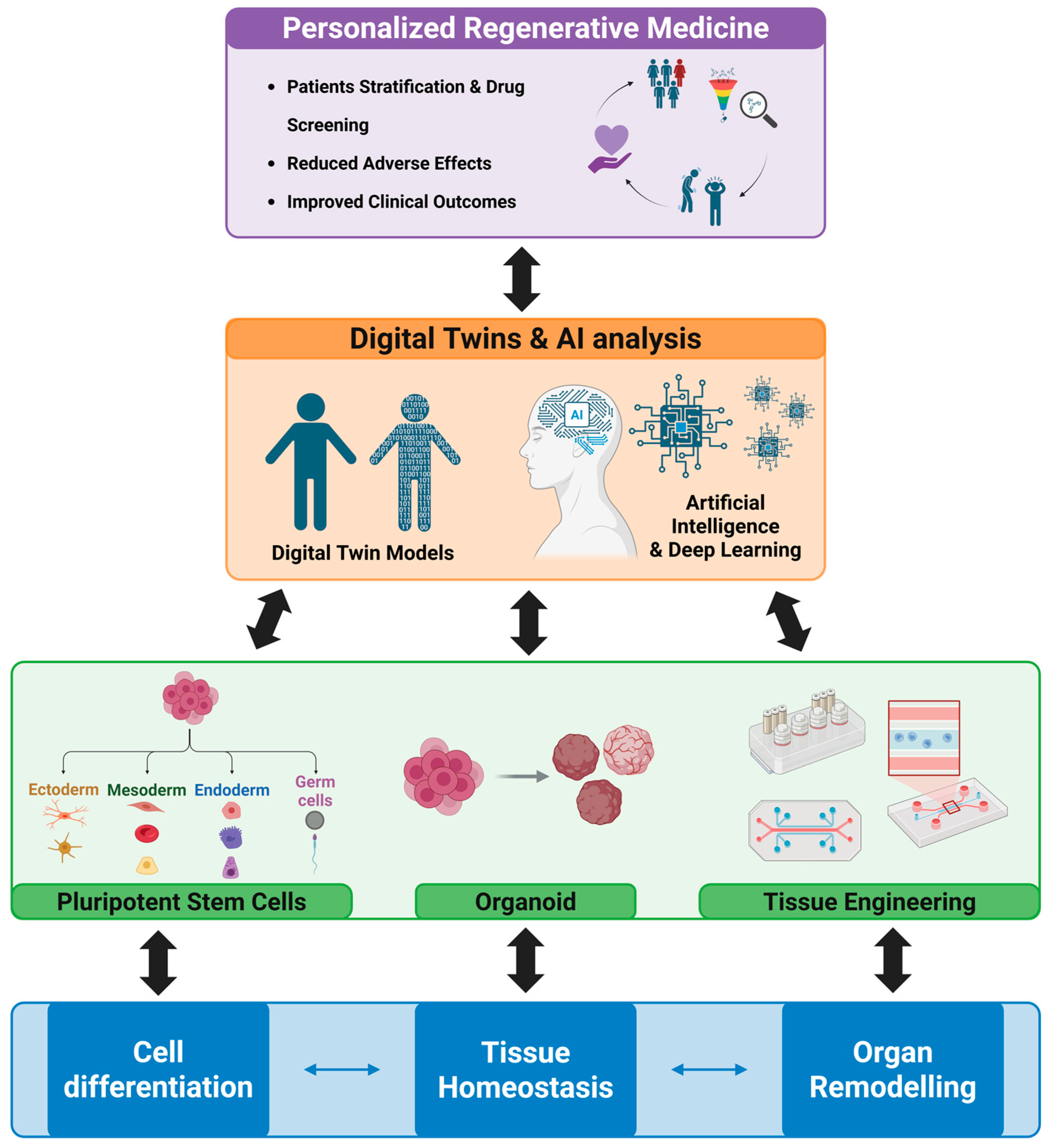
Figure 2 provides a conceptual framework for predictive and regenerative medicine, illustrating how fundamental morphological processes—cell differentiation, tissue homeostasis, and organ remodeling—inform the development of stem cell-based therapies, organoids, and tissue engineering strategies. It also highlights the role of digital twins and AI in integrating morphological and clinical data to guide personalized therapeutic interventions and optimize patient outcomes. Clinically, this integrated model enables more tailored and dynamic patient management.
Figure 2.
Conceptual framework for predictive and regenerative medicine.
The diagram illustrates the hierarchical relationship between core morphological processes (in blue), experimental models (in green), digital predictive tools (in orange), and clinical applications (in violet). It emphasizes how understanding tissue dynamics at the cellular and organ level enables the design of regenerative therapies, personalized interventions, and predictive strategies supported by AI and digital twin technologies. Created in BioRender. Bucchieri, F. (2026) https://BioRender.com/1x7xceh (accessed on 15 May 2026).
8. Conclusions
The understanding of cellular differentiation, tissue homeostasis, and organ remodeling processes today represents the beating heart of the morphological sciences and contemporary medicine. As early as the late 19th century, Rudolf Virchow had pointed the way: disease is not limited to macroscopic alterations of organs but results from dysfunctions of the cellular units [81]. Anatomy, Histology, and Embryology, integrated with molecular biology and modern imaging technologies, now provide the opportunity to interpret pathology as the visible outcome of biological programs regulated in time and space.
Building on this solid historical foundation is the legacy of Giuseppe Levi’s School, one of the greatest Italian anatomists of the 20th century (and, notably, mentor to three Nobel laureates in Medicine). For Levi, the study of morphology had no meaning unless it correlated with function [82,83]. His “form–function” vision has inspired generations of researchers and clinicians, enabling an understanding of how structural and tissue alterations can be the first step in interpreting disease onset and progression. From his school emerged scientists who applied this philosophy to experimental research, developing models that connect anatomy and histology to altered cellular differentiation, loss of tissue homeostasis, and organ remodeling, lessons that continue to inform us today.
This integrated approach makes it possible to distinguish between adaptive and maladaptive tissue responses, anticipate the onset of disease, and design targeted interventions, from traditional pharmacology to regenerative therapies based on stem cells, organoids, and tissue engineering. Predictive and precision medicine, supported by computational models, artificial intelligence, and simulation tools such as “digital twins,” as discussed in this review, further enhances the ability to forecast the progression of pathological processes and personalize therapeutic strategies.
Looking at the history of life’s evolution, we can observe that increasing biological complexity has brought not only new functionalities but also a greater number of potential vulnerabilities. In parallel, the progressive sophistication of homeostatic mechanisms and differentiation programs has made it essential to interpret disease beyond the macroscopic form, down to the molecular and cellular scale. As Giulio Bizzozero noted when classifying tissues as labile, stable, and permanent, only by understanding tissue dynamics can we correctly interpret their responses to damage, stress, and degenerative processes [84]. From a practical perspective, the proposed framework based on cell differentiation, tissue homeostasis, and organ remodeling has direct implications for both clinical practice and medical education. In the clinical setting, interpreting disease as the result of altered biological programs allows clinicians to move beyond a purely descriptive approach, supporting earlier diagnosis, more accurate prognostic stratification, and the identification of targeted therapeutic strategies. In parallel, in medical education, this integrative perspective promotes a shift from memorization-based learning toward a systems-oriented understanding of human biology, enabling students to connect structure with function and to interpret pathological processes as dynamic and interconnected events. In this sense, morphological sciences become not only foundational disciplines, but also essential tools for developing critical thinking and clinical reasoning in future physicians.
The physician of the future, as of today, will need to integrate anatomical, histological, embryological, and organogenetic knowledge to make clinical decisions grounded in the underlying biological reality. True personalized medicine does not merely address visible symptoms or lesions; it acts on the cellular and tissue programs that underlie them, optimizing therapeutic choices, reducing unnecessary interventions, and improving patients’ quality of life. Ultimately, the morphological sciences—long the foundation of medical education—are now regaining a central role: understanding the life of tissues means understanding disease itself and paves the way toward predictive, regenerative, and truly personalized medicine, balancing history, science, and innovation.
Author Contributions
Conceptualization, C.C.B., M.I.G., G.L.B. and F.C.; writing—original draft preparation, C.C.B., A.M.V., M.I.G., A.T., S.S., O.M.M., G.L.B. and F.C.; writing—review and editing, C.C.B., A.M.V. and F.C.; figure and Table preparation, C.C.B., A.M.V., M.I.G., A.T., S.S., O.M.M., G.L.B. and F.C.; supervision, C.C.B. and F.C. All authors have read and agreed to the published version of the manuscript.
Funding
This research received no external funding.
Institutional Review Board Statement
Not applicable.
Informed Consent Statement
Not applicable.
Data Availability Statement
No new data were created or analyzed in this study. Data sharing is not applicable to this article.
Acknowledgments
NextGenerationEU: through the Italian Ministry of University and Research “Avviso per la concessione di finanziamenti destinati ad Iniziative di ricerca per tecnologie e percorsi innovativi in ambito sanitario e assistenziale” finanziato a valere sulle risorse previste dal Fondo complementare al Piano Nazionale di Ripresa e Resilienza—PNC0000001—D3 4 Health Digital Driven Diagnostics, prognostics and therapeutics for sustainable Health care.” We would like to thank the Department of Biomedicine, Neuroscience, and Advanced Diagnostics at the University of Palermo and Fabio Bucchieri, director of the Institute of Human Anatomy and Histology, for providing access to Biorender.
Conflicts of Interest
The authors declare no conflicts of interest.
Abbreviations
The following abbreviations are used in this manuscript:
| AI | Artificial Intelligence |
| BDNF | Brain-derived neurotrophic factor |
| MSCs | Mesenchymal Stem Cells |
| NGF | Nerve growth factor |
References
- Breathnach, C.S. Rudolf Virchow (1821–1902) and Die Cellularpathologie (1858). J. Ir. Coll. Physicians Surg. 2002, 31, 43–46. [Google Scholar]
- Ribatti, D. Rudolf Virchow, the founder of cellular pathology. Rom. J. Morphol. Embryol. 2019, 60, 1381–1382. [Google Scholar]
- Liu, X.; Zhou, Z.; Zhang, Y.; Zhong, H.; Cai, X.; Guan, R. Recent progress on the organoids: Techniques, advantages and applications. Biomed. Pharmacother. 2025, 185, 117942. [Google Scholar] [CrossRef] [Scilit]
- Zhou, L.; Huang, J.; Li, C.; Gu, Q.; Li, G.; Li, Z.A.; Xu, J.; Zhou, J.; Tuan, R.S. Organoids and organs-on-chips: Recent advances, applications in drug development, and regulatory challenges. Med 2025, 6, 100667. [Google Scholar] [CrossRef] [Scilit]
- D’Amico, G.; Vitale, A.M.; Marco, M.D.; Lo Giudice, A.; Cecala, F.C.; Cappello, F.; Caruso Bavisotto, C. Standardized Workflow for the Generation of Patient-Derived Glioblastoma Spheroids. Methods Protoc. 2026, 9, 61. [Google Scholar] [CrossRef] [Scilit] [PubMed]
- Hu, B.; Shi, Z.; Lu, L.; Miao, Z.; Wang, H.; Zhou, Z.; Zhang, F.; Wang, R.; Luo, X.; Xu, F.; et al. A deep-learning model for intracranial aneurysm detection on CT angiography images in China: A stepwise, multicentre, early-stage clinical validation study. Lancet Digit. Health 2024, 6, e261–e271. [Google Scholar] [CrossRef] [Scilit] [PubMed]
- Amato, D.; Calderaro, S.; Lo Bosco, G.; Rizzo, R.; Vella, F. Metric Learning in Histopathological Image Classification: Opening the Black Box. Sensors 2023, 23, 6003. [Google Scholar] [CrossRef] [Scilit]
- Wang, H.; Li, X.; You, X.; Zhao, G. Harnessing the power of artificial intelligence for human living organoid research. Bioact. Mater. 2024, 42, 140–164. [Google Scholar] [CrossRef] [Scilit]
- Mazzarello, P.; Calligaro, A.L.; Calligaro, A. Giulio Bizzozero: A pioneer of cell biology. Nat. Rev. Mol. Cell Biol. 2001, 2, 776–781. [Google Scholar] [CrossRef] [Scilit] [PubMed]
- Rojas-Ríos, P.; González-Reyes, A. The plasticity of stem cell niches: A general property behind tissue homeostasis and repair. Stem Cells 2014, 32, 852–859. [Google Scholar] [CrossRef] [Scilit]
- Perrotta, C.; Cattaneo, M.G.; Molteni, R.; De Palma, C. Autophagy in the regulation of tissue differentiation and homeostasis. Front. Cell Dev. Biol. 2020, 8, 602901. [Google Scholar] [CrossRef] [Scilit]
- Dupont, J.C. Wilhelm His and mechanistic approaches to development at the time of Entwicklungsmechanik. Hist. Philos. Life Sci. 2017, 39, 21. [Google Scholar] [CrossRef] [Scilit]
- De Robertis, E.M.; Driever, W.; Mayor, R. Celebrating the centennial of the most famous experiment in embryology: Hilde Mangold, Hans Spemann and the organizer. Cells Dev. 2024, 178, 203921. [Google Scholar] [CrossRef] [Scilit]
- Perrimon, N.; Pitsouli, C.; Shilo, B.Z. Signaling mechanisms controlling cell fate and embryonic patterning. Cold Spring Harb. Perspect. Biol. 2012, 4, a005975. [Google Scholar] [CrossRef] [Scilit]
- Balasubramanian, R.; Zhang, X. Mechanisms of FGF gradient formation during embryogenesis. Semin. Cell Dev. Biol. 2016, 53, 94–100. [Google Scholar] [CrossRef] [Scilit]
- Rozario, T.; DeSimone, D.W. The extracellular matrix in development and morphogenesis: A dynamic view. Dev. Biol. 2010, 341, 126–140. [Google Scholar] [CrossRef] [Scilit]
- Mannino, G.; Russo, C.; Maugeri, G.; Musumeci, G.; Vicario, N.; Tibullo, D.; Giuffrida, R.; Parenti, R.; Lo Furno, D. Adult stem cell niches for tissue homeostasis. J. Cell. Physiol. 2022, 237, 239–257. [Google Scholar] [CrossRef] [Scilit] [PubMed]
- Blanpain, C.; Fuchs, E. Stem cell plasticity: Plasticity of epithelial stem cells in tissue regeneration. Science 2014, 344, 1242281. [Google Scholar] [CrossRef] [Scilit] [PubMed]
- Wang, Z.; Qu, Y.J.; Cui, M. Modulation of stem cell fate in intestinal homeostasis, injury and repair. World J. Stem Cells 2023, 15, 354–368. [Google Scholar] [CrossRef] [Scilit] [PubMed]
- Slack, J.M.; Tosh, D. Transdifferentiation and metaplasia—Switching cell types. Curr. Opin. Genet. Dev. 2001, 11, 581–586. [Google Scholar] [CrossRef] [Scilit]
- Giroux, V.; Rustgi, A.K. Metaplasia: Tissue injury adaptation and a precursor to the dysplasia–cancer sequence. Nat. Rev. Cancer 2017, 17, 594–604. [Google Scholar] [CrossRef] [Scilit] [PubMed]
- Ishizumi, T.; McWilliams, A.; MacAulay, C.; Gazdar, A.; Lam, S. Natural history of bronchial preinvasive lesions. Cancer Metastasis Rev. 2010, 29, 5–14. [Google Scholar] [CrossRef] [Scilit] [PubMed]
- Khazaaleh, S.; Alomari, M.; Rashid, M.U.; Castaneda, D.; Castro, F.J. Gastric intestinal metaplasia and gastric cancer prevention: Watchful waiting. Cleve. Clin. J. Med. 2024, 91, 33–39. [Google Scholar] [CrossRef] [Scilit]
- Ambrosi, T.H.; Marecic, O.; McArdle, A.; Sinha, R.; Gulati, G.S.; Tong, X.; Wang, Y.; Steininger, H.M.; Hoover, M.Y.; Koepke, L.S.; et al. Aged skeletal stem cells generate an inflammatory degenerative niche. Nature 2021, 597, 256–262. [Google Scholar] [CrossRef] [Scilit] [PubMed]
- Vitale, A.M.; D’Amico, G.; Santonocito, R.; Spinnato, G.; Di Marco, M.; Scalia, F.; Campanella, C.; Tringali, G.; Giusti, I.; Dolo, V.; et al. An overview of glioblastoma multiforme in vitro experimental models. J. Biol. Res. 2024, 97, 11920. [Google Scholar] [CrossRef] [Scilit]
- Morizane, R.; Lamers, M.M. Organoids in disease modeling and regenerative medicine. Cell. Mol. Life Sci. 2025, 82, 169. [Google Scholar] [CrossRef] [Scilit]
- Kim, J.; Koo, B.K.; Knoblich, J.A. Human organoids: Model systems for human biology and medicine. Nat. Rev. Mol. Cell Biol. 2020, 21, 571–584. [Google Scholar] [CrossRef] [Scilit]
- Lancaster, M.A.; Knoblich, J.A. Generation of cerebral organoids from human pluripotent stem cells. Nat. Protoc. 2014, 9, 2329–2340. [Google Scholar] [CrossRef] [Scilit]
- Meroueh, C.; Chen, Z.E. Artificial intelligence in anatomical pathology: Building a strong foundation for precision medicine. Hum. Pathol. 2023, 132, 31–38. [Google Scholar] [CrossRef] [Scilit]
- Chang, J.; Hatfield, B. Advancements in computer vision and pathology: Unraveling the potential of artificial intelligence for precision diagnosis and beyond. Adv. Cancer Res. 2024, 161, 431–478. [Google Scholar] [CrossRef] [Scilit]
- Honeycutt, S.E.; N’Guetta, P.Y.; O’Brien, L.L. Innervation in organogenesis. Curr. Top. Dev. Biol. 2022, 148, 195–235. [Google Scholar] [CrossRef] [Scilit]
- Keast, J.R. Innervation of developing and adult organs. Organogenesis 2013, 9, 168. [Google Scholar] [CrossRef] [Scilit] [PubMed]
- Amella, C.; Cappello, F.; Kahl, P.; Fritsch, H.; Lozanoff, S.; Sergi, C. Spatial and temporal dynamics of innervation during the development of fetal human pancreas. Neuroscience 2008, 154, 1477–1487. [Google Scholar] [CrossRef] [Scilit]
- Klein Wolterink, R.G.J.; Wu, G.S.; Chiu, I.M.; Veiga-Fernandes, H. Neuroimmune Interactions in Peripheral Organs. Annu. Rev. Neurosci. 2022, 45, 339–360. [Google Scholar] [CrossRef] [Scilit] [PubMed]
- Picoli, C.C.; Costa, A.C.; Rocha, B.G.S.; Silva, W.N.; Santos, G.S.P.; Prazeres, P.H.D.M.; Costa, P.A.C.; Oropeza, A.; da Silva, R.A.; Azevedo, V.A.C.; et al. Sensory nerves in the spotlight of the stem cell niche. Stem Cells Transl. Med. 2021, 10, 346–356. [Google Scholar] [CrossRef] [Scilit] [PubMed]
- Pei, F.; Ma, L.; Jing, J.; Feng, J.; Yuan, Y.; Guo, T.; Han, X.; Ho, T.V.; Lei, J.; He, J.; et al. Sensory nerve niche regulates mesenchymal stem cell homeostasis via FGF/mTOR/autophagy axis. Nat. Commun. 2023, 14, 344. [Google Scholar] [CrossRef] [Scilit]
- Chu, C.; Artis, D.; Chiu, I.M. Neuro-immune interactions in the tissues. Immunity 2020, 52, 464–474. [Google Scholar] [CrossRef] [Scilit]
- Klose, C.S.N.; Veiga-Fernandes, H. Neuroimmune interactions in peripheral tissues. Eur. J. Immunol. 2021, 51, 1602–1614. [Google Scholar] [CrossRef] [Scilit]
- Clevers, H. The intestinal crypt, a prototype stem cell compartment. Cell 2013, 154, 274–284. [Google Scholar] [CrossRef] [Scilit]
- Yin, L.; Li, N.; Jia, W.; Liang, M.; Yang, X.; Du, G. Skeletal muscle atrophy: From mechanisms to treatments. Pharmacol. Res. 2021, 172, 105807. [Google Scholar] [CrossRef] [Scilit]
- Villavicencio, A.; Bacallao, K.; Gabler, F.; Fuentes, A.; Albornoz, J.; Casals, A.; Vega, M. Deregulation of tissue homeostasis in endometria from patients with polycystic ovarian syndrome with and without endometrial hyperplasia. Gynecol. Oncol. 2007, 104, 290–295. [Google Scholar] [CrossRef] [Scilit]
- Zhang, S.; Xiao, X.; Yi, Y.; Wang, X.; Zhu, L.; Shen, Y.; Lin, D.; Wu, C. Tumor initiation and early tumorigenesis: Molecular mechanisms and interventional targets. Signal Transduct. Target. Ther. 2024, 9, 149. [Google Scholar] [CrossRef] [Scilit] [PubMed]
- Ford, A.J.; Rajagopalan, P. Extracellular matrix remodeling in 3D: Implications in tissue homeostasis and disease progression. WIREs Nanomed. Nanobiotechnol. 2018, 10, e1503. [Google Scholar] [CrossRef] [Scilit] [PubMed]
- Heineke, J.; Molkentin, J.D. Regulation of cardiac hypertrophy by intracellular signalling pathways. Nat. Rev. Mol. Cell Biol. 2006, 7, 589–600. [Google Scholar] [CrossRef] [Scilit]
- Savaris, R.F.; Hamilton, A.E.; Lessey, B.A.; Giudice, L.C. Endometrial gene expression in early pregnancy: Lessons from human ectopic pregnancy. Reprod. Sci. 2008, 15, 797–816. [Google Scholar] [CrossRef] [Scilit]
- Robling, A.G.; Castillo, A.B.; Turner, C.H. Biomechanical and molecular regulation of bone remodeling. Annu. Rev. Biomed. Eng. 2006, 8, 455–498. [Google Scholar] [CrossRef] [Scilit]
- Di Felice, V.; Barone, R.; Trovato, E.; D’Amico, D.; Macaluso, F.; Campanella, C.; Marino Gammazza, A.; Muccilli, V.; Cunsolo, V.; Cancemi, P.; et al. Physiactisome: A new nanovesicle drug containing heat shock protein 60 for treating muscle wasting and cachexia. Cells 2022, 11, 1406. [Google Scholar] [CrossRef] [Scilit] [PubMed]
- Eming, S.A.; Martin, P.; Tomic-Canic, M. Wound repair and regeneration: Mechanisms, signaling, and translation. Sci. Transl. Med. 2014, 6, 265sr6. [Google Scholar] [CrossRef] [Scilit]
- Hynes, R.O. The extracellular matrix: Not just pretty fibrils. Science 2009, 326, 1216–1219. [Google Scholar] [CrossRef] [Scilit]
- Medzhitov, R. Origin and physiological roles of inflammation. Nature 2008, 454, 428–435. [Google Scholar] [CrossRef] [Scilit]
- Serhan, C.N.; Savill, J. Resolution of inflammation: The beginning programs the end. Nat. Immunol. 2005, 6, 1191–1197. [Google Scholar] [CrossRef] [Scilit]
- Gurtner, G.C.; Werner, S.; Barrandon, Y.; Longaker, M.T. Wound repair and regeneration. Nature 2008, 453, 314–321. [Google Scholar] [CrossRef] [Scilit]
- Wynn, T.A. Cellular and molecular mechanisms of fibrosis. J. Pathol. 2008, 214, 199–210. [Google Scholar] [CrossRef] [Scilit]
- Porrello, E.R.; Mahmoud, A.I.; Simpson, E.; Hill, J.A.; Richardson, J.A.; Olson, E.N.; Sadek, H.A. Transient regenerative potential of the neonatal mouse heart. Science 2011, 331, 1078–1080. [Google Scholar] [CrossRef] [Scilit]
- King, T.E., Jr.; Pardo, A.; Selman, M. Idiopathic pulmonary fibrosis. Lancet 2011, 378, 1949–1961. [Google Scholar] [CrossRef] [Scilit]
- Frangogiannis, N.G. The inflammatory response in myocardial injury, repair, and remodelling. Nat. Rev. Cardiol. 2014, 11, 255–265. [Google Scholar] [CrossRef] [Scilit] [PubMed]
- Bataller, R.; Brenner, D.A. Liver fibrosis. J. Clin. Investig. 2005, 115, 209–218. [Google Scholar] [CrossRef] [PubMed]
- Humphreys, B.D. Mechanisms of renal fibrosis. Annu. Rev. Physiol. 2018, 80, 309–326. [Google Scholar] [CrossRef] [Scilit]
- Buja, L.M. The cell theory and cellular pathology: Discovery, refinements and applications fundamental to advances in biology and medicine. Exp. Mol. Pathol. 2021, 121, 104660. [Google Scholar] [CrossRef] [Scilit] [PubMed]
- Hermanek, P. Dysplasia in the gastrointestinal tract: Definition and clinical significance. Surg. Endosc. 1987, 1, 5–10. [Google Scholar] [CrossRef] [Scilit]
- Weiskirchen, R.; Weiskirchen, S.; Tacke, F. Organ and tissue fibrosis: Molecular signals, cellular mechanisms and translational implications. Mol. Asp. Med. 2019, 65, 2–15. [Google Scholar] [CrossRef] [Scilit]
- Benton, M.L.; Abraham, A.; LaBella, A.L.; Abbott, P.; Rokas, A.; Capra, J.A. The influence of evolutionary history on human health and disease. Nat. Rev. Genet. 2021, 22, 269–283. [Google Scholar] [CrossRef] [Scilit]
- Cappello, F.; Conway de Macario, E.; Macario, A.J.L. HSP60: A story as long as life on the Earth. In Heat Shock Protein 60 in Human Diseases and Disorders; Asea, A., Kaur, P., Eds.; Springer: Cham, Switzerland, 2019; Volume 18, pp. 3–14. [Google Scholar]
- Barabási, A.L.; Gulbahce, N.; Loscalzo, J. Network medicine: A network-based approach to human disease. Nat. Rev. Genet. 2011, 12, 56–68. [Google Scholar] [CrossRef] [Scilit] [PubMed]
- Singh, R.S.; Gupta, B.P. Genes and genomes and unnecessary complexity in precision medicine. NPJ Genom. Med. 2020, 5, 21. [Google Scholar] [CrossRef] [Scilit] [PubMed]
- Rathan, R.; Hamdy, H.; Kassab, S.E.; Salama, M.N.F.; Sreejith, A.; Gopakumar, A. Implications of introducing case-based radiological images in anatomy on teaching, learning and assessment of medical students: A mixed-methods study. BMC Med. Educ. 2022, 22, 723. [Google Scholar] [CrossRef] [Scilit]
- Rossant, J. Why study human embryo development? Dev. Biol. 2024, 509, 43–50. [Google Scholar] [CrossRef] [Scilit]
- Luce, E.; Duclos-Vallee, J.C. Stem cells and organoids: A paradigm shift in preclinical models toward personalized medicine. Pharmaceuticals 2025, 18, 992. [Google Scholar] [CrossRef] [Scilit]
- Wu, Y.; Ye, W.; Gao, Y.; Yi, Z.; Chen, Z.; Qu, C.; Huang, J.; Liu, F.; Liu, Z. Application of organoids in regenerative medicine. Stem Cells 2023, 41, 1101–1112. [Google Scholar] [CrossRef] [Scilit]
- Lightner, A.L.; Chan, T. Precision regenerative medicine. Stem Cell Res. Ther. 2021, 12, 39. [Google Scholar] [CrossRef] [Scilit]
- Silva, A.; Vale, N. Digital twins in personalized medicine: Bridging innovation and clinical reality. J. Pers. Med. 2025, 15, 503. [Google Scholar] [CrossRef] [Scilit]
- Chang, H.Y.; Jung, C.K.; Woo, J.I.; Lee, S.; Cho, J.; Kim, S.W.; Kwak, T.Y. Artificial intelligence in pathology. J. Pathol. Transl. Med. 2019, 53, 1–12. [Google Scholar] [CrossRef] [Scilit] [PubMed]
- Yamanaka, S. Induced pluripotent stem cells: Past, present, and future. Cell Stem Cell 2012, 10, 678–684. [Google Scholar] [CrossRef] [Scilit]
- Liu, G.; David, B.T.; Trawczynski, M.; Fessler, R.G. Advances in pluripotent stem cells: History, mechanisms, technologies, and applications. Stem Cell Rev. Rep. 2020, 16, 3–32. [Google Scholar] [CrossRef] [Scilit]
- Yang, S.; Hu, H.; Kung, H.; Zou, R.; Dai, Y.; Hu, Y.; Wang, T.; Lv, T.; Yu, J.; Li, F. Organoids: The current status and biomedical applications. MedComm 2023, 4, e274. [Google Scholar] [CrossRef] [Scilit] [PubMed]
- Hoang, V.T.; Nguyen, Q.T.; Phan, T.T.K.; Pham, T.H.; Dinh, N.T.H.; Anh, L.P.H.; Dao, L.T.M.; Bui, V.D.; Dao, H.N.; Le, D.S.; et al. Tissue engineering and regenerative medicine: Perspectives and challenges. MedComm 2025, 6, e70192. [Google Scholar] [CrossRef] [Scilit] [PubMed]
- Bulten, W.; Kartasalo, K.; Chen, P.C.; Ström, P.; Pinckaers, H.; Nagpal, K.; Cai, Y.; Steiner, D.F.; van Boven, H.; Vink, R.; et al. Artificial intelligence for diagnosis and Gleason grading of prostate cancer: The PANDA challenge. Nat. Med. 2022, 28, 154–163. [Google Scholar] [CrossRef] [Scilit]
- Sugimoto, M.A.; Sousa, L.P.; Pinho, V.; Perretti, M.; Teixeira, M.M. Resolution of inflammation: What controls its onset? Front. Immunol. 2016, 7, 160. [Google Scholar] [CrossRef] [Scilit]
- Wang, R.C.; Wang, Z. Precision medicine: Disease subtyping and tailored treatment. Cancers 2023, 15, 3837. [Google Scholar] [CrossRef] [Scilit]
- Petti, M.; Farina, L. Network medicine for patients’ stratification: From single-layer to multi-omics. WIREs Mech. Dis. 2023, 15, e1623. [Google Scholar] [CrossRef] [Scilit]
- Byers, J.M., 3rd. Rudolf Virchow—Father of cellular pathology. Am. J. Clin. Pathol. 1989, 92, S2–S8. [Google Scholar]
- Tamborini, M. Organic form and evolution: The morphological problem in twentieth-century Italian biology. Hist. Philos. Life Sci. 2022, 44, 54. [Google Scholar] [CrossRef] [Scilit] [PubMed]
- Bentivoglio, M.; Vercelli, A.; Filogamo, G. Giuseppe Levi: Mentor of three Nobel laureates. J. Hist. Neurosci. 2006, 15, 358–368. [Google Scholar] [CrossRef] [Scilit] [PubMed]
- Cossu, G. Back from the tip of the nose. Sci. World J. 2001, 1, 225–226. [Google Scholar] [CrossRef] [Scilit] [PubMed]

Disclaimer/Publisher’s Note: The statements, opinions and data contained in all publications are solely those of the individual author(s) and contributor(s) and not of MDPI and/or the editor(s). MDPI and/or the editor(s) disclaim responsibility for any injury to people or property resulting from any ideas, methods, instructions or products referred to in the content. |
© 2026 by the authors. Licensee MDPI, Basel, Switzerland. This article is an open access article distributed under the terms and conditions of the Creative Commons Attribution (CC BY) license.