Case Report

Multiple Rare Vascular Gastrointestinal Variations Present in a 97-Year-Old Female Donor: Clinical Relevance and Surgical Implications

by
Brenna Chen
1,
Guinevere Granite
2,*,
Kerrie Lashley
3,4,
Juan José Valenzuela-Fuenzalida
5 and
Maria Ximena Leighton
2
1
F. Edward Hebert School of Medicine, Uniformed Services University of Health Sciences, Bethesda, MD 20814, USA
2
Department of Surgery, Uniformed Services University of the Health Sciences, Bethesda, MD 20814, USA
3
The Henry M. Jackson Foundation for the Advancement of Military Medicine Inc., Bethesda, MD 20814, USA
4
Department of Anatomy and Cell Biology, George Washington School of Medicine & Health Sciences, Washington, DC 20037, USA
5
Departamento de Morfología, Facultad de Medicina, Universidad Andrés Bello, Santiago 8370146, Chile
*
Author to whom correspondence should be addressed.
Anatomia 2026, 5(2), 16; https://doi.org/10.3390/anatomia5020016
Submission received: 17 April 2025 / Revised: 17 March 2026 / Accepted: 7 May 2026 / Published: 2 June 2026

Abstract

Multiple rare vascular variations were identified in a 97-year-old female donor during the dissection of the gastrointestinal system. An anomalous middle colic artery originated from the celiac trunk alongside the three normal branches. The left hepatic artery, an accessory left hepatic artery, and the right hepatic artery all originated from the proper hepatic artery. The right hepatic artery exhibited a “caterpillar hump” and an abnormal tripod distal branching pattern, consisting of a very short cystic artery and two short right hepatic arteries (one superior and one inferior). Additionally, an accessory cystic artery arose from the inferior pancreaticoduodenal artery, ascending inferiorly to the common bile duct and cystic duct towards the gallbladder to join the cystic artery posteriorly. The right and middle colic arteries originated from a right colic middle colic common arterial trunk. The left colic artery and the sigmoidal arteries also originated from a left colic-sigmoidal common arterial trunk. Surgical and medical procedures in the pancreaticobiliary region are widespread, and cholecystectomy is one of the most common surgeries performed worldwide. A laparoscopic approach is the current ‘gold standard’ surgical approach. Knowledge of these potential gastrointestinal vascular variations is essential to ensure patient safety and avoid iatrogenic complications. The presence of an unidentified caterpillar-type right hepatic artery poses a significant risk of accidental ligation during cholecystectomy. Similarly, the presence of common colic arterial trunks alters the vascular landscape for laparoscopic colectomy. To ensure favorable surgical outcomes, comprehensive preoperative vascular imaging should be performed to identify such rare configurations. In this article, we discussed the frequency, embryology, physiology, and clinical and surgical implications of the anatomical variations present in this donor. To date, there have been no other reports in the literature describing this specific combination of variations.

1. Introduction

The main arterial blood supply to the foregut, midgut, and hindgut of the gastrointestinal tract is provided by the celiac trunk (CT), superior mesenteric artery (SMA), and inferior mesenteric artery (IMA), respectively [1,2]. The CT classically trifurcates, giving rise to the left gastric artery (LGA) and splenic artery, which course leftward, and the common hepatic artery (CHA), which extends rightward. The CHA sequentially branches into the gastroduodenal artery (GDA) and the proper hepatic artery (PHA), which bifurcates into the right and left hepatic arteries (RHA, LHA), supplying the liver. The cystic artery (CA) most frequently branches from the RHA [1,2]. Anatomical variations of the hepatic arteries are well documented with normal anatomy found in approximately 55–79% of individuals [3,4]. These variations were first documented by Haller in 1756 followed by Lipshutz in 1917 and Adachi in 1928 [5].
Normally, the SMA gives off three colic branches: the middle colic artery (MCA), right colic artery (RCA) and the ileocolic artery (ICA) [6]. Anatomical variations of the SMA and its colic branches are also well studied, with the expected anatomy present in 13–93% of individuals [6]. Adachi (1928) [5] was again one of the first to publish a classification system for the varying morphology of the SMA. Since Adachi (1928) [5], multiple additional classification systems have also been proposed. The IMA arises from the aorta at the level of L3-L4 and supplies blood to the distal transverse colon, descending colon, sigmoid colon, and superior rectum [7,8,9]. The classic branching pattern of the IMA is one that gives rise to the left colic artery (LCA), followed by two or three sigmoidal arteries, (SAs) and lastly, the superior rectal artery (SRA) [7,8,9,10].
In this case, we describe multiple rare anatomical variations, including an anatomical variant of the RHA known as Moynihan’s or caterpillar hump with a tripod distal branching. We also describe the presence of an accessory LHA (aLHA) and a variation of the inferior pancreaticoduodenal artery (IPDA), which gives rise to an accessory cystic artery that travels superiorly to join the CA. Furthermore, we describe a variation of the SMA in which the RCA and MCA arise from a shared trunk, as well as a variation of the IMA where the LCA and SAs arise from a shared trunk.

2. Case Description

The abdomen of a 97-year-old female donor with a cause of death of sarcopenia and failure to thrive was dissected during the graduate nursing advanced anatomy course at the Uniformed Services University of the Health Sciences. The donor was preserved via arterial embalming using a standard protocol with a 6% formaldehyde-based solution introduced through both femoral and brachial arterial access to ensure uniform tissue fixation. Following initial preparation, a systematic layer-by-layer macro dissection was performed to expose the gastrointestinal system. To ensure the integrity of the delicate neurovascular structures, a blunt dissection method was utilized to follow natural tissue planes. All anatomical structures were carefully separated from the surrounding connective tissue and fascia. This meticulous approach ensured that anatomical variations, such as the accessory cystic artery originating from the IPDA, were preserved, as these structures are often compromised during standard student dissection.
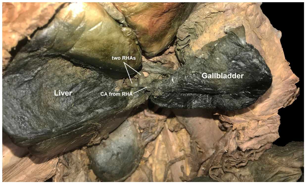
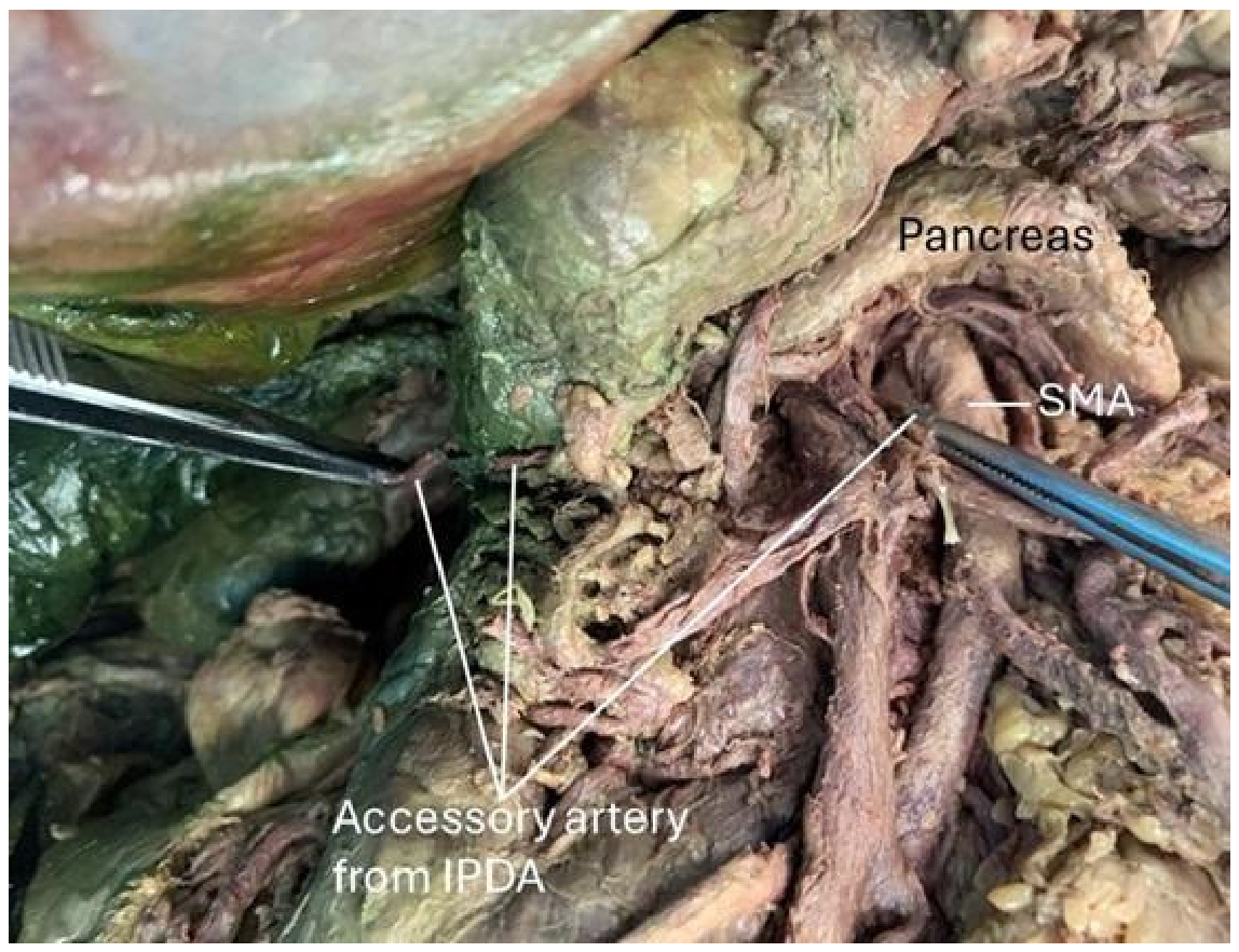
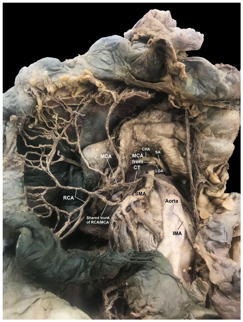
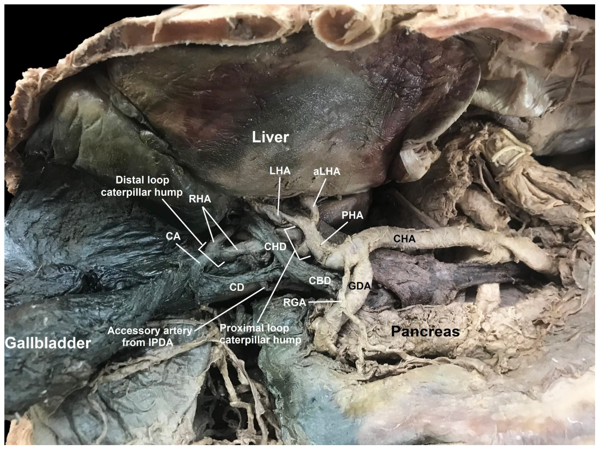
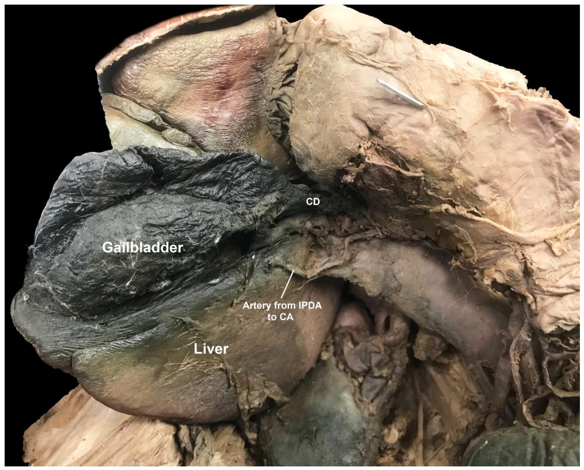
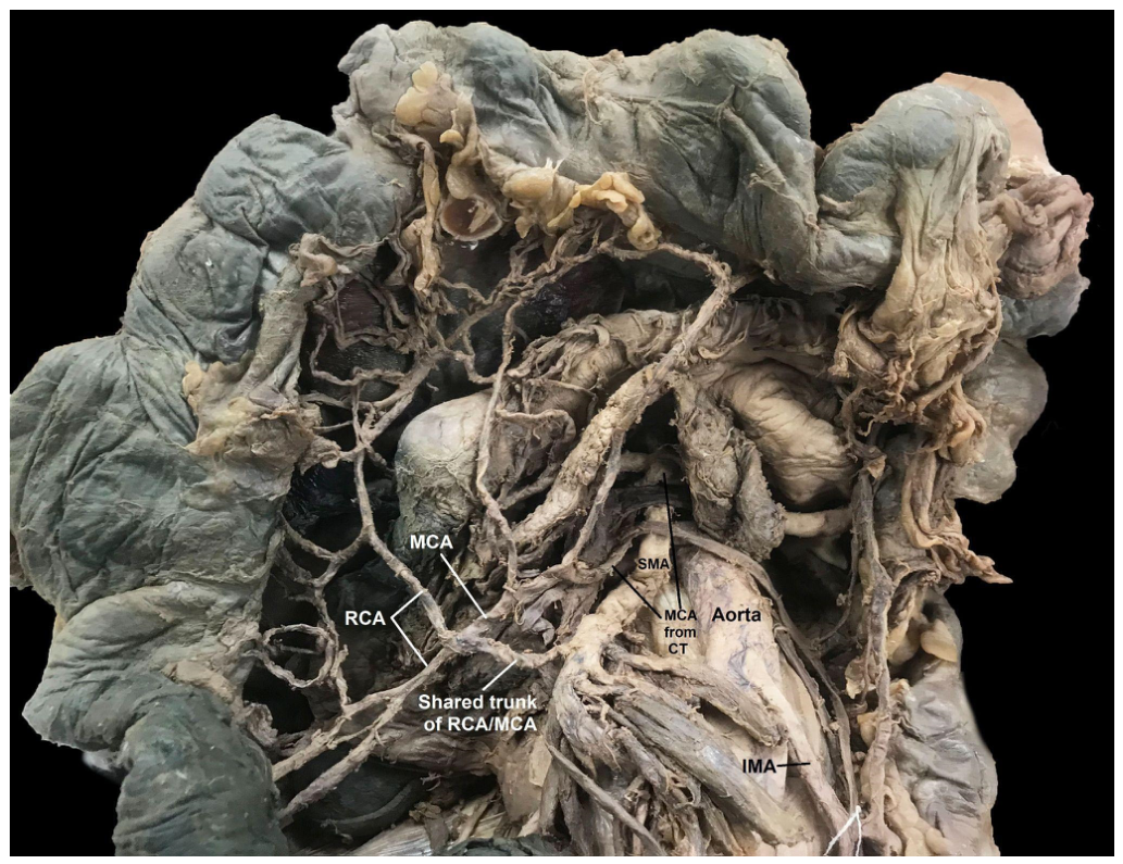
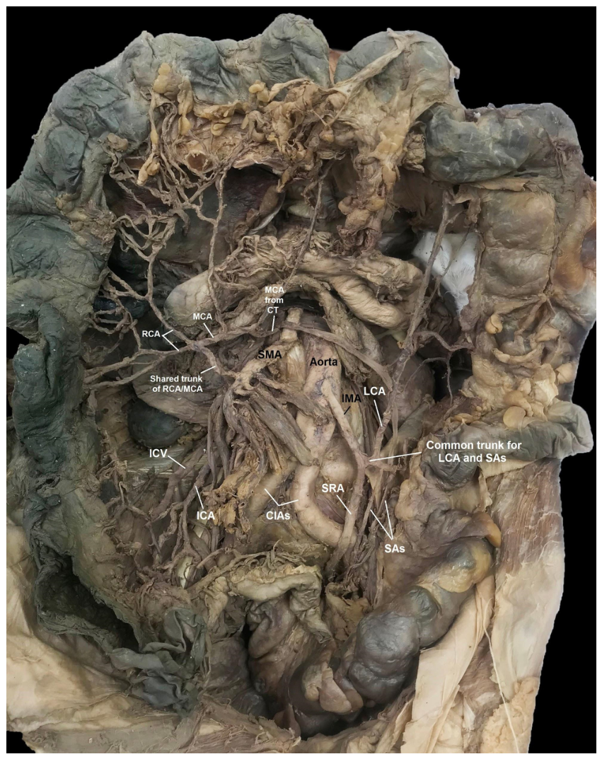
This dissection revealed multiple arterial anatomical variations involving the foregut, midgut, and hindgut. The CT was found to have an additional artery branching from its aortic origin (Figure 1). This artery was found to anastomose with the MCA that came off the SMA to provide blood supply to the colon (Figure 1 and Figure 6). Further dissection revealed the PHA had an extra branch identified as an aLHA (Figure 2). The RHA was found to have Moynihan’s hump with a tripod distal branching: a short CA and two RHAs (one superior and one inferior) (Figure 2 and Figure 3). The CA also received additional blood supply via a vessel from the IPDA (Figure 4 and Figure 5). Within the midgut, the RCA and MCA were noted to share a common trunk from the SMA, while the ICA was separate (Figure 6 and Figure 7). The IMA bifurcated into a short common trunk for the LCA and SAs while the SRA continued to descend towards the rectum (Figure 7).

3. Discussion

3.1. Celiac Trunk Variations

The trifurcation of the CT was first described in 1756 by Haller, when he described it as the “tripus Halleri” [11,12]. This classic tripus halleri or “true tripod” conventionally consists of the LGA, SA, and CHA. Variations of CT anatomy were first classified by Adachi into six categories in 1928 as follows: hepatogastrosplenic trunk, hepatosplenic trunk, gastrosplenic trunk, celiacomesenteric trunk, hepatosplenic trunk, and hepatomesenteric trunk [6]. Later, Gielecki et al. (2005) [12] described an additional CT classification system based on the number of branches in addition to the most common arteries present. The classic branching pattern of the CT has been found to be highly variable [13,14]. Donor studies have found it to be present in only 22–33% of individuals [15], whereas a recent meta-analysis performed by Triantafyllou et al. (2025) [16] found it in 83.39% of individuals.
Tetrafurcation (four branches) of the CT exhibits high variability, with reported incidences ranging from 1.8 to 32% [16,17,18,19,20]. Interestingly, Triantafyllou et al. found that tetrafurcation of the CT is more frequently identified in imaging than in cadaveric studies [16]. Gielecki et al. (2005) [12] found that tetrafurcation, which they described as quadfurcation of the CT, commonly included the LGA, SA, and CHA as three of the four branches. Multiple studies have identified the inferior phrenic artery as the most common artery to be present when tetrafurcation existed [16,19]. More specifically, Triantafyllou et al. found that the left inferior phrenic artery was most common, followed by the common inferior phrenic artery and then the right inferior phrenic artery [16]. An accessory superior RHA and dorsal pancreatic artery were additional arteries that were more frequently seen [12]. In our case, the MCA was the fourth branch of the tetrafurcation, a configuration that does not fit into the sub-variations established by Gielecki et al. (2005) [12]. However, Grigorita (2019) [11], described a similar tetrafurcation CT branching pattern to our donor of LGA, SA, CHA and MCA. Studies indicate that the MCA arises from the CT in only 0.5–2.26% of cases, duplication of the MCA occurs in approximately 3.6% of cases, and an aberrant MCA is seen in about 4% [16,21,22]. In other case reports, the MCA has also been found to originate from the CT. Yildirim et al. (2004) [21] described that the MCA from the CT descended distally and was found to split into two branches to supply the transverse colon [21]. The MCA from the SMA was absent in this case [21]. Wadhwa & Barua (2008) [22] described a very similar variation of the MCA. In both their case report and ours, the MCA was found to originate from the SMA, but an accessory MCA (aMCA) was also identified as a branch of the CT. Both of these aMCAs descended distally and posterior to the pancreas and both provided blood supply to the transverse colon.
Variations in the celiac trunk typically arise from errors during fetal development during the first trimester [11]. The aorta originates as two paired dorsal aortae with dorsal, lateral, and ventral branches [11]. During weeks four and five, the two aortae fuse to form a single aorta, which is then followed by the fusion of the pair ventral branches [11]. Under normal conditions, the ventral splanchnic vessels regress, leaving behind three persistent arteries: the celiac trunk (CT), superior mesenteric artery (SMA), and inferior mesenteric artery (IMA) [11]. These developmental variations are primarily explained by the Tandler Hypothesis (1904) [23], which proposes that four primitive ventral splanchnic branches are interconnected by a ventral longitudinal anastomosis. The persistence of additional vessels, a failure in the fusion of the paired ventral branches, or the failure of the longitudinal anastomosis to disappear leads to the anatomic variations observed in this donor [11,23]. Specifically, the presence of an accessory middle colic artery (aMCA) arising from the CT is identified as a remnant of the primitive vitelline arteries [21,22]. All these embryological events are typically completed by the 10th week of gestation, occurring simultaneously with the gut rotation and physiological herniation, both of which influence the final anatomical positioning of these aberrant vessels [11,23].
Clinically, abnormal branches from the CT can lead to accidental injury during foregut surgeries, resulting in hemorrhage, ischemia, or other unforeseen complications. In the case of an MCA arising from the CT, surgical procedures involving resection of the pancreatic head may lead to injury of the aMCA. Not only would this lead to unexpected bleeding but it may also cause transverse colon ischemia [22]. Knowledge of these anatomic variations of the CT is essential not only for surgical procedures, but also for achieving R0 resection in oncological cases [16]. Preoperative vascular imaging, such as CT angiography (CTA) or magnetitic resonance angiography (MRA), and ultrasonography are crucial for identifying these patterns. Multidetector CT Angiography (MDCTA) is the current gold standard for non-invasive vascular mapping. It allows for 3D reconstruction; in our case, this would have identified all the anatomical variations of this donor. MRA is a critical alternative for patients with renal problems who cannot tolerate CT contrast, providing high-resolution images [24]. Doppler ultrasound is a vital first-line screening tool in gallbladder disease and might alert a surgeon of the presence of an anomalous, tortuous RHA, as in this case.

3.2. Hepatic Artery Variations

Currently, the two most widely used hepatic artery variation classifications are those published by Michels in 1966 [25] and Hiatt in 1994 [26]. Michels proposed ten types of variations in hepatic blood supply (Table 1), and Hiatt then simplified the previous classification system proposed by Michel into six types of hepatic artery variants (Table 2) [26]. A ‘replaced artery’ refers to when a normal artery is absent and an additional artery arising from an alternate source functions as the arterial blood supply to a given organ [4]. An ‘accessory artery’ refers to when an additional artery is present in conjunction with the ‘normal’ artery and both provide arterial blood supply to a given organ [4]. Our dissection revealed the presence of both an aLHA and an aRHA, which would be classified as Michel’s variation VII and Hiatt’s type 4, with prevalence rates of 1% and 2.3%, respectively. In patients with a hepatic malignancy and an accessory hepatic blood supply, there is concern that there could be additional blood supply to the tumor, which could not only lead to a more aggressive malignancy but also increase the risk of bleeding during surgery [3]. In cases of liver transplantation, accessory hepatic arteries increase the number of anastomoses required, thus increasing the difficulty of the case and time under anesthesia [3]. While abnormal anatomy can cause problems during surgery, there have been cases in which it has been found to be beneficial. For instance, Malicki et al. (2022) [3] reported the case of a trauma patient who had disruption of the CHA. And, interestingly, on a repeat CT scan, an aberrant LHA was found to be the dominant hepatic artery and developed multiple collaterals, likely preventing liver ischemia in this patient [3].
Moynihan’s hump, also known as a caterpillar hump, is a rare configuration where the RHA follows a tortuous course. The incidence of this rare configuration ranges from 1 to 13% [27,28,29]. In Moynihan’s hump, the RHA may have one or two “loops” as it approaches the liver. The double-loop configuration, in which there is a more proximal and distal loop, is more common than the single loop [27,28,29]. When Moynihan’s hump is present with a double loop, the CA may arise from either the proximal or distal loop. When it arises from the proximal loop, the CA is long and may cross anterior or posterior to the RHA [27]. When it arises from the distal loop, the CA is typically extremely short [28]. The CA more commonly originates from the distal loop of Moynihan’s hump when two loops are present. Our donor was found to have a double-loop Moynihan’s hump with a very short CA originating from the distal loop, the most common variation. Surgeons should suspect Moynihan’s hump configuration if either a long, tortuous RHA or a very short CA is encountered. Identification of this requires a high index of suspicion, given how rare it is. Though uncommon, failure to identify Moynihan’s hump can have devastating consequences. The RHA is more likely to be mistaken for the CA [27,28]. Accidental ligation of the RHA may lead to liver ischemia and liver failure [28,30]. Given that the CA is usually short and branches off the distal loop, it is at increased risk of being avulsed [28,30], which may lead to unexpected bleeding. Unexpected bleeding has been shown to increase blind coagulation and clipping, which may lead to further injury, including damage to the RHA, given its proximity. Partial injury could lead to not only liver ischemia but also a pseudoaneurysm, which could lead to devastating bleeding if not identified. It could also cause stricture of the biliary tree by impacting its blood supply [28]. The etiology of Moynihan’s hump is unknown, though multiple theories have been proposed. Some studies suggested it was a result of excessive traction in surgery while others implied it may be due to cirrhosis [28]. Neither of these, however, have been supported by the literature. The most likely cause, like other gastrointestinal vascular anatomical variations, is persistence of fetal arterial vasculature.

3.3. Cystic Artery Variations

Interestingly, the CA in this individual also received additional blood supply from a vessel that branched off the IPDA. Documented CA anomalies include double CAs, present in the range of 2–25% of individuals in addition to aberrant origin of the CA [31]. A review including over 9800 cases evaluated the origin of the CA. It found that approximately 80% arose from the RHA, while 20% arose from additional arteries. Most commonly, an aberrant CA was found to arise from an aberrant RHA, LHA, or the GDA [32]. Other rare origins of the CA included the PHA, CA, proper hepatic bifurcation, superior pancreaticoduodenal artery, and SMA, in addition to other unmentioned arteries [32]. Hlaing, Thwin, and Shwe (2011) [33] had similar findings, also discovering CAs originating from the aforementioned arteries in the prior study and even the retroduodenal arteries, but again, there was no mention of the CA originating from the IPDA. However, it was difficult to find a study in which the IPDA was found as the source of the CA. The literature on the occurrence of a single CA with dual blood supply is limited. This anatomical variation was notably absent from a recent systematic review and meta-analysis on CA anatomy by Triantafyllou et al. (2025) [34]. The presence of this unusual artery is again likely the result of embryological development. The extrahepatic biliary system receives a large supply of blood via vessels from the aorta, CT and SMA during development [28]. While most of these vessels are absorbed resulting in the more commonly seen mature blood supply, failure of absorption is likely the cause of abnormal vasculature as seen here [28].

3.4. Superior Mesenteric Artery Variations

Adachi (1928) [5] was one of the first publications to propose a classification system for the variability of the colic branches of the SMA. It classified the variations of the SMA into one of three patterns: in pattern 1, the three arteries (MCA, RCA, ICA) were present and arose independently; in pattern 2, two of the arteries shared a common trunk; and in pattern 3, the RCA was absent. Others went on to present their own classification systems, including Gamo et al. (2016) [6], who proposed a new classification system. In their pattern 1, the three branches of the SMA arise separately. Their pattern 2 involves the presence of a common trunk and is divided into three subcategories based on what arteries are involved: 2a has a common trunk between the RCA and MCA, 2b is between the RCA and ICA, and 3b is a common trunk for all three branches. In pattern 3, the RCA is not present, and in pattern 4, the RCA is an accessory branch. They found pattern 1 to be present in 20% of donors and 4.28% of CTs, pattern 2 to be present in 52% of donors and approximately 20% of CTs, and pattern 3 in 8% of donors and 2% of CTs; pattern 4 was not observed. Our donor would fall under Adachi ’s pattern 2 [5] and Gamo et-al.’s pattern 2a [6].
Multiple studies have found that the RCA is the most variable of the SMA branches in terms of not only origin but also presence. The presence of the RCA is highly variable, absent in anywhere from 8 to 66% of individuals [19,20,25,35,36]. When absent, it is found that the right branch of the MCA will anastomose with the ICA, with arterial branches supplying the ascending colon [36,37,38].
Unlike the RCA, the MCA tends to be present in most individuals. However, the number of MCAs tends to vary. Studies have found anywhere from 1 to 3 MCAs in any given individual. The presence of one MCA remains the most common, which is seen in approximately 90% of people [34,35,36]. Two MCAs are seen in roughly 10% of individuals, while three MCAs are seen in less than one percent of individuals [35,36,37]. The MCA most commonly originates from the SMA. Other reported origins of the MCA with either an aberrant MCA or an additional MCA include the RCA most frequently, followed by the LCA, ICA, IPDA, HA, and SA [36,37]. Bruzzi et al. (2020) [36] even found that the MCA originated from the CT in < 1% of cases. Interestingly, a study from Singapore that evaluated fifty donors identified thirteen with tetrafurcation of the CT. In two of these thirteen donors, the fourth branch of the CT was a MCA. One of those two donors even had a double MCA, with the MCA still arising from the SMA as expected, like our donor [36].
During development, ventral segmental arteries arise from primitive dorsal aortas. Most of these arteries will regress, leaving behind the three major mesenteric vessels (CT, SMA, IMA) [8]. However, when these blood vessels fail to regress, variations in blood supply occur. Alterations in anatomy of the SMA may cause additional difficulties within the operating room. In oncologic resections of the right colon, variations of the MCA, RCA or ICA may prevent proper lymph node sampling or a R0 resection [36]. If a surgeon is unprepared, unexpected anatomy could lead to iatrogenic injury, such as unintentional bleeding, ligation of a significant artery, and possible ischemia to the organ that the aberrant vessel was supplying.

3.5. Inferior Mesenteric Artery Variations

The classic branching of the IMA is typically described as a vessel that gives off the LCA, two or three SAs, followed by the SRA [8,9,10]. Even though this is the classic teaching, this IMA branching pattern is seen in the minority of individuals; it is present in anywhere from 5 to 30% based on previous studies [9,10,39]. Multiple classifications defining the variations of the IMA have been proposed by individuals, such as Griffiths (1956) [10], Zebrowski (1971) [40], and more recently by McSweeney et al. (2020) [8] and Balcerzak (2021) [7]. Griffiths stated there were two main variations of the IMA, where the SA arose from the LCA, which was found in 30% of cases, or the SA arose from the IMA, present in 36%. He also described four less common variants including loop formation of the LCA, trifurcation, and absent LCA [10]. Zebrowski described four forms based on the premise of common trunks amongst two or more of the three main branches of the IMA. Form one describes a separate LCA with a common rectosigmoid trunk, form two describes a separate SRA with a colosigmoid trunk, form three describes both a colosigmoid and rectosigmoid trunk, and form four is trifurcation [7]. McSweeney et al. (2020) [8] proposed another classification system and categorized IMA branching into one of three categories. Type 1 includes bifurcation with either a colosigmoid trunk, rectosigmoid trunk, or both. Type 2 indicates trifurcation and type 3 indicates the absence of the LCA. Additional studies regarding the branching pattern of the IMA have found that the most common branching pattern of the IMA was one in which the LCA arose from the IMA, followed by a common rectosigmoid trunk which then gave rise to the SA and SRA [7,39]. Our dissection revealed a common colosigmoid trunk which gave rise to the LCA and two SAs, with the SRA continuing as the distal branch of the IMA, which is a more uncommon variant. Variation in the branching of the IMA is also believed to be due to remnant fetal vasculature as discussed above.
Variation of the IMA can have a large impact during operative cases, especially in colorectal surgery. High ligation of the IMA is often performed in left colon or rectal cancer operations. However, lower ligation of the IMA is essential when performing low anastomosis. Failure to identify vascular variations could lead to ligation of vessels that would have otherwise served as the blood supply to the anastomosis, leading to further complications [8]. In laparoscopic surgery, palpation which is one of the tools used to identify vasculature, is lost. In this case misidentification of a blood vessel, which increases with vascular variations, can lead to either hemodynamically significant bleeding or unintentional ischemia [7].

3.6. Physiological Implications of Vascular Variations

While anatomical variations are primarily discussed in a surgical context, they significantly influence regional hemodynamics and organ perfusion.
The presence of accessory left, and right hepatic arteries establishes a redundant physiological system, providing collateral circulation if the proper hepatic artery is compromised. Moynihan’s (caterpillar) hump introduces a tortuous path that may increase vascular resistance and alter laminar flow patterns compared to a linear RHA. Furthermore, the consolidation of colic vessels into common trunks (right-middle and left colic-sigmoidal) shifts the traditional “watershed” zones of the colon, potentially altering the distal-most reaching points of the arterial supply and their resilience to systematic hypotension. Finally, the accessory cystic artery from the IPDA creates a dual-supply system for gallbladder, ensuring perfusion even if the primary cystic artery is injured.

4. Conclusions

Most individuals with vascular variations remain asymptomatic throughout their lives. Often, these variations are inconsequential and have no clinical impact; however, in situations where a person requires operative intervention, these vascular variations can have major surgical implications. Such variations increase the complexity of cases and can lead to increased incidence of iatrogenic injuries, uncontrolled hemorrhage, damage to adjacent structures, and prolonged operation times. It is crucial for surgeons to review all available preoperative imaging carefully, as it may reveal aberrant anatomy before the procedure begins. Lastly, when unexpected bleeding or an unidentified vessel is encountered, further dissection is strongly recommended. Reliance on blind techniques such as ligation, clipping, or coagulation may result in irreversible damage to critical vascular structures.

Author Contributions

Investigation: B.C., M.X.L. and G.G.; Writing—Original Draft Preparation, B.C. and M.X.L.; Conceptualization: G.G. and M.X.L.; Writing—Reviewing and Editing: G.G., K.L., J.J.V.-F. and M.X.L.; Validation: G.G., K.L., J.J.V.-F. and M.X.L.; Supervision: G.G., K.L., J.J.V.-F. and M.X.L. All authors have read and agreed to the published version of the manuscript.

Funding

This research received no external funding.

Institutional Review Board Statement

Ethic Committee Name: HAMRC (USU Human Anatomic Material Research Committee) & PAO (Public Affairs Office) & HJF Regulatory Affairs. Approval Date: April 7th and 8th 2025.

Informed Consent Statement

Not applicable.

Data Availability Statement

Not applicable.

Acknowledgments

We would like to thank the family of our donor for their beneficent contribution. Without their generosity, this article would not have been possible.

Conflicts of Interest

The authors declare no conflicts of interest.

Disclaimer

The contents of this presentation are the sole responsibility of the author(s) and do not necessarily reflect the views, opinions or policies of Uniformed Services University of the Health Sciences (USUHS), The Henry M. Jackson Foundation for the Advancement of Military Medicine, Inc., the Department of Defense (DoD) or the Departments of the Army, Navy, or Air Force. Mention of trade names, commercial products, or organizations does not imply endorsement by the U.S. Government.

References

  1. Prakash; Mokhasi, V.; Rajini, T.; Shashirekha, M. The abdominal aorta and its branches: Anatomical variations and clinical implications. Folia Morphol. 2011, 70, 282–286. [Google Scholar]
  2. Jin, W.; Dong, M.; Pan, J.; Zhang, Q.; Li, M.; Guo, D.; Gao, Y.; Lv, Z.; Tan, T.; Ma, J. Rare combined variations of accessory left hepatic artery and accessory right hepatic artery: A case report and literature review. Surg. Radiol. Anat. 2020, 42, 443–447. [Google Scholar] [CrossRef]
  3. Malicki, M.; Marcinkowska, W.; Georgiev, G.P.; Zielinska, N.; Olewnik, Ł. Assessment of the incidence of accessory hepatic arteries: A literature review. Folia Morphol. 2022, 81, 533–543. [Google Scholar] [CrossRef]
  4. Xu, Y.C.; Yang, F.; Fu, D.L. Clinical significance of variant hepatic artery in pancreatic resection: A comprehensive review. World J. Gastroenterol. 2022, 28, 2057–2075. [Google Scholar] [CrossRef]
  5. Adachi, B. Das Arteriensystem der Japaner; Kenkyusha Press: Kyoto, Japan, 1928; Volume 2, pp. 18–71. [Google Scholar]
  6. Gamo, E.; Jiménez, C.; Pallares, E.; Simón, C.; Valderrama, F.; Sañudo, J.R.; Arrazola, J. The superior mesenteric artery and the variations of the colic patterns. A new anatomical and radiological classification of the colic arteries. Surg. Radiol. Anat. 2016, 38, 519–527. [Google Scholar] [CrossRef]
  7. Balcerzak, A.; Kwaśniewska, O.; Podgórski, M.; Olewnik, Ł.; Polguj, M. Types of inferior mesenteric artery: A proposal for a new classification. Folia Morphol. 2021, 80, 827–838. [Google Scholar] [CrossRef]
  8. McSweeney, W.; Kotakadeniya, R.; Dissabandara, A. A Comprehensive Review of the Anatomy of the Inferior Mesenteric Artery: Branching Patterns, Variant Anatomy and Clinical Significance. SN Compr. Clin. Med. 2020, 2, 2349–2359. [Google Scholar] [CrossRef]
  9. Singh, R. Variations in the branching pattern in inferior mesenteric artery: A series of five cases. J. Morphol. Sci. 2016, 33, 193–196. [Google Scholar] [CrossRef][Green Version]
  10. Griffiths, J.D. Surgical anatomy of the blood supply of the distal colon. Ann. R. Coll. Surg. Engl. 1956, 19, 241–256. [Google Scholar]
  11. Grigoriță, L.; Damen, N.S.; Vaida, M.A.; Jianu, A.M. Unusual anatomical variation: Tetrafurcation of the celiac trunk. Surg. Radiol. Anat. 2019, 41, 1399–1403. [Google Scholar] [CrossRef]
  12. Gielecki, J.; Zurada, A.; Sonpal, N.; Jabłońska, B. The clinical relevance of coeliac trunk variations. Folia Morphol. 2005, 64, 123–129. [Google Scholar]
  13. Noussios, G.; Dimitriou, I.; Chatzis, I.; Katsourakis, A. The Main Anatomic Variations of the Hepatic Artery and Their Importance in Surgical Practice: Review of the Literature. J. Clin. Med. Res. 2017, 9, 248–252. [Google Scholar] [CrossRef]
  14. Sheridan, A.; Reynolds, E.; Maynes, E.; Wind, G.; Leighton, M.X.; Granite, G. A Hepatogastrophrenic Trunk, Celiacomesenteric Trunk, and a Middle Mesenteric Artery in a 68-Year-Old White Male Donor. Diagnostics 2022, 12, 1597. [Google Scholar] [CrossRef]
  15. Beer, E.; Frazier, R.; Wind, G.; Granite, G. Branching Pattern Variations of the Celiac Trunk and Superior Mesenteric Arteries in a 94-Year-Old White Female Cadaver. FJAV 2020, 1, 1–9. [Google Scholar]
  16. Triantafyllou, G.; Belimezakis, N.; Lyros, O.; Węgiel, A.; Arkadopoulos, N.; Olewnik, Ł.; Tsakotos, G.; Zielinska, N.; Piagkou, M. Prevalence of coeliac trunk variants: A systematic review with meta-analysis. Ann. Anat. Anat. Anz. Off. Organ. Anat. Ges. 2025, 259, 152385. [Google Scholar] [CrossRef]
  17. Kumar, P.; Singh, A.; Triveni, G.S.; Chandrashekhara, S.H.; Gamanagatti, S.; Nichat, V. Celiac trunk arterial variations and their clinical implications: Role of imaging. Indian J. Gastroenterol. Off. J. Indian Soc. Gastroenterol. 2024, 43, 1099–1110. [Google Scholar] [CrossRef]
  18. Vougadiotis, I.; Karampelias, V.; Chrysikos, D.; Antonopoulos, I.; Solia, E.; Spanidis, Y.; Tsakotos, G.; Troupis, T. Anatomical Variations in the Celiac Trunk: A Short Review. Acta Med. Acad. 2023, 52, 134–141. [Google Scholar] [CrossRef]
  19. Pinal-Garcia, D.F.; Nuno-Guzman, C.M.; Gonzalez-Gonzalez, M.E.; Ibarra-Hurtado, T.R. The Celiac Trunk and Its Anatomical Variations: A Cadaveric Study. J. Clin. Med. Res. 2018, 10, 321–329. [Google Scholar] [CrossRef]
  20. Venieratos, D.; Panagouli, E.; Lolis, E.; Tsaraklis, A.; Skandalakis, P. A morphometric study of the celiac trunk and review of the literature. Clin. Anat. 2013, 26, 741–750. [Google Scholar] [CrossRef]
  21. Yíldírím, M.; Celik, H.H.; Yíldíz, Z.; Tatar, I.; Aldur, M.M. The middle colic artery originating from the coeliac trunk. Folia Morphol. 2004, 63, 363–365. [Google Scholar]
  22. Wadhwa, S.; Barua, M.P. Anomalous middle colic artery originating from common hepatic artery: A case report. Clin. Anat. 2008, 21, 798–799. [Google Scholar] [CrossRef]
  23. Tandler, J. Über die Varietäten der Arteria coeliaca und deren Entwickelung. Anat. Hefte 1904, 25, 473–500. [Google Scholar] [CrossRef]
  24. Araujo Neto, S.A.; de Mello Júnior, C.F.; Franca, H.A.; Duarte, C.M.; Borges, R.F.; de Magalhães, A.G. Multidetector computed tomography angiography of the celiac trunk and hepatic arterial system: Normal anatomy and main variants. Radiol. Bras. 2016, 49, 49–52. [Google Scholar] [CrossRef]
  25. Michels, N. Newer anatomy of the liver and its variant blood supply and collateral circulation. Am. J. Surg. 1966, 112, 337–347. [Google Scholar] [CrossRef]
  26. Hiatt, J.R.; Gabbay, J.; Busuttil, R.W. Surgical anatomy of the hepatic arteries in 1000 cases. Ann. Surg. 1994, 220, 50–52. [Google Scholar] [CrossRef]
  27. Kavitha Kamath, B. Dual Cystic Arteries in Association with Caterpillar Hump of Right Hepatic Artery—A Case Report and its Surgical Relevance. J. Clin. Diagn. Res. 2015, 9, AD01–AD02. [Google Scholar] [CrossRef]
  28. Martín Pérez, J.A.; Domínguez Rodrígues, J.A.; De Alba Cruz, I.; Lara Valdés, A.J.; Sánchez Baltazar, A.L.; Perna Lozada, L. Moynihan’s Lump as an unusual variant of right hepatic artery during a laparoscopic cholecystectomy approach. A case report. Int. J. Surg. Case Rep. 2021, 85, 106221. [Google Scholar] [CrossRef]
  29. Asghar, A.; Priya, A.; Patra, A.; Gupta, P.; Kumar, A. Moynihan’s hump of the right hepatic artery in Calot’s triangle: A systematic review and meta-analysis of its incidence and surgical importance. Surg. Radiol. Anat. 2023, 45, 643–651. [Google Scholar] [CrossRef]
  30. Marano, L.; Bartoli, A.; Polom, K.; Bellochi, R.; Spaziani, A.; Castagnoli, G. The unwanted third wheel in the Calot’s triangle: Incidence and surgical significance of caterpillar hump of right hepatic artery with a systematic review of the literature. J. Minim. Access Surg. 2019, 15, 185–191. [Google Scholar] [CrossRef]
  31. Polguj, M.; Podgórski, M.; Hogendorf, P.; Topol, M. Variations of the hepatobiliary vasculature including coexistence of accessory right hepatic artery with unusually arising double cystic arteries: Case report and literature review. Anat. Sci. Int. 2014, 89, 195–198. [Google Scholar] [CrossRef]
  32. Andall, R.G.; Matusz, P.; du Plessis, M.; Ward, R.; Tubbs, R.S.; Loukas, M. The clinical anatomy of cystic artery variations: A review of over 9800 cases. Surg. Radiol. Anat. 2016, 38, 529–539. [Google Scholar] [CrossRef] [PubMed]
  33. Hlaing, K.P.; Thwin, S.S.; Shwe, N. Unique origin of the cystic artery. Singap. Med. J. 2011, 52, E262–E264. [Google Scholar]
  34. Triantafyllou, G.; Belimezakis, N.; Lyros, O.; Tsakotos, G.; Botis, G.; Piagkou, M. Surgical anatomy of the cystic artery: A systematic review with meta-analysis. Ann. Anat. 2025, 257, 152343. [Google Scholar] [CrossRef]
  35. Cirocchi, R.; Randolph, J.; Davies, R.J.; Cheruiyot, I.; Gioia, S.; Henry, B.M.; Carlini, L.; Donini, A.; Anania, G. A systematic review and meta-analysis of variants of the branches of the superior mesenteric artery: The Achilles heel of right hemicolectomy with complete mesocolic excision. Color. Dis. 2021, 23, 2834–2845. [Google Scholar] [CrossRef]
  36. Bruzzi, M.; M’harzi, L.; Poghosyan, T.; Ben Abdallah, I.; Papadimitriou, A.; Ragot, E.; El Batti, S.; Balaya, V.; Taieb, J.; Chevallier, J.M.; et al. Arterial vascularization of the right colon with implications for surgery. Surg. Radiol. Anat. 2020, 42, 429–435. [Google Scholar] [CrossRef] [PubMed]
  37. Negoi, I.; Beuran, M.; Hostiuc, S.; Negoi, R.I.; Inoue, Y. Surgical Anatomy of the Superior Mesenteric Vessels Related to Colon and Pancreatic Surgery: A Systematic Review and Meta-Analysis. Sci. Rep. 2018, 8, 4184. [Google Scholar] [CrossRef]
  38. Haywood, M.; Molyneux, C.; Mahadevan, V.; Srinivasaiah, N. Right colic artery anatomy: A systematic review of cadaveric studies. Tech. Coloproctol. 2017, 21, 937–943. [Google Scholar] [CrossRef]
  39. Sinkeet, S.; Mwachaka, P.; Muthoka, J.; Saidi, H. Branching pattern of inferior mesenteric artery in a black african population: A dissection study. ISRN Anat. 2013, 2013, 962904. [Google Scholar] [CrossRef]
  40. Zebrowski, W.; Augustyniak, E.; Zajac, S. Variations of origin and branching of the inferior mesenteric artery and its anastomoses. Folia Morphol. 1971, 30, 575–583. [Google Scholar]
Figure 1. Tetrafurcation of the celiac trunk. CHA = common hepatic artery; CT = celiac trunk; IMA = inferior mesenteric artery; LGA = left gastric artery; MCA = middle colic artery; RCA = right colic artery; SA = splenic artery; SMA = superior mesenteric artery.
Anatomia 05 00016 g001
Figure 2. Hepatobiliary tree with the vascular variations identified. aLHA = accessory left hepatic artery; CA = cystic artery; CBD = common bile duct; CD = cystic duct; CHA = common hepatic artery; CHD = common hepatic duct; GDA = gastroduodenal artery; IPDA = inferior pancreaticoduodenal artery; LHA = left hepatic artery; PHA = proper hepatic artery; RGA = right gastric artery; RHA = right hepatic artery.
Anatomia 05 00016 g002
Figure 3. Image highlighting the tripod distal branching of the right hepatic artery. CA = cystic artery; RHA = right hepatic artery.
Anatomia 05 00016 g003
Figure 4. Accessory artery from IPDA, avulsed from CA. CA = cystic artery; CD = cystic duct; IPDA = inferior pancreaticoduodenal artery.
Anatomia 05 00016 g004
Figure 5. Course of accessory artery from the IPDA. IPDA = inferior pancreaticoduodenal artery; SMA = superior mesenteric artery.
Anatomia 05 00016 g005
Figure 6. Right colon blood supply. IMA = inferior mesenteric artery; MCA = middle colic artery; CT = celiac trunk; RCA = right colic artery; SMA = superior mesenteric artery.
Anatomia 05 00016 g006
Figure 7. Colon blood supply highlighting the arterial variations. CIAs = common iliac arteries; CT = celiac trunk; ICA = ileocolic artery; ICV = ileocolic vein; IMA = inferior mesenteric artery; LCA = left colic artery; MCA = middle colic artery; RCA = right colic artery; SAs = sigmoid arteries; SMA = superior mesenteric artery; SRA = superior rectal artery.
Anatomia 05 00016 g007
Table 1. This table was made based on Nicholas Michel’s 1966 [25] publication classifying hepatic arterial anatomical variations.
VariationDescriptionPercent
INormal55
IIReplaced LHA from LGA10
IIIReplaced RHA from SMA11
IVReplaced RHA and LHA1
VAccessory LHA8
VIAccessory RHA7
VIIAccessory RHA and LHA1
VIIIReplaced RHA and accessory LHA or replaced LHA and accessory RHA2
IXCHA from SMA2.5
XCHA from LGA0.5
Table 2. This table was made based on data from Hiatt’s publication [26].
TypeDescriptionPercentage
1Normal75.7%
2Replaced or accessory LHA9.7%
3Replaced or accessory RHA10.6%
4Replaced or accessory RHA and replaced or accessory LHA2.3%
5CHA from SMA1.5%
6CHA from aorta 0.2%
Disclaimer/Publisher’s Note: The statements, opinions and data contained in all publications are solely those of the individual author(s) and contributor(s) and not of MDPI and/or the editor(s). MDPI and/or the editor(s) disclaim responsibility for any injury to people or property resulting from any ideas, methods, instructions or products referred to in the content.