


Abstract
Skin-associated yeasts are involved in several skin disorders, while increasing resistance to conventional antifungal agents highlights the need for alternative therapeutic approaches. Essential oils (EOs) have attracted increasing scientific interest because of their broad antimicrobial properties and potential as plant-based antifungal agents. However, their activity against clinically relevant skin-associated yeasts, particularly Malassezia spp., remains insufficiently explored. The antifungal activity of six EOs—rosemary, eucalyptus, lavender, basil, sage, and thyme—was evaluated against Candida albicans (C. albicans), Candida tropicalis (C. tropicalis), and Malassezia furfur (M. furfur). Antifungal activity was assessed using disk diffusion, minimum inhibitory and fungicidal concentrations (MIC and MFC), growth inhibition curves, and biofilm formation assays. The effects of selected EOs on yeast viability and membrane integrity were further investigated by flow cytometry and fluorescence microscopy. All EOs exhibited antifungal activity, although their efficacy varied according to both EO and yeast species. Thyme EO consistently showed the strongest activity, producing the largest inhibition zones and the lowest MIC values (0.539–1.404 mg/mL), together with pronounced fungicidal and antibiofilm effects. Flow cytometry further confirmed its strong antifungal activity through extensive membrane damage. C. albicans was the most resistant yeast, whereas C. tropicalis was the most susceptible. All EOs also exhibited antifungal activity against M. furfur. Fluorescence microscopy provided visual evidence of viable M. furfur cells despite the absence of thiazole orange fluorescence observed by flow cytometry. Overall, the findings of this study support the potential of EOs, particularly thyme EO, as promising plant-based candidates for the management of skin-associated yeast infections.
1. Introduction
Human skin is a complex and dynamic ecosystem that harbors a diverse community of microorganisms, including bacteria, fungi, viruses, and microscopic arthropods [1]. This cutaneous microbiota plays a pivotal role in maintaining skin barrier integrity, regulating local immune responses, and preventing colonization by pathogens [1,2].
Fungal constituents of the skin microbiome, particularly yeasts, are generally commensal but may act as opportunistic pathogens when the host–microbe equilibrium is disrupted [3]. Among these, species of the genera Candida and Malassezia are of clinical importance due to their high prevalence on human skin and their association with a broad spectrum of skin disorders [4].
Candida species are common transient colonizers of the skin and mucosal surfaces [5]. Although typically harmless, they can cause superficial infections such as intertrigo, paronychia, and diaper dermatitis, especially in individuals with compromised immunity, diabetes mellitus, or prolonged exposure to antimicrobial therapies [6]. Their pathogenicity is mediated by several virulence factors, including adhesion to epithelial cells, morphogenetic switching, biofilm formation, and the secretion of hydrolytic enzymes, which collectively promote tissue invasion and persistent colonization [4].
In contrast, Malassezia species are lipid-dependent yeasts that dominate the healthy adult skin mycobiome, particularly in sebaceous-rich regions [7]. Although commensal under physiological conditions, these organisms are implicated in several common skin diseases, including pityriasis versicolor, seborrheic dermatitis, and atopic dermatitis [4]. Their pathogenic potential is largely attributed to lipase-mediated hydrolysis of sebum triglycerides, leading to the release of pro-inflammatory free fatty acids, as well as to complex immunomodulatory interactions with the host [8,9]. Notably, certain species, such as Malassezia furfur (M. furfur), have also been reported as causative agents of bloodstream infections in hospitalized patients, particularly those receiving parenteral nutrition [10,11].
Current management of yeast-associated skin infections relies primarily on topical or systemic antifungal agents. Azoles (e.g., ketoconazole, itraconazole) inhibit ergosterol biosynthesis; allylamines (e.g., terbinafine) target squalene epoxidase; and polyenes (e.g., nystatin, amphotericin B) bind directly to ergosterol, increasing membrane permeability [12]. Despite their widespread use, these agents present several limitations, including adverse effects, drug–drug interactions, limited efficacy against certain Malassezia species, and high recurrence rates in chronic conditions [10,13,14]. Moreover, prolonged or inappropriate antifungal use has contributed to the emergence of resistant strains, particularly among Candida spp. [15,16].
These challenges have driven growing interest in plant-derived compounds, especially essential oils (EOs), as alternative or adjuvant antifungal strategies. EOs are rich in bioactive compounds, such as terpenes, terpenoids, and phenolic compounds, which confer broad-spectrum antifungal activity. Their mechanisms of action include the disruption of fungal membrane integrity, inhibition of biofilm formation, and modulation of host inflammatory responses [4,17]. Additionally, the multitarget nature of EOs may reduce the likelihood of resistance development compared to conventional antifungals [18].
To date, research on the antifungal potential of EOs against skin-related infections has focused mainly on Candida species [19], with limited attention to other clinically relevant skin-associated yeasts. In particular, the susceptibility of Malassezia spp. to EOs remains insufficiently explored. To address this knowledge gap, the present study evaluated the antifungal activity of six EOs from rosemary (Rosmarinus officinalis L., Lamiaceae), eucalyptus (Eucalyptus globulus Labill., Myrtaceae), lavender (Lavandula hybrida Reverchon ex Briq., Lamiaceae), basil (Ocimum basilicum L., Lamiaceae), sage (Salvia officinalis L., Lamiaceae), and thyme (Thymus zygis L., Lamiaceae) against three skin-associated yeasts: Candida albicans (C. albicans), Candida tropicalis (C. tropicalis), and Malassezia furfur (M. furfur). These EOs were selected based on their previously reported antimicrobial and antifungal properties, their rich content of bioactive terpenoid and phenolic compounds linked to antifungal activity, and their relevance to skin applications [4,17,19]. In addition, the same panel of EOs was previously evaluated by the authors against clinically relevant skin-associated bacteria [20], providing a rationale for investigating their antifungal activity against skin-associated yeasts in the present study.
2. Materials and Methods
2.1. Materials
Culture media: Potato dextrose broth (PDB) and potato dextrose agar (PDA) were obtained from Condalab (Madrid, Spain), while RPMI-1640 without sodium bicarbonate and with L-glutamine was purchased from Sigma-Aldrich (St. Louis, MO, USA). M. furfur was cultivated on Dixon’s agar and on a lipid-supplemented RPMI 1640 medium (St. Louis, MO, USA), whose detailed compositions are presented in Supplementary Table S1A,B. Supplementary Figure S1 illustrates the preparation of RPMI 1640 supplemented for Malassezia spp.
Antimicrobial agents: Rosemary, eucalyptus, lavender, basil, sage, and thyme EOs were purchased from Socidestilda Lda. (Setúbal, Portugal). The oil aliquots used in this study were from the same commercial batches previously used by the authors, and their composition was reported in a previous study [20]. Terbinafine was obtained from Sigma-Aldrich (St. Louis, MO, USA).
Reagents: Phosphate-buffered saline (PBS) and 3-(N-morpholino) propane sulfonic acid (MOPS) were purchased from Sigma-Aldrich (St. Louis, MO, USA). Methanol (99% v/v), glacial acetic acid, and crystal violet were obtained from Merck (Darmstadt, Germany). The BD Cell Viability Kit for flow cytometry (consisting of thiazole orange (TO; 420 nmol/L) and propidium iodide (PI; 43 μmol/L) was acquired from BD Biosciences (Franklin Lakes, NJ, USA).
2.2. Microorganisms
The microorganisms employed in this study were C. albicans (DSM 1879), C. tropicalis (DSM 11952), and M. furfur (DSM 6170), all purchased from DSMZ (Leibniz Institute, Braunschweig, Germany). These yeast species were selected based on their natural occurrence as components of the human skin microbiota and their well-documented involvement in diverse skin pathologies [19].
The cultures were maintained in PDB for Candida spp. or Dixon’s broth for M. furfur until antimicrobial activity testing.
2.3. Disc Diffusion Assay
The disc diffusion assay was conducted following the agar disk diffusion method, as reported by the Clinical and Laboratory Standards Institute (CLSI) guidelines [21], with some modifications. For Candida spp., PDA plates (depth: 4.0 ± 0.5 mm) were prepared and inoculated with a yeast suspension in PDB containing approximately 106 colony-forming units (CFU) mL−1. The inoculum was uniformly spread on the agar surface using sterile cotton swabs. Sterile filter paper discs (6 mm diameter, Whatman no. 2) were placed on the agar, and 15 µL of each EO was applied to the discs. Plates were kept at 4 °C for 15 min to allow diffusion and subsequently incubated at 30 °C for 48 h under aerobic conditions. Zones of inhibition, expressed as clear halos surrounding the discs, were then measured. For M. furfur, Dixon’s agar plates (depth: 4.0 ± 0.5 mm) were inoculated with the fungal suspensions previously prepared in Dixon’s broth and adjusted to ~106 CFU mL−1. Inoculation, disc placement, and EO application were performed as described above. Plates were maintained at 4 °C for 15 min, followed by incubation at 30 °C for 96 h under aerobic conditions. Because M. furfur is a lipid-dependent yeast with a slower growth rate than Candida spp., a longer incubation period was required to achieve sufficient growth for reliable evaluation of antifungal activity. Zones of inhibition were subsequently recorded. An untreated growth control, consisting of plates inoculated with the microorganism but lacking an antimicrobial agent, was included to confirm normal yeast growth under the assay conditions. As expected, the yeast grew across the entire agar surface, and no inhibition zone was observed.
All conditions were tested in duplicate, and two independent experiments were performed.
2.4. Minimum Inhibitory Concentration (MIC) and Minimum Fungicidal Concentration (MFC) Determination
The minimum inhibitory concentrations (MICs) of the EOs were determined using the microdilution method, as described by the CLSI guidelines [22], with some modifications. For all assays involving EOs dispersed directly in liquid culture media, the required EO concentrations were prepared in the appropriate culture medium and vortexed vigorously immediately before inoculation to ensure uniform dispersion. For Candida spp., serial dilutions of each EO, ranging from 5% to 0.078% (v/v), were prepared in RPMI 1640 medium. Aliquots of 50 µL from each dilution were transferred to sterile 0.2 mL tubes and inoculated with 50 µL of a yeast suspension adjusted to 103 CFU mL−1. A positive control containing 50 µL of yeast inoculum and 50 µL of the appropriate medium was included; a negative control with 100 µL of medium (no yeast) was also prepared. All tubes were incubated at 30 °C for 48 h under aerobic conditions. For M. furfur, the serial dilutions of each EO ranged from 1% to 0.06% (v/v) and were prepared in RPMI 1640 medium supplemented for Malassezia spp. (Supplementary Table S1B; Supplementary Figure S1). Unlike Candida spp., which readily grow in standard RPMI 1640 medium, M. furfur is a lipophilic yeast with specific nutritional requirements, thus necessitating the use of a supplemented RPMI 1640 medium. Aliquots of 100 µL from each dilution were placed in sterile 0.2 mL tubes and inoculated with 100 µL of a yeast suspension standardized to 103 CFU mL−1. Positive controls consisted of 100 µL of inoculum in 100 µL of medium, and negative controls contained 200 µL of medium alone. All tubes were incubated at 30 °C for 96 h under aerobic conditions.
All EO dilutions and controls were tested in triplicate, and three independent tests were performed. MIC values were expressed in mg/mL and calculated based on the density of each oil.
To determine the minimum fungicidal concentrations (MFCs), 50 µL from tubes without visible growth were plated on PDA for Candida spp. or Dixon’s agar for M. furfur. Plates were incubated at 30 °C for 48 h (Candida spp.) or 96 h (M. furfur) under aerobic conditions. The MFC was defined as the lowest EO concentration that completely inhibited visible growth. Each concentration was tested in triplicate, and three independent tests were conducted. For reference purposes, the MIC and MFC of terbinafine, a conventional antifungal agent with documented in vitro activity against Candida spp. and Malassezia spp. [4,23], were determined using the same methodology.
2.5. Inhibition Curves
The growth inhibition curves for each EO were determined at three concentrations, 2 × MIC, MIC, and ½ × MIC, following the procedure described by Alexandre et al. [24] with some adaptations. The concentrations were selected based on the MIC values determined for each EO and each yeast species, allowing the antifungal response to be evaluated at the inhibitory concentration (MIC), above the inhibitory threshold (2 × MIC), and at a sub-inhibitory concentration (½ × MIC). For Candida spp., EOs were diluted to the desired concentrations in PDB. Subsequently, 50 µL of each dilution was dispensed into a 96-well microplate (Nunc, Darmstadt, Germany) and inoculated with 50 µL of a 48 h yeast suspension adjusted to 106 CFU mL−1. Optical density (OD) at 600 nm was monitored hourly for 48 h at 30 °C using a microplate reader (Multiskan GO, Thermo Scientific, Vantaa, Finland). Positive controls consisted of yeast inoculum in PDB, whereas negative controls contained PDB only. For M. furfur, the EOs were diluted in Dixon’s broth to the desired concentrations. Aliquots of 125 µL from each dilution were dispensed into a 96-well microplate and inoculated with 125 µL of a 96 h yeast suspension standardized to 106 CFU mL−1. OD at 600 nm was recorded every 4 h over 96 h at 30 °C using the same microplate reader. Positive controls contained yeast inoculum in Dixon’s broth, while negative controls contained broth only.
Yeast growth was inferred from increases in OD values. Each condition was tested in triplicate to ensure reproducibility. The inhibition percentages for each EO at all tested concentrations were calculated after 48 h for Candida spp. and 96 h for M. furfur, using Equation (1):
where ODC represents the yeast culture’s OD in the positive control at the end of the assay, and ODEO represents the OD of the yeast culture exposed to the EO. The untreated positive control was used as the reference condition and therefore corresponds to 0% inhibition. This calculation quantifies the extent of growth inhibition by expressing the percentage reduction relative to the untreated control, enabling a direct comparison of the antifungal activity of the tested EOs.
2.6. Antibiofilm Formation Activity
The antibiofilm activity of EOs was assessed at three concentrations—MIC, ½ × MIC, and ¼ × MIC—using a modified protocol based on Costa et al. [25]. These concentrations were selected based on the MIC determined for each EO and yeast species to evaluate biofilm inhibition at the MIC and at two sub-MIC concentrations (½ × MIC and ¼ × MIC). Briefly, EOs were diluted to the desired concentrations in PDB (for Candida spp.) or Dixon’s broth (for M. furfur). Then, for Candida spp., 100 µL of each dilution was pipetted into a 96-well flat-bottom microplate (Nunc, Darmstadt, Germany) and inoculated with 100 µL of a 48 h liquid inoculum at 106 CFU mL−1. For M. furfur, 125 µL of each dilution was pipetted into a 96-well flat-bottom microplate and inoculated with 125 µL of a 96 h growth inoculum at 106 CFU mL−1. Positive controls consisted of yeast inocula in the appropriate broth, while negative controls contained sterile broth only.
Plates were incubated at 30 °C for 36–40 h under aerobic conditions for Candida spp. or 88–92 h at 30 °C under aerobic conditions for M. furfur. The use of 30 °C for Candida biofilm formation aligns with previously reported biofilm models, including studies assessing the antibiofilm activity of EOs [26]. After incubation, planktonic cells were removed by discarding the well’s contents and gently washing with deionized water. Adherent biofilm cells were fixed with 200 µL of methanol for 15 min, then methanol was removed, and the wells were air-dried. Biofilms were stained with 0.1% (v/v) crystal violet solution, excess stain was rinsed off with tap water, and the plates were air-dried.
To quantify biofilm biomass, the retained crystal violet was solubilized in 33% (v/v) acetic acid for 15 min under constant agitation. The absorbance was measured at 595 nm using a Synergy H1 plate reader (BioTek Instruments, Winooski, VT, USA). The percentage inhibition of biofilm formation was calculated according to Equation (2):
Each condition was tested in quadruplicate, and three independent tests were performed.
2.7. Assessment of Yeast Cell Viability and Membrane Damage
Yeast cell viability and membrane integrity were assessed by flow cytometry using eucalyptus, lavender, and thyme EOs, selected based on their superior antifungal activity in preliminary screening assays. Each EO was tested at three concentrations: MIC, ½ × MIC, and ¼ × MIC. The experimental procedure was performed as follows:
Exposure of the yeast cells to EOs: EOs were diluted to the desired concentrations in PDB for Candida spp. and in Dixon’s broth for M. furfur. Aliquots of 250 µL of each EO dilution were transferred into 1.5 mL microcentrifuge tubes and inoculated with 250 µL of a standardized yeast suspension (106 CFU mL−1), obtained from 48 h cultures for Candida spp. and 96 h cultures for M. furfur. Growth controls were prepared by mixing 250 µL of the respective culture medium with 250 µL of the inoculum in the absence of EOs. Samples were then incubated aerobically at 30 °C for 16 h (Candida spp.) or 72 h (M. furfur), corresponding to the end of the exponential growth phase.
Flow Cytometry—staining and analysis: Following incubation with the different EOs, yeast cells were prepared for flow cytometry staining as previously described [20]. Briefly, cells were harvested by centrifugation (5000 rpm, 10 min), washed once with PBS, and centrifuged again. Cell pellets were resuspended in 500 μL PBS and stained with 5 μL of thiazole orange (TO) and 5 μL of propidium iodide (PI). Samples were incubated for at least 5 min at room temperature (RT) in the dark according to the manufacturer’s instructions (BD™ Cell Viability Kit, BD Biosciences, USA) and analyzed immediately using a BD Accuri™ C6 flow cytometer (BD Biosciences, Franklin Lakes, NJ, USA). Cell populations were distinguished using the FL1 (TO detection) and FL3 (PI detection) fluorescence channels, allowing differentiation between live, membrane-compromised, and dead cell populations in accordance with the kit guidelines.
Three independent tests were carried out for each condition.
2.8. Fluorescence Microscopy
To provide additional validation of selected flow cytometry findings, fluorescence microscopy was performed using eucalyptus EO at ¼ × MIC. Of the three EOs selected for flow cytometric analysis, eucalyptus EO was chosen because it produced the most suitable distribution of viable, membrane-compromised, and dead cells for fluorescence microscopy, enabling clear discrimination between TO- and PI-stained cell populations. This concentration was deliberately selected to confirm TO staining, as it represents a subinhibitory level that does not cause significant membrane damage or cell death, as evidenced by the weak PI-positive signal. The experimental procedure involved the following steps:
Exposure of the yeast cells to eucalyptus EO: Eucalyptus EO was diluted to the desired concentration in PDB for Candida spp. and in Dixon’s broth for M. furfur. Aliquots (250 µL) of the EO solution were pipetted into 1.5 mL microcentrifuge tubes and inoculated with 250 µL of standardized yeast suspension (106 CFU mL−1), obtained from 48 h cultures for Candida spp. and 96 h cultures for M. furfur. Growth controls were prepared by inoculating 250 µL of the respective culture medium with 250 µL of the inoculum in the absence of EO. Samples were incubated aerobically at 30 °C for 16 h (Candida spp.) or 72 h (M. furfur), corresponding to the end of the exponential growth phase.
Fluorescence staining and microscopy: Following incubation, yeast cells were harvested and washed as described for the flow cytometry protocol. Cells were then stained with 5 μL of TO and 5 μL of PI and incubated for at least 5 min at RT in the dark. Fluorescence imaging was performed using an Olympus BX51 fluorescence microscope (Olympus, Tokyo, Japan).
2.9. Statistical Analysis
Results were reported as mean values ± standard deviation (SD). Statistical analyses were carried out using GraphPad Prism version 8.0 (GraphPad Software, San Diego, CA, USA). Differences among groups were assessed using one-way analysis of variance (ANOVA), followed by Tukey’s HSD post hoc test, after verifying homoscedasticity.
3. Results and Discussion
3.1. Initial Antimicrobial Screening of EOs
The rising incidence in antifungal resistance, together with the adverse effects associated with conventional therapies, has intensified the search for alternative antifungal agents derived from natural sources, including EOs [27]. In this context, robust and efficient screening methodologies are crucial to identify compounds with potential therapeutic benefits [28]. Among these, the disc diffusion assay is commonly used due to its simplicity, cost-effectiveness, and suitability for the preliminary assessment of antifungal activity [28].
Table 1 shows the diameters of the inhibition halos obtained in the disc diffusion assay.
Table 1.
Inhibition halos (mm) of EOs for skin yeasts determined by disc diffusion.
Thyme EO exhibited the strongest antifungal activity, completely inhibiting the three tested yeasts. Eucalyptus EO was the second most effective, followed by lavender and sage. Basil EO displayed moderate activity against C. albicans but completely inhibited C. tropicalis and M. furfur. In contrast, rosemary EO was the least effective overall, producing smaller inhibition zones against C. albicans and M. furfur, although it also completely inhibited C. tropicalis.
Regarding yeast susceptibility, C. tropicalis was the most sensitive species, as it was completely inhibited by all tested EOs. C. albicans showed variable susceptibility, whereas M. furfur also displayed species-dependent responses, with thyme EO showing the strongest activity. These findings highlight the strong antifungal potential of EOs, particularly thyme EO. However, direct comparison of inhibition zone diameters between studies should be interpreted with caution, as the disc diffusion assay is influenced by methodological factors such as inoculum preparation, culture medium, agar characteristics, EO volume, and incubation conditions, as well as by the diffusion properties of individual EOs [29]. Therefore, comparisons with previous studies are more appropriately interpreted in terms of general antifungal trends rather than absolute inhibition zone diameters.
Previous studies have similarly reported antifungal activity of EOs against skin-associated yeasts, particularly C. albicans. Kozics et al. [30] reported antifungal activity of eucalyptus, lavender, sage, and thyme EOs against C. albicans, with thyme showing the strongest activity. Similarly, Gheorghita et al. [31] reported marked activity of thyme EO, whereas the activity of eucalyptus and sage was less pronounced. The overall trend observed in the present study, particularly the strong activity of thyme EO, is therefore consistent with previous reports, although differences in experimental conditions may account for variations in inhibition zone diameters.
To the authors’ knowledge, studies evaluating EO activity against C. tropicalis and M. furfur using the disc diffusion method remain scarce. Mezzomo et al. [32] reported activity of rosemary and eucalyptus EOs against both C. albicans and C. tropicalis, supporting the susceptibility of these yeasts to these EOs. Regarding M. furfur, Lee and Lee [33] reported susceptibility to thyme EO but limited activity of rosemary, eucalyptus, lavender, and sage. Similarly, another study reported activity of rosemary and lavender EOs against M. furfur [34]. Although the magnitude of the inhibition zones varied between these reports and the present study, the available evidence generally supports the susceptibility of skin-associated yeasts to several EOs, with thyme EO consistently emerging as one of the most active oils.
3.2. MIC and MFC Determination
Although the disc diffusion assay is commonly employed to screen the antimicrobial activity of EOs, it provides primarily qualitative information and does not accurately reflect the effective concentration of bioactive compounds diffusing into the agar. To address this limitation, the broth microdilution method was applied to determine the minimum inhibitory concentration (MIC) and the minimum fungicidal concentration (MFC) of each EO. The results are summarized in Table 2. For comparison, the same assay was performed using the reference antifungal drug terbinafine. This combined approach enables a more quantitative and precise assessment of EO antifungal efficacy, facilitating direct comparison with a clinically relevant standard.
Table 2.
MIC and MFC values (mg/mL) of EOs and terbinafine for skin yeasts.
The MIC values of the tested EOs ranged from 0.539 mg/mL (thyme) to 11.08 mg/mL (lavender), highlighting the pronounced differences in antifungal activity between the different skin yeasts evaluated. Notably, thyme EO consistently exhibited the strongest antifungal activity across all tested yeasts, showing the lowest MIC and MFC values. The relationship between MIC and MFC also provided insight into the nature of the antifungal activity. When MIC and MFC values were identical, the concentration required to inhibit visible growth was also sufficient to achieve fungal killing, whereas an MFC higher than the MIC indicated that a higher concentration was required for fungicidal activity after growth inhibition had been achieved. Thus, the MIC–MFC relationship provides an indication of the concentration range between growth inhibition and fungal killing.
Against C. albicans, thyme EO was the most effective, presenting the lowest MIC and MFC values, followed by eucalyptus and sage, which also demonstrated notable inhibitory capacity. In contrast, rosemary and basil required higher concentrations to achieve growth inhibition, while lavender was the least effective EO against C. albicans, displaying the highest MIC/MFC values.
For C. tropicalis, all EOs exhibited lower MIC and MFC values compared to C. albicans, indicating a higher susceptibility of this species. Thyme EO again displayed the strongest activity, followed by sage and basil, whereas lavender presented intermediate efficacy. Rosemary and eucalyptus were comparatively less active against C. tropicalis, requiring higher concentrations for inhibition and especially fungicidal activity.
In the case of M. furfur, the overall susceptibility was intermediate between C. tropicalis and C. albicans. Thyme EO remained the most potent, while eucalyptus and lavender also demonstrated relatively strong antifungal activity, albeit lower than thyme. Rosemary and sage displayed intermediate antifungal activity, whereas basil was the least effective EO against M. furfur, exhibiting the highest MIC/MFC values.
Overall, thyme EO consistently exhibited the strongest antifungal and fungicidal activity across all tested yeasts, as reflected by its markedly low MIC and MFC values. Notably, its MIC and MFC values were identical for all three yeasts, indicating that, under the experimental conditions, the concentration required to inhibit growth was also sufficient to achieve fungal killing. In contrast, basil EO demonstrated the lowest antifungal activity, particularly against M. furfur, while lavender EO displayed lower antifungal efficacy against C. albicans. Among the yeasts evaluated, C. tropicalis was the most susceptible to EOs treatment, followed by M. furfur, whereas C. albicans emerged as the least sensitive species.
Compared with the tested EOs, terbinafine generally exhibited lower MIC and MFC values, confirming its higher antifungal potency at the concentrations tested. However, its activity varied among the yeast species. Against C. albicans, terbinafine inhibited fungal growth at 0.5 mg/mL, but complete fungicidal activity was not achieved within the tested concentration range, as the MFC was greater than 0.5 mg/mL. This indicates that, under the experimental conditions, terbinafine inhibited fungal growth at the MIC, whereas a higher concentration was required to achieve complete fungal killing. In contrast, for C. tropicalis and M. furfur, fungicidal activity was achieved at concentrations only two-fold higher than the corresponding MIC values. Among the EOs, thyme exhibited the strongest fungicidal activity across all three yeasts, with identical MIC and MFC values, indicating fungal killing at the inhibitory concentration under the experimental conditions. Other EOs, such as rosemary and sage, were less effective, requiring substantially higher concentrations to achieve inhibitory or fungicidal effects.
The antifungal activity of EOs against skin-associated yeasts has been widely reported [35,36,37]. However, most of the studies have focused primarily on C. albicans [30,31,38], with relatively fewer investigations addressing other Candida species, such as C. tropicalis [32,35], or Malassezia species, including M. furfur [39]. Despite these limitations, thyme EO has consistently been reported as one of the most potent EOs against Candida spp., displaying the lowest MIC values across multiple studies [30,40,41], although there is considerable variability in the reported values. Such variability is often attributed to differences in experimental methodology and, importantly, to variations in EO chemical composition, which can be influenced by environmental conditions, geographic origin, harvest period, and plant developmental stage [35]. In agreement with previous findings, the current study also identified thyme EO as the most effective EO against all tested yeasts, further reinforcing its relevance as a promising candidate for controlling skin-related yeasts.
By contrast, rosemary, eucalyptus, and lavender EOs are generally recognized for exhibiting lower antifungal activity than thyme EO, typically requiring higher MIC and MFC values to inhibit fungal growth [37,41]. This trend was confirmed in the current study, where these oils exhibited lower antifungal activity than thyme EO.
Antifungal activity against M. furfur remains comparatively underexplored, and available MIC data are scarce. Vinciguerra et al. [39] reported a MIC of 920 µg/mL for thyme EO against M. furfur, which is substantially higher than the value observed in the present study. These discrepancies emphasize the need for more systematic and standardized investigations into the antifungal efficacy of EOs against M. furfur, to better evaluate their therapeutic potential in the context of Malassezia-associated skin disorders.
3.3. Inhibition Curves
The MIC and MFC assays provided essential information on the minimum concentrations of EOs required to inhibit or kill fungal cells. However, these endpoint measurements do not capture the dynamic responses of yeasts to EOs exposure over time. To address this limitation, growth inhibition curves were generated for each EO at three concentrations (2 × MIC, MIC, and ½ × MIC) (Figure 1A–C). This kinetic approach enabled a more comprehensive evaluation of fungal behavior, allowing clear discrimination between fungistatic and fungicidal effects across the tested concentration range.
Figure 1.
Inhibition curves of the EOs against skin-associated yeasts at 2 × MIC, MIC, and ½ × MIC. The positive control consisted of the yeast inoculum in the corresponding culture medium in the absence of EO.
For C. albicans, clear disparities in EO’s efficacy were observed. At 2 × MIC, thyme EO completely suppressed fungal growth, as evidenced by a flat growth curve, confirming a fungicidal effect. Eucalyptus and sage also demonstrated strong inhibitory activity at 2 × MIC, whereas lavender and basil caused less pronounced reductions in growth. Rosemary was the least effective EO, producing only limited inhibition even at 2 × MIC. At the MIC, thyme remained the most active EO, followed by eucalyptus, while lavender achieved only partial inhibition and the remaining oils exerted minimal effects. At ½ × MIC, thyme maintained near-complete inhibition and sage still significantly reduced growth, whereas the other EOs showed negligible activity, underscoring the relative resistance of C. albicans.
In contrast, C. tropicalis showed high susceptibility to all tested EOs. Rosemary and eucalyptus significantly inhibited growth at all concentrations, with nearly flat curves at MIC and 2 × MIC, indicating strong antifungal activity. Lavender and basil also demonstrated pronounced inhibition, with substantial growth suppression even at ½ × MIC. Sage achieved complete inhibition at the MIC and 2 × MIC, while still markedly reducing at ½ × MIC. Lastly, thyme was identified as the most potent EO, fully suppressing C. tropicalis growth even at subinhibitory concentrations (½ × MIC), confirming its superior antifungal efficacy.
For M. furfur, inhibition curves revealed an intermediate susceptibility profile. Rosemary and eucalyptus induced only partial, concentration-dependent inhibition, reducing growth at 2 × MIC and MIC without complete suppression. In contrast, lavender and basil showed strong dose-dependent activity, with significant inhibition at MIC and near-total growth suppression at 2 × MIC. Sage demonstrated marked antifungal activity, achieving complete inhibition at 2 × MIC. Thyme EO ranked among the most effective EO (as in the disk diffusion assay), causing substantial growth reduction at the MIC and more pronounced at 2 × MIC.
Taken together, the inhibition curves confirmed pronounced species-dependent susceptibility to EOs. C. tropicalis was the most sensitive strain, with all EOs exerting strong inhibitory effects even at low concentrations, especially thyme, which was able to completely suppress growth at ½ × MIC. M. furfur displayed intermediate sensitivity, responding most strongly to thyme, lavender, and sage, while rosemary, basil, and eucalyptus caused only partial inhibition. In contrast, C. albicans was the least susceptible species, requiring higher EO concentrations to achieve meaningful growth reduction, especially in the case of rosemary and lavender.
To complement the kinetic data shown in Figure 1 and allow for direct quantitative comparison, growth inhibition percentages were calculated at the end of the incubation period (48 h for Candida spp. and 96 h for M. furfur) and are summarized in Table 3.
Table 3.
Inhibition percentages of the EOs at 2 × MIC, MIC, and ½ × MIC for skin-associated yeasts.
These values confirmed a clear concentration-dependent antifungal activity of all EOs, in agreement with the inhibition curves. At subinhibitory levels (½ × MIC), most EOs produced partial inhibition, generally below 50%, consistent with a predominantly fungistatic effect. In contrast, inhibition increased markedly at the MIC and, more prominently at 2 × MIC, demonstrating that higher concentrations are required to achieve fungicidal activity.
Across all species, a consistent hierarchy of antifungal efficacy was observed:
Thyme ≈ Sage > Rosemary ≈ Lavender > Eucalyptus ≈ Basil.
Thyme and sage EOs consistently showed the highest inhibition percentages across all concentrations and species, confirming their strong antifungal potential. This activity is likely associated with their high content of bioactive monoterpenes, such as thymol and camphor, which are known to disrupt fungal membrane integrity and induce leakage of intracellular components [42,43,44]. Rosemary and lavender exhibited moderate antifungal activity, potentially linked to oxygenated monoterpenes such as 1,8-cineole, linalool, and linalyl acetate, which can promote membrane destabilization and oxidative stress [45,46]. Conversely, eucalyptus and basil generally displayed weaker antifungal effects.
Several studies have evaluated the antimicrobial activity of EOs using inhibition or time–kill curves [36,47,48], with C. albicans being the most frequently investigated yeast. However, these reports do not encompass the breadth of oils and yeast species examined in this work, underscoring its broader scope and novelty.
For instance, He et al. [48] investigated the effects of thyme EO against M. furfur at concentrations above the MIC (2 × MIC and 3 × MIC) using a kinetic approach comparable to that employed here. Their results showed a delay in fungal growth rather than complete inhibition. In contrast, our findings demonstrate that thyme EO at 2 × MIC completely inhibited M. furfur growth, indicating a stronger fungicidal effect under our experimental conditions. Similarly, Gucwa et al. [36] studied the antifungal activity of basil and thyme EOs at MIC against C. albicans using time–kill assays and reported a rapid fungicidal effect of basil EO after 2 h, while thyme EO acted predominantly as a fungistatic agent. Conversely, our results demonstrated that thyme EO fully inhibited C. albicans growth at the MIC, whereas basil showed only limited inhibition.
Overall, these contrasting outcomes highlight the substantial variability in EOs antifungal performance, which can be strongly influenced by differences in yeast strains, EO chemotypes and chemical composition, as well as experimental design and assay conditions. This variability emphasizes the importance of systematic, comparative evaluations across multiple fungal species and standardized conditions, as conducted in this study, to more accurately define and understand the antifungal potential of EOs.
3.4. Antibiofilm Formation Activity
Fungal biofilms are highly organized communities of fungal cells embedded within an extracellular polymeric matrix, mainly composed of polysaccharides [49]. These structures often develop on both biotic and abiotic surfaces, including medical devices such as catheters and prostheses, and represent a major clinical challenge. Once established, fungal biofilms facilitate dissemination into the bloodstream and internal organs, leading to severe and often life-threatening infections [50]. Notably, biofilm-associated cells exhibit markedly increased tolerance to antifungal therapies compared with their planktonic counterparts. This enhanced resistance arises from multiple, synergistic mechanisms, including the structural complexity of the biofilm, altered metabolic states, limited drug penetration due to sequestration within the extracellular matrix, upregulation of efflux pump genes, and the presence of persistent cells—a small, metabolically quiescent subpopulation capable of surviving antifungal exposure [49]. Consequently, the limited efficacy of conventional antifungals against biofilm-embedded fungi underscores the urgent need for alternative antibiofilm strategies.
In this context, EOs have attracted significant attention as natural agents with broad antibiofilm potential. Their complex composition enables them to interfere with multiple stages of biofilm development, including initial adhesion, the yeast-to-hyphal transition, and the integrity of the fungal cell wall and membrane [26,49,51]. Moreover, several EOs can disrupt the extracellular polymeric substances (EPS) that stabilize the biofilm matrix, thereby weakening its three-dimensional architecture and reducing biofilm cohesion [26,49]. Additionally, specific EO components have also been shown to modulate quorum-sensing pathways [52] further impairing biofilm formation, maturation, and maintenance.
Based on these properties, this study assessed the antibiofilm activity of rosemary, eucalyptus, lavender, basil, sage, and thyme essential oils against C. albicans, C. tropicalis, and M. furfur at three concentrations (MIC, ½ × MIC, and ¼ × MIC). Biofilm inhibition results are presented in Figure 2 as percentage reductions relative to untreated controls. To facilitate interpretation and comparison, a 50% inhibition threshold was used to classify EOs as effective biofilm inhibitors, enabling clear discrimination between strong, moderate, and weak antibiofilm activities.
Figure 2.
Effect of MIC and sub-MIC concentrations of EOs on biofilm formation in three skin-associated yeasts. Biofilm inhibition is expressed as a percentage. Different letters represent the statistically significant differences between the same concentration of the different EOs (p < 0.05), with red letters indicating significant differences between ½ × MIC, and blue letters indicating significant differences between ¼ × MIC. No significant differences were found between the MICs for the different oils. Symbols represent the significant differences between different concentrations of the same EO. * represents the significative differences between MIC and ¼ × MIC (* p < 0.05, ** p < 0.01, *** p < 0.001, **** p < 0.0001); # indicates the differences between MIC and ½ × MIC (# p < 0.05, ## p < 0.01, ### p < 0.001, #### p < 0.0001), and + represents the significative differences between ½ × MIC and ¼ × MIC (+ p < 0.05, ++ p < 0.01, +++ p < 0.001, ++++ p < 0.0001).
C. albicans remains the most prevalent pathogenic fungus associated with biofilm-related infections, and most clinical cases of candidiasis are directly associated with its strong biofilm-forming capacity [38]. Moreover, biofilm-forming C. albicans isolates exhibit significantly increased resistance to standard antifungal therapies, underscoring the need for alternative antibiofilm strategies [38]. In this study, all tested EOs inhibited C. albicans biofilm formation by at least 50% at all evaluated concentrations, highlighting their broad antibiofilm potential. Eucalyptus, lavender, and thyme were the most effective, achieving nearly complete (≈100%) inhibition across all concentrations tested and clearly standing out as the most potent agents. Sage also displayed strong antibiofilm activity, achieving almost complete inhibition at the MIC and maintaining high efficacy at subinhibitory concentrations (with ≈85% at ½ × MIC and ≈80% at ¼ × MIC). In contrast, rosemary and basil were the least effective oils. Although basil almost fully inhibited biofilm formation at the MIC (≈100%), its activity decreased significantly (p < 0.05) with lower concentrations (≈75% at ½ × MIC and 85% at ¼ × MIC). Similarly, rosemary showed high inhibition at the MIC (≈95%) and a slight increase at ½ × MIC, but its efficacy declined significantly (p < 0.05) at ¼ × MIC, and the inhibition dropped to approximately 70%.
These findings are consistent with previous studies describing the antibiofilm activity of EOs against C. albicans [35,38,53]. In line with our results, thyme EO is often identified as the most effective inhibitor, producing the greatest reduction in biofilm formation [26,35,53]. Strong antibiofilm activity of lavender EO has also been reported [35], aligning with our findings. Sage EO has been described as exhibiting robust, albeit slightly lower, antibiofilm efficacy compared to thyme [35,53], which closely matches the trend observed here. Likewise, rosemary EO is generally reported as less effective than thyme, a pattern also reflected in the present study [38,53].
For C. tropicalis, all EOs inhibited biofilm formation by at least 50% at both the MIC and ½ × MIC, with this effect being maintained at ¼ × MIC for all oils except basil. Eucalyptus, lavender, and thyme again emerged as the most potent EOs, inhibiting biofilm formation by nearly 100% at all tested concentrations. Sage also demonstrated high efficacy, achieving almost complete inhibition at the MIC and ½ × MIC, with only a slight, non-significant decrease at ¼ × MIC (p > 0.05). Rosemary showed nearly 100% of biofilm formation at the MIC, followed by a moderate reduction at ½ × MIC and a more pronounced decrease at ¼ × MIC (≈80%). Basil exhibited strong activity at the MIC and ½ × MIC; however, inhibition dropped significantly (p < 0.05) at ¼ × MIC, falling below the 50% threshold.
Jafri and Ahmad [54] previously reported that thyme EO-mediated inhibition of C. tropicalis biofilms of approximately 68% and 43% at ½ × MIC and ¼ × MIC, respectively. In contrast, thyme EO in the present study achieved almost complete inhibition at all concentrations tested, indicating higher antibiofilm efficacy under the conditions employed.
Against M. furfur, all EOs inhibited biofilm formation by at least 50% at both the MIC and ¼ × MIC, while rosemary was the only EO to fall below this threshold at ½ × MIC. Specifically, rosemary showed strong inhibition at the MIC (≈95%), but its activity declined sharply at lower concentrations, dropping below 50% at ½ × MIC, reaching only slightly above 50% at ¼ × MIC. Eucalyptus, sage, and thyme displayed the strongest antibiofilm effects, each achieving nearly 100% inhibition at the MIC. Thyme EO exhibited a clear dose-dependent response, with inhibition decreasing as the concentration decreased. Eucalyptus and sage also showed reduced activity at ½ × MIC, but at ¼ × MIC their inhibition levels increased slightly relative to ½ × MIC, although the differences were not statistically significant (p > 0.05). Basil EO followed a dose–response trend, maintaining near-complete inhibition at the MIC and ½ × MIC but decreasing significantly (p < 0.05) to approximately 75% at ¼ × MIC. Lavender EO was the least effective against M. furfur, with inhibition of approximately 80%, 75%, and 55% at the MIC, ½ × MIC, and ¼ × MIC, respectively. To the best of the authors’ knowledge, this is the first study that specifically evaluated the antibiofilm activity of these EOs against M. furfur, highlighting the significance of the findings of the present work. Nevertheless, comparisons can be drawn with studies evaluating other EOs against this yeast. For example, Wang et al. [55] reported concentration-dependent antibiofilm activity of Cinnamomum burmannii EO against M. furfur, with higher concentrations yielding in stronger inhibition. A similar dose-dependent behavior was observed here for thyme, lavender, and basil EOs.
In summary, all tested EOs effectively inhibited biofilm formation of the three yeast species, although their potency varied depending on both EO type and fungal species. Thyme EO consistently demonstrated the strongest antibiofilm activity, achieving near complete inhibition even at sub-inhibitory concentrations. Regarding species susceptibility, C. albicans was the most resistant to EO treatment, while C. tropicalis was the most susceptible, showing the highest levels of inhibition across concentrations. Overall, these results highlight the strong potential of selected EOs—especially thyme—as promising natural agents for the control of fungal biofilms.
3.5. Fungal Viability and Membrane Integrity Assessment
To complement the antifungal and antibiofilm assays and gain mechanistic insight into the mode of action of the EOs against skin-associated yeasts, fungal cell viability was further evaluated following EO exposure. Based on their superior antifungal performance, eucalyptus, lavender, and thyme EOs were selected for detailed analysis.
Fungal viability and membrane integrity were assessed by flow cytometry using a TO/PI live–dead staining protocol (Figure 3), which allows the discrimination of live, injured, and dead cell populations. This approach also enables direct evaluation of EO-induced membrane damage, a key mechanism underlying antifungal activity. Cells with intact membranes exclude PI penetration, whereas compromised membranes allow PI penetration, resulting in DNA binding and fluorescence emission [56]. Accordingly, PI uptake serves as a robust indicator of loss of membrane integrity and cellular injury, providing quantitative insight into the extent of EO-mediated membrane disruption.
Figure 3.
Quantification of live, injured, and dead yeast cells after exposure to three concentrations of eucalyptus, lavender, and thyme EOs. A yeast sample without treatment with EOs was also included as a control. Yeast cells were stained with PI and TO using a live/dead assay, and their viability was quantified by flow cytometry. Statistical significance is indicated as follows: * represent significant differences in live cell percentages between the control and EO-treated groups (* p < 0.05, ** p < 0.01, *** p < 0.001, **** p < 0.0001); + represent significant differences in injured cell percentages between the control and EO-treated groups (+ p < 0.05, ++ p < 0.01, +++ p < 0.001, ++++ p < 0.0001); and # represent significant differences in dead cell percentages between the control and EO-treated groups (# p < 0.05, ## p < 0.01, ### p < 0.001, #### p < 0.0001).
Flow cytometry analysis confirmed that thyme EO exerted the strongest antifungal activity against C. albicans, significantly reducing the proportion of live cells and concomitantly increasing the percentages of injured and dead populations at all tested concentrations (p < 0.05). Lavender and eucalyptus EOs also significantly decreased the proportion of viable cells at ½ × MIC and MIC compared to the untreated control. Additionally, lavender EO induced a significant increase in injured cells at both concentrations (p < 0.05), while eucalyptus EO caused a significant rise in dead cells at ½ × MIC (p < 0.05). These results indicate that thyme EO most effectively compromised membrane integrity in C. albicans, while lavender and eucalyptus exert more moderate, concentration-dependent effects.
Overall, C. tropicalis exhibited greater susceptibility to EOs than C. albicans. Thyme EO again showed the highest activity, inducing significant increases in injured and dead cells at all concentrations (p < 0.05) and nearly eliminating live cells. Similarly, lavender EO caused a significant reduction in live cells at all tested concentrations (p < 0.05) and significantly increased the proportion of injured cells compared to the control. Moreover, lavender EO significantly elevated the percentage of dead cells at ½ × MIC and MIC, but not at ¼ × MIC. Eucalyptus EO also reduced the proportion of live cells and significantly increased the proportion of injured cells across all concentrations; however, the corresponding increase in dead cells did not reach statistical significance. Together, these data suggest that EO-mediated antifungal activity against C. tropicalis predominantly involves membrane injury, with thyme EO inducing the most severe damage.
The application of flow cytometry to assess the antifungal activity of EOs against skin-associated yeasts remains largely unexplored, with only a few studies using this approach [57,58,59]. Moreover, most existing research mainly focuses on C. albicans [57,58,60]. To the authors’ knowledge, no previous work has used TO/PI live–dead staining or examined the specific combination of EOs and yeast species examined here, underscoring the novelty and relevance of this study. Despite the scarcity of literature, meaningful comparisons can still be drawn with studies that evaluate EO-induced membrane damage using flow cytometry or PI staining alone.
In this line, Chen et al. [57] reported that exposure of C. albicans to Anethum graveolens EO resulted in nearly complete PI-positive cells, indicative of extensive cell wall or membrane disruption. A comparable effect was observed in the present study following treatment of C. albicans with thyme EO at the MIC. Likewise, Pinto et al. [58] and Khan et al. [60] demonstrated a dose-dependent increase in PI-positive C. albicans cells after exposure to Thymus villosus subsp. lusitanicus and Ocimum sanctum EOs, respectively. These findings align with current results, in which EO concentrations consistently led to higher proportions of injured and dead cells.
Silva et al. [59] further reported concentration-dependent increases in PI-positive cells for both C. albicans and C. tropicalis treated with Coriandrum sativum EO at MIC, ½ × MIC, and ¼ × MIC. However, they observed a higher proportion of PI-positive cells in C. albicans than in C. tropicalis at the lower concentrations. This contrasts with the present results, in which C. tropicalis consistently showed higher percentages of PI-positive (injured and dead) cells than C. albicans across all EOs tested, indicating greater intrinsic susceptibility of C. tropicalis to EO-mediated membrane damage.
For M. furfur, the presence of PI-positive cells indicated that all tested EOs disrupted the cell wall and plasma membrane. To the authors’ knowledge, no previous studies have applied TO/PI live–dead staining on this yeast species, and investigations using flow cytometry to assess EOs’ antifungal activity against M. furfur remain scarce. Wang et al. [55] examined the effects of Cinnamomum burmannii EO on M. furfur using PI staining and found a clear dose-dependent increase in PI-positive cells, with higher EO concentrations inducing greater membrane damage. Similarly, the present study also observed an increased proportion of PI-stained (dead) cells at higher EO concentrations. However, precise discrimination between live, injured, and dead populations was limited, as no TO staining was detected for M. furfur under the flow cytometry conditions employed.
3.6. Absence of TO Staining in Untreated M. furfur Cells Confirmed by Fluorescence Microscopy
Because live, injured, and dead M. furfur cells could not be reliably distinguished by flow cytometry—primarily due to the absence of a detectable TO signal even in untreated cells—fluorescence microscopy was employed as a complementary approach. Untreated and eucalyptus EO-treated (¼ × MIC) cells of C. albicans, C. tropicalis, and M. furfur were stained either with TO alone or with combined TO/PI protocol and subsequently analyzed by fluorescence microscopy (Figure 4A). This analysis was designed to clarify whether the lack of TO signal observed in the flow cytometry analysis resulted from insufficient staining of untreated control cells or from intrinsic limitations in TO uptake and/or nucleic acid binding in M. furfur cells.
Figure 4.
Fluorescence microscopy analysis of TO and TO/PI staining in C. albicans, C. tropicalis, and M. furfur cells, along with a schematic representation of the Malassezia cell wall. (A) Representative bright-field and fluorescence microscopy images of C. albicans, C. tropicalis, and M. furfur cells, either untreated or treated with eucalyptus EO at ¼ × MIC. Untreated cells are shown in panels 1–3 and 13–15 (bright-field) and 7–9 (TO staining), while EO-treated cells are in panels 4–6 and 16–18 (bright-field) and 10–12 (TO staining). Combined PI/TO staining results are displayed in panels 19–21 for untreated cells and 22–24 for EO-treated cells. All images were acquired at 100× magnification. (B) A schematic of the Malassezia spp. cell wall shows a lipid-rich outer layer composed of fatty acids, phospholipids, and triglycerides that cover the polysaccharide cell wall (containing β-glucans, chitin, and galactomannans) and the plasma membrane.
Untreated C. albicans and C. tropicalis cells exhibited strong TO fluorescence when stained with TO alone, indicating efficient nucleic acid staining in viable cells. Consistently, combined TO/PI staining of untreated controls showed minimal PI uptake, confirming intact membrane integrity. Following exposure to eucalyptus EO, Candida cells stained with TO alone displayed reduced TO fluorescence relative to untreated controls. In EO-treated samples stained with TO/PI, increased PI fluorescence accompanied heterogeneous TO staining, with cells exhibiting green (live), yellow (injured), and red (dead) fluorescence patterns. These staining profiles support membrane damage as a primary antifungal mechanism of eucalyptus EO, consistent with the membrane-disrupting activity of its major constituent, eucalyptol, which has been widely reported to increase fungal membrane permeability and compromise cell integrity [61]. Additionally, fluorescence microscopy observations for Candida spp. were fully consistent with antifungal activity and flow cytometry data, confirming the reliability of the TO/PI staining assay.
Only a limited number of studies have reported live–dead fluorescence microscopy images of C. albicans and C. tropicalis cells exposed to EOs [62], and most rely exclusively on PI staining without performing comprehensive live–dead discrimination using combined staining protocols [63,64]. Nonetheless, these reports consistently describe increased PI-positive (red) cells following EO exposure, in agreement with the results observed here.
Regarding M. furfur, the bright-field microscopy revealed that untreated cells maintained their normal morphology. However, when stained with TO alone, these cells did not exhibit the bright green fluorescence observed in Candida cells, instead appearing yellowish. Under combined TO/PI staining, most untreated M. furfur cells displayed a yellowish fluorescence, with a small fraction showing yellow/orange fluorescence. These results indicate that untreated M. furfur cells remain viable, but TO does not readily penetrate or bind to their nucleic acids under basal conditions. Thus, the absence of TO fluorescence does not reflect cell death or staining failure, but rather an intrinsic limitation in TO uptake or binding, explaining the TO-negative signal observed by flow cytometry in untreated controls.
After exposure to eucalyptus EO at ¼ × MIC, a subset of M. furfur cells exhibited red or orange fluorescence, indicative of PI uptake and increased membrane permeability, leading to cell death. In contrast, TO fluorescence remained weak or absent, with many cells retaining a yellowish appearance. These findings demonstrate that PI uptake by M. furfur cells occurs normally after membrane disruption, while TO uptake remains restricted. Unlike Candida spp., which display the typical green-yellow-red live–dead staining pattern, M. furfur cells predominantly exhibited yellowish and orange/red fluorescence after EO exposure. This atypical staining pattern further corroborates the flow cytometry results.
Collectively, these findings indicate that the absence of TO fluorescence in M. furfur, observed by both flow cytometry and fluorescence microscopy, arises from structural characteristics of the yeast rather than from reduced viability. As illustrated in Figure 4B, the Malassezia cell wall presents a distinct hydrophobic, multilayered architecture, including a thick, lipid-rich outer layer [65,66,67,68], which markedly reduces cell wall porosity compared to Candida species [67]. Given that TO is a cationic, hydrophilic nucleic acid dye that relies on cell wall permeability and electrostatic interactions for cellular uptake and binding [69,70], the lipid-dense Malassezia barrier likely restricts TO diffusion and stable intracellular binding to nucleic acids. In contrast, PI penetrates only cells with severely compromised membranes, explaining the clear detection of PI-positive M. furfur cells after EO treatment despite the persistent lack of TO fluorescence.
4. Conclusions
The present study demonstrates that EOs exhibit promising antifungal activity against clinically relevant skin-associated yeasts, although the magnitude of this activity varies with both the EO and the yeast species. Among the six EOs investigated, thyme EO consistently showed the most pronounced activity across the assays, combining strong growth inhibition with fungicidal and antibiofilm effects. These findings identify thyme EO as a particularly promising candidate for further investigation as a plant-based antifungal agent targeting skin-associated yeasts. The differential susceptibility observed among the yeast species also highlights the importance of considering the specific microorganism when evaluating the antifungal potential of EOs.
The complementary assessment of growth inhibition, biofilm formation, and cell viability provided a broader characterization of the antifungal potential of the tested EOs than a single endpoint assay would. The results against M. furfur are particularly relevant, given the limited information available on the activity of EOs against this lipid-dependent, skin-associated yeast. At the same time, the atypical flow-cytometric staining observed for M. furfur emphasizes the need for careful interpretation of viability assays when investigating microorganisms with distinctive cell wall properties. Overall, the findings support further exploration of EOs as potential components of strategies aimed at managing fungal dysbiosis of the skin.
Despite these promising results, this study was limited to a single reference strain per species. Future research should include multiple strains and clinical isolates to better understand inter-strain differences and to evaluate the broader translational potential of EO-based antifungal strategies. Additionally, examining different chemotypes of the studied EOs is advisable, as variations in chemical composition may significantly affect their antifungal activity. Practical issues related to EO application—such as volatility, limited aqueous solubility, potential skin irritation, and the risk of tolerance developing with prolonged use—must also be considered. Therefore, future studies should aim to develop advanced delivery systems that improve stability, bioavailability, and safety, thereby enhancing their potential as effective topical antifungal agents.
Supplementary Materials
The following supporting information can be downloaded at https://www.mdpi.com/article/10.3390/applbiosci5040100/s1, Figure S1: Preparation of the RPMI 1640 medium supplemented for Malassezia spp. Table S1: Detailed composition of culture media employed for the cultivation of M. furfur. (A) Dixon’s agar medium composition. (B) Detailed composition of RPMI 1640 medium supplemented for Malassezia spp.
Author Contributions
Conceptualization, M.E.P. and F.K.T.; methodology, A.I.L.; validation, M.E.P. and F.K.T.; formal analysis, A.I.L. and C.S.O.; investigation, A.I.L. and C.S.O.; resources, M.E.P. and F.K.T.; writing—original draft preparation, A.I.L.; writing—review and editing, M.E.P. and F.K.T.; supervision, M.E.P. and F.K.T.; funding acquisition, M.E.P. and F.K.T. All authors have read and agreed to the published version of the manuscript.
Funding
This work was supported by national funds from FCT—Fundação para a Ciência e a Tecnologia through the projects UIDB/50016/2025, LA/P/0076/2020 (https://doi.org/10.54499/LA/P/0076/2020), and DETEX (https://doi.org/10.54499/2023.12365.PEX). Ana I. Lopes acknowledges FCT for financial support through her PhD fellowship (2023.01154.BD; https://doi.org/10.54499/2023.01154.BD). Cláudia S. Oliveira acknowledges FCT for the Individual Assistant Researcher Contract (2024. 09806.CEECIND). The authors also acknowledge financial support from the integrated project Be@t—Textile Bioeconomy (TC-C12-i01, Sustainable Bioeconomy, 02/C12-i01.01/2022), promoted under the Recovery and Resilience Plan (RRP) and Next Generation EU, for the period 2021–2026.
Institutional Review Board Statement
Not applicable.
Informed Consent Statement
Not applicable.
Data Availability Statement
All the data generated in this research work are available in the main text, tables and figures. Additionally, any data in this study can be obtained from the corresponding author upon reasonable request.
Acknowledgments
The graphical abstract, Figure 4B, and Supplementary Figure S1 were created using BioRender.com.
Conflicts of Interest
The authors declare no conflicts of interest.
Abbreviations
The following abbreviations are used in this manuscript:
| EOs | Essential oils |
| C. albicans | Candida albicans |
| C. tropicalis | Candida tropicalis |
| M. furfur | Malassezia furfur |
| PDB | Potato dextrose broth |
| PDA | Potato dextrose agar |
| PBS | Phosphate-buffered saline |
| MOPS | 3-(N-morpholino) propane sulfonic acid |
| PI | Propidium iodide |
| TO | Thiazole orange |
| MFC | Minimum fungicidal concentration |
| MIC | Minimum inhibitory concentration |
| CFU | Colony-forming units |
| CLSI | Clinical and Laboratory Standards Institute |
| OD | Optical density |
| RT | Room temperature |
References
- Grice, E.A.; Segre, J.A. The Skin Microbiome. Nat. Rev. Microbiol. 2011, 9, 244–253, Erratum in Nat. Rev. Microbiol. 2011, 9, 626. [Google Scholar] [CrossRef] [Scilit]
- Joglekar, P.; Conlan, S.; Lee-Lin, S.Q.; Deming, C.; Kashaf, S.S.; Kong, H.H.; Segre, J.A. Integrated Genomic and Functional Analyses of Human Skin–Associated Staphylococcus Reveal Extensive Inter- and Intra-Species Diversity. Proc. Natl. Acad. Sci. USA 2023, 120, e2310585120. [Google Scholar] [CrossRef] [Scilit]
- Leong, C.; Schmid, B.; Toi, M.J.; Wang, J.; Irudayaswamy, A.S.; Goh, J.P.Z.; Bosshard, P.P.; Glatz, M.; Dawson, T.L. Geographical and Ethnic Differences Influence Culturable Commensal Yeast Diversity on Healthy Skin. Front. Microbiol. 2019, 10, 1891. [Google Scholar] [CrossRef] [Scilit]
- Lopes, A.I.; Pintado, M.M.; Tavaria, F.K. Possible Plant-Based Solutions for Skin Yeast Infections. Fungal Biol. Rev. 2024, 48, 100354. [Google Scholar] [CrossRef] [Scilit]
- Bona, E.; Cantamessa, S.; Pavan, M.; Novello, G.; Massa, N.; Rocchetti, A.; Berta, G.; Gamalero, E. Sensitivity of Candida Albicans to Essential Oils: Are They an Alternative to Antifungal Agents? J. Appl. Microbiol. 2016, 121, 1530–1545. [Google Scholar] [CrossRef] [Scilit]
- Rai, M.; Ingle, A.; Pandit, R.; Paralikar, P.; Gupta, I.; Anasane, N.; Voljč, M. Nanotechnology for the Treatment of Fungal Infections on Human Skin. In The Microbiology of Skin, Soft Tissue, Bone and Joint Infections; Elsevier: Amsterdam, The Netherlands, 2017; pp. 169–184. [Google Scholar]
- Chandra, S.; Srinivas, R.; Dawson, T.; Common, J. Cutaneous Malassezia: Commensal, Pathogen, or Protector? Front. Cell. Infect. Microbiol. 2021, 10, 614446. [Google Scholar] [CrossRef] [Scilit]
- Cheng, Y.; Cong, J.; Xu, J.; Tang, L.; Zhou, Z.; Yang, X.; Hu, Y.; Li, Y.; He, R.; Xiang, Q. Research Progress on the Exacerbation of Lipid Metabolism by Malassezia and Its Impact on the Skin Barrier Function. Cosmetics 2025, 12, 67. [Google Scholar] [CrossRef] [Scilit]
- Park, M.; Park, S.; Jung, W.H. Skin Commensal Fungus Malassezia and Its Lipases. J. Microbiol. Biotechnol. 2021, 31, 637–644. [Google Scholar] [CrossRef] [Scilit]
- Rhimi, W.; Theelen, B.; Boekhout, T.; Otranto, D.; Cafarchia, C. Malassezia spp. Yeasts of Emerging Concern in Fungemia. Front. Cell. Infect. Microbiol. 2020, 10, 370. [Google Scholar] [CrossRef] [Scilit]
- Barber, G.R.; Brown, A.E.; Kiehn, T.E.; Edwards, F.F.; Armstrong, D. Catheter-Related Malassezia Furfur Fungemia in Immunocompromised Patients. Am. J. Med. 1993, 95, 365–370. [Google Scholar] [CrossRef] [Scilit]
- Scorzoni, L.; de Paula e Silva, A.C.A.; Marcos, C.M.; Assato, P.A.; de Melo, W.C.M.A.; de Oliveira, H.C.; Costa-Orlandi, C.B.; Mendes-Giannini, M.J.S.; Fusco-Almeida, A.M. Antifungal Therapy: New Advances in the Understanding and Treatment of Mycosis. Front. Microbiol. 2017, 8, 36. [Google Scholar] [CrossRef] [Scilit]
- Chebil, W.; Haouas, N.; Eskes, E.; Vandecruys, P.; Belgacem, S.; Belhadj Ali, H.; Babba, H.; Van Dijck, P. In Vitro Assessment of Azole and Amphotericin B Susceptibilities of Malassezia spp. Isolated from Healthy and Lesioned Skin. J. Fungi 2022, 8, 959. [Google Scholar] [CrossRef] [Scilit]
- Gupta, A.K.; Kohli, Y.; Li, A.; Faergemann, J.; Summerbell, R.C. In Vitro Susceptibility of the Seven Malassezia Species to Ketoconazole, Voriconazole, Itraconazole and Terbinafine. Br. J. Dermatol. 2000, 142, 758–765. [Google Scholar] [CrossRef] [Scilit]
- Eddouzi, J.; Parker, J.E.; Vale-Silva, L.A.; Coste, A.; Ischer, F.; Kelly, S.; Manai, M.; Sanglard, D. Molecular Mechanisms of Drug Resistance in Clinical Candida Species Isolated from Tunisian Hospitals. Antimicrob. Agents Chemother. 2013, 57, 3182–3193. [Google Scholar] [CrossRef] [Scilit]
- Tortorano, A.M.; Prigitano, A.; Morroni, G.; Brescini, L.; Barchiesi, F. Candidemia: Evolution of Drug Resistance and Novel Therapeutic Approaches. Infect. Drug Resist. 2021, 14, 5543–5553. [Google Scholar] [CrossRef] [Scilit]
- Câmara, J.S.; Perestrelo, R.; Ferreira, R.; Berenguer, C.V.; Pereira, J.A.M.; Castilho, P.C. Plant-Derived Terpenoids: A Plethora of Bioactive Compounds with Several Health Functions and Industrial Applications—A Comprehensive Overview. Molecules 2024, 29, 3861. [Google Scholar] [CrossRef] [Scilit]
- Zuzarte, M.; Gonçalves, M.; Canhoto, J.; Salgueiro, L. Antidermatophytic Activity of Essential Oils. Sci. Against Microb. Pathog. Commun. Curr. Res. Technol. Adv. 2011, 1, 1167–1178. [Google Scholar]
- Imam, M.W.; Luqman, S. Unveiling the Mechanism of Essential Oil Action against Skin Pathogens: From Ancient Wisdom to Modern Science. Arch. Microbiol. 2024, 206, 347. [Google Scholar] [CrossRef] [Scilit]
- Lopes, A.I.; Oliveira, C.S.; Pintado, M.E.; Tavaria, F.K. Can Essential Oils Effectively Control Skin Bacteria? Unveiling Their Powerful Antimicrobial Effects. Microbiol. Spectr. 2025, 14, e01723-25. [Google Scholar] [CrossRef] [Scilit]
- CLSI Standard M44; Performance Standards for Antifungal Disk Diffusion Susceptibility Testing of Yeasts, 3rd Edition. Clinical and Laboratory Standards Institute (CLSI): Wayne, PA, USA, 2018.
- CLSI Document M27-A3; Reference Method for Broth Dilution Antifungal Susceptibility Testing of Yeasts; Approved Standard—Third Edition. Clinical and Laboratory Standards Institute (CLSI): Wayne, PA, USA, 2008.
- Ryder, N.S.; Wagner, S.; Leitner, I. In Vitro Activities of Terbinafine against Cutaneous Isolates of Candida Albicans and Other Pathogenic Yeasts. Antimicrob. Agents Chemother. 1998, 42, 1057–1061. [Google Scholar] [CrossRef] [Scilit]
- Alexandre, E.M.C.; Silva, S.; Santos, S.A.O.; Silvestre, A.J.D.; Duarte, M.F.; Saraiva, J.A.; Pintado, M. Antimicrobial Activity of Pomegranate Peel Extracts Performed by High Pressure and Enzymatic Assisted Extraction. Food Res. Int. 2019, 115, 167–176. [Google Scholar] [CrossRef] [Scilit]
- Costa, E.; Silva, S.; Tavaria, F.; Pintado, M. Antimicrobial and Antibiofilm Activity of Chitosan on the Oral Pathogen Candida Albicans. Pathogens 2014, 3, 908–919. [Google Scholar] [CrossRef] [Scilit]
- Rajkowska, K.; Nowicka-Krawczyk, P.; Kunicka-Styczyńska, A. Effect of Clove and Thyme Essential Oils on Candida Biofilm Formation and the Oil Distribution in Yeast Cells. Molecules 2019, 24, 1954. [Google Scholar] [CrossRef] [Scilit]
- Chávez-González, M.L.; Rodríguez-Herrera, R.; Aguilar, C.N. Essential Oils: A Natural to Combat Resistance. In Antibiotic Resistance; Elsevier: Amsterdam, The Netherlands, 2016; pp. 227–237. [Google Scholar]
- Ersanli, C.; Tzora, A.; Skoufos, I.; Fotou, K.; Maloupa, E.; Grigoriadou, K.; Voidarou, C.; Zeugolis, D.I. The Assessment of Antimicrobial and Anti-Biofilm Activity of Essential Oils against Staphylococcus Aureus Strains. Antibiotics 2023, 12, 384. [Google Scholar] [CrossRef] [Scilit]
- Balouiri, M.; Sadiki, M.; Ibnsouda, S.K. Methods for in Vitro Evaluating Antimicrobial Activity: A Review. J. Pharm. Anal. 2016, 6, 71–79. [Google Scholar] [CrossRef] [Scilit]
- Kozics, K.; Bučková, M.; Puškárová, A.; Kalászová, V.; Cabicarová, T.; Pangallo, D. The Effect of Ten Essential Oils on Several Cutaneous Drug-Resistant Microorganisms and Their Cyto/Genotoxic and Antioxidant Properties. Molecules 2019, 24, 4570. [Google Scholar] [CrossRef] [Scilit]
- Gheorghita, D.; Robu, A.; Antoniac, A.; Antoniac, I.; Ditu, L.M.; Raiciu, A.-D.; Tomescu, J.; Grosu, E.; Saceleanu, A. In Vitro Antibacterial Activity of Some Plant Essential Oils against Four Different Microbial Strains. Appl. Sci. 2022, 12, 9482. [Google Scholar] [CrossRef] [Scilit]
- Mezzomo, L.; Ferrão, S.K.; Butzge, J.; Calil, L.N.; Appel, M.; Luisi, S.B.; Limberger, R.P.; Mezzari, A. Evaluation of Antifungal Activity of Essential Oils against Different Candida spp. Clinical Isolates. J. Innov. Pharm. Biol. Sci. 2021, 8, 1–9. [Google Scholar]
- Lee, J.-H.; Lee, J.-S. Chemical Composition and Antifungal Activity of Plant Essential Oils against Malassezia Furfur. Korean J. Microbiol. Biotechnol. 2010, 38, 315–321. [Google Scholar]
- Lee, M.Y.; Na, E.Y.; Yun, S.J.; Lee, S.C.; Won, Y.H.; Lee, J.B. In Vitro Study and Clinical Trial of Natural Essential Oils and Extract against Malassezia Species. J. Mycol. Infect. 2018, 23, 91–98. [Google Scholar] [CrossRef] [Scilit]
- Karpiński, T.M.; Ożarowski, M.; Seremak-Mrozikiewicz, A.; Wolski, H. Anti-Candida and Antibiofilm Activity of Selected Lamiaceae Essential Oils. Front. Biosci.-Landmark 2023, 28, 28. [Google Scholar] [CrossRef] [Scilit]
- Gucwa, K.; Milewski, S.; Dymerski, T.; Szweda, P. Investigation of the Antifungal Activity and Mode of Action of Thymus Vulgaris, Citrus Limonum, Pelargonium Graveolens, Cinnamomum Cassia, Ocimum Basilicum, and Eugenia Caryophyllus Essential Oils. Molecules 2018, 23, 1116. [Google Scholar] [CrossRef] [Scilit]
- Ebani, V.; Nardoni, S.; Bertelloni, F.; Najar, B.; Pistelli, L.; Mancianti, F. Antibacterial and Antifungal Activity of Essential Oils against Pathogens Responsible for Otitis Externa in Dogs and Cats. Medicines 2017, 4, 21. [Google Scholar] [CrossRef] [Scilit]
- El-Baz, A.M.; Mosbah, R.A.; Goda, R.M.; Mansour, B.; Sultana, T.; Dahms, T.E.S.; El-Ganiny, A.M. Back to Nature: Combating Candida Albicans Biofilm, Phospholipase and Hemolysin Using Plant Essential Oils. Antibiotics 2021, 10, 81. [Google Scholar] [CrossRef] [Scilit]
- Vinciguerra, V.; Rojas, F.; Tedesco, V.; Giusiano, G.; Angiolella, L. Chemical Characterization and Antifungal Activity of Origanum Vulgare, Thymus Vulgaris Essential Oils and Carvacrol against Malassezia Furfur. Nat. Prod. Res. 2019, 33, 3273–3277. [Google Scholar] [CrossRef] [Scilit]
- Sakkas, H.; Petsios, S.; Gousia, P.; Economou, V.; Papadopoulou, C. Antifungal Activity of Four Essential Oils against Candida Clinical Isolates. Asian J. Ethnopharmacol. Med. Foods 2016, 2, 22–25. [Google Scholar] [CrossRef] [Scilit]
- Tampieri, M.P.; Galuppi, R.; Macchioni, F.; Carelle, M.S.; Falcioni, L.; Cioni, P.L.; Morelli, I. The Inhibition of Candida Albicans by Selected Essential Oils and Their Major Components. Mycopathologia 2005, 159, 339–345. [Google Scholar] [CrossRef] [Scilit]
- Alshaikh, N.A.; Perveen, K. Susceptibility of Fluconazole-Resistant Candida Albicans to Thyme Essential Oil. Microorganisms 2021, 9, 2454. [Google Scholar] [CrossRef] [Scilit]
- Blanc, A.R.; Sortino, M.A.; Butassi, E.; Svetaz, L.A. Synergistic Effects of Thymus Vulgaris Essential Oil in Combination with Antifungal Agents and Inhibition of Virulence Factors of Candida Albicans. Phytomed. Plus 2023, 3, 100481. [Google Scholar] [CrossRef] [Scilit]
- Ivanov, M.; Kannan, A.; Stojković, D.S.; Glamočlija, J.; Calhelha, R.C.; Ferreira, I.C.F.R.; Sanglard, D.; Soković, M. Camphor and Eucalyptol—Anticandidal Spectrum, Antivirulence Effect, Efflux Pumps Interference and Cytotoxicity. Int. J. Mol. Sci. 2021, 22, 483. [Google Scholar] [CrossRef] [Scilit]
- Adaszyńska-Skwirzyńska, M.; Dzięcioł, M.; Szczerbińska, D. Lavandula Angustifolia Essential Oils as Effective Enhancers of Fluconazole Antifungal Activity against Candida Albicans. Molecules 2023, 28, 1176. [Google Scholar] [CrossRef] [Scilit]
- Shahina, Z.; Al Homsi, R.; Price, J.D.W.; Whiteway, M.; Sultana, T.; Dahms, T.E.S. Rosemary Essential Oil and Its Components 1,8-Cineole and α-Pinene Induce ROS-Dependent Lethality and ROS-Independent Virulence Inhibition in Candida Albicans. PLoS ONE 2022, 17, e0277097. [Google Scholar] [CrossRef] [Scilit]
- Budzyńska, A.; Sadowska, B.; Lipowczan, G.; Maciąg, A.; Kalemba, D.; Różalska, B. Activity of Selected Essential Oils against Candida Spp. Strains. Evaluation of New Aspects of Their Specific Pharmacological Properties, with Special Reference to Lemon Balm. Adv. Microbiol. 2013, 3, 317–325. [Google Scholar] [CrossRef]
- He, D.; Wu, X.; Wu, K.; Chai, X.; Liang, Y.; Zhang, X.; Cha, Q.; Xie, W. Synergistic Activity of Clove Essential Oil and Thyme Essential Oil and Their Interaction against Malassezia furfur, Escherichia coli, Staphylococcus aureus. LWT 2024, 204, 116431. [Google Scholar] [CrossRef] [Scilit]
- Nazzaro, F.; Fratianni, F.; Coppola, R.; Feo, V. De Essential Oils and Antifungal Activity. Pharmaceuticals 2017, 10, 86. [Google Scholar] [CrossRef] [Scilit]
- Costa-Orlandi, C.; Sardi, J.; Pitangui, N.; De Oliveira, H.; Scorzoni, L.; Galeane, M.; Medina-Alarcón, K.; Melo, W.; Marcelino, M.; Braz, J.; et al. Fungal Biofilms and Polymicrobial Diseases. J. Fungi 2017, 3, 22. [Google Scholar] [CrossRef] [Scilit]
- De Toledo, L.; Ramos, M.; Spósito, L.; Castilho, E.; Pavan, F.; Lopes, É.; Zocolo, G.; Silva, F.; Soares, T.; Dos Santos, A.; et al. Essential Oil of Cymbopogon nardus (L.) Rendle: A Strategy to Combat Fungal Infections Caused by Candida Species. Int. J. Mol. Sci. 2016, 17, 1252. [Google Scholar] [CrossRef] [Scilit]
- Touati, A.; Mairi, A.; Ibrahim, N.A.; Idres, T. Essential Oils for Biofilm Control: Mechanisms, Synergies, and Translational Challenges in the Era of Antimicrobial Resistance. Antibiotics 2025, 14, 503. [Google Scholar] [CrossRef] [Scilit]
- Proškovcová, M.; Čonková, E.; Váczi, P.; Harčárová, M.; Malinovská, Z. Antibiofilm Activity of Selected Plant Essential Oils from the Lamiaceae Family against Candida Albicans Clinical Isolates. Ann. Agric. Environ. Med. 2021, 28, 260–266. [Google Scholar] [CrossRef] [Scilit]
- Jafri, H.; Ahmad, I. Thymus Vulgaris Essential Oil and Thymol Inhibit Biofilms and Interact Synergistically with Antifungal Drugs against Drug Resistant Strains of Candida Albicans and Candida Tropicalis. J. Mycol. Med. 2020, 30, 100911. [Google Scholar] [CrossRef] [Scilit]
- Wang, W.; Cai, S.; Wang, Y.; Tan, Y.; Xu, J.; Xiong, P. Cinnamomum Burmannii Essential Oil as a Promising Antimicrobial Agent Against Cutaneous Pathogens: Mechanistic Insights into Its Anti-Malassezia Furfur Activity. Microorganisms 2025, 13, 1241. [Google Scholar] [CrossRef] [Scilit]
- Li, L.; He, Y.; Zou, Q.; Chen, W.; Liu, Y.; He, H.; Zhang, J. In Vitro and in Vivo Synergistic Inhibition of Malassezia Furfur Targeting Cell Membranes by Rosa Rugosa Thunb. and Coptidis Rhizoma Extracts. Front. Microbiol. 2024, 15, 1456240. [Google Scholar] [CrossRef] [Scilit]
- Chen, Y.; Zeng, H.; Tian, J.; Ban, X.; Ma, B.; Wang, Y. Antifungal Mechanism of Essential Oil from Anethum Graveolens Seeds against Candida Albicans. J. Med. Microbiol. 2013, 62, 1175–1183. [Google Scholar] [CrossRef] [Scilit]
- Pinto, E.; Gonçalves, M.J.; Hrimpeng, K.; Pinto, J.; Vaz, S.; Vale-Silva, L.A.; Cavaleiro, C.; Salgueiro, L. Antifungal Activity of the Essential Oil of Thymus villosus Subsp. Lusitanicus against Candida, Cryptococcus, Aspergillus and Dermatophyte Species. Ind. Crops Prod. 2013, 51, 93–99. [Google Scholar] [CrossRef] [Scilit]
- Silva, F.; Ferreira, S.; Duarte, A.; Mendonça, D.I.; Domingues, F.C. Antifungal Activity of Coriandrum Sativum Essential Oil, Its Mode of Action against Candida Species and Potential Synergism with Amphotericin B. Phytomedicine 2011, 19, 42–47. [Google Scholar] [CrossRef] [Scilit]
- Khan, A.; Ahmad, A.; Akhtar, F.; Yousuf, S.; Xess, I.; Khan, L.A.; Manzoor, N. Ocimum Sanctum Essential Oil and Its Active Principles Exert Their Antifungal Activity by Disrupting Ergosterol Biosynthesis and Membrane Integrity. Res. Microbiol. 2010, 161, 816–823. [Google Scholar] [CrossRef] [Scilit]
- Gupta, P.; Pruthi, V.; Poluri, K.M. Mechanistic Insights into Candida Biofilm Eradication Potential of Eucalyptol. J. Appl. Microbiol. 2021, 131, 105–123. [Google Scholar] [CrossRef] [Scilit]
- Mukhtar, N.I.; Abllah, Z.; Mohamad, A.N.; Shahdan, I.A.; Long, K.; Haron, U.A. Application of Direct Fluorescence-Based Live/Dead Staining for Assessment of Antifungal Activity of Coconut Oil against Candida albicans. Malays. J. Anal. Sci. 2019, 23, 812–817. [Google Scholar]
- Malveira, E.A.; Souza, P.F.N.; Neto, N.A.S.; Aguiar, T.K.B.; Rodrigues, N.S.; Henrique, C.W.B.; Silva, A.F.B.; Lima, L.B.; Albuquerque, C.C.; Freitas, C.D.T. Essential Oil from Croton Blanchetianus Leaves: Anticandidal Potential and Mechanisms of Action. J. Fungi 2022, 8, 1147. [Google Scholar] [CrossRef] [Scilit]
- Rodriguez-Canales, M.; Hernandez-Hernandez, A.B.; Nava-Solis, U.; Rodriguez-Monroy, M.A.; Canales-Martinez, M.M. Targeting Candida Albicans Pathogenicity: A Multifactorial Approach Using Lippia Graveolens Essential Oil. Int. J. Mol. Sci. 2025, 27, 166. [Google Scholar] [CrossRef] [Scilit]
- Celis Ramírez, A.M.; Amézquita, A.; Cardona Jaramillo, J.E.C.; Matiz-Cerón, L.F.; Andrade-Martínez, J.S.; Triana, S.; Mantilla, M.J.; Restrepo, S.; Barrios, A.F.G.; Cock, H. de Analysis of Malassezia Lipidome Disclosed Differences Among the Species and Reveals Presence of Unusual Yeast Lipids. Front. Cell. Infect. Microbiol. 2020, 10, 338. [Google Scholar] [CrossRef] [Scilit]
- Park, Y.; Yu, B.S.; Heo, Y.M.; Kyung, S.; Lee, K.-E.; Kim, S.; Kang, S.; Han, K.; Kim, D.H. Characteristics of Malassezia Furfur at Various PH and Effects of Malassezia Lipids on Skin Cells. Appl. Microbiol. Biotechnol. 2024, 108, 455. [Google Scholar] [CrossRef] [Scilit]
- Sasikumar, J.; Laha, S.; Naik, B.; Das, S.P. Enhanced Visualization of Nuclear Staining and Cell Cycle Analysis for the Human Commensal Malassezia. Sci. Rep. 2024, 14, 20936. [Google Scholar] [CrossRef] [Scilit]
- Kim, S.-H.; Ko, H.-C.; Kim, M.-B.; Kwon, K.-S.; Oh, C.-K. The Effect of Detergents on the Morphology and Immunomodulatory Activity of Malassezia Furfur. Ann. Dermatol. 2009, 21, 130. [Google Scholar] [CrossRef] [Scilit]
- McFarland, A.W.; Fernando, L.P.; Kellish, P.; Story, S.P.; Schober, G.B.; Kumar, S.; Gong, C.; King, A.; Gong, X.; Leutou, A.S.; et al. Nucleic Acid Specificity, Cellular Localization and Reduced Toxicities of Thiazole Orange-Neomycin Conjugates. ChemistryOpen 2025, 14, e202400189. [Google Scholar] [CrossRef] [Scilit]
- Suss, O.; Motiei, L.; Margulies, D. Broad Applications of Thiazole Orange in Fluorescent Sensing of Biomolecules and Ions. Molecules 2021, 26, 2828. [Google Scholar] [CrossRef] [Scilit]
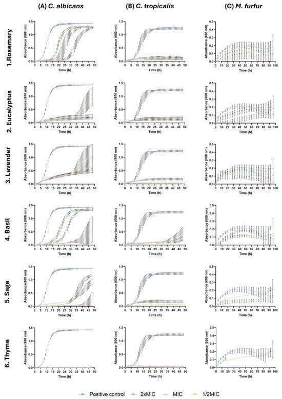
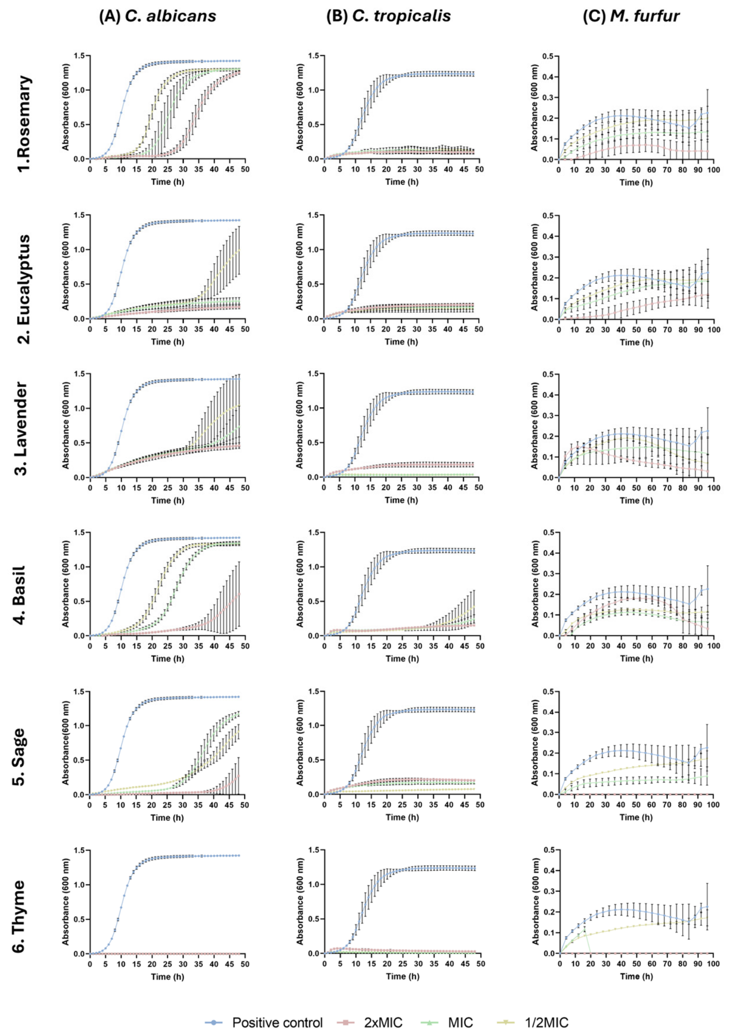
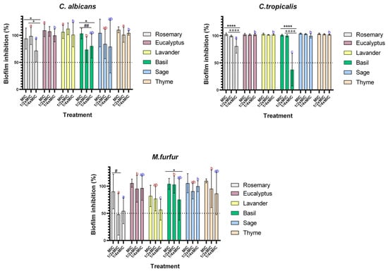
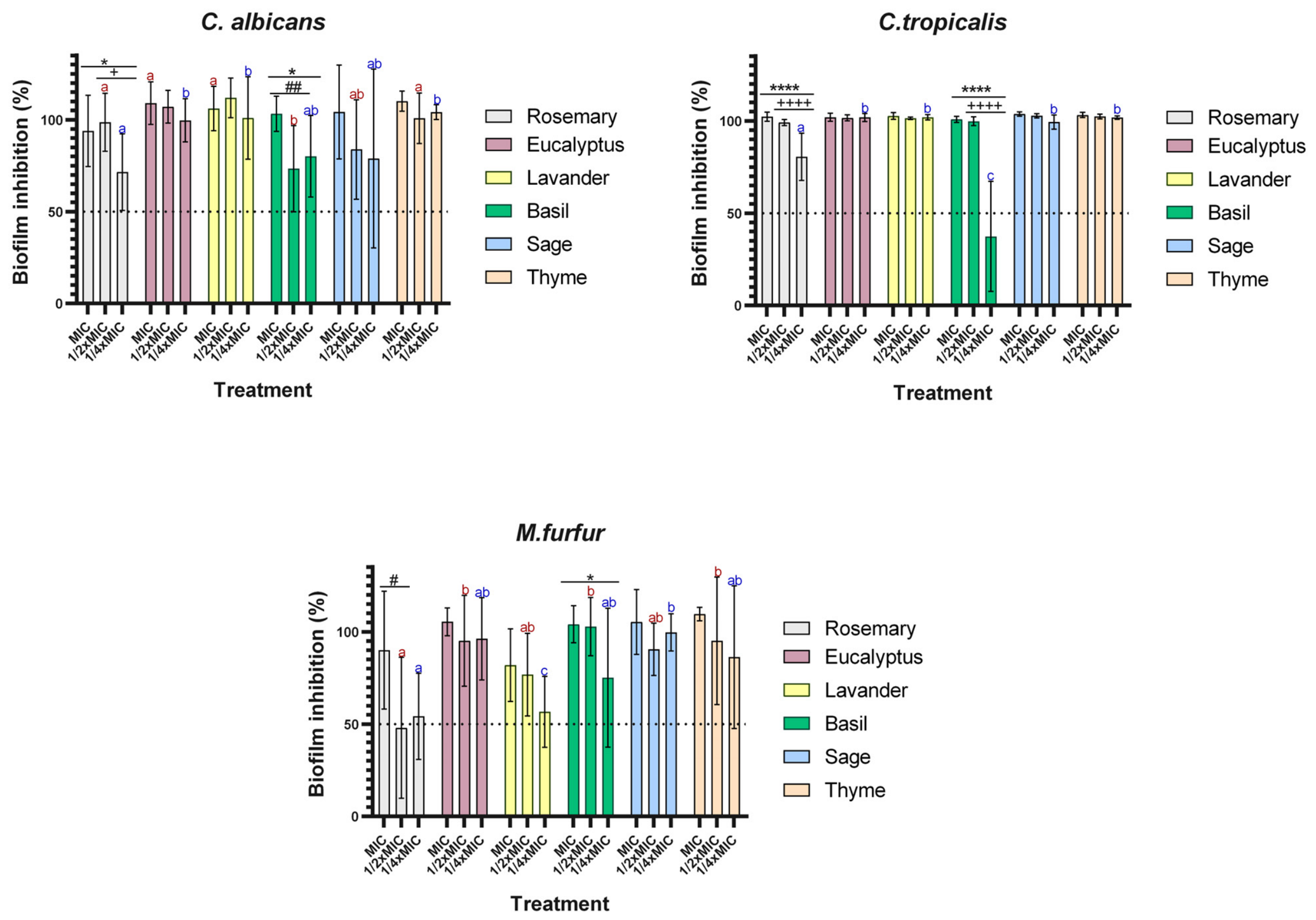
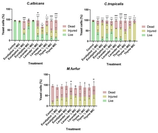
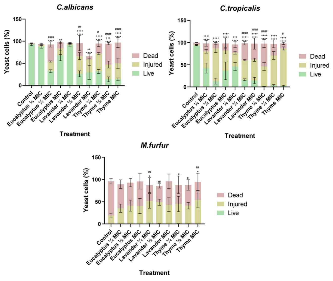
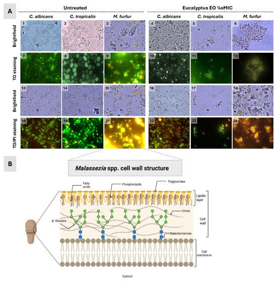
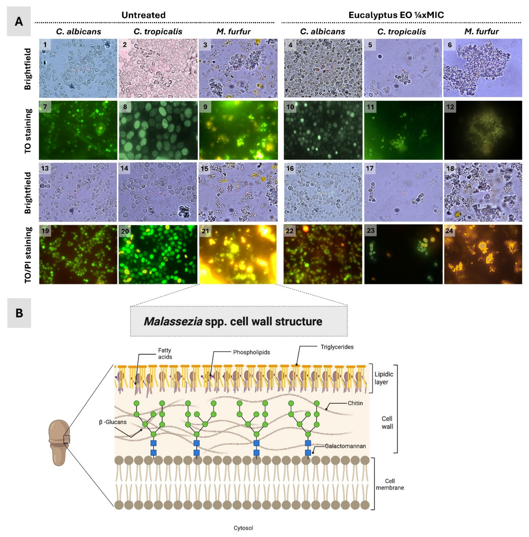

Disclaimer/Publisher’s Note: The statements, opinions and data contained in all publications are solely those of the individual author(s) and contributor(s) and not of MDPI and/or the editor(s). MDPI and/or the editor(s) disclaim responsibility for any injury to people or property resulting from any ideas, methods, instructions or products referred to in the content. |
© 2026 by the authors. Licensee MDPI, Basel, Switzerland. This article is an open access article distributed under the terms and conditions of the Creative Commons Attribution (CC BY) license.