


Abstract
Cancer is an alarming health concern and economic burden in both developed and developing countries. Recently, there has been a growing demand for new alternative medications with more effectiveness and fewer harmful effects. During the past decades, a set of chemotherapeutic agents has been developed to fight against a large spectrum of cancer types. Unfortunately, their use is associated with a high level of toxicity; they are expensive, also, and their deployment is restricted by the emergence of cellular resistance. Plant-based components are garnering attention due to their low toxicity, selectivity, efficiency, and ease of accessibility. Alkaloids are one of these targeted compounds. Indeed, they are a highly diverse group with basic heterocyclic nitrogen-containing alkaloids that exhibit potent anticancer effects against a large panel of solid and liquid tumors, such as lung, breast, leukemia, liver, and colon cancer. The main molecular mechanisms involved in alkaloids’ anticancer effect are the induction of apoptosis via the extrinsic and intrinsic pathways, DNA damage, and the inhibition of cell cycle progression. Amazingly, these auspicious compounds exhibited strenuous inhibitory effects against a whole range of key enzymes involved in cancer progression and metastasis, such as Cytochrome P450 (CYP450), Cyclooxygenase-2 (Cox-2), Lysine-Specific Demethylase 1 (LSD1), Poly [ADP-ribose] polymerase (PARP), and topoisomerase, mainly through two action modes, namely irreversible and reversible inhibition. Furthermore, several conventional extraction methods have been developed to extract bioactive compounds from natural matrices, such as Soxhlet and hot water extraction. However, these techniques have many drawbacks, as they require a large amount of organic solvents, which not only affect human health but also generate severe environmental issues. To overcome these limitations, multiple eco-extraction techniques have emerged as potential alternatives to traditional extraction methods such as ultrasonic extraction, microwave-assisted extraction, and supercritical fluid extraction. In fact, they are considered eco-friendly and efficient technologies with less time and solvent consumption. Overall, this review aims to provide an updated overview of the most prominent anticancer alkaloids that have not been well reviewed already, as well as the main green extraction techniques relevant to the extraction of antineoplastic alkaloids.
1. Introduction
Cancer is a disease characterized by the swift growth of abnormal cells or tissues in the body, which tend to proliferate in an uncontrolled manner and sometimes metastasize, such as in the case of malignant tumors (invasive). The second tumor type does not invade nearby tissues (stays in situ) and is referred to as being benign [1]. To date, 600 types of cancer have been identified. Most of them require unique diagnoses and management protocols [2]. It is worthwhile emphasizing that, over the next 25 years, the number of new cancer cases worldwide is projected to increase by 61%, reaching 30.5 million in 2050, whereas annual global cancer deaths are expected to rise by almost 75% to 18.6 million among the elderly [3]. Chemotherapy and radiotherapy are the most notorious and effective therapies for dealing with cancer diseases. However, they both have severe side effects, mainly the non-selective effects of the antitumor drugs, as they damage normal cells and tissues; they cause nausea, hair loss, fatigue, bone marrow problems, etc. [4]. Hence, the development of novel natural antineoplastic agents with more efficiency and fewer harmful effects is urgently required. Thereby, medicinal plants have caught the great focus of researchers during the past decades since they represent a valuable repository for new drug discovery and have been playing a pivotal role in the prevention and treatment of cancer.
Phytoalkaloids have recently gained particular attention as propitious secondary metabolites for the treatment of a wide range of chronic diseases, among them cancers; in fact, they are a highly diverse group of natural molecules derived from amino acids with a ring structure containing nitrogen (mostly the nitrogen atom is located inside the heterocycle ring) [5,6]. Alkaloids are mainly produced by the plant kingdom, especially in the higher plants, for the most part those belonging to the families of Ranunculaceae, Leguminosae, Papaveraceae, and Loganiaceae. They could also be found in lower plants, insects, marine organisms, and microorganisms [5]. To date, more than 21,000 alkaloids have been identified and are classified based on their chemical structures, biosynthesis, and ecological activities [6,7]. Alkaloids exhibit tremendous therapeutic potentials, for instance, the powerful analgesic effect of morphine, the relieving action of ephedrine, the anti-hyperglycemic effect of piperine, and so on [5]. The oldest breakthroughs in the hunt for natural anticancer drugs date back to the 1950s with the discovery and isolation of the vinca alkaloids, vinblastine and vincristine, as well as camptothecin, a topoisomerase I inhibitor. These molecules have been successfully developed into clinically active agents [6].
To boot, a plethora of traditional extraction techniques have been used to extract dozens of bioactive compounds from plant material, such as Soxhlet and maceration. However, these techniques present various drawbacks, as they require relatively large quantities of solvent, which not only increases operating costs but also generates serious environmental issues [8]. Additionally, during the extraction, plant samples are subjected to high pressure and temperature, which may entail thermal destruction of sensitive compounds [9]. To go around these impediments, several nonconventional methods have emerged during the last 50 years and are considered environmentally friendly and attention-grabbing, mainly due to their greater efficiency: ultrasound, pulsed electric field, supercritical fluids, enzyme digestion, and microwave irradiation [10].
Herein, this review critically summarizes and elucidates the anticancer mechanisms of several notable plant-derived alkaloids, with particular emphasis on apoptosis, antimetastatic activity, and synergistic combination strategies. In parallel, we compare conventional extraction procedures with emerging eco-friendly techniques (e.g., subcritical and microwave-assisted water extraction) that can influence yield, purity, and ultimately the pharmacological behavior of these compounds. By integrating mechanistic, technological, and translational aspects, this article aims to define the current opportunities and limitations of these alkaloids as candidates for future oncological therapies and to highlight key priorities for clinical development. To achieve this, we conducted a comprehensive and selective literature search in PubMed, Google Scholar, and Europe PMC to identify the most promising alkaloid candidates from medicinal plants that exhibit antitumor activity across diverse cancer types.
2. Interest and Relevance in Emerging Plant-Derived Alkaloids with Anticancer Potential
The bibliometric analysis of the anticancer activity of isolated alkaloids from medicinal plants reveals significant insights into their therapeutic potential [11,12]. Based on the PubMed platform [13], the bibliometric data of publications on the anticancer activity of common alkaloids isolated from medicinal plants is presented in Figure 1. In silico pharmacokinetic and drug-likeness evaluation of the selected alkaloids was conducted using the SwissADME web server [14], with canonical SMILES retrieved from PubChem [15]. SwissADME was employed to predict key ADME parameters, including gastrointestinal absorption, blood–brain barrier permeability, P-glycoprotein substrate status, cytochrome P450 interactions, and relevant physicochemical descriptors. Drug-likeness was assessed using the Lipinski, Ghose, Veber, Egan, and Muegge filters, together with the bioavailability score to estimate oral drug potential.
Figure 1.
The record of publications on the anticancer activity of studied alkaloids from medicinal plants. (Based on the PubMed platform [13]).
The high number concerning colchicine (4645 publications) suggests extensive research interest and clinical exploration, especially due to colchicine’s well-established use in cancer treatments like chemotherapy. Sanguinarine (287 publications) and chelerythrine (349 publications) are also notable with moderate numbers of publications, indicating substantial research into their potential as anticancer agents. On the other hand, alkaloids like Coptisine (96 publications), palmatine (115 publications), and chelidonine (47 publications) show growing interest in the scientific community, particularly in preclinical or early-stage studies for cancer therapies. Finally, alkaloids with fewer publications, such as liriodenine (50 publications) and antofine (25 publications), indicate emerging or niche research areas. These lower values could highlight new avenues for research and clinical trials.
The chemical structures of different plant-derived alkaloids help them to exhibit various mechanisms of action against cancer cells, including apoptosis induction and cell cycle arrest (Figure 2 and Table 1). Alkaloids like chelidonine and colchicine have been shown to trigger apoptosis in cancer cells, with chelidonine causing apoptosis at low doses and autophagy at high doses [16]. Moreover, many alkaloids interfere with cell cycle progression, effectively halting cancer cell proliferation [17]. According to different clinical relevance, these alkaloids are being explored as adjuncts to conventional therapies, enhancing the efficacy of treatments like chemotherapy [16]. Furthermore, the structural diversity of alkaloids presents opportunities for discovering new anticancer agents [18].
Figure 2.
Chemical structure of the plant-derived alkaloids chelidonine, coptisine, sanguinarine, and colchicine.
Table 1.
General overview of the other isolated alkaloids with prominent anticancer activity.
3. Anticancer Activity of Main Isolated Alkaloids from Medicinal Plants
3.1. Chelidonine
Chelidonine is a major benzophenanthridine alkaloid isolated from Chelidonium majus L. (C. majus), a widely prescribed Chinese plant known as the greater celandine (family Papaveraceae), and widely spread in Europe and Western Asia [32]. The chemical structure of the chelidonine molecule is presented in Figure 2. Chelidonine has a broad spectrum of pharmacological activities: antibacterial, anticancer [33], anti-inflammatory [34], spasmolytic [35], hepatoprotective, analgesic [36], antioxidant, immunomodulatory, radioprotective, and nephroprotective activities [37]. Indeed, it is considered one of the major components of the anticancer drug Ukrain, which has been clinically used to treat a large panel of tumors, mainly lung, breast, leukemia, colon, and prostate cancer. Figure 3 presents the proposed mechanism of the effect of chelidonine on numerous cancer cell types. Chelidonine is also used to enhance the quality of life and life expectancy for patients with advanced pancreatic cancer [38,39]. Importantly, the in vitro investigations have shown that this alkaloid could exhibit an anticancer effect in less than one micromole. Table 2 summarizes reported IC50 values of chelidonine across different cancer cell lines, depending on assay conditions and exposure time, as reported in the literature.
Figure 3.
The proposed mechanism of the effect of chelidonine on numerous cancer cell types. Bax: Bcl-2-associated X protein; Bcl-2: B-cell lymphoma 2; ERK: extracellular signal-regulated kinase; Top II: topoisomerase II; ROS: reactive oxygen species; STAT-3: signal transducer and activator of transcription 3; APAF-1: apoptotic protease-activating factor-1; PARP: poly (ADP-ribose) polymerase; PI3K: phosphoinositide 3-kinase; P38: mitogen-activated protein kinases; Tumor protein P53; p21: cyclin-dependent kinase inhibitor 1.
Table 2.
Reported IC50 values of chelidonine in different cancer cell lines.
The in vitro growth of breast cancer MCF-7 cells was roughly inhibited after a treatment with 0.1 μM of chelidonine [40]. Interestingly, it has also been demonstrated that chelidonine sensitizes tumor cells to cancer therapy and enhances the cytotoxicity of chemotherapeutic agents [41,44]. In this context, chemotherapy uses several drugs in the treatment of metastatic or recurrent non-small cell lung cancer (NSCLC), mostly Osimertinib, a tyrosine kinase inhibitor (TKI) that is hampered, unfortunately, by the occurrence of the T790M secondary mutation in the epidermal growth factor receptor gene (EGFR) [45]. A study has reported that chelidonine could effectively interact with mutated EGFR, inhibiting selectively its phosphorylation and the downstream targets in cells with mutated EGFR, and abolishes, as a consequence, the in vitro and in vivo growth of NSCLC cells [35]. Furthermore, it has been shown that chelidonine contributes to overcoming drug resistance through the inhibition of p-glycoprotein (MDR-1) expression and other enzymes of the cytochrome P450 system implicated in the metabolism of xenobiotics in colon and leukemia cancer cells [44]. Another study has concluded that chelidonine reduces the expression of epithelial–mesenchymal transition and improves the apoptotic effect of Lenvatinib against hepatocellular carcinoma (HCC) in vivo [46]. Additionally, chelidonine imparts cell growth inhibitory responses in various cancer cell lines, such as HeLa and T98G cells, by inducing cell arrest at the G2/M phase, triggering apoptosis through the production of reactive oxygen species (ROS). Also, it can alter several signaling pathways and induce apoptosis via the upregulation of expressions of p21, p38, and p53 and downregulation of AKT, PI3K, JAK3, STAT3, E6, and E7 [35,47,48]. In addition, it was found that chelidonine provokes T98G cell death mediated by the activation of caspase-3 and -9 (caspase-dependent pathway) [48], prevents microtubule polymerization, destabilizes microtubule structures, and arrests cell cycle progression in stage M in SGC-7901 human gastric cancer cells through the increase in the phosphorylation of histone H3 at Ser10 [38]. At low concentrations, chelidonine is capable of remarkably reducing telomerase activity in HepG2 human liver cancer, mainly through the downregulation of the expression of the catalytic subunit of telomerase (hTERT) [49]. Moreover, it has been proven by proteomics analysis that chelidonine significantly arrests the mitochondrial respiratory chain in NSCLC cells [35]. In the same way, this alkaloid induces a slight release of cytochrome in human CEM T-leukemia cells accompanied with the activation of caspase-3 [50], causes the loss of mitochondrial membrane potential (MMP) during the early phase of apoptosis in NSCLC cells [35], and induces Mcl-1 degradation in chelidonine-treated T98G cells, leading, as a consequence, to mitochondrial instability increase [48]. Chelidonine intercalates with DNA, causing its fragmentation, and inhibits RNA polymerase, topoisomerase, telomerase, and even ribosomes [41]. However, physical methods such as optical spectroscopy have demonstrated that the interaction between chelidonine and the DNA macromolecules (DNA–chelidonine) is actually flimsy [51]. Moreover, chelidonine is a potent inhibitor of NF-κB pathway activation induced by TNF-α. In addition, it prevents the expression of COX-2, MMP-9, cyclin D1, and ICAM-1 in HCT116 cells [52]. Likewise, it is a selective inhibitor of STK19 serine/threonine kinase (ATP-competitive inhibitor of STK19), which has been proposed as a cancer driver [53,54]. Chelidonine exhibited antiproliferative effects against various Neuroblastoma RAS (NRAS)-mutant cancer cells in vitro, prevented NRAS-driven cancer growth, and displayed minimal toxicity in mice models [53]. These studies suggest that chelidonine may be a prominent natural therapeutic agent for the management of various cancer types.
3.2. Coptisine
Coptisine (COP) is a natural small isoquinoline alkaloid isolated from the rhizome of the Coptis chinensis plant, recognized as ‘huanglian’ in traditional Chinese medicine [55]. The chemical structure of the coptisine molecule is presented in Figure 2. Coptisine has been shown to possess a wide variety of pharmacological and biological activities, essentially as antitumor [56], antibacterial, bactericidal [57], anti-hyperlipidemic and anti-cachectic [58], neuroprotective, antioxidant [59], and anti-inflammatory [60]. Coptisine has been proven to inhibit cancer cell growth via the induction of apoptosis through intrinsic and extrinsic pathways in numerous cancer cells derived from the lung [56], colon [61] and liver [62]. Figure 4 presents a proposed mechanism of coptisine-induced apoptosis in a variety of cancer types. A thorough examination proved that coptisine exerted in vitro antiproliferative and cytotoxic effects against various cancer cells, such as hepatocellular carcinoma cells Hep3B, human colon cancer cells HCT-116, and non-small cell lung cancer cellular models A549, H460, and H2170, largely through the increase in intracellular and mitochondrial ROS generation. The upregulation of expression of the pro-apoptotic proteins BAX, Bad, Apaf-1, and AIF; the downregulation of expression of the anti-apoptotic Bcl-2 protein; and the decline in mitochondrial membrane potential (MMP) (Δψm) led to cytochrome c release from mitochondria into the cytosol concomitantly with the sequential activation of caspase-9 and -3, and subsequently cleavage of PARP in a dose-dependent approach [56,63,64]. Moreover, it was found that coptisine induces apoptosis via upregulation of ROS-dependent JNK phosphorylation in Hep3B cells [63], and it triggers in vitro apoptosis in colon cancer cells through the attenuation of PI3K, Akt, and p-Akt levels. The same study demonstrated that the tumor growth rate was significantly inhibited in HCT-116 xenograft nude mice after being treated with 150 mg/kg of coptisine [64]. COP disturbed the mitochondrial electron transport chain and increased the expression and recruitment of the two-mitophagy proteins PINK-1 and Parkin from the cytoplasm to mitochondria in a dose-dependent manner in hepatocellular carcinoma cells [65]. Moreover, this compound inhibited the growth of non-small cell lung cancer cells by blocking cell cycle progression at the G2/M phase and decreasing the expression of cyclin B1, cdc2, and cdc25C. Meanwhile, it upregulated p21 [56], and COP evidently decreased the expression of cyclin D1 and cyclin E, leading to cell cycle arrest at G1 phase, downregulated the expression of AKT and ERK in a concentration-dependent approach, induced apoptosis in colon cancer cells in vitro, and displayed low toxicity in mice [61]. Also, it increased the expression of death receptor 4 (DR4) and death receptor 5 (DR5) in Hep3B cells [63]. Coptisine affects cell viability and inhibits cell growth of HepG2 through the inhibition of the secretion of exosomal circCCT3, which upregulates hexokinase II (HK2), a key enzyme involved in glycolysis, and via triggering apoptosis [62]. This alkaloid is able to induce DNA damage in Hep3B and A549 cells, assessed by the increase in expression of pH2AX protein, a marker for DNA damage [56,65]. It is worth noting that coptisine also increased comet tail length, TUNEL-positive cells, and p-γH2AX, which are indicators of DNA damage in hepatocellular carcinoma cells [63]. Multispectroscopic, electrochemical, and molecular modeling techniques have revealed that coptisine causes a localized destabilization of the DNA double helix through intercalation between two adjacent base pairs in the DNA, creating a sort of sandwich-like structure. The interaction between coptisine and the DNA double helix takes place by non-covalent stacking interactions with DNA base pairs [55]. Thus, coptisine exhibits a high affinity to DNA due to the strong stacking interactions between an aromatic chromophore of this alkaloid and the base pairs [56,66]. Some studies have speculated that the development of coptisine-based clinical drugs may exert a cardioprotective effect and attenuate the oxidative stress in cardiomyocytes triggered by anticancer drugs [67]. Thereby, these studies suggest that coptisine may be a potential candidate for various human cancer therapies.
Figure 4.
A proposed mechanism of coptisine-induced apoptosis in a variety of cancer types. APAF-1: apoptotic protease-activating factor-1; ROS: reactive oxygen species; JNK: c-Jun N-terminal kinases; p21: cyclin-dependent kinase inhibitor 1.
3.3. Sanguinarine
Sanguinarine (SNG), also known as 13-methylbenzodi-oxolo[5,6-]-1,3-dioxolo[4,5-I] phenanthridinium, is a benzophenanthridine belonging to the group of benzylisoquinoline alkaloids. It is isolated from several plant species, namely Argemone mexicana, Chelidonium majus, Macleaya cordata, Poppy fumaria, Bocconia frutescens (Papaveraceae), and predominantly from the roots of the plant Sanguinaria canadensis, which is largely used in toothpaste and mouthwashes to remove dental plaque formation and alleviate gingival inflammation [5,68,69,70]. The chemical structure of the SNG molecule is presented in Figure 2. This alkaloid has been found to possess an extensive assortment of pharmacological and biological activities, such as anti-diabetic [71], anti-microbial, antioxidant, anti-inflammatory [5], antifungal [72], and antiviral [73]. A set of recent studies reported that SNG has an antitumor potential against numerous cancer types, both in vitro and in vivo. Among others, lymphoma [74], leukemia [68], breast [69], prostate [75], thyroid [76], and colon cancer [77]. Figure 5 presents a proposed mechanism of SNG-induced apoptosis in multiple cancer cell lines. Impressively, numerous studies have drawn attention to the anticancer effect of SNG at micromolar concentrations (less than 10 µM), along with the absence of side effects on healthy normal cells (selectivity), suggesting, therefore, the possible use of this alkaloid as an anticancer agent [68,78]. These multiple targets are conveyed by a wide variety of molecular mechanisms triggered by this alkaloid. SNG exhibited in vitro antiproliferative effects against a large panel of cancer cell lines, such as multiple myeloma cell lines (U266, IM9, MM1S, and RPMI-8226), MDA-MB-231 human breast carcinoma cells, HT-29 human colon cancer cells, and acute lymphoblastic leukemia (Pre-ALL) cell lines, mainly by apoptosis through the generation of ROS such as superoxide radical (O2•−), which occurs due to the depletion of glutathione. SNG also increased the ratio of Bax/Bcl-2, triggered the translocation of Bax into the mitochondrial membrane simultaneously with the induction of loss of mitochondrial membrane potential (MMP), and stimulated the release of cytochrome c into the cytosolic compartment, which in turn combined with APAF-1, ATP, and procaspase 9, forming a multimeric complex named apoptosome, and activating caspase cascades (caspase 9, 3), and subsequently PARP cleavage [68,69,70,77]. SNG suppresses activated AKT and downregulates the expression of the anti-apoptotic genes cIAP1, cIAP2, and XIA [68]. It was found that in vitro treatment with SNG 1 µM improves the cytotoxicity of the anticancer drug cisplatin in ovarian cancer (A2780/R) cells through depletion of intracellular glutathione (GSH). Additionally, both SNG and cisplatin increased the entry of cells into apoptosis better than each compound alone [79].
Figure 5.
A proposed mechanism of sanguinarine-induced apoptosis in multiple cancer cell lines. SNG: sanguinarine, APAF-1: apoptotic protease-activating factor-1, HIF-1α: hypoxia inducible factor-1α; SHP-1: The tyrosine phosphatase; STAT-3: signal transducer and activator of transcription 3; DR4: Death receptor 4: JAK2: Janus kinase 2; Mir-16-2: MicroRNA 16-2.
Beyond its previously described combinations with cisplatin and piperlongumine (PL), SNG has also demonstrated synergistic activity with the proteasome inhibitor bortezomib in multiple myeloma models. At sub-cytotoxic concentrations, the co-administration of both agents markedly enhances caspase-3 activation and PARP cleavage, leading to a significant reduction in cell viability. Mechanistically, this potentiation has been associated with suppression of the JAK2/STAT3 signaling pathway and concomitant upregulation of the tyrosine phosphatase SHP-1, thereby reinforcing mitochondrial, caspase-dependent apoptosis [70]. These findings suggest that SNG can sensitize tumor cells to proteasome inhibition through modulation of pro-survival STAT3 signaling. In A549 non-small cell lung cancer cells, the PL–SNG combination (4:1 ratio) not only augments intracellular ROS accumulation and apoptotic cell death but also exerts a more pronounced inhibitory effect on migration and invasion compared to either compound alone. This enhanced antimetastatic activity correlates with substantial disruption of cytoskeletal organization, including altered F-actin architecture and decreased vimentin expression, as reflected in impaired wound closure kinetics [80]. Importantly, PL itself is known to interfere with metastatic behavior in several cancer models through ROS-dependent mechanisms, inhibition of epithelial–mesenchymal transition (EMT) markers, and reorganization or depolymerization of F-actin and vimentin—two key structural determinants of cell motility [80,81]. When combined with SNG, which activates apoptosis via ROS amplification and STAT3 inhibition, these complementary mechanisms likely, converge to produce synergistic suppression of both survival and metastatic potential.
Collectively, accumulating evidence indicates that SNG-based combinations—particularly with ROS-modulating or STAT3-targeting agents—can induce synergistic pro-apoptotic and antimetastatic effects. However, the magnitude and even the direction of these interactions remain highly context-dependent, varying according to tumor type, molecular background, and drug concentration. This mechanistic multiplicity aligns with the signaling complexity illustrated in Figure 5, where modulation of ROS generation, JAK2/STAT3 activity, SHP-1 expression, and cytoskeleton-related pathways provides several nodes for pharmacological synergy with other alkaloids such as PL [70,80,81].
SNG induced G1 phase cell cycle arrest and apoptosis in HCC cells through the upregulation of miR-16 expression, which resulted in the downregulation of its target genes bcl2 and cyclin D in a P53-dependent manner (in both wild-type and mutant p53) [82]. It was found that SNG reduced cell viability and induced apoptosis in human oral squamous cell carcinoma KB cells through the activation of the extrinsic apoptosis pathways hallmarked by TRAIL and DR4 increase, caspase 8 activation, truncation of Bid, DNA fragmentation, and the inactivation of the PI3K/Akt pathway [83]. On the other hand, it has been demonstrated that HIF-1α and STAT3 are overexpressed in various cancers. They are both involved in hypoxia-induced epithelial–mesenchymal transition (EMT) and play a key role in cancer progression and invasion. In this context, a study showed that SNG successfully prevents the nuclear interaction between HIF-1α and both p-STAT3-Tyr and p-STAT3-Ser in vivo and in vitro, prohibits the formation of the complex HIF-1α/STAT3, and prevents the induction of its target proteins in TNBC cells during hypoxia [84,85]. In the same way, SNG increased SHP-1, a protein tyrosine phosphatase (PTP), which subsequently led to the inhibition of the phosphorylation and activation of JAK2 and STAT3 in multiple myeloma cells [70], and so forth.
3.4. Colchicine
Colchicine is an alkaloid purified from the plant Colchicum autumnale, commonly known as meadow saffron, and usually prescribed for the treatment of several diseases, including familial Mediterranean fever, acute and chronic gout, osteoarthritis, scleroderma, amyloidosis, Behcet’s syndrome, Sweet’s syndrome, and pericarditis [86,87]. The chemical structure of the colchicine molecule is presented in Figure 2. This alkaloid exhibits a wide array of pharmacological and biological properties, like anti-inflammatory, cardioprotective [88], and antiviral, and has been recently approved by the Food and Drug Administration (FDA) for the prevention and treatment of gout [89]. Currently, many studies have brought attention to the possible use of this alkaloid in the management of a plethora of cancer types, such as cholangiocarcinoma (CC) [90], liver cancer [91], cervical cancer, esophageal cancer [92], colon cancer [93], and pancreatic cancer [94]. However, the accurate mechanism by which this alkaloid exerts its effect is still elusive and yet to be established [95]. Figure 6 presents the underlying mechanism of colchicine (a microtubule destabilizer)-induced apoptosis on various cancer cell lines. The use of colchicine in medicine as an anticancer agent has been hindered by its toxicity on normal cells. Nevertheless, some recent studies have suggested the safe employment of this alkaloid at clinically controlled concentrations [96]. It is universally agreed that colchicine binds at Cys-354, near the interface located between the α and β subunits of tubulin, by H-bonding and hydrophobic interactions, and consequently markedly decreases the incorporation of tubulin-colchicine into microtubules (destabilizes microtubule polymerization). Whatever the nucleotide (GTP or GDP), this leads to the disruption of mitosis in cancerous cells [87,97,98]. Additionally, several studies have shown that colchicine exhibits potent in vitro antitumor activity against NCI-N87 gastric cancer cells; normal liver L-02 cells; human cervical cancer cell lines CaSki, HeLa, and C-33A; and colon cancer cells HT-29, mainly by blocking the cell cycle at G2/M. The triggering of apoptosis via the induction of loss of mitochondrial membrane potential Δψm, Bax/Bcl2 ratio increase, and the translocation of pro-apoptotic protein Bax to the outer membrane of mitochondria, followed by the cytochrome c release to the cytoplasm, with which Apaf-1, pro-caspase 9, and ATP could associate to form the apoptosome complex and subsequently activate caspase 9,3 [92,93,99,100]. The administration of colchicine (after seven days of the inoculation of NCI-N87 gastric carcinoma cells) at a dose of 0.05 mg/kg once every 24 h subcutaneously in BALB/c nude mice remarkably reduced the tumor volume and weight as compared to the control group. More importantly, the inhibition of cancer growth during the treatment period was achieved without side effects on liver or renal tissue and did not notably change the body weight of mice [100]. Similarly, a group of mice (BALB/c-nu) inoculated with the cancer cell line NCI-N87 was treated with 0.07 mg of colchicine/kg once every day for 14 days, which reduced the tumor weight significantly without affecting the body weight of the mice during the treatment. The study recommended this alkaloid as a potential candidate for palliative treatment of gastric cancer [101]. Moreover, colchicine upregulates the tumor suppressor proteins P53 and Rb, while it downregulates the expression of retinoblastoma protein (pRb) and triggers, as a consequence, downstream cell cycle arrest in human cervical cancer cell lines in vitro [92]. On the other hand, the in vitro treatment of breast cancer cells MCF-7 with colchicine induces apoptosis and cell cycle arrest at G2/M in a time- and dose-dependent approach [102]. This alkaloid also increases ROS production and activates p38 by phosphorylation in colon cancer cells [93]. It exerts an antiproliferative effect on gastric cancer cells through the upregulation of the gene (DUSP1) [101] and causes DNA fragmentation in NCI-N87 cells at lower concentrations (less than 10 nM) [100]. Additionally, colchicine exhibits a strong antimitotic potential at a concentration of 40 nM, inhibiting cell cycle progression in lung cancer cell line A549 at the G2/M phase. In the same mode, while it induces cell cycle arrest at the same phase in breast cancer cells MDA-MB231 at a concentration of 60 nM [103], it was found that colchicine affected intracellular Ca2+ homeostasis. Apparently, colchicine-induced Ca2+ release from the endoplasmic reticulum was mainly mediated by the PLC pathway. The same study reported that colchicine caused cell death in the human oral cancer cell line OC2 independently of Ca2+ [104]. Further preclinical studies based on in vivo and in vitro models are recommended to fill some gaps in the previous investigations and extend the knowledge about the anticancer mechanisms of this alkaloid, such as whether the colchicine-induced Ca2+ released from the endoplasmic reticulum contributes somehow to cytotoxicity in tumor cells, and precisely the mechanisms involved in it.
Figure 6.
The underlying mechanism of colchicine (a microtubule destabilizer)-induced apoptosis in various cancer cell lines. GPCR: G protein-coupled receptor; Bax: Bcl-2-associated X protein; APAF-1: apoptotic protease-activating factor-1; Bcl-2: B-cell lymphoma 2; Rb: The retinoblastoma protein; IP3: inositol trisphosphate; DAG: diacylglycerol.
3.5. Other Isolated Alkaloids with Prominent Anticancer Activity
The other isolated alkaloids with prominent anticancer activity include chelerythrine, liriodenine, antofine, and palmatine. Table 1 displays a general overview of such alkaloids based on plant precursors, chemical structure, and type of target cancer, as well as the effects and mechanisms of action.
Chelerythrine, isolated from Chelidonium majus L. and Toddalia asiatica L., exhibits significant anticancer activity across multiple cancer types, including cervical, lung, breast, and liver cancers. In cervical cancer, it induces apoptosis, disrupts mitochondrial function, and generates reactive oxygen species (ROS) in HeLa and C33A cell lines. In lung cancer, chelerythrine targets cancer stem cells (CSCs) by reducing the expression of β-catenin, SOX2, and MYC, thereby inhibiting migration and invasion. It also induces ROS production, apoptosis, and autophagy in NSCLC A549 and NCI-H1299 cells. In breast cancer, chelerythrine binds to quadruplex complexes and downregulates BCL2, VEGFA, and KRAS gene expression, showcasing anti-angiogenic and anti-invasive properties in MCF-7 cells. Additionally, it demonstrates antiproliferative effects and apoptosis induction in human liver cancer cells, inhibiting the migration and invasion of Hep3B cells. These mechanisms highlight its broad anticancer potential [19,20,21,22,23].
Liriodenine, derived from Fissistigma glaucescens, Annona glabra, Liriodendron tulipifera, and other plants, is effective against esophageal, breast, laryngeal, and ovarian cancers. In esophageal cancer, liriodenine triggers apoptosis, causes G2/M cell cycle arrest, and enhances radiosensitivity in ECA-109 cells by increasing the Bax/Bcl2 ratio and caspase-3 levels while delaying DNA damage repair. In breast cancer, it prevents cell growth by inducing apoptosis; downregulating Bcl-2, cyclin D1, and VEGF; and upregulating tumor suppressor proteins like p53 and caspase-3. For laryngeal cancer, it inhibits cell migration and induces apoptosis both in vivo and in vitro, with increased p53 expression in HEp-2 cells. In ovarian cancer, liriodenine activates the intrinsic apoptotic pathway, decreases mitochondrial membrane potential, triggers cytochrome c release, and blocks the cell cycle at the S phase. These effects collectively inhibit cancer cell proliferation and survival [24,25,26].
Antofine, found in Vincetoxicum nigrum, Cynanchum vincetoxicum, and other Asclepiadaceae family plants, displays potent anticancer effects against lung cancer and leukemia. In lung cancer, antofine inhibits cancer cell proliferation, sensitizes cells to chemotherapy agents, and suppresses angiogenesis by modulating key signaling pathways such as AKT/mTOR/AMPK and ERK in endothelial cells. Its anti-angiogenic activity further inhibits migration and tube formation in HUVECs. In leukemia, antofine selectively inhibits cell proliferation and induces apoptosis without affecting normal cells, with a slight suppression of NF-κB activation. These results emphasize its selectivity and therapeutic promise in cancer treatment [27,28,29].
Palmatine, isolated from Rhizoma coptidis and Rutidea parviflora D.C., demonstrates anticancer activity against colorectal and ovarian cancers. In colorectal cancer, it selectively suppresses proliferation both in vitro and in vivo, induces G2/M cell cycle arrest through aurora kinase A (AURKA) downregulation, generates ROS, and impacts mitochondrial membrane potential. Additionally, palmatine upregulates pro-apoptotic proteins such as P53, P73, caspase-3, and caspase-9. In ovarian cancer, it displays strong anticancer potential by sensitizing A2780 cells to cisplatin and inducing apoptosis in OVCAR-4 cells through the activation of caspase-3/7, PARP, and annexin V. These findings underscore palmatine’s potential for targeted anticancer therapy [30,31].
4. Pharmacokinetic Properties of Major Alkaloids
In silico pharmacokinetic evaluation using SwissADME indicated that all major alkaloids analyzed (chelidonine, coptisine, sanguinarine, colchicine, chelerythrine, liriodenine, antofine, palmatine, sophocarpine, mitragynine, and epiberberine) exhibit high predicted gastrointestinal absorption. This favorable absorption is consistent with their generally suitable physicochemical properties, including moderate molecular weight, limited hydrogen-bonding capacity, and optimal lipophilicity (Table 3). All compounds complied with major drug-likeness filters (Lipinski, Ghose, Veber, Egan, and Muegge) and showed a uniform bioavailability score (0.55), supporting their oral drug-like potential.
Table 3.
Physicochemical parameters of the major alkaloids.
Moreover, all compounds were predicted to exhibit high gastrointestinal absorption, which can be explained by their moderate molecular weights, balanced lipophilicity, low hydrogen-bonding capacity, and generally acceptable polar surface areas. In addition, all alkaloids fulfilled the major drug-likeness filters (Lipinski, Veber, Ghose, Egan, and Muegge) and displayed a uniform bioavailability score of 0.55, indicating favorable oral drug-like properties. Experimental observations for colchicine, mitragynine, and isoquinoline alkaloids such as palmatine and coptisine further support efficient intestinal uptake, although systemic exposure may be limited by post-absorptive processes. In particular, colchicine exhibits an experimentally determined oral bioavailability of approximately 45% in humans, characterized by rapid gastrointestinal absorption but reduced systemic exposure due to first-pass metabolism and P-glycoprotein-mediated efflux [105]. Similarly, chelidonine has been reported to exhibit good intestinal permeability and favorable oral absorption in experimental models, which can be attributed to its moderate molecular weight, balanced lipophilicity, and limited hydrogen-bonding capacity, thereby supporting the in silico ADME predictions [106].
On the other hand, most alkaloids were predicted to be blood–brain barrier permeant, except colchicine, suggesting potential central nervous system distribution (Table 4). This is consistent with the reported neuroactive effects of compounds such as mitragynine, sanguinarine, and chelerythrine. Several alkaloids were also identified as P-glycoprotein substrates, indicating that efflux transport may restrict intracellular accumulation and contribute to low plasma concentrations despite good absorption, as documented for berberine-type alkaloids.
Table 4.
Pharmacokinetic properties and CYP inhibition potential of the major alkaloids.
Metabolic predictions highlighted frequent interactions with cytochrome P450 enzymes, with several compounds showing inhibitory potential toward key isoforms (CYP1A2, CYP2C19, CYP2D6, and CYP3A4). This suggests a possible risk of herb–drug interactions, especially for mitragynine, which has been experimentally shown to inhibit CYP2D6 and CYP3A4. Although colchicine was not predicted as a strong inhibitor, it is extensively metabolized by CYP3A4, and its toxicity is known to increase in the presence of CYP3A4 inhibitors.
Elimination is expected to occur mainly through hepatic metabolism followed by biliary and/or renal excretion. More lipophilic alkaloids with broader tissue distribution may display slower clearance, as reported for mitragynine, whereas colchicine undergoes both renal and biliary elimination with enterohepatic recycling.
In summary, SwissADME predictions indicate that these alkaloids share common pharmacokinetic features, including good oral absorption, frequent CNS accessibility, transporter-mediated efflux, and substantial CYP involvement. These characteristics support their therapeutic potential while emphasizing the importance of metabolic interactions and safety evaluation in future pharmacological development.
5. Safety, Toxicity, and Side-Effect Considerations of Anticancer Alkaloids
Despite their promising anticancer activity, plant-derived alkaloids present safety and toxicity challenges that are critical for clinical translation. Their toxicological profiles are highly compound-specific and depend on dose, route of administration, metabolic stability, and treatment duration.
For instance, colchicine is the most clinically characterized alkaloid. Evidence from randomized trials and meta-analyses indicates that gastrointestinal adverse effects, notably diarrhea and nausea, are the most common, whereas serious hepatic, hematological, or neuromuscular toxicities are uncommon at therapeutic doses. Hence, careful dose control is particularly important in patients with renal or hepatic impairment or when colchicine is co-administered with CYP3A4 or P-glycoprotein inhibitors [107].
In contrast, chelidonine and related alkaloids from C. majus show selective cytotoxicity toward cancer cells in vitro, with limited toxicity to normal cells at low micromolar concentrations. Safety concerns reported clinically are largely associated with crude plant extracts, which have been linked to idiosyncratic hepatotoxicity, suggesting that purified alkaloids may offer improved safety profiles under controlled dosing conditions [108]. Coptisine and palmatine generally exhibit low acute toxicity and good tolerability in preclinical studies. Their poor oral bioavailability may limit systemic toxicity but also complicates dose optimization. Potential cytochrome P450 inhibition raises the possibility of herb–drug interactions when combined with conventional anticancer therapies [109]. In contrast, benzophenanthridine alkaloids, such as sanguinarine, display potent anticancer effects but higher intrinsic cytotoxicity, with mechanisms involving oxidative stress and mitochondrial dysfunction, resulting in a narrow therapeutic margin. Their clinical potential may therefore rely on targeted delivery or combination strategies to minimize systemic toxicity [110]. Overall, these differences disclose the importance of compound-specific toxicological evaluation and rational dosing strategies. Notably, the ability of several alkaloids to enhance chemosensitivity supports their potential use at sub-toxic doses as adjuvant agents, thereby improving efficacy while minimizing adverse effects.
6. Enzymatic Inhibition by Alkaloids in Suppressing Tumor Growth and Proliferation
The living organisms around us, including humans, plants, bacteria, etc., possess an arsenal of enzymes that play a vital role in monitoring the matrix chemical reactions inside the cells. In fact, they are mainly natural proteins that act as biological catalysts (biocatalysts) that activate the rate of chemical reactions in biological systems (sometimes up to a million times faster) due to their ability to lower the energy of activation [111,112]. Enzyme inhibitors play a crucial role in the drug discovery process since inhibiting key enzymes helps to defeat chronic diseases, including cancers. Indeed, they are molecules that modulate the catalytic properties of an enzyme once they bind to the active site in order to reduce the reaction rate or even block the catalysis process radically [113]. Enzyme inhibitors have been categorized mainly based on their action mode, namely, irreversible and reversible. The latter type of inhibitor includes three major groups: competitive, non-competitive, and uncompetitive inhibitors [114]. As some enzymes are highly induced in cancer cells, such as hexokinase II and glucose-6- phosphate isomerase, enzyme inhibition is considered a promising therapeutic strategy to stop cancer progression [115].
Alkaloids have proven their efficiency by blocking many key enzymes involved in cancer progression, both in vivo and in vitro. Figure 7 summarizes the key natural alkaloids that exhibit inhibitory effects against a range of enzymes involved in cancer development and metastasis.
Figure 7.
Natural alkaloids that exhibit inhibitory effects against a range of enzymes involved in cancer development and metastasis. MMPs: metalloproteinases; HK2: Hexokinase 2; Topo II: topoisomerases II; LSD1: lysine specific demethylase 1; CYP450: Cytochrome P450; MAOs: Monoamine oxidases; COX-2/1: Cyclooxygenase-2/1; PARP: Poly (ADP-ribose) polymerase.
Sophocarpine, a matrine-type alkaloid, was found to exhibit inhibitory effects on two human liver CYP isoforms (CYP3A4, CYP2C9). This alkaloid was reported to be a non-competitive inhibitor of the isoform CYP3A4, while it is a competitive inhibitor of CYP2C9 with IC50 values of 12.22 and 15.96 µM, and Ki values were found to be 6.74 and 9.19 µM, respectively [116]. Mitragynine and corynantheidine alkaloids were identified as potent inhibitors of CYP2D6, with reported IC50 values of 2.2 and 4.2 µM and Ki values of 1.1 and 2.8 µM, respectively, while both compounds were recognized as competitive inhibitors of CYP2D6 [117]. Epiberberine, a natural protoberberine alkaloid, was characterized as a selective inhibitor of lysine-specific demethylase1 (LSD1), with an IC50 value of 0.14 µM [118].
Matrine, a pleiotropic alkaloid, showed a strong inhibitory effect against hexokinase 2 (HK2) overexpressed in human myeloid leukemia [119]. Interestingly, the enzyme inhibition modes summarized in Table 5 have direct clinical relevance. For instance, competitive inhibitors (e.g., nitidine chloride against CYP1B1 or mitragynine against CYP3A4) may exhibit substrate-dependent effects, potentially reducing inhibitory efficacy at high substrate concentrations. In contrast, non-competitive and mixed inhibitors (e.g., peimine, cepharanthine, and mitragynine against CYP2C9 and CYP2D6) suppress enzyme activity independently of substrate levels, which may lead to more sustained inhibition in vivo. Moreover, reversible inhibition of cancer-related targets, as exemplified by epiberberine acting on LSD1 with sub-micromolar potency, is generally associated with a lower risk of toxicity than irreversible inhibition, supporting its translational potential in anticancer therapy. Table 5 recaps the main enzyme inhibitors that may be involved in cancer growth.
Table 5.
Alkaloids exert inhibitory effects against selected enzymes.
7. Extraction of Phytoalkaloids by Green Extraction Techniques
The extraction of phytoalkaloids using green techniques is gaining traction due to its efficiency and reduced environmental impact. Green extraction techniques, such as supercritical fluid extraction, microwave-assisted extraction, and ultrasound-assisted extraction, minimize solvent use and waste generation, addressing significant environmental concerns associated with traditional methods [128]. These techniques not only enhance extraction yields of bioactive compounds but also ensure the preservation of their therapeutic properties. By minimizing the use of hazardous solvents, which aligns with green chemistry principles, these methods can decrease solvent-related waste by up to 80% [129], as well as require less energy compared to conventional methods [130].
Based on the literature, Table 6 provides a comparison of the pros and cons of various green extraction techniques for phytoalkaloids exhibiting anticancer effects. It is worth noting that the effectiveness of each technique can vary depending on the specific phytoalkaloids and plant materials being extracted. The choice of extraction method should be based on the target compounds, plant matrix, and desired end use of the extract.
Table 6.
Comparison of the pros and cons of various green extraction techniques for phytoalkaloids exhibiting anticancer effects.
7.1. Supercritical Carbon Dioxide Extraction Technique
During the past few decades, the use of carbon dioxide (CO2) in the supercritical state has emerged as one of the most promising technologies to mitigate atmospheric emissions of CO2 due to its advantageous thermodynamic and transport properties in supercritical conditions. Also, this technology is considered environmentally friendly [142]. Generally, and for a pure substance, the supercritical fluid is referred to as a form of matter in which the liquid and gaseous states are indistinguishable; in other words, the matter behaves as a heavy gas and expands to fill the shape of its container. Also, it has the typical density of a liquid (between 0.1 and 1.0 g/mL), which explains its relevant dissolution characteristic [143]. In a phase diagram, the solid, liquid, and gas areas are separated by curves, known as equilibrium curves, which characterize given temperatures and pressures, and where two phases coexist in equilibrium in the same diagram (Figure 8). The three states of pure matter (such as CO2) may exist in a single area called the triple point [144,145]. Beyond the triple point, and across the gas–liquid coexistence curve, as the temperature and the pressure rise, the material becomes supercritical; thus, the boundaries between liquid and gas disappear [146]. Carbon dioxide is the major customarily used supercritical fluid solvent in the extraction [147].
Figure 8.
Example of a typical phase diagram of carbon dioxide. (Reprinted from Liao (2020) [148], Copyright© 2020 Shen-Kung Liao. Licensed under the terms of the Creative Commons Attribution License).
Its utilization at the supercritical state is mainly justified by its low critical pressure (7.39 MPa) and temperature (31 °C), which makes it easily manageable. It is also non-flammable and nontoxic, with the possibility of recycling for successive extractions [149]. Additionally, CO2 is inexpensive, safe, and commercially available. Compared to conventional extraction techniques like Soxhlet, carbon dioxide supercritical extraction provides a wide spectrum of useful properties. On the one hand, it reduces the problems associated with the storage of organic solvents, especially those linked to their disposal and their environmental impacts. On the other hand, it could accelerate the extraction process and penetrate into small pores of the sample matrix that are unattainable to liquid due to the absence of surface tension and its low viscosity [149,150]. Eventually, throughout the extraction, the supercritical carbon dioxide extraction technique (SC-CO2) affords the possibility of using a lower temperature, which makes it possible to avoid thermal destruction of the labile compounds and protects the extracts from oxidation and loss of biological activity thanks to the lack of light and oxygen [151]. Several physicochemical parameters seem to influence the yield of extraction with SC-CO2. In the first place, it increases slightly with increasing SC-CO2 flow rate. Moreover, with a pressure hovering around 150–250 bar, the oil yield decreases as the temperature is raised at constant pressure. While at a pressure of 300 bar, the yield of the extraction increases with increasing temperature due to the high oil solubility [152]. Finally, the high moisture content in the sample matrix decreases the extraction efficiency. In this sense, an experimental investigation has concluded that moisture contents between 7 and 18% barely affected the extractability of oil, but as the moisture content increased (>18%), the extraction efficiency was strongly impacted. Interestingly, at the moisture content (∼85%), the extraction was nearly blocked [150,153]. However, this technique has some drawbacks, especially those related to the complexity of the instrumentation and the cost of the equipment [154]. Carbon dioxide in the supercritical state is an optimal solvent for lipophilic substances (non-polar) such as essential oils, lipids, flavor, and fragrance ingredients, whereas it presents a low affinity with polar compounds (hydrophilic) [143,155]. Thereby, the addition of polar co-solvents, such as 5–10% ethanol or methanol, helps to increase mobile phase polarity and the solubility of polar compounds [151,156].
To the best of our knowledge, CO2 has been extensively used for the supercritical fluid extraction (SFE) of phytochemicals from medicinal plants. Effectively, SC-CO2 has some relevant applications in the extraction of alkaloids endowed with anticancer activity from different natural products. The isoquinoline alkaloids, chelidonine, berberine, coptisine, SNG, and chelerythrine, which are widely known for their anti-inflammatory, antitumor, and cytotoxic properties, were effectively extracted from both aerial and root parts of Chelidonium majus L. using either supercritical carbon dioxide alone or with a co-solvent mixture. According to the results of the study [157], chelidonine was found to be remarkably soluble in SC-CO2 (increasing up to the density of 850 kg/m3 and diminishing after that). Contrary to the other alkaloids, the optimum conditions for the extraction of chelidonine from both parts were determined to be 30 MPa and 328 K (density of 850 kg/m3). At the moment, isopropanol as a co-solvent is recommended to be the best option to extract the main alkaloids. Balkrishna et al. [103] have used supercritical CO2 to extract colchicine from Gloriosa superba seeds, with 3% of water as a co-solvent to improve the extraction efficiency under optimized conditions of extraction: pressure: 400 bar; temperature: 60 °C; CO2 flow rate: 100 g/min; and time: 3 h. They obtained an optimum yield of 27% colchicine content (w/w). Falcão et al. [158] have used supercritical CO2 to extract the potent antineoplastic alkaloid vinblastine from Catharanthus roseus. As presented in Figure 9, the extraction was performed by adding 2% ethanol as a co-solvent. The optimum conditions were determined to be a pressure of 300 bars and a temperature of 40 °C. On the other hand, this technique allowed for the extraction of vinblastine with a yield of up to 92%, which is higher compared to traditional extraction techniques like Soxhlet.
Figure 9.
Schematic illustration of the optimum conditions of supercritical fluid extraction of vinblastine from Catharanthus roseus, as described by Falcão et al. (2017) [158]. (Schema designed and created from raw data provided in Ref. [158]).
Dutta et al. [159] successfully extracted the bioactive alkaloid piperine from black pepper (Piper nigrum L.) using CO2 supercritical fluid. The optimal yield of piperine was determined to be 131.7 ± 6 mg/100 g dry black pepper. It was obtained at the following extraction conditions: pressure of 300 bars, temperature of 60 °C, CO2 flow rate of 2 L/min, and time of 45 min.
7.2. Microwave-Assisted Extraction Technique
Microwave-assisted extraction technique (MAE) is an automated green extraction technique used to extract, among other things, active compounds from medicinal plants. Since 1986, this technology has combined the deployment of microwave energy and traditional solvent extraction [160]. Moreover, this technology finds multiple uses in the chemical industry, such as catalysis, sample digestion, solvent extraction, sample drying, adsorption and desorption, cleaning up samples, chromogenic reactions, and so on [161]. Microwaves are a form of electromagnetic wave that consists of two perpendicular oscillating fields: an electric and a magnetic field, with frequencies ranging from 300 MHz to 300 GHz. Microwave heating is quite a different process from conventional heating. In fact, the two fundamental mechanisms by which microwaves heat up polar solvent molecules and may occur simultaneously are dipole rotation and ionic conduction [162]. Dipole rotation is related to the alignment of polar molecules in the direction of the electric field (the rotational motion of these molecules is directed in the electric field), while ionic conduction refers to the migration of ions through a solution and results in a homogeneous heat in the medium due to the resistance of the solvent to the ionic migration [160,163]. Thus, the microwave heating mechanism promotes the partition of the target compounds of interest from the sample into the solvent [160]. MAE efficiency depends on several factors, among them the sample material, solvent properties, and the extracted component’s dielectric constant. The penetration of microwaves into molecules is proportional to their dielectric constant (ε) [154,164]. MAE technology is a good method to extract antineoplastic alkaloids from medicinal plants.
Javad et al. [165] used the MAE and Soxhlet techniques to extract vinblastine from the leaves of Catharanthus roseus G. Don. The amount of vinblastine extracted by MAE was significantly higher compared to the vinblastine content isolated by the Soxhlet method. As well, MAE is faster (a few seconds) and requires a small amount of solvent to successfully extract vinblastine alkaloids, with excellent yields compared to Soxhlet.
Belwal et al. [166] used methanol and MAE to extract berberine and palmatine alkaloids from Berberis jaeschkeana roots: Berberine (46.38 mg/g) and palmatine (20.54 mg/g) under optimized MAE conditions were obtained, which are significantly higher yields compared to those previously reported using conventional methods (Figure 10).
Figure 10.
Schematic illustration of experimental process to extract berberine and palmatine alkaloids from Berberis jaeschkeana roots, as described by Belwal et al. (2020) [166]. (Schema designed and created from raw data provided in Ref. [166]).
In comparison to the conventional extraction methods, the use of MAE reduced the time of extraction and organic solvent use, and it increased the release of compounds from the targeted matrix into the extraction solvents [161,163]. In addition, this technology offers the possibility of performing simultaneous multiple extractions. In addition, it drastically decreases waste generation, energy consumption, and solvent dumping into the environment, and thus human exposure [167].
7.3. Ultrasound-Assisted Extraction Technique
Ultrasound-assisted extraction (UAE) is a promising and innovative technique based on applying ultrasonic waves with frequencies ranging between 20 kHz and 100 MHz, allowing the extraction of tremendous compounds from different matrices, mainly medicinal plant samples [168]. The propagation of ultrasound waves through the medium causes bubble gas formation in the areas of varying pressure [169]. Bubbles grow up in size until they reach a critical size, ending up collapsing violently and releasing large amounts of energy and shock waves (the cavitation phenomenon), which disrupt the sample’s microporous particles [9,169]. The shockwave induces damage to plant cell membranes and increases the mass transfer of target analytes from the solid phase to the extraction solvent [170].
Wang et al. [171] used UAE to extract 10 different types of antioxidants from sesame (Sesamum indicum L.). Among isolated compounds, l-hyoscyamine alkaloid is known for its anticancer activity. They obtained an optimum extraction yield of l-hyoscyamine (0.53 mg/kg) from black sesame seeds, while, from white ones, the optimum yield was 0.04 mg/kg under optimized conditions: 75% methanol, a liquid-to-material ratio of 20:1 mL/g, an extraction power of 410 W, a temperature of 50 °C, and a time of 65 min (Figure 11).
Figure 11.
Schematic illustration of extraction process for different types of antioxidants (among isolated compounds, l-hyoscyamine alkaloid is known for its anticancer activity) from sesame (Sesamum indicum L.), as described by Wang et al. (2019) [171]. (Schema designed and created from raw data provided in Ref. [171]).
UAE can be used as an alternative to conventional extraction methods due to its numerous advantages, mostly the possibility of performing simultaneous extractions, a lower amount of solvent, and reduced extraction time. Also, this technique increases the yield and quality of extracts, preserving target compounds from oxidation and decomposition since the extraction is performed at lower temperatures [172,173].
7.4. Microwave-Assisted Subcritical Water Extraction Technique
Subcritical water extraction (SWE) has recently gained particular focus as a new, powerful technique to extract various classes of compounds from natural matrices [137]. In this technique, subcritical water substitutes for organic solvents, being less expensive, safer, and providing better quality and quantitative performance extraction. To keep water in the liquid state, it is subjected to temperatures below its critical point (120–350 °C); meanwhile, the system is maintained at high pressures in the range of 20 to 150 bars [138,139]. Under these conditions, the solvation of water is improved, clearly due to the increase in water polarity and surface tension, while its viscosity and dielectric constant decrease steadily. Hence, the solubility of analytes increases owing to the great subcritical water diffusion inside the extraction samples [138,140]. Interestingly, at room temperature, the dielectric constant of water is around 80. However, once the temperature is raised to 250 °C, and the pressure is 25 bars, the dielectric constant drops to 25, which is nearer to that of some organic solvents like methanol and ethanol. Thus, the solubility of non-polar compounds may increase, and the extraction efficiency is improved [139,140]. Consequently, this technique has been employed to extract a wide spectrum of bioactive compounds.
When SWE is combined with microwave irradiation, the resulting microwave-assisted subcritical water extraction (MA-SWE) markedly accelerates heat and mass transfer because microwaves couple directly with water and polar components inside the plant matrix, producing rapid volumetric heating and cell disruption with lower energy input and shorter extraction times than conventional heating [174]. In recent biorefinery applications, MA-SWE has enabled efficient recovery of diverse phytochemicals under relatively mild conditions while maintaining the green, solvent-free character of SWE [175].
Manikyam et al. employed MA-SWE to optimize berberine extraction from Berberis aristata roots using a pressurized microwave system (Figure 12). In this method, 2 g of milled root (0.05–0.65 mm) was suspended in subcritical water at controlled solid-to-solvent ratios and irradiated at 110–170 °C under pressure to keep water in the liquid state. A Harmony Search Algorithm-based design was used to evaluate the statistical influence of extraction temperature, time (5–70 min), particle size, extraction repeatability (1–7 cycles), and solvent/meal ratio (4–16 v/w), revealing that all five factors significantly affected berberine yield and that increasing temperature and solvent volume enhanced extraction efficiency. Under the optimal conditions (170 °C, 70 min, particle size 0.65 mm, solvent/sample ratio around 16:1 v/w, up to three extraction repetitions), the extract reached a berberine concentration of approximately 220–270 µg mL−1, indicating that MA-SWE can provide high yields with good reproducibility while using only water as the extraction medium [141].
Figure 12.
Schematic illustration of experimental setup to optimize the extraction conditions of the anti-neoplastic alkaloid berberine from roots of Berberis aristata, as described by Manikyam et al. (2017) [141]. (Schema designed and created from raw data provided in Ref. [141]).
8. Challenges, Gaps, and Future Research
Alkaloids exhibit significant anticancer activity while also demonstrating multifunctional roles, particularly in targeting pathways associated with inflammation and oxidative stress. This dual action enhances their therapeutic potential in cancer treatment. While the multifunctional roles of alkaloids present promising avenues for cancer therapy, it is essential to consider the complexity of their interactions within biological systems, which may lead to varying outcomes in clinical settings. Further, by addressing challenges and leveraging future opportunities, the field of phytoalkaloid research can progress toward developing sustainable, effective, and multifunctional anticancer therapies. Such advancements will not only validate their therapeutic potential but also ensure their accessibility and scalability for global health needs (Figure 13).
Figure 13.
Summary of challenges and leveraging future opportunities in the field of phytoalkaloid extraction research, which can progress toward developing sustainable, effective, and multifunctional anticancer therapies.
8.1. Key Challenges
The integration of phytoalkaloids into pharmaceutical applications presents several challenges, primarily related to extraction techniques, bioavailability, and sustainability. Green extraction methods, including ionic liquid-based ultrasound-assisted extraction and hydrotropic extraction, are eco-friendly alternatives that have garnered significant attention. However, these techniques often yield lower quantities of alkaloids and face difficulties in scalability for industrial use [176,177]. Additionally, the structural complexity and diversity of alkaloids further complicate their isolation and characterization, increasing the cost and time required for research and development [178]. These factors collectively hinder the broader adoption of phytoalkaloids in pharmaceutical applications.
Green extraction techniques have shown potential in addressing some of these challenges but still require further refinement. For example, ionic liquid-based extraction methods enhance the efficiency of alkaloid isolation while maintaining product quality. Nevertheless, their application on an industrial scale remains limited due to challenges in cost and scalability [176]. Similarly, advanced hydrotropic extraction techniques, such as ultrasound-assisted hydrotropic extraction (UAHE) and microwave-assisted hydrotropic extraction (MAHE), have demonstrated extraction yields of up to 99%. Despite their promising performance, these methods require additional optimization to ensure their applicability across a wider range of alkaloids and medicinal plants [177].
Another critical challenge lies in the bioavailability and toxicity of phytoalkaloids. Although these compounds exhibit significant anticancer activity in vitro, achieving effective in vivo delivery poses substantial difficulties [178]. Factors such as poor solubility, rapid metabolism, and limited absorption impact their bioavailability. Moreover, ensuring that these compounds are delivered safely while minimizing side effects remains a major hurdle in developing effective pharmaceutical formulations. Toxicity concerns further complicate the process, as improper dosing or delivery mechanisms could lead to adverse effects [179].
Sustainability issues also represent a significant barrier to the widespread use of phytoalkaloids. The increasing demand for medicinal plants, driven by their pharmacological potential, raises concerns about overharvesting and the depletion of natural resources. This overexploitation not only threatens biodiversity but also undermines the long-term availability of key plant species required for alkaloid extraction. To mitigate these risks, responsible sourcing practices and conservation efforts are essential [180].
Despite these challenges, green extraction methods offer innovative solutions that could transform the sustainability and efficiency of phytoalkaloid extraction. By refining these techniques and addressing bioavailability and sustainability concerns, significant advancements in the pharmaceutical application of phytoalkaloids may be achieved, paving the way for their broader integration into therapeutic strategies.
8.2. Critical Gaps
Research on phytoalkaloids highlights critical gaps, particularly in understanding their synergistic effects with conventional chemotherapies and the molecular mechanisms underlying their anticancer activities. Bridging these gaps could lead to enhanced therapeutic outcomes and more effective targeted therapies.
Despite evidence suggesting that combining alkaloids with standard chemotherapeutics could reduce side effects and enhance efficacy, limited studies have investigated these combinations. Natural products, including alkaloids, have demonstrated potential in modulating signaling pathways that promote apoptosis and inhibit tumor growth when paired with chemotherapeutics [181]. Exploring these synergistic interactions could revolutionize cancer treatment approaches by improving patient outcomes and minimizing adverse effects.
The precise mechanisms through which alkaloids exert their anticancer effects remain largely uncharted, posing a significant barrier to their optimization for targeted therapies. Studies indicate that alkaloids possess diverse structures capable of interacting uniquely with cancer pathways, but comprehensive investigations are scarce [182]. Understanding these molecular interactions is crucial for leveraging their full therapeutic potential and advancing precision oncology [18].
Current research primarily focuses on a small subset of well-studied plants, overlooking the vast potential of biodiversity-rich regions. Many underexplored flora may harbor novel alkaloids with unique bioactivities that could serve as promising anticancer agents [183]. Expanding research to include underutilized plants could unlock new opportunities for drug discovery and enhance the pipeline of natural product-derived pharmaceuticals [184].
While the therapeutic potential of phytoalkaloids is undeniable, challenges such as low solubility and bioavailability continue to hinder their clinical application. Addressing these limitations through advanced formulation strategies is essential to maximize their efficacy and ensure their integration into modern oncology [18].
8.3. Future Research and Directions
Advancing research in green extraction techniques is essential for improving the efficiency, cost-effectiveness, and scalability of alkaloid extraction processes. Novel technologies, such as MAE and SFE, offer innovative solutions to achieve these objectives. For instance, MAE utilizes microwaves to heat samples, resulting in faster extraction times and higher yields while significantly reducing solvent usage [185]. Similarly, SFE employs supercritical fluids, such as carbon dioxide, to extract bioactive compounds, providing a nontoxic and environmentally friendly alternative to traditional solvents [158].
Further advancements necessitate rigorous preclinical and clinical studies to validate the anticancer properties of alkaloids. These investigations should focus on optimizing dosing regimens and exploring the pharmacokinetics and safety profiles of alkaloids to ensure efficacy and minimize risks in human applications [186]. Such studies are critical for transitioning from laboratory findings to clinically relevant therapies.
Bioengineering approaches also hold great potential in addressing sustainability challenges. Techniques such as synthetic biology can enable the sustainable production of alkaloids, reducing reliance on large-scale harvesting of medicinal plants and promoting environmental conservation [187]. These strategies could pave the way for more accessible and eco-friendly pharmaceutical applications.
Moreover, expanding research into the multifunctional properties of alkaloids could uncover their potential in treating a broader range of diseases, such as neurodegenerative disorders and metabolic syndromes. This diversification would not only enhance their therapeutic value but also widen their pharmaceutical applications [188].
Despite the promise of these advancements, challenges remain, including the degradation of bioactive compounds during extraction and the requirement for specialized equipment, which may hinder the widespread adoption of these techniques. Addressing these issues will be crucial for successfully implementing innovative extraction methods and unlocking the full potential of alkaloids in pharmaceutical research.
9. Conclusions and Perspectives
The current review provides an updated overview of several alkaloids with a prominent anticancer effect, for the most part, chelidonine, coptisine, and palmatine. In fact, these compounds have displayed a potent antitumor inhibitory effect against a large panel of solid and liquid tumor cells, both in vivo and in vitro. Growing evidence based on numerous studies supports the approach that the antineoplastic alkaloids, combined with traditional anticancer therapies, are likely to expose clinical benefits and reduce the side effects of chemotherapy drugs. The other emerging antitumor strategy consists of the simultaneous use of a cocktail of combined alkaloids. Hence, a synergetic effect against a range of cancer cells has been documented by several studies. The main molecular mechanisms involved in alkaloids’ anticancer effects are the induction of apoptosis through the extrinsic pathway mediated by death receptors 4 and 5 and caspase 8 activation, or through the intrinsic pathway hallmarked by an increase in intracellular ROS. There is a loss of mitochondrial potential concomitant with cytochrome c release, followed by caspase 9/3 activation, the upregulation of the pro-apoptotic protein BAX, and the downregulation of Bcl-2. The other mechanisms involved in the anticancer effect of alkaloids are the induction of DNA damage, the inhibition of cell cycle progression at the G2/M phases and the downregulation of the NF-κB survival pathway. These encouraging results suggest the possible use of these suitable compounds to deal with a wide variety of cancer types or at least in cancer palliative care to manage the pain. Further preclinical and toxicological trials based on in vivo and in vitro models are strongly recommended to explore additional action mechanisms of these prodigious molecules.
To extract bioactive compounds from natural products, many conventional techniques like Soxhlet, maceration, and hydrodistillation have been used despite being impeded by dozens of disadvantages. In fact, they require a large amount of organic solvents, which not only impact human health but also cause severe environmental issues. Additionally, they may destroy sensitive compounds, as these fragile molecules have been subjected to high temperatures during the whole extraction procedure. For this reason, several nonconventional techniques have been developed to overcome these limitations, like ultrasound, pulsed electric field, supercritical fluids, and microwave-assisted subcritical water, which are good alternatives to traditional extraction methods. They are considered environmentally friendly and efficient technologies, as more often the solvents used in these techniques are nontoxic, non-flammable, and recyclable, with high solvation and diffusivity increasing the compound’s solubility and the extraction yield.
Finally, from a clinical perspective, the anticancer relevance of plant-derived alkaloids extends beyond direct cytotoxic activity. Several alkaloids discussed in this review, including chelidonine, coptisine, palmatine, and colchicine, have been reported to enhance tumor sensitivity to conventional chemotherapeutic agents, modulate drug-resistance pathways, and exert dose-dependent anticancer effects with comparatively lower toxicity in preclinical models. These properties suggest a potential role for alkaloids as adjuvant or supportive agents in oncology, contributing to improved treatment tolerability and reduced chemotherapy-associated adverse effects. Moreover, alkaloids with anti-inflammatory, analgesic, and cytoprotective properties may offer additional benefits in palliative cancer care, especially for symptom management and quality-of-life improvement. Although these clinical implications are currently supported mainly by preclinical evidence, they provide a strong rationale for further translational research and well-designed clinical investigations.
Author Contributions
L.B.: Conceptualization; Investigation; Validation; Revision and Editing; Supervision. A.F.: Investigation; Validation; Revision and Editing; Supervision. S.K.: Writing—original draft; Investigation. R.E.B.: Writing—original draft; Investigation. V.T.: Visualization. All authors have read and agreed to the published version of the manuscript.
Funding
This research received no external funding.
Institutional Review Board Statement
Not applicable.
Informed Consent Statement
Not applicable.
Data Availability Statement
No new data were created or analyzed in this study.
Acknowledgments
The authors are thankful to Sultan Moulay Slimane University, Morocco, for their partial support of this work. During the preparation of this manuscript, the authors used the Gemini with Banana AI tool for the purposes of generating generic, non-branded illustrations to illustrate specific laboratory equipment in the figures. Specifically, the tool was used to generate a generic representation of the Multiwave-3000® microwave reaction system for Figure 10; both a generic ultrasonic bath (DTC-27J®) and centrifuge for Figure 11; and a generic microwave digester ETHOS EX® for Figure 12. The authors have reviewed and edited the output and take full responsibility for the content of this publication.
Conflicts of Interest
The authors declare no conflicts of interest.
References
- Roy, A.; Attre, T.; Bharadvaja, N. Anticancer agent from medicinal plants: A review. In New Aspects in Medicinal Plants and Pharmacognosy, 1st ed.; Tiezzi, A., Karpiński, T.M., Eds.; JBBooks: Poznań, Poland, 2017; pp. 54–73. [Google Scholar] [CrossRef]
- Mohamed, M.F.A.; Abuo-Rahma, G.E.-D.A. Molecular targets and anticancer activity of quinoline–chalcone hybrids: Literature review. RSC Adv. 2020, 10, 31139–31155. [Google Scholar] [CrossRef] [Scilit]
- Institute for Health Metrics and Evaluation (IHME). The Lancet: Cancer Deaths Expected to Rise to Over 18 Million in 2050—An Increase of Nearly 75% from 2024, Study Forecasts. Available online: https://www.healthdata.org/news-events/newsroom/news-releases/lancet-cancer-deaths-expected-rise-over-18-million-2050-increase#:~:text=There%20has%20been%20a%20rapid,over%20that%20same%20time%20period (accessed on 29 January 2026).
- Sumaira, K.; Haroon, K. Anti-cancer Potential of Phyto-alkaloids: A Prospective Review. Curr. Cancer Ther. Rev. 2016, 12, 66–75. [Google Scholar] [CrossRef] [Scilit]
- Kuete, V. Health Effects of Alkaloids from African Medicinal Plants. In Toxicological Survey of African Medicinal Plants; Kuete, V., Ed.; Elsevier: Amsterdam, The Netherlands, 2014; pp. 611–633. [Google Scholar] [CrossRef] [Scilit]
- Gupta, A.P.; Pandotra, P.; Kushwaha, M.; Khan, S.; Sharma, R.; Gupta, S. Alkaloids: A Source of Anticancer Agents from Nature. In Studies in Natural Products Chemistry; Atta ur, R., Ed.; Elsevier: Amsterdam, The Netherlands, 2015; Volume 46, pp. 341–445. [Google Scholar] [CrossRef] [Scilit]
- Desgagné-Penix, I. Distribution of alkaloids in woody plants. Plant Sci. Today 2017, 4, 137–142. [Google Scholar] [CrossRef] [Scilit]
- Tatke, P.; Rajan, M. Comparison of Conventional and Novel Extraction Techniques for the Extraction of Scopoletin from Convolvulus Pluricaulis. Indian J. Pharm. Educ. Res. 2014, 48, 27–31. [Google Scholar] [CrossRef] [Scilit]
- Roohinejad, S.; Nikmaram, N.; Brahim, M.; Koubaa, M.; Khelfa, A.; Greiner, R. Potential of Novel Technologies for Aqueous Extraction of Plant Bioactives. In Water Extraction of Bioactive Compounds; Dominguez González, H., González Muñoz, M.J., Eds.; Elsevier: Amsterdam, The Netherlands, 2017; pp. 399–419. [Google Scholar] [CrossRef] [Scilit]
- Azmir, J.; Zaidul, I.S.M.; Rahman, M.M.; Sharif, K.M.; Mohamed, A.; Sahena, F.; Jahurul, M.H.A.; Ghafoor, K.; Norulaini, N.A.N.; Omar, A.K.M. Techniques for extraction of bioactive compounds from plant materials: A review. J. Food Eng. 2013, 117, 426–436. [Google Scholar] [CrossRef] [Scilit]
- Yang, G.; Xie, W.; Ding, Y.; Wang, W.; Huang, C.; Zhao, T.; Li, Y. A Worldwide Bibliometric Analysis of Tetrandrine Research in Recent Two Decades. Front. Pharmacol. 2022, 13, 896050. [Google Scholar] [CrossRef] [Scilit]
- Ai, S.; Li, Y.; Zheng, H.; Zhang, M.; Tao, J.; Liu, W.; Peng, L.; Wang, Z.; Wang, Y. Collision of herbal medicine and nanotechnology: A bibliometric analysis of herbal nanoparticles from 2004 to 2023. J. Nanobiotechnol. 2024, 22, 140. [Google Scholar] [CrossRef] [Scilit]
- National Library of Medicine. National Center for Biotechnology Information—PubMed®. Available online: https://pubmed.ncbi.nlm.nih.gov/advanced (accessed on 25 October 2024).
- Swiss Institute of Bioinformatics (SIB). SwissADME—Swiss Drug Design. Available online: https://www.swissadme.ch/index.php (accessed on 4 February 2026).
- Daina, A.; Michielin, O.; Zoete, V. SwissADME: A free web tool to evaluate pharmacokinetics, drug-likeness and medicinal chemistry friendliness of small molecules. Sci. Rep. 2017, 7, 42717. [Google Scholar] [CrossRef] [Scilit]
- Pavlenko, O.; Strokina, I. The influences of alkaloids of Chelidonium majus L., Colchicum autumnale L, Catharanthus roseus (L.) G.Don and Vinca minor L. on malignant neoplasms, the review of modern researches. Ukr. Sci. Med. Youth J. 2023, 142, 145–152. [Google Scholar] [CrossRef] [Scilit]
- Varela, C.; Silva, F.; Costa, G.; Cabral, C. Alkaloids: Their relevance in cancer treatment. In New Insights Into Glioblastoma; Vitorino, C., Balaña, C., Cabral, C., Eds.; Academic Press: Cambridge, MA, USA, 2023; pp. 361–401. [Google Scholar] [CrossRef] [Scilit]
- Olofinsan, K.; Abrahamse, H.; George, B.P. Therapeutic Role of Alkaloids and Alkaloid Derivatives in Cancer Management. Molecules 2023, 28, 5578. [Google Scholar] [CrossRef] [Scilit]
- Warowicka, A.; Popenda, Ł.; Bartkowiak, G.; Musidlak, O.; Litowczenko-Cybulska, J.; Kuźma, D.; Nawrot, R.; Jurga, S.; Goździcka-Józefiak, A. Protoberberine compounds extracted from Chelidonium majus L. as novel natural photosensitizers for cancer therapy. Phytomedicine 2019, 64, 152919. [Google Scholar] [CrossRef] [Scilit] [PubMed]
- Heng, W.S.; Cheah, S.-C. Chelerythrine Chloride Downregulates β-Catenin and Inhibits Stem Cell Properties of Non-Small Cell Lung Carcinoma. Molecules 2020, 25, 224. [Google Scholar] [CrossRef] [Scilit] [PubMed]
- Jana, J.; Mondal, S.; Bhattacharjee, P.; Sengupta, P.; Roychowdhury, T.; Saha, P.; Kundu, P.; Chatterjee, S. Chelerythrine down regulates expression of VEGFA, BCL2 and KRAS by arresting G-Quadruplex structures at their promoter regions. Sci. Rep. 2017, 7, 40706. [Google Scholar] [CrossRef] [Scilit] [PubMed]
- Zhu, Y.; Pan, Y.; Zhang, G.; Wu, Y.; Zhong, W.; Chu, C.; Qian, Y.; Zhu, G. Chelerythrine Inhibits Human Hepatocellular Carcinoma Metastasis in Vitro. Biol. Pharm. Bull. 2018, 41, 36–46. [Google Scholar] [CrossRef] [Scilit]
- Tang, Z.-H.; Cao, W.-X.; Wang, Z.-Y.; Lu, J.-H.; Liu, B.; Chen, X.; Lu, J.-J. Induction of reactive oxygen species-stimulated distinctive autophagy by chelerythrine in non-small cell lung cancer cells. Redox Biol. 2017, 12, 367–376. [Google Scholar] [CrossRef] [Scilit]
- Wu, G.; Chen, G.; Zhou, J.; Zhu, H.; Chu, J.; Zhang, F. Liriodenine enhances radiosensitivity in esophageal cancer ECA-109 cells by inducing apoptosis and G2/M arrest. Oncol. Lett. 2018, 16, 5020–5026. [Google Scholar] [CrossRef] [Scilit]
- Li, Z.H.; Gao, J.; Hu, P.H.; Xiong, J.P. Anticancer effects of liriodenine on the cell growth and apoptosis of human breast cancer MCF-7 cells through the upregulation of p53 expression. Oncol. Lett. 2017, 14, 1979–1984. [Google Scholar] [CrossRef] [Scilit]
- Nordin, N.; Abdul Majid, N.; Mohd Hashim, N.; Abd Rahman, M.; Hassan, Z.; Mohd Ali, H. Liriodenine, an aporphine alkaloid from Enicosanthellum pulchrum, inhibits proliferation of human ovarian cancer cells through induction of apoptosis via the mitochondrial signaling pathway and blocking cell cycle progression. Drug Des. Dev. Ther. 2015, 9, 1437–1448. [Google Scholar] [CrossRef] [Scilit]
- Oh, J.; Kim, G.D.; Kim, S.; Lee, S.K. Antofine, a natural phenanthroindolizidine alkaloid, suppresses angiogenesis via regulation of AKT/mTOR and AMPK pathway in endothelial cells and endothelial progenitor cells derived from mouse embryonic stem cells. Food Chem. Toxicol. 2017, 107, 201–207. [Google Scholar] [CrossRef] [Scilit]
- Kim, E.-H.; Min, H.-Y.; Chung, H.-J.; Song, J.; Park, H.-J.; Kim, S.; Lee, S.K. Anti-proliferative activity and suppression of P-glycoprotein by (−)-antofine, a natural phenanthroindolizidine alkaloid, in paclitaxel-resistant human lung cancer cells. Food Chem. Toxicol. 2012, 50, 1060–1065. [Google Scholar] [CrossRef] [Scilit]
- Tania, B.; Xianwen, Y.; Weihong, L.; Francois, B.; Tony, K.; Arnaud, M.; Laurent, V.; Andre, S. Differential Effects of Antofine N-Oxide on Solid Tumor and Leukemia Cells. Anti-Cancer Agents Med. Chem. 2014, 14, 1315–1323. [Google Scholar] [CrossRef] [Scilit]
- Liu, X.; Zhang, Y.; Wu, S.; Xu, M.; Shen, Y.; Yu, M.; Fan, J.; Wei, S.; Xu, C.; Huang, L.; et al. Palmatine induces G2/M phase arrest and mitochondrial-associated pathway apoptosis in colon cancer cells by targeting AURKA. Biochem. Pharmacol. 2020, 175, 113933. [Google Scholar] [CrossRef] [Scilit] [PubMed]
- Johnson-Ajinwo, O.R.; Richardson, A.; Li, W.-W. Palmatine from Unexplored Rutidea parviflora Showed Cytotoxicity and Induction of Apoptosis in Human Ovarian Cancer Cells. Toxins 2019, 11, 237. [Google Scholar] [CrossRef] [Scilit] [PubMed]
- Kim, O.; Hwangbo, C.; Kim, J.; Li, D.H.; Min, B.S.; Lee, J.H. Chelidonine suppresses migration and invasion of MDA-MB-231 cells by inhibiting formation of the integrin-linked kinase/PINCH/α-parvin complex. Mol. Med. Rep. 2015, 12, 2161–2168. [Google Scholar] [CrossRef] [Scilit] [PubMed]
- Jesionek, W.; Fornal, E.; Majer-Dziedzic, B.; Móricz, Á.M.; Nowicky, W.; Choma, I.M. Investigation of the composition and antibacterial activity of Ukrain™ drug using liquid chromatography techniques. J. Chromatogr. A 2016, 1429, 340–347. [Google Scholar] [CrossRef] [Scilit]
- Liao, W.; He, X.; Yi, Z.; Xiang, W.; Ding, Y. Chelidonine suppresses LPS-Induced production of inflammatory mediators through the inhibitory of the TLR4/NF-κB signaling pathway in RAW264.7 macrophages. Biomed. Pharmacother. 2018, 107, 1151–1159. [Google Scholar] [CrossRef] [Scilit]
- Xie, Y.-J.; Gao, W.-N.; Wu, Q.-B.; Yao, X.-J.; Jiang, Z.-B.; Wang, Y.-W.; Wang, W.-J.; Li, W.; Hussain, S.; Liu, L.; et al. Chelidonine selectively inhibits the growth of gefitinib-resistant non-small cell lung cancer cells through the EGFR-AMPK pathway. Pharmacol. Res. 2020, 159, 104934. [Google Scholar] [CrossRef] [Scilit]
- Yilmaz, B.; Özbek, H.; Çitoğlu, G.; Ugras, S.; Bayram, I.; Erdogan, E. Analgesic and hepatoprotective effects of Chelidonium majus L. Ank. Univ. Eczaci. Fak. Derg. 2007, 36, 9–12. [Google Scholar] [CrossRef] [Scilit]
- Heo Jee, I.; Kim Jeong, H.; Lee Jeong, M.; Lim Soon, S.; Kim Sung, C.; Park Jae, B.; Kim Jae, B.; Lee Jae, Y. Antioxidant Activity and Its Mechanism of Chelidonium majus Extract. Korean J. Med. Crop Sci. 2013, 21, 136–141. [Google Scholar] [CrossRef] [Scilit]
- Qu, Z.; Zou, X.; Zhang, X.; Sheng, J.; Wang, Y.; Wang, J.; Wang, C.; Ji, Y. Chelidonine induces mitotic slippage and apoptotic-like death in SGC-7901 human gastric carcinoma cells. Mol. Med. Rep. 2016, 13, 1336–1344. [Google Scholar] [CrossRef] [Scilit]
- Zemskov, V.; Prokopchuk, O.; Susak, Y.; Zemskov, S.; Tkachenko, O.; Hodysh, Y.; Nowicky, W. Efficacy of Ukrain in the treatment of pancreatic cancer. Langenbeck’s Arch. Surg. 2002, 387, 84–89. [Google Scholar] [CrossRef] [Scilit] [PubMed]
- Kazemi Noureini, S.; Fatemi, L.; Wink, M. Telomere shortening in breast cancer cells (MCF7) under treatment with low doses of the benzylisoquinoline alkaloid chelidonine. PLoS ONE 2018, 13, e0204901. [Google Scholar] [CrossRef] [Scilit] [PubMed]
- El-Readi, M.Z.; Eid, S.; Ashour, M.L.; Tahrani, A.; Wink, M. Modulation of multidrug resistance in cancer cells by chelidonine and Chelidonium majus alkaloids. Phytomedicine 2013, 20, 282–294. [Google Scholar] [CrossRef] [Scilit] [PubMed]
- Havelek, R.; Seifrtova, M.; Kralovec, K.; Krocova, E.; Tejkalova, V.; Novotny, I.; Cahlikova, L.; Safratova, M.; Opletal, L.; Bilkova, Z.; et al. Comparative cytotoxicity of chelidonine and homochelidonine, the dimethoxy analogues isolated from Chelidonium majus L. (Papaveraceae), against human leukemic and lung carcinoma cells. Phytomedicine 2016, 23, 253–266. [Google Scholar] [CrossRef] [Scilit]
- Capistrano, I.R.; Wouters, A.; Lardon, F.; Gravekamp, C.; Apers, S.; Pieters, L. In vitro and in vivo investigations on the antitumour activity of Chelidonium majus. Phytomedicine 2015, 22, 1279–1287. [Google Scholar] [CrossRef] [Scilit]
- Herrmann, R.; Roller, J.; Polednik, C.; Schmidt, M. Effect of chelidonine on growth, invasion, angiogenesis and gene expression in head and neck cancer cell lines. Oncol. Lett. 2018, 16, 3108–3116. [Google Scholar] [CrossRef] [Scilit]
- Nishino, M.; Suda, K.; Kobayashi, Y.; Ohara, S.; Fujino, T.; Koga, T.; Chiba, M.; Shimoji, M.; Tomizawa, K.; Takemoto, T.; et al. Effects of secondary EGFR mutations on resistance against upfront osimertinib in cells with EGFR-activating mutations in vitro. Lung Cancer 2018, 126, 149–155. [Google Scholar] [CrossRef] [Scilit]
- Hou, F.-J.; Guo, L.-X.; Zheng, K.-Y.; Song, J.-N.; Wang, Q.; Zheng, Y.-G. Chelidonine enhances the antitumor effect of lenvatinib on hepatocellular carcinoma cells. OncoTargets Ther. 2019, 12, 6685–6697. [Google Scholar] [CrossRef] [Scilit]
- Paul, A.; Bishayee, K.; Ghosh, S.; Mukherjee, A.; Sikdar, S.; Chakraborty, D.; Boujedaini, N.; Khuda-Bukhsh, A.R. Chelidonine isolated from ethanolic extract of Chelidonium majus promotes apoptosis in HeLa cells through p38-p53 and PI3K/AKT signalling pathways. J. Chin. Integr. Med. 2012, 10, 1025–1038. [Google Scholar] [CrossRef] [Scilit]
- Lee, Y.-K.; Lee, K.W.; Kim, M.; Lee, Y.; Yoo, J.; Hwangbo, C.; Park, K.H.; Kim, K.D. Chelidonine Induces Caspase-Dependent and Caspase-Independent Cell Death through G2/M Arrest in the T98G Human Glioblastoma Cell Line. Evid.-Based Complement. Altern. Med. 2019, 2019, 6318179. [Google Scholar] [CrossRef] [Scilit]
- Kazemi, N.S.; Wink, M. Transcriptional down regulation of hTERT and senescence induction in HepG2 cells by chelidonine. World J. Gastroenterol. 2009, 15, 3603–3610. [Google Scholar] [CrossRef] [Scilit]
- Kaminskyy, V.; Kulachkovskyy, O.; Stoika, R. A decisive role of mitochondria in defining rate and intensity of apoptosis induction by different alkaloids. Toxicol. Lett. 2008, 177, 168–181. [Google Scholar] [CrossRef] [Scilit]
- Bashmakova, N.; Hovorun, D.; Kutovyy, S.; Losytskyy, M.; Yashchuk, V.; Zaika, L. The DNA Influence on Spectral Properties of the Berberine, Chelidonine and Sanguinarine Alkaloids. WDS’08 Proc. Contrib. Pap. 2008, 3, 163–167. [Google Scholar]
- Zhang, Z.H.; Mi, C.; Wang, K.S.; Wang, Z.; Li, M.Y.; Zuo, H.X.; Xu, G.H.; Li, X.; Piao, L.X.; Ma, J.; et al. Chelidonine inhibits TNF-α-induced inflammation by suppressing the NF-κB pathways in HCT116 cells. Phytother. Res. 2018, 32, 65–75. [Google Scholar] [CrossRef] [Scilit]
- Qian, L.; Chen, K.; Wang, C.; Chen, Z.; Meng, Z.; Wang, P. Targeting NRAS-Mutant Cancers with the Selective STK19 Kinase Inhibitor Chelidonine. Clin. Cancer Res. 2020, 26, 3408–3419. [Google Scholar] [CrossRef] [Scilit]
- Rodríguez-Martínez, M.; Boissiére, T.; Noe Gonzalez, M.; Litchfield, K.; Mitter, R.; Walker, J.; Kjœr, S.; Ismail, M.; Downward, J.; Swanton, C.; et al. Evidence That STK19 Is Not an NRAS-dependent Melanoma Driver. Cell 2020, 181, 1395–1405.e1311. [Google Scholar] [CrossRef] [Scilit]
- Mi, R.; Bai, X.-T.; Tu, B.; Hu, Y.-J. Unraveling the coptisine–ctDNA binding mechanism by multispectroscopic, electrochemical and molecular docking methods. RSC Adv. 2015, 5, 47367–47376. [Google Scholar] [CrossRef] [Scilit]
- Rao, P.C.; Begum, S.; Sahai, M.; Sriram, D.S. Coptisine-induced cell cycle arrest at G2/M phase and reactive oxygen species–dependent mitochondria-mediated apoptosis in non-small-cell lung cancer A549 cells. Tumor Biol. 2017, 39, 1010428317694565. [Google Scholar] [CrossRef] [Scilit]
- Li, C.; Huang, P.; Wong, K.; Xu, Y.; Tan, L.; Chen, H.; Lu, Q.; Luo, C.; Tam, C.; Zhu, L.; et al. Coptisine-induced inhibition of Helicobacter pylori: Elucidation of specific mechanisms by probing urease active site and its maturation process. J. Enzym. Inhib. Med. Chem. 2018, 33, 1362–1375. [Google Scholar] [CrossRef] [Scilit]
- Fu, S.; Ni, S.; Wang, D.; Hong, T. Coptisine Suppresses Mast Cell Degranulation and Ovalbumin-Induced Allergic Rhinitis. Molecules 2018, 23, 3039. [Google Scholar] [CrossRef] [Scilit]
- Jung, H.A.; Min, B.-S.; Yokozawa, T.; Lee, J.-H.; Kim, Y.S.; Choi, J.S. Anti-Alzheimer and Antioxidant Activities of Coptidis Rhizoma Alkaloids. Biol. Pharm. Bull. 2009, 32, 1433–1438. [Google Scholar] [CrossRef] [Scilit]
- Chen, H.-B.; Luo, C.-D.; Liang, J.-L.; Zhang, Z.-B.; Lin, G.-S.; Wu, J.-Z.; Li, C.-L.; Tan, L.-H.; Yang, X.-B.; Su, Z.-R.; et al. Anti-inflammatory activity of coptisine free base in mice through inhibition of NF-κB and MAPK signaling pathways. Eur. J. Pharmacol. 2017, 811, 222–231. [Google Scholar] [CrossRef] [Scilit]
- Huang, T.; Xiao, Y.; Yi, L.; Li, L.; Wang, M.; Tian, C.; Ma, H.; He, K.; Wang, Y.; Han, B.; et al. Coptisine from Rhizoma coptidis Suppresses HCT-116 Cells-related Tumor Growth in vitro and in vivo. Sci. Rep. 2017, 7, 38524. [Google Scholar] [CrossRef] [Scilit] [PubMed]
- Lv, B.; Zhu, W.; Feng, C. Coptisine Blocks Secretion of Exosomal circCCT3 from Cancer-Associated Fibroblasts to Reprogram Glucose Metabolism in Hepatocellular Carcinoma. DNA Cell Biol. 2020, 39, 2281–2288. [Google Scholar] [CrossRef] [Scilit]
- Kim, S.Y.; Hwangbo, H.; Lee, H.; Park, C.; Kim, G.-Y.; Moon, S.-K.; Yun, S.J.; Kim, W.-J.; Cheong, J.; Choi, Y.H. Induction of Apoptosis by Coptisine in Hep3B Hepatocellular Carcinoma Cells through Activation of the ROS-Mediated JNK Signaling Pathway. Int. J. Mol. Sci. 2020, 21, 5502. [Google Scholar] [CrossRef] [Scilit]
- Han, B.; Jiang, P.; Li, Z.; Yu, Y.; Huang, T.; Ye, X.; Li, X. Coptisine-induced apoptosis in human colon cancer cells (HCT-116) is mediated by PI3K/Akt and mitochondrial-associated apoptotic pathway. Phytomedicine 2018, 48, 152–160. [Google Scholar] [CrossRef] [Scilit]
- Kim, S.Y.; Hwangbo, H.; Kim, M.Y.; Ji, S.Y.; Lee, H.; Kim, G.-Y.; Kwon, C.-Y.; Leem, S.-H.; Hong, S.H.; Cheong, J.; et al. Coptisine induces autophagic cell death through down-regulation of PI3K/Akt/mTOR signaling pathway and up-regulation of ROS-mediated mitochondrial dysfunction in hepatocellular carcinoma Hep3B cells. Arch. Biochem. Biophys. 2021, 697, 108688. [Google Scholar] [CrossRef] [Scilit]
- Ma, L.; Song, F.; Liu, Z.; Liu, S. Study on noncovalent complexes of alkaloids with DNA duplex using electrospray ionization mass spectrometry. Rapid Commun. Mass Spectrom. 2013, 27, 51–58. [Google Scholar] [CrossRef] [Scilit]
- Dan, W.; Liu, J.; Guo, X.; Zhang, B.; Qu, Y.; He, Q. Study on Medication Rules of Traditional Chinese Medicine against Antineoplastic Drug-Induced Cardiotoxicity Based on Network Pharmacology and Data Mining. Evid.-Based Complement. Altern. Med. 2020, 2020, 7498525. [Google Scholar] [CrossRef] [Scilit]
- Kuttikrishnan, S.; Siveen, K.S.; Prabhu, K.S.; Khan, A.Q.; Akhtar, S.; Mateo, J.M.; Merhi, M.; Taha, R.; Omri, H.E.; Mraiche, F.; et al. Sanguinarine suppresses growth and induces apoptosis in childhood acute lymphoblastic leukemia. Leuk. Lymphoma 2019, 60, 782–794. [Google Scholar] [CrossRef] [Scilit]
- Choi, W.Y.; Kim, G.-Y.; Lee, W.H.; Choi, Y.H. Sanguinarine, a Benzophenanthridine Alkaloid, Induces Apoptosis in MDA-MB-231 Human Breast Carcinoma Cells through a Reactive Oxygen Species-Mediated Mitochondrial Pathway. Chemotherapy 2008, 54, 279–287. [Google Scholar] [CrossRef] [Scilit] [PubMed]
- Akhtar, S.; Achkar, I.W.; Siveen, K.S.; Kuttikrishnan, S.; Prabhu, K.S.; Khan, A.Q.; Ahmed, E.I.; Sahir, F.; Jerobin, J.; Raza, A.; et al. Sanguinarine Induces Apoptosis Pathway in Multiple Myeloma Cell Lines via Inhibition of the JaK2/STAT3 Signaling. Front. Oncol. 2019, 9, 285. [Google Scholar] [CrossRef] [Scilit] [PubMed]
- Govindasami, C.; Chakkaravarthy, E.; Kavisa, G.; Subramaniyan, S. Determination of Antidiabetic Compounds from Helicteres Isora fruits by Oral Glucose Tolerance Test. J. Appl. Pharm. Sci. 2016, 6, 172–174. [Google Scholar] [CrossRef] [Scilit]
- Kim, S.; Lee, T.-J.; Leem, J.; Choi, K.S.; Park, J.-W.; Kwon, T.K. Sanguinarine-induced apoptosis: Generation of ROS, down-regulation of Bcl-2, c-FLIP, and synergy with TRAIL. J. Cell. Biochem. 2008, 104, 895–907. [Google Scholar] [CrossRef] [Scilit]
- Wink, M. Potential of DNA Intercalating Alkaloids and Other Plant Secondary Metabolites against SARS-CoV-2 Causing COVID-19. Diversity 2020, 12, 175. [Google Scholar] [CrossRef] [Scilit]
- Hussain, A.R.; Al-Jomah, N.A.; Siraj, A.K.; Manogaran, P.; Al-Hussein, K.; Abubaker, J.; Platanias, L.C.; Al-Kuraya, K.S.; Uddin, S. Sanguinarine-Dependent Induction of Apoptosis in Primary Effusion Lymphoma Cells. Cancer Res. 2007, 67, 3888–3897. [Google Scholar] [CrossRef] [Scilit]
- Rahman, A.; Pallichankandy, S.; Thayyullathil, F.; Galadari, S. Critical role of H2O2 in mediating sanguinarine-induced apoptosis in prostate cancer cells via facilitating ceramide generation, ERK1/2 phosphorylation, and Par-4 cleavage. Free Radic. Biol. Med. 2019, 134, 527–544. [Google Scholar] [CrossRef] [Scilit]
- Khan, A.Q.; Mohamed, E.A.N.; Hakeem, I.; Nazeer, A.; Kuttikrishnan, S.; Prabhu, K.S.; Siveen, K.S.; Nawaz, Z.; Ahmad, A.; Zayed, H.; et al. Sanguinarine Induces Apoptosis in Papillary Thyroid Cancer Cells via Generation of Reactive Oxygen Species. Molecules 2020, 25, 1229. [Google Scholar] [CrossRef] [Scilit]
- Lee, J.S.; Jung, W.-K.; Jeong, M.H.; Yoon, T.R.; Kim, H.K. Sanguinarine Induces Apoptosis of HT-29 Human Colon Cancer Cells via the Regulation of Bax/Bcl-2 Ratio and Caspase-9-Dependent Pathway. Int. J. Toxicol. 2012, 31, 70–77. [Google Scholar] [CrossRef] [Scilit]
- Ahmad, N.; Gupta, S.; Husain, M.M.; Heiskanen, K.M.; Mukhtar, H. Differential antiproliferative and apoptotic response of sanguinarine for cancer cells versus normal cells. Clin. Cancer Res. 2000, 6, 1524–1528. [Google Scholar]
- Sarkhosh-Inanlou, R.; Molaparast, M.; Mohammadzadeh, A.; Shafiei-Irannejad, V. Sanguinarine enhances cisplatin sensitivity via glutathione depletion in cisplatin-resistant ovarian cancer (A2780) cells. Chem. Biol. Drug Des. 2020, 95, 215–223. [Google Scholar] [CrossRef] [Scilit]
- Hałas-Wiśniewska, M.; Zielińska, W.; Izdebska, M.; Grzanka, A. The Synergistic Effect of Piperlongumine and Sanguinarine on the Non-Small Lung Cancer. Molecules 2020, 25, 3045. [Google Scholar] [CrossRef] [Scilit]
- Liu, D.; Qiu, X.Y.; Wu, X.; Hu, D.X.; Li, C.Y.; Yu, S.B.; Pan, F.; Chen, X.Q. Piperlongumine suppresses bladder cancer invasion via inhibiting epithelial mesenchymal transition and F-actin reorganization. Biochem. Biophys. Res. Commun. 2017, 494, 165–172. [Google Scholar] [CrossRef] [Scilit]
- Zhang, B.; Wang, X.; Deng, J.; Zheng, H.; Liu, W.; Chen, S.; Tian, J.; Wang, F. p53-dependent upregulation of miR-16-2 by sanguinarine induces cell cycle arrest and apoptosis in hepatocellular carcinoma. Cancer Lett. 2019, 459, 50–58. [Google Scholar] [CrossRef] [Scilit]
- Lee, T.K.; Park, C.; Jeong, S.-J.; Jeong, M.-J.; Kim, G.-Y.; Kim, W.-J.; Choi, Y.H. Sanguinarine Induces Apoptosis of Human Oral Squamous Cell Carcinoma KB Cells via Inactivation of the PI3K/Akt Signaling Pathway. Drug Dev. Res. 2016, 77, 227–240. [Google Scholar] [CrossRef] [Scilit]
- Su, Q.; Wang, J.; Fan, M.; Ghauri, M.A.; Ullah, A.; Wang, B.; Dai, B.; Zhan, Y.; Zhang, D.; Zhang, Y. Sanguinarine disrupts the colocalization and interaction of HIF-1α with tyrosine and serine phosphorylated-STAT3 in breast cancer. J. Cell. Mol. Med. 2020, 24, 3756–3761. [Google Scholar] [CrossRef] [Scilit]
- Cui, Y.; Li, Y.-Y.; Li, J.; Zhang, H.-Y.; Wang, F.; Bai, X.; Li, S.-S. STAT3 regulates hypoxia-induced epithelial mesenchymal transition in oesophageal squamous cell cancer. Oncol. Rep. 2016, 36, 108–116. [Google Scholar] [CrossRef] [Scilit]
- Leung, Y.Y.; Yao Hui, L.L.; Kraus, V.B. Colchicine—Update on mechanisms of action and therapeutic uses. Semin. Arthritis Rheum. 2015, 45, 341–350. [Google Scholar] [CrossRef] [Scilit]
- Kumar, A.; Sharma, P.R.; Mondhe, D.M. Potential anticancer role of colchicine-based derivatives: An overview. Anti-Cancer Drugs 2017, 28, 250–262. [Google Scholar] [CrossRef] [Scilit]
- Gasparyan, A.Y.; Ayvazyan, L.; Yessirkepov, M.; Kitas, G.D. Colchicine as an anti-inflammatory and cardioprotective agent. Expert Opin. Drug Metab. Toxicol. 2015, 11, 1781–1794. [Google Scholar] [CrossRef] [Scilit]
- Schlesinger, N.; Firestein, B.L.; Brunetti, L. Colchicine in COVID-19: An Old Drug, New Use. Curr. Pharmacol. Rep. 2020, 6, 137–145. [Google Scholar] [CrossRef] [Scilit]
- Wu, C.-C.; Lin, Z.-Y.; Kuoc, C.-H.; Chuang, W.-L. Clinically acceptable colchicine concentrations have potential for the palliative treatment of human cholangiocarcinoma. Kaohsiung J. Med. Sci. 2015, 31, 229–234. [Google Scholar] [CrossRef] [Scilit]
- Lin, Z.-Y.; Wu, C.-C.; Chuang, Y.-H.; Chuang, W.-L. Anti-cancer mechanisms of clinically acceptable colchicine concentrations on hepatocellular carcinoma. Life Sci. 2013, 93, 323–328. [Google Scholar] [CrossRef] [Scilit]
- Yan, L.; Huang, H.; Zhang, Y.; Yuan, X.; Yan, Z.; Cao, C.; Luo, X. Involvement of p53-dependent apoptosis signal in antitumor effect of Colchicine on human papilloma virus (HPV)-positive human cervical cancer cells. Biosci. Rep. 2020, 40, BSR20194065. [Google Scholar] [CrossRef] [Scilit] [PubMed]
- Huang, Z.; Xu, Y.; Peng, W. Colchicine induces apoptosis in HT-29 human colon cancer cells via the AKT and c-Jun N-terminal kinase signaling pathways. Mol. Med. Rep. 2015, 12, 5939–5944. [Google Scholar] [CrossRef] [Scilit] [PubMed]
- Kumar, A.; Singh, B.; Mahajan, G.; Sharma, P.R.; Bharate, S.B.; Mintoo, M.J.; Mondhe, D.M. A novel colchicine-based microtubule inhibitor exhibits potent antitumor activity by inducing mitochondrial mediated apoptosis in MIA PaCa-2 pancreatic cancer cells. Tumor Biol. 2016, 37, 13121–13136. [Google Scholar] [CrossRef] [Scilit] [PubMed]
- National Library of Medicine. National Center for Biotechnology Information—PubChem: Colchicine (C22H25NO6). Available online: https://pubchem.ncbi.nlm.nih.gov/compound/colchicine (accessed on 25 November 2024).
- Bhattacharya, S.; Das, A.; Datta, S.; Ganguli, A.; Chakrabarti, G. Colchicine induces autophagy and senescence in lung cancer cells at clinically admissible concentration: Potential use of colchicine in combination with autophagy inhibitor in cancer therapy. Tumor Biol. 2016, 37, 10653–10664. [Google Scholar] [CrossRef] [Scilit]
- Chen, Z.-W.; Chen, L.-H.; Akentieva, N.; Lichti, C.F.; Darbandi, R.; Hastings, R.; Covey, D.F.; Reichert, D.E.; Townsend, R.R.; Evers, A.S. A neurosteroid analogue photolabeling reagent labels the colchicine-binding site on tubulin: A mass spectrometric analysis. Electrophoresis 2012, 33, 666–674. [Google Scholar] [CrossRef] [Scilit]
- Dorléans, A.; Gigant, B.; Ravelli, R.B.G.; Mailliet, P.; Mikol, V.; Knossow, M. Variations in the colchicine-binding domain provide insight into the structural switch of tubulin. Proc. Natl. Acad. Sci. USA 2009, 106, 13775–13779. [Google Scholar] [CrossRef] [Scilit]
- Chen, X.-m.; Liu, J.; Wang, T.; Shang, J. Colchicine-induced apoptosis in human normal liver L-02 cells by mitochondrial mediated pathways. Toxicol. In Vitro 2012, 26, 649–655. [Google Scholar] [CrossRef] [Scilit]
- Zhang, T.; Chen, W.; Jiang, X.; Liu, L.; Wei, K.; Du, H.; Wang, H.; Li, J. Anticancer effects and underlying mechanism of Colchicine on human gastric cancer cell lines in vitro and in vivo. Biosci. Rep. 2019, 39, BSR20181802. [Google Scholar] [CrossRef] [Scilit] [PubMed]
- Lin, Z.-Y.; Kuo, C.-H.; Wu, D.-C.; Chuang, W.-L. Anticancer effects of clinically acceptable colchicine concentrations on human gastric cancer cell lines. Kaohsiung J. Med. Sci. 2016, 32, 68–73. [Google Scholar] [CrossRef] [Scilit] [PubMed]
- Sun, Y.; Lin, X.; Chang, H. Proliferation inhibition and apoptosis of breast cancer MCF-7 cells under the influence of colchicine. J. Balk. Union Oncol. 2016, 21, 570–575. [Google Scholar]
- Balkrishna, A.; Das, S.K.; Pokhrel, S.; Joshi, A.; Laxmi; Verma, S.; Sharma, V.K.; Sharma, V.; Sharma, N.; Joshi, C.S. Colchicine: Isolation, LC–MS QTof Screening, and Anticancer Activity Study of Gloriosa superba Seeds. Molecules 2019, 24, 2772. [Google Scholar] [CrossRef] [Scilit]
- Sun, G.C.; Chen, H.H.; Liang, W.-Z.; Jan, C.-R. Exploration of the effect of the alkaloid colchicine on Ca2+ handling and its related physiology in human oral cancer cells. Arch. Oral Biol. 2019, 102, 179–185. [Google Scholar] [CrossRef] [Scilit]
- Sadiq, N.M.; Robinson, K.J.; Terrell, J.M. Colchicine. In StatPearls; StatPearls Publishing: Treasure Island, FL, USA, 2025. [Google Scholar]
- Kanmodi, R.; Oddiri, R.; Arowosegbe, M.; Rahmon, S. Exploring the pharmacokinetic properties and inhibitory potentials of plant-derived alkaloids against nuclear protein targets in triple-negative breast cancer: An In Silico approach. Sci. Afr. 2023, 22, e01940. [Google Scholar] [CrossRef] [Scilit]
- Stewart, S.; Yang, K.C.K.; Atkins, K.; Dalbeth, N.; Robinson, P.C. Adverse events during oral colchicine use: A systematic review and meta-analysis of randomised controlled trials. Arthritis Res. Ther. 2020, 22, 28. [Google Scholar] [CrossRef] [Scilit]
- Li, X.-L.; Sun, Y.-P.; Wang, M.; Wang, Z.-B.; Kuang, H.-X. Alkaloids in Chelidonium majus L: A review of its phytochemistry, pharmacology and toxicology. Front. Pharmacol. 2024, 15, 1440979. [Google Scholar] [CrossRef] [Scilit]
- Wu, J.; Luo, Y.; Deng, D.; Su, S.; Li, S.; Xiang, L.; Hu, Y.; Wang, P.; Meng, X. Coptisine from Coptis chinensis exerts diverse beneficial properties: A concise review. J. Cell. Mol. Med. 2019, 23, 7946–7960. [Google Scholar] [CrossRef] [Scilit]
- Zhang, J.; Liang, Y.; Ren, L.; Zhang, T. In vitro Anti-Inflammatory Potency of Sanguinarine and Chelerythrine via Interaction with Glucocorticoid Receptor. eFood 2020, 1, 392–398. [Google Scholar] [CrossRef] [Scilit]
- Raja, R.R.; Sreenivasulu, M. Enzymes and Protein Drugs—An Overview. Int. J. Pharm. Sci. Med. 2016, 1, 31–53. [Google Scholar]
- Arya, A.; Kumar, A.; Jha, J. Understanding Enzymes: An Introductory Text; Drawing Pin Publishing: New Delhi, India, 2016. [Google Scholar]
- Gomes, A.R.; Rocha-Santos, T.A.P. Bioassays—Enzyme Assays. In Encyclopedia of Analytical Science, 3rd ed.; Worsfold, P., Poole, C., Townshend, A., Miró, M., Eds.; Academic Press: Oxford, UK, 2019; pp. 271–277. [Google Scholar] [CrossRef] [Scilit]
- Singh, R.; Geetanjali; Singh, V. Exploring Alkaloids as Inhibitors of Selected Enzyme. Asian J. Chem. 2011, 23, 483–490. [Google Scholar]
- Scatena, R.; Bottoni, P.; Pontoglio, A.; Mastrototaro, L.; Giardina, B. Glycolytic enzyme inhibitors in cancer treatment. Expert Opin. Investig. Drugs 2008, 17, 1533–1545. [Google Scholar] [CrossRef] [Scilit]
- Zhang, J.; Li, C.; Zhang, J.; Zhang, F. In vitro inhibitory effects of sophocarpine on human liver cytochrome P450 enzymes. Xenobiotica 2019, 49, 1127–1132. [Google Scholar] [CrossRef] [Scilit]
- Kamble, S.H.; Sharma, A.; King, T.I.; Berthold, E.C.; León, F.; Meyer, P.K.L.; Kanumuri, S.R.R.; McMahon, L.R.; McCurdy, C.R.; Avery, B.A. Exploration of cytochrome P450 inhibition mediated drug-drug interaction potential of kratom alkaloids. Toxicol. Lett. 2020, 319, 148–154. [Google Scholar] [CrossRef] [Scilit]
- Li, Z.-R.; Suo, F.-Z.; Guo, Y.-J.; Cheng, H.-F.; Niu, S.-H.; Shen, D.-D.; Zhao, L.-J.; Liu, Z.-Z.; Maa, M.; Yu, B.; et al. Natural protoberberine alkaloids, identified as potent selective LSD1 inhibitors, induce AML cell differentiation. Bioorg. Chem. 2020, 97, 103648. [Google Scholar] [CrossRef] [Scilit]
- Lin, G.; Wu, Y.; Cai, F.; Li, Z.; Su, S.; Wang, J.; Cao, J.; Ma, L. Matrine Promotes Human Myeloid Leukemia Cells Apoptosis Through Warburg Effect Mediated by Hexokinase 2. Front. Pharmacol. 2019, 10, 1069. [Google Scholar] [CrossRef] [Scilit]
- Mao, X.; Wang, J.; Wang, Q.; Yang, L.; Li, Y.; Lin, H.; Peng, Y.; Zheng, J. Nitidine Chloride–Induced CYP1 Enzyme Inhibition and Alteration of Estradiol Metabolism. Drug Metab. Dispos. 2019, 47, 919–927. [Google Scholar] [CrossRef] [Scilit]
- Kim, H.G.; Lee, H.S.; Jeon, J.S.; Choi, Y.J.; Choi, Y.J.; Yoo, S.-Y.; Kim, E.-Y.; Lee, K.; Park, I.; Na, M.; et al. Quasi-Irreversible Inhibition of CYP2D6 by Berberine. Pharmaceutics 2020, 12, 916. [Google Scholar] [CrossRef] [Scilit]
- Li, M.; Liu, X.; Wang, Y.; Ju, X. In vitro effects of peimine on the activity of cytochrome P450 enzymes. Xenobiotica 2020, 50, 1202–1207. [Google Scholar] [CrossRef] [Scilit]
- Zhang, X.; Feng, P.; Gao, X.; Wang, B.; Gou, C.; Bian, R. In vitro inhibitory effects of cepharanthine on human liver cytochrome P450 enzymes. Pharm. Biol. 2020, 58, 247–252. [Google Scholar] [CrossRef] [Scilit]
- Baek, S.C.; Ryu, H.W.; Kang, M.-G.; Lee, H.; Park, D.; Cho, M.-L.; Oh, S.-R.; Kim, H. Selective inhibition of monoamine oxidase A by chelerythrine, an isoquinoline alkaloid. Bioorg. Med. Chem. Lett. 2018, 28, 2403–2407. [Google Scholar] [CrossRef] [Scilit] [PubMed]
- Li, L.; Abraham, A.D.; Zhou, Q.; Ali, H.; #039; Brien, J.V.; Hamill, B.D.; Arcaroli, J.J.; Messersmith, W.A.; LaBarbera, D.V. An Improved High Yield Total Synthesis and Cytotoxicity Study of the Marine Alkaloid Neoamphimedine: An ATP-Competitive Inhibitor of Topoisomerase IIα and Potent Anticancer Agent. Mar. Drugs 2014, 12, 4833–4850. [Google Scholar] [CrossRef] [Scilit] [PubMed]
- Kong, L.D.; Cheng, C.H.K.; Tan, R.X. Inhibition of MAO A and B by some plant-derived alkaloids, phenols and anthraquinones. J. Ethnopharmacol. 2004, 91, 351–355. [Google Scholar] [CrossRef] [Scilit]
- Hanapi, N.A.; Ismail, S.; Mansor, S.M. Inhibitory effect of mitragynine on human cytochrome P450 enzyme activities. Pharmacogn. Res. 2013, 5, 241–246. [Google Scholar] [CrossRef] [Scilit] [PubMed]
- Masne, T.; Bansode, D.A. Exploring Diverse Techniques for Phytochemical Extraction from Plant Sources: A Comprehensive Review. Int. J. Pharmacogn. Chin. Med. 2024, 8, 1–11. [Google Scholar] [CrossRef] [Scilit]
- Bincy, R.; Soosamma, J.; Venkatesh, C.; Gajula Harini, K. Green Extraction Techniques for Phytoconstituents from Natural Products. In Medicinal Plants; Sanjeet, K., Ed.; IntechOpen: Rijeka, Croatia, 2022; Chapter 16. [Google Scholar] [CrossRef] [Scilit]
- Naqvi, S.A.R.; Akbar, N.; Shah, S.M.A.; Ali, S.; Abbas, A. Green approaches for the extraction of bioactive from natural sources for pharmaceutical applications. In Green Sustainable Process for Chemical and Environmental Engineering and Science; Inamuddin, Boddula, R., Ahamed, M.I., Asiri, A.M., Eds.; Elsevier: Amsterdam, The Netherlands, 2021; pp. 269–291. [Google Scholar] [CrossRef] [Scilit]
- Ahmed, H.Y.; Kareem, S.M.; Atef, A.; Safwat, N.A.; Shehata, R.M.; Yosri, M.; Youssef, M.; Baakdah, M.M.; Sami, R.; Baty, R.S.; et al. Optimization of Supercritical Carbon Dioxide Extraction of Saussurea costus Oil and Its Antimicrobial, Antioxidant, and Anticancer Activities. Antioxidants 2022, 11, 1960. [Google Scholar] [CrossRef] [Scilit]
- Yıldırım, M.; Erşatır, M.; Poyraz, S.; Amangeldinova, M.; Kudrina, N.O.; Terletskaya, N.V. Green Extraction of Plant Materials Using Supercritical CO2: Insights into Methods, Analysis, and Bioactivity. Plants 2024, 13, 2295. [Google Scholar] [CrossRef] [Scilit]
- Choommongkol, V.; Punturee, K.; Klumphu, P.; Rattanaburi, P.; Meepowpan, P.; Suttiarporn, P. Microwave-Assisted Extraction of Anticancer Flavonoid, 2′,4′-Dihydroxy-6′-methoxy-3′,5′-dimethyl Chalcone (DMC), Rich Extract from Syzygium nervosum Fruits. Molecules 2022, 27, 1397. [Google Scholar] [CrossRef] [Scilit]
- Aleman, R.S.; Marcia, J.; Duque-Soto, C.; Lozano-Sánchez, J.; Montero-Fernández, I.; Ruano, J.A.; Hoskin, R.T.; Moncada, M. Effect of Microwave and Ultrasound-Assisted Extraction on the Phytochemical and In Vitro Biological Properties of Willow (Salix alba) Bark Aqueous and Ethanolic Extracts. Plants 2023, 12, 2533. [Google Scholar] [CrossRef] [Scilit]
- Akhtar, I.; Javad, S.; Yousaf, Z.; Iqbal, S.; Jabeen, K. Review: Microwave assisted extraction of phytochemicals an efficient and modern approach for botanicals and pharmaceuticals. Pak. J. Pharm. Sci. 2019, 32, 223–230. [Google Scholar]
- Pham, D.-C.; Nguyen, H.-C.; Nguyen, T.-H.L.; Ho, H.-L.; Trinh, T.-K.; Riyaphan, J.; Weng, C.-F. Optimization of Ultrasound-Assisted Extraction of Flavonoids from Celastrus hindsii Leaves Using Response Surface Methodology and Evaluation of Their Antioxidant and Antitumor Activities. BioMed Res. Int. 2020, 2020, 3497107. [Google Scholar] [CrossRef] [Scilit] [PubMed]
- Nastić, N.; Švarc-Gajić, J.; Delerue-Matos, C.; Barroso, M.F.; Soares, C.; Moreira, M.M.; Morais, S.; Mašković, P.; Gaurina Srček, V.; Slivac, I.; et al. Subcritical water extraction as an environmentally-friendly technique to recover bioactive compounds from traditional Serbian medicinal plants. Ind. Crops Prod. 2018, 111, 579–589. [Google Scholar] [CrossRef] [Scilit]
- Li, B.; Akram, M.; Al-Zuhair, S.; Elnajjar, E.; Munir, M.T. Subcritical water extraction of phenolics, antioxidants and dietary fibres from waste date pits. J. Environ. Chem. Eng. 2020, 8, 104490. [Google Scholar] [CrossRef] [Scilit]
- Herrero, M.; Castro-Puyana, M.; Rocamora-Reverte, L.; Ferragut, J.A.; Cifuentes, A.; Ibáñez, E. Formation and relevance of 5-hydroxymethylfurfural in bioactive subcritical water extracts from olive leaves. Food Res. Int. 2012, 47, 31–37. [Google Scholar] [CrossRef] [Scilit]
- Zhang, J.; Wen, C.; Zhang, H.; Duan, Y.; Ma, H. Recent advances in the extraction of bioactive compounds with subcritical water: A review. Trends Food Sci. Technol. 2020, 95, 183–195. [Google Scholar] [CrossRef] [Scilit]
- Manikyam, H.K.; Ramesh, C.; Poluri, K.M.; Assad, A. Microwave assisted Subcritical water extraction of Berberine hydrochloride from the roots of Berberis Aristata using Harmony search algorithm. J. Herb. Med. Res. 2017, 2, 19. [Google Scholar] [CrossRef] [Scilit]
- Nikolai, P.; Rabiyat, B.; Aslan, A.; Ilmutdin, A. Supercritical CO2: Properties and Technological Applications—A Review. J. Therm. Sci. 2019, 28, 394–430. [Google Scholar] [CrossRef] [Scilit]
- Kim, J.; Mae Choi, Y.; Yoo, K.-P. Supercritical fluid extraction of alkaloids. In Alkaloids: Chemical and Biological Perspectives; Pelletier, S.W., Ed.; Pergamon: Berlin, Germany, 2001; Volume 15, pp. 415–431. [Google Scholar] [CrossRef] [Scilit]
- Luthria, D.L. Oil Extraction and Analysis: Critical Issues and Competitive Studies, 1st ed.; AOCS Publishing: Champaign, IL, USA, 2004. [Google Scholar] [CrossRef] [Scilit]
- Williams, J.R.; Clifford, A.A.; Al-Saidi, S.H.R. Supercritical fluids and their applications in biotechnology and related areas. Mol. Biotechnol. 2002, 22, 263–286. [Google Scholar] [CrossRef] [Scilit]
- O’Neil, S.A. Supercritical Fluids as Antisolvents and Transport Media; University of Nottingham: Nottingham, UK, 2003. [Google Scholar]
- Yang, Y.; Hu, B. Bio-based chemicals from biorefining: Lipid and wax conversion and utilization. In Advances in Biorefineries; Waldron, K., Ed.; Woodhead Publishing: Cambridge, UK, 2014; pp. 693–720. [Google Scholar] [CrossRef] [Scilit]
- Liao, S.-K. Looking For Eco-Friendly Clothing That Coexists With The Environment. Trends Text. Eng. Fash. Technol. 2020, 6, 707–709. [Google Scholar] [CrossRef] [Scilit]
- Soares, G.C.; Learmonth, D.A.; Vallejo, M.C.; Davila, S.P.; González, P.; Sousa, R.A.; Oliveira, A.L. Supercritical CO2 technology: The next standard sterilization technique? Mater. Sci. Eng. C 2019, 99, 520–540. [Google Scholar] [CrossRef] [Scilit] [PubMed]
- King, J.W. Supercritical fluid extraction: Present status and prospects. Grasas Y Aceites 2002, 53, 8–21. [Google Scholar] [CrossRef] [Scilit]
- Pieczykolan, A.; Pietrzak, W.; Rój, E.; Nowak, R. Effects of Supercritical Carbon Dioxide Extraction (SC-CO2) on the content of tiliroside in the extracts from Tilia L. flowers. Open Chem. 2019, 17, 302–312. [Google Scholar] [CrossRef] [Scilit]
- He, H.-P.; Corke, H.; Cai, J.-G. Supercritical Carbon Dioxide Extraction of Oil and Squalene from Amaranthus Grain. J. Agric. Food Chem. 2003, 51, 7921–7925. [Google Scholar] [CrossRef] [Scilit]
- Nagy, B.; Simándi, B. Effects of particle size distribution, moisture content, and initial oil content on the supercritical fluid extraction of paprika. J. Supercrit. Fluids 2008, 46, 293–298. [Google Scholar] [CrossRef] [Scilit]
- Costa, R. The Chemistry of Mushrooms: A Survey of Novel Extraction Techniques Targeted to Chromatographic and Spectroscopic Screening. In Studies in Natural Products Chemistry; Atta-ur, R., Ed.; Elsevier: Amsterdam, The Netherlands, 2016; Volume 49, pp. 279–306. [Google Scholar] [CrossRef] [Scilit]
- Ibáñez, E.; Mendiola, J.A.; Castro-Puyana, M. Supercritical Fluid Extraction. In Encyclopedia of Food and Health; Caballero, B., Finglas, P.M., Toldrá, F., Eds.; Academic Press: Oxford, UK, 2016; pp. 227–233. [Google Scholar] [CrossRef] [Scilit]
- Kayathi, A.; Chakrabarti, P.P.; Bonfim-Rocha, L.; Cardozo-Filho, L.; Jegatheesan, V. Selective extraction of polar lipids of mango kernel using Supercritical Carbon dioxide (SC–CO2) extraction: Process optimization of extract yield/phosphorous content and economic evaluation. Chemosphere 2020, 260, 127639. [Google Scholar] [CrossRef] [Scilit]
- Gañán, N.A.; Dias, A.M.A.; Bombaldi, F.; Zygadlo, J.A.; Brignole, E.A.; de Sousa, H.C.; Braga, M.E.M. Alkaloids from Chelidonium majus L.: Fractionated supercritical CO2 extraction with co-solvents. Sep. Purif. Technol. 2016, 165, 199–207. [Google Scholar] [CrossRef] [Scilit]
- Falcão, M.A.; Scopel, R.; Almeida, R.N.; do Espirito Santo, A.T.; Franceschini, G.; Garcez, J.J.; Vargas, R.M.F.; Cassel, E. Supercritical fluid extraction of vinblastine from Catharanthus roseus. J. Supercrit. Fluids 2017, 129, 9–15. [Google Scholar] [CrossRef] [Scilit]
- Dutta, S.; Bhattacharjee, P. Modeling of supercritical carbon dioxide extraction of piperine from Malabar black pepper. Mater. Today Proc. 2016, 3, 3238–3252. [Google Scholar] [CrossRef] [Scilit]
- Llompart, M.; Garcia-Jares, C.; Celeiro, M.; Dagnac, T. Extraction | Microwave-Assisted Extraction☆. In Encyclopedia of Analytical Science, 3rd ed.; Worsfold, P., Poole, C., Townshend, A., Miró, M., Eds.; Academic Press: Oxford, UK, 2019; pp. 67–77. [Google Scholar] [CrossRef] [Scilit]
- Singh, A.K.; Rana, H.K.; Pandey, A.K. Analysis of chlorophylls. In Recent Advances in Natural Products Analysis; Sanches Silva, A., Nabavi, S.F., Saeedi, M., Nabavi, S.M., Eds.; Elsevier: Amsterdam, The Netherlands, 2020; pp. 635–650. [Google Scholar] [CrossRef] [Scilit]
- Chan, C.-H.; Yusoff, R.; Ngoh, G.-C.; Kung, F.W.-L. Microwave-assisted extractions of active ingredients from plants. J. Chromatogr. A 2011, 1218, 6213–6225. [Google Scholar] [CrossRef] [Scilit]
- Gomez, L.; Tiwari, B.; Garcia-Vaquero, M. Emerging extraction techniques: Microwave-assisted extraction. In Sustainable Seaweed Technologies; Torres, M.D., Kraan, S., Dominguez, H., Eds.; Elsevier: Amsterdam, The Netherlands, 2020; pp. 207–224. [Google Scholar] [CrossRef] [Scilit]
- Kataoka, H. Pharmaceutical Analysis|Sample Preparation☆. In Encyclopedia of Analytical Science, 3rd ed.; Worsfold, P., Poole, C., Townshend, A., Miró, M., Eds.; Academic Press: Oxford, UK, 2019; pp. 231–255. [Google Scholar] [CrossRef] [Scilit]
- Javad, S.; Sarwar, S.; Jabeen, K.; Iqbal, S.; Tariq, A. Enhanced extraction of an anticancer drug, Vinblastine, from Catharanthus roseus. Bolan Soc. Pure Appl. Biol. 2016, 5, 608–614. [Google Scholar] [CrossRef] [Scilit]
- Belwal, T.; Pandey, A.; Bhatt, I.D.; Rawal, R.S. Optimized microwave assisted extraction (MAE) of alkaloids and polyphenols from Berberis roots using multiple-component analysis. Sci. Rep. 2020, 10, 917. [Google Scholar] [CrossRef] [Scilit] [PubMed]
- Llompart, M.; Celeiro, M.; Dagnac, T. Microwave-assisted extraction of pharmaceuticals, personal care products and industrial contaminants in the environment. TrAC Trends Anal. Chem. 2019, 116, 136–150. [Google Scholar] [CrossRef] [Scilit]
- Reddy, A.V.B.; Moniruzzaman, M.; Madhavi, V.; Jaafar, J. Recent improvements in the extraction, cleanup and quantification of bioactive flavonoids. In Studies in Natural Products Chemistry; Atta ur, R., Ed.; Elsevier: Amsterdam, The Netherlands, 2020; Volume 66, pp. 197–223. [Google Scholar] [CrossRef] [Scilit]
- Rutkowska, M.; Namieśnik, J.; Konieczka, P. Ultrasound-Assisted Extraction. In The Application of Green Solvents in Separation Processes; Pena-Pereira, F., Tobiszewski, M., Eds.; Elsevier: Amsterdam, The Netherlands, 2017; pp. 301–324. [Google Scholar] [CrossRef] [Scilit]
- Al Jitan, S.; Alkhoori, S.A.; Yousef, L.F. Phenolic Acids From Plants: Extraction and Application to Human Health. In Studies in Natural Products Chemistry; Atta ur, R., Ed.; Elsevier: Amsterdam, The Netherlands, 2018; Volume 58, pp. 389–417. [Google Scholar] [CrossRef] [Scilit]
- Wang, D.; Zhang, L.; Xu, Y.; Qi, X.; Wang, X.; Wang, X.; Zhang, Q.; Li, P. Optimization of an Ultrasound-Assisted Extraction for Simultaneous Determination of Antioxidants in Sesame with Response Surface Methodology. Antioxidants 2019, 8, 321. [Google Scholar] [CrossRef] [Scilit]
- Louie, K.B.; Kosina, S.M.; Hu, Y.; Otani, H.; de Raad, M.; Kuftin, A.N.; Mouncey, N.J.; Bowen, B.P.; Northen, T.R. Mass Spectrometry for Natural Product Discovery. In Comprehensive Natural Products III; Liu, H.-W., Begley, T.P., Eds.; Elsevier: Oxford, UK, 2020; pp. 263–306. [Google Scholar] [CrossRef] [Scilit]
- Duarte, K.; Justino, C.I.L.; Gomes, A.M.; Rocha-Santos, T.; Duarte, A.C. Green Analytical Methodologies for Preparation of Extracts and Analysis of Bioactive Compounds. In Comprehensive Analytical Chemistry; Rocha-Santos, T., Duarte, A.C., Eds.; Elsevier: Amsterdam, The Netherlands, 2014; Volume 65, pp. 59–78. [Google Scholar] [CrossRef] [Scilit]
- Chemat, A.; Chaji, S.; Cravotto, C.; Capaldi, G.; Boffa, L.; Grillo, G.; Fabiano-Tixier, A.-S.; Cravotto, G. Microwave-Assisted Subcritical Water Extraction of Hemp Seeds for the Simultaneous Recovery of Proteins and Phenolic Compounds. Foods 2025, 14, 4201. [Google Scholar] [CrossRef] [Scilit]
- Cheng, Y.; Xue, F.; Yu, S.; Du, S.; Yang, Y. Subcritical Water Extraction of Natural Products. Molecules 2021, 26, 4004. [Google Scholar] [CrossRef] [Scilit]
- Fiq’rah Lestari, A.; Donny, L.; Raditya, I.; Abdul, M.N. Ionic liquid-based ultrasound-assisted extraction: A novel insights of green extraction techniques for bioactive compounds from herbal plants. Indones. J. Pharm. Sci. 2024, 22, 147–162. [Google Scholar] [CrossRef] [Scilit]
- Usman, M.; Nakagawa, M.; Cheng, S. Emerging Trends in Green Extraction Techniques for Bioactive Natural Products. Processes 2023, 11, 3444. [Google Scholar] [CrossRef] [Scilit]
- Martins, R.; Barbosa, A.; Advinha, B.; Sales, H.; Pontes, R.; Nunes, J. Green Extraction Techniques of Bioactive Compounds: A State-of-the-Art Review. Processes 2023, 11, 2255. [Google Scholar] [CrossRef] [Scilit]
- Malyushevskaya, A.; Koszelnik, P.; Yushchishina, A.; Mitryasova, O.; Gruca-Rokosz, R. Green Approach to Intensify the Extraction Processes of Substances from Plant Materials. J. Ecol. Eng. 2022, 23, 197–204. [Google Scholar] [CrossRef] [Scilit]
- Jani, S.; Jethava, P.; Patel, N. Novel Approaches in Green Extraction for Natural Medicines. Bioequivalence Bioavailab. Int. J. 2022, 6, 1–7. [Google Scholar] [CrossRef] [Scilit]
- Ayaz, M.; Nawaz, A.; Ahmad, S.; Mosa, O.F.; Eisa Hamdoon, A.A.; Khalifa, M.A.; Sadiq, A.; Ullah, F.; Wadood, A.; Kabra, A.; et al. Underlying Anticancer Mechanisms and Synergistic Combinations of Phytochemicals with Cancer Chemotherapeutics: Potential Benefits and Risks. J. Food Qual. 2022, 2022, 1189034. [Google Scholar] [CrossRef] [Scilit]
- Kadir, A.K.M.S.; Murugappan, S. Alkaloid Containing Natural Products for Therapeutic Use in Cancer. In Handbook of Research on Natural Products and Their Bioactive Compounds as Cancer Therapeutics; Pandurangan, A.K., Anandasadagopan, S.K., Alhumaydhi, F.A., Eds.; IGI Global: Hershey, PA, USA, 2022; pp. 352–383. [Google Scholar] [CrossRef] [Scilit]
- Bajpai, P.; Usmani, S.; Kumar, R.; Prakash, O. Recent advances in anticancer approach of traditional medicinal plants: A novel strategy for cancer chemotherapy. Intell. Pharm. 2024, 2, 291–304. [Google Scholar] [CrossRef] [Scilit]
- Gnanaselvan, S.; Yadav, S.A.; Manoharan, S.P.; Pandiyan, B. Uncovering the Anticancer Potential of Phytomedicine and Polyherbal’s Synergism against Cancer—A Review. Biointerface Res. Appl. Chem. 2023, 16, 356. [Google Scholar] [CrossRef] [Scilit]
- Nithya, S.; Krishnan, R.R.; Rao, N.R.; Naik, K.; Praveen, N.; Vasantha, V.L. Microwave-Assisted Extraction of Phytochemicals. In Drug Discovery and Design Using Natural Products; Cruz, J.N., Ed.; Springer Nature: Cham, Switzerland, 2023; pp. 209–238. [Google Scholar] [CrossRef] [Scilit]
- Nguyen Thu, H.; Than Thi Kieu, M.; Nguyen Van, P. Green extraction: Concepts, principles, solutions, future prospects and challenges. J. Pharm. Res. Drug Inf. 2023, 14, 47–61. [Google Scholar] [CrossRef] [Scilit]
- Jain, I.; Kaur, R.; Kumar, A.; Paul, M.; Singh, N. Emerging protein sources and novel extraction techniques: A systematic review on sustainable approaches. Int. J. Food Sci. Technol. 2024, 59, 6797–6820. [Google Scholar] [CrossRef] [Scilit]
- Koina, I.M.; Sarigiannis, Y.; Hapeshi, E. Green Extraction Techniques for the Determination of Active Ingredients in Tea: Current State, Challenges, and Future Perspectives. Separations 2023, 10, 121. [Google Scholar] [CrossRef] [Scilit]
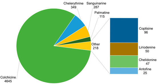
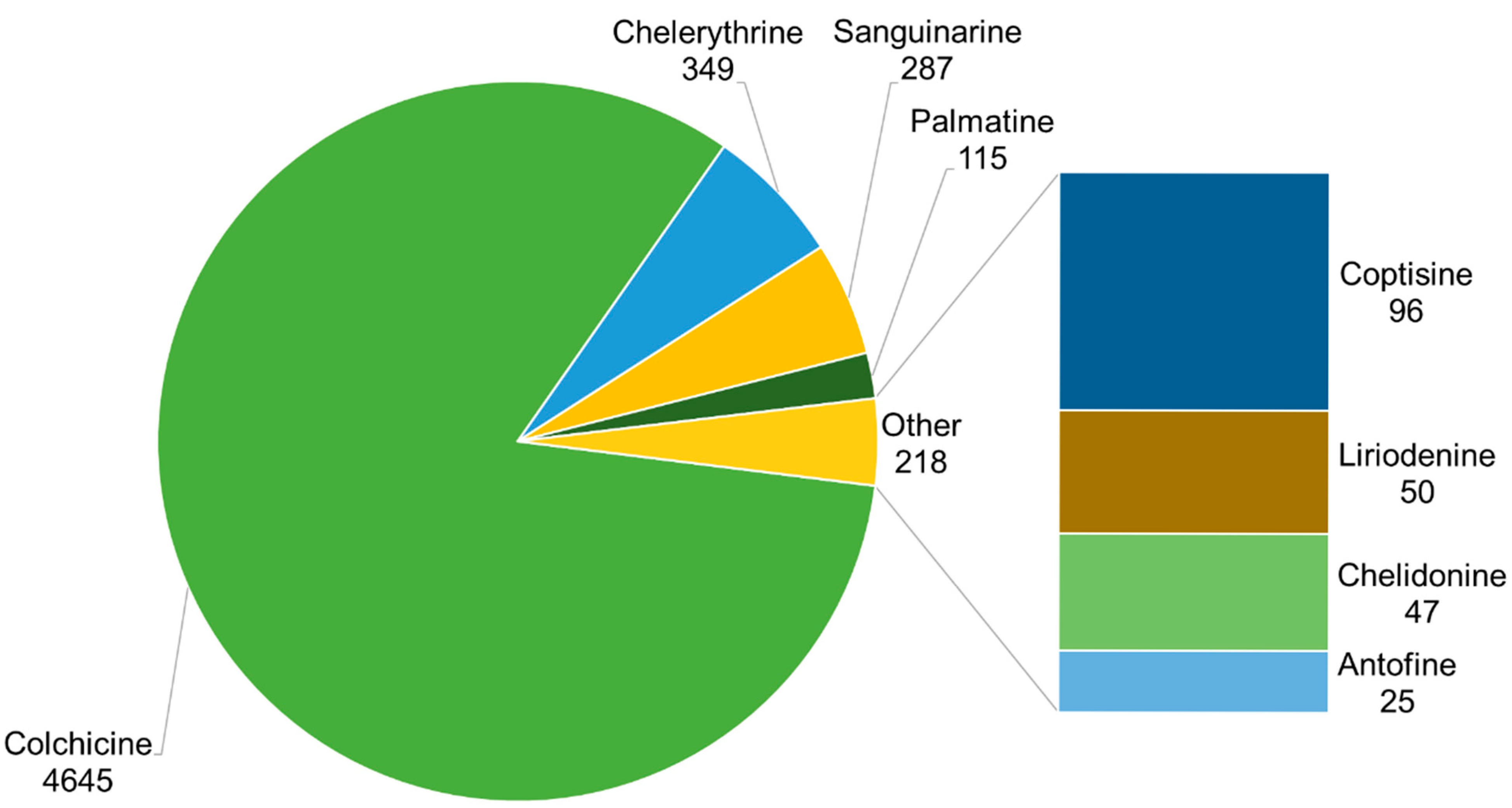
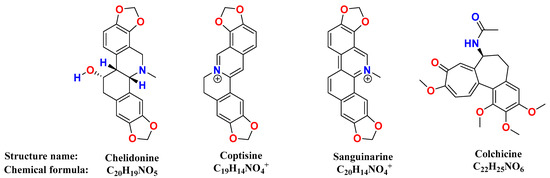
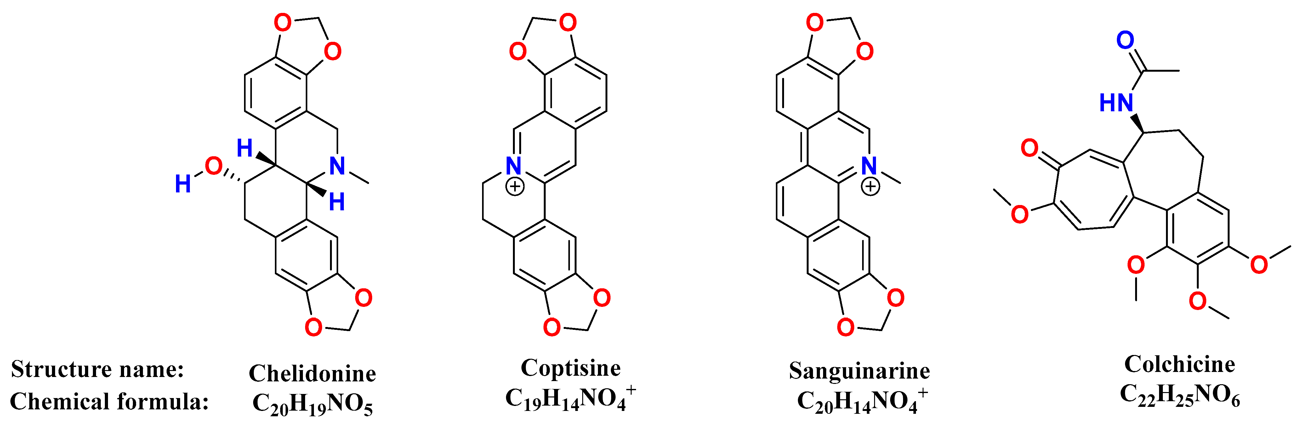
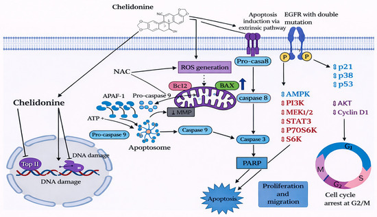
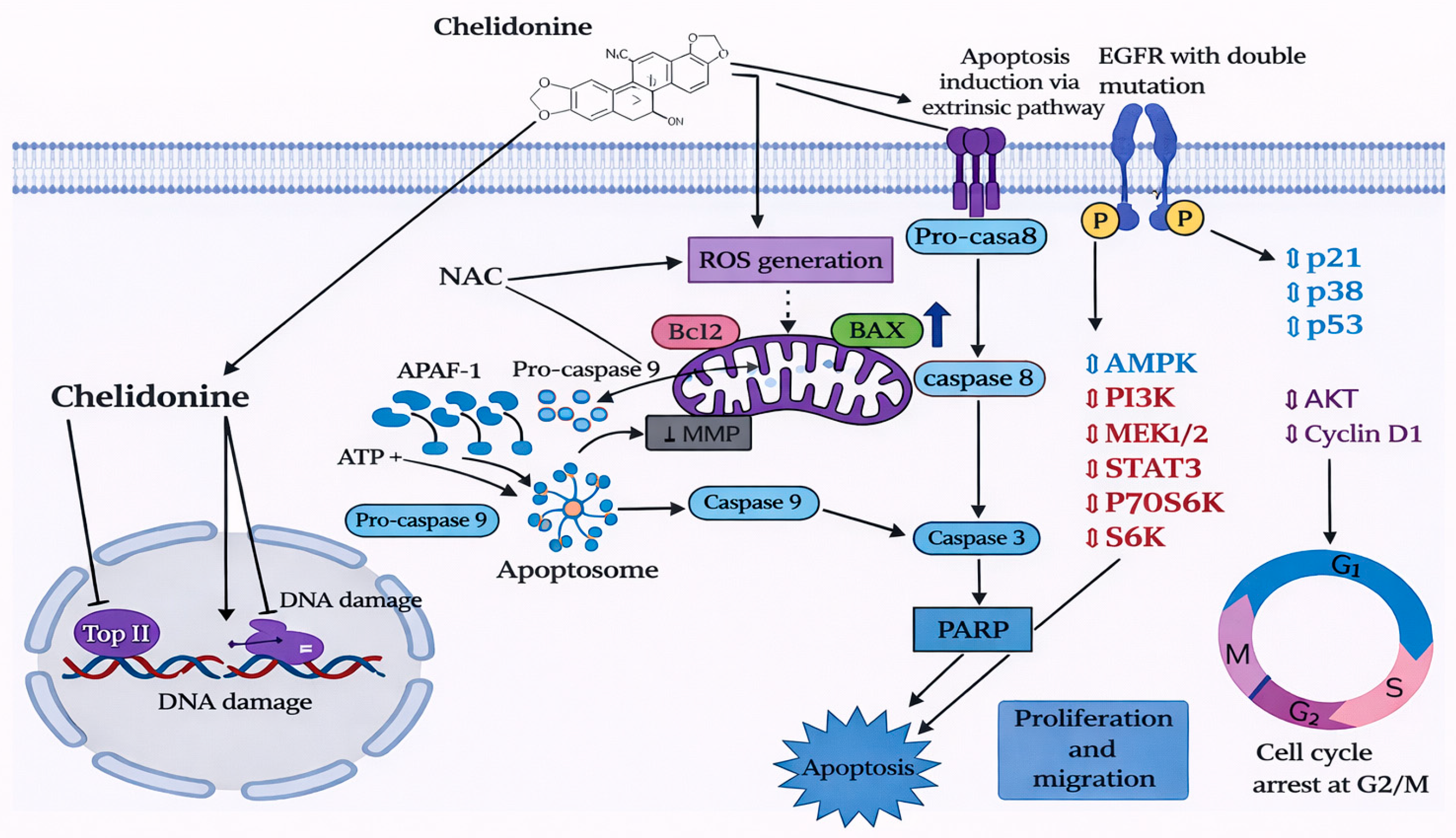
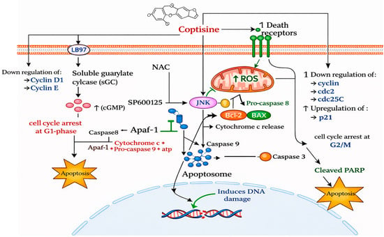
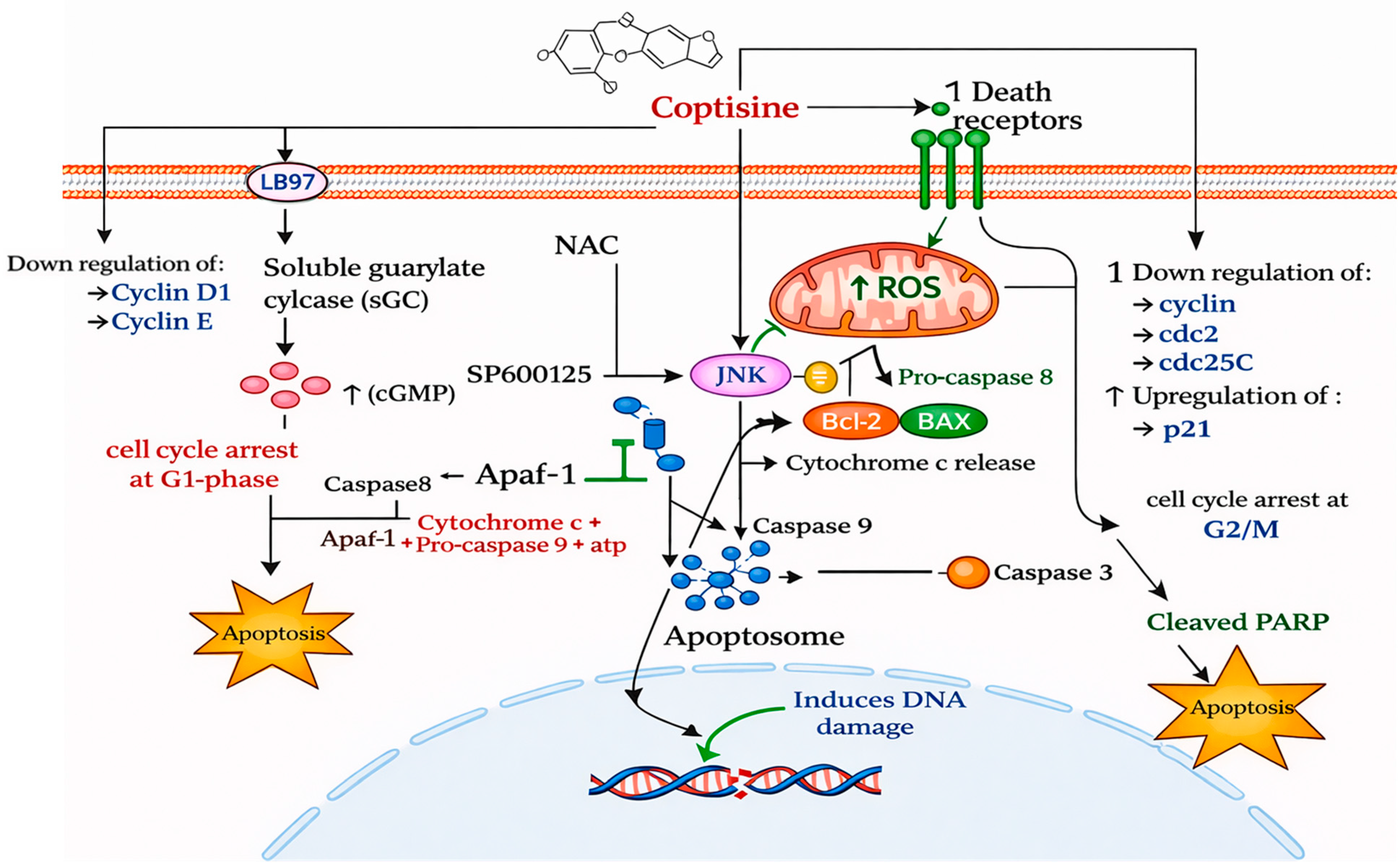
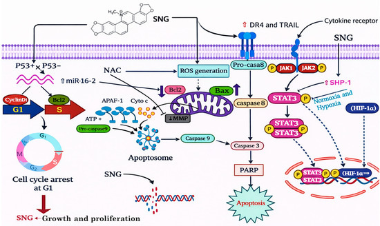
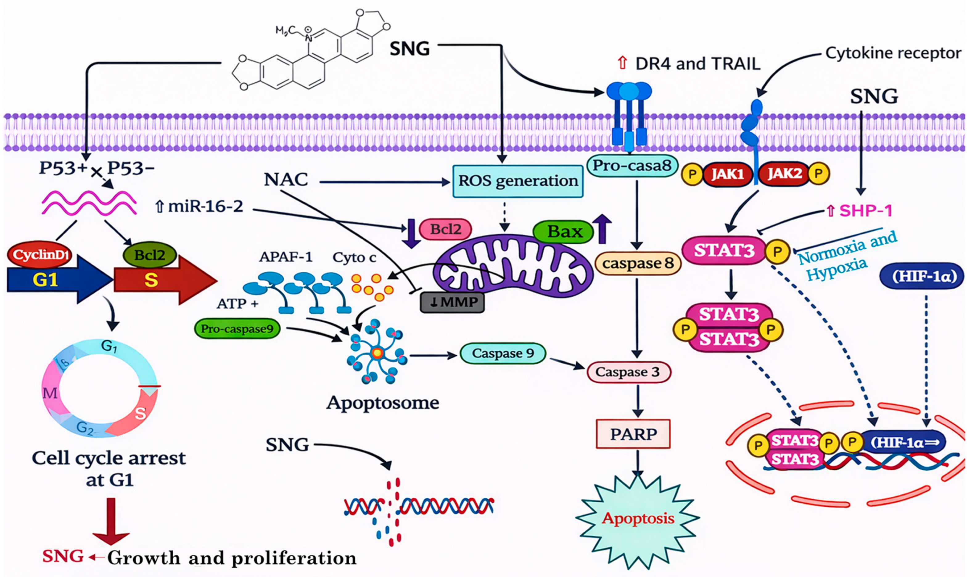
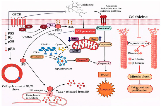
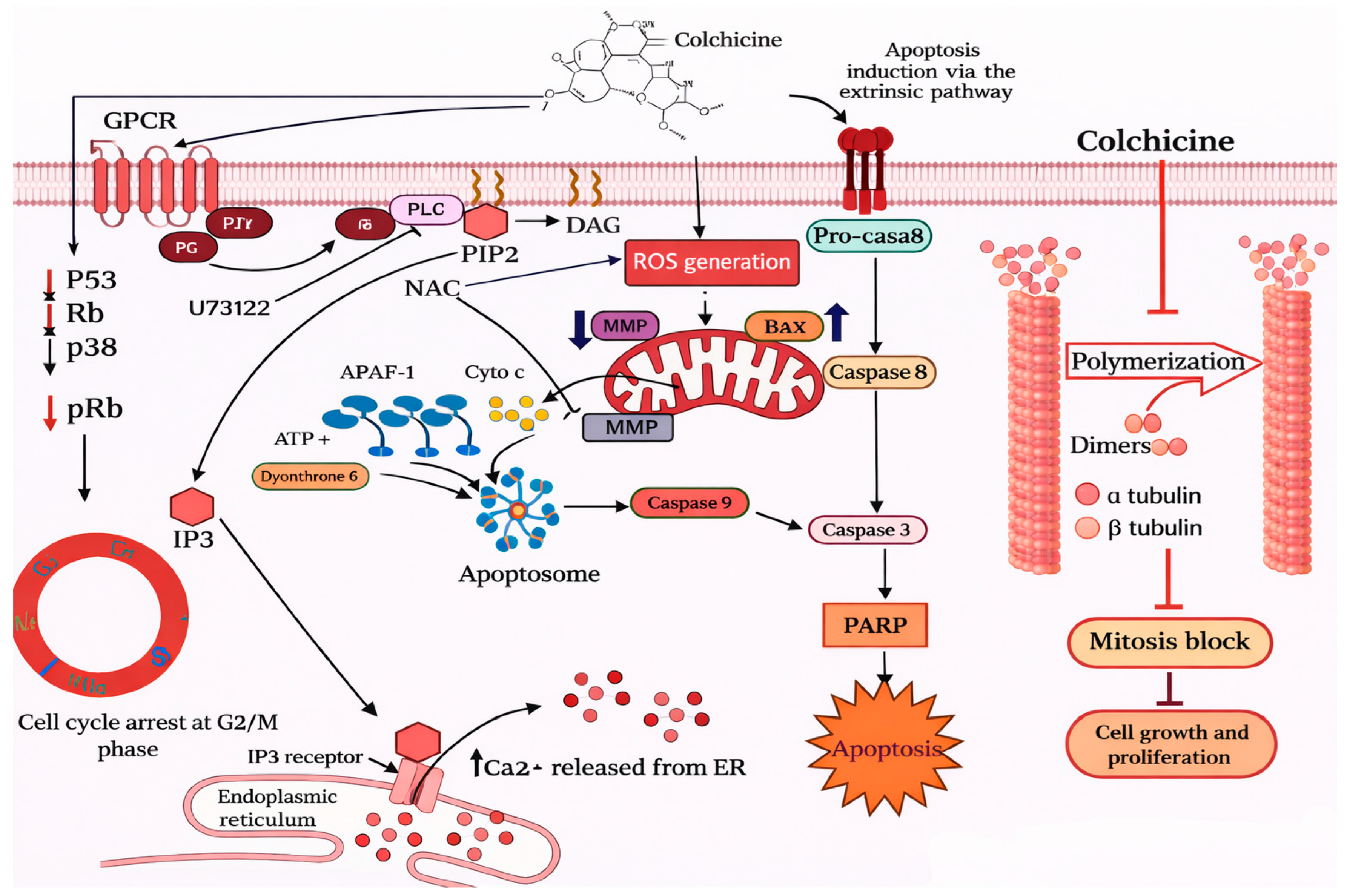
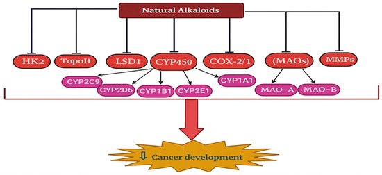
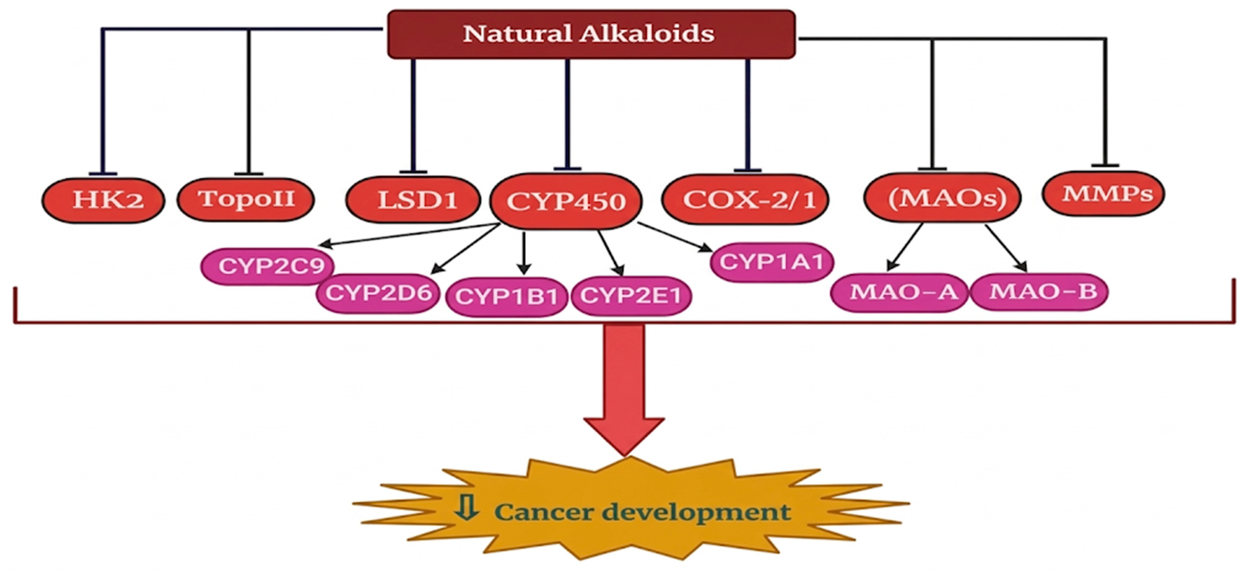
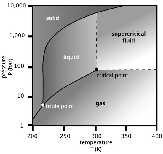
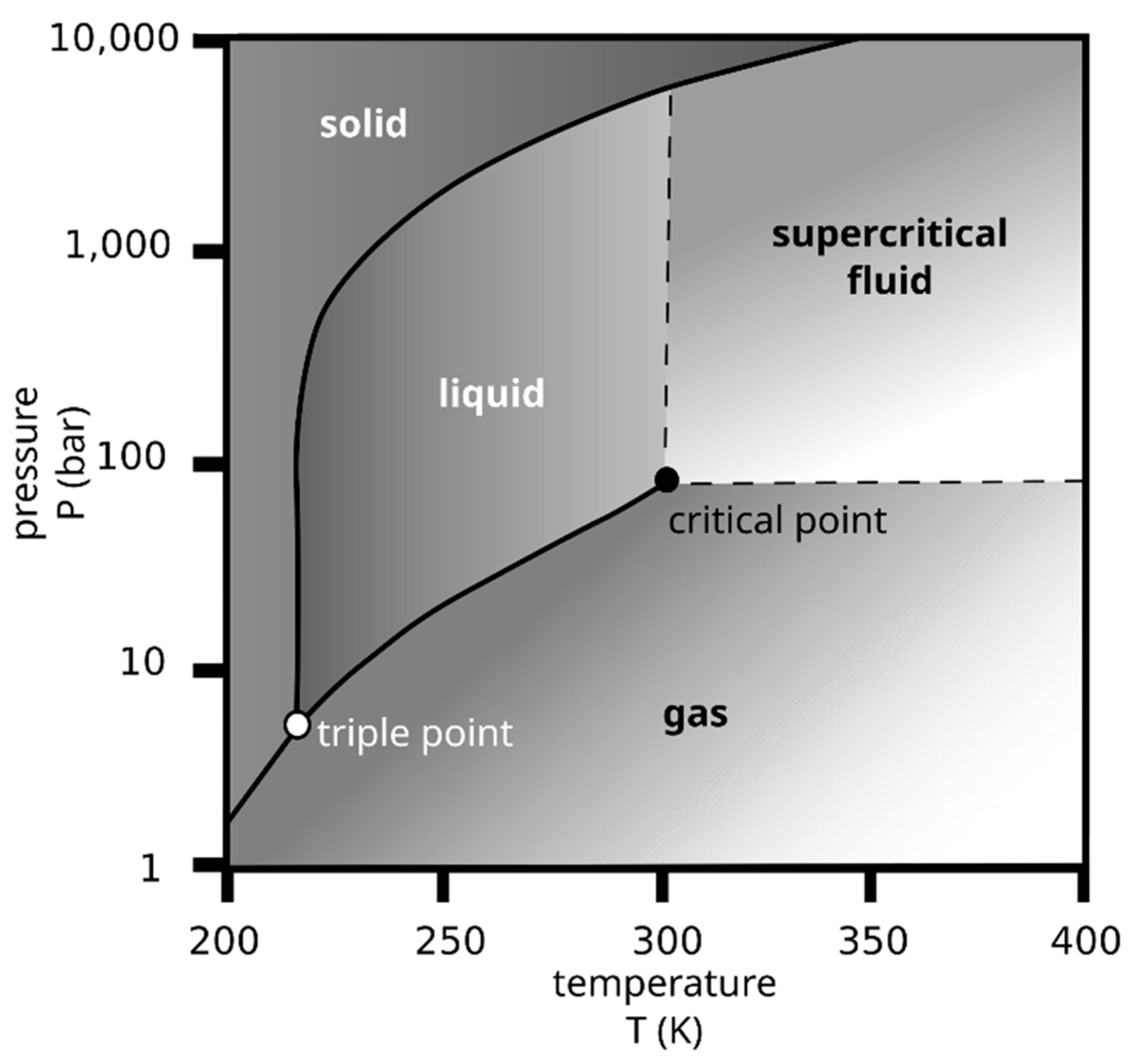
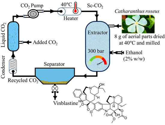
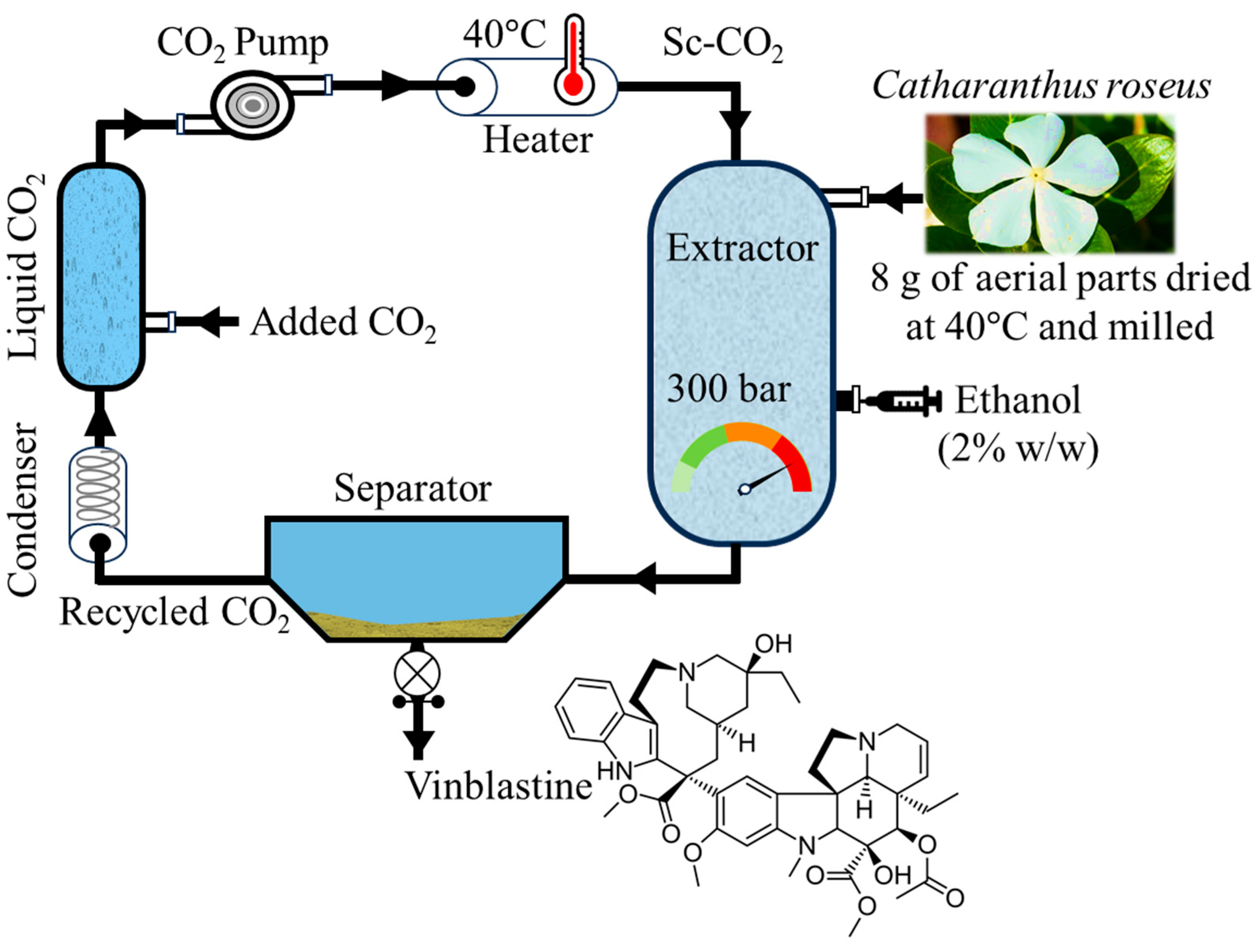
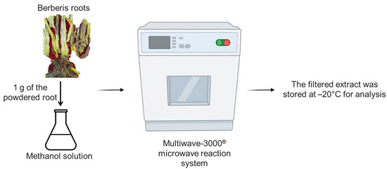
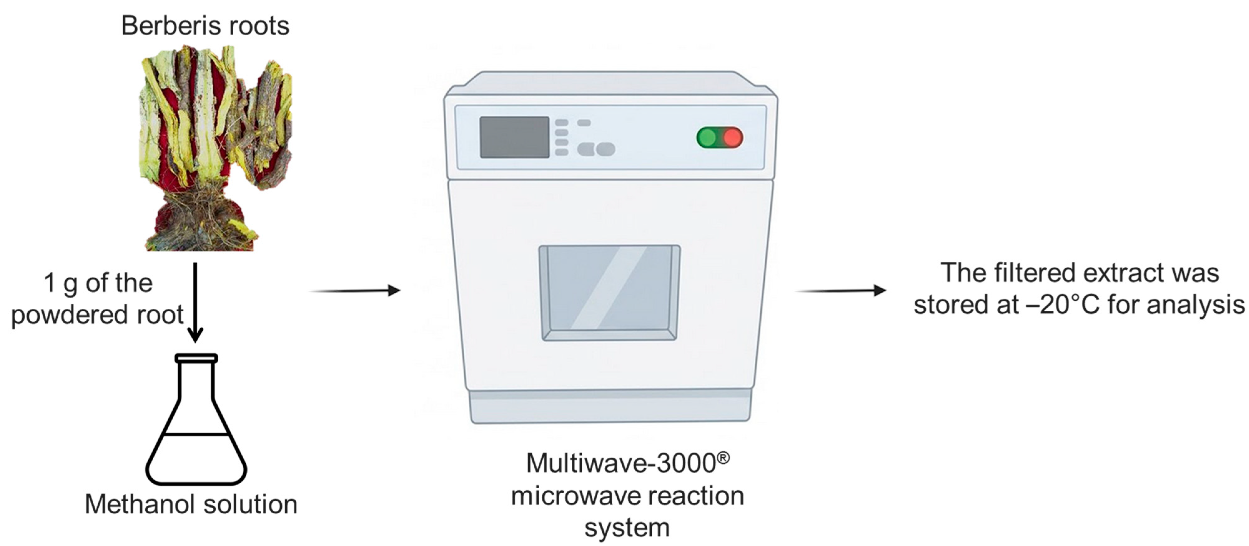
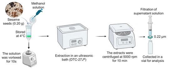
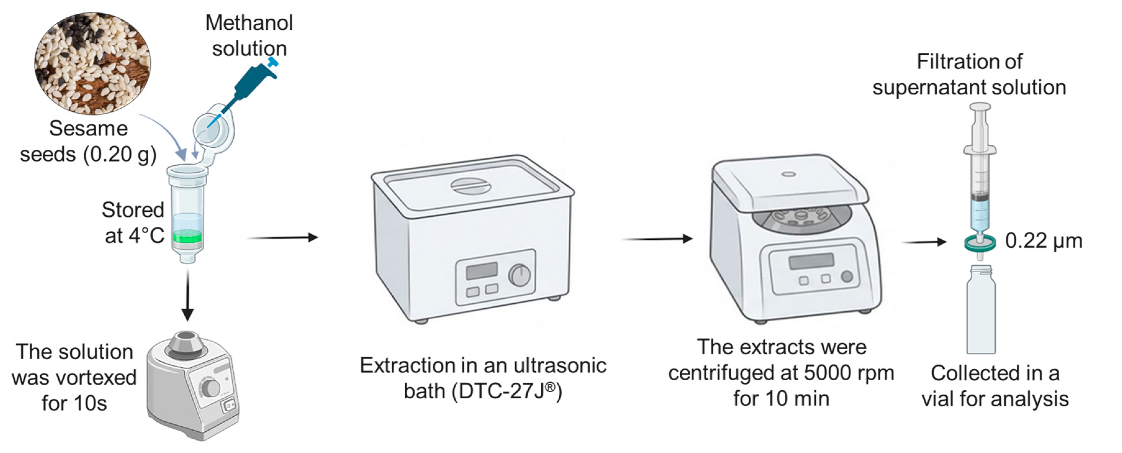
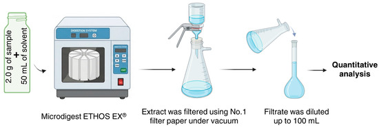
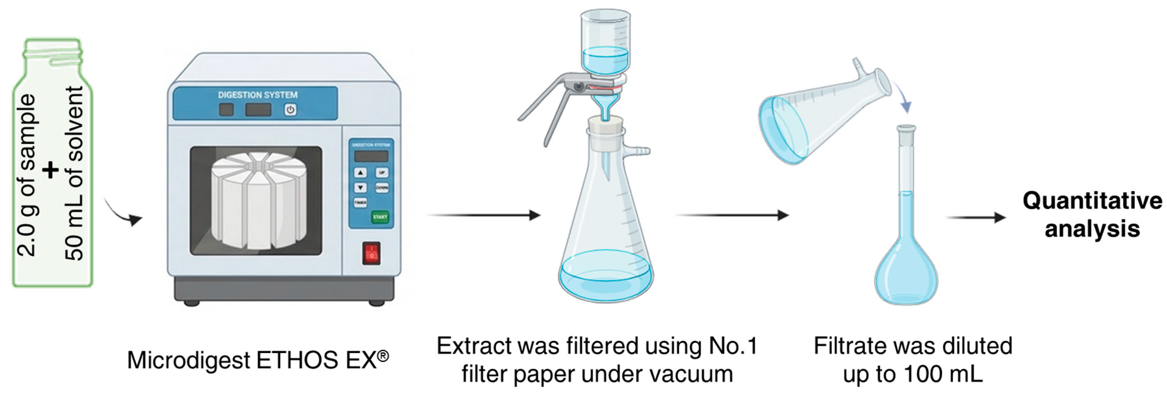
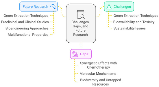
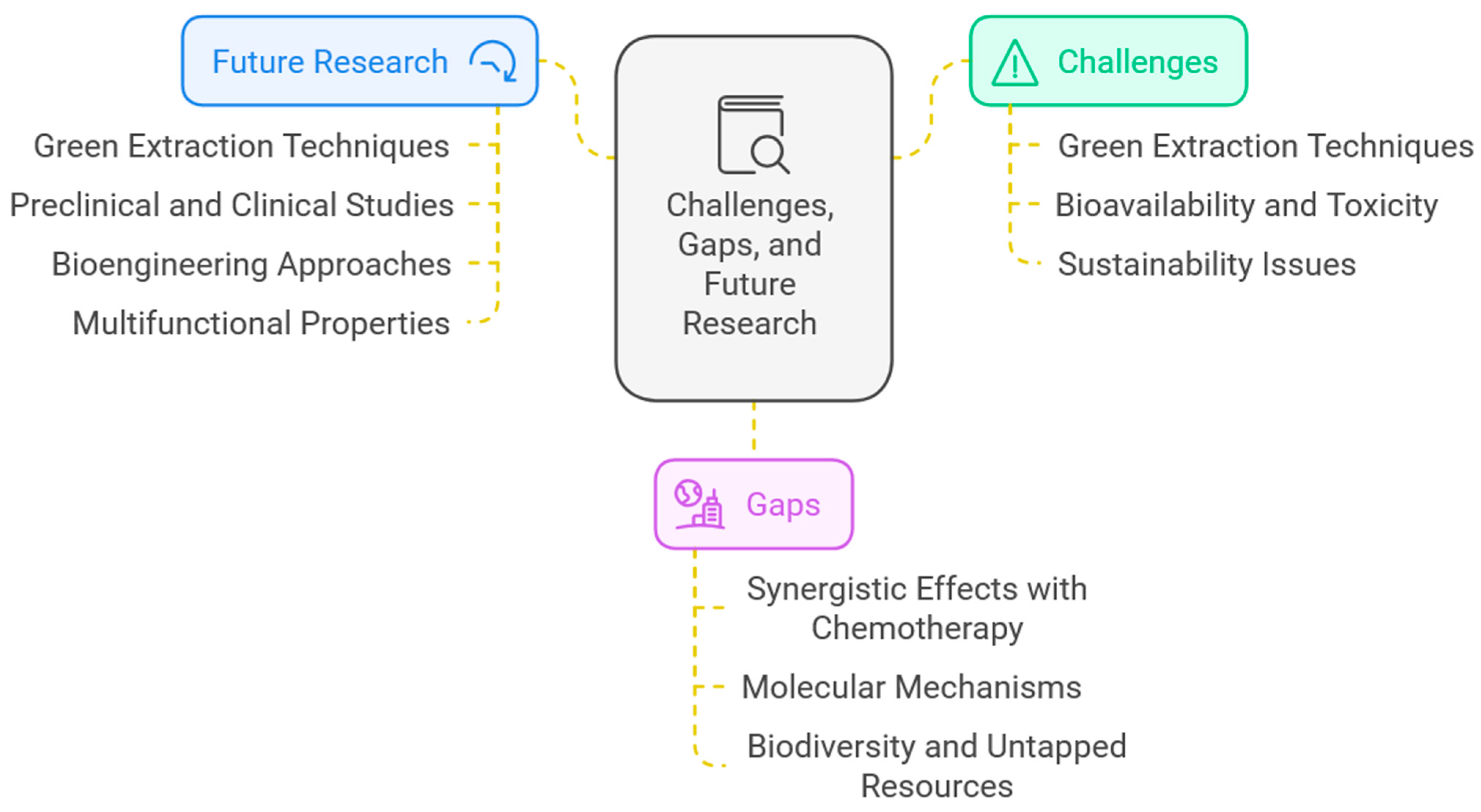

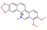
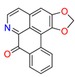
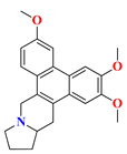
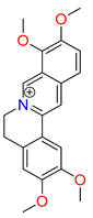
Disclaimer/Publisher’s Note: The statements, opinions and data contained in all publications are solely those of the individual author(s) and contributor(s) and not of MDPI and/or the editor(s). MDPI and/or the editor(s) disclaim responsibility for any injury to people or property resulting from any ideas, methods, instructions or products referred to in the content. |
© 2026 by the authors. Licensee MDPI, Basel, Switzerland. This article is an open access article distributed under the terms and conditions of the Creative Commons Attribution (CC BY) license.