


Simple Summary
Bats host a wide diversity of viruses, including some that may affect animal and human health. Monitoring viruses circulating in bat populations is therefore important for understanding viral diversity and identifying potential emerging pathogens. In this study, we analyzed bat samples originally collected for routine rabies surveillance using a metagenomic sequencing approach. This strategy allowed the detection and genomic characterization of a hepatitis B-like virus in the bat species Molossus rufus. The recovered viral genome showed the typical organization of viruses belonging to the Hepadnaviridae family but also displayed genetic differences compared with previously described bat viruses. These findings expand current knowledge about the diversity of hepatitis B-related viruses in bats and highlight the value of integrating metagenomic approaches into existing public health surveillance systems for the discovery of previously unknown viruses.
Abstract
Bats are recognized reservoirs for a vast array of viral diversity, including members of the Hepadnaviridae family. Within a One Health framework, genomic surveillance of these animals is fundamental to understanding viral diversity and the potential risks of zoonotic spillover in high-density human population areas. This study describes the detection of a bat hepadnavirus through agnostic viral metagenomics in samples from passive surveillance collected in urban and peri-urban areas in Brazil. Sequencing was performed using the Oxford Nanopore Technologies (MinION) platform, and the bioinformatics pipeline involved de novo assembly and taxonomic identification against viral databases. We identified several contigs with similarity to the Tent-making bat hepatitis B virus (TBHBV) in a single liver sample. The largest contig (3182 bp) represents the complete genome, exhibiting a nucleotide identity of 80.93% with the original reference isolate. Our findings document the circulation of this viral lineage in a new epidemiological setting (the Brazilian urban interface), underscoring the importance of continuous surveillance to monitor the evolution and geographic distribution of bat orthohepadnaviruses and their relevance to public health.
1. Introduction
Bats are recognized as important reservoirs of viral diversity [1]. Due to their broad ecological range, wide geographic distribution, and social behavior, these animals play a significant role in the maintenance and transmission of several viral families, including viruses with zoonotic potential [2]. Consequently, surveillance programs targeting bat populations, especially in urban settings, have become increasingly important for the early detection and characterization of such viruses [3,4].
In recent years, several hepadnaviruses have been described in bat species from different geographic regions, expanding the known host range and genetic diversity of these viruses [5,6,7,8]. In Brazil, hepadnaviruses have also been reported in bats, although genomic data remain limited [6].
Members of the family Hepadnaviridae are small enveloped DNA viruses with partially double-stranded circular genomes of 3.0 to 3.4 kb [9]. Viruses within the genus Orthohepadnavirus infect not only bats, but a wide range of mammalian hosts, including humans, primates, rodents, and cats [10,11,12,13].
In this regard, virus discovery efforts have been greatly facilitated by advances in high-throughput sequencing [14]. One strategy is the metagenomic approaches which allow the unbiased detection of viral genomes directly from biological samples and have become valuable tools for pathogen surveillance and viral discovery [15]. In the context of public health surveillance, samples collected for diagnostic purposes can also be explored using metagenomics, enabling the identification of previously unknown viruses [16]. In this study, we report the identification and genomic characterization of a potentially divergent hepadnavirus detected in a bat specimen collected in an urban setting in São Paulo, Brazil, and submitted for rabies diagnosis through passive surveillance. Using genome reconstruction, followed by comparative and phylogenetic analyses, we investigated the relationship of the identified virus with previously described members of the genus Orthohepadnavirus.
2. Materials and Methods
2.1. Sample Origin and Surveillance Context
As part of routine surveillance activities, the Laboratory of Zoonoses and Vector-Borne Diseases of the Zoonoses Surveillance Division of the municipality of São Paulo (LabZoo/DVZ/COVISA/SEABEVS/SMS) employs agnostic viral metagenomic sequencing to investigate viral diversity in wildlife specimens submitted for diagnostic purposes within a One Health framework. Therefore, 24 carcasses of bats obtained through passive surveillance and submitted to LabZoo for rabies diagnosis in the year of 2025 were subsequently repurposed for research aimed at investigating viruses of zoonotic interest.
The samples originated from different municipalities within the state of São Paulo, Brazil. Maps showing sampling sites were generated using QGIS v.3.40.0-Bratislava (QGIS.org, http://www.qgis.org, accessed on 15 March 2026).
This study was performed in compliance with the guidelines established by the Brazilian Ministry of Health’s Ordinance No. 1138 regarding the use of these specimens. According to article 3, Zoonoses Surveillance Units are authorized to: receive animal carcasses (item XI), perform laboratory diagnostics for zoonoses of public health relevance (item IV), and collect biological samples for diagnostic purposes beyond rabies (item XIV).
2.2. Nucleic Acid Extraction and Metagenomic Sequencing
Because the objective of this study was unbiased viral discovery, liver tissue was used for shotgun metagenomic analysis due to its potential to harbor viruses circulating systemically in the host. Approximately 10 mg of liver tissue from each bat was individually homogenized using L-Beader 24 (Loccus, Cotia, SP, Brazil) and subsequently filtered through a 0.22 µm syringe filter. The filtrate was treated with TURBO DNase (2 U/µL; Invitrogen, Carlsbad, CA, USA), and total nucleic acids were extracted using TRIzol® Reagent (Thermo Fisher Scientific, Carlsbad, CA, USA) according to the manufacturer’s instructions.
Metagenomic sequencing was performed using the SMART-9N protocol as described elsewhere [17]. The resulting amplicons were purified with AMPure XP beads (Beckman Coulter, Amersham, UK) at a 1:1 ratio, quantified via Qubit 3.0 fluorometry (Life Technologies, Carlsbad, CA, USA), and normalized to 65 ng per sample. Libraries were prepared using the NEBNext Ultra II End Repair/dA-tailing and Ligation modules (New England Biolabs [NEB], Ipswich, MA, USA), with multiplexing handled by the Native Barcoding Kit 24 V14 (Oxford Nanopore Technologies [ONT], Oxford, UK). Sequencing was conducted on a MinION Mk1C platform (ONT) using R10.4.1 flow cells loaded with 33 ng of the final library and operated for 72 h to maximize depth of coverage.
2.3. Genome Assembly and Consensus Reconstruction
Raw signal processing, including high-accuracy (HAC) basecalling and demultiplexing, was performed using Guppy v.6.5.7 (ONT). Following adapter, primer removal, and quality filtering via Cutadapt v4.8 (NBIS, Uppsala, Sweden) and Porechop v0.3.2 (University of Melbourne, Melbourne, Australia), genomes were reconstructed through de novo assembly with MEGAHIT v1.2.9 (The University of Hong Kong, Hong Kong, China) [18], followed by polishing with Medaka v1.11.3 (ONT) to generate high-fidelity consensus sequences.
Sequence alignment was performed against the NCBI RefSeq viral protein database (updated 10 February 2026) via DIAMOND v2.1.9 (University of Tübigen, Tübigen, Germany) [19] (blastx mode, more-sensitive setting). To ensure the accuracy of the viral identification and filter out spurious hits, all potential viral sequences underwent a secondary screening against the NCBI non-redundant (nr) protein database.
To refine the viral genome, sequencing reads were mapped back to the assembled contig and a consensus genome sequence was generated. Variant calling and consensus reconstruction were performed using BCFtools v1.17 (Genome Research Ltd., Wellcome Sanger Institute, Hinxton, UK) [20], and the resulting sequence was further repolished using Medaka v.1.11.3 to correct residual errors associated with long-read sequencing data.
The resulting repolished consensus sequence was visually inspected by mapping the filtered reads back to the genome using Minimap2 2.28-r1209 (Dana-Farber Cancer Institute, Boston, MA, USA) [21] and examining the alignment in Tablet 1.17.08.17 (James Hutton Institute, Dundee, UK) [22] to verify read support across the genome and to confirm the integrity of predicted open reading frames.
2.4. Molecular Identification of the Host Species
Host species identification was performed using mitochondrial sequences recovered from the metagenomic dataset. Nanopore reads were mapped against a dataset of bat mitochondrial gene sequences retrieved from GenBank using Minimap2 2.28-r1209 [21]. The resulting alignments were processed with SAMtools v1.18 (Genome Research Ltd., Wellcome Sanger Institute, Hinxton, UK) [23], and variant calling was performed using BCFtools v1.17 [20]. A consensus mitochondrial sequence was generated from the mapped reads. The resulting consensus sequence was compared with sequences available in the NCBI nucleotide database using BLASTn v2.17.0 (NCBI, Bethesda, MD, USA) to determine the host species identity.
2.5. Genome Annotation
Open reading frames (ORFs) were predicted and annotated based on similarity to previously described members of the genus Orthohepadnavirus through BLASTn v2.17.0 searches. The presence and organization of the canonical hepadnavirus genes, including polymerase, surface, precore/core, and X proteins, were confirmed through sequence comparison and translation analysis.
2.6. Sequence Similarity and Phylogenetic Analysis
The viral genome sequence and predicted proteins were compared to sequences available in public databases using similarity searches as well representative sequences of hepadnavirus available at International Committee on Taxonomy of Viruses (ICTV) homepage. Multiple sequence alignments were performed using MAFFT v7.505 (Osaka University, Osaka, Japan) [24]. Phylogenetic relationships between the divergent identified virus and representative members of the family Hepadnaviridae were inferred based on the amino acid sequences of the polymerase gene, using maximum-likelihood methods implemented in IQ-TREE v2.0.7 (University of Vienna, Vienna, Austria) [25].
The polymerase gene was selected due to its relatively conserved nature among members of the Orthohepadnavirus genus, enabling reliable alignment and comparison across divergent lineages. Amino acid-based analysis was performed to minimize effects of synonymous substitutions and mutational saturation.
Statistical support for tree topology was assessed using bootstrap resampling. The tree was visualized using Interactive Tree of Life (iTOL) v7.4.2 (European Molecular Biology Laboratory [EMBL], Heidelberg, Germany) [26].
2.7. Genome Organization and Sequencing Depth Visualization
The genomic organization of the recovered hepadnavirus sequence was visualized using the software Circos v0.69-8 (Canada’s Michael Smith Genome Sciences Centre, Vancouver, BC, Canada) [27]. ORFs were identified based on annotation of the assembled genome and included the polymerase (Pol), surface (S), precore/core (preC/C), and X genes. Sequencing reads were mapped to the assembled viral genome to determine coverage depth across genomic positions. The depth of coverage was calculated using SAMtools v1.18 [23], and the resulting coverage data were used to generate a circular genome plot.
2.8. Sliding Window Analysis of Nucleotide Identity
Nucleotide identity between the recovered viral genome and the closest related sequence, Tent-making bat hepatitis B virus (TBHBV—Accession Number: KC790381.1), was evaluated using a sliding window approach implemented in R v4.4.0 (R Foundation for Statistical Computing, Vienna, Austria) within the RStudio environment (v2025.05.1, Posit Software, PBC, Boston, MA, USA). The aligned nucleotide sequences were imported from a FASTA alignment file produced after MAFFT v7.505 [24]. Sequence identity was calculated across the genome using a window size of 200 nucleotides and a step size of 20 nucleotides. For each window, the percentage of identical nucleotides between the two sequences was computed. The resulting identity values were plotted against genomic position using the ggplot2 package v4.0.2 to generate a line graph representing sequence similarity across the genome. Annotated ORF positions were overlaid on the plot to indicate the genomic locations of the polymerase, surface, precore/core, and X genes.
2.9. Pairwise Nucleotide Identity Analysis
Pairwise nucleotide identities among complete viral genomes were calculated using global pairwise alignments implemented in Python v3.10 (Python Software Foundation, Wilmington, DE, USA) with the BioPython library v.1.86 (pairwise2 module). For each pair of sequences, nucleotide identity was calculated as the proportion of identical positions in the alignment relative to the alignment length. Identity values were compiled into a similarity matrix and visualized as a heatmap using the Python libraries pandas v2.2.3, seaborn v0.13.2, and matplotlib v3.10.0.
3. Results
3.1. Geographic Distribution of Sampling Sites
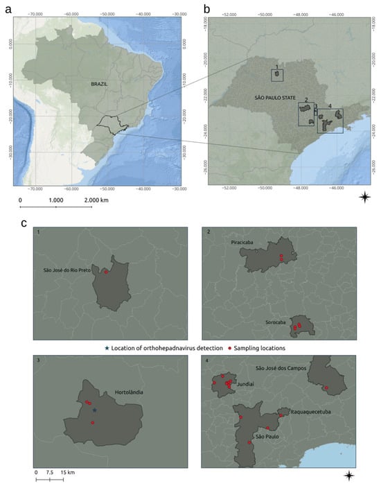
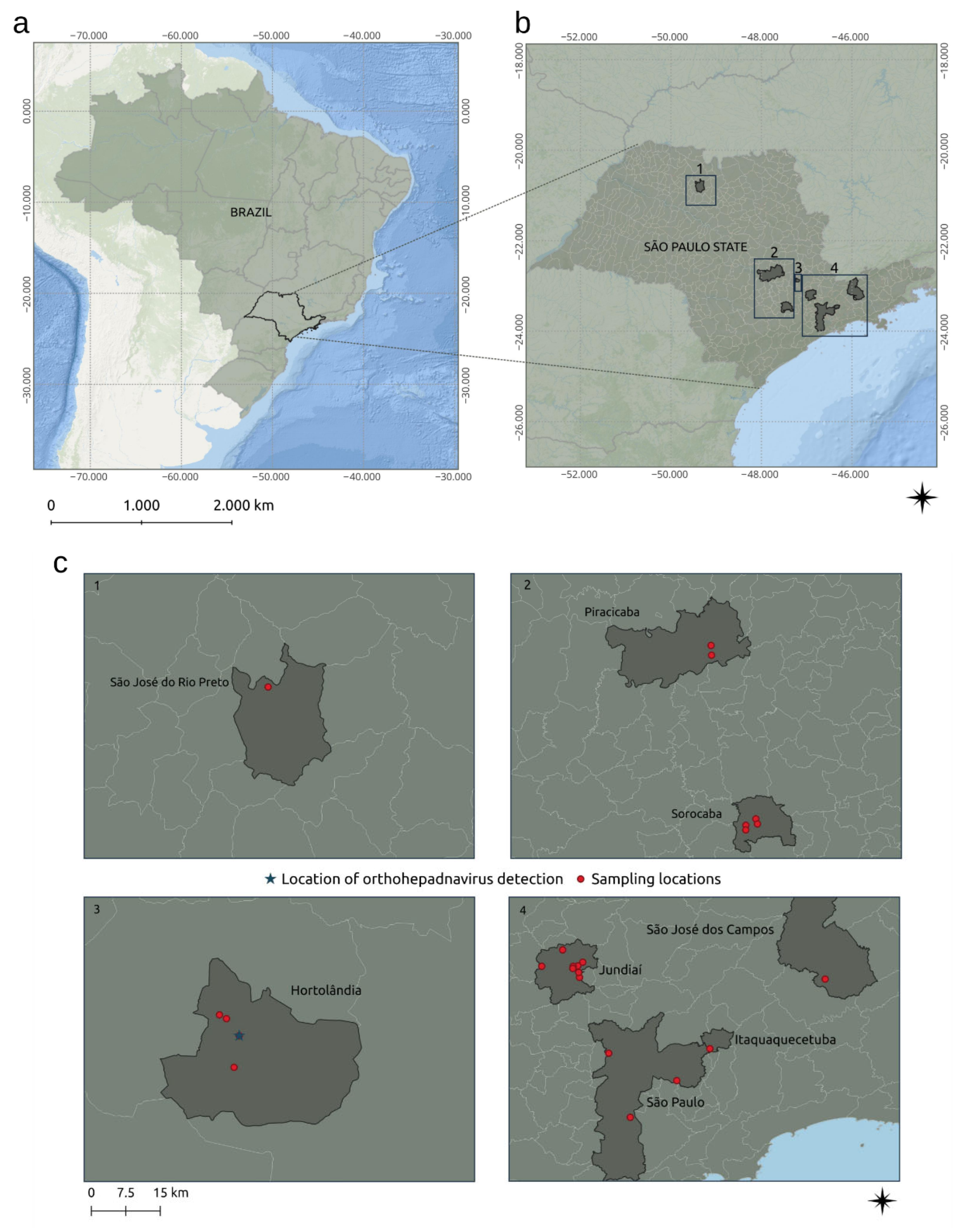
A total of 24 bat carcasses were obtained through passive surveillance from different municipalities within the state of São Paulo, Brazil. The spatial distribution of sampling sites is shown in Figure 1.
Figure 1.
Geographic distribution of bat sampling sites in Brazil. (a) Location of the study area within Brazil, highlighting the state of São Paulo. (b) Distribution of sampling municipalities within the state. Numbered boxes (1–4) indicate the specific regions shown in higher magnification in panel (c). (c) Detailed view of sampling sites across municipalities, with red dots indicating collection points and the blue star representing the location where the orthohepadnavirus was detected.
3.2. General Results
A metagenomic library was generated from the 24 bat samples. All specimens tested negative for Rabies virus detection using the Direct Fluorescence Antibody Test (DFA), the gold standard for post-mortem laboratory diagnosis of rabies [28].
Metagenomic sequencing generated 404,056 raw reads, of which 380,600 were retained after primer and adapter trimming as well as quality filtering. The filtered reads had a mean length of 417 bp and an N50 length of 450 bp (Supplementary Table S1).
Viral sequences related to the family Hepadnaviridae were identified in one sample derived from the liver of a bat originated from an urban area of Sao Paulo State, Brazil (Sample ID 3848/25—Supplementary Table S2).
3.3. Host Species Identification
A mitochondrial 12S rRNA fragment was successfully reconstructed from the metagenomic reads. The consensus sequence showed complete coverage across the reference fragment and, when compared with the NCBI nucleotide database using BLASTn, exhibited 100% nucleotide identity and 100% query coverage with sequences of Molossus rufus.
3.4. Genome Characterization of the Bat Hepadnavirus
The assembled viral genome, named Molossus rufus bat hepadnavirus (MRBHV), was 3182 nucleotides (nt) in length with a GC content of 46.86%. Mapping of sequencing reads to the reconstructed genome identified 16,300 reads, corresponding to approximately 4.3% of the dataset. These reads provided 100% genome coverage with an average sequencing depth of 3135×, supporting the reconstruction of a complete viral genome (Supplementary Table S1).
Sequencing reads were mapped across the entire genome, with a minimum depth of 29× and a maximum depth of 7138×, indicating complete genome recovery without uncovered regions (Supplementary Figure S1).
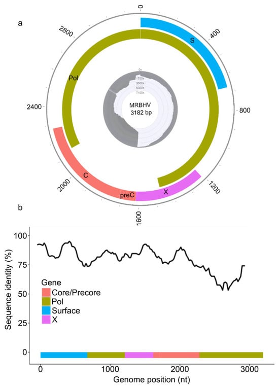
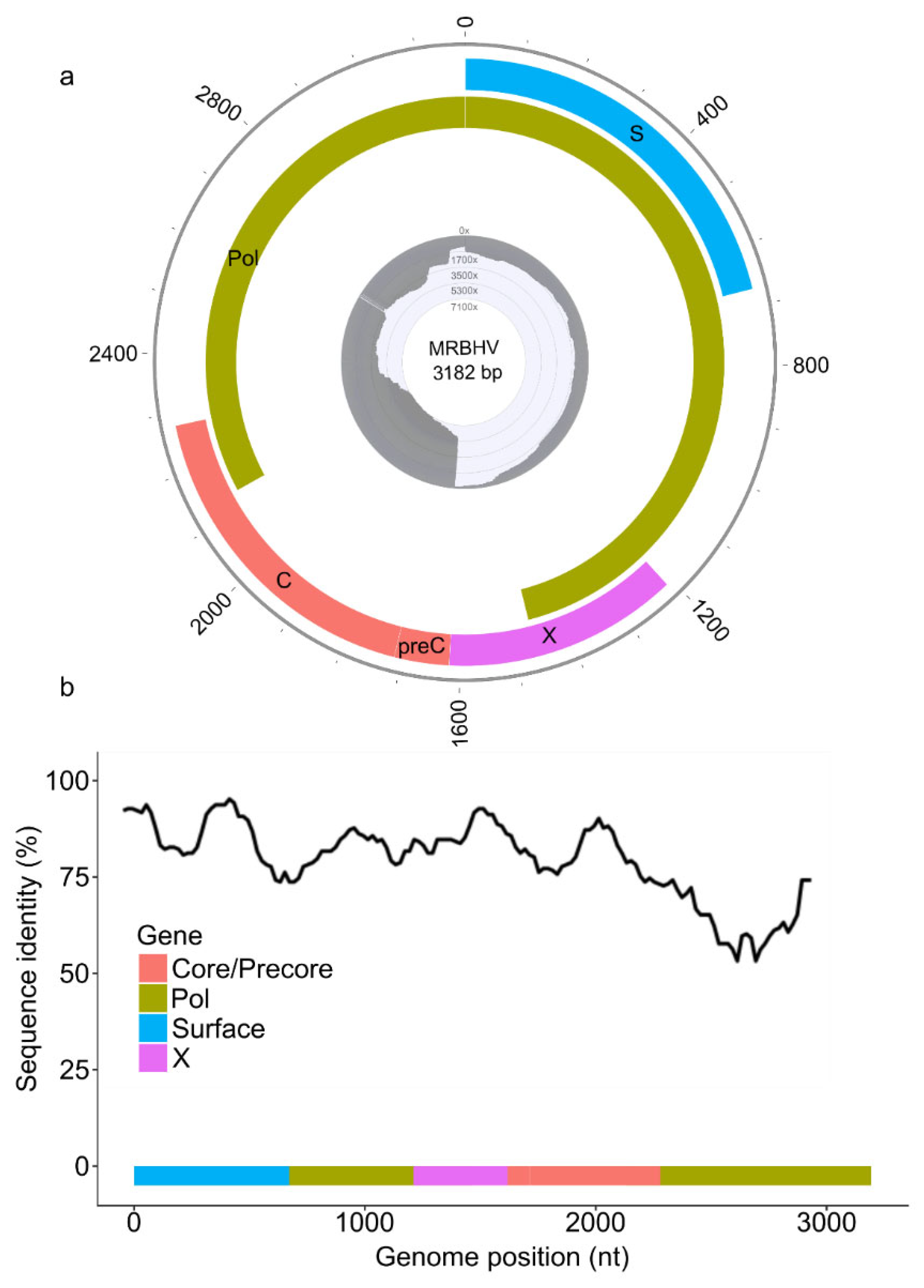
Genome annotation revealed the typical hepadnavirus genomic organization, including the polymerase (Pol), surface (S), precore/core (preC/C), and X ORFS. The polymerase ORF spanned nucleotides 2132–3182 and 1–1466 and encoded a predicted protein of 838 amino acids (aa). The surface gene was located between nucleotides 1–672 and encoded a protein of 223 aa. The core ORF was located between nucleotides 1714–2280 and encoded a predicted capsid protein of 188 aa. An upstream region preceding the core gene (nt 1618–1713) was identified and likely represents a precore region encoding a 32 aa peptide. The X gene spanned nucleotides 1211–1618 and encoded a predicted protein of 135 aa (Figure 2a).
Figure 2.
Genome organization and sequencing coverage of the bat hepadnavirus identified in this study (MRBHV). (a) Genome organization of the divergent bat hepadnavirus MRBHV showing the four overlapping open reading frames encoding the polymerase (Pol), surface (S), precore/core (preC/C), and X proteins. Gray histogram indicates sequencing coverage depth obtained from metagenomic reads. Complete genome of the bat hepadnavirus 3182 kb. (b) Sliding window analysis of nucleotide identity between MRBHV genome and the closest related virus (TBHBV—Accession Number: KC790381.1). Colors indicate the genomic regions corresponding to each gene.
Comparative analysis using BLASTn revealed that the genome shared the highest similarity with TBHBV, showing 80.93% nucleotide identity with 99% query coverage and an E-value of 0.0 (Supplementary Table S3).
3.5. Genomic Similarity Analysis
Sliding window analysis of nucleotide identity between the MRBHV genome and the TBHBV revealed identity values ranging from approximately 55% to 95% across the genome. The identity plot revealed heterogeneous similarity, with higher nucleotide identity observed in the surface gene and parts of the polymerase region. The lowest similarity was detected toward the terminal portion of the polymerase gene, indicating increased divergence in this region (Figure 2b).
3.6. Phylogenetic Analysis
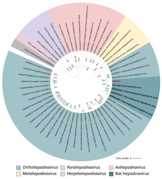
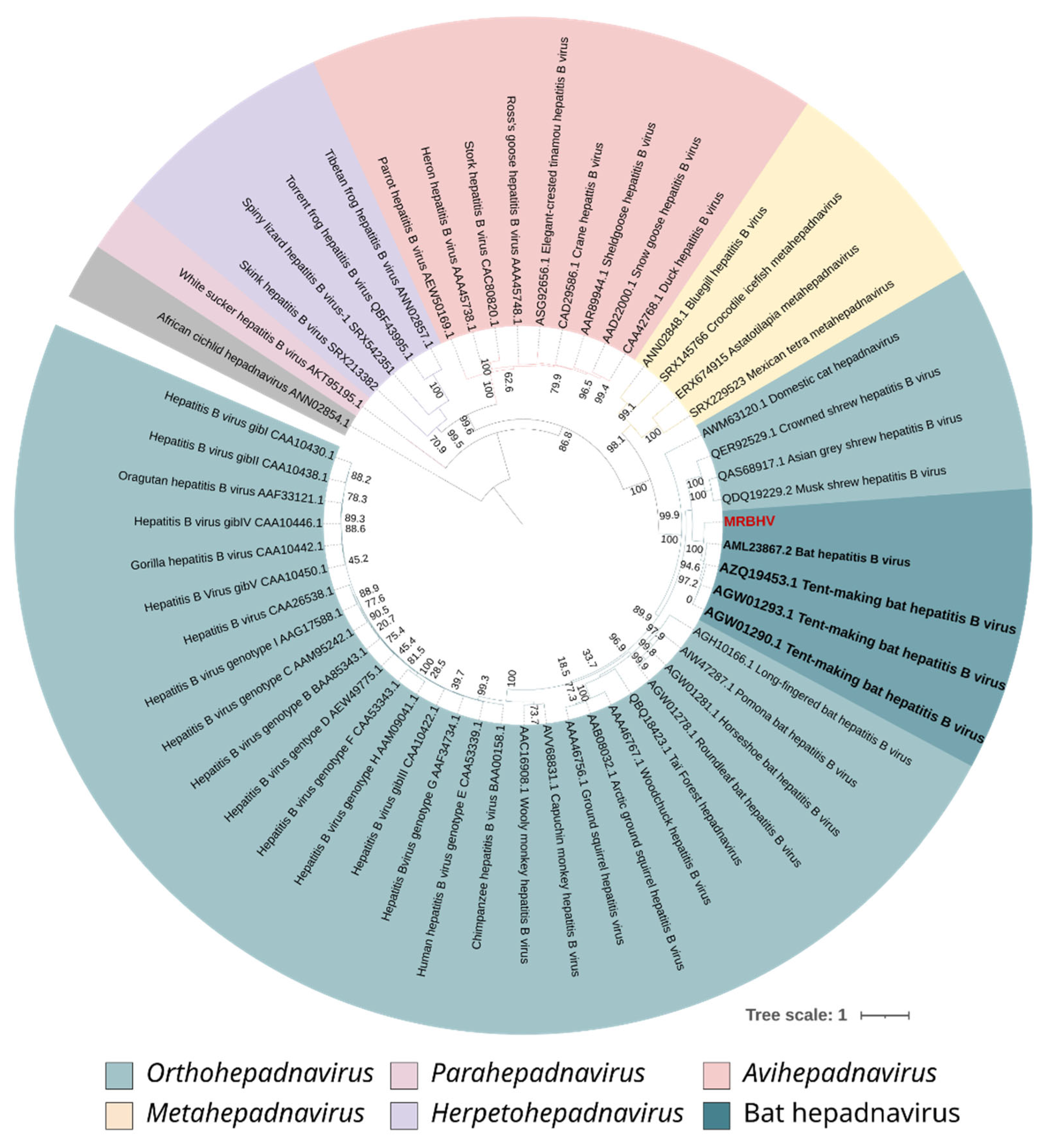
Phylogenetic reconstruction based on the amino acid sequence of the polymerase protein was performed using the maximum likelihood method under the Q.pfam+F+R5 evolutionary model (Supplementary Table S4; Supplementary Data S1). The resulting tree showed that the MRBHV clustered within the genus Orthohepadnavirus and within the clade of bat-assocaited hepadnaviruses, forming a well-supported branch with an ultrafast bootstrap value of 100. Within this clade, MRBHV was positioned as a distinct lineage, sister to a subclade including Tent-making bat hepatitis B virus sequences and the previously reported Brazilian sequence (AML23867.2)
These results indicate that the virus identified in this study likely represents a distinct lineage within the bat-associated hepadnaviruses, with strong statistical support (Figure 3).
Figure 3.
Phylogenetic analysis of the bat hepadnavirus identified in this study (MRBHV) based on polymerase amino acid sequences. The phylogenetic tree was reconstructed using representative polymerase amino acid sequences of the Hepadnaviridae family. The sequence identified in this study (MRBHV) is highlighted in red. Bat hepadnaviruses are highlighted in bold. Node values represent bootstrap support percentages. The tree was rooted using sequence African cichlid hepadnavirus ANN02854.1. The scale bar indicates the number of nucleotide substitutions per site.
3.7. -Genomic Identity Comparisons
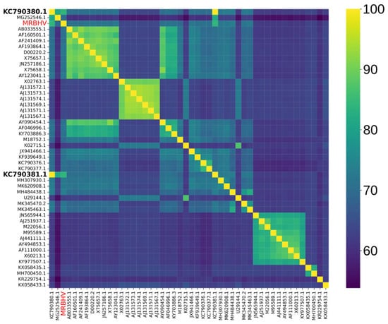
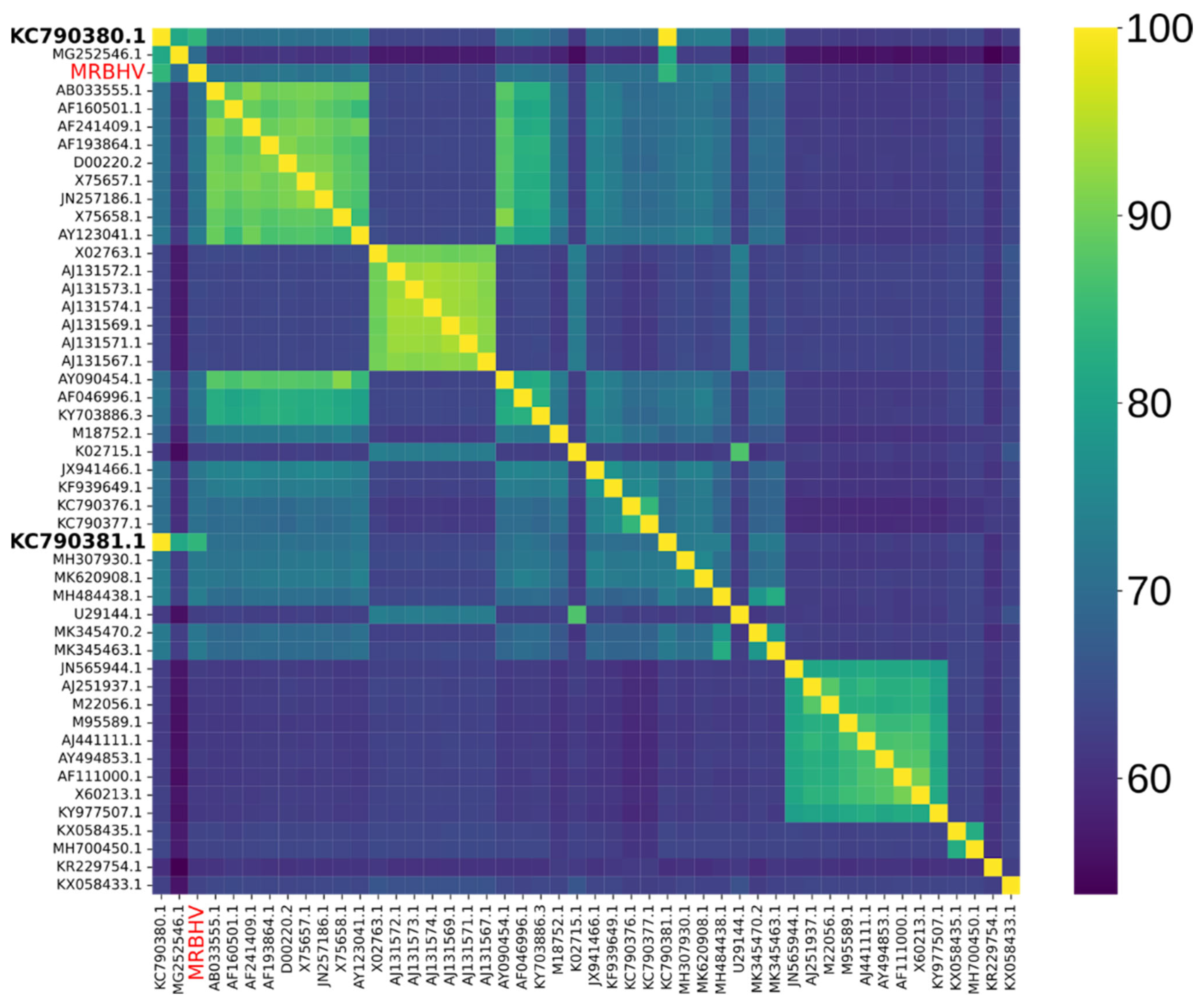
Pairwise nucleotide identity analysis based on complete genome sequences showed that the virus identified in this study shares the highest similarity with previously reported bat hepadnaviruses (TBHBV—GenBank Accession Numbers KC790380.1 and KC790381.1) while lower identity values were observed when compared with more distantly related hepadnaviruses. This pattern is consistent with the phylogenetic results and further supports that the detected virus represents a potentially distinct lineage within bat-associated hepadnaviruses (Figure 4).
Figure 4.
Pairwise nucleotide identity among complete genomes of representative hepadnaviruses. Heatmap showing pairwise nucleotide sequence identity calculated from complete genome sequences of representative members of the family Hepadnaviridae. The virus identified in this study (MRBHV) is highlighted in red on both axes. Sequences showing the highest similarity are in bold. Colors represent the percentage of nucleotide identity, ranging from low identity (purple/blue) to high identity (yellow). Clusters of closely related viruses are visible as blocks of higher similarity along the diagonal.
4. Discussion
In this study, we report the detection and genomic characterization of a hepadnavirus identified in liver tissue from a Molossus rufus bat. The recovered genome displayed the canonical genomic organization typical of members of the family Hepadnaviridae, including the polymerase, surface, precore/core, and X open reading frames arranged in an overlapping configuration [9]. The genome length (3182 nt) is consistent with those reported for other bat-associated hepadnaviruses [29].
Comparative sequence analysis revealed that the virus detected in this study is most closely related to Tent-making bat hepatitis B virus, sharing approximately 80.9% nucleotide identity across the genome The observed nucleotide identity indicates a considerable level of genetic divergence. Similar levels of divergence have been reported among distinct lineages of bat-associated hepadnaviruses, reflecting the substantial evolutionary diversity of this viral group. The genomic differences observed here may represent host-specific evolutionary adaptation or long-term diversification of hepadnaviruses detected in bat populations [8,30]. On that matter, the virus identified in this study is conservatively interpreted as a potentially divergent lineage as the observed nucleotide identity lies near proposed species demarcation thresholds for members of the Orthohepadnavirus genus, according to ICTV criteria.
With respect to the phylogenetic analysis, it was based on amino acid sequences of the polymerase gene, a relatively conserved region among Orthohepadnavirus members [31]. While this approach enables robust comparisons across divergent viruses and reduces the impact of synonymous substitutions [32,33], it may limit phylogenetic resolution at finer evolutionary scales, particularly among closely related sequences [34]. Therefore, caution is warranted when interpreting subtle evolutionary relationships.
Nevertheless, this analysis revealed a clade of bat-associated hepadnaviruses. A previously reported bat-associated hepadnavirus partial polymerase sequence from Brazil (AML23867.2), identified in a Platyrrhinus lineatus bat from the state of São Paulo [6], was included in the analysis. This sequence clustered closely with Tent-making bat hepatitis B virus, sharing a more recent common ancestor, whereas MRBHV formed a distinct and more divergent lineage within the bat-associated hepadnaviruses. Notably, Tent-making bat hepatitis B virus has been previously detected in bats belonging to the family Phyllostomidae [29], whereas the virus described here was identified in a member of the Molossidae family.
Despite the limitations imposed by the partial nature of the available Brazil sequence, this comparison highlights both similarities and divergences among hepadnaviruses circulating in this country. From an evolutionary perspective, these findings suggest the co-circulation of multiple genetically distinct lineages. From a host perspective, the detection of related but distinct viruses in bats from different families (Molossidae and Phyllostomidae) supports the notion that hepadnaviruses may infect a broad range of bat hosts [5]. Furthermore, the fact that both viruses were detected in the same geographic region (São Paulo state) indicates that this diversity is not only host-associated but also occurs locally, emphasizing the complexity of hepadnaviruses circulation in bat populations [8].
The high bootstrap support observed for the bat-associated hepadnavirus clade indicates the existence of a robust evolutionary relationship between these viruses. Nevertheless, sliding window analysis demonstrated heterogeneous levels of nucleotide identity across the genome, with values ranging from approximately 55% to 95%, indicating the presence of both conserved and more divergent genomic regions. This pattern is consistent with the evolutionary dynamics commonly observed in hepadnaviruses, whose compact genomes and extensive gene overlap impose distinct selective pressures across coding regions [35].
Genome annotation revealed a region upstream of the core gene consistent with a predicted precore sequence. In orthohepadnaviruses, the precore region encodes the precursor of the secreted hepatitis B e antigen (HBeAg), a protein involved in modulation of host immune responses [36]. Although functional characterization was not performed in the present study, the presence of this genomic feature further supports the structural similarity between the virus identified here and other members of the Hepadnaviridae.
Regarding the host, the detection of a potentially divergent hepadnavirus in a Molossus rufus bat expands the known host range of bat-associated hepadnaviruses. Given that bats harbor a diverse range of viruses within the family Hepadnaviridae, this finding further contributes to the understanding of the diversity and evolutionary history of this viral group [29,30].
The genus Molossus comprises insectivorous bats widely distributed throughout the Americas [37]. Molossus rufus, in particular, has been reported to inhabit urban environments in Brazil [38]. Noteworthy, the bats analyzed in this study came from urban areas where they frequently occupy buildings and other human-modified structures. This ecological context places these animals as synanthropic fauna as they are in close proximity to human populations and domestic animals, increasing opportunities for viral detection at the human–animal interface [39].
Despite these findings, the zoonotic potential of bat-associated hepadnaviruses remains incompletely understood. Previous studies have demonstrated that certain bat hepadnaviruses are antigenically related to human hepatitis B virus and may share functional similarities with members of the genus Orthohepadnavirus [29]. However, there is currently no evidence that bat hepadnaviruses are capable of infecting humans under natural conditions. The discovery of genetically diverse hepadnaviruses in bats nonetheless highlights the importance of continued surveillance, particularly in synanthropic species that frequently occur in close proximity to human populations. Monitoring the diversity and evolution of these viruses as well as others may contribute to a better understanding of their host range and potential for cross-species transmission [5,29] especially in urban settings [40,41].
Importantly, the virus described in this study was identified through a metagenomic surveillance strategy applied to samples obtained from passive surveillance activities. The analyzed bats were originally submitted to the public health diagnostic network for rabies testing and subsequently repurposed for viral discovery using shotgun metagenomics. This approach highlights the value of integrating research-oriented viral surveillance with existing public health diagnostic workflows [42]. Passive surveillance programs, particularly those focused on rabies diagnostics, routinely receive wildlife specimens and therefore represent an important and wasted resource for the detection of additional viral agents circulating in animal populations. In this context, the application of shotgun metagenomics within public health surveillance frameworks represents a powerful tool for the detection of potentially zoonotic viruses before spillover events occur [43,44].
Although this study provides important insights, it has several limitations that should be considered when interpreting the results. First, molecular confirmation using PCR or Sanger sequencing was not performed. Although such approaches would further strengthen the findings, the viral genome was reconstructed from high-throughput metagenomic sequencing data with consistent coverage and sufficient read depth, supporting the reliability of the assembled sequence. In addition, the use of carcass-derived samples may have limited the availability and integrity of nucleic acids for further targeted amplification. Moreover, this study has an exploratory nature. Nevertheless, future studies incorporating targeted validation approaches would be valuable to further corroborate these findings. Second, the virus was detected in a single bat specimen, which precluded any inference regarding its prevalence or distribution in bat populations. These findings should be interpreted as preliminary evidence, highlighting the need for broader surveillance studies. Furthermore, only liver tissue was analyzed, preventing evaluation of viral tissue tropism or systemic infection. Finally, the use of random amplification during metagenomic library preparation may introduce biases in genome representation. Future studies including targeted screening of additional bat samples and molecular confirmation approaches will be important to better understand the distribution, host range, and evolutionary diversity of this virus.
Taken together, the identification and genomic characterization of the Molossus rufus bat hepadnavirus underscores the importance of integrating wildlife surveillance, diagnostic infrastructure, and metagenomic technologies to improve our understanding of viral diversity and to strengthen preparedness for emerging zoonotic threats.
Supplementary Materials
The following supporting information can be downloaded at: https://www.mdpi.com/article/10.3390/zoonoticdis6020015/s1, Figure S1: Sequencing depth across the complete viral genome of MRBHV and distribution of coverage values; Table S1: Sequencing statistics; Table S2: Samples analyzed; Table S3: BLASTn similarity results; Table S4: Polymerase amino acid sequences used for phylogenetic analysis; Data S1: Polymerase amino acid sequences used in phylogenetic analysis.
Author Contributions
Conceptualization, J.A.C.; Formal analysis, J.A.C. and A.A.R.-M.; Methodology, J.A.C.; Supervision, J.A.C. and A.A.R.-M.; Writing—original draft, J.A.C.; Writing—review and editing, J.A.C. and A.A.R.-M. All authors have read and agreed to the published version of the manuscript.
Funding
This work was funded by the Zoonoses Surveillance Division (Divisão de Vigilância de Zoonoses—DVZ) of the Health Surveillance Coordination (Coordenadoria de Vigilância em Saúde—COVISA) of the municipality of São Paulo, SP, Brazil.
Institutional Review Board Statement
Ethical review and approval were not required for this study as it involved exclusively the use of post-mortem biological samples obtained from carcasses collected during official public health surveillance (Federal Ordinance No. 1.138/2014). The origin of the samples was validated as secondary use of material from the rabies diagnostic routine of the Municipality of São Paulo, thus not constituting animal experimentation under Law No. 11.794/2008.
Informed Consent Statement
Not applicable.
Data Availability Statement
The original data presented in the study are openly available. The complete viral genome sequence generated in this study has been deposited in GenBank under accession number PZ158833. Raw sequencing reads have been deposited in the Sequence Read Archive under BioProject PRJNA14366300; BioSamples from SAMN56479928 to SAMN56479951. The scripts used for the pairwise identity analysis and figures generation are available at GitHub (https://github.com/julianaamorim-arch/bat-hepadnavirus-genome-analysis, accessed on 15 March 2026).
Acknowledgments
The authors acknowledge the Zoonoses Surveillance Division (Divisão de Vigilância de Zoonoses—DVZ) of the Health Surveillance Coordination (Coordenadoria de Vigilância em Saúde—COVISA) of the municipality of São Paulo for providing the infrastructure and diagnostic activities that enabled the development of this study. The authors also acknowledge the support of Instituto Todos pela Saúde (ITpS) for funding the pilot project that validated the metagenomic approach used in this work.
Conflicts of Interest
The authors declare no conflicts of interest.
References
- Tsai, V.; Lai, Y.C.; Contreras, G.P.; Yeh, T.Y. Impact of anthropogenic activities on the ecosystem and emergence of bat-borne zoonotic diseases. Virology 2026, 617, 110811. [Google Scholar] [CrossRef] [Scilit] [PubMed]
- Wang, L.F.; Anderson, D.E. Viruses in bats and potential spillover to animals and humans. Curr. Opin. Virol. 2019, 34, 79–89. [Google Scholar] [CrossRef] [Scilit]
- Phelps, K.L.; Hamel, L.; Alhmoud, N.; Ali, S.; Bilgin, R.; Sidamonidze, K.; Urushadze, L.; Karesh, W.; Olival, K.J. Bat Research Networks and Viral Surveillance: Gaps and Opportunities in Western Asia. Viruses 2019, 11, 240. [Google Scholar] [CrossRef] [Scilit]
- Szabó, A.; Lanszki, Z.; Kemenesi, G.; Nándori, A.; Malik, P.; Bányai, K.; Károlyi, H.F.; Nagy, Á.; Sós, E.; Banović, P.; et al. Multipurpose Passive Surveillance of Bat-Borne Viruses in Hungary: Lyssaviruses and Filoviruses in Focus. Animals 2025, 15, 3590. [Google Scholar] [CrossRef] [Scilit] [PubMed]
- Nie, F.Y.; Lin, X.D.; Hao, Z.Y.; Chen, X.N.; Wang, Z.X.; Wang, M.R.; Wu, J.; Wang, H.W.; Zhao, G.; Ma, R.Z.; et al. Extensive diversity and evolution of hepadnaviruses in bats in China. Virology 2018, 514, 88–97. [Google Scholar] [CrossRef] [Scilit] [PubMed]
- de Souza, A.J.S.; Malheiros, A.P.; Chagas, A.A.C.D.; Paiva, V.L.G.D.S.; Lauri, L.S.; Scheffer, K.C.; Mori, E.; Gomes-Gouvêa, M.S.; Pinho, J.R.R.; Sá, L.R.M. Orthohepadnavirus infection in a neotropical bat (Platyrrhinus lineatus). Comp. Immunol. Microbiol. Infect. Dis. 2021, 79, 101713. [Google Scholar] [CrossRef] [Scilit]
- Koumba Mavoungou, D.S.; Bohou Kombila, L.; Longo Pendy, N.M.; Koumba Moukouama, S.E.; Lekana-Douki, S.E.; Maganga, G.D.; Leroy, E.M.; Aghokeng, A.F.; N’dilimabaka, N. Prevalence and Genetic Diversity of Bat Hepatitis B Viruses in Bat Species Living in Gabon. Viruses 2024, 16, 1015. [Google Scholar] [CrossRef] [Scilit]
- Rasche, A.; Souza, B.F.C.D.; Drexler, J.F. Bat hepadnaviruses and the origins of primate hepatitis B viruses. Curr. Opin. Virol. 2016, 16, 86–94. [Google Scholar] [CrossRef] [Scilit]
- Magnius, L.; Mason, W.S.; Taylor, J.; Kann, M.; Glebe, D.; Dény, P.; Sureau, C.; Norder, H.; ICTV Report Consortium. ICTV Virus Taxonomy Profile: Hepadnaviridae. J. Gen. Virol. 2020, 101, 571–572. [Google Scholar] [CrossRef] [Scilit]
- World Health Organization. WHO Guidelines on Hepatitis B and C Testing; World Health Organization: Geneva, Switzerland, 2017. [Google Scholar]
- Grethe, S.; Heckel, J.O.; Rietschel, W.; Hufert, F.T. Molecular epidemiology of hepatitis B virus variants in nonhuman primates. J. Virol. 2000, 74, 5377–5381. [Google Scholar] [CrossRef]
- Nie, F.Y.; Tian, J.H.; Lin, X.D.; Yu, B.; Xing, J.G.; Cao, J.H.; Holmes, E.C.; Ma, R.Z.; Zhang, Y.Z. Discovery of a highly divergent hepadnavirus in shrews from China. Virology 2019, 531, 162–170. [Google Scholar] [CrossRef] [Scilit]
- Aghazadeh, M.; Shi, M.; Barrs, V.R.; McLuckie, A.J.; Lindsay, S.A.; Jameson, B.; Hampson, B.; Holmes, E.C.; Beatty, J.A. A Novel Hepadnavirus Identified in an Immunocompromised Domestic Cat in Australia. Viruses 2018, 10, 269. [Google Scholar] [CrossRef] [Scilit]
- Kawasaki, J.; Tomonaga, K.; Horie, M. Large-scale investigation of zoonotic viruses in the era of high-throughput sequencing. Microbiol. Immunol. 2023, 67, 1–13. [Google Scholar] [CrossRef] [Scilit]
- Ko, K.K.K.; Chng, K.R.; Nagarajan, N. Metagenomics-enabled microbial surveillance. Nat. Microbiol. 2022, 7, 486–496. [Google Scholar] [CrossRef] [Scilit] [PubMed]
- Russell, T.; Formiconi, E.; Casey, M.; McElroy, M.; Mallon, P.W.G.; Gautier, V.W. Viral Metagenomic Next-Generation Sequencing for One Health Discovery and Surveillance of (Re)Emerging Viruses: A Deep Review. Int. J. Mol. Sci. 2025, 26, 9831. [Google Scholar] [CrossRef] [Scilit]
- Claro, I.M.; Ramundo, M.S.; Coletti, T.M.; da Silva, C.A.M.; Valenca, I.N.; Candido, D.S.; Sales, F.C.S.; Manuli, E.R.; de Jesus, J.G.; de Paula, A.; et al. Rapid viral metagenomics using SMART-9N amplification and nanopore sequencing. Wellcome Open Res. 2023, 6, 241. [Google Scholar] [CrossRef] [Scilit] [PubMed]
- Li, D.; Liu, C.M.; Luo, R.; Sadakane, K.; Lam, T.W. MEGAHIT: An ultra-fast single-node solution for large and complex metagenomics assembly via succinct de Bruijn graph. Bioinformatics 2015, 31, 1674–1676. [Google Scholar] [CrossRef] [Scilit] [PubMed]
- Buchfink, B.; Xie, C.; Huson, D.H. Fast and sensitive protein alignment using DIAMOND. Nat. Methods 2015, 12, 59–60. [Google Scholar] [CrossRef] [Scilit]
- Danecek, P.; Bonfield, J.K.; Liddle, J.; Marshall, J.; Ohan, V.; Pollard, M.O.; Whitwham, A.; Keane, T.; McCarthy, S.A.; Davies, R.M.; et al. Twelve years of SAMtools and BCFtools. Gigascience 2021, 10, giab008. [Google Scholar] [CrossRef] [Scilit]
- Li, H. Minimap2: Pairwise alignment for nucleotide sequences. Bioinformatics 2018, 34, 3094–3100. [Google Scholar] [CrossRef] [Scilit]
- Milne, I.; Stephen, G.; Bayer, M.; Cock, P.J.A.; Pritchard, L.; Cardle, L.; Shaw, P.D.; Marshall, D. Using Tablet for visual exploration of second-generation sequencing data. Brief. Bioinform. 2013, 14, 193–202. [Google Scholar] [CrossRef] [Scilit]
- Li, H.; Handsaker, B.; Wysoker, A.; Fennell, T.; Ruan, J.; Homer, N.; Marth, G.; Abecasis, G.; Durbin, R.; 1000 Genome Project Data Processing Subgroup. The Sequence Alignment/Map format and SAMtools. Bioinformatics 2009, 25, 2078–2079. [Google Scholar] [CrossRef] [Scilit]
- Katoh, K.; Standley, D.M. MAFFT multiple sequence alignment software version 7: Improvements in performance and usability. Mol. Biol. Evol. 2013, 30, 772–780. [Google Scholar] [CrossRef] [Scilit]
- Minh, B.Q.; Schmidt, H.A.; Chernomor, O.; Schrempf, D.; Woodhams, M.D.; von Haeseler, A.; Lanfear, R. IQ-TREE 2: New Models and Efficient Methods for Phylogenetic Inference in the Genomic Era. Mol. Biol. Evol. 2020, 37, 1530–1534. [Google Scholar] [CrossRef] [Scilit] [PubMed]
- Letunic, I.; Bork, P. Interactive Tree Of Life (iTOL) v6: Recent updates to the phylogenetic tree display and annotation tool. Nucleic Acids Res. 2024, 52, W78–W82. [Google Scholar] [CrossRef] [Scilit]
- Krzywinski, M.; Schein, J.; Birol, I.; Connors, J.; Gascoyne, R.; Horsman, D.; Jones, S.J.; Marra, M.A. Circos: An information aesthetic for comparative genomics. Genome Res. 2009, 19, 1639–1645. [Google Scholar] [CrossRef] [Scilit]
- Dean, D.; Abelseth, M.K.; Atanasiu, P. The fluorescent antibody test. In Laboratory Techniques in Rabies, 4th ed.; Mesli, F.X., Kaplan, M.M., Koprowski, H., Eds.; World Health Organization: Geneva, Switzerland, 1996. [Google Scholar]
- Drexler, J.F.; Geipel, A.; König, A.; Corman, V.M.; van Riel, D.; Leijten, L.M.; Bremer, C.M.; Rasche, A.; Cottontail, V.M.; Maganga, G.D.; et al. Bats carry pathogenic hepadnaviruses antigenically related to hepatitis B virus and capable of infecting human hepatocytes. Proc. Natl. Acad. Sci. USA 2013, 110, 16151–16156. [Google Scholar] [CrossRef] [Scilit] [PubMed]
- Hong, X.; Menne, S.; Hu, J. Constrained evolution of overlapping genes in viral host adaptation: Acquisition of glycosylation motifs in hepadnaviral precore/core genes. PLoS Pathog. 2022, 18, e1010739. [Google Scholar] [CrossRef] [Scilit] [PubMed]
- Lytras, S.; Arriagada, G.; Gifford, R.J. Ancient evolution of hepadnaviral paleoviruses and their impact on host genomes. Virus Evol. 2021, 7, veab012. [Google Scholar] [CrossRef] [Scilit]
- Opperdoes, F.R.; Lemey, P. Phylogenetic analysis using protein sequences. In The Phylogenetic Handbook: A Practical Approach to Phylogenetic Analysis and Hypothesis Testing; Lemey, P., Salemi, M., Vandamme, A.-M., Eds.; Cambridge University Press: Cambridge, UK, 2009; pp. 313–342. [Google Scholar]
- Holmes, E.C. The Evolution and Emergence of RNA Viruses; Oxford University Press: Oxford, UK, 2009. [Google Scholar]
- Felsenstein, J. Inferring Phylogenies; Sinauer Associates: Sunderland, MA, USA, 2004. [Google Scholar]
- Milich, D.; Liang, T.J. Exploring the biological basis of hepatitis Be antigen in hepatitis B virus infection. Hepatology 2003, 38, 1075–1086. [Google Scholar] [CrossRef] [Scilit]
- Wang, B.; Yang, X.L.; Li, W.; Zhu, Y.; Ge, X.Y.; Zhang, L.B.; Zhang, Y.Z.; Bock, C.T.; Shi, Z.L. Detection and genome characterization of four novel bat hepadnaviruses and a hepevirus in China. Virol. J. 2017, 14, 40. [Google Scholar] [CrossRef] [Scilit] [PubMed]
- Loureiro, L.O.; Engstrom, M.D.; Lim, B.K. Comparative phylogeography of mainland and insular species of Neotropical molossid bats (Molossus). Ecol. Evol. 2019, 10, 389–409. [Google Scholar] [CrossRef] [Scilit] [PubMed]
- Pacheco, S.M.; Sodré, M.; Gama, A.R.; Bredt, A.; Cavallini, E.M.; Sanches, R.V.; Marques, V.; Guimarães, M.M.; Bianconi, G. Morcegos Urbanos: Status do Conhecimento e Plano de Ação para a Conservação no Brasil. Chiropt. Neotrop. 2010, 16, 629. [Google Scholar]
- Voigt, C.C.; Phelps, K.L.; Aguirre, L.F.; Corrie Schoeman, M.; Vanitharani, J.; Zubaid, A. Bats and Buildings: The Conservation of Synanthropic Bats. In Bats in the Anthropocene: Conservation of Bats in a Changing World; Voigt, C., Kingston, T., Eds.; Springer: Cham, Switzerland, 2016. [Google Scholar]
- Plowright, R.K.; Eby, P.; Hudson, P.J.; Smith, I.L.; Westcott, D.; Bryden, W.L.; Middleton, D.; Reid, P.A.; McFarlane, R.A.; Martin, G.; et al. Ecological dynamics of emerging bat virus spillover. Proc. Biol. Sci. 2015, 282, 20142124. [Google Scholar] [CrossRef] [Scilit]
- Davy, C.M.; Banerjee, A.; Korine, C.; Guy, C.; Mubareka, S. Urban Bats, Public Health, and Human-Wildlife Conflict. In Urban Bats. Fascinating Life Sciences; Moretto, L., Coleman, J.L., Davy, C.M., Fenton, M.B., Korine, C., Patriquin, K.J., Eds.; Springer: Cham, Switzerland, 2022. [Google Scholar]
- Conselheiro, J.A.; Barone, G.T.; Reis-Menezes, A.A.; da Rosa, A.R.; de Oliveira, D.C.; Moreira, F.R.R.; Chaves, B.A.; Sampaio, V.S.; Rocha, F.; Vigilato, M.A.N.; et al. Viral metagenomics of synanthropic urban bats: A surveillance strategy for uncovering potentially zoonotic viruses. bioRxiv 2025. [Google Scholar] [CrossRef] [Scilit]
- Mokili, J.L.; Rohwer, F.; Dutilh, B.E. Metagenomics and future perspectives in virus discovery. Curr. Opin. Virol. 2012, 2, 63–77. [Google Scholar] [CrossRef] [Scilit]
- Carroll, D.; Daszak, P.; Wolfe, N.D.; Gao, G.F.; Morel, C.M.; Morzaria, S.; Pablos-Méndez, A.; Tomori, O.; Mazet, J.A.K. The Global Virome Project. Science 2018, 359, 872–874. [Google Scholar] [CrossRef] [Scilit]
Disclaimer/Publisher’s Note: The statements, opinions and data contained in all publications are solely those of the individual author(s) and contributor(s) and not of MDPI and/or the editor(s). MDPI and/or the editor(s) disclaim responsibility for any injury to people or property resulting from any ideas, methods, instructions or products referred to in the content. |
© 2026 by the authors. Licensee MDPI, Basel, Switzerland. This article is an open access article distributed under the terms and conditions of the Creative Commons Attribution (CC BY) license.