


Abstract
Human-induced pluripotent stem cell (hiPSC)-derived retinal organoids (ROs) have emerged as a transformative 3D in vitro model that closely recapitulates human retinogenesis and the layered cellular architecture of the native retina. This review highlights their profound utility as a precision platform for ophthalmic research. ROs, particularly those derived from patients with inherited retinal diseases (IRDs), provide an effective model for investigating disease pathogenesis, while retaining the patient’s genetic background. This specificity is crucial for the modeling of monogenic disorders like Retinitis Pigmentosa (RP) and Leber Congenital Amaurosis (LCA) and complex conditions like Age-Related Macular Degeneration (AMD), Diabetic Retinopathy (DR), and battle-related ocular trauma in the battlefield. Furthermore, ROs serve as a scalable and high-throughput tool for drug screening and toxicology testing, accelerating the identification of therapeutic compounds. Finally, they represent a renewable source for generating purified, functional retinal cells (e.g., photoreceptors and retinal ganglion cells) for precision cell therapies aimed at retinal repair. While challenges remain in achieving true vascularization and long-term maturation, RO technology is poised to rapidly advance personalized medicine and curative strategies for blinding retinal disorders.
1. Introduction
Retinal diseases, including Inherited Retinal Diseases (IRDs), Age-Related Macular Degeneration (AMD), and Diabetic Retinopathy (DR), are leading causes of irreversible vision loss globally [1]. Traditional research models, primarily two-dimensional cell cultures and animal models, often fail to accurately replicate the complex human retinal environment, layered structure, and unique pathobiology of these human-specific genetic diseases. Retinal organoids (ROs) overcome these limitations by allowing human stem cell-derived retinal cells to self-organize into a stratified, three-dimensional structure. This provides a system that is highly relevant to human pathophysiology.
Current RO protocols can generate the seven major retinal cell types: photoreceptors (rods and cones), bipolar cells, amacrine cells, horizontal cells, Müller glia, and retinal ganglion cells (RGCs) [2,3]. In addition to generating the major retinal cell types, ROs have demonstrated functional properties such as light sensitivity [4,5,6,7]. Patient-derived ROs have been established for numerous IRD-causing gene mutations [8,9]. They are increasingly used to investigate disease-specific phenotypes, therapeutic responses, and pathogenic mechanisms across the spectrum of more than 300 IRD-associated genes [10]. Advanced co-culture and bioengineering techniques are enabling the development of more complex models that incorporate additional essential cell types [5,11].
The main challenges associated with ROs include incomplete maturation, lack of vascularization, and limited reproducibility. Achieving the full morphological and functional maturity of native adult photoreceptors and RGCs remains difficult, limiting the ability of ROs to model late-onset diseases. Additionally, current ROs lack a functional blood–retina barrier and an intrinsic vasculature, limiting their use in modeling diseases such as Neovascular AMD and advanced DR. Furthermore, current RO protocols have reproducibility issues that reduce efficiency and increase variability across induced pluripotent stem cell (iPSC) lines and production batches, particularly in large-scale drug screening applications [12,13].
The core rationale for utilizing ROs derived from human embryonic stem cells (hESCs) and hiPSC is to provide a patient-specific, three-dimensional, and developmentally accurate model. ROs derived from patient-derived cells (Table 1) inherently retain the donor’s unique genetic background, allowing researchers to observe disease mechanisms specific to individual mutations and genetic modifiers [9]. The 3D architecture of the RO facilitates cell-to-cell communication and stratification, which is vital for studying complex mechanisms of disease that rely on layered retinal organization, such as those involving the photoreceptor–RPE interface (AMD, Stargardt disease) or inner retinal circuits (glaucoma, DR). The human context offers a more reliable predictor of a drug’s efficacy and potential toxicity in human iPSC-derived ROs compared to animal or 2D models.
Table 1.
Common Origins for iPSCs.
Retinal organoid technology is dramatically reducing reliance on less predictive animal models and accelerating the pace of translational research. It provides a unique time window into the earliest stages of human retinogenesis and disease onset, which are often inaccessible in vivo. The long-term goal is to develop a fully bio-engineered integral retinal unit (Table 2) that includes the neural retina, RPE, and a functional vascular supply to serve as an ultimate preclinical testing platform and a scalable source of transplantable cells for personalized regenerative medicine.
Table 2.
In Vitro 3D Retinal Structural Approaches.
The proposed innovations for leveraging ROs are focused on enhancing model complexity and clinical application. These include the development of vascularized and microglia-integrated ROs through advanced co-cultures to model the inflammatory and vascular components of complex diseases like AMD, DR, and glaucoma [29,30]. To measure RO health non-invasively, implementing advanced electrophysiological and optical tools (e.g., multielectrode arrays, calcium imaging) provides quantitative, functional measurements, rather than relying solely on morphology [25,31].
ROs may be leveraged using CRISPR/Cas9 to correct a patient’s genetic mutation in the hiPSCs to generate isogenic control organoids. These allow for definitive attribution of the observed developmental pathology to the specific gene variants, thus increasing the model’s robustness for personalized drug screening.
Patient-derived ROs with genetic mutations for IRDs (e.g., RPGR) can accurately reproduce key disease phenotypes, such as cilial defects and accelerated photoreceptor loss, that are not present in isogenic gene-corrected control ROs [32,33]. By using RO-based high-throughput screens, therapeutic compounds may achieve a higher success rate in preventing retinopathies compared to compounds identified using traditional 2D models. Additionally, photoreceptors and RGCs purified from mature, gene-corrected hiPSC-derived ROs may have the capacity to perform superior integration and long-term functional survival after transplantation into recipient retina models compared to cells derived from traditional 2D culture methods.
The impact of the advancing RO technologies is profound and multi-layered. For one, ROs fundamentally transform the study of human retinogenesis and disease pathobiology, moving from correlational animal data to direct, human-relevant data. Also, ROs accelerate the transition to personalized medicine by enabling testing of therapeutic interventions on an individual patient’s retinal cells in vitro. For example, ROs may be used to validate vectors and screen for toxicity in AAV-mediated gene therapies [34]. By increasing the success rate of preclinical drug development and providing a scalable, non-controversial source of cells for transplantation, the RO platform may be able to reduce societal and healthcare burdens associated with irreversible blindness.
2. Retinal Organoid Differentiation Protocols
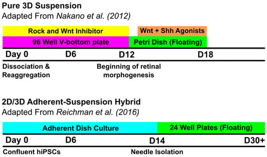
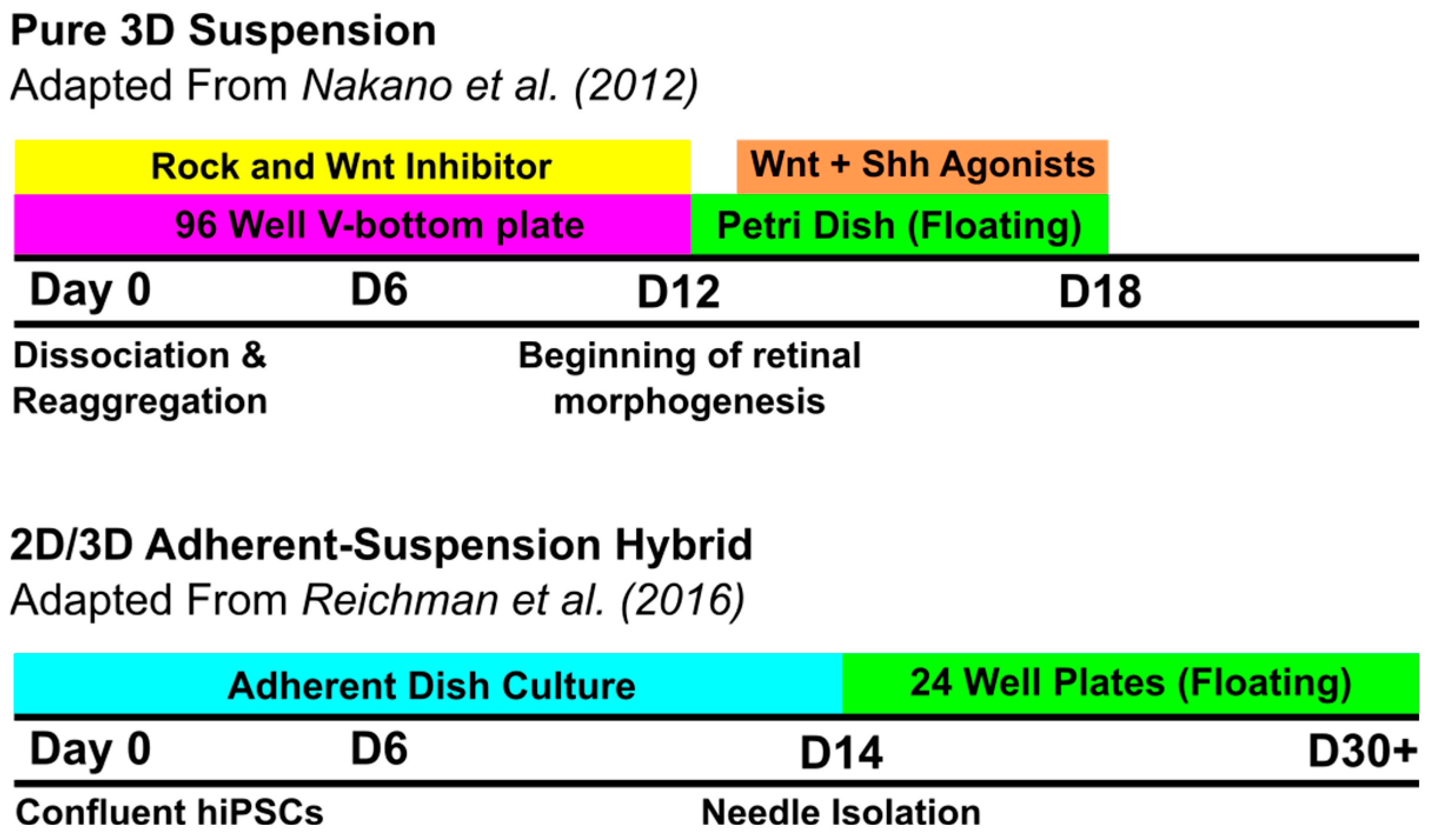
To provide structural and functional platforms suitable for high-throughput drug screening and precision medicine, the field relies on two distinct types of differentiation protocols (Figure 1).
Figure 1.
Overview of Timeline for Retinal Organoid Differentiation Method Types.
2.1. Pure 3D Suspension Protocols
These methods rely on the intrinsic self-organizing capacity of hiPSCs. Single-cell suspensions are re-aggregated in low-attachment V-bottom or U-bottom wells to form uniform embryoid bodies (EBs). Remarkably, these cultures showed self-organization and retinal differentiation with little exogenous growth factors, such as Wnt and BMP inhibitors, when first shown in mouse ESCs [23]. In human ESCs, culture medium was optimized for neural retina formation with the addition of various exogenous modulators, such as Wnt inhibitors and BMP antagonists [24]. Recent studies investigated the addition of other growth factors in these suspension cultures [35,36].
2.2. 2D/3D Adherent-Suspension Hybrid Protocols
To circumvent the efficiency limitations of pure suspension protocols, transitional protocols introduced an adherent phase to facilitate the manual selection of eye field structures. Notably, this method did not require the use of signaling factors like Wnt/BMP antagonists to achieve efficient differentiation of neural retina and RPE cell types [37,38,39]. Other protocols began with the formation of EBs that were then seeded to form an adherent culture for the generation of eye domains [4,40]. Each of these methods mechanically removed eye field structures from the 2D culture. These structures were transferred to suspension culture for long-term maturation. As they matured, they experienced stratification of retinal layers [4].
3. Ideal Retinal Organoids
The search for an ‘ideal’ RO has resulted in the procurement of a variety of methods to enhance the physiological likeness of these models using the addition of various tissues and biomechanical devices. There appears to be 5 major goals for the use of ROs. These can be classified as isogenic control, drug testing, cell therapy, disease modeling, and structural definition.
With gene editing technologies, ROs can undergo disease-relevant phenotypic shifts [41]. This has many implications for the use of ROs in disease and pharmaceutical research. These include gene identification of pathogenic variants [42] and testing for corrective therapies [32].
ROs have the potential to be used in drug testing environments. They display similar physiologic responses to known retinotoxins as are seen in the native human retina [43], supporting their use to screen novel drugs for toxicity. Further, ROs have already been used to measure drug efficacy and toxicity profiles using cellular responses to candidate anti-tumor drugs [44]. In combination with emerging microfluidic technology [25], ROs may provide a high-throughput platform to reliably improve drug development and screening.
Novel cellular therapies [45,46,47,48] are a notable indication for the procurement of mature and selective ROs. There are a variety of approaches to these therapies including sheet integration [45,46], injection of purified cellular suspensions [47], and bio-engineered scaffold implantation [48]. These ROs are optimized for their function by improving maturity and structural organization to maximize their integrative and therapeutic potential.
Disease modeling can be performed using the RO platform. In general, diseases modeled with ROs can be grouped into inherited diseases [41,42], multifactorial diseases [49,50], and trauma [51]. The generation of these ROs is highly variable across diseases of interest.
Structural definition of tissue is important to many studies, and ROs can provide great versatility in methods of histological analysis. They can be supported by additional tissues, as seen in cases of co-culturing methods, to enhance maturation [11]. Additionally, gene-edited progenitors allow for the inclusion of knock-in reporter sequences to follow sequences such as self-organization [52,53]. These may be helpful in standardizing RO generation protocols and following disease progression.
4. Cell Replacement Therapies
One potential use for human-derived ROs is the procurement of human retinal cells. Many retinal diseases result in the loss of cells in the layers of the retina and cause progressive loss of vision. Future therapeutic treatments for vision loss may include the administration of cells cultured in vitro that are descended from a patient or a human donor. With the versatility and control of RO development in a lab environment, procedures may be adjusted to increase the yield of desired cell types [54,55]. These therapies leverage the organoid’s ability to produce mature and stratified retinal tissue or highly purified progenitor cells for transplantation.
4.1. Photoreceptor Precursor Cells
These cells are sourced from fully laminated retinal organoids, which are differentiated long-term in 3D suspension cultures. These organoids self-organize into an optic cup-like structure, consisting of the major retinal layers. Importantly, the Outer Nuclear Layer (ONL) is formed, containing both rod and cone photoreceptors. From the organoids, a single cell suspension is produced, in which various procedures for the selective enrichment of photoreceptor precursor cells (PRPCs) may take place [56,57]. These cells can be purposed to replace damaged or lost photoreceptors in diseases such as Retinitis Pigmentosa, where inner retinal neurons (bipolar, amacrine, and retinal ganglion cells) may survive. This could be achieved through an injection of the transplanted PRPCs into the subretinal space, where they may be able to integrate into the host retina. To isolate the PRPCs from the organoids, various techniques have been attempted and refined for clinical-grade manufacturing, including cell sorting with flow cytometry and selective disassociation with enzymes. Isolation of photoreceptor precursor cells using flow cytometry combines negative selection of the CD29 and SSEA-19 biomarkers and positive selection of CD73 (CD29-/SSEA-19-/CD73+) [56,57]. Enzymatic isolation of photoreceptors leverages the organization of layers of the RO by removing photoreceptor cell-containing supernatant at earlier timepoints after the introduction of dissociative enzymes [58].
4.2. Retinal Pigment Epithelium (RPE) Monolayers/Sheets
While RPE cells can be differentiated in 2D culture, they are also derived from the same early retinal lineage as neural retinal cells in the 3D-shaped ROs. In some RO differentiation protocols, it is possible to derive RPE cells in patches adjacent to the neural retina. These cells may be isolated and expanded in 2D culture to produce an RPE monolayer, which has potential to replace degenerated RPE [59]. Certain disease stages are notable for RPE degeneration, such as the geographic atrophy stage of age-related macular degeneration (AMD) [60]. Transplantation of an RPE monolayer into the subretinal space has been proposed as a method to replace the degenerated native tissue [61]. There are several clinical trials investigating the safety of this surgical procedure in patients [62]. Integration of the layer has the potential to restore the outer blood–retina barrier and provide phagocytic and metabolic support to overlying photoreceptors, preventing future cell stress.
4.3. Retinal Progenitor Cells (RPCs)
RPCs belong to immature cell types that are dominant in early ROs. Progenitors are a source of multipotent cells that can be banked with cryopreservation for later use [63]. These cells are both highly proliferative and versatile, making them useful for long-term storage while maintaining multipotency and the ability to expand. RPCs derived from hiPSCs have shown integrative capacity when transplanted in mice [64], suggesting that they may provide a source of functional cells for tissue regeneration. A potential drawback to the increased pluripotency of these cells is the associated risk of uncontrolled proliferation and teratoma formation.
4.4. Utilizing Retinal Organoids in Clinical Settings
The utilization of ROs for patient therapies involves a multitude of translational and logistical challenges. For one, delivering individualized organoid-derived treatment requires validation of cGMP, which can become highly cost-prohibitive. Beyond costs, variability plays a central role in achieving good outcomes. Particularly, photoreceptor rosette formation has been shown to be problematic in the process of subretinal transplantation of RO-derived sheets [65]. Rossette formation and other reproducibility issues are being addressed through the utilization of bioreactors to standardize RO generation [66].
5. Retinal Organoids for Drug Development and Testing
There is evidence that retinal organoids accurately model drug effects that occur in vivo, providing indication for their use in toxicological screening [43]. 3D ROs for drug testing are categorized by their accuracy in recapitulating a particular disease process or reproducing a specific physiological function. Additionally, these ROs can be formed from patient-derived hiPSCs. Patient-specific drug testing can be performed by measuring physiologic responses to available or novel drugs before administration to the patient. This facet of personalized medicine could have major implications for the advancement of patient safety and outcome improvement in the settings of experimental drugs or drugs with narrow therapeutic windows. These advantages may be limited by the time required to observe changes in mature ROs.
5.1. Standard Laminated Retinal Organoids
These are standard organoids that contain the key cell types of the neural retina in laminated layers. These types include photoreceptors, retinal ganglion cells, bipolar cells, horizontal cells, amacrine cells, and Müller glia. The applications of these organoids are widespread and include retinotoxicity screening, IRD modeling, and drug efficacy testing. Introducing toxic compounds to ROs can reveal cellular stress responses, as shown in a study evaluating retinal exposure to bisphenols [67]. IRDs may be studied with the model organoids (Figure 2) derived from hiPSCs of the patients with those diseases, including Retinitis Pigmentosa and Leber Congenital Amaurosis [32,68]. Efficacy testing can be used to determine a proposed therapeutic’s ability to promote and protect visual cycle function. The limitations of standard laminated retinal organoids include the lack of a native blood supply, heterogeneity of cells, and the extensive time required for cultures [69].
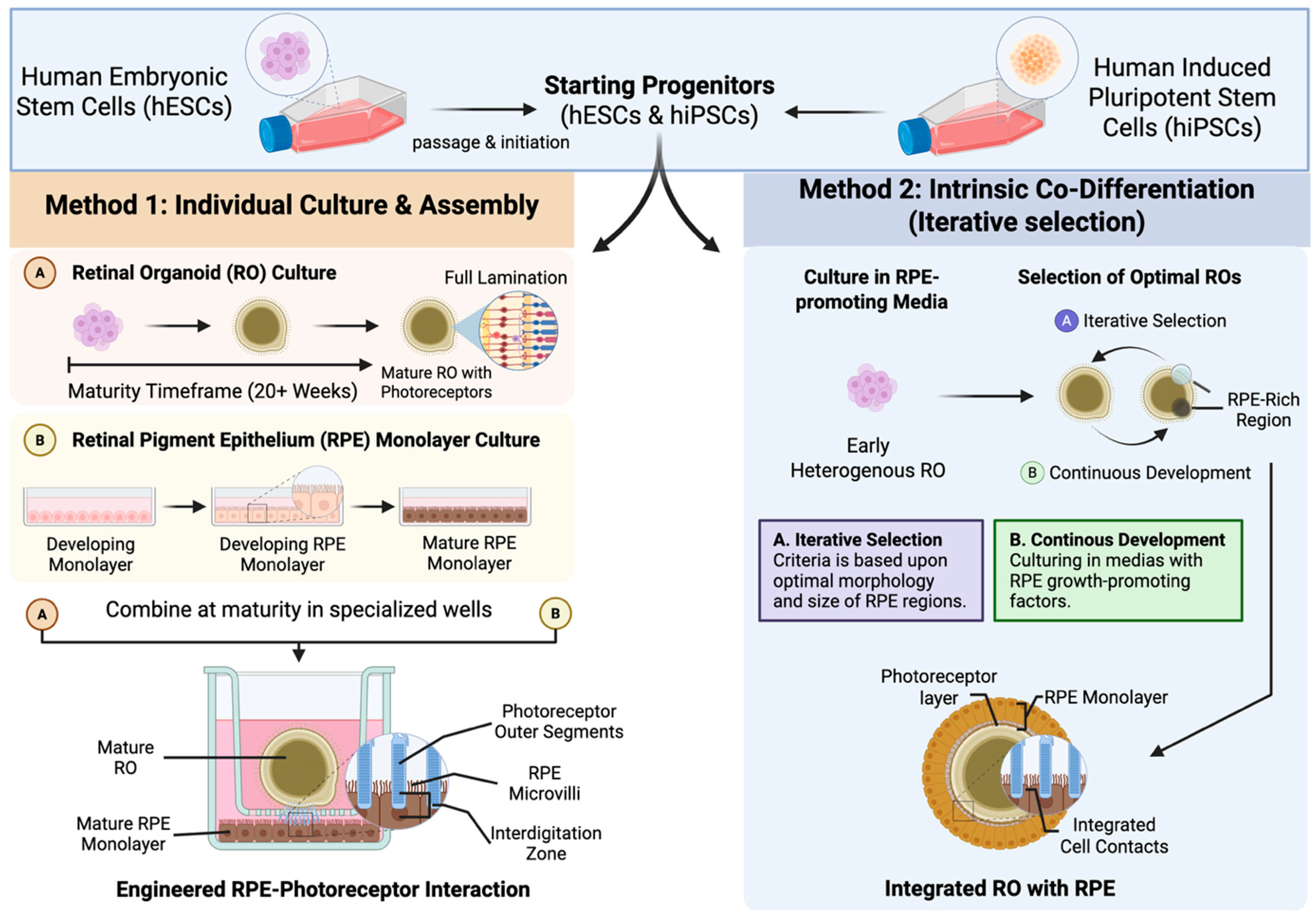
Figure 2.
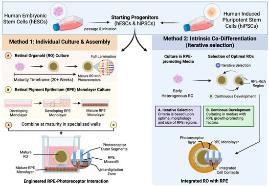
Methods for Producing Retinal Organoids with RPE. Method 1 involves individually culturing pluripotent stem cells (ESCs and iPSCs) to generate separate retinal organoids and RPE monolayers. Once mature, these cultures are combined in specialized wells designed to facilitate interaction between the RPE and photoreceptors. In contrast, Method 2 utilizes pluripotent stem cells to iteratively select retinal organoids with larger, RPE-rich regions. This approach relies on specialized culture media supplemented with factors that promote both RPE growth and its characteristic interaction with photoreceptors.
5.2. Co-Culture Models
These are models consisting of the neural retina and RPE layer that are designed to capture the functional relationship between the two layers. These cultures are formed through the direct co-culture of an RPE monolayer with the RO [11], or with the use ofTf protocols that favor the spontaneous generation of RPE patches [59] (Figure 2, Table 3). As a crucial part of the visual cycle, this layer recycles important signal transduction molecules and phagocytoses the outer segments of photoreceptors. AMD is a disease marked by the degeneration of photoreceptors, whose modeling accuracy may be improved with accelerated differentiation in co-culture [70]. Additionally, these co-culture models may recapitulate a more physiologically accurate microenvironment for drug development [71].
Table 3.
Comparison of Methods to Produce Retinal Organoids with RPE.
5.3. Vascularized Retinal Organoids (VROs)
These cultures seek to address the limitation that standard retinal organoids lack a native blood supply. They do so by introducing vascular endothelial cells and pericytes to initiate the formation of blood vessel-like structures in the retinal organoid or by co-culturing ROs with blood vessel/vascular organoids [5,6]. These organoids provide pathways for studying the breakdown of the blood–retina barrier and neovascularization (as seen in proliferative diabetic retinopathy). VROs may be used to identify mechanisms and discover drugs to advance the treatment of retinal vascular disorders [30]. Similarly to RPE co-cultures, these organoids are limited by the increased complexity of their cultures. Additionally, they present challenges in controlling perfusion of natural blood flow. Lack of perfusion limits the modeling of thrombotic events as seen in vein and artery occlusions of the retina [30]. Some techniques for the creation of VROs include co-aggregation [30] and discrete fusion [5]. These techniques differ based on the maturity of the RO at the time of combination. In coaggregation, RO differentiation begun with the inclusion of vascular cells, while discrete fusion added vascular organoids to mature ROs.
5.4. Retinal Organoids Integrating Microglial Cells
Cultures involving microglial cells are valuable to the study of drug development as they can display characteristics of immune competency [29]. As many pathologies may include immune systems, it is vital that these processes are present in RO models [72]. Additionally, immune responses can be relevant to drug screening processes [73] and may affect candidate therapies due to limited acceptable doses.
5.5. Retinal Organoids in Micro-Physiological Systems (MPSs)
Devices such as organ on a chip or microfluidic devices can be paired with retinal organoids to provide control over nutrient delivery, waste removal, and mechanical force [28]. These models provide higher throughput than traditional methods due to their compactness and automated perfusion of tissue [25,26]. They can be used to screen large quantities of compounds. Additionally, they may be integrated with multiple-organ-chip devices to perform systemic toxicity screening [74].
6. Types of 3D Retinal Organoids for Disease Modeling
6.1. Inherited Retinal Disease (IRD) Models
This is a common application for retinal organoids, as patient-derived iPSCs provide the precise genetic makeup to recapitulate the disease process for observation of the underlying mechanisms (Table 4).
Table 4.
Genetic Profiles and Cellular Features of Key Inherited Retinopathies.
6.2. Multifactorial Disease Models
These models induce pathological conditions in the organoid or incorporate additional cell types to accurately reflect the complexity of the disease in vivo (Table 5) [85,86,87].
Table 5.
Pathological Features and Induction Methods of Multifactorial Disease Models.
6.3. Composite Organoid Models
These are more complex co-culture models that aim to overcome the limitations of the basic retinal organoid, including the lack of a vascular supply, a robust RPE-PR interaction, and immune surveillance (Table 6).
Table 6.
Features and Applications of Composite Organoid Models.
7. 3D Retinal Organoids for Battlefield Ocular Trauma Modeling
The transition from anterior segment trauma to permanent retinal vision loss is a complex pathological cascade often initiated by battlefield injuries. There are a variety of injuries associated with the battlefield environment (Table 7) [95]. While corneal lacerations and blast-induced ocular surface damage represent the immediate clinical presentation in the field, these injuries frequently trigger a generalized ocular inflammatory response.
Table 7.
Common Ocular Injuries in the Battlefield Environment.
7.1. Corneal Trauma Modeling
Corneal injury models, such as those utilizing photoactivated Rose Bengal for laceration sealing, highlight the importance of immediate stabilization [96]. However, without rapid intervention, anterior segment trauma initiates a cascade of inflammatory cytokines and growth factors that migrate to the posterior segment. Using Microglia-integrated Retinal Organoids (Table 6), researchers could model how these systemic and anterior-derived inflammatory signals trigger retinal microglia activation. The “Injury-on-a-dish” approach may allow for the study of treatments for Retinal Fibrosis and Proliferative Vitreoretinopathy (PVR), conditions that Dr. Wang’s research has identified as major complications following combat ocular trauma [97,98]. Such an approach could involve similar methods performed in animals, such as scratch assays, to accurately model traumatic conditions. Modeling trauma with human ROs could provide many advantages, especially in the development of anti-inflammatory and anti-fibrotic therapies due to the availability of high-throughput systems for ROs. While these models may be useful for the retina’s responses to injury, there are currently no organoids that accurately include a cornea–aqueous–vitreous–retina connection to model the origination and propagation of the inflammatory cascade related to corneal laceration.
7.2. Ultrasound and Blast-Induced Retinal Injury
In addition to mechanical trauma, the battlefield environment exposes service members to blast overpressure and high-intensity waves. Experimental models have utilized ultrasound-induced injury and blast shock tubes to simulate these non-penetrating traumas. These models demonstrate that repetitive low-level blast exposure leads to significant apoptosis in the neuroretina and optic nerve [51]. Retinal organoids provide a unique platform to subject neural tissue to controlled ultrasonic or mechanical stress, allowing for the quantification of RGC loss and the testing of neuroprotective small molecules, such as RUNX1 inhibitors, to mitigate post-traumatic damage [99].
8. Overcoming Current Limitations in Organoid Fidelity
Despite their potential, the current generation of retinal organoids faces structural and functional hurdles that impact their model fidelity to resemble the adult human retina [100]. A primary limitation is the inability to achieve complete structural maturation. Due to the lack of endogenous vascularization and variable spatial integration with the retinal pigment epithelium (RPE), photoreceptors often fail to reach the outer segment maturity seen in vivo. This constraint has historically limited the modeling of late-onset degenerative diseases and the long-term evaluation of cell therapies [101].
Recent bioengineering advancements are beginning to address these gaps. The integration of endothelial cells has shown significant promise in extending organoid maturity. For instance, ROs have been successfully vascularized through co-culture with vascular organoids, mimicking the retina’s complex nutrient delivery system [4,5,6]. Furthermore, because microglia are not derived from retinal neuroectoderm, traditional ROs lack the machinery to model neuroinflammation [102]. However, the addition of iPSC-derived microglia to RO cultures now provides a robust platform for studying immune-involved pathologies such as Age-Related Macular Degeneration (AMD) and Diabetic Retinopathy (DR) [29].
The lack of an optic nerve head, which is a major barrier in glaucoma research using ROs, is being addressed through multisystem engineering. Recent studies have reported the projection of optic nerve-like fiber bundles when ROs are cultured in proximity to brain organoids, paving the way for a novel co-culture [103]. Overall, the integration of non-neural retinal cells into culture with ROs presents opportunities to study additional ocular disease mechanisms. They introduce the ability to study the interaction of tissues with the neural retina and establish targets to prevent retinal degeneration in these diseases [103].
Finally, a major limitation of ROs is the lack of major ocular structures including the cornea, lens, lacrimal glands, etc. Some studies have attempted to introduce these structures to construct ‘assembloids’; however, these still lack structural similarities to the human eye or do not mature beyond primitive states [94,104]. Thus, the procurement of a fully integrated model of the human eye remains an important goal for the advancement of organoid models.
9. Conclusions
Human-induced pluripotent stem cell-derived ROs represent a definitive shift in ophthalmic research, moving away from the less representative animal models and moving towards a robust, patient-specific 3D system. By recapitulating disease-specific phenotypes and generating isogenic controls, these organoids provide an unprecedented view into elusive pathologies and the linkages between specific genes and retinal degeneration. They are ideal for modeling and are continually being analyzed with new chemical, functional, and histological parameters. Beyond their utility as discovery tools, ROs are foundational to the future of clinical translation. They offer a platform for high-throughput therapeutic screening and a self-renewing source of purified cells for transplantation in patients with IRDs.
The evolution from simple developmental models to these complex, multicellular systems marks a paradigm shift in our ability to address combat-related ocular trauma. The transition toward point-of-care diagnostic and therapeutic platforms represents the next frontier for military medicine.
While a fully engineered retina remains a long-term goal, the integration of vascular, immune, and neural components is making that vision increasingly feasible. Both individual and co-culture RO generation protocols are being evaluated and standardized, which can improve their efficiency and accessibility. Achieving this will solidify the RO platform as an indispensable tool in the fight against blinding diseases, leading directly to the implementation of personalized and curative strategies for both civilian and military populations.
Author Contributions
Conceptualization, Y.Z.; investigation, Y.Z., E.P.Z., S.S.; resources, Y.Z., M.J.S., H.H., A.A., J.-X.M.; writing—original draft preparation, E.P.Z., S.S., Y.Z.; writing—review and editing, Y.Z., M.J.S., H.H.; visualization, E.P.Z., Y.Z.; supervision, Y.Z., A.A., J.-X.M.; funding acquisition, Y.Z. All authors have read and agreed to the published version of the manuscript.
Funding
NIH/NEI R21EY037262, R21EY035833, DoD HT94252510856, and Translational Eye and Vision Research Center (i.e., TrEVR Pilot Research Grant 2024–2026) (YZ, PI).
Institutional Review Board Statement
Not applicable.
Informed Consent Statement
Not applicable.
Data Availability Statement
No new data were created or analyzed in this study. Data sharing is not applicable to this article.
Conflicts of Interest
The authors declare no conflict of interest.
References
- Flaxman, S.R.; Bourne, R.R.A.; Resnikoff, S.; Ackland, P.; Braithwaite, T.; Cicinelli, M.V.; Das, A.; Jonas, J.B.; Keeffe, J.; Kempen, J.H.; et al. Global causes of blindness and distance vision impairment 1990–2020: A systematic review and meta-analysis. Lancet Glob. Health 2017, 5, e1221–e1234. [Google Scholar] [CrossRef] [Scilit] [PubMed]
- Cowan, C.S.; Renner, M.; De Gennaro, M.; Gross-Scherf, B.; Goldblum, D.; Hou, Y.; Munz, M.; Rodrigues, T.M.; Krol, J.; Szikra, T.; et al. Cell Types of the Human Retina and Its Organoids at Single-Cell Resolution. Cell 2020, 182, 1623–1640.e34. [Google Scholar] [CrossRef] [Scilit] [PubMed]
- Collin, J.; Queen, R.; Zerti, D.; Dorgau, B.; Hussain, R.; Coxhead, J.; Cockell, S.; Lako, M. Deconstructing Retinal Organoids: Single Cell RNA-Seq Reveals the Cellular Components of Human Pluripotent Stem Cell-Derived Retina. Stem Cells 2019, 37, 593–598. [Google Scholar] [CrossRef] [Scilit] [PubMed]
- Zhong, X.; Gutierrez, C.; Xue, T.; Hampton, C.; Vergara, M.N.; Cao, L.-H.; Peters, A.; Park, T.S.; Zambidis, E.T.; Meyer, J.S.; et al. Generation of three-dimensional retinal tissue with functional photoreceptors from human iPSCs. Nat. Commun. 2014, 5, 4047. [Google Scholar] [CrossRef] [Scilit] [PubMed]
- Chen, H.; Liang, Y.; Sun, X.; Xiong, W.; Yang, T.; Liang, Y.; Ye, X.; Li, X.; Wang, W.; Gu, J.; et al. Generation of vascularized retinal organoids containing microglia based on a PDMS microwell platform. Sci. Adv. 2025, 11, eady6410. [Google Scholar] [CrossRef] [Scilit] [PubMed]
- Chen, Y.; Zhang, L.; Jiang, F.; Fu, Y.; Yang, J.; Zhang, M. Human retinal organoids: From developmental biology to translational applications. Exp. Eye. Res. 2026, 269, 111051. [Google Scholar] [CrossRef] [Scilit]
- Saha, A.; Capowski, E.; Zepeda, M.A.F.; Nelson, E.C.; Gamm, D.M.; Sinha, R. Cone photoreceptors in human stem cell-derived retinal organoids demonstrate intrinsic light responses that mimic those of primate fovea. Cell Stem Cell 2022, 29, 487–489. [Google Scholar] [CrossRef] [Scilit] [PubMed]
- Seah, I.; Goh, D.; Banerjee, A.; Su, X. Modeling inherited retinal diseases using human induced pluripotent stem cell derived photoreceptor cells and retinal pigment epithelial cells. Front. Med. 2024, 11, 1328474. [Google Scholar] [CrossRef] [Scilit] [PubMed]
- Lee, Y.J.; Jo, D.H. Retinal Organoids from Induced Pluripotent Stem Cells of Patients with Inherited Retinal Diseases: A Systematic Review. Stem Cell Rev. Rep. 2025, 21, 167–197. [Google Scholar] [CrossRef] [Scilit] [PubMed]
- Ashworth, K.E.; Weisbrod, J.; Ballios, B.G. Inherited Retinal Diseases and Retinal Organoids as Preclinical Cell Models for Inherited Retinal Disease Research. Genes 2024, 15, 705. [Google Scholar] [CrossRef] [Scilit] [PubMed]
- Akhtar, T.; Xie, H.; Khan, M.I.; Zhao, H.; Bao, J.; Zhang, M.; Xue, T. Accelerated photoreceptor differentiation of hiPSC-derived retinal organoids by contact co-culture with retinal pigment epithelium. Stem Cell Res. 2019, 39, 101491. [Google Scholar] [CrossRef] [Scilit] [PubMed]
- O’Hara-Wright, M.; Gonzalez-Cordero, A. Retinal organoids: A window into human retinal development. Development 2020, 147, dev189746. [Google Scholar] [CrossRef] [Scilit] [PubMed]
- Galindo-Cabello, N.; Caballano-Infantes, E.; Benites, G.; Pastor-Idoate, S.; Diaz-Corrales, F.J.; Usategui-Martin, R. Retinal Organoids: Innovative Tools for Understanding Retinal Degeneration. Int. J. Mol. Sci. 2025, 26, 3263. [Google Scholar] [CrossRef] [Scilit] [PubMed]
- Raab, S.; Klingenstein, M.; Liebau, S.; Linta, L. A Comparative View on Human Somatic Cell Sources for iPSC Generation. Stem Cells Int. 2014, 2014, 768391. [Google Scholar] [CrossRef] [Scilit] [PubMed]
- Loh, Y.H.; Hartung, O.; Li, H.; Guo, C.; Sahalie, J.M.; Manos, P.D.; Urbach, A.; Heffner, G.C.; Grskovic, M.; Vigneault, F.; et al. Reprogramming of T cells from human peripheral blood. Cell Stem Cell 2010, 7, 15–19. [Google Scholar] [CrossRef] [Scilit] [PubMed]
- Beiner, D.; Zhu, H.; Bosholm, C.C.; Wang, H.C.; Criswell, T.; Atala, A.; Ma, J.X.; Zhang, Y. Derivation and characterization of retinal pigment epithelium from urine-derived iPSCs. FEBS Open Bio 2026, 16, 1691–1707. [Google Scholar] [CrossRef] [Scilit]
- Guan, X.; Mack, D.L.; Moreno, C.M.; Strande, J.L.; Mathieu, J.; Shi, Y.; Markert, C.D.; Wang, Z.; Liu, G.; Lawlor, M.W.; et al. Dystrophin-deficient cardiomyocytes derived from human urine: New biologic reagents for drug discovery. Stem Cell Res. 2014, 12, 467–480. [Google Scholar] [CrossRef] [Scilit]
- Liu, Y.; Zheng, Y.; Li, S.; Xue, H.; Schmitt, K.; Hergenroeder, G.W.; Wu, J.; Zhang, Y.; Kim, D.H.; Cao, Q. Human neural progenitors derived from integration-free iPSCs for SCI therapy. Stem Cell Res. 2017, 19, 55–64. [Google Scholar] [CrossRef] [Scilit] [PubMed]
- Sacco, A.M.; Belviso, I.; Romano, V.; Carfora, A.; Schonauer, F.; Nurzynska, D.; Montagnani, S.; Di Meglio, F.; Castaldo, C. Diversity of dermal fibroblasts as major determinant of variability in cell reprogramming. J. Cell. Mol. Med. 2019, 23, 4256–4268. [Google Scholar] [CrossRef] [Scilit] [PubMed]
- Lowry, W.E.; Richter, L.; Yachechko, R.; Pyle, A.D.; Tchieu, J.; Sridharan, R.; Clark, A.T.; Plath, K. Generation of human induced pluripotent stem cells from dermal fibroblasts. Proc. Natl. Acad. Sci. USA 2008, 105, 2883–2888. [Google Scholar] [CrossRef] [Scilit] [PubMed]
- Gore, M.; Tiwari, A.; Jahagirdar, D.; Narayanasamy, A.; Jain, R.; Dandekar, P. Three-dimensional spheroids of choroid-retinal vascular endothelial cells as an in-vitro model for diabetic retinopathy: Proof-of-concept investigation. Curr. Res. Pharmacol. Drug Discov. 2022, 3, 100111. [Google Scholar] [CrossRef] [Scilit] [PubMed]
- Usui, H.; Nishiwaki, A.; Landiev, L.; Kacza, J.; Eichler, W.; Wako, R.; Kato, A.; Takase, N.; Kuwayama, S.; Ohashi, K.; et al. In vitro drusen model—Three-dimensional spheroid culture of retinal pigment epithelial cells. J. Cell Sci. 2018, 132, jcs.215798. [Google Scholar] [CrossRef] [Scilit] [PubMed]
- Eiraku, M.; Takata, N.; Ishibashi, H.; Kawada, M.; Sakakura, E.; Okuda, S.; Sekiguchi, K.; Adachi, T.; Sasai, Y. Self-organizing optic-cup morphogenesis in three-dimensional culture. Nature 2011, 472, 51–56. [Google Scholar] [CrossRef] [Scilit] [PubMed]
- Nakano, T.; Ando, S.; Takata, N.; Kawada, M.; Muguruma, K.; Sekiguchi, K.; Saito, K.; Yonemura, S.; Eiraku, M.; Sasai, Y. Self-formation of optic cups and storable stratified neural retina from human ESCs. Cell Stem Cell 2012, 10, 771–785. [Google Scholar] [CrossRef] [Scilit] [PubMed]
- Achberger, K.; Probst, C.; Haderspeck, J.; Bolz, S.; Rogal, J.; Chuchuy, J.; Nikolova, M.; Cora, V.; Antkowiak, L.; Haq, W.; et al. Merging organoid and organ-on-a-chip technology to generate complex multi-layer tissue models in a human retina-on-a-chip platform. eLife 2019, 8, e46188. [Google Scholar] [CrossRef] [Scilit] [PubMed]
- Drabbe, E.; Pelaez, D.; Agarwal, A. Retinal organoid chip: Engineering a physiomimetic oxygen gradient for optimizing long term culture of human retinal organoids. Lab Chip 2025, 25, 1626–1636. [Google Scholar] [CrossRef] [Scilit] [PubMed]
- Nair, D.S.R.; Seiler, M.J.; Patel, K.H.; Thomas, V.; Camarillo, J.C.M.; Humayun, M.S.; Thomas, B.B. Tissue Engineering Strategies for Retina Regeneration. Appl. Sci. 2021, 11, 2154. [Google Scholar] [CrossRef] [Scilit] [PubMed]
- Marcos, L.F.; Wilson, S.L.; Roach, P. Tissue engineering of the retina: From organoids to microfluidic chips. J. Tissue Eng. 2021, 12, 20417314211059876. [Google Scholar] [CrossRef] [Scilit] [PubMed]
- Chichagova, V.; Georgiou, M.; Carter, M.; Dorgau, B.; Hilgen, G.; Collin, J.; Queen, R.; Chung, G.; Ajeian, J.; Moya-Molina, M.; et al. Incorporating microglia-like cells in human induced pluripotent stem cell-derived retinal organoids. J. Cell. Mol. Med. 2023, 27, 435–445. [Google Scholar] [CrossRef] [Scilit] [PubMed]
- Inagaki, S.; Nakamura, S.; Kuse, Y.; Aoshima, K.; Funato, M.; Shimazawa, M.; Hara, H. Establishment of vascularized human retinal organoids from induced pluripotent stem cells. Stem Cells 2025, 43, sxae093. [Google Scholar] [CrossRef] [Scilit] [PubMed]
- Lee, S.; Chung, W.G.; Jeong, H.; Cui, G.; Kim, E.; Lim, J.A.; Seo, H.; Kwon, Y.W.; Byeon, S.H.; Lee, J.; et al. Electrophysiological Analysis of Retinal Organoid Development Using 3D Microelectrodes of Liquid Metals. Adv. Mater. 2024, 36, e2404428. [Google Scholar] [CrossRef] [Scilit] [PubMed]
- Lane, A.; Jovanovic, K.; Shortall, C.; Ottaviani, D.; Panes, A.B.; Schwarz, N.; Guarascio, R.; Hayes, M.J.; Palfi, A.; Chadderton, N.; et al. Modeling and Rescue of RP2 Retinitis Pigmentosa Using iPSC-Derived Retinal Organoids. Stem Cell Rep. 2020, 15, 67–79. [Google Scholar] [CrossRef] [Scilit] [PubMed]
- Athanasiou, D.; Afanasyeva, T.A.V.; Chai, N.; Ziaka, K.; Jovanovic, K.; Guarascio, R.; Boldt, K.; Corral-Serrano, J.C.; Kanuga, N.; Roepman, R.; et al. Small molecule treatment alleviates photoreceptor cilia defects in LCA5-deficient human retinal organoids. Acta Neuropathol. Commun. 2025, 13, 26. [Google Scholar] [CrossRef] [Scilit] [PubMed]
- Boon, N.; Lu, X.; Andriessen, C.A.; Moustakas, I.; Buck, T.M.; Freund, C.; Arendzen, C.H.; Bohringer, S.; Mei, H.; Wijnholds, J. AAV-mediated gene augmentation therapy of CRB1 patient-derived retinal organoids restores the histological and transcriptional retinal phenotype. Stem Cell Rep. 2023, 18, 1123–1137. [Google Scholar] [CrossRef] [Scilit] [PubMed]
- Mellough, C.B.; Collin, J.; Khazim, M.; White, K.; Sernagor, E.; Steel, D.H.; Lako, M. IGF-1 Signaling Plays an Important Role in the Formation of Three-Dimensional Laminated Neural Retina and Other Ocular Structures from Human Embryonic Stem Cells. Stem Cells 2015, 33, 2416–2430. [Google Scholar] [CrossRef] [Scilit] [PubMed]
- Kuwahara, A.; Ozone, C.; Nakano, T.; Saito, K.; Eiraku, M.; Sasai, Y. Generation of a ciliary margin-like stem cell niche from self-organizing human retinal tissue. Nat. Commun. 2015, 6, 6286. [Google Scholar] [CrossRef] [Scilit] [PubMed]
- Reichman, S.; Terray, A.; Slembrouck, A.; Nanteau, C.; Orieux, G.; Habeler, W.; Nandrot, E.F.; Sahel, J.A.; Monville, C.; Goureau, O. From confluent human iPS cells to self-forming neural retina and retinal pigmented epithelium. Proc. Natl. Acad. Sci. USA 2014, 111, 8518–8523. [Google Scholar] [CrossRef] [Scilit] [PubMed]
- Reichman, S.; Goureau, O. Production of Retinal Cells from Confluent Human iPS Cells. Methods Mol. Biol. 2016, 1357, 339–351. [Google Scholar] [PubMed]
- Gonzalez-Cordero, A.; Kruczek, K.; Naeem, A.; Fernando, M.; Kloc, M.; Ribeiro, J.; Goh, D.; Duran, Y.; Blackford, S.J.I.; Abelleira-Hervas, L.; et al. Recapitulation of Human Retinal Development from Human Pluripotent Stem Cells Generates Transplantable Populations of Cone Photoreceptors. Stem Cell Rep. 2017, 9, 820–837. [Google Scholar] [CrossRef] [Scilit] [PubMed]
- Meyer, J.S.; Howden, S.E.; Wallace, K.A.; Verhoeven, A.D.; Wright, L.S.; Capowski, E.E.; Pinilla, I.; Martin, J.M.; Tian, S.; Stewart, R.; et al. Optic vesicle-like structures derived from human pluripotent stem cells facilitate a customized approach to retinal disease treatment. Stem Cells 2011, 29, 1206–1218. [Google Scholar] [CrossRef] [Scilit] [PubMed]
- Deng, W.L.; Gao, M.L.; Lei, X.L.; Lv, J.N.; Zhao, H.; He, K.W.; Xia, X.X.; Li, L.Y.; Chen, Y.C.; Li, Y.P.; et al. Gene Correction Reverses Ciliopathy and Photoreceptor Loss in iPSC-Derived Retinal Organoids from Retinitis Pigmentosa Patients. Stem Cell Rep. 2018, 10, 1267–1281. [Google Scholar] [CrossRef] [Scilit] [PubMed]
- Buskin, A.; Zhu, L.; Chichagova, V.; Basu, B.; Mozaffari-Jovin, S.; Dolan, D.; Droop, A.; Collin, J.; Bronstein, R.; Mehrotra, S.; et al. Disrupted alternative splicing for genes implicated in splicing and ciliogenesis causes PRPF31 retinitis pigmentosa. Nat. Commun. 2018, 9, 4234. [Google Scholar] [CrossRef] [Scilit] [PubMed]
- Dorgau, B.; Georgiou, M.; Chaudhary, A.; Moya-Molina, M.; Collin, J.; Queen, R.; Hilgen, G.; Davey, T.; Hewitt, P.; Schmitt, M.; et al. Human Retinal Organoids Provide a Suitable Tool for Toxicological Investigations: A Comprehensive Validation Using Drugs and Compounds Affecting the Retina. Stem Cells Transl. Med. 2022, 11, 159–177. [Google Scholar] [CrossRef] [Scilit] [PubMed]
- Srimongkol, A.; Laosillapacharoen, N.; Saengwimol, D.; Chaitankar, V.; Rojanaporn, D.; Thanomchard, T.; Borwornpinyo, S.; Hongeng, S.; Kaewkhaw, R. Sunitinib efficacy with minimal toxicity in patient-derived retinoblastoma organoids. J. Exp. Clin. Cancer Res. 2023, 42, 39. [Google Scholar] [CrossRef] [Scilit] [PubMed]
- Hirami, Y.; Mandai, M.; Sugita, S.; Maeda, A.; Maeda, T.; Yamamoto, M.; Uyama, H.; Yokota, S.; Fujihara, M.; Igeta, M.; et al. Safety and stable survival of stem-cell-derived retinal organoid for 2 years in patients with retinitis pigmentosa. Cell Stem Cell 2023, 30, 1585–1596 e6. [Google Scholar] [CrossRef] [Scilit] [PubMed]
- Lin, B.; McLelland, B.T.; Aramant, R.B.; Thomas, B.B.; Nistor, G.; Keirstead, H.S.; Seiler, M.J. Retina Organoid Transplants Develop Photoreceptors and Improve Visual Function in RCS Rats with RPE Dysfunction. Investig. Ophthalmol. Vis. Sci. 2020, 61, 34. [Google Scholar] [CrossRef] [Scilit] [PubMed]
- Ribeiro, J.; Procyk, C.A.; West, E.L.; O’Hara-Wright, M.; Martins, M.F.; Khorasani, M.M.; Hare, A.; Basche, M.; Fernando, M.; Goh, D.; et al. Restoration of visual function in advanced disease after transplantation of purified human pluripotent stem cell-derived cone photoreceptors. Cell Rep. 2021, 35, 109022. [Google Scholar] [CrossRef] [Scilit] [PubMed]
- Lee, I.K.; Ludwig, A.L.; Phillips, M.J.; Lee, J.; Xie, R.; Sajdak, B.S.; Jager, L.D.; Gong, S.; Gamm, D.M.; Ma, Z. Ultrathin micromolded 3D scaffolds for high-density photoreceptor layer reconstruction. Sci. Adv. 2021, 7, eabf0344. [Google Scholar] [CrossRef] [Scilit] [PubMed]
- de Lemos, L.; Antas, P.; Ferreira, I.S.; Santos, I.P.; Felgueiras, B.; Gomes, C.M.; Brito, C.; Seabra, M.C.; Tenreiro, S. Modelling neurodegeneration and inflammation in early diabetic retinopathy using 3D human retinal organoids. Vitr. Model 2024, 3, 33–48. [Google Scholar] [CrossRef] [Scilit] [PubMed]
- Hao, X.; Du, L.; Liu, G.; Li, Z.; Wang, S. Human retinal organoids for modelling dry age-related macular degeneration and screening drugs. Genes Dis. 2025, 12, 101593. [Google Scholar] [CrossRef] [Scilit] [PubMed]
- Wang, H.-C.H.; Choi, J.-H.; Greene, W.A.; Plamper, M.L.; Cortez, H.E.; Chavko, M.; Li, Y.; Dalle Lucca, J.J.; Johnson, A.J. Pathophysiology of blast-induced ocular trauma with apoptosis in the retina and optic nerve. Mil. Med. 2014, 179, 34–40. [Google Scholar] [CrossRef] [Scilit] [PubMed]
- Capowski, E.E.; Samimi, K.; Mayerl, S.J.; Phillips, M.J.; Pinilla, I.; Howden, S.E.; Saha, J.; Jansen, A.D.; Edwards, K.L.; Jager, L.D.; et al. Reproducibility and staging of 3D human retinal organoids across multiple pluripotent stem cell lines. Development 2019, 146, dev.171686. [Google Scholar] [CrossRef] [Scilit] [PubMed]
- Bai, J.; Koos, D.S.; Stepanian, K.; Fouladian, Z.; Shayler, D.W.H.; Aparicio, J.G.; Fraser, S.E.; Moats, R.A.; Cobrinik, D. Episodic live imaging of cone photoreceptor maturation in GNAT2-EGFP retinal organoids. Dis. Model. Mech. 2023, 16, dmm050193. [Google Scholar] [CrossRef] [Scilit] [PubMed]
- Zerti, D.; Dorgau, B.; Felemban, M.; Ghareeb, A.E.; Yu, M.; Ding, Y.; Krasnogor, N.; Lako, M. Developing a simple method to enhance the generation of cone and rod photoreceptors in pluripotent stem cell-derived retinal organoids. Stem Cells 2020, 38, 45–51. [Google Scholar] [CrossRef] [Scilit] [PubMed]
- Lim, B.Y.; Chen, C.; Fredericks, A.; Nilli, E.; Mesa Mora, S.; Weatherstone, M.; Loi, T.H.; Newman, P.; Aryamanesh, N.; Zreiqat, H.; et al. Retinoic acid drives cell fate specification, maturation and retinal regionality in human retinal organoids. Nat. Commun. 2026, 17, 5702. [Google Scholar] [CrossRef] [Scilit] [PubMed]
- Lakowski, J.; Welby, E.; Budinger, D.; Di Marco, F.; Di Foggia, V.; Bainbridge, J.W.B.; Wallace, K.; Gamm, D.M.; Ali, R.R.; Sowden, J.C. Isolation of Human Photoreceptor Precursors via a Cell Surface Marker Panel from Stem Cell-Derived Retinal Organoids and Fetal Retinae. Stem Cells 2018, 36, 709–722. [Google Scholar] [CrossRef] [Scilit] [PubMed]
- Susaimanickam, P.J.; Capowski, E.; Xiong, M.N.; Edwards, K.L.; Phanse, Y.; Welch, M.J.; Pier, A.; Mayerl, S.J.; Zepeda, M.A.F.; Peterson, K.D.; et al. Identification and enrichment of human retinal organoid-derived red/green cone-competent precursors with enhanced axon dynamics. Stem Cell Res. Ther. 2026, 17, 336. [Google Scholar] [CrossRef] [Scilit] [PubMed]
- Stone, N.E.; Bohrer, L.R.; Mullin, N.K.; Berthold, A.; Wright, A.T.; Han, I.C.; Stone, E.M.; Mullins, R.F.; Tucker, B.A. Device-free isolation of photoreceptor cells from patient iPSC-derived retinal organoids. JCI Insight 2025, 10, e186338. [Google Scholar] [CrossRef] [Scilit] [PubMed]
- Flores-Bellver, M.; Canto-Soler, M.V. Generation of Induced-Primary Retinal Pigment Epithelium from Human Retinal Organoids. In Ocular Regeneration: Methods and Protocols; Gross, J.M., Ed.; Springer: New York, NY, USA, 2025; pp. 197–214. [Google Scholar]
- Li, M.; Huisingh, C.; Messinger, J.; Dolz-Marco, R.; Ferrara, D.; Freund, K.B.; Curcio, C.A. Histology of Geographic Atrophy Secondary to Age-Related Macular Degeneration: A Multilayer Approach. Retina 2018, 38, 1937–1953. [Google Scholar] [CrossRef] [Scilit] [PubMed]
- Liu, Z.; Parikh, B.H.; Tan, Q.S.W.; Wong, D.S.L.; Ong, K.H.; Yu, W.; Seah, I.; Holder, G.E.; Hunziker, W.; Tan, G.S.W.; et al. Surgical Transplantation of Human RPE Stem Cell-Derived RPE Monolayers into Non-Human Primates with Immunosuppression. Stem Cell Rep. 2021, 16, 237–251. [Google Scholar] [CrossRef] [Scilit] [PubMed]
- Liu, H.; Huang, S.S.; Lingam, G.; Kai, D.; Su, X.; Liu, Z. Advances in retinal pigment epithelial cell transplantation for retinal degenerative diseases. Stem Cell Res. Ther. 2024, 15, 390. [Google Scholar] [CrossRef] [Scilit] [PubMed]
- Gozlan, S.; Batoumeni, V.; Fournier, T.; Nanteau, C.; Potey, A.; Clémençon, M.; Orieux, G.; Sahel, J.-A.; Goureau, O.; Roger, J.E.; et al. Bankable human iPSC-derived retinal progenitors represent a valuable source of multipotent cells. Commun. Biol. 2023, 6, 762. [Google Scholar] [CrossRef] [Scilit] [PubMed]
- Iwama, Y.; Nomaru, H.; Masuda, T.; Kawamura, Y.; Matsumura, M.; Murata, Y.; Teranishi, K.; Nishida, K.; Ota, S.; Mandai, M.; et al. Label-free enrichment of human pluripotent stem cell-derived early retinal progenitor cells for cell-based regenerative therapies. Stem Cell Rep. 2024, 19, 254–269. [Google Scholar] [CrossRef] [Scilit] [PubMed]
- Malhotra, S.; Seiler, M.J.; Browne, A.W. Challenges and Advances in the Production of Transplantable Retinal Tissue from Retinal Organoids. J. Ophthalmic Vis. Res. 2025, 20, 10-18502. [Google Scholar] [CrossRef] [Scilit] [PubMed]
- Cobb, H.; Aparicio-Domingo, S.; Canto-Soler, M.V. Transitioning into GMP-Compliance: Alternative Methods for Producing Retinal Organoids for Transplantation. Transl. Vis. Sci. Technol. 2021, 10, 9. [Google Scholar] [CrossRef] [Scilit] [PubMed]
- Li, M.; Gong, J.; Ge, L.; Gao, H.; Yang, J.; Yang, C.; Kang, J.; Fang, Y.; Xu, H. Development of human retinal organoid models for bisphenol toxicity assessment. Ecotoxicol. Environ. Saf. 2022, 245, 114094. [Google Scholar] [CrossRef] [Scilit] [PubMed]
- Kruczek, K.; Qu, Z.; Gentry, J.; Fadl, B.R.; Gieser, L.; Hiriyanna, S.; Batz, Z.; Samant, M.; Samanta, A.; Chu, C.J.; et al. Gene Therapy of Dominant CRX-Leber Congenital Amaurosis using Patient Stem Cell-Derived Retinal Organoids. Stem Cell Rep. 2021, 16, 252–263. [Google Scholar] [CrossRef] [Scilit] [PubMed]
- Palakkan, A.A.; Kumar, G.S. Retinal organoids in age-related macular degeneration: Promise, pitfalls, and progress. Exp. Eye Res. 2026, 264, 110831. [Google Scholar] [CrossRef] [Scilit] [PubMed]
- Sharma, K.; Pawlick, J.S.; Busskamp, V. Retinal Organoid Development: Disease Modeling and Therapeutic Strategies. In Advances in Using Stem Cells to Treat Neural Diseases; Springer Nature: Berlin, Germany, 2025; pp. 151–173. [Google Scholar]
- Liu, C.F.; Wong, N.K.; Yip, S.P.; Huang, C.-L. Retinal Organoid Differentiation Strategies and Their Application in Retinal Degenerative Disease Modeling. In Advances in Using Stem Cells to Treat Neural Diseases; Springer Nature: Berlin, Germany, 2025; pp. 175–187. [Google Scholar]
- Usui-Ouchi, A.; Giles, S.; Harkins-Perry, S.; Mills, E.A.; Bonelli, R.; Wei, G.; Ouchi, Y.; Ebihara, N.; Nakao, S.; Friedlander, M.; et al. Integrating human iPSC-derived macrophage progenitors into retinal organoids to generate a mature retinal microglial niche. Glia 2023, 71, 2372–2382. [Google Scholar] [CrossRef] [Scilit] [PubMed]
- Schmied, V.; Korkut-Demirbas, M.; Venturino, A.; Maya-Arteaga, J.P.; Siegert, S. Microglia determine an immune-challenged environment and facilitate ibuprofen action in human retinal organoids. J. Neuroinflamm. 2025, 22, 98. [Google Scholar] [CrossRef] [Scilit] [PubMed]
- Zhang, B.; Korolj, A.; Lai, B.F.L.; Radisic, M. Advances in organ-on-a-chip engineering. Nat. Rev. Mater. 2018, 3, 257–278. [Google Scholar] [CrossRef] [Scilit]
- Georgiou, M.; Fujinami, K.; Michaelides, M. Inherited retinal diseases: Therapeutics, clinical trials and end points—A review. Clin. Exp. Ophthalmol. 2021, 49, 270–288. [Google Scholar] [CrossRef] [Scilit] [PubMed]
- West, E.L.; Majumder, P.; Naeem, A.; Fernando, M.; O’Hara-Wright, M.; Lanning, E.; Kloc, M.; Ribeiro, J.; Ovando-Roche, P.; Shum, I.O.; et al. Antioxidant and lipid supplementation improve the development of photoreceptor outer segments in pluripotent stem cell-derived retinal organoids. Stem Cell Rep. 2022, 17, 775–788. [Google Scholar] [CrossRef] [Scilit] [PubMed]
- Ashworth, K.E.; Zhang, J.; D’Amata, C.; Heon, E.; Ballios, B.G. USH2A-Mutated Human Retinal Organoids Model Rod-Cone Dystrophy. Investig. Ophthalmol. Vis. Sci. 2025, 66, 2. [Google Scholar] [CrossRef] [Scilit] [PubMed]
- Bellapianta, A.; Qi, J.; Giugliano, M.; Ouaidat, S.; El Rawas, R.; Bolz, M.; Salti, A. Single-Cell Transcriptomics on PRPF31-Mutated Retinal Organoids Reveal Early Muller Glial Activation and Progressive Photoreceptor Degeneration. Biomedicines 2025, 14, 45. [Google Scholar] [CrossRef] [Scilit] [PubMed]
- Li, G.; Gao, G.; Wang, P.; Song, X.; Xu, P.; Xie, B.; Zhou, T.; Pan, G.; Peng, F.; Zhang, Q.; et al. Generation and Characterization of Induced Pluripotent Stem Cells and Retinal Organoids from a Leber’s Congenital Amaurosis Patient with Novel RPE65 Mutations. Front. Mol. Neurosci. 2019, 12, 212. [Google Scholar] [CrossRef] [Scilit] [PubMed]
- Perdigao, P.R.L.; Ollington, B.; Sai, H.; Leung, A.; Sacristan-Reviriego, A.; van der Spuy, J. Retinal Organoids from an AIPL1 CRISPR/Cas9 Knockout Cell Line Successfully Recapitulate the Molecular Features of LCA4 Disease. Int. J. Mol. Sci. 2023, 24, 5912. [Google Scholar] [CrossRef] [Scilit] [PubMed]
- Watson, A.; Queen, R.; Ferrandez-Peral, L.; Dorgau, B.; Collin, J.; Nelson, A.; Hussain, R.; Coxhead, J.; McCorkindale, M.; Atkinson, R.; et al. Unravelling genotype-phenotype correlations in Stargardt disease using patient-derived retinal organoids. Cell Death Dis. 2025, 16, 108. [Google Scholar] [CrossRef] [Scilit] [PubMed]
- Sanjurjo-Soriano, C.; Jimenez-Medina, C.; Erkilic, N.; Cappellino, L.; Lefevre, A.; Nagel-Wolfrum, K.; Wolfrum, U.; Van Wijk, E.; Roux, A.F.; Meunier, I.; et al. USH2A variants causing retinitis pigmentosa or Usher syndrome provoke differential retinal phenotypes in disease-specific organoids. HGG Adv. 2023, 4, 100229. [Google Scholar] [CrossRef] [Scilit] [PubMed]
- Ivanchenko, M.V.; Hathaway, D.M.; Mulhall, E.M.; Booth, K.T.; Wang, M.; Peters, C.W.; Klein, A.J.; Chen, X.; Li, Y.; Gyorgy, B.; et al. PCDH15 dual-AAV gene therapy for deafness and blindness in Usher syndrome type 1F models. J. Clin. Investig. 2024, 134, e177700. [Google Scholar] [CrossRef] [Scilit] [PubMed]
- Huang, K.C.; Wang, M.L.; Chen, S.J.; Kuo, J.C.; Wang, W.J.; Nguyen, P.N.N.; Wahlin, K.J.; Lu, J.F.; Tran, A.A.; Shi, M.; et al. Morphological and Molecular Defects in Human Three-Dimensional Retinal Organoid Model of X-Linked Juvenile Retinoschisis. Stem Cell Rep. 2019, 13, 906–923. [Google Scholar] [CrossRef] [Scilit] [PubMed]
- Swaroop, A.; Chew, E.Y.; Rickman, C.B.; Abecasis, G.R. Unraveling a multifactorial late-onset disease: From genetic susceptibility to disease mechanisms for age-related macular degeneration. Annu. Rev. Genom. Hum. Genet. 2009, 10, 19–43. [Google Scholar] [CrossRef] [Scilit] [PubMed]
- Capitao, M.; Soares, R. Angiogenesis and Inflammation Crosstalk in Diabetic Retinopathy. J. Cell. Biochem. 2016, 117, 2443–2453. [Google Scholar] [CrossRef] [Scilit] [PubMed]
- VanderWall, K.B.; Huang, K.C.; Pan, Y.; Lavekar, S.S.; Fligor, C.M.; Allsop, A.R.; Lentsch, K.A.; Dang, P.; Zhang, C.; Tseng, H.C.; et al. Retinal Ganglion Cells with a Glaucoma OPTN(E50K) Mutation Exhibit Neurodegenerative Phenotypes When Derived from Three-Dimensional Retinal Organoids. Stem Cell Rep. 2020, 15, 52–66. [Google Scholar] [CrossRef] [Scilit] [PubMed]
- Volkner, M.; Wagner, F.; Steinheuer, L.M.; Carido, M.; Kurth, T.; Yazbeck, A.; Schor, J.; Wieneke, S.; Ebner, L.J.A.; Del Toro Runzer, C.; et al. HBEGF-TNF induce a complex outer retinal pathology with photoreceptor cell extrusion in human organoids. Nat. Commun. 2022, 13, 6183. [Google Scholar] [CrossRef] [Scilit] [PubMed]
- Polesovska, L.; Trmacova, S.; Celiker, C.; Hruba, E.; Gambin, F.M.; Chochola, V.; Matuskova, V.; Beli, E.; Barta, T. Unveiling the cellular and molecular mechanisms of diabetic retinopathy with human retinal organoids. Cell Death Dis. 2025, 16, 892. [Google Scholar] [CrossRef] [Scilit] [PubMed]
- Liu, H.; Zhang, Y.; Zhang, Y.Y.; Li, Y.P.; Hua, Z.Q.; Zhang, C.J.; Wu, K.C.; Yu, F.; Zhang, Y.; Su, J.; et al. Human embryonic stem cell-derived organoid retinoblastoma reveals a cancerous origin. Proc. Natl. Acad. Sci. USA 2020, 117, 33628–33638. [Google Scholar] [CrossRef] [Scilit] [PubMed]
- Saengwimol, D.; Rojanaporn, D.; Chaitankar, V.; Chittavanich, P.; Aroonroch, R.; Boontawon, T.; Thammachote, W.; Jinawath, N.; Hongeng, S.; Kaewkhaw, R. A three-dimensional organoid model recapitulates tumorigenic aspects and drug responses of advanced human retinoblastoma. Sci. Rep. 2018, 8, 15664. [Google Scholar] [CrossRef] [Scilit] [PubMed]
- Sharma, K.; Habibey, R.; Ribeiro, M.M.; Cui, B.; Siwicki, R.A.; Striebel, J.; Pawlick, J.S.; Zorn, J.; Utz, L.; Renner, M.; et al. Retinal ganglion cell survival and functional maturation in transiently vascularized human retinal organoids. Cell Stem Cell 2026, 33, 253–271 e13. [Google Scholar] [CrossRef] [Scilit] [PubMed]
- Ghareeb, A.E.; Lako, M.; Steel, D.H. Coculture techniques for modeling retinal development and disease, and enabling regenerative medicine. Stem Cells Transl. Med. 2020, 9, 1531–1548. [Google Scholar] [CrossRef] [Scilit] [PubMed]
- Isla-Magrane, H.; Veiga, A.; Garcia-Arumi, J.; Duarri, A. Multiocular organoids from human induced pluripotent stem cells displayed retinal, corneal, and retinal pigment epithelium lineages. Stem Cell Res. Ther. 2021, 12, 581. [Google Scholar] [CrossRef] [Scilit] [PubMed]
- Scott, R. The injured eye. Philos. Trans. R Soc. Lond. B Biol. Sci. 2011, 366, 251–260. [Google Scholar] [CrossRef] [Scilit] [PubMed]
- Soeken, T.A.; Zhu, H.; DeMartelaere, S.; Davies, B.W.; Kim, M.; Wang, H.-C.; Aden, J.; Grimm, R.; Alt, C.; Kochevar, I.E.; et al. Sealing of Corneal Lacerations Using Photoactivated Rose Bengal Dye and Amniotic Membrane. Cornea 2018, 37, 211–217. [Google Scholar] [CrossRef] [Scilit] [PubMed]
- Blackford, B.G.; Justin, G.A.; Baker, K.M.; Brooks, D.I.; Wang, H.-C.H.; Ryan, D.S.; Weichel, E.D.; Colyer, M.H. Proliferative Vitreoretinopathy After Combat Ocular Trauma in Operation Iraqi Freedom and Operation Enduring Freedom: 2001 to 2011. Ophthalmic Surg. Lasers Imaging Retin. 2020, 51, 556–564. [Google Scholar] [CrossRef] [Scilit] [PubMed]
- Greene, W.; Burke, T.; Bramblett, G.L.T.C.; Wang, H.-C. Detection of Retinal Fibrosis in a Rabbit Model of Penetrating Eye Injury. Mil. Med. 2020, 185, 443–447. [Google Scholar] [CrossRef] [Scilit] [PubMed]
- Delgado-Tirado, S.; Amarnani, D.; Zhao, G.; Rossin, E.J.; Eliott, D.; Miller, J.B.; Greene, W.A.; Ramos, L.; Arevalo-Alquichire, S.; Leyton-Cifuentes, D.; et al. Topical delivery of a small molecule RUNX1 transcription factor inhibitor for the treatment of proliferative vitreoretinopathy. Sci. Rep. 2020, 10, 20554. [Google Scholar] [CrossRef] [Scilit] [PubMed]
- Fathi, M.; Ross, C.T.; Hosseinzadeh, Z. Functional 3-Dimensional Retinal Organoids: Technological Progress and Existing Challenges. Front. Neurosci. 2021, 15, 668857. [Google Scholar] [CrossRef] [Scilit] [PubMed]
- Singh, R.K.; Nasonkin, I.O. Limitations and Promise of Retinal Tissue from Human Pluripotent Stem Cells for Developing Therapies of Blindness. Front. Cell. Neurosci. 2020, 14, 179. [Google Scholar] [CrossRef] [Scilit] [PubMed]
- Achberger, K.; Haderspeck, J.C.; Kleger, A.; Liebau, S. Stem cell-based retina models. Adv. Drug Deliv. Rev. 2019, 140, 33–50. [Google Scholar] [CrossRef] [Scilit] [PubMed]
- Fernando, M.; Lee, S.; Wark, J.R.; Xiao, D.; Lim, B.Y.; O’Hara-Wright, M.; Kim, H.J.; Smith, G.C.; Wong, T.; Teber, E.T.; et al. Differentiation of brain and retinal organoids from confluent cultures of pluripotent stem cells connected by nerve-like axonal projections of optic origin. Stem Cell Rep. 2022, 17, 1476–1492. [Google Scholar] [CrossRef] [Scilit] [PubMed]
- Gabriel, E.; Albanna, W.; Pasquini, G.; Ramani, A.; Josipovic, N.; Mariappan, A.; Schinzel, F.; Karch, C.M.; Bao, G.; Gottardo, M.; et al. Human brain organoids assemble functionally integrated bilateral optic vesicles. Cell Stem Cell 2021, 28, 1740–1757 e8. [Google Scholar] [CrossRef] [Scilit] [PubMed]

Disclaimer/Publisher’s Note: The statements, opinions and data contained in all publications are solely those of the individual author(s) and contributor(s) and not of MDPI and/or the editor(s). MDPI and/or the editor(s) disclaim responsibility for any injury to people or property resulting from any ideas, methods, instructions or products referred to in the content. |
© 2026 by the authors. Licensee MDPI, Basel, Switzerland. This article is an open access article distributed under the terms and conditions of the Creative Commons Attribution (CC BY) license.