


Abstract
Glioblastoma (GBM) remains one of the most lethal primary brain malignancies. Current treatment protocols have modestly improved the survival rate, while recurrence remains high. Conventional two-dimensional (2D) cell lines and animal xenografts have been essential for mechanistic discovery, yet they frequently fail to preserve patient-specific architecture, stem-like cell states, immune and stromal interactions, oxygen gradients, and clinically relevant drug responses. Organoid-based GBM models, including patient-derived glioblastoma organoids (GBOs) and related three-dimensional (3D) models, have therefore emerged as translational platforms that more closely approximate tumour biology while remaining experimentally tractable. This review summarises major trends in organoid-based GBM model development, from explant organoids to 3D bioprinted micro-physiological systems. It summarises how these models are being used to study GBM biology, the tumour microenvironment, therapeutic responses and their clinical translation. Furthermore, patient-derived GBM organoid models could become key tools for personalised medicine, as some can be generated within clinically meaningful time frames, preserve inter- and intra-tumoural heterogeneity, model treatment responses, and identify resistance pathways. However, organoid platforms are not yet standardised, and many still lack vascular perfusion, mature immune components, reproducible extracellular matrix composition, matched normal brain controls, and validated clinical endpoints. The next phase of research will require harmonised biobanking, integrated single-cell and spatial omics, automation, organoid-on-chip perfusion, vascular and immune engineering, ethical governance, and prospective clinical trials that test whether organoid-guided treatment improves outcomes. Overall, organoid-based GBM models should be viewed not as replacements for all preclinical models but as patient-focused human experimental systems that can bridge molecular profiling, functional drug testing, and precision neuro-oncology.
1. Introduction: Hallmarks of GBM and the Need for Human-Relevant Models
Glioblastoma (GBM) is an isocitrate dehydrogenase (IDH) wild-type, Grade IV astrocytoma, defined clinically by rapid progression, diffuse infiltration, profound therapeutic resistance, and tumour recurrence [1,2]. Its biological hallmarks include intra-tumoural genetic diversity, phenotypic plasticity, glioma stem-like cells, hypoxia, necrosis, invasive migration along white matter tracts and blood vessels, angiogenesis, immune suppression, and metabolic adaptation [3,4,5]. These features account for in vitro drug testing often failing when translated to patients [2,6]. A monolayer culture can be used to identify cytotoxic compounds, but it cannot accurately reproduce spatial gradients of oxygen and nutrients, extracellular matrix components, tumour-stroma crosstalk, or drug penetration across a brain-like interface [6,7,8]. On the other hand, mouse xenografts provide whole-organism pharmacology but are slow, costly, and confounded by species-specific brain, vascular, and immune biology [9,10].
Organoids are self-organising three-dimensional (3D) cultures derived from neural stem cells, pluripotent stem cells (including embryonic and induced pluripotent stem cells), or patient tumour tissue [11,12]. In cancer research, patient-derived tumour organoids are valued because they can retain genetic, histological, and phenotypic features of the parental tumour while allowing experimental conditions to be controlled and manipulated [13,14,15]. Brain organoid technology has expanded rapidly since Lancaster and colleagues from the Institute of Molecular Biotechnology, Vienna, Austria, reported the first robust cerebral organoid protocols in 2013 [16,17]. GBM researchers have adapted the approach in several ways: expanding tumour pieces directly, engineering oncogenic lesions in cerebral organoids, introducing patient-derived glioma cells into normal cerebral organoids, and constructing vascularised or bioprinted tissue-like systems [18,19]. These platforms are attractive for personalised therapy since they can be produced from an individual patient, linked with sequencing, tested against drug panels, and, in some protocols, tested within a potential decision window [20,21,22].
The central premise of this review is that GBM organoid development has moved from proof-of-concept modelling to early translational applications [20,23,24]. Functional precision oncology tests therapeutic vulnerability directly in patient-derived material, complementing the potential targets identified by molecular profiling [25]. A molecular alteration, an ex vivo drug response and a patient outcome are different observations. Sampling bias, clonal selection during culture, incomplete stromal, vascular and immune compartments, and the absence of systemic pharmacokinetics can separate these observations [13,25,26,27]. GBO results should therefore inform an integrated assessment, rather than independently determine expected clinical efficacy [13,25]. The key questions are which platform and endpoint suit a therapy, whether resistant populations are represented, and how the assay should be validated before guiding care [2,19,28].
This review considers the broader family of organoid-based GBM models. Section 2 defines the model classes and their conventional comparators. Section 3 examines GBM biology and the tumour microenvironment; Section 4 brings together drug screening, mechanism-specific therapeutic testing and preclinical validation; and Section 5 addresses clinical translation and personalised medicine. Section 6, Section 7 and Section 8 discuss limitations, future perspectives, and conclusions.
2. Establishment of Organoid-Based GBM Models
GBM organoid platforms have developed alongside conventional in vitro and in vivo models [18,19]. Each model captures a different part of the disease, and no single system currently represents the full biological and clinical complexity of GBM [2,18,19]. In this review, we distinguish models according to their source material, culture principle, time to generation, preservation of microenvironment, and intended application (Figure 1 and Table 1). Section 2.1 and Section 2.2 provide context on conventional in vitro and in vivo models, while Section 2.3 distinguishes organoid-based platforms and related tissue models. Times in Table 1 are protocol-dependent preparation intervals, not guaranteed clinical reporting times. Cerebral-organoid maturation, cell expansion, quality control, and downstream testing may add to the stated interval.
Figure 1.
Overview of GBM model systems. GBM model systems are classified into three main categories: in vitro models, in vitro–in vivo bridge models, and in vivo models. Within the in vitro category, organoid-based GBM models include patient-derived glioblastoma organoids (GBOs), patient-derived organoid/explant models, cerebral organoid–GBM co-cultures (GLICO), genetically engineered cerebral organoids, and 3D-bioprinted GBM organoids. The schematic illustrates the origins and principal characteristics of these model systems. In the schematic, the patient-derived organoid/explant branch is an umbrella grouping for dissociation-derived PDOs and non-dissociated patient-derived explants (PDEs).
Table 1.
Comparison of experimental approaches used in preclinical GBM research.
2.1. In Vitro GBM Models
GBM model systems can be obtained through different methods, as shown in Figure 1. Traditional in vitro GBM research has depended on immortalised adherent cell lines and, later, primary cells maintained under neurosphere conditions (free-floating 3D culture via non-adhesive plates and serum-free media with additional growth factors) [43,44]. Two-dimensional (2D) cultures (Figure 1) are inexpensive, reproducible, and compatible with high-throughput screening, but long-term passage in serum-containing media can select cell states that no longer resemble primary GBM [8,44]. Cells flatten on plastic, receive uniform exposure to oxygen and soluble factors, and lose the architecture that shapes cell-cycle dynamics, drug diffusion, and resistance [6,8,45]. For this reason, 2D cellular assays are best regarded as early discovery tools rather than clinically predictive models [6,8].
Three-dimensional spheroids (Figure 1) partially overcome these limitations by restoring cell–cell interaction and diffusion gradients [8,46]. Scaffold-free spheroids formed in ultra-low attachment plates are simple and scalable, while scaffold-based systems allow manipulation of extracellular matrix composition and stiffness [46,47,48].
These models are useful for viability assays, invasion studies, and comparative drug response, yet many are derived from a limited number of cell lines and do not adequately preserve patient-specific heterogeneity [6,8,49]. Glioma stem-like cells (GSC) are tumour cells with self-renewing and tumour-propagating properties and are associated with tumour initiation, diffuse invasion, recurrence, and treatment resistance [50,51]. Patient-derived GSC neurosphere cultures (Figure 1) allow enrichment and expansion of these populations but may underrepresent non-stem tumour populations and provide limited representation of the surrounding tumour microenvironment [43,44,50]. The organoid field emerged from the need to combine the experimental tractability of in vitro culture with stronger preservation of tumour architecture and heterogeneity [6,52].
2.2. In Vivo GBM Models
In vivo models remain important as they provide vascular perfusion, pharmacokinetics, host toxicity, intracranial growth constraints, and complex tissue interactions [10,41,53]. Genetically engineered mouse models (Figure 1) are useful for testing defined driver mutations and tumour evolution, whereas patient-derived xenografts (Figure 1) preserve many features of human tumours, especially when implanted orthotopically [10,41,53]. However, patient-derived xenograft generation may take months, requires immunocompromised animals, is expensive, and may select for clones that adapt to the murine brain [41,42,53]. These constraints limit the use of xenografts for rapid personalised decision-making [10].
Organoid-derived xenografts and glioma stem cell xenografts (Figure 1) can bridge in vitro and in vivo approaches [10,41]. For example, patient-derived organoids can be transplanted into a mouse brain to test invasion and treatment response in a living system, while the matched in vitro organoids remain available for drug screening and molecular profiling [20,41]. Nevertheless, the clinical usefulness of a model depends not only on fidelity but also on the speed of model establishment [10,29]. A model that requires several months to generate may be valuable for research but is unlikely to guide first-line treatment for a newly diagnosed patient [42,54]. This is why patient-derived organoid and explant approaches, with faster establishment times, have become a major focus [29,54].
2.3. Categories of Organoid-Based GBM Models
The umbrella term “organoid-based GBM models” (Figure 1) is used in this review as a broad category that includes several related but distinct three-dimensional platforms [18,19,55]. These include patient-derived GBM organoids (GBOs), dissociation-derived patient-derived organoids (PDOs), genetically engineered cerebral organoids that develop GBM-like lesions, cerebral organoid–GBM co-cultures (GLICOs), and bioprinted or microfluidic organoid-like systems that reproduce selected tumour microenvironment features [18,19]. Therefore, GBOs should be understood as one subtype within the broader group of organoid-based GBM models, rather than as a synonym for the entire field.
Organoid-based GBM models differ in the questions they can address [19]. The directly expanded patient-derived GBOs approach is strong at preserving intrinsic sensitivity and tumour heterogeneity, whereas the engineered cerebral organoids approach is strong at testing defined oncogenic events in a human neurodevelopmental context [20,33,34]. GLICO models examine invasion and tumour–neural interactions [36,56]. The co-culture approaches are well suited for invasion and tumour–normal brain interactions, while bioprinted systems are suited for reproducibility, vascular mimicry, and high-throughput testing [36,38,39].
A key decision when designing organoid experiments is whether to preserve the tumour as it exists, reconstruct the tumour from selected components, or engineer tumour initiation within a controlled normal brain-like organoid [20,33,38,57,58]. Model selection should therefore begin with the biological or therapeutic question. Preserving the tumour can retain local cellular relationships but limit expansion [20,26,29]; reconstruction permits control over selected components but may omit relevant biology [38,59]; and engineering allows causal studies without reproducing the full history of an adult GBM [33,34,60]. Added immune or vascular complexity is useful when it tests a specific mechanism but can reduce reproducibility, scalability, and throughput [25,38,59]. A simpler, well-characterised assay may be preferable for a focused tumour-intrinsic screen, with complementary models used to assess delivery or immune effects [20,21,38]. The following sections therefore examine these organoid-based GBM model classes separately, beginning with patient-derived GBOs as the most direct tumour tissue-preserving platform.
2.3.1. Glioblastoma Organoids (GBOs)
Throughout this review, “glioblastoma organoids (GBOs)” (Figure 1) refers specifically to patient-derived organoids established from freshly resected GBM tissue, usually by culturing intact tumour fragments or minimally processed tumour material under serum-free 3D conditions [20,29]. The term GBOs does not include engineered cerebral organoids, tumour–brain co-cultures, bioprinted systems, or other microphysiological organoid-like platforms.
Patient-derived GBOs are usually generated from small fragments of freshly resected GBM tissue in serum-free conditions, often under agitation [20,29]. Unlike dissociation-based cultures, these organoids preserve local tissue architecture, hypoxic gradients, necrotic regions, cancer stem-like populations, and multiple tumour cell states [20,52]. Jacob and colleagues (2020) reported rounded GBO formation within 1–2 weeks by culturing small tumour fragments (approx. 1 mm) in suspension on an orbital shaker with serum-free medium not supplemented with Epidermal Growth Factor (EGF) or Fibroblast Growth Factor 2 (FGF2) [20]. The media facilitates the preservation of the different cell populations obtained by the tumour sample, which preserves the inter- and intra-tumoral heterogeneity [20]. While the subsequent protocol describes approximately 2–4 weeks to generate established GBO cultures [29]. These are protocol-specific generation intervals, not necessarily consecutive stages within a single protocol or the time needed for downstream assays and reporting [20,29]. Molecular and histological fidelity must also be assessed at the time of testing, rather than inferred from early establishment alone [20,26,29].
GBOs have demonstrated strong concordance with parental tumours at genomic, transcriptomic, and histological levels [20,24,61]. They can retain amplifications, mutations, pathway activation, cellular diversity, and variable treatment sensitivity [20,61,62]. More recent studies have used GBOs to examine standard-of-care resistance [62,63]. Majc and colleagues (2024) showed that patient-derived tumour organoids can mimic treatment-induced DNA damage responses after irradiation and temozolomide (TMZ), including activation of p53-associated and DNA repair pathways [62]. Avenel and colleagues (2024) identified GDNF/GFRA1 signalling as a contributor to chemo- and radio resistance in patient-derived GBM spheroid/organoid systems [64]. Such studies show that organoids can move beyond simple viability testing and reveal adaptive molecular programs induced by therapy [62,64].
GBOs still encounter several limitations, which impacts their clinical use and relevance to varied degrees. The success of GBO establishment and quantity depends on tissue quality and surgical availability [20,23,29]. Some specimens generate too few organoids for broad drug panels to incorporate the full range of available treatments across several concentrations and technical replicates. These panels may include TMZ, Lomustine, Irinotecan, Epidermal Growth Factor Receptor (EGFR) or PI3K/mTOR pathway inhibitors, PARP inhibitors, and selected repurposed drugs, with each treatment tested at multiple concentrations using technical replicates [20,23,65]. Culture conditions may progressively alter immune, endothelial, and stromal compartments [20,26,29]. In matrix-supported protocols, undefined matrices can introduce batch variability [66]. GBOs also lack an intact blood–brain barrier; engineered barrier models instead incorporate brain microvascular endothelial cells, pericytes, and astrocytes, often within a perfused microfluidic system, to create a tight-junction forming, blood–brain barrier (BBB)-like vascular interface for testing drug penetration [67,68]. Thus, GBOs provide patient-derived functional measurements, but clinical interpretation requires quality control, replicate testing, and clinical validation [13,25,29].
2.3.2. Patient-Derived Organoids and Explants
Dissociation-Derived Patient-Derived Organoids (PDOs)
Patient-derived organoid (PDO) terminology overlaps with GBO terminology [14,19]. PDO is a broad term for organoid cultures established from patient-derived cells or tissue and does not, by itself, mean that the starting sample was dissociated [13,14,26,32,54]. For clarity, this review uses “tissue-fragment GBOs” for the minimally processed tumour-fragment cultures described in Section 2.3.1, and “dissociation-derived PDOs” for organoids assembled from mechanically or enzymatically dispersed patient tumour cells [20,26,29,69]. Both may be described more broadly as patient-derived organoids, but their preparation methods are not interchangeable. Dissociation-derived PDOs are formed by allowing isolated tumour cells to reaggregate in 3D culture; unlike tissue-fragment GBOs, they do not initially retain the intact spatial relationships of the resected fragments [13,14,19]. Culture conditions must also be considered: the tissue-fragment GBO protocol described above uses serum-free, non-adherent suspension culture under agitation, without added epidermal growth factor (EGF) or fibroblast growth factor 2 (FGF2) [20,29]. In contrast, dissociation-derived cultures require cell isolation and reaggregation; whether they use suspension or matrix-supported conditions and which growth factors are added depend on the individual protocol [13,14,19]. These tumour-derived approaches should not be confused with the human embryonic stem cell (hESC)- or induced pluripotent stem cells (hiPSCs)-derived cerebral organoids discussed in Section 2.3.3 [16,17].
Patient-Derived Explants (PDEs)
Patient-derived explants (PDEs) preserve intact tumour slices or fragments without dissociation or reaggregation [20,26,29,69]. They can retain tumour architecture and resident microenvironment for short periods and are suited to early treatment-response, spatial, and immune studies [30,31,32,69,70]. Unlike tissue-fragment GBOs grown in agitated, non-adherent suspension, explant preparations are generally maintained without agitation, for example, on membrane inserts at an air–liquid interface or embedded in a matrix such as Matrigel; the matrix and culture medium depend on the specific explant protocol [20,26,30,32,69].
Explant protocols also differ in matrix, medium and subsequent outgrowth [26,32,69]. LeBlanc and colleagues (2022) embedded tumour fragments in Matrigel and compared the resulting explants with the parent tumours and matched cell lines; finding that retention of transcriptional states varied between samples [26]. An expansion or maintenance interval should not be treated as the time required to prepare fresh tis-sue or deliver a treatment recommendation. Fresh-tissue preparation, expansion, experimental culture duration and completion of a drug assay are separate stages [26,29,31]. Explants can support early functional measurements, while expansion-based protocols take longer and require their own fidelity assessment [26,31,70].
Moreover, PDEs can preserve immune cells and stromal architecture better than passaged organoids, although culture duration is often limited [32,54]. Shekarian and colleagues (2022) used glioblastoma explants to study immunotherapy-induced interferon-gamma responses and spatial immune-cell rearrangements [32]. Morelli and colleagues (2022) used live tumour explants and metabolic imaging to measure early ex vivo treatment responses, highlighting an approach that may fit clinical time frames as it avoids long expansion [70]. Explants therefore occupy an important position between fresh tissue assays and longer-term organoids: they are less scalable, but they may be more faithful to the immediate tumour ecosystem [30,32,54].
2.3.3. Genetically Manipulated Cerebral Organoids (Neoplastic Cerebral Organoid (neoCOR) and Related Models)
Cerebral organoids are 3D culture models that mimic various brain regions and aspects of the human cerebral cortex [16,17]. Cerebral organoids can be generated in vitro from either human embryonic stem cells (hESCs) or induced pluripotent stem cells (hiPSCs) via methods first developed by Lancaster et al. (2013) [16,17]. Cerebral organoid development dependents on the formation of 3D cell aggregates called embryoid bodies, and their subsequent differentiation into neural tissue within a protein gel matrix, where they are kept in culture in a spinning bioreactor or shaker for maturation, with long-term maintenance possible under suitable conditions [16,17]. Genetically engineered cerebral organoids (Figure 1) model GBM by introducing oncogenic alterations into human pluripotent stem cell-derived brain organoids, commonly via CRISPR/Cas9 manipulation [33,34]. Bian and colleagues (2018) developed neoplastic cerebral organoids (neoCOR) using transposon- and CRISPR/Cas9-based manipulation of tumour suppressor genes and oncogenes [33]. Ogawa and colleagues (2018) used CRISPR-mediated engineering to create GBM-like lesions inside cerebral organoids, preserving surrounding non-malignant neural tissue [34]. These systems are powerful tools because the initiating GBM mutation is known, the timing of transformation can be controlled, and tumour growth can be observed in a human neural context [33,34,35,71].
neoCOR-like models are particularly useful for studying tumour initiation, genotype-phenotype relationships, early invasion, and drug sensitivity to targeted agents [33,35,60]. As normal neural cells are present in the same organoid, they allow preliminary assessment of tumour selectivity and neurotoxicity [33,34]. Their limitations include a lack of functional vasculature and incomplete representation of immune components, including microglia, which restrict their ability to reproduce the native tumour microenvironment [33,34,72,73]. They also often resemble foetal or immature brain tissue more than the adult brain in which GBM usually arises [16,34,60]. Importantly, they represent selected driver combinations rather than the full evolutionary history of patient GBM [33,34,60]. In a related paediatric application, Leva et al. (2026) used human forebrain organoids expressing BRAF V600E or the KIAA1549::BRAF fusion to model paediatric low-grade glioma heterogeneity [73]. This study extends the application of engineered brain organoids to a distinct disease context; its findings do not establish a prediction of treatment response in adult GBM.
To investigate the functional consequences of defined genetic alterations, Wang et al. (2024) established the laboratory-engineered GBM-like organoids (LEGOs) from human iPSCs with selected tumour-suppressor gene alterations, enabling comparisons of genotype-associated molecular networks and drug responses [60]. Such panels support reproducible genotype–phenotype studies and candidate-drug testing, but their responses should not be interpreted as predictions for an individual patient [13,60]. They complement patient-derived models by allowing controlled mechanistic comparisons [33,60]. However, vasculature and immunogenic limitations remain.
2.3.4. Cerebral Organoid-Glioblastoma Co-Cultures (GLICO)
GLICO models are generated by introducing patient-derived glioma stem cells or tumour spheroids into cerebral organoids [36,37]. Their main strength is modelling invasion into human brain-like tissue [36,56]. GBM lethality is driven not only by proliferation but also by diffuse infiltration that prevents complete resection [74,75]. A system in which tumour cells migrate through neural tissue can therefore reveal biology that spheroids alone cannot capture [36,56].
Linkous and colleagues (2019) demonstrated that patient-derived glioma stem cells invade cerebral organoids, form tumours resembling the donor tumour, and develop tumour microtube networks that support communication, proliferation, and resistance [36]. Other studies have shown spontaneous spheroid infiltration into early cerebral organoids and successful use of co-cultures to assess therapies such as 5-aminolevulinic acid photodynamic therapy [37]. GLICO models can also help distinguish tumour-selective effects from toxicity to surrounding neural cells [36,37].
The principal disadvantage is the time required [36,37]. Cerebral organoids may require weeks to months to mature before co-culture, making them less suitable for immediate treatment selection [36,37]. They also vary between batches and often resemble a developing brain [16,36]. Even so, GLICO systems are useful in vitro platforms for studying invasion, microtube-mediated networks, and tumour–normal brain interaction [36,37,56].
2.3.5. 3D Bioprinted GBM Organoids and Microphysiological Systems
Three-dimensional bioprinting offers a route to reproducible, spatially controlled GBM models [38,40,67]. Bioinks can combine patient-derived tumour cells, astrocytes, microglia, endothelial cells, pericytes, and extracellular matrix components in defined patterns [38,67]. Common bioink formulations include fibrinogen–gelatine and collagen–hyaluronic acid, while removable sacrificial inks such as Pluronic F127 can be used to create perfusable vascular channels [38,39]. This is particularly relevant because many organoids lack perfusable vasculature and standardised geometry [2,72]. Neufeld and colleagues (2021) developed a perfusable 3D-bioprinted GBM model with patient-derived cells and a vascular channel lined with endothelial cells and pericytes, producing drug responses closer to in vivo models than monolayer culture [38]. Ozturk and colleagues (2020) used a bioprinted model for long-term tomographic monitoring and demonstrated TMZ response followed by resistance [76]. Maloney and colleagues (2020) showed that immersion bioprinting can generate patient-derived GBM organoids in multi-well plates for chemotherapy screening [39].
Recent microphysiological systems combine 3D printing, hydrogels, and microfluidics [38,40]. GlioFlow3D integrates GBM cell lines and primary human cells within hydrogel microchannels, enabling investigation of vascular-like architecture, heterogeneity, drug resistance, and cytokine secretion [40]. Neurovascular unit and blood-tumour barrier models address the additional question of delivery to tumour tissue [67,68]. Their value depends on demonstrating the relevant function, such as permeability or exposure under flow, rather than cellular complexity alone [67,68]. The major challenges are cost, technical complexity, bioink standardisation, scalability with small patient samples, and validation against clinical outcomes [25,38,66].
3. Modelling GBM Biology and the Tumour Microenvironment
The value of organoid-based GBM models lies in their ability to connect mechanisms of action and therapy [6,18]. They are increasingly used to investigate biological function, drug response, glioma stem cell states, personalised medicine, and the tumour microenvironment [50,51]. Some applications of organoid models in GBM research are summarised in Table 2. (* “PDOs” in Table 2 is a broad designation for patient-derived organoid models; it does not imply that all cited models were generated by dissociation). This section focuses on biological and microenvironmental aspects across these model classes. Therapeutic testing and clinical translation are considered separately in Section 4 and Section 5.
Table 2.
Selected organoid applications in GBM research.
3.1. Biological Function Studies
Organoids enable functional studies of aggressive growth, hypoxia, metabolism, inflammation, quiescence, and niche interactions under more realistic spatial constraints [77,78,79]. Hypoxic cores and nutrient gradients can induce gene-expression programs resembling treatment-resistant regions of patient tumours [20,52]. Shakya and colleagues (2021) showed that lipid metabolism differs between stem and non-stem GBM cell compartments and that lipid enrichment can occur in hypoxic organoid cores, linking metabolism to spatial niches [77]. Tejero and colleagues (2019) identified quiescent GBM cell signatures associated with mesenchymal shift and niche interaction, supporting the idea that dormancy and recurrence should be studied in spatially organised models rather than homogeneous monolayers [78].
Organoids also allow the study of selected aspects of inflammation and glia-tumour communication [79]. These models can capture glial inflammatory signalling, but those lacking T and B cells cannot reproduce adaptive immune responses mediated by these cells [59,79]. Human cerebral organoids have been used to test anti-inflammatory nanotherapeutics such as dendritic polyglycerol sulphate, which reduces inflammatory markers and GBM invasiveness through microglial modulation [79]. Engineered cerebral organoids have been used to investigate EGFRvIII-induced astrogenesis and proliferation during human brain development [80]. Together, these studies show that organoids are not simply drug-screening tools; they are experimental systems for observing how tumour cells respond to and reshape brain-like tissue [79,80].
3.2. Organoid Applications in GSC Research
Organoid models enable investigation of niche-dependent GSC behaviour, including self-renewal, quiescence, invasion, and treatment resistance [51,78]. The GSC phenotype is not a permanent or genetically fixed state [50,81]. Instead, self-renewal, quiescence, invasion, and treatment resistance can change in response to local oxygen and nutrient levels, extracellular-matrix composition, cell–cell contact, and signals released by neighbouring neural, vascular and immune cells. These responses illustrate the niche-dependent nature of GSC behaviour [51,81,82]. Hypoxia, for example, can promote patient-specific invasive and mesenchymal-like programmes, rather than producing an identical response in all GSC populations [83].
Organoid models provide a more suitable spatial setting in which these interactions can be examined [20,36,78,84]. However, their cellular composition depends on the method used [20,36,59]. Patient-derived GBOs can retain stem-like and more differentiated malignant populations together with aspects of the original tumour architecture, whereas GLICO models, described in Section 2.3.4, introduce a neural environment containing progenitors, neurons, and astrocyte-like cells [20,36,56]. Microglia, functional blood vessels, and endothelial cells are not present in every model and usually require specific co-culture or bioengineering approaches [38,59,67]. Therefore, organoids do not reproduce the complete GSC niche, but they allow selected components of that niche to be studied under controlled conditions [20,51].
Direct organoid studies illustrate the importance of the microenvironment [5,78,85]. Tejero and colleagues (2019) identified a slowly dividing GSC population within 3D GBM organoids that retained self-renewal capacity but showed greater treatment resistance and a stronger mesenchymal transcriptional profile than proliferating cells [78]. Hypoxia, TGF-β signalling, and extracellular-matrix interactions contributed to this quiescent state [78]. More recently, Nicholson and colleagues (2024) found that chronic hypoxia and oxidative stress altered astrocyte-like cells in long-term glioma–cerebral organoids, increasing the release of tumour-supportive ligands such as FGF1 and promoting later GSC expansion [85]. Watanabe and colleagues (2024) also identified HOPX- and SATB2-associated progenitor populations with intrinsic immune-like programmes and resistance to conventional treatment in GSC-derived organoids [86].
These findings are particularly relevant to recurrence and mesenchymal-like transition [50,51,78]. Treatment may remove rapidly proliferating cells while leaving quiescent, invasive, or adaptable stem-like populations capable of repopulating the tumour [78,86]. Combining organoids with single-cell sequencing and spatial transcriptomics can reveal where these populations are located and how their transcriptional states change following treatment [26,87]. Future studies integrating longitudinal live imaging, cell-lineage tracing, CRISPR-based perturbation, and drug testing should help distinguish pre-existing resistant GSC populations from cells that acquire stem-like properties in response to therapy [35,51,78]. Such approaches may identify niche-dependent vulnerabilities that are lost when tumour tissue is dissociated and cultured as conventional monolayers or tumourspheres [44,63].
3.3. Modelling the GBM Microenvironment
The GBM microenvironment includes extracellular matrix, vascular niches, perivascular spaces, hypoxia, necrosis, astrocytes, neurons, oligodendrocyte lineage cells, microglia, macrophages, endothelial cells, pericytes, and infiltrating lymphocytes [2,3,5]. Organoids have become important because they can preserve or reconstruct parts of this ecosystem [20,38,59].
The origin of non-malignant cells is an important distinction between models. Fresh explants and minimally processed tissue fragment GBOs can initially retain endogenous immune and stromal cells from the resected tumour, although their abundance varies with sampling and culture duration [20,32]. By comparison, the neural compartment of engineered cerebral organoids and GLICOs develop in culture and does not preserve the patient’s original stromal compartment [34,36]. In reconstituted immune co-cultures and many bioprinted or microfluidic systems, selected immune, astrocytic, endothelial, or perivascular cells are added experimentally [38,40,59]. These systems provide control over cell composition, but reproducing a cell type does not establish preservation of its original patient-specific state. Reports should therefore state which components are retained, generated in culture, or introduced experimentally.
3.3.1. Mimicking GBM Inter- and Intra-Tumour Microenvironmental Heterogeneity
GBM heterogeneity refers to variation in the cellular composition and local conditions of the GBM microenvironment that exists between patients (inter-tumour heterogeneity) and between regions within a single tumour (intra-tumour heterogeneity) [20,26]. Distinct regions can differ in driver alterations, copy-number changes, differentiation state, stromal cell composition, hypoxia, immune infiltration, and treatment sensitivity [20,26,61]. The focus here is on these microenvironmental differences and their relationships with malignant cells, rather than on tumour-cell genetic diversity alone.
GBOs generated from multiple regions or multiple fragments can preserve this diversity better than clonal cell lines [20,26]. Single-cell sequencing has shown that patient-derived organoids can retain malignant cell states and variable non-malignant components, although some immune and stromal populations decline over time [20,26,29]. Consequently, preservation of malignant-cell diversity alone does not establish that an organoid represents the full tumour microenvironment. The ability to model microenvironmental heterogeneity depends on which non-malignant components and local conditions are retained or reconstructed in culture [20,26,59]. Accounting for tumour heterogeneity is important when evaluating personalised therapies, as a drug that is effective against one tumour subclone but leaves others unaffected may provide only temporary benefit [20,61]. Multi-region organoid sampling and spatial profiling can therefore help characterise the microenvironmental features represented in these models and improve the design of combination therapies [20,24,26].
3.3.2. Mimicking the Extracellular Matrix
The brain extracellular matrix differs from collagen-rich peripheral tissues [88]. It contains hyaluronan, proteoglycans, glycoproteins, and a soft mechanical environment that influences invasion and therapeutic resistance [88,89]. Many organoids are grown in Matrigel, which is useful but derived from mouse sarcoma and poorly defined [66]. Synthetic hydrogels, hyaluronic-acid-based matrices, decellularised brain matrix, and tuneable biomaterials are being explored to make ECM conditions more physiologically relevant and reproducible [66,88,89]. Bioprinting further allows control of stiffness, pore size, and ligand distribution [38,90]. A key future requirement is matching matrix composition to the biological question: invasion assays need a permissive matrix, drug screens need reproducibility, and vascular models need materials compatible with endothelial lining and perfusion [38,88,89].
3.3.3. Mimicking Brain Vasculature
GBM is highly vascularised, and perivascular niches support stem-like states, invasion, and resistance [50,81]. Conventional GBOs often show hypoxia and necrosis but lack perfused vessels [20,29]. Vascularisation strategies include endothelial co-culture, addition of pericytes, angiogenic factor engineering, microfluidic perfusion, in vivo implantation, and 3D bioprinting of channels [38,67,72,91]. Recent reviews of vascularised brain organoids emphasise that vascular features can improve nutrient delivery, maturation, neurovascular interaction, and disease modelling [72]. For GBM, vascularised models are also needed to study anti-angiogenic therapy and perivascular invasion [38,67,72]. The challenge is to create vessels that are not merely endothelial tubes but functionally resemble brain microvasculature with barrier properties, pericyte coverage, and flow-dependent signalling [38,67,68,72].
3.3.4. Mimicking the Blood-Brain and Blood-Tumour Barriers
The BBB and blood–tumour barrier restrict the delivery of many therapeutic agents, but permeability depends on the compound, efflux transport, and regional barrier integrity [27,75]. A disrupted tumour core does not imply adequate exposure to the infiltrated brain [27,75]. Consequently, activity after direct addition to culture medium cannot establish that a systemically administered drug reaches the tumour at an effective un-bound concentration [25,27].
Barrier models can be integrated with organoids through microfluidic chips, endothelialised channels, and neurovascular constructs [67,68]. Cakir and colleagues (2019) induced vascular-like features in human cortical organoids through ETV2 expression, while Neufeld and colleagues (2021) engineered perfusable channels in a bioprinted GBM model [38,91]. These approaches address specific gaps: controlled flow permits dynamic exposure and washout, a validated endothelial interface permits permeability measurements, and vascular immune co-cultures can examine selected recruitment steps [25,38,68]. Endothelial markers or vessel-like morphology alone do not establish a functional BBB. Comparisons should therefore include barrier integrity, permeability, and intratissue exposure alongside the tumour response [27,67,68,92]. As discussed in the recent functional precision oncology review, chip integration can improve mechanistic assessment of delivery and trafficking; it does not reproduce whole-body distribution, metabolism or clearance, or by itself establish clinical predictive validity [25,27,68].
3.3.5. Mimicking Immune Response
GBM has a highly complex and immunosuppressive microenvironment, characterised by abundant microglia and macrophages, limited infiltration of effector T cells, elevated immunosuppressive cytokines, and multiple checkpoint- and myeloid-mediated resistance mechanisms [3,93,94].
Standard GBOs may initially preserve resident immune cells but often lose them with passaging [20,29]. Strategies to model immunity include short-term explants, co-culture with peripheral blood mononuclear cells, incorporation of microglia-like cells, macrophage co-culture, and testing of patient-matched immune therapeutics [23,32,59]. CAR-T studies using GBOs are particularly promising because they can measure cytolysis, antigen loss, and cytokines in a patient-specific tumour architecture [23]. However, immune-organoid assays must account for donor matching, human leukocyte antigen compatibility, immune-cell activation state, and culture duration [29,32,59].
Endogenous immune-cell persistence is time- and protocol-dependent. Jacob and colleagues (2020) found similar expression levels of several immune-related genes in macrophage/microglial cells from parental tumours and GBOs cultured for two weeks, although macrophage-like cell states were enriched in culture [20]. Their subsequent protocol reports IBA1-positive macrophages/microglia for at least six weeks, with declining abundance through cell death and dilution by proliferating tumour cells [29]. Cell retention does not establish phenotypic or functional stability. Immunotherapy studies should therefore report culture age and passage and assess immune-cell viability, abundance, and activation state at the start and end of the assay. Use of early cultures is preferable when the question depends on the native immune compartment, added immune cells require separate characterisation.
Recent reconstructed models illustrate a different approach. Baisiwala and colleagues (2026) combined patient-derived tumour cells and matched peripheral blood mono-nuclear cells with preformed cortical organoids in the iHOTT model [59]. Short-term assays reproduced selected immune and tumour-cell responses to checkpoint blockades. This provides a controlled tumour–immune model but does not demonstrate long-term preservation of the endogenous immune compartment or validated prediction of individual treatment benefit [59].
4. Drug Screening and Preclinical Therapeutic Testing
Organoid-based drug screening can be organised around therapeutic mechanisms rather than only compound libraries [6,23,60,62]. The aim should be to identify treatments that account for molecular heterogeneity, invasion, angiogenesis, and immune response [2,19,95]. This section integrates screening approaches with mechanism-specific assays across organoid-based GBM models. It addresses experimental treatment response and preclinical validation; evidence for patient treatment guidance is also discussed in Section 5.
4.1. Drug Screening and Treatment Response
Drug discovery in GBM has been limited by the poor translation of findings from conventional cell cultures into clinically effective treatments [2,6,75,96]. Many compounds induce GBM cytotoxicity and apoptosis in 2D cell studies but fail in animals or patients because they do not penetrate the brain, do not target resistant subclones, or are ineffective in hypoxic and invasive niches [6,27,63]. Organoid models offer a middle step in which drug effects can be measured across heterogeneous cell populations and spatial compartments [20,63]. Viability, invasion, apoptosis, DNA damage, hypoxia markers, cytokines, live imaging, and single-cell sequencing can all be incorporated [23,26,62]. Examples include testing standard-of-care radiotherapy and TMZ, metabolic inhibitors, dopamine receptor antagonists, mitochondrial and lipid metabolism inhibitors, COX-2-related immunosuppressive pathways, photodynamic therapy, and CAR-T cells using organoids and related 3D models [23,37,62,77,97,98,99,100].
Several studies demonstrate the value of direct organoid-based treatment testing. Jacob and colleagues (2020) established a living biobank of PDOs and observed genotype-associated responses to targeted agents, including gefitinib in EGFR-altered organoids, trametinib in an NF1-mutant model, and everolimus in a PI3K-pathway-altered model [20]. The same platform also showed antigen-dependent activity of EGFRvIII-directed CAR-T cells [20]. Sundar and colleagues (2022) compared matched organoid and tumoursphere cultures and found that organoids displayed stronger and more patient-specific resistance to TMZ, radiotherapy, lomustine, ibrutinib, and ruxolitinib [63]. This finding indicates that tumoursphere assays may over-estimate treatment sensitivity [63]. In recurrent GBM, Ratliff and colleagues (2022) screened 41 approved drugs in PDOs and identified possible treatment options for three of four samples within 13–15 days; however, no clinical outcomes were reported yet [65].
Verduin and colleagues (2023) showed that organoid responses to TMZ broadly reflected O6-methylguanine-DNA methyltransferase (MGMT) promoter methylation and identified JNK-pathway activation following treatment [61]. Combining TMZ with the JNK inhibitor SP600125 produced synergistic effects, although the effective concentrations differed among PDOs [61]. Similarly, it was found that radiotherapy and TMZ produced little reduction in organoid viability or invasion but activated ATM, ATR, MDM2, and CDKN1A, demonstrating a coordinated DNA-damage response associated with treatment resistance [62].
GBO biobanks allow comparison across molecular subtypes and cellular states, but clinically oriented pharmacotyping requires more than a viability measurement [13,101]. Reports should specify tissue input and sampled region, organoid size and passage, culture age, medium and matrix, drug concentration and exposure schedule, biological and technical replicate numbers, assay timing, controls, and predefined response thresholds [21,29,101]. Concentration should be interpreted against achievable unbound exposure in the relevant compartment, including brain tissue where data are available. Readouts answer different questions: ATP or metabolic signals estimate viable biomass, invasion assays measure motility, DNA-damage markers indicate a pharmacodynamic response, and cytokines reflect immune activity. None are interchangeable with durable tumour killing or patient benefit. Target engagement and a mechanism-appropriate functional endpoint should be assessed together [13,21,23,62]. Jungwirth and colleagues (2026) screened 166 approved oncology drugs within eight days after surgery using an automated GBM organoid platform [21]. This demonstrates technical feasibility under that workflow, rather than a universal turnaround or established clinical utility.
4.2. Drug Penetration and Interpretation of Apparent Resistance
Apparent resistance in large GBOs may partly reflect limited drug diffusion into the tissue, in addition to cell-intrinsic resistance or hypoxia-associated cell states. The concentration in the culture medium therefore cannot be assumed to equal exposure in the organoid core. Intratissue penetration should be distinguished from transport across the blood–brain or blood–tumour barrier, as discussed in Section 3.3.4. As a practical implication, comparisons should control organoid size and exposure duration and, where feasible, measure drug distribution or target engagement in the rim and core [7,29,63]. Diffusion is not an explanation for every resistant response: Sundar and colleagues (2022) detected doxorubicin throughout their organoids within 48 h and observed persistent proliferation at the exposed rim, supporting a biological contribution to resistance in that system [63]. This result does not establish equivalent penetration for every compound, organoid size or formulation.
4.3. Therapeutic Agents Targeting Molecular Heterogeneity
GBM contains alterations in receptor tyrosine kinase signalling, PI3K/AKT/mTOR, MAPK, TP53, RB, telomere maintenance, DNA repair, and epigenetic regulation [60,75]. Organoids allow testing of targeted inhibitors in tumours that preserve these pathways [20,60]. For example, EGFR-amplified or EGFRvIII-positive organoids may be used to test EGFR-directed agents, while DNA damage response markers can guide combinations with radiotherapy, PARP inhibitors, or ATR/ATM pathway inhibitors [20,62,65]. The most useful screens will combine genomic annotation with functional response because the presence of a mutation does not guarantee drug dependency [20,25,65].
4.4. Organoid-Based GBM Models and the Study of Anti-Invasive Drugs
Invasion is difficult to study in 2D viability assays [8]. GBOs, GLICO, neoCOR models, and matrix-embedded organoids can quantify migration distance, invasion speed, microtube formation, and interaction with neural tissue [33,36,56,62]. Anti-invasive therapies may not immediately reduce tumour viability but could improve resectability, reduce recurrence, or increase sensitivity to radiotherapy [30,37]. Organoid invasion assays therefore require endpoints beyond cell number [30,56]. Live imaging, fluorescent labelling, and automated tracking can identify invasive subpopulations and responses to compounds that target cytoskeleton, integrins, extracellular matrix remodelling, neural activity, or microtube communication [30,35,36,56].
4.5. Organoid-Based GBM Models and the Study of Anti-Angiogenesis Agents
Anti-angiogenic therapy such as bevacizumab can reduce oedema and radiographic enhancement but has limited survival benefits across general GBM patients that were not selected using predictive biomarkers [102,103]. Organoid models with vascular or endothelial compartments can help distinguish between vascular normalisation, vessel regression, hypoxia induction, and adaptive invasion [38,67]. Bioprinted vascular channels and microfluidic systems are particularly suitable for testing endothelial–tumour interactions and perfusion-dependent drug effects [38,40,67]. These models may clarify why anti-angiogenic pressure can sometimes increase invasive behaviour and how it might be combined with metabolic, anti-invasive, or immune therapies [38,104].
4.6. Organoid-Based GBM Models for the Study of Immunotherapy
Checkpoint blockade, vaccines, oncolytic viruses, bispecific antibodies, and CAR-T cells have all been explored in GBM, but an effective response remains limited [93]. Organoids can support immunotherapy testing by preserving antigen heterogeneity and 3D tumour architecture [20,23,59]. CAR-T organoid assays can reveal antigen escape, incomplete infiltration, cytokine production, and tumour-cell killing [20,23]. Explants can show spatial immune rearrangement after treatment [32]. For oncolytic viruses, organoids can assess viral spread through tissue-like structures [105]. The next challenge is incorporating vasculature and immune trafficking because immune entry into GBM is a major determinant of response [25,93].
Logun and colleagues (2025) treated GBOs derived from six patients with the same autologous, dual-target EGFR–IL13Rα2 CAR-T-cell products administered intrathecally to the corresponding patients in a phase I trial [23]. Tumour-cell cytolysis within GBOs correlated with CAR-T-cell engraftment in patients’ cerebrospinal fluid, while cytokine-release patterns resembled those measured in patient cerebrospinal fluid [23]. These findings should be interpreted in the context of the direct co-culture design. Because CAR-T cells were added directly to GBO cultures, the assays assessed local infiltration and tumour-cell killing after access tumour tissue had been provided [23,29]. Such assays do not reproduce systemic T-cell trafficking, expansion and persistence in the circulation, recruitment from peripheral sites, endothelial adhesion and extravasation, or passage across the blood–brain and blood–tumour barriers [23,25,27]. Organoid infiltration therefore does not establish successful tumour delivery following systemic administration. The delivery route also matters when interpreting agreement with clinical responses. Perfused vascular co-cultures may model selected recruitment steps, but whole-body biodistribution, systemic toxicity, and sustained immune recruitment still require complementary in vivo or clinical evaluation.
4.7. Integration into Preclinical Validation
In preclinical research, organoid-based GBM models can fill the gap between reductionist cell assays and animal models [19,41]. They are useful for target validation, biomarker discovery, combination testing, resistance modelling, and toxicity screening against neural tissue. They can also help prioritise therapeutic candidates for subsequent xenograft studies or clinical testing [33,34,41]. A rational pipeline might begin with genomic and histological characterisation of the patient’s tumour, generate organoid-based GBM models, perform focused drugs or combination testings, use single-cell/spatial analysis to identify resistant populations, and validate the most promising treatment in an orthotopic model or clinical trial [24,25,41,60].
Organoid data should not be interpreted in isolation [13,25]. A drug that shows strong organoid activity but poor brain penetration may require delivery optimisation [27,106,107]. A drug that fails in a GBO may still work in vivo if its major effect is immune-mediated and the organoid lacks immune components [32,59]. Conversely, a drug that is toxic to normal cerebral organoids may raise safety concerns even if it induces apoptosis in GBOs [33,34,37]. Therefore, the best preclinical use of organoids is integrated: GBOs for patient-specific tumour response, GLICO for invasion and neurotoxicity, vascularised, or barrier models for delivery, explants for short-term immune/spatial response, and xenografts for systemic pharmacology [19,41,54,67].
5. Clinical Translation and Personalised Medicine
The strongest clinical promise of patient-derived GBM organoid models is functional precision medicine [24,25,65]. Molecular profiling can identify targetable alterations, but genotype alone rarely predicts response in GBM because redundancy, heterogeneity, and drug delivery limit efficacy [25,27,75]. Organoid testing adds a functional layer: a patient tumour can be exposed to a panel of approved drugs, trial agents, or rational combinations, and the response can be evaluated in the context of that patient’s tumour architecture [20,21,65]. The focus here is patient-specific treatment guidance and clinical concordance, building on the preclinical assays described in Section 4.
Four levels of evidence should be distinguished. Technical feasibility means obtaining an interpretable assay within the available time and tissue constraints [13,21]. Biological concordance means retaining relevant tumour features or observing treatment effects consistent with patient measurements [20,23]. Predictive clinical validity requires a prespecified assay to anticipate a defined patient outcome, with performance assessed prospectively in an independent, adequately sized cohort [13,108]. Clinical utility requires evidence that using the result to choose treatment improves patient outcomes compared with the relevant standard decision process [13,108]. Progress at one level does not establish the next [13,25].
Several studies suggest that clinically relevant timelines are achievable [21,22,29,65]. Chen and colleagues (2022) used patient-derived brain tumour organoids to identify treatment options within four weeks and reported favourable outcomes in selected cases [22]. Ratliff and colleagues (2022) used patient-derived tumour organoids to guide drug therapy in recurrent GBM [65]. As summarised in Table 3, by the end of September 2026, ClinicalTrials.gov listed a small number of studies using patient-derived glioma or GBM organoids for drug sensitivity and personalised treatment strategies [109,110,111,112,113]. These studies indicate that the field is beginning to move from laboratory validation towards prospective clinical evaluation, although most remain at the feasibility, observational, or early validation stage [13,24].
Table 3.
ClinicalTrials.gov studies using patient-derived GBM organoids. Statuses are posted on ClinicalTrials.gov and retrieved on 23 September 2026. No results were posted in these five records at retrieval. Registration or treatment assignment does not establish clinical utility [109,110,111,112,113].
A striking recent development is the use of GBOs as real-time avatars in immunotherapy [23]. Recently, GBOs adapted from the protocol published by Jacob et al. (2020) have been utilised by Logun and colleagues (2025) in a phase 1 clinical trial CAR-T trial for recurrent GBM patients [23,29]. In the trial, the patient-derived GBOs were treated in parallel with the patients to provide an early assessment of treatment efficacy [23]. The authors found that the cytokine release and the degree of cytolysis in GBOs correlated with patient metrics [23]. Regardless of the success of the study, limitations should be kept in mind. The authors found that the limited time for GBO expansion and sparse surgical samples limited the number of GBOs they could generate, preventing the authors from performing all sets of experiments for all patients which lowers the scope of the experiment [23].
Organoid cytolysis, antigen reduction, and cytokine patterns reflected patient cerebrospinal fluid and clinical metrics, supporting the concept that GBOs may help interpret or even predict immunotherapy response [23]. This is important because immunotherapy in GBM has been difficult to evaluate using conventional immunodeficient xenografts [23,53,93]. These correlations support local bioactivity assessment but do not establish that GBOs reproduce systemic T-cell trafficking, as discussed in Section 4.6.
More broadly, Smabers and colleagues’ (2025) multicentre prospective OPTIC study in metastatic colorectal cancer assessed PDO responses against outcomes of standard systemic treatment [108]. This provides an example of prospective clinical validation in another solid tumour, rather than evidence for clinical utility in GBM. Peng et al. (2025) developed individualised patient tumour organoids that preserve features of human brain tumour ecosystems and evaluated their relationship with patient treatment responses [24]. Tissue access, culture selection, assay standardisation, and timely reporting are shared challenges; the BBB, infiltrative brain disease and constraints on repeat sampling add particular difficulties in GBM [22,65,68].
Before routine adoption, organoid-guided therapy must address four questions. First, what quantity and quality of viable tumour tissue are required to generate sufficient organoids for treatment controls, technical replicates, and clinically relevant drug panels? Current studies report the use of small tumour fragments and, in one protocol, total specimens of approximately 0.5–2 mL, but they do not establish a validated minimum tissue requirement [20,23,29,65]. Second, what response metric best predicts clinical benefit: viability, invasion, DNA damage, cytokine release, single-cell state change, or a composite score [13,25]? Third, how should an organoid response be interpreted when treatment failure may result from limitations not represented by the model, particularly blood–brain barrier penetration and systemic pharmacokinetics [25,27]? Fourth, can organoid-guided decisions improve survival or quality of life compared with genomic profiling and physician-directed treatment alone [13,23,65]? These questions require prospective, standardised, and multi-institutional trials.
6. Limitations in Organoid-Based GBM Models and Emerging Improvement
Despite rapid progress, organoid-based GBM models have important limitations [19,114]. The first limitation is reproducibility. Differences in tissue processing, media composition, matrix, agitation, organoid size, passage, and endpoint timing can change assay results [29,101,114]. Small surgical samples also constrain controls and replicates. Standard operating procedures, prespecified acceptance criteria and reporting of failed or insufficient cultures are needed. Defined matrices, automation and imaging can reduce selected sources of variation without removing patient-to-patient heterogeneity [21,39,66].
The second limitation is biological fidelity. Sampling and culture selection can alter malignant cell states, while immune, endothelial, and stromal populations may decline over time [20,26]. Engineered neural tissue may remain immature, and endothelial structures need not be perfused or barrier competent [36,38,67,72]. Histology and genomic or single-cell comparisons should therefore assess the compartments required by the study. Retaining parental mutations demonstrates one aspect of fidelity, not a complete tumour ecosystem or clinical predictivity [13,26]. Protocol differences are summarised in Table 4.
Table 4.
Comparison of organoid-based GBM models and related comparator systems, culture protocols, applications, advantages, and limitations.
The third limitation is pharmacological predictivity. Drug diffusion, matrix binding, exposure duration, and the absence of systemic metabolism or BBB transport can separate ex vivo sensitivity from achievable tumour effects [27,63,68]. Concentration response data should be interpreted with pharmacokinetic constraints and mechanism-appropriate pharmaco-dynamic measurements [21,25,27]. Perfused or barrier-containing models address selected delivery questions but require their own functional validation [38,67,68].
The fourth limitation is clinical utility. Agreement between organoid and patient responses does not show that choosing treatment with the assay improves outcomes. Clinical utility requires prospective comparison with the relevant standard decision process, accounting for assay failures, turnaround, drug access, and treatment related harm [13,23,65]. This is distinct from reproducibility, biological fidelity, and pharmacological predictivity.
However, improvements are already emerging [19,28]. To improve consistency across studies, standardised reporting should include tissue source, diagnosis and molecular markers, time from surgery to culture, dissociation or fragment size, media, matrix, passage number, organoid size distribution, viability criteria, histology, genomic concordance, and assay endpoints [29,101]. Alongside these reporting standards, automation and imaging pipelines can reduce observer bias [21,39], while synthetic or defined matrices can improve reproducibility [66,88]. The choice of culture duration should also reflect the intended application: short-term explants can preserve immune components, while longer-term organoids can support expansion and biobanking [29,32]. Vascularisation and organoid-on-chip systems can further improve perfusion, delivery modelling [38,68,72], and multi-omics, which can verify whether organoids retain the parental tumour state [26,60,87]. Ultimately, clinically meaningful validation requires matching organoid response with actual patient treatment and outcome [13,24,108].
7. Future Perspectives and Future Applications
A clinical implementation pathway should begin at tissue acquisition: record the sampled region, treatment history, transport interval and viable tumour input while retaining material for diagnosis and molecular profiling. Before screening, predefined quality criteria should cover identity, contamination, viability, organoid size, passage and the cell compartments required by the assay [20,29]. The full turnaround should be measured from surgery to an interpretable report, including failed cultures and repeat tests, rather than from the start of drug exposure alone [21,63,65]. Drug panels, exposure schedules, response thresholds, and pharmacodynamic endpoints should be set before patient outcomes are examined.
The future of organoid-based GBM models will be shaped by five developments [19,25,101]. The first is prospective clinical validation [13,24,108]. Retrospective concordance is encouraging, but the field must show that organoid-guided treatment changes management and improves outcomes [13]. Trials should predefine success metrics, turnaround time, assay thresholds, and how organoid data will be integrated with molecular tumour boards [13,25,101].
The second is microenvironment engineering [19,28]. Mature, perfusable vascular networks, blood–brain barrier interfaces, microglia, macrophages, T cells, astrocytes, and neurons need to be incorporated in a modular way [38,59,67,68]. Not every experiment requires all components, but clinically predictive platforms should reproduce the compartments relevant to the therapy being tested [19,25].
The third is multi-omics integration [60,87]. Single-cell RNA sequencing, spatial transcriptomics, proteomics, metabolomics, and imaging can transform organoids from screening objects into mechanistic maps [26,60,87]. These data can identify why a tumour responds, which subclones survive and what combination might prevent escape [60,61]. Computational models and machine learning may help link organoid phenotypes to patient outcomes, but they will require well-annotated biobanks [25,101].
The fourth is scalability [21,101]. Small patient samples, variable culture success, and cost remain obstacles [23,29,101]. Large-scale organoid production, automated liquid handling, miniaturised assays, droplet or immersion bioprinting, and standardised biobanks could make organoid testing more accessible [21,39].
The fifth is governance [14,101]. Patient-derived living models raise consent, privacy, data sharing, and commercial use issues [14,101]. Consent processes should explain organoid generation, genomic analysis, storage, sharing and possible future use [14,101].
In the long term, patient-derived GBM organoid models may become part of a precision neuro-oncology workflow in which surgery yields tissue for diagnosis, sequencing, organoid generation, and functional testing [21,24,25]. Within weeks, clinicians could receive an integrated report combining molecular markers, drug sensitivity, invasion phenotype, immune vulnerability, and delivery considerations [21,25,65]. This vision is not yet routine, but recent real-time CAR-T avatar studies that support further evaluation of organoids for predicting patient responses [23], and ongoing organoid-based clinical trials show that it is no longer speculative.
8. Conclusions
Organoid-based GBM models have transformed the experimental landscape for one of the most treatment-resistant human cancers [19]. Patient-derived models preserve patient-specific features more effectively than conventional monolayers and can be generated faster than many xenografts. Together, the different platforms provide a human 3D context for studying heterogeneity, invasion, quiescence, metabolism, therapy resistance and immunotherapy [20,36,52,59,78]. Patient-derived GBOs are especially promising for functional precision medicine, while engineered cerebral organoids, GLICO models, explants, vascularised systems, and 3D bioprinted platforms address complementary questions [19]. The field now needs standardisation and prospective clinical validation [13,25,101]. If these challenges are met, organoids could become practical tools for selecting therapies, designing trials, understanding resistance, and improving the translation of GBM research into patient benefit.
Author Contributions
Conceptualization, J.W., N.B., G.K.K.; Investigation, J.W. and N.B.; writing—original draft preparation, J.W. and N.B.; writing—review and editing, J.W., N.B., D.B., P.B., G.K.K.; visualization, J.W. and N.B.; supervision, P.B. and G.K.K.; project administration, D.B., P.B., G.K.K.; funding acquisition, D.B., P.B., G.K.K.; All authors have read and agreed to the published version of the manuscript.
Funding
This research was funded by Research Ireland (formerly Science Foundation Ireland), grant number 21/FFP-A/9189) and Research Ireland (formerly Irish Research Council) Government of Ireland Postdoctoral Fellowship 2023, Project ID: GOIPD/2023/1288.
Institutional Review Board Statement
Not applicable.
Informed Consent Statement
Not applicable.
Data Availability Statement
No data was used for the research described in the article.
Acknowledgments
The authors also thank Technological University Dublin and Sustainability and Health Research Hub for the use of facilities and support of technical staff.
Conflicts of Interest
The authors declare no conflicts of interest.
References
- Louis, D.N.; Perry, A.; Wesseling, P.; Brat, D.J.; Cree, I.A.; Figarella-Branger, D.; Hawkins, C.; Ng, H.K.; Pfister, S.M.; Reifenberger, G.; et al. The 2021 WHO Classification of Tumors of the Central Nervous System: A summary. Neuro Oncol. 2021, 23, 1231–1251. [Google Scholar] [CrossRef] [Scilit] [PubMed]
- Pawlowski, K.D.; Duffy, J.T.; Babak, M.V.; Balyasnikova, I.V. Modeling glioblastoma complexity with organoids for personalized treatments. Trends Mol. Med. 2023, 29, 282–296. [Google Scholar] [CrossRef] [Scilit] [PubMed]
- Bayona, C.; Ranđelović, T.; Ochoa, I. Tumor microenvironment in glioblastoma: The central role of the hypoxic-necrotic core. Cancer Lett. 2026, 639, 218216. [Google Scholar] [CrossRef] [Scilit] [PubMed]
- Boylan, J.; Byers, E.; Kelly, D.F. The Glioblastoma Landscape: Hallmarks of Disease, Therapeutic Resistance, and Treatment Opportunities. Med. Res. Arch. 2023, 11, 1–22. [Google Scholar] [CrossRef] [Scilit] [PubMed]
- White, J.; White, M.P.J.; Wickremesekera, A.; Peng, L.; Gray, C. The tumour microenvironment, treatment resistance and recurrence in glioblastoma. J. Transl. Med. 2024, 22, 540. [Google Scholar] [CrossRef] [Scilit] [PubMed]
- Wanigasekara, J.; Cullen, P.J.; Bourke, P.; Tiwari, B.; Curtin, J.F. Advances in 3D culture systems for therapeutic discovery and development in brain cancer. Drug Discov. Today 2023, 28, 103426. [Google Scholar] [CrossRef] [Scilit] [PubMed]
- Wanigasekara, J.; Mondala, J.R.M.; Cullen, P.J.; Tiwari, B.K.; Kinsella, G.K.; Curtin, J.F. Ultrasound-Mediated Drug Diffusion, Uptake, and Cytotoxicity in a Glioblastoma 3D Tumour Sphere Model. Cells 2025, 14, 886. [Google Scholar] [CrossRef] [Scilit] [PubMed]
- Kapałczyńska, M.; Kolenda, T.; Przybyła, W.; Zajączkowska, M.; Teresiak, A.; Filas, V.; Ibbs, M.; Bliźniak, R.; Łuczewski, Ł.; Lamperska, K. 2D and 3D cell cultures—A comparison of different types of cancer cell cultures. Arch. Med. Sci. 2018, 14, 910–919. [Google Scholar] [CrossRef] [Scilit] [PubMed]
- Long, J.E.; Jankovic, M.; Maddalo, D. Drug discovery oncology in a mouse: Concepts, models and limitations. Future Sci. OA 2021, 7, FSO737. [Google Scholar] [CrossRef] [Scilit] [PubMed]
- Patrizii, M.; Bartucci, M.; Pine, S.R.; Sabaawy, H.E. Utility of Glioblastoma Patient-Derived Orthotopic Xenografts in Drug Discovery and Personalized Therapy. Front. Oncol. 2018, 8, 23. [Google Scholar] [CrossRef] [Scilit] [PubMed]
- Clevers, H. Modeling Development and Disease with Organoids. Cell 2016, 165, 1586–1597. [Google Scholar] [CrossRef] [Scilit] [PubMed]
- Tang, X.Y.; Wu, S.; Wang, D.; Chu, C.; Hong, Y.; Tao, M.; Hu, H.; Xu, M.; Guo, X.; Liu, Y. Human organoids in basic research and clinical applications. Signal Transduct. Target. Ther. 2022, 7, 168. [Google Scholar] [CrossRef] [Scilit] [PubMed]
- Wensink, G.E.; Elias, S.G.; Mullenders, J.; Koopman, M.; Boj, S.F.; Kranenburg, O.W.; Roodhart, J.M.L. Patient-derived organoids as a predictive biomarker for treatment response in cancer patients. npj Precis. Oncol. 2021, 5, 30. [Google Scholar] [CrossRef] [Scilit] [PubMed]
- Taurin, S.; Alzahrani, R.; Aloraibi, S.; Ashi, L.; Alharmi, R.; Hassani, N. Patient-derived tumor organoids: A preclinical platform for personalized cancer therapy. Transl. Oncol. 2025, 51, 102226. [Google Scholar] [CrossRef] [Scilit] [PubMed]
- Lago, C.; Federico, A.; Leva, G.; Mack, N.L.; Schwalm, B.; Ballabio, C.; Gianesello, M.; Abballe, L.; Giovannoni, I.; Reddel, S.; et al. Patient- and xenograft-derived organoids recapitulate pediatric brain tumor features and patient treatments. EMBO Mol. Med. 2023, 15, e18199. [Google Scholar] [CrossRef] [Scilit] [PubMed]
- Lancaster, M.A.; Renner, M.; Martin, C.A.; Wenzel, D.; Bicknell, L.S.; Hurles, M.E.; Homfray, T.; Penninger, J.M.; Jackson, A.P.; Knoblich, J.A. Cerebral organoids model human brain development and microcephaly. Nature 2013, 501, 373–379. [Google Scholar] [CrossRef] [Scilit] [PubMed]
- Lancaster, M.A.; Knoblich, J.A. Generation of cerebral organoids from human pluripotent stem cells. Nat. Protoc. 2014, 9, 2329–2340. [Google Scholar] [CrossRef] [Scilit] [PubMed]
- Xu, C.; Yuan, X.; Hou, P.; Li, Z.; Wang, C.; Fang, C.; Tan, Y. Development of glioblastoma organoids and their applications in personalized therapy. Cancer Biol. Med. 2023, 20, 353–368. [Google Scholar] [CrossRef] [Scilit] [PubMed]
- Wang, X.; Sun, Y.; Zhang, D.Y.; Ming, G.L.; Song, H. Glioblastoma modeling with 3D organoids: Progress and challenges. Oxf. Open Neurosci. 2023, 2, kvad008. [Google Scholar] [CrossRef] [Scilit] [PubMed]
- Jacob, F.; Salinas, R.D.; Zhang, D.Y.; Nguyen, P.T.T.; Schnoll, J.G.; Wong, S.Z.H.; Thokala, R.; Sheikh, S.; Saxena, D.; Prokop, S.; et al. A Patient-Derived Glioblastoma Organoid Model and Biobank Recapitulates Inter- and Intra-tumoral Heterogeneity. Cell 2020, 180, 188–204.e22. [Google Scholar] [CrossRef] [Scilit] [PubMed]
- Jungwirth, G.; Paul, A.; Wöllner, A.; Warta, R.; Cao, J.; Fermi, V.; Jassowicz, L.; Dao Trong, P.; von Deimling, A.; Debus, J.; et al. Automated and personalized glioblastoma tumor organoid drug screening platform exposes sensitivity to proteasome and HDAC inhibitors. npj Precis. Oncol. 2026, 10, 228. [Google Scholar] [CrossRef] [Scilit] [PubMed]
- Chen, C.C.; Li, H.W.; Wang, Y.L.; Lee, C.C.; Shen, Y.C.; Hsieh, C.Y.; Lin, H.L.; Chen, X.X.; Cho, D.Y.; Hsieh, C.L.; et al. Patient-derived tumor organoids as a platform of precision treatment for malignant brain tumors. Sci. Rep. 2022, 12, 16399. [Google Scholar] [CrossRef] [Scilit] [PubMed]
- Logun, M.; Wang, X.; Sun, Y.; Bagley, S.J.; Li, N.; Desai, A.; Zhang, D.Y.; Nasrallah, M.P.; Pai, E.L.; Oner, B.S.; et al. Patient-derived glioblastoma organoids as real-time avatars for assessing responses to clinical CAR-T cell therapy. Cell Stem Cell 2025, 32, 181–190.e4. [Google Scholar] [CrossRef] [Scilit] [PubMed]
- Peng, T.; Ma, X.; Hua, W.; Wang, C.; Chu, Y.; Sun, M.; Fermi, V.; Hamelmann, S.; Lindner, K.; Shao, C.; et al. Individualized patient tumor organoids faithfully preserve human brain tumor ecosystems and predict patient response to therapy. Cell Stem Cell 2025, 32, 652–669.e11. [Google Scholar] [CrossRef] [Scilit] [PubMed]
- Caruso, A.; Delvecchio, A.; Memeo, R.; Lanzino, M.; Martinotti, S. Patient-derived organoids in functional precision oncology: From experimental models to clinical decision-making. Front. Endocrinol. 2026, 17, 1872361. [Google Scholar] [CrossRef] [Scilit] [PubMed]
- LeBlanc, V.G.; Trinh, D.L.; Aslanpour, S.; Hughes, M.; Livingstone, D.; Jin, D.; Ahn, B.Y.; Blough, M.D.; Cairncross, J.G.; Chan, J.A.; et al. Single-cell landscapes of primary glioblastomas and matched explants and cell lines show variable retention of inter- and intratumor heterogeneity. Cancer Cell 2022, 40, 379–392.e9. [Google Scholar] [CrossRef] [Scilit] [PubMed]
- Wu, D.; Chen, Q.; Chen, X.; Han, F.; Chen, Z.; Wang, Y. The blood-brain barrier: Structure, regulation, and drug delivery. Signal Transduct. Target. Ther. 2023, 8, 217. [Google Scholar] [CrossRef] [Scilit] [PubMed]
- Ko, J.; Hyung, S.; Cheong, S.; Chung, Y.; Li Jeon, N. Revealing the clinical potential of high-resolution organoids. Adv. Drug Deliv. Rev. 2024, 207, 115202. [Google Scholar] [CrossRef] [Scilit] [PubMed]
- Jacob, F.; Ming, G.L.; Song, H. Generation and biobanking of patient-derived glioblastoma organoids and their application in CAR T cell testing. Nat. Protoc. 2020, 15, 4000–4033. [Google Scholar] [CrossRef] [Scilit] [PubMed]
- Parker, J.J.; Lizarraga, M.; Waziri, A.; Foshay, K.M. A Human Glioblastoma Organotypic Slice Culture Model for Study of Tumor Cell Migration and Patient-specific Effects of Anti-Invasive Drugs. J. Vis. Exp. 2017, 125, 53557. [Google Scholar] [CrossRef] [Scilit] [PubMed]
- Mann, B.; Zhang, X.; Bell, N.; Adefolaju, A.; Thang, M.; Dasari, R.; Kanchi, K.; Valdivia, A.; Yang, Y.; Buckley, A.; et al. A living ex vivo platform for functional, personalized brain cancer diagnosis. Cell Rep. Med. 2023, 4, 101042. [Google Scholar] [CrossRef] [Scilit] [PubMed]
- Shekarian, T.; Zinner, C.P.; Bartoszek, E.M.; Duchemin, W.; Wachnowicz, A.T.; Hogan, S.; Etter, M.M.; Flammer, J.; Paganetti, C.; Martins, T.A.; et al. Immunotherapy of glioblastoma explants induces interferon-γ responses and spatial immune cell rearrangements in tumor center, but not periphery. Sci. Adv. 2022, 8, eabn9440. [Google Scholar] [CrossRef] [Scilit] [PubMed]
- Bian, S.; Repic, M.; Guo, Z.; Kavirayani, A.; Burkard, T.; Bagley, J.A.; Krauditsch, C.; Knoblich, J.A. Genetically engineered cerebral organoids model brain tumor formation. Nat. Methods 2018, 15, 631–639. [Google Scholar] [CrossRef] [Scilit] [PubMed]
- Ogawa, J.; Pao, G.M.; Shokhirev, M.N.; Verma, I.M. Glioblastoma Model Using Human Cerebral Organoids. Cell Rep. 2018, 23, 1220–1229. [Google Scholar] [CrossRef] [Scilit] [PubMed]
- Lago, C.; Gianesello, M.; Santomaso, L.; Leva, G.; Ballabio, C.; Anderle, M.; Antonica, F.; Tiberi, L. Medulloblastoma and high-grade glioma organoids for drug screening, lineage tracing, co-culture and in vivo assay. Nat. Protoc. 2023, 18, 2143–2180. [Google Scholar] [CrossRef] [Scilit] [PubMed]
- Linkous, A.; Balamatsias, D.; Snuderl, M.; Edwards, L.; Miyaguchi, K.; Milner, T.; Reich, B.; Cohen-Gould, L.; Storaska, A.; Nakayama, Y.; et al. Modeling Patient-Derived Glioblastoma with Cerebral Organoids. Cell Rep. 2019, 26, 3203–3211.e5. [Google Scholar] [CrossRef] [Scilit] [PubMed]
- Pedrosa, L.; Bedia, C.; Diao, D.; Mosteiro, A.; Ferrés, A.; Stanzani, E.; Martínez-Soler, F.; Tortosa, A.; Pineda, E.; Aldecoa, I.; et al. Preclinical Studies with Glioblastoma Brain Organoid Co-Cultures Show Efficient 5-ALA Photodynamic Therapy. Cells 2023, 12, 1125. [Google Scholar] [CrossRef] [Scilit] [PubMed]
- Neufeld, L.; Yeini, E.; Reisman, N.; Shtilerman, Y.; Ben-Shushan, D.; Pozzi, S.; Madi, A.; Tiram, G.; Eldar-Boock, A.; Ferber, S.; et al. Microengineered perfusable 3D-bioprinted glioblastoma model for in vivo mimicry of tumor microenvironment. Sci. Adv. 2021, 7, eabi9119. [Google Scholar] [CrossRef] [Scilit] [PubMed]
- Maloney, E.; Clark, C.; Sivakumar, H.; Yoo, K.; Aleman, J.; Rajan, S.A.P.; Forsythe, S.; Mazzocchi, A.; Laxton, A.W.; Tatter, S.B.; et al. Immersion Bioprinting of Tumor Organoids in Multi-Well Plates for Increasing Chemotherapy Screening Throughput. Micromachines 2020, 11, 208. [Google Scholar] [CrossRef] [Scilit] [PubMed]
- Pun, S.; Prakash, A.; Demaree, D.; Krummel, D.P.; Sciumè, G.; Sengupta, S.; Barrile, R. Rapid Biofabrication of an Advanced Microphysiological System Mimicking Phenotypical Heterogeneity and Drug Resistance in Glioblastoma. Adv. Healthc. Mater. 2024, 13, e2401876. [Google Scholar] [CrossRef] [Scilit] [PubMed]
- Golebiewska, A.; Hau, A.C.; Oudin, A.; Stieber, D.; Yabo, Y.A.; Baus, V.; Barthelemy, V.; Klein, E.; Bougnaud, S.; Keunen, O.; et al. Patient-derived organoids and orthotopic xenografts of primary and recurrent gliomas represent relevant patient avatars for precision oncology. Acta Neuropathol. 2020, 140, 919–949. [Google Scholar] [CrossRef] [Scilit] [PubMed]
- Kerstetter-Fogle, A.E.; Harris, P.L.R.; Brady-Kalnay, S.M.; Sloan, A.E. Generation of Glioblastoma Patient-Derived Intracranial Xenografts for Preclinical Studies. Int. J. Mol. Sci. 2020, 21, 5113. [Google Scholar] [CrossRef] [Scilit] [PubMed]
- Wan, F.; Zhang, S.; Xie, R.; Gao, B.; Campos, B.; Herold-Mende, C.; Lei, T. The utility and limitations of neurosphere assay, CD133 immunophenotyping and side population assay in glioma stem cell research. Brain Pathol. 2010, 20, 877–889. [Google Scholar] [CrossRef] [Scilit] [PubMed]
- Lee, J.; Kotliarova, S.; Kotliarov, Y.; Li, A.; Su, Q.; Donin, N.M.; Pastorino, S.; Purow, B.W.; Christopher, N.; Zhang, W.; et al. Tumor stem cells derived from glioblastomas cultured in bFGF and EGF more closely mirror the phenotype and genotype of primary tumors than do serum-cultured cell lines. Cancer Cell 2006, 9, 391–403. [Google Scholar] [CrossRef] [Scilit] [PubMed]
- Zhang, S.; Wanigasekara, J.; Boehm, D.; Curtin, J.F.; Bourke, P.; Zhang, T.; Kinsella, G.K. Cold Atmospheric Plasma for Prostate Cancer Therapy—Potential for Clinical Translation. Plasma Process. Polym. 2026, 23, e70129. [Google Scholar] [CrossRef] [Scilit]
- Wanigasekara, J.; Carroll, L.J.; Cullen, P.J.; Tiwari, B.; Curtin, J.F. Three-Dimensional (3D) in vitro cell culture protocols to enhance glioblastoma research. PLoS ONE 2023, 18, e0276248. [Google Scholar] [CrossRef] [Scilit] [PubMed]
- Wanigasekara, J. Novel Therapeutic Approaches to Treat Brain Cancer Combining 3D Cell Culture Models, Cold Atmospheric Plasma and Airborne Acoustic. Ph.D. Thesis, Technological University Dublin, Dublin, Ireland, 2023. [Google Scholar] [CrossRef]
- Carroll, L.J.; Tiwari, B.; Curtin, J.F.; Wanigasekara, J. U-251MG Spheroid Generation Using Low Attachment Plate Method Protocol. protocols.io 2021. [Google Scholar] [CrossRef] [Scilit]
- Sharma, K.; Dey, S.; Karmakar, R.; Rengan, A.K. A comprehensive review of 3D cancer models for drug screening and translational research. Cancer Innov. 2024, 3, e102. [Google Scholar] [CrossRef] [Scilit] [PubMed]
- Rodriguez, S.M.B.; Staicu, G.A.; Sevastre, A.S.; Baloi, C.; Ciubotaru, V.; Dricu, A.; Tataranu, L.G. Glioblastoma Stem Cells-Useful Tools in the Battle against Cancer. Int. J. Mol. Sci. 2022, 23, 4602. [Google Scholar] [CrossRef] [Scilit] [PubMed]
- Azzarelli, R. Organoid Models of Glioblastoma to Study Brain Tumor Stem Cells. Front. Cell Dev. Biol. 2020, 8, 220. [Google Scholar] [CrossRef] [Scilit] [PubMed]
- Hubert, C.G.; Rivera, M.; Spangler, L.C.; Wu, Q.; Mack, S.C.; Prager, B.C.; Couce, M.; McLendon, R.E.; Sloan, A.E.; Rich, J.N. A Three-Dimensional Organoid Culture System Derived from Human Glioblastomas Recapitulates the Hypoxic Gradients and Cancer Stem Cell Heterogeneity of Tumors Found In Vivo. Cancer Res. 2016, 76, 2465–2477. [Google Scholar] [CrossRef] [Scilit] [PubMed]
- da Hora, C.C.; Schweiger, M.W.; Wurdinger, T.; Tannous, B.A. Patient-Derived Glioma Models: From Patients to Dish to Animals. Cells 2019, 8, 1177. [Google Scholar] [CrossRef] [Scilit] [PubMed]
- Skarne, N.; D’Souza, R.C.J.; Palethorpe, H.M.; Bradbrook, K.A.; Gomez, G.A.; Day, B.W. Personalising glioblastoma medicine: Explant organoid applications, challenges and future perspectives. Acta Neuropathol. Commun. 2025, 13, 6. [Google Scholar] [CrossRef] [Scilit] [PubMed]
- Ishan, M.; Jimenez-Macias, J.L.; Read, R.D. Modeling gliomas with organoids: Reconstructing the human neural microenvironment for translational neuro-oncology. Front. Oncol. 2026, 16, 1803705. [Google Scholar] [CrossRef] [Scilit] [PubMed]
- Krieger, T.G.; Tirier, S.M.; Park, J.; Jechow, K.; Eisemann, T.; Peterziel, H.; Angel, P.; Eils, R.; Conrad, C. Modeling glioblastoma invasion using human brain organoids and single-cell transcriptomics. Neuro Oncol. 2020, 22, 1138–1149. [Google Scholar] [CrossRef] [Scilit] [PubMed]
- Gao, Q.; Wu, Y.; Mao, Y.; Chen, X.; Geng, Z.; Su, J. Organoids: A new frontier in precision medicine and engineering. Precis. Med. Eng. 2025, 2, 100049. [Google Scholar] [CrossRef] [Scilit]
- Kast, V.J.; Navaee, F.; Halvaei, M.; Liebau, S.; Azoitei, N.; Breunig, M.; Loskill, P.; Kleger, A. Reconstructing tumor tissues in 3D: From organoids to bioengineered niches. Cell Stem Cell 2026, 33, 546–570. [Google Scholar] [CrossRef] [Scilit] [PubMed]
- Baisiwala, S.; Fazzari, E.; Li, M.X.; Martija, A.; Azizad, D.J.; Sun, L.; Herrera, G.; Phan, T.; Monteleone, A.; Kan, R.L.; et al. A human tumor-immune organoid model of glioblastoma. Cell Rep. 2026, 45, 116790. [Google Scholar] [CrossRef] [Scilit] [PubMed]
- Wang, C.; Sun, M.; Shao, C.; Schlicker, L.; Zhuo, Y.; Harim, Y.; Peng, T.; Tian, W.; Stöffler, N.; Schneider, M.; et al. A multidimensional atlas of human glioblastoma-like organoids reveals highly coordinated molecular networks and effective drugs. npj Precis. Oncol. 2024, 8, 19. [Google Scholar] [CrossRef] [Scilit] [PubMed]
- Verduin, M.; Hoosemans, L.; Vanmechelen, M.; van Heumen, M.; Piepers, J.A.F.; Astuti, G.; Ackermans, L.; Schijns, O.E.M.G.; Kampen, K.R.; Tjan-Heijnen, V.C.G.; et al. Patient-derived glioblastoma organoids reflect tumor heterogeneity and treatment sensitivity. Neurooncol. Adv. 2023, 5, vdad152. [Google Scholar] [CrossRef] [Scilit] [PubMed]
- Majc, B.; Habič, A.; Malavolta, M.; Vittori, M.; Porčnik, A.; Bošnjak, R.; Mlakar, J.; Matjašič, A.; Zupan, A.; Vidmar, M.S.; et al. Patient-derived tumor organoids mimic treatment-induced DNA damage response in glioblastoma. iScience 2024, 27, 110604. [Google Scholar] [CrossRef] [Scilit] [PubMed]
- Sundar, S.J.; Shakya, S.; Barnett, A.; Wallace, L.C.; Jeon, H.; Sloan, A.; Recinos, V.; Hubert, C.G. Three-dimensional organoid culture unveils resistance to clinical therapies in adult and pediatric glioblastoma. Transl. Oncol. 2022, 15, 101251. [Google Scholar] [CrossRef] [Scilit] [PubMed]
- Avenel, I.C.N.; Ewald, J.D.; Ariey-Bonnet, J.; Kristensen, I.H.; Petterson, S.A.; Thesbjerg, M.N.; Burton, M.; Thomassen, M.; Wennerberg, K.; Michaelsen, S.R.; et al. GDNF/GFRA1 signaling contributes to chemo- and radioresistance in glioblastoma. Sci. Rep. 2024, 14, 17639. [Google Scholar] [CrossRef] [Scilit] [PubMed]
- Ratliff, M.; Kim, H.; Qi, H.; Kim, M.; Ku, B.; Azorin, D.D.; Hausmann, D.; Khajuria, R.K.; Patel, A.; Maier, E.; et al. Patient-Derived Tumor Organoids for Guidance of Personalized Drug Therapies in Recurrent Glioblastoma. Int. J. Mol. Sci. 2022, 23, 6572. [Google Scholar] [CrossRef] [Scilit] [PubMed]
- Shao, Y.; Wang, J.; Jin, A.; Jiang, S.; Lei, L.; Liu, L. Biomaterial-assisted organoid technology for disease modeling and drug screening. Mater. Today Bio 2025, 30, 101438. [Google Scholar] [CrossRef] [Scilit] [PubMed]
- Tung, Y.T.; Chen, Y.C.; Derr, K.; Wilson, K.; Song, M.J.; Ferrer, M. A 3D Bioprinted Human Neurovascular Unit Model of Glioblastoma Tumor Growth. Adv. Healthc. Mater. 2024, 13, e2302831. [Google Scholar] [CrossRef] [Scilit] [PubMed]
- Santillán-Cortez, D.; Castell-Rodríguez, A.E.; González-Arenas, A.; Suárez-Cuenca, J.A.; Pérez-Koldenkova, V.; Añorve-Bailón, D.; Toledo-Lozano, C.G.; García, S.; Escamilla-Tilch, M.; Mondragón-Terán, P. A Versatile Microfluidic Device System that Lacks a Synthetic Extracellular Matrix Recapitulates the Blood-Brain Barrier and Dynamic Tumor Cell Interaction. Bioengineering 2024, 11, 1008. [Google Scholar] [CrossRef] [Scilit] [PubMed]
- Shimizu, F.; Hovinga, K.E.; Metzner, M.; Soulet, D.; Tabar, V. Organotypic explant culture of glioblastoma multiforme and subsequent single-cell suspension. Curr. Protoc. Stem Cell Biol. 2011, 19, Unit 3.5. [Google Scholar] [CrossRef] [Scilit] [PubMed]
- Morelli, M.; Lessi, F.; Barachini, S.; Liotti, R.; Montemurro, N.; Perrini, P.; Santonocito, O.S.; Gambacciani, C.; Snuderl, M.; Pieri, F.; et al. Metabolic-imaging of human glioblastoma live tumors: A new precision-medicine approach to predict tumor treatment response early. Front. Oncol. 2022, 12, 969812. [Google Scholar] [CrossRef] [Scilit] [PubMed]
- Singh, S.K.; Wang, Y.; Habib, A.; Priyadarshini, M.; Kodavali, C.V.; Chen, A.; Ma, W.; Wang, J.; Hameed, N.U.F.; Hu, B.; et al. TP53-PTEN-NF1 depletion in human brain organoids produces a glioma phenotype in vitro. Front. Oncol. 2023, 13, 1279806. [Google Scholar] [CrossRef] [Scilit] [PubMed]
- Kistemaker, L.; van Bodegraven, E.J.; de Vries, H.E.; Hol, E.M. Vascularized human brain organoids: Current possibilities and prospects. Trends Biotechnol. 2025, 43, 1275–1285. [Google Scholar] [CrossRef] [Scilit] [PubMed]
- Leva, G.; Santomaso, L.; Gianesello, M.; Patrizi, S.; Ress, F.; Cocchini, F.; Antonacci, C.; Gianno, F.; Abballe, L.; Lago, C.; et al. Modeling pediatric low-grade glioma heterogeneity using human forebrain organoids. Mol. Cancer 2026, 25, 133. [Google Scholar] [CrossRef] [Scilit] [PubMed]
- Rodgers, L.T.; Villano, J.L.; Hartz, A.M.S.; Bauer, B. Glioblastoma Standard of Care: Effects on Tumor Evolution and Reverse Translation in Preclinical Models. Cancers 2024, 16, 2638. [Google Scholar] [CrossRef] [Scilit] [PubMed]
- Wu, W.; Klockow, J.L.; Zhang, M.; Lafortune, F.; Chang, E.; Jin, L.; Wu, Y.; Daldrup-Link, H.E. Glioblastoma multiforme (GBM): An overview of current therapies and mechanisms of resistance. Pharmacol. Res. 2021, 171, 105780. [Google Scholar] [CrossRef] [Scilit] [PubMed]
- Ozturk, M.S.; Lee, V.K.; Zou, H.; Friedel, R.H.; Intes, X.; Dai, G. High-resolution tomographic analysis of in vitro 3D glioblastoma tumor model under long-term drug treatment. Sci. Adv. 2020, 6, eaay7513. [Google Scholar] [CrossRef] [Scilit] [PubMed]
- Shakya, S.; Gromovsky, A.D.; Hale, J.S.; Knudsen, A.M.; Prager, B.; Wallace, L.C.; Penalva, L.O.F.; Brown, H.A.; Kristensen, B.W.; Rich, J.N.; et al. Altered lipid metabolism marks glioblastoma stem and non-stem cells in separate tumor niches. Acta Neuropathol. Commun. 2021, 9, 101. [Google Scholar] [CrossRef] [Scilit] [PubMed]
- Tejero, R.; Huang, Y.; Katsyv, I.; Kluge, M.; Lin, J.Y.; Tome-Garcia, J.; Daviaud, N.; Wang, Y.; Zhang, B.; Tsankova, N.M.; et al. Gene signatures of quiescent glioblastoma cells reveal mesenchymal shift and interactions with niche microenvironment. EBioMedicine 2019, 42, 252–269. [Google Scholar] [CrossRef] [Scilit] [PubMed]
- Zhang, I.; Lépine, P.; Han, C.; Lacalle-Aurioles, M.; Chen, C.X.; Haag, R.; Durcan, T.M.; Maysinger, D. Nanotherapeutic Modulation of Human Neural Cells and Glioblastoma in Organoids and Monocultures. Cells 2020, 9, 2434. [Google Scholar] [CrossRef] [Scilit] [PubMed]
- Kim, H.M.; Lee, S.H.; Lim, J.; Yoo, J.; Hwang, D.Y. The epidermal growth factor receptor variant type III mutation frequently found in gliomas induces astrogenesis in human cerebral organoids. Cell Prolif. 2021, 54, e12965. [Google Scholar] [CrossRef] [Scilit] [PubMed]
- Tang, J.; Amin, M.A.; Campian, J.L. Glioblastoma Stem Cells at the Nexus of Tumor Heterogeneity, Immune Evasion, and Therapeutic Resistance. Cells 2025, 14, 562. [Google Scholar] [CrossRef] [Scilit] [PubMed]
- He, J.; Xiong, L.; Li, Q.; Lin, L.; Miao, X.; Yan, S.; Hong, Z.; Yang, L.; Wen, Y.; Deng, X. 3D modeling of cancer stem cell niche. Oncotarget 2018, 9, 1326–1345. [Google Scholar] [CrossRef] [Scilit] [PubMed]
- Marallano, V.J.; Ughetta, M.E.; Tejero, R.; Nanda, S.; Ramalingam, R.; Stalbow, L.; Sattiraju, A.; Huang, Y.; Ramakrishnan, A.; Shen, L.; et al. Hypoxia drives shared and distinct transcriptomic changes in two invasive glioma stem cell lines. Sci. Rep. 2024, 14, 7246. [Google Scholar] [CrossRef] [Scilit] [PubMed]
- Park, S.; Avera, A.D.; Kim, Y. Biomanufacturing of glioblastoma organoids exhibiting hierarchical and spatially organized tumor microenvironment via transdifferentiation. Biotechnol. Bioeng. 2022, 119, 3252–3274. [Google Scholar] [CrossRef] [Scilit] [PubMed]
- Nicholson, J.G.; Cirigliano, S.; Singhania, R.; Haywood, C.; Shahidi Dadras, M.; Yoshimura, M.; Vanderbilt, D.; Liechty, B.; Fine, H.A. Chronic hypoxia remodels the tumor microenvironment to support glioma stem cell growth. Acta Neuropathol. Commun. 2024, 12, 46. [Google Scholar] [CrossRef] [Scilit] [PubMed]
- Watanabe, F.; Hollingsworth, E.W.; Bartley, J.M.; Wisehart, L.; Desai, R.; Hartlaub, A.M.; Hester, M.E.; Schiapparelli, P.; Quiñones-Hinojosa, A.; Imitola, J. Patient-derived organoids recapitulate glioma-intrinsic immune program and progenitor populations of glioblastoma. PNAS Nexus 2024, 3, pgae051. [Google Scholar] [CrossRef] [Scilit] [PubMed]
- Li, T.; Yin, J.; Hao, Y.; Gao, W.; Li, Q.; Feng, Q.; Tao, B.; Hao, M.; Liu, Y.; Lin, C.; et al. Single-cell sequencing and organoids: Applications in organ development and disease. Mol. Biomed. 2025, 6, 134. [Google Scholar] [CrossRef] [Scilit] [PubMed]
- Ananthanarayanan, B.; Kim, Y.; Kumar, S. Elucidating the mechanobiology of malignant brain tumors using a brain matrix-mimetic hyaluronic acid hydrogel platform. Biomaterials 2011, 32, 7913–7923. [Google Scholar] [CrossRef] [Scilit] [PubMed]
- Yi, H.G.; Jeong, Y.H.; Kim, Y.; Choi, Y.J.; Moon, H.E.; Park, S.H.; Kang, K.S.; Bae, M.; Jang, J.; Youn, H.; et al. A bioprinted human-glioblastoma-on-a-chip for the identification of patient-specific responses to chemoradiotherapy. Nat. Biomed. Eng. 2019, 3, 509–519. [Google Scholar] [CrossRef] [Scilit] [PubMed]
- Ruchika; Bhardwaj, N.; Yadav, S.K.; Saneja, A. Recent advances in 3D bioprinting for cancer research: From precision models to personalized therapies. Drug Discov. Today 2024, 29, 103924. [Google Scholar] [CrossRef] [Scilit] [PubMed]
- Cakir, B.; Xiang, Y.; Tanaka, Y.; Kural, M.H.; Parent, M.; Kang, Y.J.; Chapeton, K.; Patterson, B.; Yuan, Y.; He, C.S.; et al. Engineering of human brain organoids with a functional vascular-like system. Nat. Methods. 2019, 16, 1169–1175. [Google Scholar] [CrossRef] [Scilit] [PubMed]
- Avera, A.D.; Schnorbus, T.N.; Kim, Y. Exogenous matrix-free biomanufacturing of glioblastoma organoids enables autonomous assembly of neurovascular-like structures and extracellular matrix. Bioeng. Transl. Med. 2026, 11, e70166. [Google Scholar] [CrossRef] [Scilit] [PubMed]
- Sarantopoulos, A.; Ene, C.; Aquilanti, E. Therapeutic approaches to modulate the immune microenvironment in gliomas. npj Precis. Oncol. 2024, 8, 241. [Google Scholar] [CrossRef] [Scilit] [PubMed]
- Yang, M.; Oh, I.Y.; Mahanty, A.; Jin, W.L.; Yoo, J.S. Immunotherapy for Glioblastoma: Current State, Challenges, and Future Perspectives. Cancers 2020, 12, 2334. [Google Scholar] [CrossRef] [Scilit] [PubMed]
- Rybin, M.J.; Ivan, M.E.; Ayad, N.G.; Zeier, Z. Organoid Models of Glioblastoma and Their Role in Drug Discovery. Front. Cell. Neurosci. 2021, 15, 605255. [Google Scholar] [CrossRef] [Scilit] [PubMed]
- Jezierzański, M.; Nafalska, N.; Stopyra, M.; Furgoł, T.; Miciak, M.; Kabut, J.; Gisterek-Grocholska, I. Temozolomide (TMZ) in the Treatment of Glioblastoma Multiforme-A Literature Review and Clinical Outcomes. Curr. Oncol. 2024, 31, 3994–4002. [Google Scholar] [CrossRef] [Scilit] [PubMed]
- Kim, S.J.; Park, J.; Shim, J.K.; Choi, R.J.; Moon, J.H.; Kim, E.H.; Teo, W.Y.; Chang, J.H.; Kang, S.G. Disruption of bioenergetics enhances the radio-sensitivity of patient-derived glioblastoma tumorspheres. Transl. Oncol. 2025, 51, 102197. [Google Scholar] [CrossRef] [Scilit] [PubMed]
- Hong, J.P.; Choi, R.J.; Shim, J.K.; Kim, K.; Kim, R.N.; Cho, H.; Kim, S.J.; Kim, S.; Kim, N.H.; Park, H.H.; et al. Synergistic combination of perphenazine and temozolomide suppresses patient-derived glioblastoma tumorspheres. Neuro Oncol. 2025, 27, 654–667. [Google Scholar] [CrossRef] [Scilit] [PubMed]
- Lombardi, F.; Augello, F.R.; Artone, S.; Ciafarone, A.; Topi, S.; Cifone, M.G.; Cinque, B.; Palumbo, P. Involvement of Cyclooxygenase-2 in Establishing an Immunosuppressive Microenvironment in Tumorspheres Derived from TMZ-Resistant Glioblastoma Cell Lines and Primary Cultures. Cells 2024, 13, 258. [Google Scholar] [CrossRef] [Scilit] [PubMed]
- Park, J.; Shim, J.K.; Kang, J.H.; Choi, J.; Chang, J.H.; Kim, S.Y.; Kang, S.G. Regulation of bioenergetics through dual inhibition of aldehyde dehydrogenase and mitochondrial complex I suppresses glioblastoma tumorspheres. Neuro Oncol. 2018, 20, 954–965. [Google Scholar] [CrossRef] [Scilit] [PubMed]
- Di Paola, F.J.; Calafato, G.; Piccaluga, P.P.; Tallini, G.; Rhoden, K.J. Patient-Derived Organoid Biobanks for Translational Research and Precision Medicine: Challenges and Future Perspectives. J. Pers. Med. 2025, 15, 394. [Google Scholar] [CrossRef] [Scilit] [PubMed]
- Gilbert, M.R.; Dignam, J.J.; Armstrong, T.S.; Wefel, J.S.; Blumenthal, D.T.; Vogelbaum, M.A.; Colman, H.; Chakravarti, A.; Pugh, S.; Won, M.; et al. A randomized trial of bevacizumab for newly diagnosed glioblastoma. N. Engl. J. Med. 2014, 370, 699–708. [Google Scholar] [CrossRef] [Scilit] [PubMed]
- Chinot, O.L.; Wick, W.; Mason, W.; Henriksson, R.; Saran, F.; Nishikawa, R.; Carpentier, A.F.; Hoang-Xuan, K.; Kavan, P.; Cernea, D.; et al. Bevacizumab plus radiotherapy-temozolomide for newly diagnosed glioblastoma. N. Engl. J. Med. 2014, 370, 709–722. [Google Scholar] [CrossRef] [Scilit] [PubMed]
- Pàez-Ribes, M.; Allen, E.; Hudock, J.; Takeda, T.; Okuyama, H.; Viñals, F.; Inoue, M.; Bergers, G.; Hanahan, D.; Casanovas, O. Antiangiogenic therapy elicits malignant progression of tumors to increased local invasion and distant metastasis. Cancer Cell 2009, 15, 220–231. [Google Scholar] [CrossRef] [Scilit] [PubMed]
- Yang, F.; Wang, H.; Wang, Y. Organoid-based platforms for oncolytic virus discovery and optimization: Bridging in vitro innovation and clinical translation. Biochim. Biophys. Acta Rev. Cancer 2026, 1881, 189652. [Google Scholar] [CrossRef] [Scilit] [PubMed]
- Mainprize, T.; Lipsman, N.; Huang, Y.; Meng, Y.; Bethune, A.; Ironside, S.; Heyn, C.; Alkins, R.; Trudeau, M.; Sahgal, A.; et al. Blood-Brain Barrier Opening in Primary Brain Tumors with Non-invasive MR-Guided Focused Ultrasound: A Clinical Safety and Feasibility Study. Sci. Rep. 2019, 9, 321. [Google Scholar] [CrossRef] [Scilit] [PubMed]
- Wanigasekara, J.; de Carvalho, A.M.A.; Cullen, P.J.; Tiwari, B.; Curtin, J.F. Converging technologies: Targeting the hallmarks of cancer using ultrasound and microbubbles. Trends Cancer 2021, 7, 886–890. [Google Scholar] [CrossRef] [Scilit] [PubMed]
- Smabers, L.P.; Wensink, G.E.; Verissimo, C.S.; Doorn, M.; Yang, T.; Voskuilen, T.; Huismans, M.A.; Valkenburg-van Iersel, L.B.J.; Cirkel, G.A.; Gootjes, E.C.; et al. Patient-Derived Organoids Predict Treatment Response in Metastatic Colorectal Cancer. Clin. Cancer Res. 2025, 31, 5015–5026. [Google Scholar] [CrossRef] [Scilit] [PubMed]
- ClinicalTrials.gov. HiLoGlio Organoid Study: ‘A Living Tissue Bank of Patient-Derived Organoids From Glioma Tumors’ (NCT04865315). Available online: https://clinicaltrials.gov/study/NCT04865315 (accessed on 29 September 2026).
- ClinicalTrials.gov. Glioma Stem Cell Organoids: Preclinical Model of Glioblastoma Heterogeneity to Explore Resistance Mechanisms to Conventional Treatment Schedules. (NCT04868396). Available online: https://clinicaltrials.gov/study/NCT04868396 (accessed on 29 September 2026).
- ClinicalTrials.gov. Biomarker and Tumor Cell Culture-Driven Pilot Trial for Treatment of Recurrent Glioblastoma (NCT05432518). Available online: https://clinicaltrials.gov/study/NCT05432518 (accessed on 29 September 2026).
- ClinicalTrials.gov. Evaluation of Ex Vivo Drug Combination Optimization Platform in Recurrent High Grade Astrocytic Glioma (NCT05532397). Available online: https://clinicaltrials.gov/study/NCT05532397 (accessed on 29 September 2026).
- ClinicalTrials.gov. Development and Characterization of Patient’s Derived Organoids as a Platform for the Screening of Novel Therapeutic Treatments for Glioblastoma Multiforme (NCT06781372). Available online: https://clinicaltrials.gov/study/NCT06781372 (accessed on 29 September 2026).
- Soubéran, A.; Tchoghandjian, A. Practical Review on Preclinical Human 3D Glioblastoma Models: Advances and Challenges for Clinical Translation. Cancers 2020, 12, 2347. [Google Scholar] [CrossRef] [Scilit] [PubMed]
- Kong, B.H.; Park, N.R.; Shim, J.K.; Kim, B.K.; Shin, H.J.; Lee, J.H.; Huh, Y.M.; Lee, S.J.; Kim, S.H.; Kim, E.H.; et al. Isolation of glioma cancer stem cells in relation to histological grades in glioma specimens. Childs Nerv. Syst. 2013, 29, 217–229. [Google Scholar] [CrossRef] [Scilit] [PubMed]
- Oh, H.C.; Shim, J.K.; Park, J.; Lee, J.H.; Choi, R.J.; Kim, N.H.; Kim, H.S.; Moon, J.H.; Kim, E.H.; Chang, J.H.; et al. Combined effects of niclosamide and temozolomide against human glioblastoma tumorspheres. J. Cancer Res. Clin. Oncol. 2020, 146, 2817–2828. [Google Scholar] [CrossRef] [Scilit] [PubMed]
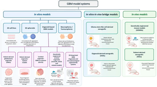
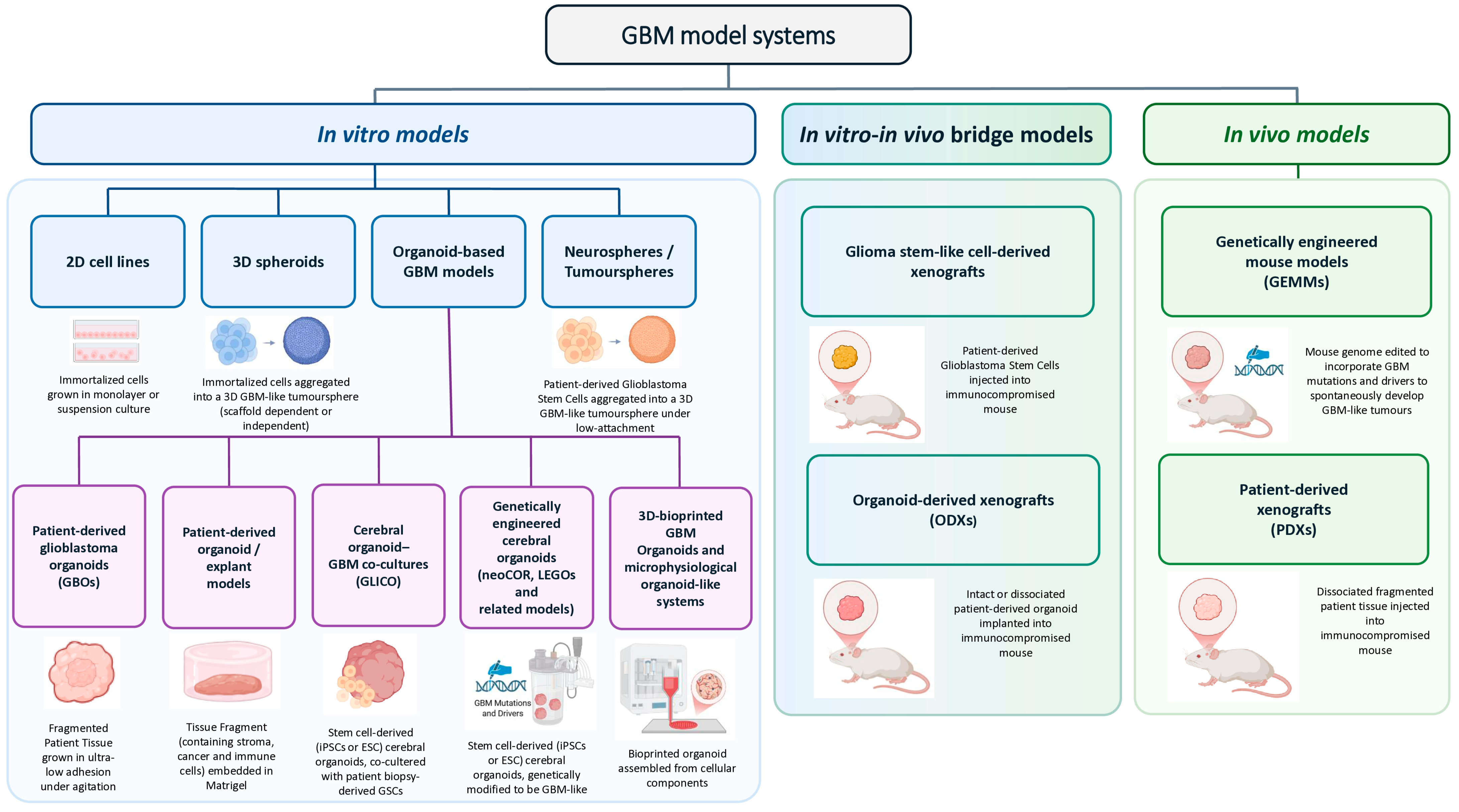

Disclaimer/Publisher’s Note: The statements, opinions and data contained in all publications are solely those of the individual author(s) and contributor(s) and not of MDPI and/or the editor(s). MDPI and/or the editor(s) disclaim responsibility for any injury to people or property resulting from any ideas, methods, instructions or products referred to in the content. |
© 2026 by the authors. Licensee MDPI, Basel, Switzerland. This article is an open access article distributed under the terms and conditions of the Creative Commons Attribution (CC BY) license.