


Abstract
Organoids are three-dimensional multicellular structures that mimic key aspects of native tissues consisting ideal tools to study organ development and pathophysiology when incorporated in customized bioscaffolds. In vivo, the extracellular matrix (ECM) maintains tissue integrity and regulates cell adhesion, migration, differentiation, and survival through biochemical and mechanical signals. Tissue-derived decellularized extracellular matrix (dECM) can preserve organ-specific biochemical signals and cell-adhesive motifs, creating a bioactive environment that supports physiologically relevant organoid growth. 3D bioprinting technology marks a transformative phase in organoid research by enhancing the structural and functional complexity of organoid models and expanding their application in pharmacology and regenerative medicine. These systems enhance tissue modeling and drug testing while adhering to the principles of animal replacement, reduction, and refining (3Rs) in research. Remaining challenges include donor variability, limited mechanical stability, and the lack of standardized decellularization protocols that can be addressed by adopting quality and safety metrics. The combination of dECM-based biomaterials and 3D bioprinting holds great potential for the development of human-relevant, customizable, and ethically sound in vitro models for regenerative medicine and personalized therapies. In this review, we discuss the latest (2021–2025) developments in applying extracellular matrix bioprinting techniques to organoid technology, presenting examples for the most commonly referenced organoid types.
1. Introduction
The Directive 2010/63/EU on animal experimentation within the European Union established the EU Reference Laboratory for Alternatives to Animal Testing (EURL ECVAM) to work on the promotion of non-animal approaches in research, in light of the 3Rs principle—Reduce, Refine, Replace animal experimentation. Among the priorities of ECVAM has been the coordination of the CEN-CENELEC focus group on Organ on Chip, recognizing the transformative effect of this technology in research and regulatory testing utilizing non-animal models [1]. On the other side of the planet, the US Food and Drug Administration (FDA) announced, earlier this year, a comprehensive plan for phasing out animal testing for the development of monoclonal antibodies and other drugs [2]. The FDA commissioner comments on the importance of organoids and organ-on-chip systems as key tools to achieve the goals of the 3Rs, serving not only as a means to this end but also as a more efficient pipeline for novel treatments. UK’s very recent announcement on accelerating the phasing out of animal testing was also largely applauded by the scientific community. UK’s roadmap includes three main directions—organ-on-chip systems, drug efficacy predictions using AI, and 3D-bioprinted tissues to create realistic human tissue samples [3].
Back in 2009, Prof. Hans Clever’s research group at the Hubrecht Institute in Utrecht, the Netherlands, were intrigued by the intestinal tract’s ability for self-repair. They suspected stem cell activity and managed to prove their theory by locating adult stem cells in the intestinal crypts. Later on, they pioneered a technique to grow these cells in the lab and, this way, the first ‘intestinoids’ were generated and applied to test drugs safely on active human intestinal tissue [4]. Organoids are three-dimensional (3D) structures that appear with multicellular complexity and some level of tissue structure and function [5]. In principle, they can expand robustly and for extended periods because of their stem cell nature and can also be genetically manipulated, thus constituting ideal tools for studying organ development and pathophysiology in vitro. Until today, all major organs have been recreated in the form of organoids, including skin, heart, retina, brain, lung, pancreas, kidney, intestine, and liver [6].
In nature, organs and tissues are surrounded by the extracellular matrix (ECM). The ECM is responsible for tissue integrity and elasticity, while transmitting biochemical signals that will regulate cell adhesion, migration, proliferation, differentiation, and survival. It is a two-way function: the cells secrete and remodel their matrix, while the ECM shapes cell identity. Organoids in the lab are developed under 3D culture systems to aggregate by adhesion, self-organize, and differentiate into 3D cell structures with the corresponding tissue morphology. The matrix that will support organoid growth in vitro should replicate, as closely as possible, the native extracellular matrix that would surround the specific tissue in the body. This task is multi-faceted: we not only need to think about biological signaling but also take into consideration the mechanical integrity of the matrix, while also looking out for tissue-specific factors that will affect organoid fate. Diverse scaffold systems, including Matrigel, natural polymers, and synthetic polymers, have been tailored to meet the requirements of organoid cultures. While Matrigel is readily available as a commercialized product, its origin from a mouse tumor raises concerns about the potential transmission of animal pathogens that could impact the immune system [7]. Previous reports have highlighted the issues of data reproducibility due to batch variations of Matrigel, along with pathogenic contamination risks, like the lactate dehydrogenase-elevating virus, and growth factors bearing preparations that affect the cultured cells’ microenvironment [8,9]. Natural or synthetic polymers, as alternatives to Matrigel, are currently studied for cell culture applications. Commonly used matrices of natural origin include collagen, fibrin, gelatin, and hyaluronic acid-based materials, while synthetic matrices are mostly based on polyethylene glycol (PEG) and Pluronic. With a semisynthetic origin, gelatin methacrylate (GelMa) is the most widely used matrix, combining bioactivity and tunability [10]. The problem with natural polymers is the variability in gelation kinetics, which directly affects the uniformity and reproducibility of organoid cultures [7]. Synthetic hydrogels on the other hand, although resolving the variability of natural matrices, exhibit low bioactivity and insufficient tissue-specific ECM composition. Despite the developments in artificial scaffold systems, no single biomaterial fully replaces the diverse functions and complex composition of native ECM. Organ- or tissue-specific hydrogels created solely from decellularized extracellular matrix (dECM) are increasingly used in tissue engineering and, more recently, in clinical applications, due to their preserved bio-instructiveness and inherent cell-adhesive moieties [11]. The tissue-specific nature of the dECM delivers unique biochemical, biophysical, and biomechanical cues to organoids resembling the in vivo cell-matrix communication systems [7]. Due to the diverse composition of ECM across different organs, the use of tissue-specific dECM hydrogels is expected to offer a customized approach for organoid culture.
Three-dimensional (3D) bioprinting technology marks a transformative phase in organoid research by enhancing the structural and functional complexity of organoid models and expanding their application in pharmacology and regenerative medicine. Despite the extraordinary advances in the field of tissue engineering, there are still important limitations such as uncontrollable cell placement, improper perfusability of the microchannels in organ-on-chip research, and difficulty in the identification of biologically relevant materials that will promote cell survival and growth. Three-dimensional bioprinting can resolve such challenges, by controlled cell placement and structure of vascular networks as well as microchannels with suitable permeability [12]. Every x, y, and z axes coordinate, along with cell seeding and material deposition, is precisely regulated and easily modified. In comparison with other scaffolding techniques, 3D bioprinting offers the benefits of automation and high cell density in intricate geometries [13]. Ongoing research in 3D printable materials is expected to guide organoid growth, providing scientists with in vitro study platforms of extraordinary value.
In this review, we aim to bring together the current knowledge on extracellular matrix-based materials for organoid growth and 3D bioprinting applications. We will go through the composition and the functionality of ECM as a biomaterial ex vivo, and we will report on the latest examples of ECM utilization as a growth-supporting matrix for organoid culture. Next, we will see how ECM can be transformed into a bioink for 3D printing applications and the current research on 3D printable ECM materials for organoid culture. Our reviewing method was based on SCOPUS database search with the terms ‘organoid’, ‘organoid culture’, ‘bioink’, ‘bioprinting’, ‘extracellular matrix’, and all the possible combinations of them. From the search results, the document types ‘review’ and ‘conference paper’ were excluded to focus only on research articles containing thorough experimental evidence. Although not limiting the publication year in our initial search, the chosen articles were published in the years 2021–2025, with one exception published in 2019. While papers on the use of extracellular matrix for organoid culture are already dated after the invention and establishment of organoid culture in the lab, the use of extracellular matrix as a bioink component for 3D bioprinting applications is a very young field of study; hence, the chosen articles to be reviewed are dated only in the last 5 years. The reviewed articles were chosen to represent every organoid type that has been reported until today and utilized native extracellular matrix either for the culturing method or as a bioink component.
We want to refer the reader to the review of Guo et al. on dECM-based materials for organoids and engineered organ culture, where the authors report extensively on the different organ and organoid systems that were engineered up to this day [14]. Wang et al. have also reported on the different ECM-based materials for 3D bioprinting and their application for bone, skin, heart, liver, blood vessel, and neuronal tissues [15]. In our review, we will give a bioengineering aspect on the topic and focus on the potential of engineered organoids as in vitro study systems that can substitute animal experimentation. We hope to convince the reader on the capabilities of 3D bioprinting and ECM-based materials for the development of physiologically-relevant organoid systems for disease modeling and drug testing.
2. Extracellular Matrix In- and Ex Vivo: A Multipurpose Biomaterial
In vivo, the extracellular matrix is nature’s scaffolding material. It is composed of two main classes of macromolecules, proteoglycans (PGs) and fibrous proteins, i.e., collagen, elastin, fibronectin, and laminin. PGs fill most of the extracellular interstitial space within a tissue in the form of a hydrated gel, while they serve a variety of functions that vary significantly between different tissues [16]. Structurally, the main element of ECM is collagen, constituting up to 30% of the total protein mass of an organism. The different types of collagens are responsible for fundamental functions, such as tensile strength, cell adhesion, chemotaxis, and migration. An important note here is that ECM directs essential functions—morphological and physiological—by binding growth factors (GFs). These ECM-bound GFs are part of a tightly controlled feedback circuit that is essential to maintain tissue homeostasis. One step further, ECM’s components like GFs can be substantially different from one tissue to another (e.g., bone vs. skin), within one tissue (e.g., renal cortex vs. renal medulla), and even between one physiological state to another (normal vs. cancerous). In a healthy tissue, the ECM is highly dynamic and undergoes continuous remodeling. Type I and III collagens, elastin, and fibronectin form a flexible network of fibers that allow the ECM to resist tensile stresses. A normal tissue can also resist compressive stresses with the help of the glycosaminoglycan (GAG) network that is bonded to the fibrous ECM molecules and influences the overall ECM stiffness. GAGs also bind growth factors that are indispensable in cell signaling and differentiation. For this reason, the GAG content is an indicative factor for the efficacy of ECM extraction and decellularization processes towards the development of ECM-based materials. GAGs’ role in ECM structure and function has been nicely reported by Sodhi and Panitch [17]. Further ECM homeostasis is regulated by the secretion of metalloproteinases from fibroblasts and other enzymes that crosslink and regulate tissue stiffness. These processes are regulated by various GFs. Frantz et al. have comprehensively reviewed ECM structure and function in normal, aged, and diseased states [18].
Ex vivo, ECM accounts for the perfect biological template with a multitude of engineering possibilities. When extracted from a source organism, ECM undergoes a typical procedure for decellularization (dECM) whilst maintaining 3D structure and part of biological activity, such as the preservation of growth factors. Most importantly, the ECM processing ex vivo results in a non-immunogenic material. Thus, we end up with a biocompatible scaffold that can be harnessed in a variety of applications. We can divide the use of extracellular matrix in three broad categories, all of which have been encountered in organoid research: (a) used as a matrix for 3D cell culture, (b) used for biological studies, and (c) used as a bioink component for 3D bioprinting applications. Each category is described below.
- (a)
- dECM as a 3D cell culture matrix—The development of organoids is achieved with three main components: seeded stem cells, culture medium tailored to the tissue-specific needs, and a matrix that will provide structural support and additional nutritional components for the 3D culture [14]. Currently, the commonly used commercial matrix is Matrigel, but it comes with limitations on composition and performance that limit clinical applications of the developed organoids. Research efforts are dedicated to developing dECM matrices derived from tissue-specific ECM, hence showing organ specificity that can be further enhanced with factors that will improve organoid development towards physiologically relevant models. During the extraction and decellularization process, ECM loses its native patterning. Nevertheless, researchers are taking advantage of the preserved collagen content, GAG content, and growth factors that can be present in the final material and drive organoid growth in higher or lower levels depending on the efficacy of the process. To ensure safety and efficacy, dECM should meet standardized quality metrics post-decellularization; the key criteria are listed in Table 1. It is, however, worth mentioning that no universal standardization for this type of product exists up to date [19]. In the following paragraphs, we will present examples of the utilization of dECM for 3D cell culture applications and their importance in regulating organoid microenvironment.
- (b)
- dECM for biological studies—The ECM microenvironment serves as reservoir of several growth factors and other biochemical signals that influence the developmental trajectory of cells and direct pattern formation. During decellularization protocols of tissue-derived ECM, researchers have to carefully select reagents and tissue processing methods to preserve not only ECM’s structural integrity but also its bioactive components. Engineered dECM scaffolds have demonstrated enhanced stimulatory effects for immunomodulation, angiogenesis, and osteogenesis in bone and cartilage reconstruction studies [20].
- (c)
- dECM as a bioink component—Essentially, dECM comes in the form of a hydrogel. This hydrogel can be further modified to induce its gel-forming capabilities, light-activation properties, or any other relevant chemistry that would make it printable in common 3D printers. First reported in 2014 by Pati et al., dECM bioinks were used for printing tissue analogs for adipose, cartilage, and cardiac tissues [21]. The researchers emphasized the adaptability and versatility of the bioprinting methodology utilizing dECM materials, while they observed the effect of the bioink on cell implantation, viability, and sustained functionality. In the following paragraphs, we will report on examples on the development of dECM bioinks relative to organoid applications.
Table 1.
dECM quality and safety metrics commonly used in the field.
3. Applications of dECM-Based Materials for Organoids Culture
In this section we will visit some literature examples on organoids culture in extracellular matrix to understand how the matrix-specific characteristics have influenced organoid development. Table 2 summarizes characteristic examples of studies that demonstrated the potential of ECM as an organoid culture matrix alternative to Matrigel or other synthetic matrices. The most abundant organoid types that we encountered in the recent literature are intestine, kidney, hepatic, brain, retinal, and bone organoids. Tumor organoids of any tissue will be presented in a separate paragraph.
To start with, we will refer to the work of Kim et al. who claimed that Matrigel, the gold standard in organoid culture and murine tumor-derived matrix, can be readily replaced by intestine-derived and stomach-derived extracellular matrix (GI ECM) for the development of intestinal and gastric organoids, accordingly [32]. For the direct comparison of GI ECM with Matrigel, the authors investigated organoid morphology, formation efficiency, organ-specific gene expression level, and organoid functionality. The results demonstrated that ECM hydrogels can address batch-to-batch variability, safety, and cost-issues encountered with Matrigel. Organoids cultured in GI ECMs exhibited higher expression of stem cell markers than Matrigel organoids, although the budding in Matrigel was much better than in GI ECM cultures. Importantly, the developed matrices could be adjusted to clinical settings unlike Matrigel and thus address translational limitations. More specifically, the endotoxin levels and immunogenicity results of the GI-tissue derived materials verified their biocompatibility and safety, making them considerable for clinical applications according to FDA guidelines for implantable medical devices. On the contrary, Matrigel or Matrigel-derived products have not been approved by the FDA up to date, when commercialized products based on decellularized tissue ECM have already been approved (e.g., AlloDerm®, Symvess®, InnovaMatrix®) [33,34,35]. Interested also in a clinically relevant scaffold, Giobbe et al. utilized small intestinal mucosa/submucosa to obtain an ECM scaffold. They showed that intestinal-derived ECM can support the formation and growth of not only intestinal but other endoderm-derived tissue such as liver, stomach, and pancreas [36]. We can also refer the reader to the review of Rezakhani et al. on the extracellular matrix requirements for intestinal, hepatic, and pancreatic organoid culture, presenting significant insights for the development of physiologically relevant organoid systems with biomaterial-based approaches [37].
In kidney organoid research, researchers from Korea have demonstrated that kidney organoids cultured in dECM kidney-derived matrix exhibited vascularization and maturation patterns of glomerular development [38]. Transplantation of the kidney organoids into mouse kidney showed vascular integrity and accelerated recruitment of endothelial cells. In another approach, Garetta et al. engineered the vascularization of kidney organoids in vitro through the assembly of endothelial cell spheroids with kidney organoids, a combination of 2D and 3D approaches [39]. Wang et al. have recently reviewed on extracellular matrix manipulation for kidney organoid research, referring to the ways that ECM refines the microenvironment of organoids but also to the induction of pathological microenvironments in vitro for disease modeling [40]. Brain extracellular matrix has also shown great promise for brain or spinal cord organoid development. Wu et al. isolated brain ECM from mouse and showed that organoids cultured in the tissue-specific matrix exhibited higher expression levels of markers from different parts of the natural spinal cord, compared to the control Matrigel culture [41]. Cho et al. attempted the use of human brain ECM to study its effects on human brain organoids [42]. They found significant differences from the typical organoid culture, among them being the increased neuronal population and enhanced neurogenesis. Very recently, Gai et al. developed bone organoids that were vascularized and mineralized utilizing bone decellularized ECM [43]. The bionic matrix, composed of dECM, calcium phosphate, and salmon DNA facilitated the osteogenesis and angiogenesis of bone marrow mesenchymal stromal cells. The bionic matrix was also successfully used for the in vivo bone repair of cranial defect. A research group from UK demonstrated that the retinal extracellular matrix enhances the expression of synaptic markers and light responsiveness of retinal organoids [44]. The study attempts to address the challenge of robust light responses from retinal organoids so that they can be utilized for drug screening and regenerative medicine. In liver organoid research, van Tienderen et al. demonstrated a novel technique for the scalable production of healthy and tumor liver organoids utilizing microfluidics and liver extracellular matrix [45]. Their organoid microcapsule models were amenable to drug screening, showing a dose-dependent response to clinically relevant anticancer drugs. In this case, the ECM served as a powerful platform to study ECM–organoid interactions and was manipulated for personalized medicine applications.
Tumor organoids or tumoroids are disassociated tumor tissues that mimic the primary tissues in architecture and function. Most importantly, they retain the histopathological features, genetic profile, mutational landscape, and even response to therapy [46]. Compared to the patient-derived xenograft models, organoid establishment requires less time and tissue material, while tumoroids stably maintain the key characteristics of primary tumors even after long-term passaging. Tumoroid research has come to resolve the challenges faced with cancer cells and xenografts, namely the absence of an organism-specific tumor microenvironment and extracellular matrix culturing systems. The extracellular matrix has been a target for therapeutic interventions for cancer treatment, and thus, the study of interactions between ECM and cancer cells would give great insights for treatment protocols. A research group from Italy tried to study this interplay between ECM and metastatic cells of peritoneal metastases from colorectal cancer [47]. Organoids were grown on scaffolds obtained from neoplastic peritoneum, while an impact of ECM on the response to standard chemotherapy treatments was also observed. Importantly, the researchers compared the biochemical factors found in Matrigel with the patient-derived tumor dECM and highlighted how commercially available matrices fail to fully reproduce the in vivo context. This year, scientists developed, for the first time, patient-derived ECM for the growth of glioblastoma multiforme organoids [48]. The study indicated that natively synthesized ECMs eliminate artifacts and variability from xenogeneic scaffolds and provide insights for ECM-targeted drug development.
Table 2.
Applications of dECM-based materials for organoids culture.
4. Beyond Static: dECM as a Bioink
After gaining some insights into organoid culture in dECM-based matrices, we can now explore the concept of dECM as a bioink. As discussed previously, decellularization processes yield ECM materials in the form of a hydrogel. This material can be used as a feeding or base material for a 3D printer. The 3D printing process will yield a 3D model with the shape and other physical characteristics pre-defined by the user using computer-aided design (CAD). When we mix biological material—most often cells—into a dECM hydrogel, we create a bioink that can be used for 3D bioprinting, making it possible to print living tissue structures. Three-dimensional bioprinting technology offers the privilege to control when and where the cells will grow in the printed structure and, this way, generate realistic biological models of tissues and organs. The organoids’ research examples we saw previously necessitate the use of a transplantation organism, since it is not possible to run complex biological studies in vitro. Three-dimensional bioprinting gives us the opportunity to refine, reduce, and replace animal experimentation by developing tissue and organ-like systems with high levels of complexity, precise cell localization and vascularization, and thus perform advanced studies in vitro.
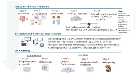
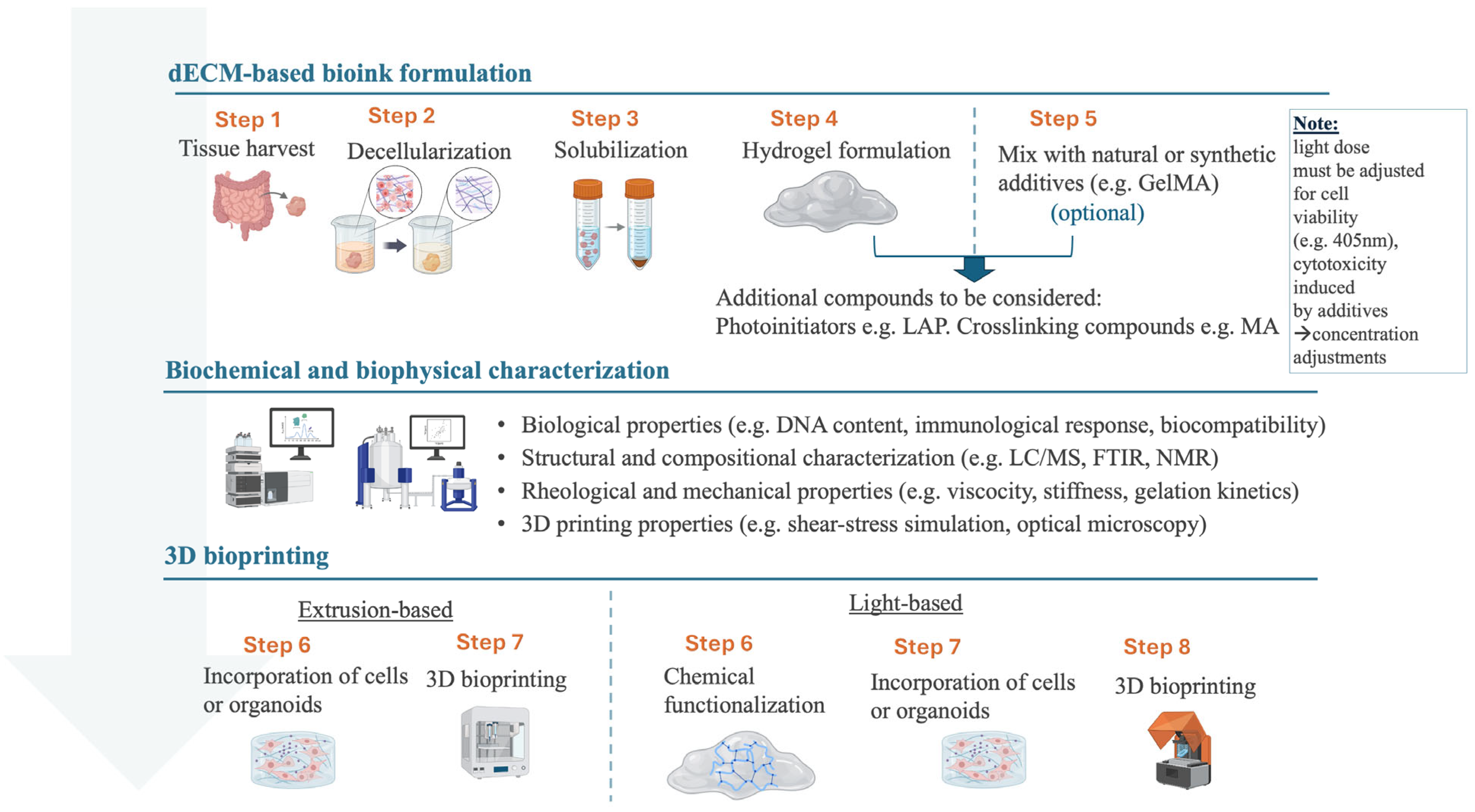
To transform a tissue-derived ECM into a bioink, three main steps must be followed: (1) tissue decellularization with removal of immunogenic factors, (2) tissue solubilization with chemical or enzymatic treatments (pre-gel), and (3) hydrogel formulation with printability (Figure 1). Throughout the bioink production process, several biochemical and biophysical characterization techniques should be employed for the quality control and standardization of the final material. The resulting material can be used directly for extrusion 3D bioprinting, where the bioink is fed through a nozzle and extruded onto a surface in the pre-defined shape layer-by-layer. A careful assessment of printability is necessary when adopting extrusion-based 3D printing techniques, testing several system parameters like the nozzle diameter, the system pressure, and the dispensing rate. The cell viability in this technique can be compromised during the printing process due to mechanical stress. The second-most famous 3D printing technique is light-based 3D bioprinting or vat photopolymerization, where a vat is filled with the liquid material (hydrogel) and the 3D model is produced through light-mediated curing. This technique does not require extensive parameter optimization, but it is necessary to modify the dECM material with photocrosslinkable compounds and a photoinitiator. These additions can raise toxicity issues of the material and, thus, a careful optimization in terms of concentration is needed. Commonly, scientists incorporate additives into the dECM hydrogels to enhance its printability and mechanical stability, or change its physical properties like stiffness, to replicate more closely the different biological components. Such additives can be natural, like glycerol, or synthetic, like methacrylate compounds for enhanced crosslinking. Elomaa et al. have comprehensively reviewed the methods, techniques, and composition of tissue-specific dECM bioinks for 3D bioprinting applications [11]. In the following section, we will examine some literature examples of dECM-based materials that were used as bioinks for enhancing organoid culture.
Figure 1.
The main steps and processes for the transformation of extracellular matrix into a bioink for 3D bioprinting applications. Three main phases (in blue) that contain several steps (in orange) are followed to reach from dECM bioink formulation to a 3D-printed biological model. Steps 1–4 are essential, while the specific steps during the 3D bioprinting phase (6–8) may vary depending on the characteristics of the final material and its intended application. Part of the figure was created with BioRender.
5. Combining Technologies: dECM in 3D Bioprinting for Organoids Studies
Three-dimensional bioprinting has been utilized for organoid studies either through the direct printing of organoids embedded in a bioink or through printing bioscaffolds that will be used as organoid growth platforms. Either way, 3D bioprinting has shown remarkable potential in tissue engineering and regenerative medicine applications through the customized fabrication of biological scaffolds to recapitulate organ and tissue microanatomy. The examples presented in the following paragraphs are summarized in Table 3.
Table 3.
Applications of dECM-based bioinks for organoids culture and 3D bioprinting.
Starting with intestinal organoids applications, Elomaa et al. used decellularized small intestine submucosa for in vitro mimics of intestinal crypts [50]. The researchers modified the dECM material with methacrylic anhydride (MA) and a photoinitiator so that light-based 3D printing would be possible. After thorough characterization on the rheology of the material and its biocompatibility, the optimum concentration (1.5 wt% dECM-MA) was chosen for 3D printing villi-like structures with a low-cost DLP 3D printer. The printed structures showed enhanced mechanical stability over an 11-day cell culture along with physiologically relevant stiffness (3.5 kPa over 2.9 kPa that corresponds to the human small intestine stiffness). The structures were seeded with primary cells derived from human ileal organoids and formed proliferative epithelial cell monolayers that resembled the anatomy of small intestinal epithelium. An important note is that the dECM material preserved its bioactivity regarding the sGAG molecules and several growth factors that are critical for the regulation of intestinal cells and ECM. Additionally, the dECM material supported a higher number of cells that differentiated into goblet cells than the one achieved with the control Matrigel-coated membranes. Xu et al. showcased the potential of intestinal dECM bioinks at different levels of tissue engineering research [51]. After thorough characterization of the decellularized ECM from porcine, the researchers prepared dECM-based bioinks with variable rheological characteristics to investigate the mechanical properties of the material. They used an extrusion-based 3D printing technique with a pneumatic pressure-assisted dispenser and blue light source. The nozzle diameter was 200 μm and the printing speed 10 mm/min. For optimum printing behavior, the dECM bioinks were modified with hyaluronic acid and gelatin, the concentration of which could tune the elastic modulus and thus the stiffness of the produced bioscaffolds. The researchers explored the efficacy of intestinal organoid culture after embedding them in the prepared bioinks and printing them in the shape of intestinal crypts. The results showed that the tissue-specific matrix could deliver biochemical stimulus and maintain stemness more effectively than Matrigel. Importantly, it was also shown that the seeded cells in the bioink could differentiate into enterocytes, goblet cells, enteroendocrine cells, and Paneth cells, exhibiting a distinct differentiation pattern compared to Matrigel cultures. More specifically, Paneth cells and proliferative cells were found increased compared to Matrigel, whereas enterocytes, goblet cells, and enteroendocrine cells were found with reduced proportions. The authors conclude that the tunability in dECM stiffness is related to the promotion of expansion over differentiation in short-lived epithelial cells. Transcriptomic analysis revealed regulatory biological processes supported by the dECM matrix leading to the buildup of organoids specific niche environment. As a next step, co-culture systems were established by feeding fibroblasts and macrophages in the dECM pre-gel along with the intestinal crypts already studied, so 3D bioprinting yielded crypt-like 3D constructs with different cell types that were altogether cultured. Formation of organoids was observed to be higher in the co-culture system compared to the control group (intestinal organoids only). Representative intestinal markers were followed with qPCR to reveal the proliferation and differentiation pattern in the co-culture systems, indicating that the terminal-differentiated enterocytes and the epithelial barrier are more vulnerable to inflammation. This work consists one of the few that demonstrated in detail the potential of dECM bioinks to support intestinal organoids and co-culture systems with quite promising expansion and differentiation levels. The 3D-bioprinted constructs were also implanted in mice showing their potential for regenerative therapies.
In kidney organoid research, Shin et al. investigated the potential of a kidney dECM bioink for 3D bioprinting and kidney microtissue development [52]. In this study, 3D bioprinting was employed as a means to control the material stiffness, a crucial aspect in kidney tissue applications. The researchers prepared a kidney dECM material modified with methacrylic anhydride (MA), achieving mechanical tunability down to 0.67 kPa, a first-time reported value. As a first characterization step, the researchers confirmed cell removal and preservation of collagen fibers as the primary structural component, while confirming that the functionalization process with MA did not affect the total protein content of dECM. In a thorough photo-crosslinking and mechanical properties characterization study, it was revealed that the developed bioink undergoes a rapid crosslinking process (within 10 s of light exposure), that can be tunable depending on the dECM concentration. Compressive modulus could also be tuned, achieving 0.67, 3.19, and 4.81 kPa, respectively. This tunable properties of the kidney dECM make it a promising biomaterial for various applications with different requirements. Pathological conditions such as fibrosis could also be recapitulated with this technique. In the current study, the authors were interested in soft materials since nephron progenitor cells thrive in a range of 0.1–3 kPa, supporting at the same time kidney organoid formation. To investigate bioprinting of iPSC-derived kidney organoids, a customized DLP-SLA bioprinter was used. A 405 nm light beam projector was adjusted to a light stand for precise exposure control, while a Z-axis stage was also precisely positioned for uniform thickness of the 3D models. HEK cells were incorporated in the bioink and at least 90% viability throughout a 14-day culture period was observed post-printing. Structural integrity was assumed to be consistent due to the use of light-based 3D bioprinting that enhances printing resolution and fabrication precision. Over two weeks culture time, 3D mini kidney-like structures were developed. Well-defined cytoplasmic structures with distinct nuclei were revealed during histology assessment. To highlight the versatility of the developed bioink, the authors performed further rheological analysis for extrusion-based 3D bioprinting. Extrusion, droplet-based bioprinting was achieved with a 500 μL Hamilton syringe coupled with a 25 G nozzle. The rotation cycles were optimized for droplet volume control. Post-printing, a 405 nm light source was used for crosslinking the models that would ensure their structural stability. Upon application of shear stress in extrusion bioprinting, the developed bioink exhibited shear-thinning behavior which ensured uniform bioink flow. Cell elongation and proliferation was observed post-printing through fluorescence microscopy; however, cell viability is not reported. In conclusion, the researchers demonstrated that the developed bioink is suitable for both extrusion-based and light-based 3D bioprinting since it can be both stiffness-tunable and photocrosslinkable, making it an ideal scaffold for various renal tissue engineering applications.
The potential of 3D bioprinting to process tailored bioinks for precisely patterned scaffolds has been indispensable for the development of islet organoids towards pancreatic tissue engineering and diabetes research. Zhu et al. designed a customized bioink containing pancreatic ECM that exhibited excellent biocompatibility, pro-angiogenic property, and immunomodulatory ability both in vitro and in vivo [53]. Light-based 3D bioprinting was employed for the layered and partitioned printing of islet organoids. The prepared hydrogel containing mouse islet β cells was printed as a 6 × 6 × 2 square scaffold with a total of 20 layers, 100 μm thickness per layer, and an irradiation time of 40 s with a printing temperature of 37 °C. Biochemical analysis based on mRNA expression indicated insulin sensitivity to glucose during organoid culture at the 3D-printed constructs. In a follow-up study of the same research group, 3D-bioprinted pancreatic islet organoids maintained islet cell adhesion and morphology, mimicking the pancreas microenvironment [54]. The study demonstrated the specific biochemical pathways involved in the promotion of function and activity of pancreatic islets through the designed ECM hydrogel. In vivo transplantation of the scaffolded organoids showed insulin expression and glucagon genes even 12 weeks after transplantation.
In cancer organoid research, Dai et al. demonstrated the extraction of colorectal cancer organoids from patient-derived tumor samples to be used in a high-throughput organoid drug screening platform constructed via 3D bioprinting [55]. The carrier material for organoids would be a composite dECM-GelMa hydrogel that would be used for light-based 3D bioprinting of organoid-loaded gel spheres. At first, dECM was extracted from porcine adipose tissue and mixed with in-house prepared GelMa. This way, the researchers aimed at a material that would both preserve biochemical cues of the extracellular matrix and show excellent printability without compromising cell viability throughout the printing process. A microextrusion 3D bioprinter was used to obtain an array of gel spheres. The prepared material was mixed with the digested colorectal cancer organoids at a cell density of 5 × 10⁵ cells mL−1 and printed in 48-well plates. A post-printing gelation step with exposure to 405 nm light followed. Viable organoid cells post-printing reached 96.9%, whereas Matrigel organoid cultures after 5 days showed 99.26% viability. The dECM-GelMa bioink was also compared with pure GelMa, the conventional printable material, where organoid viability already decreased significantly at the beginning of the culture period (65.14%). This result indicates the significant enhancement in biogenic properties offered by blending dECM with synthetic matrices for optimum 3D bioprinting results. Further, the authors studied the spatial morphology of the 3D-bioprinted organoids. The advantage of tumor organoids over tumor spheroids is their capacity to generate heterogeneous spatial structures, closely resembling the authentic tumor tissue. Therefore, colorectal cancer organoids in this study were characterized with imaging techniques to observe their growth pattern. The organoids displayed a conspicuous glandular lumen-like structure. Overall, 3D bioprinting could obtain multicellular polar structures consistent with traditional culture methods. Notably, the bioprinted organoids showed enhanced proliferative activity than the Matrigel-cultured ones after immunofluorescence staining. The VEGF levels were also found increased in the bioprinted organoids compared to the Matrigel-cultured ones. VEGF is a specifically interested factor in cancer research, not only for its angiogenic activity, but also due to the targeting for known anticancer drugs. The epidermal growth factor (EGFR), again an important biomarker associated with cancer prognosis, was also found elevated in the bioprinted constructs. The developed 3D-bioprinted organoids were also assessed for their sensitivity to oxaliplatin, one of the first-line drugs in the treatment of colorectal cancer. The results indicated remarkable superiority of tumor organoids compared to traditional 2D models in the authenticity of drug evaluation, with the bioprinted embedded organoids responding even better than Matrigel cultures (65.8% cell survival rate in Matrigel over 74.35% in the composite bioink). Live/dead staining of cells indicated no significant barriers to drug penetration, while the actual drug concentration on organoids was identical to the theoretical value. The authors conclude that the developed bioink offered an optimal microenvironment for colorectal tumoroids’ growth and drug evaluation. This research is an excellent example of personalized therapy protocols development with 3D-bioprinted patient-derived tumor organoids, eliminating the use of animal models.
6. Limitations and Challenges
While dECM-based materials continue to attract significant interest, several technical hurdles still limit their reliable translation. A major concern is donor variability: age, pathology, and tissue origin can alter matrix composition and mechanics, leading to inconsistent batches. To mitigate donor-to-donor functional differences of dECM, standardization and stabilization of the product post-decellularization is essential. Common sterilization methods include irradiation with gamma ray or an electron beam, ethylene oxide gassing, or 0.05–0.5% peracetic acid soaking with subsequent thorough dialysis [54], while crosslinking uses methacrylation (DoF 46–96%) followed by photopolymerization using lithium phenyl-2,4,6-trimethylbenzoylphosphinate (LAP) under UV light [56,57]. These preserve printability and growth factor binding while minimizing bioactivity loss. Strategies such as organ pooling, pre-defined statistical batch-release criteria, and the development of standardized base-ECM formulations can help stabilize composition and reduce dependence on products like Matrigel. However, organ-derived ECM should be carefully assessed for its topological differences and specific reporting on the parts of the organ used is needed. For example, intestinal-derived ECM approved by the FDA concerns the small intestinal submucosa region, thus the preparation process for this type of ECM should be attentive if one wants to obtain a clinically relevant matrix.
Incomplete decellularization represents another persistent issue, as residual DNA or membrane fragments may provoke immune responses or affect cell behavior, especially in multicellular systems. At the opposite extreme, harsh processing steps can damage structural proteins, growth factors, and glycosaminoglycans and compromise bioactivity. To reduce these risks, workflows increasingly incorporate process analytics, including LC/MS to monitor retained growth factors, DNA fragment size profiling, and stepwise biochemical assays to fine-tune decellularization conditions for each tissue type. Greater harmonization of protocols across laboratories is essential, as current variation in decellularization and solubilization methods hampers reproducibility and complicates regulatory assessment.
Mechanical limitations remain a constraint for many applications. dECM hydrogels typically offer limited stiffness and poor shape fidelity, which restricts their use in load-bearing contexts and precision fabrication techniques such as 3D bioprinting. Mechanical performance can be improved by blending dECM with defined polymers such as GelMA or HA-MA, enabling more predictable tuning of modulus, crosslinking behavior, and printability. Current research is also exploring methacrylate-free methods, exploiting the inherent functional groups of dECM for crosslinking [58].
Additional challenges arise from the use of xenogeneic tissues, which introduce regulatory, biosafety, and ethical questions, as well as from scale-up barriers, since producing large, compositionally stable batches remains time- and resource-intensive. Implementing controlled pooling, establishing quantitative release specifications, and integrating in-line quality monitoring can ease scale-up and support more reliable manufacturing pipelines. These limitations point out the need for standardized processing, quantitative quality control, and hybrid material strategies that preserve ECM bioactivity while improving mechanical robustness and clinical readiness (Box 1).
Box 1. Best practices in dECM research.
- Usage of organ donor pooling and statistical release criteria to reduce donor-to-donor variability.
- Application of process analytics at key steps (e.g., LC/MS for retained growth factors, DNA fragment analysis, immunological response, GAG assays).
- Optimization of decellularization conditions with tissue-specific response profiling to avoid under- or over-processing.
- Strengthening of mechanics by blending dECM with defined polymers (e.g., GelMA, HA-MA) for tunable stiffness and improved printability.
- Standardization of protocols and documentation of batch-level metadata to support reproducibility and regulatory review.
7. Conclusions and Future Perspectives
Organoid culture necessitates an appropriate matrix that will provide structural support and nutritional components. The matrix is also responsible for biochemical signaling that will drive cell organization and differentiation. The common lab practice for organoid culture involves the use of Matrigel, a commercially available, mouse tumor-derived matrix. However, utilizing dECM derived from tissue-specific ECM displays tissue and organ specificity, while it can be enhanced with relevant proteins and other compounds to foster nature-like organoid models development. The ECM can also be modified chemically towards a printable hydrogel for 3D bioprinting applications, further enhancing the physiological relevance of organoid models. This way, the actual culture matrix can be printed into the specific organ-like structure, whereas Matrigel cannot be readily manipulated for tuning its mechanical or biochemical properties. The growth of the organoid in a custom-designed scaffold that contains biochemical cues leads to organ-like microstructures that can be used for disease modeling and drug screening. Further bioengineering research is expected to give insights into the complex impact of ECM on organoid development and enhance the clinical relevance of scaffolded organoids while eliminating reliance upon animals in research.
Looking ahead, human-derived extracellular matrix from discarded tissue like placenta is currently investigated and will pave the way for organism-specific organoid development tackling the incompatibility issues that arise from animal-sourced materials. The advancement in 3D bioprinting technologies is soon expected to provide realistic models of human organs with vascular branches. One step further, 4D bioprinting brings the potential to manipulate the organoid microenvironment and induce biological processes in real time. The coming decade is bringing organ engineering within reach like never before.
Funding
This research received no external funding.
Institutional Review Board Statement
Not applicable.
Informed Consent Statement
Not applicable.
Data Availability Statement
No new data were created or analyzed in this study. Data sharing is not applicable to this article.
Acknowledgments
Elena Gkantzou acknowledges the support of the European Molecular Biology Organization (EMBO) in the context of the EMBO Research Exchange Grant (Nr. 11419).
Conflicts of Interest
The authors declare no conflicts of interest.
Abbreviations
| 3D | Three-dimensional |
| CAD | Computer-aided design |
| ECM | Extracellular matrix |
| dECM | Decellularized extracellular matrix |
| DLP | Digital-light processing |
| FDA | Food and Drug Administration |
| GelMa | Gelatin methacrylate |
| GAGs | Glycosaminoglycans |
| GFs | Growth factors |
| VEGF | Vascular endothelial growth factor |
| MA | Methacrylic anhydride |
| HA | Hyaluronic acid |
| PEG | Polyethylene glycol |
| PGs | Proteoglycans |
| iPSCs | Induced pluripotent stem cells |
| DoF | Degree of functionalization |
| RT | Room temperature |
| LC/MS | Liquid chromatography/mass spectrometry |
References
- European Commission. 2024 ECVAM Status Report: Biotechnology Boom Accompanies Continuous Drop in Animal Testing—European Commission. Available online: https://joint-research-centre.ec.europa.eu/jrc-news-and-updates/2024-ecvam-status-report-biotechnology-boom-accompanies-continuous-drop-animal-testing-2025-04-02_en (accessed on 20 December 2025).
- US Food and Drug Administration. FDA Announces Plan to Phase Out Animal Testing Requirement for Monoclonal Antibodies and Other Drugs. Available online: https://www.fda.gov/news-events/press-announcements/fda-announces-plan-phase-out-animal-testing-requirement-monoclonal-antibodies-and-other-drugs (accessed on 20 December 2025).
- Animal Testing to Be Phased Out Faster as UK Unveils Roadmap for Alternative Methods—GOV.UK. Available online: https://www.gov.uk/government/news/animal-testing-to-be-phased-out-faster-as-uk-unveils-roadmap-for-alternative-methods (accessed on 20 December 2025).
- Sato, T.; Vries, R.G.; Snippert, H.J.; Van De Wetering, M.; Barker, N.; Stange, D.E.; Van Es, J.H.; Abo, A.; Kujala, P.; Peters, P.J.; et al. Single Lgr5 Stem Cells Build Crypt-Villus Structures in Vitro without a Mesenchymal Niche. Nature 2009, 459, 262–265. [Google Scholar] [CrossRef] [Scilit] [PubMed]
- Takebe, T.; Wells, J.M. Organoids by Design. Science 2019, 364, 956–959. [Google Scholar] [CrossRef] [Scilit] [PubMed]
- Yang, S.; Hu, H.; Kung, H.; Zou, R.; Dai, Y.; Hu, Y.; Wang, T.; Lv, T.; Yu, J.; Li, F. Organoids: The Current Status and Biomedical Applications. MedComm 2023, 4, e274. [Google Scholar] [CrossRef] [Scilit] [PubMed]
- Yoon, J.; Yoon, G.; Jang, J. Transformative Potential of Three-Dimensional Bioprinting Technology for Advanced Organoid Research. Organoid 2024, 4, e8. [Google Scholar] [CrossRef] [Scilit]
- Kawase, E.; Nakatsuji, N. Development of Substrates for the Culture of Human Pluripotent Stem Cells. Biomater. Sci. 2023, 11, 2974–2987. [Google Scholar] [CrossRef] [Scilit]
- Aisenbrey, E.A.; Murphy, W.L. Synthetic Alternatives to Matrigel. Nat. Rev. Mater. 2020, 5, 539. [Google Scholar] [CrossRef] [Scilit]
- Barrs, R.W.; Jia, J.; Silver, S.E.; Yost, M.; Mei, Y. Biomaterials for Bioprinting Microvasculature. Chem. Rev. 2020, 120, 10887–10949. [Google Scholar] [CrossRef] [Scilit]
- Elomaa, L.; Almalla, A.; Keshi, E.; Hillebrandt, K.H.; Sauer, I.M.; Weinhart, M. Rise of Tissue- and Species-Specific 3D Bioprinting Based on Decellularized Extracellular Matrix-Derived Bioinks and Bioresins. Biomater. Biosyst. 2023, 12, 100084. [Google Scholar] [CrossRef] [Scilit]
- Jafarkhani, M.; Salehi, Z.; Aidun, A.; Shokrgozar, M.A. Bioprinting in Vascularization Strategies. Iran. Biomed J. 2019, 23, 9–20. [Google Scholar] [CrossRef] [Scilit] [PubMed]
- De Spirito, M.; Palmieri, V.; Perini, G.; Papi, M. Bridging the Gap: Integrating 3D Bioprinting and Microfluidics for Advanced Multi-Organ Models in Biomedical Research. Bioengineering 2024, 11, 664. [Google Scholar] [CrossRef] [Scilit]
- Guo, X.; Liu, B.; Zhang, Y.; Cheong, S.; Xu, T.; Lu, F.; He, Y. Decellularized Extracellular Matrix for Organoid and Engineered Organ Culture. J. Tissue Eng. 2024, 15, 20417314241300386. [Google Scholar] [CrossRef] [Scilit] [PubMed]
- Wang, H.; Yu, H.; Zhou, X.; Zhang, J.; Zhou, H.; Hao, H.; Ding, L.; Li, H.; Gu, Y.; Ma, J.; et al. An Overview of Extracellular Matrix-Based Bioinks for 3D Bioprinting. Front. Bioeng. Biotechnol. 2022, 10, 905438. [Google Scholar] [CrossRef] [Scilit] [PubMed]
- Chen, D.; Smith, L.R.; Khandekar, G.; Patel, P.; Yu, C.K.; Zhang, K.; Chen, C.S.; Han, L.; Wells, R.G. Distinct Effects of Different Matrix Proteoglycans on Collagen Fibrillogenesis and Cell-Mediated Collagen Reorganization. Sci. Rep. 2020, 10, 19065. [Google Scholar] [CrossRef] [Scilit] [PubMed]
- Sodhi, H.; Panitch, A. Glycosaminoglycans in Tissue Engineering: A Review. Biomolecules 2020, 11, 29. [Google Scholar] [CrossRef] [Scilit]
- Frantz, C.; Stewart, K.M.; Weaver, V.M. The Extracellular Matrix at a Glance. J. Cell Sci. 2010, 123, 4195. [Google Scholar] [CrossRef] [Scilit]
- Tao, M.; Ao, T.; Mao, X.; Yan, X.; Javed, R.; Hou, W.; Wang, Y.; Sun, C.; Lin, S.; Yu, T.; et al. Sterilization and Disinfection Methods for Decellularized Matrix Materials: Review, Consideration and Proposal. Bioact. Mater. 2021, 6, 2927–2945. [Google Scholar] [CrossRef] [Scilit]
- Yuan, Y.; Xu, Y.; Mao, Y.; Liu, H.; Ou, M.; Lin, Z.; Zhao, R.; Long, H.; Cheng, L.; Sun, B.; et al. Three Birds, One Stone: An Osteo-Microenvironment Stage-Regulative Scaffold for Bone Defect Repair through Modulating Early Osteo-Immunomodulation, Middle Neovascularization, and Later Osteogenesis. Adv. Sci. 2024, 11, e2306428. [Google Scholar] [CrossRef] [Scilit]
- Pati, F.; Jang, J.; Ha, D.H.; Won Kim, S.; Rhie, J.W.; Shim, J.H.; Kim, D.H.; Cho, D.W. Printing Three-Dimensional Tissue Analogues with Decellularized Extracellular Matrix Bioink. Nat. Commun. 2014, 5, 3935. [Google Scholar] [CrossRef] [Scilit]
- Crapo, P.M.; Gilbert, T.W.; Badylak, S.F. An Overview of Tissue and Whole Organ Decellularization Processes. Biomaterials 2011, 32, 3233–3243. [Google Scholar] [CrossRef] [Scilit]
- Dortaj, H.; Vaez, A.; Hassanpour-Dehnavie, A.; Alizadeh, A.A. An Update on Technical Method of Cartilage Decellularization: A Physical-Based Protocol. Bioimpacts 2024, 15, 30047. [Google Scholar] [CrossRef] [Scilit]
- Fernández-Pérez, J.; Ahearne, M. The Impact of Decellularization Methods on Extracellular Matrix Derived Hydrogels. Sci. Rep. 2019, 9, 14933. [Google Scholar] [CrossRef] [Scilit] [PubMed]
- Koley, D.; Bard, A.J. Triton X-100 Concentration Effects on Membrane Permeability of a Single HeLa Cell by Scanning Electrochemical Microscopy (SECM). Proc. Natl. Acad. Sci. USA 2010, 107, 16783–16787. [Google Scholar] [CrossRef] [Scilit] [PubMed]
- Keane, T.J.; Swinehart, I.T.; Badylak, S.F. Methods of Tissue Decellularization Used for Preparation of Biologic Scaffolds and in Vivo Relevance. Methods 2015, 84, 25–34. [Google Scholar] [CrossRef] [Scilit] [PubMed]
- Yang, Y.G.; Sykes, M. Xenotransplantation: Current Status and a Perspective on the Future. Nat. Rev. Immunol. 2007, 7, 519–531. [Google Scholar] [CrossRef] [Scilit]
- Jiang, Y.; Li, R.; Han, C.; Huang, L. Extracellular Matrix Grafts: From Preparation to Application (Review). Int. J. Mol. Med. 2021, 47, 463–474. [Google Scholar] [CrossRef] [Scilit]
- Nomura, Y.; Fukui, C.; Morishita, Y.; Haishima, Y. A Biological Study Establishing the Endotoxin Limit for in Vitro Proliferation of Human Mesenchymal Stem Cells. Regen. Ther. 2017, 7, 45–51. [Google Scholar] [CrossRef] [Scilit]
- Guidance for Industry: Pyrogen and Endotoxins Testing: Questions and Answers|FDA. Available online: https://www.fda.gov/regulatory-information/search-fda-guidance-documents/guidance-industry-pyrogen-and-endotoxins-testing-questions-and-answers (accessed on 20 December 2025).
- Sueters, J.; de Boer, L.; Groenman, F.; Huirne, J.A.F.; Smit, T.H.; Zaat, S.A.J. A Sterilization Method for Human Decellularized Vaginal Matrices. Sci. Rep. 2024, 14, 31728. [Google Scholar] [CrossRef] [Scilit]
- Kim, S.; Min, S.; Choi, Y.S.; Jo, S.H.; Jung, J.H.; Han, K.; Kim, J.; An, S.; Ji, Y.W.; Kim, Y.G.; et al. Tissue Extracellular Matrix Hydrogels as Alternatives to Matrigel for Culturing Gastrointestinal Organoids. Nat. Commun. 2022, 13, 1692. [Google Scholar] [CrossRef] [Scilit]
- FDA. Clears First Particulate Placental Extracellular Matrix Medical Device for Wound Management—BioSpace. Available online: https://www.biospace.com/fda-clears-first-particulate-placental-extracellular-matrix-medical-device-for-wound-management?utm_source=chatgpt.com (accessed on 20 December 2025).
- FDA. Approves First Acellular Tissue Engineered Vessel to Treat Vascular Trauma in Extremities|FDA. Available online: https://www.fda.gov/news-events/press-announcements/fda-approves-first-acellular-tissue-engineered-vessel-treat-vascular-trauma-extremities?utm_source=chatgpt.com (accessed on 20 December 2025).
- Dussoyer, M.; Michopoulou, A.; Rousselle, P. Decellularized Scaffolds for Skin Repair and Regeneration. Appl. Sci. 2020, 10, 3435. [Google Scholar] [CrossRef] [Scilit]
- Giobbe, G.G.; Crowley, C.; Luni, C.; Campinoti, S.; Khedr, M.; Kretzschmar, K.; De Santis, M.M.; Zambaiti, E.; Michielin, F.; Meran, L.; et al. Extracellular Matrix Hydrogel Derived from Decellularized Tissues Enables Endodermal Organoid Culture. Nat. Commun. 2019, 10, 5658. [Google Scholar] [CrossRef] [Scilit]
- Rezakhani, S.; Gjorevski, N.; Lutolf, M.P. Extracellular Matrix Requirements for Gastrointestinal Organoid Cultures. Biomaterials 2021, 276, 121020. [Google Scholar] [CrossRef] [Scilit] [PubMed]
- Kim, J.W.; Nam, S.A.; Yi, J.; Kim, J.Y.; Lee, J.Y.; Park, S.Y.; Sen, T.; Choi, Y.M.; Lee, J.Y.; Kim, H.L.; et al. Kidney Decellularized Extracellular Matrix Enhanced the Vascularization and Maturation of Human Kidney Organoids. Adv. Sci. 2022, 9, 2103526. [Google Scholar] [CrossRef] [Scilit] [PubMed]
- Garreta, E.; Moya-Rull, D.; Marco, A.; Amato, G.; Ullate-Agote, A.; Tarantino, C.; Gallo, M.; Esporrín-Ubieto, D.; Centeno, A.; Vilas-Zornoza, A.; et al. Natural Hydrogels Support Kidney Organoid Generation and Promote In Vitro Angiogenesis. Adv. Mater. 2024, 36, 2400306. [Google Scholar] [CrossRef] [Scilit] [PubMed]
- Wang, R.; Sui, Y.; Liu, Q.; Xiong, Y.; Li, S.; Guo, W.; Xu, Y.; Zhang, S. Recent Advances in Extracellular Matrix Manipulation for Kidney Organoid Research. Front. Pharmacol. 2024, 15, 1472361. [Google Scholar] [CrossRef] [Scilit]
- Wu, W.; Liu, Y.; Liu, R.; Wang, Y.; Zhao, Y.; Li, H.; Lu, B.; Ju, C.; Gao, X.; Xu, H.; et al. Decellularized Brain Extracellular Matrix Hydrogel Aids the Formation of Human Spinal-Cord Organoids Recapitulating the Complex Three-Dimensional Organization. ACS Biomater. Sci. Eng. 2024, 10, 3203–3217. [Google Scholar] [CrossRef] [Scilit]
- Cho, A.N.; Jin, Y.; An, Y.; Kim, J.; Choi, Y.S.; Lee, J.S.; Kim, J.; Choi, W.Y.; Koo, D.J.; Yu, W.; et al. Microfluidic Device with Brain Extracellular Matrix Promotes Structural and Functional Maturation of Human Brain Organoids. Nat. Commun. 2021, 12, 4730. [Google Scholar] [CrossRef] [Scilit]
- Gai, T.; Zhang, H.; Hu, Y.; Li, R.; Wang, J.; Chen, X.; Wang, J.; Chen, Z.; Jing, Y.; Wang, C.; et al. Sequential Construction of Vascularized and Mineralized Bone Organoids Using Engineered ECM-DNA-CPO-Based Bionic Matrix for Efficient Bone Regeneration. Bioact. Mater. 2025, 49, 362–377. [Google Scholar] [CrossRef] [Scilit]
- Dorgau, B.; Felemban, M.; Hilgen, G.; Kiening, M.; Zerti, D.; Hunt, N.C.; Doherty, M.; Whitfield, P.; Hallam, D.; White, K.; et al. Decellularised Extracellular Matrix-Derived Peptides from Neural Retina and Retinal Pigment Epithelium Enhance the Expression of Synaptic Markers and Light Responsiveness of Human Pluripotent Stem Cell Derived Retinal Organoids. Biomaterials 2019, 199, 63–75. [Google Scholar] [CrossRef] [Scilit]
- van Tienderen, G.S.; Willemse, J.; van Loo, B.; van Hengel, E.V.A.; de Jonge, J.; van der Laan, L.J.W.; Leijten, J.; Verstegen, M.M.A. Scalable Production of Size-Controlled Cholangiocyte and Cholangiocarcinoma Organoids within Liver Extracellular Matrix-Containing Microcapsules. Cells 2022, 11, 3657. [Google Scholar] [CrossRef] [Scilit]
- Xu, H.; Jiao, D.; Liu, A.; Wu, K. Tumor Organoids: Applications in Cancer Modeling and Potentials in Precision Medicine. J. Hematol. Oncol. 2022, 15, 58. [Google Scholar] [CrossRef] [Scilit]
- Varinelli, L.; Guaglio, M.; Brich, S.; Zanutto, S.; Belfiore, A.; Zanardi, F.; Iannelli, F.; Oldani, A.; Costa, E.; Chighizola, M.; et al. Decellularized Extracellular Matrix as Scaffold for Cancer Organoid Cultures of Colorectal Peritoneal Metastases. J. Mol. Cell Biol. 2023, 14, mjac064. [Google Scholar] [CrossRef] [Scilit] [PubMed]
- Avera, A.D.; Gibson, D.J.; Birge, M.L.; Schnorbus, T.N.; Concannon, I.M.; Kim, Y. Characterization of Native Extracellular Matrix of Patient-Derived Glioblastoma Multiforme Organoids. Tissue Eng. Part A 2025, 31, 17–18. [Google Scholar] [CrossRef] [Scilit]
- Tevlek, A. The Role of Decellularized Cell Derived Extracellular Matrix in the Establishment and Culture of in Vitro Breast Cancer Tumor Model. Biomed. Mater. 2024, 19, 025037. [Google Scholar] [CrossRef] [Scilit] [PubMed]
- Elomaa, L.; Gerbeth, L.; Almalla, A.; Fribiczer, N.; Daneshgar, A.; Tang, P.; Hillebrandt, K.; Seiffert, S.; Sauer, I.M.; Siegmund, B.; et al. Bioactive Photocrosslinkable Resin Solely Based on Refined Decellularized Small Intestine Submucosa for Vat Photopolymerization of in Vitro Tissue Mimics. Addit. Manuf. 2023, 64, 103439. [Google Scholar] [CrossRef] [Scilit]
- Xu, Z.Y.; Huang, J.J.; Liu, Y.; Chen, C.W.; Qu, G.W.; Wang, G.F.; Zhao, Y.; Wu, X.W.; Ren, J.A. Extracellular Matrix Bioink Boosts Stemness and Facilitates Transplantation of Intestinal Organoids as a Biosafe Matrigel Alternative. Bioeng. Transl. Med. 2023, 8, e10327. [Google Scholar] [CrossRef] [Scilit]
- Shin, J.; Tabatabaei Rezaei, N.; Choi, S.; Li, Z.; Kim, D.H.; Kim, K. Photocrosslinkable Kidney Decellularized Extracellular Matrix-Based Bioink for 3D Bioprinting. Adv. Healthc. Mater. 2025, 14, 2501616. [Google Scholar] [CrossRef] [Scilit]
- Zhu, B.; Wang, D.; Pan, H.; Gong, T.; Ren, Q.; Wang, Z.; Guo, Y. Three-in-One Customized Bioink for Islet Organoid: GelMA/ECM/PRP Orchestrate pro-Angiogenic and Immunoregulatory Function. Colloids Surf. B Biointerfaces 2023, 221, 113017. [Google Scholar] [CrossRef] [Scilit]
- Wang, D.; Guo, Y.; Zhu, J.; Liu, F.; Xue, Y.; Huang, Y.; Zhu, B.; Wu, D.; Pan, H.; Gong, T.; et al. Hyaluronic Acid Methacrylate/Pancreatic Extracellular Matrix as a Potential 3D Printing Bioink for Constructing Islet Organoids. Acta Biomater. 2023, 165, 86–101. [Google Scholar] [CrossRef] [Scilit]
- Dai, R.; Chen, W.; Chen, Y.; Jin, J.; Zhang, S.; Zhang, C.; Liu, J. 3D Bioprinting Platform Development for High-Throughput Cancer Organoid Models Construction and Drug Evaluation. Biofabrication 2024, 16, 035026. [Google Scholar] [CrossRef] [Scilit]
- Altunbek, M.; Gezek, M.; Buck, P.; Camci-Unal, G. Development of Human-Derived Photocrosslinkable Gelatin Hydrogels for Tissue Engineering. Biomacromolecules 2023, 25, 165–176. [Google Scholar] [CrossRef] [Scilit]
- Lee, J.Y.; Lee, H.; Jin, E.J.; Ryu, D.; Kim, G.H. 3D Bioprinting Using a New Photo-Crosslinking Method for Muscle Tissue Restoration. NPJ Regen. Med. 2023, 8, 18. [Google Scholar] [CrossRef] [Scilit]
- Kim, M.; Kang, D.; Han, H.; Jang, J. Light-Activated Decellularized Extracellular Matrix-Based Bioinks for Enhanced Mechanical Integrity. Mater. Today Bio 2025, 32, 101859. [Google Scholar] [CrossRef] [Scilit]

Disclaimer/Publisher’s Note: The statements, opinions and data contained in all publications are solely those of the individual author(s) and contributor(s) and not of MDPI and/or the editor(s). MDPI and/or the editor(s) disclaim responsibility for any injury to people or property resulting from any ideas, methods, instructions or products referred to in the content. |
© 2026 by the authors. Licensee MDPI, Basel, Switzerland. This article is an open access article distributed under the terms and conditions of the Creative Commons Attribution (CC BY) license.