


Abstract
Background: Gastrointestinal graft-versus-host disease (GI-GVHD) remains a major cause of morbidity and mortality after allogeneic hematopoietic cell transplantation. Conditioning directly injures the intestinal epithelium, whereas antibiotics, nutritional disruption, and immunosuppression remodel the intestinal ecosystem; together, these exposures are associated with loss of obligate anaerobes and altered availability of microbiota- associated metabolites. This review examines how host tissues sense these metabolic changes and translate them into divergent biological outcomes. Methods: We integrate evidence on directly produced microbial metabolites, including short-chain fatty acids, tryptophan-derived indoles, and desaminotyrosine, as well as microbially modified bile acids, host–microbial co-metabolites such as trimethylamine N-oxide, and emerging microbiota-associated compounds. We then examine receptor-mediated pathways involving FFAR2/3, HCAR2, AhR, PXR, FXR, TGR5, and VDR, as well as receptor-independent mechanisms such as HDAC inhibition and mTORC1-STING-dependent regeneration. Results: We propose that metabolite activity in GI-GVHD is determined not by abundance alone, but by chemical form, concentration, tissue distribution, sensor identity, responding cell type, and disease stage. This framework explains how a metabolite may reinforce epithelial tolerance while promoting inflammatory myeloid or donor T-cell programs in another compartment. We further evaluate postbiotic replacement, receptor modulation, precision nutrition, defined microbial consortia, and fecal microbiota transplantation, highlighting pharmacokinetic, pharmacodynamic, infectious, and graft-versus-leukemia considerations. Conclusions: Cell- and compartment-resolved metabolite pharmacology may enable tissue-protective strategies that limit GI-GVHD without broadly suppressing antitumor immunity.
1. Introduction
1.1. Clinical Relevance of Gastrointestinal GVHD
Allogeneic hematopoietic cell transplantation (allo-HCT) can provide definitive treatment for numerous malignant and nonmalignant hematologic diseases, yet graft-versus-host disease (GVHD) continues to limit its overall benefit. This review specifically addresses gastrointestinal graft-versus-host disease occurring after allogeneic hematopoietic cell transplantation; GVHD arising after solid-organ transplantation or transfusion is outside its primary scope. In GVHD, immunocompetent donor cells recognize recipient antigens and mediate tissue injury [1]. The gastrointestinal tract is both a major target of acute GVHD and a site at which local damage can intensify systemic alloimmunity [1,2]. Injury caused by conditioning compromises tight junctions, the mucus layer, intestinal stem cells (ISCs), and Paneth cells. Damage-associated signals and translocated microbial products then activate host and donor antigen-presenting cells (APCs), expand alloreactive donor T-cells, and increase inflammatory cytokine release. Cytotoxic donor T-cells further injure the crypt epithelium, creating a feed-forward process in which barrier disruption, microbial translocation, and immune activation perpetuate one another. Concurrent loss of regenerative pathways, including interleukin-22-mediated support of ISCs, further weakens epithelial resistance to alloimmune injury [3,4]. Systemic corticosteroids remain the accepted first-line therapy for acute GVHD, although responses are frequently inadequate, especially in severe gastrointestinal involvement [1]. Intensifying immunosuppression may increase opportunistic infection, delay immune reconstitution, facilitate malignant relapse, and raise treatment-related mortality. Nonspecific inhibition of donor immunity can also attenuate the therapeutically desirable graft-versus-leukemia (GVL) response. These trade-offs motivate approaches that reinforce epithelial repair and tissue tolerance without broadly disabling antitumor immunity. The intestinal microbiota and its metabolites are particularly relevant in this context because they jointly influence barrier restoration, innate activation, antigen presentation, and donor T-cell behavior.
1.2. From Microbial Composition to Microbial Metabolic Function
Allo-HCT imposes profound and sustained ecological stress on the intestinal microbiota. Conditioning, broad-spectrum antimicrobials, diminished enteral feeding, dietary change, mucosal injury, and immunosuppressive treatment together reshape community structure. Across clinical cohorts, low microbial diversity, depletion of obligate anaerobes, and overgrowth by facultative pathogens have been linked to bacteremia, severe GVHD, and transplant-related death [5,6,7,8,9]. By contrast, a greater abundance of commensals such as Blautia correlates with lower GVHD-related mortality, whereas lactose-dependent Enterococcus expansion exacerbates GVHD in experimental models and is associated with adverse outcomes in clinical cohorts [7,8]. These observations identify dysbiosis as an important feature of allo-HCT outcomes, but taxonomic profiles alone do not capture the full biology of microbiota–host communication. Distantly related organisms may share metabolic capabilities, while strains assigned to one species can differ markedly in substrate use and metabolite output.
Strain-level functional variation is further shaped by genomic plasticity and ecological adaptation. Accessory genes, plasmids, prophages, pathogenicity islands, and other mobile genetic elements can confer distinct metabolic, antimicrobial-resistance, colonization, and virulence traits among strains assigned to the same species. Horizontal gene transfer contributes to the acquisition and dissemination of these functions, whereas phase variation, epigenetic regulation, and the rewiring of stress-response or two-component regulatory systems can alter microbial phenotypes without requiring conventional gene acquisition. Niche specialization further determines whether these traits support dietary glycan utilization, mucosal persistence, or exploitation of an inflamed intestinal environment. Consequently, commensal and pathogenic behavior should be understood as a context-dependent continuum rather than as a fixed taxonomic property. In allo-HCT recipients, antibiotics, epithelial injury, altered nutrient availability, inflammation, and immunosuppression may act as ecological filters that select for or unmask pre-existing pathobiont-associated functions. Accordingly, strain-resolved genomic and functional analyses are required to distinguish taxonomic persistence from preservation of beneficial metabolic activity. However, direct evidence that de novo horizontal gene transfer or strain-level genomic evolution causally drives GI-GVHD remains limited; these concepts should therefore be regarded as an ecological and evolutionary framework for future investigation rather than as established GI-GVHD mechanisms [10].
Here, “microbiota-associated metabolites” encompasses directly produced microbial metabolites, microbially modified host molecules, and host–microbial co-metabolites. These categories distinguish microbial production from the microbial transformation of host substrates and subsequent host conversion of microbial precursors. Short-chain fatty acids (SCFAs), especially butyrate and propionate, contribute to colonocyte bioenergetics, histone acetylation, junctional stability, and immune regulation through receptor-mediated and intracellular mechanisms [11,12]. Bacterial tryptophan products can engage metabolite sensors and protective interferon-related programs, thereby reducing epithelial leakiness and experimental GVHD [13]. In parallel, microbial conversion of primary bile acids produces unconjugated and secondary species with distinct activity at nuclear and membrane receptors. GVHD-associated T-cell inflammation suppresses these bacterial transformations, and the altered bile acid milieu can in turn reinforce alloimmune activation [14,15]. Dysbiosis therefore encompasses more than shifts in individual taxa; it also involves depletion, redistribution, or the maladaptive remodeling of community metabolic functions.
Recent mechanistic work further demonstrates that host responses to microbial cues are organized by cellular compartment. Microbiota-dependent regulation of epithelial major histocompatibility complex class II can alter GVHD independently of donor–recipient genetic disparity [16], and microbial antigenic inputs can influence the clonal repertoire selected from donor T-cells after transplantation [17,18]. The aromatic amino acid product desaminotyrosine, meanwhile, stimulates intestinal repair through host mTORC1- and STING-dependent programs rather than a conventional metabolite receptor [19]. These findings caution against assigning an invariant protective or harmful label to a metabolite. Biological activity instead reflects its chemical species and local exposure together with sensor availability, target-cell identity, anatomical distribution, and phase of GVHD.
This review examines how allo-HCT-associated ecological disruption changes host exposure to microbiota-associated metabolites, how intestinal epithelial, myeloid, innate lymphoid, and donor T-cell compartments sense these signals, and how the resulting pathways may be pharmacologically manipulated to prevent or treat acute GI-GVHD. We specifically distinguish human clinical associations, causal evidence from GVHD models, direct receptor or pathway validation, and mechanisms extrapolated from nontransplant disorders.
1.3. Scope and Evidence Framework
Throughout this review, the language used to describe microbiota–metabolite–GVHD relationships is aligned with the strength of the underlying evidence. Findings from human cohorts and multi-omics studies are described as associations unless supported by an interventional design. Metabolite supplementation, depletion, microbial transfer, or pharmacological perturbation in GVHD models is interpreted as evidence that an intervention modifies disease severity, but does not by itself establish that endogenous metabolite deficiency initiates GVHD. Statements that a receptor or signaling pathway is required for, or mediates, a biological effect are reserved for receptor-specific genetic manipulation, pharmacological blockade with evidence of target engagement, or rescue and epistasis experiments. Evidence derived from non-GVHD intestinal models is used only to support biological plausibility and is explicitly identified as mechanistic extrapolation or a candidate pathway. Accordingly, reduced metabolite abundance alone is not considered evidence that metabolite loss causes GI-GVHD.
This narrative review primarily addresses acute gastrointestinal GVHD because the most substantial mechanistic and clinical evidence linking the microbiota, microbiota-associated metabolites, and host-sensing pathways has been generated in this setting. Evidence from chronic GVHD is included only when a direct relationship with microbial composition or metabolite exposure has been demonstrated, such as the association between circulating or fecal SCFAs and protection from chronic GVHD. Mechanistic observations from inflammatory bowel disease, infection, metabolic disease, or steady-state intestinal homeostasis are used only to clarify plausible signaling pathways and are explicitly distinguished from direct GVHD evidence.
The relevant literature, published through 31 July 2026, was considered using combinations of terms related to “graft-versus-host disease,” “gastrointestinal GVHD,” “gut microbiota,” “microbial metabolites,” “short-chain fatty acids,” “bile acids,” “tryptophan metabolites,” “indoles,” “metabolite-sensing receptors,” and “postbiotics.” Evidence is summarized along three complementary dimensions: human clinical findings, experimental disease-modification evidence, and sensor or pathway validation. These dimensions are considered separately rather than combined into a single hierarchy.
Accordingly, this review traces the transition from allo-HCT-associated ecological disruption to microbial metabolic dysfunction, defines the major receptor-dependent and receptor-independent sensing pathways, and examines how these signals are integrated by intestinal epithelial cells, myeloid cells, innate lymphoid cells, and donor T-cells. The central proposition is that the biological effects of microbiota-associated metabolites in gastrointestinal GVHD are determined by the integration of metabolite exposure, receptor identity, responding cell type, tissue compartment, and disease stage. This cell- and context-resolved framework provides a pharmacological basis for developing postbiotics, metabolite-receptor modulators, and microbiota-directed interventions that promote intestinal tolerance while preserving antimicrobial defense and GVL activity.
2. From Intestinal Dysbiosis to Metabolic Dysfunction After Allogeneic Hematopoietic Cell Transplantation
2.1. Conditioning-Induced Epithelial Injury and Ecological Disruption
Intestinal dysbiosis after allo-HCT is initiated before gastrointestinal GVHD is clinically evident. Cytotoxic conditioning and total-body irradiation injure rapidly cycling epithelial cells, distort crypt architecture, and weaken mucus and tight-junction barriers [1,2,3]. Because ISCs and their niche are highly susceptible, regenerative capacity is already reduced when subsequent alloimmune injury develops. Loss of Paneth cells further diminishes α-defensins and other antimicrobial peptides, eroding the normal spatial separation between mucosal surfaces and luminal organisms and enabling expansion of potentially pathogenic taxa [3]. The intestine is thereby converted from a homeostatic microbial habitat into an inflammatory niche that favors translocation.
Once the barrier is breached, lipopolysaccharide, peptidoglycan, bacterial DNA, and other microbial-associated molecular patterns gain access to the lamina propria and circulation. Together with danger signals released by injured host cells, they activate innate and antigen-presenting populations, increase inflammatory cytokines, and facilitate donor T-cell priming [1,2]. The local ecology changes concurrently. Disturbed epithelial oxidative metabolism and perfusion, inflammatory oxygen and nitrogen radicals, and impaired nutrient uptake alter both luminal redox conditions and available substrates. These changes preferentially support facultative anaerobes over obligate anaerobic commensals adapted to a stable hypoxic lumen and complex-carbohydrate fermentation. Epithelial damage is therefore not only an outcome of GVHD; it is an upstream ecological event that permits dysbiosis and metabolic failure to emerge.
2.2. Ecological Selection by Antibiotics, Diet, and Transplantation-Related Therapies
Among transplantation-related exposures, antibiotics exert one of the strongest effects on the intestinal microbiome. Regimens with broad anaerobic coverage remove obligate anaerobes that normally supply colonization resistance and key metabolites. Early use of these agents has been associated with greater intestinal injury, GVHD-related death, and unfavorable transplant outcomes in animal models and patient cohorts [5,20]. Community collapse often culminates in domination by a single organism, commonly Enterococcus species or Enterobacteriaceae, and such domination may precede bacteremia caused by the same bacterial group [21]. These data support microbiome-conscious antimicrobial stewardship, but interpretation must account for confounding by indication, infection severity, neutropenia, center-level prescribing, and other time-varying clinical factors.
Nutrient availability constitutes a second, potentially adjustable ecological pressure. Poor oral intake, parenteral feeding, mucositis, and dietary restriction reduce fermentable carbohydrate delivery to the colon and thereby disadvantage SCFA-producing anaerobes. Conversely, particular substrates can selectively promote disease-associated taxa: lactose supports Enterococcus growth, and either dietary or genetic limitation of lactose reduces enterococcal expansion and mitigates experimental GVHD [8]. Experimental pretransplant obesity is likewise accompanied by microbiota alteration, barrier leakage, and more severe acute GVHD [22]. Excessive fructose consumption has more recently been reported to worsen acute GVHD by perturbing the microbiota, damaging the epithelial barrier, and inducing HIF-1α-associated inflammatory macrophage programs [23]. Resistant starch or prebiotic oligosaccharides may instead sustain butyrate-producing organisms and fecal butyrate, although available clinical observations remain exploratory and do not yet prove preventive efficacy [24]. Non-nutritive sweeteners may constitute an additional, but insufficiently characterized, dietary selection pressure. Experimental models and studies in nontransplant populations have reported compound-specific effects of saccharin, sucralose, aspartame, acesulfame-K, and related sweeteners on microbial composition, SCFA production, and host metabolic responses; however, the direction and magnitude of these effects vary according to sweetener formulation, dose, exposure duration, background diet, and experimental model [25]. Human studies have generally demonstrated smaller or inconsistent effects, and no direct evidence currently establishes non-nutritive sweetener exposure as a determinant of microbiome dysfunction or GI-GVHD after allo-HCT.
Transplant-associated treatments should consequently be analyzed as interacting ecological forces. Antibiotics, glucocorticoids, calcineurin inhibitors, gastric acid suppression, opioids, and changes in nutritional support can each modify microbial proliferation, motility, mucosal immunity, or substrate access. The microbiota can reciprocally influence drug transformation and host pharmacokinetics. Because patients with the most severe illness generally receive the most intensive treatment, studies require longitudinal sampling and time-updated exposure models to separate therapy-driven changes from dysbiosis produced by GVHD itself.
2.3. Loss and Reconfiguration of Microbial Metabolic Functions
Dysbiosis has functional consequences that cannot be reduced to the loss of named taxa. One prominent defect is diminished microbial fermentation. After allo-HCT, butyrate availability falls in part because obligate anaerobic producers are depleted. Replenishing butyrate increases epithelial histone acetylation and junctional protein expression and attenuates experimental GVHD; signaling through epithelial GPR43 adds a receptor-dependent layer of protection [11,12]. Thus, the aggregate capacity of a community to generate SCFAs may carry more biological information than the relative abundance of any individual producer.
The network of microbial transformations of host-derived bile acids is similarly remodeled. Bile salt hydrolases (BSH) deconjugate glycine- or taurine-conjugated primary bile acids, whereas bile acid-inducible (bai) enzymes and hydroxysteroid dehydrogenases (HSDH) catalyze dehydroxylation, oxidation, and epimerization. Depletion of these activities changes both bile acid abundance and the receptor activity encoded by the pool. Experimental and integrated multi-omics studies show that GVHD inflammation impairs bacterial bile acid conversion and that the resulting imbalance can reciprocally intensify epithelial and donor T-cell pathology [14,15]. Longitudinal patient studies likewise report fewer secondary bile acids and reduced representation of microbial transformation genes in individuals who develop GI-GVHD, although diarrhea, antibiotics, hepatic dysfunction, and nutritional status can influence these measures [26].
Tryptophan metabolism is disrupted in parallel. Indoxyl sulfate is a host–microbial co-metabolite formed through host conversion of microbiota-derived indole. Low urinary indoxyl sulfate after allo-HCT has been used as an indirect marker of bacterial tryptophan metabolism and is associated with inferior clinical outcomes [27], although urinary levels also depend on host biotransformation and renal handling. Human metabolomics also demonstrates coordinated changes across microbiota- and host-derived tryptophan products during acute GVHD [28]. In experimental systems, bacterial indoles engage type I interferon-associated tissue-protective programs, implying that impaired indole production may directly reduce epithelial resilience rather than simply accompany declining community diversity [13].
Other aromatic amino acid metabolites may participate in mucosal restitution. Desaminotyrosine activates regenerative programs that require mTORC1 and STING, illustrating metabolite action through intracellular stress and repair pathways rather than a classical receptor [19]. Dysbiosis can also favor inflammatory products. Trimethylamine N-oxide (TMAO), a host–microbial co-metabolite generated through the host oxidation of bacterially produced trimethylamine, drives M1-like macrophage polarization and worsens experimental GVHD through nuclear factor κB and NLRP3 inflammasome signaling [29]. Post-transplant metabolic disturbance therefore includes two complementary processes: disappearance of metabolites required for tissue protection and increased exposure to products that amplify innate inflammation.
Taxonomic resemblance should not be equated with metabolic equivalence. Strains within a single species may differ in carbohydrate fermentation, bile acid conversion, aromatic amino acid pathways, or antimicrobial resistance, whereas unrelated taxa can provide redundant functions. Establishing a biologically important metabolic defect consequently requires functional metagenomics, quantitative community measurements, and both targeted and discovery metabolomics rather than compositional profiling alone.
2.4. A Self-Amplifying Metabolic–Immune Circuit
Taken together, available data favor a reciprocal model of microbial involvement in GI-GVHD. Conditioning first damages the epithelium and destabilizes its microbial niche. Antibiotics, reduced enteral substrates, and inflammatory selection then remove metabolically active anaerobes and enrich facultative or pathogenic organisms. Reduced concentrations of SCFAs, secondary bile acids, indole derivatives, and reparative aromatic amino acid metabolites are associated with impaired epithelial recovery and diminished mucosal immune restraint after allo-HCT. Supplementation experiments in GVHD models indicate that selected metabolites can attenuate tissue injury or inflammatory responses; however, these interventions do not establish that endogenous depletion of the corresponding metabolites initiates disease. Conversely, translocated microbial components and selected inflammatory metabolites can amplify myeloid- and donor T-cell responses, thereby aggravating established epithelial injury [11,13,14,15,19,29].
Experimental transfer of disease-associated microbial communities can reproduce or intensify features of GVHD in murine models, and continuous exposure across the pre- and post-transplantation periods may be required to generate the full phenotype [30]. Nevertheless, temporal ordering and transferability alone cannot determine whether an individual metabolite initiates disease, amplifies established inflammation, or serves as a marker of reduced intake, altered transit, or tissue injury. The proposed self-amplifying metabolic-immune circuit should therefore be interpreted as a conceptual synthesis containing both experimentally validated links and association-based connections, rather than as a fully resolved linear causal pathway. (Figure 1).
Figure 1.
Intestinal dysbiosis and metabolic dysfunction after allo-HCT. Conditioning, antibiotics and reduced enteral nutrition can injure the epithelial niche and alter microbial ecology. Changes in SCFA, indole, DAT and bile-acid profiles may alter epithelial, donor T-cell and myeloid responses, impair repair and amplify intestinal immune injury. These pools include microbial products, microbially modified host molecules and host–microbial co-metabolites. The central crypt illustrates the barrier and stem-cell niche. Human associations between microbial communities or metabolite profiles and transplant outcomes are distinguished from experimental GVHD-model findings involving epithelial injury, metabolite responses and immune damage. Gray dashed arrows indicate a conceptual sequence and feedback, with immune injury returning to epithelial injury. They do not establish that every link, or the complete circuit, has been causally validated in patients. Gray solid arrows in the origin schematic indicate microbial production or subsequent host conversion. Dose, compartment, responding cell and disease stage modify exposure and response. Blue identifies transplant exposures, rose/red injury, ochre dysbiosis, purple metabolite dysfunction and teal cellular responses. Green niche cells represent stem cells. Other cell, microbial and particle colors distinguish schematic biological elements, not evidence strength or specific taxa. Upward/downward symbols indicate increases/decreases, and ≠ means ‘does not establish.’ Metabolite depletion alone does not demonstrate disease causation. allo-HCT, allogeneic hematopoietic cell transplantation; GVHD, graft-versus-host disease; GI-GVHD, gastrointestinal graft-versus-host disease; ISC, intestinal stem cell; T-cell (T), T lymphocyte. DAT, desaminotyrosine; SCFA/SCFAs, short-chain fatty acid/acids.
3. Molecular Pathways for Host Sensing of Microbiota-Associated Metabolites
Host sensing of microbiota-associated metabolites involves G protein-coupled receptors, nuclear receptors, ligand-regulated transcription factors, and receptor-independent intracellular mechanisms. This section distinguishes direct ligand–sensor interactions from metabolic actions and downstream signaling requirements. The cellular and compartmental consequences of these mechanisms are examined in Section 4.
3.1. Short-Chain Fatty Acid Sensing
Acetate, propionate, and butyrate are the predominant SCFAs generated by the bacterial fermentation of nondigestible carbohydrates, with a smaller contribution from amino acid fermentation. Their host effects involve extracellular receptor signaling, intracellular enzyme inhibition, and metabolic utilization [11,31,32,33].
SCFAs are interpreted through extracellular receptors and intracellular metabolic mechanisms. The principal G protein-coupled sensors are free fatty acid receptor 2 (FFAR2/GPR43), free fatty acid receptor 3 (FFAR3/GPR41), and hydroxycarboxylic acid receptor 2 (HCAR2/GPR109A) [34]. Their ligand spectra overlap but are not identical. FFAR2 responds to acetate, propionate, and butyrate. FFAR3 is relatively more responsive to propionate and butyrate, whereas HCAR2 is activated by butyrate, β-hydroxybutyrate, and nicotinic acid. These receptors also differ in their distribution across epithelial, myeloid, neuronal, adipose, and other immune compartments. SCFA biology therefore cannot be represented by a simple one-ligand–one-receptor scheme.
FFAR2/GPR43 has direct mechanistic support in experimental GVHD. GPR43-dependent protection involves ERK signaling and an NLRP3–IL-18 program, and receptor deficiency worsens gastrointestinal injury and mortality [12]. Evidence for HCAR2-mediated intestinal regulation derives mainly from nontransplant models [32,35]. whereas a cell-defined causal role for FFAR3 in GI-GVHD remains unestablished. The consequences for epithelial and immune compartments are discussed in Section 4.
Transport through monocarboxylate carriers permits intracellular SCFA actions distinct from surface-receptor signaling. Butyrate, and propionate at higher exposure, inhibit histone deacetylases and alter histone acetylation and chromatin accessibility. Experimental GVHD studies support an association between epithelial butyrate availability and histone acetylation [11], whereas the resulting epithelial and T-cell responses are considered in Section 4.1 and Section 4.3.
In differentiated colonocytes, butyrate also serves as a dominant oxidative fuel. Mitochondrial utilization generates adenosine triphosphate, consumes oxygen, and helps sustain a hypoxic epithelial interface favorable to obligate anaerobes. Impaired butyrate oxidation may therefore oxygenate the epithelial surface and support facultative anaerobic expansion, linking host energy metabolism back to community ecology.
SCFAs can therefore act as receptor ligands, epigenetic enzyme inhibitors, and respiratory substrates. Distinguishing these mechanisms is necessary before attributing an observed response to a specific SCFA receptor. SCFA exposure differs markedly between anatomical compartments. In a human postmortem study, total SCFA concentrations ranged from 80 ± 11 mmol/kg in descending-colon contents to 131 ± 9 mmol/kg in caecal contents, whereas portal and peripheral venous concentrations were 375 ± 70 and 79 ± 22 μmol/L, respectively [36]. By comparison, Mathewson et al. used 1 mM butyrate in primary intestinal epithelial-cell experiments associated with their GVHD study [11]. This nominal concentration exceeds the reported peripheral concentration of total SCFAs, supporting caution when extrapolating epithelial culture findings to circulating cells. However, total SCFA measurements cannot be equated with butyrate concentrations, and neither colonic contents nor culture medium directly represents mucosal cellular exposure. These postmortem data provide compartmental context rather than reference ranges for allo-HCT recipients.
3.2. Sensing of Tryptophan-Derived Indoles
Tryptophan is partitioned among host kynurenine and serotonin metabolism and bacterial pathways that produce indole, indole-3-aldehyde, indole-3-acetic acid, indole-3-propionic acid, indoleacrylic acid, and related molecules. Some of these products remain concentrated in the intestine, whereas others enter portal blood or undergo additional host conversion. Microbiota-derived indole undergoes host-mediated oxidation to indoxyl and subsequent sulfation, predominantly in the liver, to form indoxyl sulfate. Indoxyl sulfate is therefore a host–microbial co-metabolite, and its urinary concentration provides an indirect readout of bacterial tryptophan metabolism that also depends on host biotransformation and renal handling. For unsubstituted indole, a study of 53 healthy adults reported fecal concentrations of 0.30–6.64 mM, with a mean of 2.59 mM [37]. In an epithelial-cell study, HCT-8 cells were exposed to 1 mM indole, with transcriptional responses examined after 4 and 24 h [38]. This experimental concentration falls within the reported fecal range, providing a quantitative rationale for investigating local intestinal effects. Nevertheless, fecal measurements do not establish epithelial or lamina propria exposure, and these values should not be extrapolated to other indole derivatives or interpreted as reference concentrations after allo-HCT.
A central sensor for bacterial indole derivatives is the aryl hydrocarbon receptor (AhR). Without ligands, AhR is retained in a cytoplasmic protein complex. Ligand engagement promotes its nuclear entry, association with AhR nuclear translocator, and regulation of xenobiotic response elements. The resulting programs affect IL-22 production, antimicrobial defenses, epithelial regeneration, immune lineage differentiation, and xenobiotic handling. Indole-3-aldehyde provides an example of a microbial ligand engaging AhR in experimental intestinal models [39]. Its implications for epithelial protection and innate-cell-mediated responses are discussed in Section 4.1.
AhR ligands differ in affinity, metabolic persistence, tissue distribution, and their effects on receptor turnover. AhR activation should therefore be characterized at the level of individual ligands rather than treated as a uniform pharmacological state. Cell-specific differentiation outcomes are discussed in Section 4.
Indole compounds can additionally engage the pregnane X receptor (PXR), although activity depends on the metabolite, species, and ligand context. In reporter assays, indole-3-propionic acid (IPA) alone activated human PXR with an EC50 of 120 μM, compared with 0.55 μM for mouse PXR; human PXR activation was enhanced when IPA was combined with 1 mM indole [40]. The same study linked this signaling axis to reduced TLR4-associated epithelial dysfunction. These results highlight substantial species differences, but assay-derived EC50 values do not establish effective tissue concentrations or therapeutic thresholds in GI-GVHD. Supporting evidence derives principally from non-GVHD intestinal models, and receptor-specific validation in allo-HCT remains limited. The potential epithelial consequences are considered in Section 4.1.
In experimental GVHD, indole-mediated protection requires intact host type I interferon signaling [13]. This establishes a downstream signaling requirement rather than the direct binding of indoles to the interferon receptor. Tissue protection and preservation of GVL activity are discussed in Section 4.1 and Section 4.3, respectively.
3.3. Bile Acid Sensing
Bile acids originate from host cholesterol metabolism, but intestinal microorganisms greatly expand their chemical repertoire. Secondary bile acids and their derivatives are therefore described here as microbially modified host-derived molecules rather than exclusively microbial products. Hepatic primary bile acids are usually conjugated to glycine or taurine before entering bile. In the intestine, BSH remove these conjugates, HSDH catalyze oxidation and epimerization, and enzymes encoded by the bai operon mediate 7α-dehydroxylation. These reactions generate secondary and other microbially modified bile acids whose receptor profiles differ from those of the original host molecules.
Remodeling of the bile acid pool changes the balance of agonists, partial agonists, and antagonists available to host receptors [14,15,26]. The farnesoid X receptor (FXR/NR1H4) is a bile acid-activated nuclear receptor. After ligand binding, FXR partners with retinoid X receptor to control bile acid synthesis and transport, detoxification, lipid handling, and epithelial homeostasis. Donor T-cell FXR has direct experimental support in GVHD [14]. Its cellular consequences are discussed in Section 4.3.
Takeda G protein-coupled receptor 5 (TGR5/GPBAR1) is a membrane bile acid sensor distributed across epithelial, enteroendocrine, myeloid, neural, and other cell types. Its activation raises intracellular cyclic adenosine monophosphate and can suppress nuclear factor κB-dependent inflammation. In intestinal stem-cell models, TGR5 also promotes regeneration and epithelial recovery [41,42].
The vitamin D receptor (VDR) and PXR provide additional nuclear sensing routes for bile acids [43]. Lithocholic acid and selected derivatives activate VDR, an intestinal bile acid sensor that coordinates detoxification and barrier-associated transcription [44]. PXR recognizes several hydrophobic bile acids and regulates xenobiotic processing and epithelial defense. Because microbial indoles also activate PXR, this receptor may integrate signals from distinct microbial metabolic networks rather than report bile acid exposure alone [40].
Bile acids may also influence cellular stress through mechanisms not fully explained by canonical receptor activation. Accordingly, the efficacy of TUDCA should not be assigned to FXR or TGR5 without receptor-specific validation [45]. Its epithelial effects are discussed in Section 4.1.
The receptor signal encoded by a bile acid pool depends on molecular composition rather than total concentration. Conjugation controls passive permeability and transporter requirements, whereas hydroxylation and epimerization modify affinity and efficacy. Individual bile acids may act at several receptors; a weak FXR ligand may, for example, be a more effective agonist at TGR5 or VDR. Exposure also differs among the lumen, mucosa, portal blood, liver, and systemic circulation. Fecal bile acid data alone therefore cannot identify receptor engagement in a particular host compartment. In addition, substantial differences between murine and human bile acid pools limit direct quantitative extrapolation from experimental models. Mechanistic studies should therefore combine targeted bile acid profiling with transporter abundance, receptor activity, anatomical localization, and downstream pharmacodynamic responses.
Quantitative measurements are particularly important for individual bile acid derivatives. Hang et al. reported mean fecal levels of 3-oxoLCA and isoalloLCA of 23 and 2 pmol/mg, respectively, in patients with ulcerative colitis; isoalloLCA values ranged from 0 to 17 pmol/mg [46]. In the same study, representative T-cell differentiation experiments used 20 μM 3-oxoLCA or isoalloLCA. These findings demonstrate endogenous occurrence and experimental activity, but fecal mass-normalized measurements cannot be directly equated with culture-medium molarity or free concentrations at intestinal T-cells. Moreover, these human measurements were obtained outside allo-HCT and do not establish whether comparable cellular exposure occurs in GI-GVHD.
3.4. Noncanonical and Emerging Metabolite-Sensing Pathways
A recognized membrane or nuclear receptor is not required for every effect of microbiota-associated metabolites. Some products are used as fuels or biosynthetic substrates, some inhibit enzymes, and others alter intracellular stress or repair networks without directly binding the signaling protein implicated downstream. Correctly distinguishing these modes of action is essential when a sensor is proposed for a newly studied metabolite.
Desaminotyrosine illustrates the distinction between a downstream signaling requirement and a direct molecular sensor. Its protective response in experimental GVHD requires mTORC1 and STING, but direct binding to either component has not been established [19]. The regenerative phenotype is discussed in Section 4.1.
TMAO is a host–microbial co-metabolite whose proximal recognition mechanism remains uncertain [47]. Its formation involves microbial trimethylamine production and host oxidation, while circulating exposure also depends on renal elimination. Its inflammatory effects in experimental GVHD are discussed in Section 4.2.
Other candidate mechanisms are derived mainly from intestinal models outside GVHD. Spermine, spermidine, and related polyamines affect chromatin, autophagy, mitochondrial activity, and inflammasome function; microbiota-dependent polyamine signals have been connected to NLRP6 regulation and epithelial antimicrobial defenses [43,48], Because polyamines can arise from dietary, microbial, and host sources, their abundance alone does not establish microbial origin. Bacterial lactate can stimulate intestinal stem-cell proliferation and development through combined receptor and metabolic effects [42,49]. Whether either pathway materially contributes to crypt recovery after allo-HCT remains unknown.
Succinate may rise during dysbiosis, tissue hypoxia, or impaired mitochondrial metabolism. Extracellular succinate activates succinate receptor 1, while intracellular accumulation can stabilize HIF-1α and enhance interleukin-1β production [50,51]. Because microbial and host pathways both generate this intermediate, source-resolved approaches such as isotope tracing are necessary before a GVHD phenotype can be attributed specifically to bacterial succinate.
Bacterial sphingolipids and vitamin-derived antigens represent further signaling categories. Sphingolipids synthesized by symbiotic organisms alter host lipid networks and help regulate intestinal natural killer T-cell homeostasis [52]. Riboflavin intermediates can be displayed by the nonclassical antigen-presenting molecule MR1 and recognized by mucosal-associated invariant T-cells, providing a direct connection between microbial vitamin metabolism and lymphocyte activation [53]. Evidence directly linking either mechanism to GI-GVHD is currently sparse; they should therefore be treated as testable hypotheses rather than validated intervention targets.
Emerging metabolite pathways should be categorized by the type of evidence supporting them: verified binding to a receptor, transporter-dependent intracellular action, use as an energetic or anabolic substrate, modulation of a metabolic enzyme, or requirement for a downstream signaling module. This scheme avoids describing downstream dependencies as receptors and keeps GVHD-specific causality separate from mechanisms inferred from other inflammatory disorders. It also provides the necessary foundation for comparing how epithelial, myeloid, innate lymphoid, and donor T-cell compartments interpret the same metabolic exposure.
4. Cell-Specific Integration of Metabolite Sensing in Gastrointestinal GVHD
Building on the molecular pathways described in Section 3, this section examines how epithelial, stem-cell, myeloid, and donor T-cell compartments integrate metabolite signals. Particular attention is given to differences between tissue protection and immune suppression, the cellular attribution of experimentally observed effects, and the distinction between direct GVHD evidence and mechanistic extrapolation (Figure 2).
Figure 2.
Cell-specific sensing of microbiota-associated metabolites. The upper panels summarize experimental GVHD findings: GPR43-associated intestinal protection through ERK/NLRP3/IL-18, selected indole-dependent protection requiring host type I IFN signaling, and donor T-cell FXR signaling that can enhance inflammation. DAT requires mTORC1 and epithelial STING for the studied regenerative response, whereas TMAO promotes downstream NF-κB/NLRP3-associated inflammation. Direct DAT binding remains unproven, and the proximal TMAO sensor is unresolved. Solid arrows in response pathways indicate functional relationships tested in GVHD models, not necessarily direct ligand binding. Solid source-conversion arrows depict microbial production, microbial modification of host bile acids, or microbial TMA followed by host oxidation to TMAO. Gray dashed arrows identify mechanisms established outside GVHD: HCAR2/FFAR3 and T-cell programs, AhR/IL-22 and PXR-associated barrier regulation, and TGR5/VDR/PXR biology. These mechanisms are not presented as verified mediators of clinical GI-GVHD responses. Green result labels/arrows indicate repair or protection, red inflammation, and up arrow means an increase. Green, ochre and purple headings distinguish SCFAs, indoles and bile acids, respectively. Other illustration colors identify biological elements, not evidence strength. Epithelial icons indicate tissue protection without assigning every signal exclusively to epithelial cells. The asterisk restricts retained GVL to tested regimens. Human metabolite associations do not establish these causal pathways or clinical efficacy. AhR, aryl hydrocarbon receptor; DAT, desaminotyrosine; ERK, extracellular signal-regulated kinase; FFAR3, free fatty acid receptor 3; FXR, farnesoid X receptor; GPR43, G protein-coupled receptor 43; GVHD, graft-versus-host disease; GI-GVHD, gastrointestinal graft-versus-host disease; GVL, graft-versus-leukemia; HCAR2, hydroxycarboxylic acid receptor 2; IFN, interferon; IL-18/IL-22, interleukin-18/interleukin-22; ISC, intestinal stem cell; mTORC1, mechanistic target of rapamycin complex 1; NF-κB, nuclear factor kappa B; NLRP3, NLR family pyrin domain containing 3; PXR, pregnane X receptor; SCFAs, short-chain fatty acids; STING, stimulator of interferon genes; T-cell, T lymphocyte; TGR5, G protein-coupled bile acid receptor 1; TMA, trimethylamine; TMAO, trimethylamine N-oxide; VDR, vitamin D receptor.
4.1. Intestinal Epithelial and Stem Cells: Barrier Integrity and Tissue Tolerance
Differentiated epithelial cells and crypt stem/progenitor cells may respond differently to the same metabolite exposure. Butyrate restoration improves epithelial survival and barrier integrity in experimental GVHD [11]. However, non-GVHD crypt models show that the exposure of normally protected stem/progenitor cells to butyrate can inhibit proliferation [54]. This distinction highlights the importance of barrier architecture and epithelial differentiation state; it should not be interpreted as an established inhibitory effect on intestinal regeneration during GVHD.
FFAR2-associated protection and indole-dependent host responses provide additional evidence for tissue-protective metabolite signaling [12,13]. The molecular pathways are described in Section 3.1 and Section 3.2. A remaining question is whether protection reflects direct epithelial sensing, signals from neighboring innate cells, or coordinated responses across these compartments. In particular, AhR- and PXR-associated barrier mechanisms established outside transplantation require further cell-specific validation in allo-HCT [39,40].
TUDCA reduces epithelial apoptosis and MHC class II expression, thereby limiting intestinal antigen presentation [45], whereas desaminotyrosine supports intestinal regeneration [19]. TGR5-dependent stem-cell regeneration provides a further candidate mechanism, although the supporting evidence largely derives from non-GVHD models [41]. These findings identify epithelial survival, local antigen presentation, and regenerative capacity as distinct therapeutic outcomes. Their implications for preservation of GVL activity are considered in Section 4.3.
4.2. Myeloid Cells and APCs: Inflammatory Amplification
After conditioning, recipient macrophages and dendritic cells are exposed to microbial products in the lamina propria, draining lymph nodes, and bloodstream. Barrier failure permits translocation of microbial-associated molecular patterns, while damaged tissue releases extracellular adenosine triphosphate, uric acid, and other danger signals. Metabolite sensing consequently occurs against a highly activated inflammatory background and can either increase or reduce the threshold for myeloid-cell activation.
SCFAs and selected bile acid or AhR ligands can favor less inflammatory macrophage and dendritic-cell states in nontransplant models [32,42]. Their relevance to GI-GVHD requires demonstration that myeloid-cell sensing contributes to disease modification. Inflammasome responses also require cellular attribution: epithelial repair-associated signaling should be distinguished from inflammatory IL-1β production in recipient immune compartments. Thus, an anti-inflammatory effect in another disease model does not establish a validated myeloid therapeutic target in GVHD.
TMAO provides direct experimental evidence of a metabolite-associated inflammatory myeloid response in GVHD. Exposure promotes an M1-like macrophage state, NF-κB/NLRP3-associated signaling, and inflammatory cytokine release, accompanying increased disease severity [29]. Its host–microbial formation and determinants of systemic exposure are described in Section 3.4.
Professional APCs and intestinal epithelial cells contribute differently to alloantigen presentation. Recipient dendritic cells support donor T-cell priming, whereas epithelial MHC class II provides a local route for donor CD4+ T-cell activation within the intestine [55]. Metabolite-associated changes in these compartments should therefore be assessed separately: reduced APC activation, reduced epithelial antigen presentation, and improved barrier integrity represent distinct outcomes and may require different intervention windows.
4.3. Donor T-Cells: Differentiation, Cytotoxicity and GVL Activity
Donor T-cells can encounter microbiota-derived metabolites during priming in lymphoid organs, circulation, and subsequent entry into the intestine. SCFAs promote regulatory T-cell differentiation in nontransplant models [31,32,33,56], but this does not establish that the direct stabilization of donor-derived Tregs mediates protection in GVHD. Studies must distinguish direct donor T-cell effects from secondary changes caused by epithelial protection or altered antigen presentation.
Microbially modified bile acid derivatives exert structure-dependent control over T-cell fate. In non-GVHD models, 3-oxo-lithocholic acid suppresses RORγt-dependent Th17 differentiation, isoallo-lithocholic acid promotes regulatory T-cell development, and other secondary species support RORγt+ peripheral regulatory T-cells [46,57,58]. These experiments establish a mechanistic link between bacterial bile acid conversion and the Th17/Treg balance, but sufficient exposure of relevant tissues after allo-HCT has not been demonstrated for many of these molecules.
Donor T-cell FXR has direct mechanistic support in experimental GVHD. Altered microbial bile acid conversion changes the ligand environment and can enhance FXR-dependent inflammatory responses in donor T-cells [14]. This finding establishes the donor lymphocyte compartment as a relevant site of bile acid sensing, while leaving the effects of epithelial or hepatobiliary FXR manipulation to be evaluated separately.
AhR-associated T-cell differentiation is context dependent, with regulatory, Th17, and Tr1 outcomes reported under different experimental conditions. These findings should not be interpreted as evidence that a microbial indole produces the same differentiation program in donor T-cells during GVHD. Translation requires direct assessment of donor-cell phenotype and function rather than aggregate AhR activity alone.
Donor CD8+ T-cells warrant separate evaluation. Metabolite-dependent changes in mitochondrial metabolism, chromatin acetylation, cytokines, or homing receptors may alter their expansion and cytotoxicity. Excessive inhibition could alleviate GVHD while compromising pathogen defense or the GVL response. Notably, indole-mediated tissue protection and TUDCA each reduced experimental GVHD without abolishing GVL activity [13,45]. These findings suggest that increasing tissue tolerance may separate GVHD control from GVL more effectively than broad donor T-cell suppression. Even so, GVL preservation must be tested directly for each metabolite or receptor intervention.
4.4. Spatial, Temporal and Cellular Determinants of Biological Response
The examples discussed in Section 4.1, Section 4.2 and Section 4.3 show that metabolite activity cannot be inferred from ligand identity alone. Responding-cell identity, differentiation state, receptor expression, and barrier integrity influence which biological response predominates. An effect demonstrated in one compartment should therefore not be assumed to occur in another.
Fecal concentrations are incomplete surrogates for cellular exposure. Levels in the lumen, mucus, intestinal tissue, portal blood, and systemic circulation can differ because of microbial utilization, epithelial uptake, host biotransformation, and clearance. Paired metabolite measurements and cell-resolved pharmacodynamic analyses are needed to connect exposure with the proposed response.
The relevant target compartment also changes over the disease course. Donor T-cell priming and recipient APC activity are prominent during initiation, whereas epithelial injury, local antigen presentation, and inflammatory effector responses sustain established disease. Regenerative programs become particularly relevant during recovery. Metabolite-directed interventions should consequently specify the intended target cell, exposure compartment, and therapeutic window rather than assume a uniform effect throughout GI-GVHD (Table 1).
Table 1.
Selected evidence for host sensing of microbiota-associated metabolites relevant to GI-GVHD after allo-HCT.
5. Pharmacological Exploitation of Microbiota–Metabolite–Host Sensing Pathways
The microbiota–metabolite–host-sensor axis offers several distinct intervention points: replacing a depleted metabolite, modulating its host sensor, restoring microbial metabolite production and transformation of host-derived substrates, or altering the ecology that supports the relevant pathway. These approaches differ in controllability and biological breadth. A chemically defined postbiotic permits reproducible dosing, but may not recapitulate the location and kinetics of community production. Fecal microbiota transplantation (FMT) and live biotherapeutics can restore multiple functions at once, yet engraftment, metabolite output, and biosafety are less predictable. Translation should therefore emphasize correction of a measurable metabolic and sensing defect rather than restoration of taxonomy as an end in itself.
5.1. Direct Metabolite Replacement and Postbiotics
SCFAs are among the most extensively investigated candidates for postbiotic therapy. In murine acute GVHD, butyrate supplementation or restoration of epithelial butyrate exposure increases histone acetylation, strengthens tight-junction integrity, and attenuates intestinal injury [11]. Propionate attenuated experimental GVHD through GPR43-dependent signaling, whereas acetate did not improve GVHD in the tested regimen [12]. By contrast, the principal human evidence linking butyrate and propionate to GVHD protection derives from observational studies of chronic GVHD. These chronic GVHD associations provide clinical relevance but do not demonstrate that SCFA supplementation prevents or treats acute GI-GVHD. Prospective intervention studies specifically enrolling patients with, or at risk of, acute GI-GVHD are therefore required [59].
Practical delivery of SCFAs is constrained by taste, rapid proximal absorption, extensive utilization by colonocytes and liver, and difficulty sustaining distal intestinal exposure. Oral sodium butyrate may therefore generate inconsistent mucosal concentrations. Potential alternatives include tributyrin or other esterified prodrugs, delayed-release and microencapsulated preparations, rectal formulations, and fermentable substrates that produce SCFAs locally. Formulation should follow the intended compartment: colon-targeted delivery may suit epithelial and lamina propria effects, whereas modulation of circulating or lymphoid cells may require systemic exposure. The latter may also produce metabolic effects not encountered during physiological local generation.
Bile acid-based strategies benefit from greater prior clinical use. Orally administered ursodeoxycholic acid (UDCA) has long been used to prevent hepatobiliary complications after allo-HCT; in a randomized multicenter trial, prophylaxis reduced hepatic events, severe acute GVHD, and nonrelapse mortality [60]. Mechanistic studies now suggest that UDCA can reduce FXR activity, inhibit proliferation of human T-cells, and associate with lower GVHD-related mortality [14]. In experimental models, TUDCA decreases epithelial apoptosis and MHC class II expression while preserving GVL activity. UDCA and TUDCA should not be treated as interchangeable [45]. Their effects may involve FXR modulation, reduction in endoplasmic reticulum stress, membrane stabilization, and broader remodeling of the bile acid pool to different extents.
Microbial indole derivatives represent another postbiotic opportunity. In experimental allo-HCT, they engage host type I interferon-dependent tissue protection, reduce GVHD, and retain GVL activity [13]. Translation is complicated by heterogeneity in AhR and PXR potency, rapid oxidation and conjugation by the host, and conversion into products with distinct biology. Colon-restricted analogues, controlled-release prodrugs, or more stable derivatives may provide more consistent exposure than the parent indole. Persistent systemic AhR stimulation should nonetheless be avoided until ligand-specific toxicology is adequately defined.
Desaminotyrosine is compelling because it promotes mTORC1–STING-dependent epithelial regeneration rather than imposing broad donor immune suppression [19]. Development will require the systematic characterization of oral absorption, metabolic stability, tissue distribution, the exposure range that supports repair, and possible interference with antiviral or antitumor STING functions. Current efficacy evidence remains preclinical.
A combination postbiotic could theoretically better approximate physiological microbial signaling by supplying complementary epithelial fuels, regenerative indoles, and immunoregulatory bile acids. Such combinations cannot be presumed additive. Their development requires factorial dose exploration and the direct assessment of synergistic or antagonistic receptor engagement. A fixed formulation should be justified by nonoverlapping mechanisms and compatible pharmacokinetics rather than by the independent efficacy of each component.
5.2. Pharmacological Modulation of Metabolite-Sensing Receptors
Direct receptor modulation may reduce dependence on variable microbial production. FFAR2 agonists could reproduce selected acetate- or propionate-associated epithelial effects, while FFAR3 and HCAR2 ligands may target immune, neural, or epithelial responses. The protective effect of GPR43 signaling in experimental GVHD provides direct validation of FFAR2 as a candidate target [12]. Studies outside allo-HCT also implicate FFAR2 and HCAR2 in inflammasome-regulated barrier homeostasis and regulatory immunity [32,35]. Synthetic FFAR2 agonists, including allosteric and dual FFAR2/FFAR3 compounds, have not yet been tested adequately in allo-HCT. Nicotinic-acid activation of HCAR2 is clinically feasible but may be limited by systemic lipid effects and prostaglandin-associated flushing.
AhR offers a promising yet unusually complex pharmacological target. Ligands vary in affinity, metabolic persistence, transcriptional bias, and capacity to promote receptor degradation. Depending on chemical structure and the cytokine environment, AhR can support epithelial repair, ILC3-derived IL-22, Th17 cells, conventional regulatory T-cells, or Tr1 differentiation [39,56,61]. Candidate modulators should therefore be defined by ligand-specific transcriptional signatures rather than the generic label of agonist. Local, rapidly metabolized, or partial agonists may provide greater control than durable high-affinity compounds with systemic distribution.
PXR activation may reinforce barrier integrity and limit TLR4-dependent epithelial inflammation [40]. PXR activation can induce both cytochrome P450 3A4 (CYP3A4) and ATP-binding cassette subfamily B member 1 (ABCB1), which encodes P-glycoprotein (P-gp). CYP3A4 catalyzes the oxidative metabolism of numerous transplantation-related drugs, whereas intestinal P-gp limits net oral absorption by transporting susceptible substrates back into the intestinal lumen [62]. Their coordinated induction may reduce systemic exposure to CYP3A4 and/or P-gp substrates, including tacrolimus, cyclosporine, and sirolimus; PXR-mediated CYP3A4 induction may also reduce ruxolitinib exposure. Conversely, concomitant CYP3A inhibition by azole antifungals may increase drug exposure and toxicity. PXR-directed interventions would therefore require prospective pharmacokinetic and drug–drug interaction assessment. Application in allo-HCT would require prospective drug–drug interaction studies and, preferably, an intestinally restricted ligand with minimal hepatic activation.
TGR5 agonism could combine stem-cell regeneration with the suppression of myeloid nuclear factor κB signaling. Experimental agents such as INT-777 are active in non-GVHD systems, and TGR5 stimulation can enhance intestinal stem-cell recovery [41,42]. Direct efficacy and safety in GI-GVHD have not been established. Effects on gallbladder filling, enteroendocrine hormone release, pruritus, and energy metabolism argue for development of gut-restricted or signaling-biased agonists.
FXR requires the greatest caution because therapeutic direction varies by compartment. The FXR agonist GW4064 worsened experimental GVHD, whereas deletion of FXR in donor T-cells improved survival [14]. Systemic agonists developed for cholestatic or metabolic liver disease therefore cannot be presumed beneficial in GVHD. Conversely, indiscriminate antagonism could disturb hepatic and intestinal bile acid homeostasis. More rational options include transient blockade during donor T-cell expansion, T-cell-selective inhibition, or delivery that spares epithelial and hepatic FXR.
5.3. Restoration of Endogenous Metabolite Production
Microbiome-directed interventions differ substantially in biological breadth, manufacturing control, therapeutic purpose, and evidentiary maturity. Studies in inflammatory bowel disease support the general biological plausibility that dietary substrates, probiotics, and fecal microbiota transplantation can modify microbial composition, metabolic activity, barrier function, and mucosal immune responses [63]. However, these findings cannot be directly extrapolated to allo-HCT recipients, who frequently have profound immunosuppression, epithelial injury, neutropenia, and concurrent broad-spectrum antimicrobial exposure. Accordingly, microbiome restoration strategies are considered separately below according to their intended purpose, stage of evidence, functional or pharmacodynamic endpoints, clinical endpoints, and principal safety limitations.
Restoring microbial metabolite production and transformation of host-derived substrates may provide more physiological and durable metabolite exposure than direct administration. The prerequisite is appropriate antibiotic stewardship, not withdrawal of necessary antimicrobial treatment from neutropenic or otherwise immunocompromised patients. In practice, this means avoiding redundant anaerobic coverage, shortening unnecessary courses, de-escalating when clinical data permit, and weighing infectious efficacy against collateral loss of metabolically important commensals.
Precision nutrition can provide substrates to surviving microbial pathways. Resistant starch, fermentable fibers, inulin-type fructans, and oligosaccharides may increase SCFA production, but only if compatible fermenters are present. In a study using historical controls, combined resistant starch and a glutamine–fiber–oligosaccharide formulation was associated with preservation of the gut microbiota and lower overall aGVHD incidence [24], The difference in GI-aGVHD incidence was not statistically significant, and the contribution of each component remains uncertain. Mucositis, nausea, diarrhea, ileus, and poor intake may nevertheless limit feasibility. Interventions should therefore be matched to enteral tolerance and baseline functional capacity; supplying a substrate to a community lacking the necessary genes may be ineffective or drive undesirable cross-feeding.
Probiotics or defined consortia could restore discrete functions such as butyrate synthesis, indole production from tryptophan, bile salt deconjugation, or secondary bile acid formation. Compared with conventional FMT, defined products offer tighter manufacturing control, although successful activity still requires engraftment and suitable substrates. A clinically informed experimental study linked the carbapenem-associated loss of Bacteroides ovatus to corticosteroid nonresponse; reintroduction of B ovatus limited mucus degradation and improved GVHD, illustrating restoration of an ecological function rather than delivery of a single metabolite [64]. Translation requires strain-level evaluation of virulence, antimicrobial susceptibility, translocation, and horizontal resistance-gene transfer.
Autologous FMT uses a recipient sample banked before intensive conditioning or antimicrobial exposure and may reduce exogenous pathogen transmission. After antibiotic treatment in allo-HCT, this approach restored diversity and recipient-derived strains [65]. Its use requires prospective stool collection, and the stored pretransplant community may already contain dysbiosis or multidrug-resistant organisms.
Third-party donor FMT can provide a broader microbial and metabolic repertoire than autologous FMT, but its clinical interpretation depends on the therapeutic objective and disease status of the enrolled population. An initial single-group study conducted after allo-HCT primarily established feasibility, donor-strain acquisition, and recovery of microbial diversity; it was not designed to demonstrate efficacy against GI-GVHD [66]. In a prospective single-center, single-arm study, van Lier et al. administered donor FMT to 15 allo-HCT recipients with steroid-refractory or steroid-dependent acute or late acute intestinal GVHD. Ten patients achieved a complete clinical response within one month, but the absence of a concurrent control group precludes definitive attribution of these responses to FMT [67]. MaaT013, a standardized pooled allogeneic microbiota ecosystem product, was subsequently evaluated in 24 patients with grade III–IV steroid-refractory GI-aGVHD in a multicenter, single-arm phase 2 study. The primary endpoint was gastrointestinal complete response or very good partial response at day 28, achieved by 7 of 24 patients (29%). The gastrointestinal overall response rate, including partial responses, was 9 of 24 (38%) [68].
Randomized FMT studies must also be interpreted according to their prespecified objectives. The randomized, double-blind trial conducted by Rashidi et al. primarily evaluated the four-month rate of all-cause infection in allo-HCT and acute myeloid leukemia populations rather than the prevention or treatment of GI-GVHD [69]. Similarly, the preplanned interim analysis reported by Reddi et al. primarily evaluated donor-dependent differences in microbial engraftment in the aGVHD-prevention setting [70]. Neither study should be cited as definitive evidence that FMT prevents or treats GI-GVHD. Donor selection should therefore incorporate colonization fitness, metabolic function, and biosafety rather than microbial diversity alone.
Microbial engraftment, increased alpha diversity, and the restoration of metabolite-producing taxa should be interpreted as functional or pharmacodynamic endpoints rather than evidence of clinical benefit. Clinical efficacy requires prespecified patient-centered outcomes, including GI-GVHD incidence or severity, treatment response, corticosteroid exposure, infection, nonrelapse mortality, overall survival, and preservation of immune reconstitution and GVL activity. Prevention studies in patients without established GI-GVHD should therefore be evaluated separately from therapeutic studies enrolling patients with active or steroid-refractory disease.
5.4. Therapeutic Timing, Delivery and Patient Stratification
Intervention goals change across the transplant timeline. Before conditioning, the priorities are nutritional optimization, avoidance of unnecessary antibiotics, preservation of ecological resilience, and, where appropriate, collection of autologous stools. At this stage, interventions should emphasize the safety and maintenance of baseline microbial function rather than the suppression of alloimmunity.
During the early period after conditioning, barrier preservation becomes the dominant objective. Locally delivered SCFAs, indole derivatives, TUDCA, or desaminotyrosine could in principle limit apoptosis and support repair before donor T-cell expansion reaches its maximum [11,13,19,45]. Around neutrophil engraftment, enteral nutrition, function-directed prebiotics, and cautious microbial restoration may prevent the prolonged depletion of obligate anaerobes. FMT in this interval requires clinical stability, control of active infection, and coordination with antibiotics, because ongoing broad-spectrum therapy may block donor-community engraftment.
Once GI-GVHD is established, treatment should be aligned with the prevailing mechanism. Predominant epithelial injury with limited systemic inflammation may be suited to tissue-protective postbiotics, whereas established donor T-cell activation is likely to require concomitant glucocorticoid or JAK-pathway therapy. In steroid-refractory disease, FMT, pooled microbial products, and regenerative metabolites should be evaluated as adjuncts to evidence-based immunomodulation rather than replacements for it.
Stratification must extend beyond relative taxonomic abundance. A useful clinical model would integrate absolute microbial load, genes for butyrate synthesis, BSH and bai activity, tryptophan metabolism, fecal and plasma metabolites, recent antimicrobial and nutritional exposures, and epithelial injury markers such as REG3α and ST2. Receptor activity could add a pharmacodynamic layer. For example, loss of secondary bile acids together with increased T-cell FXR activity could identify a candidate subgroup for FXR-directed intervention, whereas preserved SCFA biosynthetic genes in the setting of substrate deficiency could favor a prebiotic over direct SCFA replacement (Figure 3) [14,26,27,28].
Figure 3.
Therapeutic translation after allo-HCT. The upper timeline locates selected human clinical studies within transplant phases. The gray solid arrow indicates temporal progression, not treatment efficacy, a recommended sequence or validated optimal treatment windows. Stage colors progress from blue through green and ochre to red. UDCA prophylaxis has clinical trial evidence. The RS + GFO study used historical controls and mucositis as its primary outcome. Early FMT studies assessed microbiota restoration or feasibility; donor FMT for active intestinal GVHD provided an uncontrolled treatment signal. MaaT013, a pooled donor-derived microbial ecosystem product, has single-arm clinical evidence in steroid-refractory disease. These human study designs provide different levels of evidence, and restoration or engraftment does not itself prove clinical benefit. The green middle row and mouse icon identify direct preclinical GVHD-model interventions: butyrate/propionate, selected indoles, DAT, TUDCA and GPR43 agonism. TUDCA is distinct from clinically studied UDCA. The ochre lower row and dashed arrows denote proposed local delivery, receptor modulation, microbial products and combinations requiring prospective validation. Illustrative particle colors distinguish interventions without implying chemical structure, evidence strength or clinical efficacy. Tissue/cell illustrations indicate disease context rather than proven mechanisms of FMT or MaaT013. ‘+’ indicates a proposed combination. Allo-HCT, allogeneic hematopoietic cell transplantation; DAT, desaminotyrosine; FMT, fecal microbiota transplantation; GFO, glutamine, fiber and oligosaccharides; GVHD, graft-versus-host disease; GI-GVHD, gastrointestinal graft-versus-host disease; GPR43, G protein-coupled receptor 43; RS, resistant starch; TUDCA, tauroursodeoxycholic acid; UDCA, ursodeoxycholic acid. MaaT013 is a product designation, not an abbreviation.
5.5. Pharmacokinetic, Pharmacodynamic and Safety Considerations
Translational status, intended therapeutic window, principal outcomes, and major safety limitations of these interventions are summarized in Table 2. Microbiota-associated metabolites require pharmacokinetic analysis by anatomical compartment. Stool concentration represents net production minus microbial utilization, epithelial uptake, and transit losses; it is not equivalent to mucosal exposure. Plasma values additionally reflect barrier permeability, portal extraction, hepatic biotransformation, and renal elimination. Translational studies should therefore obtain serial stool and blood samples and, whenever feasible, intestinal tissue. Across these comparisons, culture-medium concentrations represent nominal exposures rather than measured free concentrations at target cells. Fecal or tissue amounts expressed per unit mass cannot be directly converted to molarity without additional information, and animal dosing regimens should be distinguished from measured tissue concentrations. The values presented in Section 3.1, Section 3.2 and Section 3.3 therefore provide contextual benchmarks rather than established physiological or therapeutic ranges for GI-GVHD. Target-cell exposure after allo-HCT remains insufficiently characterized for many of the metabolites discussed.
Table 2.
Clinical studies, preclinical interventions and proposed strategies targeting the microbiota–metabolite–host axis after allo-HCT.
Chemical forms should also be resolved. SCFA salts, ester prodrugs, and protein-bound molecules differ in absorption and distribution. Bile acids undergo transporter-dependent enterohepatic cycling, microbial deconjugation, hepatic reconjugation, and structure-dependent receptor activation. Indoles are rapidly oxidized, sulfated, and glucuronidated. Hepatic or renal impairment may therefore change systemic exposure even if bacterial production remains constant.
Pharmacodynamic studies should demonstrate pathway engagement rather than rely on concentration alone. Potential measures include epithelial histone acetylation and IL-18 for SCFA treatment, CYP1A1-associated transcription for AhR ligands, FGF19- or SHP-related responses for FXR modulation, cyclic adenosine monophosphate signaling for TGR5, and regenerative epithelial programs for desaminotyrosine. Because opposite effects can occur in epithelium and lymphocytes, bulk blood or fecal markers should not substitute for target-cell-resolved measurements.
Prospective assessment of drug interactions is essential. AhR and PXR ligands may regulate CYP enzymes and transport proteins, while altered bile acids or barrier permeability can change oral absorption [62]. Tacrolimus, cyclosporine, sirolimus, ruxolitinib, glucocorticoids, and azole antifungals deserve particular attention because exposure is often CYP3A dependent or the therapeutic window is narrow.
FMT and live biotherapeutics carry hazards distinct from those of purified postbiotics. Potentially transmissible agents include enteric bacteria, fungi, parasites, viruses, multidrug-resistant organisms, and uncharacterized pathogenic strains. FMT has transmitted extended-spectrum β-lactamase-producing Escherichia coli bacteremia to immunocompromised recipients, including one fatal infection [71]. Donor qualification must therefore cover resistant organisms, enteric and blood-borne pathogens, relevant emerging infections, and the resistome. Traceable manufacturing, quarantine, batch testing, and prolonged safety monitoring are also required.
Defined metabolites are easier to control but are not inherently benign. Bile acid derivatives may cause hepatobiliary, cholestatic, or metabolic toxicity; sustained AhR stimulation can be immunotoxic or carcinogenic depending on ligand structure; and systemic TGR5 agonism may affect gallbladder and enteroendocrine physiology. Safety endpoints must extend beyond short-term improvement in GVHD to infection, organ toxicity, immune reconstitution, relapse, and maintenance of GVL. A time-limited, tissue-restricted intervention that replaces a documented missing signal is likely to offer the most favorable therapeutic index.
6. Translational Challenges, Future Directions and Conclusions
6.1. Causality Versus Consequence
A major translational problem is determining whether an abnormality in microbiota-associated metabolite profiles precedes GI-GVHD, amplifies inflammation after it begins, or simply reflects treatment and tissue damage. Most human studies associate fecal or circulating metabolite profiles with subsequent clinical outcomes. These analyses remain vulnerable to reverse causation. Important time-varying confounders include conditioning intensity, antibiotic exposure, diet, diarrhea, organ dysfunction, immunosuppression, and the underlying malignancy [26,27,28,72]. Depletion of a metabolite considered protective therefore does not by itself establish that the depletion contributes causally to disease.
The evidence instead supports a reciprocal and evolving relationship. Conditioning damage and donor T-cell inflammation alter the intestinal niche and suppress microbial functions such as SCFA generation and secondary bile acid conversion. The resulting loss of metabolic support can then impair repair or strengthen inflammatory signaling, converting an initial consequence into a feed-forward amplifier [14,15]. T-cell-driven inflammation, for example, reduces microbial BSH activity and remodels the bile acid pool; the altered ligand environment can subsequently enhance donor T-cell activation through FXR-dependent mechanisms [14].
Demonstrating causality requires concordant evidence from several complementary systems. Serial clinical cohorts should show that the metabolic change precedes symptomatic GI-GVHD and persists after adjustment for time-updated exposures. Germ-free or gnotobiotic animals bearing defined communities can isolate a pathway from broader taxonomic disruption. Stable-isotope experiments are needed to apportion dietary, bacterial, and host contributions. Depletion and replacement studies should establish exposure–response relationships in the lumen, mucosa, portal blood, and systemic circulation. Finally, rescue designs and conditional receptor deletion must test whether the proposed host pathway is required. Work on butyrate, indoles, bile acids, and desaminotyrosine provides strong precedents [11,12,13,14,19], but reproducibility across transplant protocols, microbial backgrounds, and clinically relevant treatments remains necessary.
6.2. Methodological and Model Limitations
Animal models do not fully reproduce human metabolite exposure. Bile acids illustrate this limitation: conventional mice possess abundant CYP2C70-derived muricholic acids, whereas humans have pools dominated by cholic acid, chenodeoxycholic acid, and their microbially modified derivatives. Because bile acid structures differ in activity at FXR, TGR5, PXR, and VDR, species composition can materially change receptor occupancy and drug response [73]. Humanized bile acid models may therefore be required before receptor-directed therapies are translated to GVHD trials.
Microbiota-dependent phenotypes also vary with supplier, cage, housing, diet, baseline community, and antibiotic protocol [74]. These factors can alter disease severity and metabolite exposure even in genetically identical animals. Reproducibility requires the transparent reporting of diet and housing, characterization of baseline microbiota and absolute bacterial burden, precise antimicrobial schedules, and replication in independently maintained colonies.
Human studies face additional technical constraints. Amplicon sequencing of the 16S rRNA gene has limited strain and functional resolution and generally reports relative rather than absolute abundance; an apparently enriched taxon may simply remain after the depletion of others [75,76]. Shotgun metagenomics, quantitative profiling, metatranscriptomics, and direct functional-gene assays should therefore complement compositional surveys. Even these methods measure genetic potential rather than pathway flux, so metabolomics and, when possible, isotope tracing remain indispensable.
Stool metabolites likewise cannot be treated as direct measures of the exposure experienced by host cells. Fecal values integrate synthesis, microbial consumption, host uptake, transit, and biotransformation, and may diverge from concentrations at the epithelial surface, in lamina propria, or in portal and peripheral blood. Direct comparisons of luminal and mucosa-associated communities further show that fecal samples incompletely represent mucosal colonization and host interaction [77]. Assigning a metabolite effect to a compartment therefore requires paired stool, plasma, and intestinal tissue analyses.
Mechanisms defined in acute GI-GVHD should not automatically be extended to chronic GVHD. Acute disease is characterized by epithelial damage, alloimmune activation, and inflammatory cytotoxicity, whereas chronic disease incorporates prolonged immune dysregulation, abnormal repair, and fibrosis. A metabolite that protects the acute epithelial barrier may therefore have a different function during chronic remodeling [59].
6.3. Conclusions
Microbiota-derived metabolites cannot be assigned fixed protective or pathogenic identities. Their activity in GI-GVHD emerges from chemical form, local concentration, tissue distribution, sensor identity, responding cell, and phase of disease. A pathway that repairs epithelium in one compartment may increase donor T-cell or inflammatory myeloid activity in another. Stool abundance alone therefore cannot predict biological effect, and systemic receptor modulation may not reproduce the actions of a locally generated microbial ligand.
The next phase of the field should move from nonspecific increases in microbial diversity to mechanism-defined, cell- and receptor-resolved postbiotic pharmacology. Progress will require causal models, anatomically resolved exposure data, validated markers of target engagement, and delivery systems that place a metabolite or receptor modulator in the intended intestinal compartment. Stratification should also incorporate antimicrobial history, nutritional state, baseline microbial functions, epithelial injury, and the current stage of GVHD.
The clinical value of modifying host responses to microbiota-associated metabolites will ultimately depend on protecting tissue without imposing unacceptable infectious, metabolic, or oncological risk. GI-GVHD control, GVL activity, immune reconstitution, infection, and malignant relapse should be evaluated as interconnected outcomes rather than as isolated efficacy and safety domains. Under these conditions, metabolite-sensing pathways offer promising but highly context-dependent targets for the precision prevention and treatment of GI-GVHD.
Author Contributions
Conceptualization, D.Y. and R.Z.; methodology, T.S.; software, Y.L. (Yong Luo); validation, D.Y., R.Z. and S.S.; formal analysis, Y.L. (Yani Liu); investigation, S.S.; resources, S.S.; data curation, T.W.; writing—original draft preparation, D.Y.; writing—review and editing, R.Z.; visualization, D.Y.; supervision, S.S.; project administration, S.S.; funding acquisition, S.S. All authors have read and agreed to the published version of the manuscript.
Funding
This work was supported by the National Natural Science Foundation of China (82675127, 82474008 and 82173902 to S.S.; 82104274 to R.Z.).
Institutional Review Board Statement
Not applicable.
Informed Consent Statement
Not applicable.
Data Availability Statement
No new data were created or analyzed in this study. Data sharing is not applicable to this article.
Conflicts of Interest
The author declares no conflicts of interest.
References
- Zeiser, R.; Blazar, B.R. Acute Graft-versus-Host Disease—Biologic Process, Prevention, and Therapy. N. Engl. J. Med. 2017, 377, 2167–2179. [Google Scholar] [CrossRef] [Scilit]
- Hill, G.R.; Ferrara, J.L. The primacy of the gastrointestinal tract as a target organ of acute graft-versus-host disease: Rationale for the use of cytokine shields in allogeneic bone marrow transplantation. Blood 2000, 95, 2754–2759. [Google Scholar] [CrossRef] [Scilit]
- Eriguchi, Y.; Takashima, S.; Oka, H.; Shimoji, S.; Nakamura, K.; Uryu, H.; Shimoda, S.; Iwasaki, H.; Shimono, N.; Ayabe, T.; et al. Graft-versus-host disease disrupts intestinal microbial ecology by inhibiting Paneth cell production of α-defensins. Blood 2012, 120, 223–231. [Google Scholar] [CrossRef] [Scilit]
- Hanash, A.M.; Dudakov, J.A.; Hua, G.; O’Connor, M.H.; Young, L.F.; Singer, N.V.; West, M.L.; Jenq, R.R.; Holland, A.M.; Kappel, L.W.; et al. Interleukin-22 protects intestinal stem cells from immune-mediated tissue damage and regulates sensitivity to graft versus host disease. Immunity 2012, 37, 339–350. [Google Scholar] [CrossRef] [Scilit]
- Shono, Y.; Docampo, M.D.; Peled, J.U.; Perobelli, S.M.; Velardi, E.; Tsai, J.J.; Slingerland, A.E.; Smith, O.M.; Young, L.F.; Gupta, J.; et al. Increased GVHD-related mortality with broad-spectrum antibiotic use after allogeneic hematopoietic stem cell transplantation in human patients and mice. Sci. Transl. Med. 2016, 8, 339ra71. [Google Scholar] [CrossRef] [Scilit]
- Taur, Y.; Jenq, R.R.; Perales, M.A.; Littmann, E.R.; Morjaria, S.; Ling, L.; No, D.; Gobourne, A.; Viale, A.; Dahi, P.B.; et al. The effects of intestinal tract bacterial diversity on mortality following allogeneic hematopoietic stem cell transplantation. Blood 2014, 124, 1174–1182. [Google Scholar] [CrossRef] [Scilit]
- Jenq, R.R.; Taur, Y.; Devlin, S.M.; Ponce, D.M.; Goldberg, J.D.; Ahr, K.F.; Littmann, E.R.; Ling, L.; Gobourne, A.C.; Miller, L.C.; et al. Intestinal Blautia Is Associated with Reduced Death from Graft-versus-Host Disease. Biol. Blood Marrow Transplant. 2015, 21, 1373–1383. [Google Scholar] [CrossRef] [Scilit]
- Stein-Thoeringer, C.K.; Nichols, K.B.; Lazrak, A.; Docampo, M.D.; Slingerland, A.E.; Slingerland, J.B.; Clurman, A.G.; Armijo, G.; Gomes, A.L.C.; Shono, Y.; et al. Lactose drives Enterococcus expansion to promote graft-versus-host disease. Science 2019, 366, 1143–1149. [Google Scholar] [CrossRef] [Scilit]
- Peled, J.U.; Gomes, A.L.C.; Devlin, S.M.; Littmann, E.R.; Taur, Y.; Sung, A.D.; Weber, D.; Hashimoto, D.; Slingerland, A.E.; Slingerland, J.B.; et al. Microbiota as Predictor of Mortality in Allogeneic Hematopoietic-Cell Transplantation. N. Engl. J. Med. 2020, 382, 822–834. [Google Scholar] [CrossRef] [Scilit]
- Ramadan, Y.N.; Bukhari, S.Q.; Alatawi, Z.; Oriquat, G.; Ellah, N.H.A.; Mohamedosman, E.H.A.; Ahmed, R.; Hetta, H.F. Evolutionary Genomics of Human Gut Bacteria: Ecological Plasticity Across the Mutualism-Pathogenicity Spectrum. Int. J. Mol. Sci. 2026, 27, 5009. [Google Scholar] [CrossRef] [Scilit]
- Mathewson, N.D.; Jenq, R.; Mathew, A.V.; Koenigsknecht, M.; Hanash, A.; Toubai, T.; Oravecz-Wilson, K.; Wu, S.-R.; Sun, Y.; Rossi, C.; et al. Gut microbiome-derived metabolites modulate intestinal epithelial cell damage and mitigate graft-versus-host disease. Nat. Immunol. 2016, 17, 505–513, Correction in Nat. Immunol. 2016, 17, 1235. [Google Scholar] [CrossRef] [Scilit]
- Fujiwara, H.; Docampo, M.D.; Riwes, M.; Peltier, D.; Toubai, T.; Henig, I.; Wu, S.J.; Kim, S.; Taylor, A.; Brabbs, S.; et al. Microbial metabolite sensor GPR43 controls severity of experimental GVHD. Nat. Commun. 2018, 9, 3674. [Google Scholar] [CrossRef] [Scilit]
- Swimm, A.; Giver, C.R.; DeFilipp, Z.; Rangaraju, S.; Sharma, A.; Ulezko Antonova, A.; Sonowal, R.; Capaldo, C.; Powell, D.; Qayed, M.; et al. Indoles derived from intestinal microbiota act via type I interferon signaling to limit graft-versus-host disease. Blood 2018, 132, 2506–2519. [Google Scholar] [CrossRef] [Scilit]
- Lindner, S.; Miltiadous, O.; Ramos, R.J.F.; Paredes, J.; Kousa, A.I.; Dai, A.; Fei, T.; Lauder, E.; Frame, J.; Waters, N.R.; et al. Altered microbial bile acid metabolism exacerbates T cell-driven inflammation during graft-versus-host disease. Nat. Microbiol. 2024, 9, 614–630. [Google Scholar] [CrossRef] [Scilit]
- Han, L.; Sun, X.; Kong, J.; Li, J.; Feng, K.; Bai, Y.; Wang, X.; Zhu, Z.; Yang, F.; Chen, Q.; et al. Multi-omics analysis reveals a feedback loop amplifying immune responses in acute graft-versus-host disease due to imbalanced gut microbiota and bile acid metabolism. J. Transl. Med. 2024, 22, 746. [Google Scholar] [CrossRef] [Scilit]
- Masetti, R.; Zama, D.; Leardini, D.; Muratore, E.; Turroni, S.; Brigidi, P.; Pession, A. Microbiome-Derived Metabolites in Allogeneic Hematopoietic Stem Cell Transplantation. Int. J. Mol. Sci. 2021, 22, 1197. [Google Scholar] [CrossRef] [Scilit]
- Yue, X.; Zhou, H.; Wang, S.; Chen, X.; Xiao, H. Gut microbiota, microbiota-derived metabolites, and graft-versus-host disease. Cancer Med. 2024, 13, e6799. [Google Scholar] [CrossRef] [Scilit]
- Yeh, A.C.; Koyama, M.; Waltner, O.G.; Minnie, S.A.; Boiko, J.R.; Shabaneh, T.B.; Takahashi, S.; Zhang, P.; Ensbey, K.S.; Schmidt, C.R.; et al. Microbiota dictate T cell clonal selection to augment graft-versus-host disease after stem cell transplantation. Immunity 2024, 57, 1648–1664.e9. [Google Scholar] [CrossRef] [Scilit]
- Göttert, S.; Thiele Orberg, E.; Fan, K.; Heinrich, P.; Matthe, D.M.; Khalid, O.; Klostermeier, L.; Suriano, C.; Strieder, N.; Gebhard, C.; et al. The microbial metabolite desaminotyrosine protects against graft-versus-host disease via mTORC1 and STING-dependent intestinal regeneration. Nat. Commun. 2025, 16, 9282. [Google Scholar] [CrossRef] [Scilit]
- Weber, D.; Jenq, R.R.; Peled, J.U.; Taur, Y.; Hiergeist, A.; Koestler, J.; Dettmer, K.; Weber, M.; Wolff, D.; Hahn, J.; et al. Microbiota Disruption Induced by Early Use of Broad-Spectrum Antibiotics Is an Independent Risk Factor of Outcome after Allogeneic Stem Cell Transplantation. Biol. Blood Marrow Transplant. 2017, 23, 845–852. [Google Scholar] [CrossRef] [Scilit]
- Taur, Y.; Xavier, J.B.; Lipuma, L.; Ubeda, C.; Goldberg, J.; Gobourne, A.; Lee, Y.J.; Dubin, K.A.; Socci, N.D.; Viale, A.; et al. Intestinal domination and the risk of bacteremia in patients undergoing allogeneic hematopoietic stem cell transplantation. Clin. Infect. Dis. 2012, 55, 905–914. [Google Scholar] [CrossRef] [Scilit]
- Khuat, L.T.; Le, C.T.; Pai, C.S.; Shields-Cutler, R.R.; Holtan, S.G.; Rashidi, A.; Parker, S.L.; Knights, D.; Luna, J.I.; Dunai, C.; et al. Obesity induces gut microbiota alterations and augments acute graft-versus-host disease after allogeneic stem cell transplantation. Sci. Transl. Med. 2020, 12, eaay7713. [Google Scholar] [CrossRef] [Scilit]
- Wu, K.; Yu, H.; Cao, K.; Dai, B.; Yuan, Y.; Qian, X.; Zhong, H.; Qu, Y.; Jiang, H.; Chen, T. Overconsumption of fructose aggravates acute GVHD by inducing gut dysbiosis and promoting macrophage-mediated inflammatory response. Gut Microbes 2026, 18, 2642459. [Google Scholar] [CrossRef] [Scilit]
- Yoshifuji, K.; Inamoto, K.; Kiridoshi, Y.; Takeshita, K.; Sasajima, S.; Shiraishi, Y.; Yamashita, Y.; Nisaka, Y.; Ogura, Y.; Takeuchi, R.; et al. Prebiotics protect against acute graft-versus-host disease and preserve the gut microbiota in stem cell transplantation. Blood Adv. 2020, 4, 4607–4617. [Google Scholar] [CrossRef] [Scilit]
- Hetta, H.F.; Sirag, N.; Elfadil, H.; Salama, A.; Aljadrawi, S.F.; Alfaifi, A.J.; Alwabisi, A.N.; AbuAlhasan, B.M.; Alanazi, L.S.; Aljohani, Y.A.; et al. Artificial Sweeteners: A Double-Edged Sword for Gut Microbiome. Diseases 2025, 13, 115. [Google Scholar] [CrossRef] [Scilit]
- Cagle, R.; Proll, S.; Minot, S.S.; Purcell, H.; Zhu, W.; Djukovic, D.; Liu, C.; Fiedler, T.; DeMeules, M.; Mielcarek, M.; et al. Acute gastrointestinal graft-versus-host disease is associated with reductions of secondary bile acids following allogeneic hematopoietic cell transplantation. Front. Microbiol. 2026, 17, 1818647. [Google Scholar] [CrossRef] [Scilit]
- Weber, D.; Oefner, P.J.; Hiergeist, A.; Koestler, J.; Gessner, A.; Weber, M.; Hahn, J.; Wolff, D.; Stämmler, F.; Spang, R.; et al. Low urinary indoxyl sulfate levels early after transplantation reflect a disrupted microbiome and are associated with poor outcome. Blood 2015, 126, 1723–1728. [Google Scholar] [CrossRef] [Scilit]
- Michonneau, D.; Latis, E.; Curis, E.; Dubouchet, L.; Ramamoorthy, S.; Ingram, B.; de Latour, R.P.; Robin, M.; de Fontbrune, F.S.; Chevret, S.; et al. Metabolomics analysis of human acute graft-versus-host disease reveals changes in host and microbiota-derived metabolites. Nat. Commun. 2019, 10, 5695. [Google Scholar] [CrossRef] [Scilit]
- Wu, K.; Yuan, Y.; Yu, H.; Dai, X.; Wang, S.; Sun, Z.; Wang, F.; Fei, H.; Lin, Q.; Jiang, H.; et al. The gut microbial metabolite trimethylamine N-oxide aggravates GVHD by inducing M1 macrophage polarization in mice. Blood 2020, 136, 501–515. [Google Scholar] [CrossRef] [Scilit]
- Han, J.E.; Lee, D.S.; Jeong, S.W.; Yun, J.H.; Kang, S.; Jang, S.; Lee, E.; Baek, J.H.; Jeon, C.O.; Bae, J. Pre-Administration of Akkermansia Muciniphila Prevents the Development of Severe Acute Graft-Versus-Host Disease in Systemic Organs. Adv. Sci. 2026, 13, e15982. [Google Scholar] [CrossRef] [Scilit]
- Smith, P.M.; Howitt, M.R.; Panikov, N.; Michaud, M.; Gallini, C.A.; Bohlooly, Y.M.; Glickman, J.N.; Garrett, W.S. The microbial metabolites, short-chain fatty acids, regulate colonic Treg cell homeostasis. Science 2013, 341, 569–573. [Google Scholar] [CrossRef] [Scilit]
- Singh, N.; Gurav, A.; Sivaprakasam, S.; Brady, E.; Padia, R.; Shi, H.; Thangaraju, M.; Prasad, P.D.; Manicassamy, S.; Munn, D.H.; et al. Activation of Gpr109a, receptor for niacin and the commensal metabolite butyrate, suppresses colonic inflammation and carcinogenesis. Immunity 2014, 40, 128–139. [Google Scholar] [CrossRef] [Scilit]
- McManus, D.; Copsel, S.N.; Pfeiffer, B.J.; Wolf, D.; Barreras, H.; Ma, S.; Khodor, A.; Komai, S.; da Silva, M.B.; Hazime, H.; et al. Pretransplant targeting of TNFRSF25 and CD25 stimulates recipient Tregs in target tissues, ameliorating GVHD post-HSCT. Blood 2025, 146, 2710–2727. [Google Scholar] [CrossRef] [Scilit]
- Brown, A.J.; Goldsworthy, S.M.; Barnes, A.A.; Eilert, M.M.; Tcheang, L.; Daniels, D.; Muir, A.I.; Wigglesworth, M.J.; Kinghorn, I.; Fraser, N.J.; et al. The Orphan G protein-coupled receptors GPR41 and GPR43 are activated by propionate and other short chain carboxylic acids. J. Biol. Chem. 2003, 278, 11312–11319. [Google Scholar] [CrossRef] [Scilit]
- Macia, L.; Tan, J.; Vieira, A.T.; Leach, K.; Stanley, D.; Luong, S.; Maruya, M.; McKenzie, C.l.; Hijikata, A.; Wong, C.; et al. Metabolite-sensing receptors GPR43 and GPR109A facilitate dietary fibre-induced gut homeostasis through regulation of the inflammasome. Nat. Commun. 2015, 6, 6734. [Google Scholar] [CrossRef] [Scilit]
- Cummings, J.H.; Pomare, E.W.; Branch, W.J.; Naylor, C.P.; Macfarlane, G.T. Short chain fatty acids in human large intestine, portal, hepatic and venous blood. Gut 1987, 28, 1221–1227. [Google Scholar] [CrossRef] [Scilit]
- Darkoh, C.; Chappell, C.; Gonzales, C.; Okhuysen, P. A rapid and specific method for the detection of indole in complex biological samples. Appl. Environ. Microbiol. 2015, 81, 8093–8097. [Google Scholar] [CrossRef] [Scilit]
- Bansal, T.; Alaniz, R.C.; Wood, T.K.; Jayaraman, A. The bacterial signal indole increases epithelial-cell tight-junction resistance and attenuates indicators of inflammation. Proc. Natl. Acad. Sci. USA 2010, 107, 228–233. [Google Scholar] [CrossRef] [Scilit]
- Zelante, T.; Iannitti, R.G.; Cunha, C.; De Luca, A.; Giovannini, G.; Pieraccini, G.; Zecchi, R.; D’Angelo, C.; Massi-Benedetti, C.; Fallarino, F.; et al. Tryptophan catabolites from microbiota engage aryl hydrocarbon receptor and balance mucosal reactivity via interleukin-22. Immunity 2013, 39, 372–385. [Google Scholar] [CrossRef] [Scilit]
- Venkatesh, M.; Mukherjee, S.; Wang, H.; Li, H.; Sun, K.; Benechet, A.P.; Qiu, Z.; Maher, L.; Redinbo, M.R.; Phillips, R.S.; et al. Symbiotic bacterial metabolites regulate gastrointestinal barrier function via the xenobiotic sensor PXR and Toll-like receptor 4. Immunity 2014, 41, 296–310. [Google Scholar] [CrossRef] [Scilit]
- Sorrentino, G.; Perino, A.; Yildiz, E.; El Alam, G.; Bou Sleiman, M.; Gioiello, A.; Pellicciari, R.; Schoonjans, K. Bile Acids Signal via TGR5 to Activate Intestinal Stem Cells and Epithelial Regeneration. Gastroenterology 2020, 159, 956–968.e8. [Google Scholar] [CrossRef] [Scilit]
- Pols, T.W.; Nomura, M.; Harach, T.; Lo Sasso, G.; Oosterveer, M.H.; Thomas, C.; Rizzo, G.; Gioiello, A.; Adorini, L.; Pellicciari, R.; et al. TGR5 activation inhibits atherosclerosis by reducing macrophage inflammation and lipid loading. Cell Metab. 2011, 14, 747–757. [Google Scholar] [CrossRef] [Scilit]
- Staudinger, J.L.; Goodwin, B.; Jones, S.A.; Hawkins-Brown, D.; MacKenzie, K.I.; LaTour, A.; Liu, Y.; Klaassen, C.D.; Brown, K.K.; Reinhard, J.; et al. The nuclear receptor PXR is a lithocholic acid sensor that protects against liver toxicity. Proc. Natl. Acad. Sci. USA 2001, 98, 3369–3374. [Google Scholar] [CrossRef] [Scilit]
- Makishima, M.; Lu, T.T.; Xie, W.; Whitfield, G.K.; Domoto, H.; Evans, R.M.; Haussler, M.R.; Mangelsdorf, D.J. Vitamin D receptor as an intestinal bile acid sensor. Science 2002, 296, 1313–1316. [Google Scholar] [CrossRef] [Scilit]
- Haring, E.; Uhl, F.M.; Andrieux, G.; Proietti, M.; Bulashevska, A.; Sauer, B.; Braun, L.M.; Gomez, E.d.V.; Esser, P.R.; Martin, S.F.; et al. Bile acids regulate intestinal antigen presentation and reduce graft-versus-host disease without impairing the graft-versus-leukemia effect. Haematologica 2021, 106, 2131–2146. [Google Scholar] [CrossRef] [Scilit]
- Hang, S.; Paik, D.; Yao, L.; Kim, E.; Trinath, J.; Lu, J.; Ha, S.; Nelson, B.N.; Kelly, S.P.; Wu, L.; et al. Bile acid metabolites control T(H)17 and T(reg) cell differentiation. Nature 2019, 576, 143–148. [Google Scholar] [CrossRef] [Scilit]
- Li, X.; Lin, Y.; Li, X.; Xu, X.; Zhao, Y.; Xu, L.; Gao, Y.; Li, Y.; Tan, Y.; Qian, P.; et al. Tyrosine supplement ameliorates murine aGVHD by modulation of gut microbiome and metabolome. EBioMedicine 2020, 61, 103048. [Google Scholar] [CrossRef] [Scilit]
- Levy, M.; Thaiss, C.A.; Zeevi, D.; Dohnalová, L.; Zilberman-Schapira, G.; Mahdi, J.A.; David, E.; Savidor, A.; Korem, T.; Herzig, Y.; et al. Microbiota-Modulated Metabolites Shape the Intestinal Microenvironment by Regulating NLRP6 Inflammasome Signaling. Cell 2015, 163, 1428–1443. [Google Scholar] [CrossRef] [Scilit]
- Lee, Y.S.; Kim, T.Y.; Kim, Y.; Lee, S.H.; Kim, S.; Kang, S.W.; Yang, J.-Y.; Baek, I.-J.; Sung, Y.H.; Park, Y.-Y.; et al. Microbiota-Derived Lactate Accelerates Intestinal Stem-Cell-Mediated Epithelial Development. Cell Host Microbe 2018, 24, 833–846.e6. [Google Scholar] [CrossRef] [Scilit]
- Tannahill, G.M.; Curtis, A.M.; Adamik, J.; Palsson-McDermott, E.M.; McGettrick, A.F.; Goel, G.; Frezza, C.; Bernard, N.J.; Kelly, B.; Foley, N.H.; et al. Succinate is an inflammatory signal that induces IL-1β through HIF-1α. Nature 2013, 496, 238–242. [Google Scholar] [CrossRef] [Scilit]
- He, W.; Miao, F.J.; Lin, D.C.; Schwandner, R.T.; Wang, Z.; Gao, J.; Chen, J.-L.; Tian, H.; Ling, L. Citric acid cycle intermediates as ligands for orphan G-protein-coupled receptors. Nature 2004, 429, 188–193. [Google Scholar] [CrossRef] [Scilit]
- An, D.; Oh, S.F.; Olszak, T.; Neves, J.F.; Avci, F.Y.; Erturk-Hasdemir, D.; Lu, X.; Zeissig, S.; Blumberg, R.S.; Kasper, D.L. Sphingolipids from a symbiotic microbe regulate homeostasis of host intestinal natural killer T cells. Cell 2014, 156, 123–133. [Google Scholar] [CrossRef] [Scilit]
- Kjer-Nielsen, L.; Patel, O.; Corbett, A.J.; Le Nours, J.; Meehan, B.; Liu, L.; Bhati, M.; Chen, Z.; Kostenko, L.; Reantragoon, R.; et al. MR1 presents microbial vitamin B metabolites to MAIT cells. Nature 2012, 491, 717–723. [Google Scholar] [CrossRef] [Scilit]
- Kaiko, G.E.; Ryu, S.H.; Koues, O.I.; Collins, P.L.; Solnica-Krezel, L.; Pearce, E.J.; Oltz, E.M.; Stappenbeck, T.S. The Colonic Crypt Protects Stem Cells from Microbiota-Derived Metabolites. Cell 2016, 165, 1708–1720, Correction in Cell 2016, 167, 1137. [Google Scholar] [CrossRef] [Scilit]
- Koyama, M.; Mukhopadhyay, P.; Schuster, I.S.; Henden, A.S.; Hülsdünker, J.; Varelias, A.; Vetizou, M.; Kuns, R.D.; Robb, R.J.; Zhang, P.; et al. MHC Class II Antigen Presentation by the Intestinal Epithelium Initiates Graft-versus-Host Disease and Is Influenced by the Microbiota. Immunity 2019, 51, 885–898.e7. [Google Scholar] [CrossRef] [Scilit]
- Arpaia, N.; Campbell, C.; Fan, X.; Dikiy, S.; van der Veeken, J.; deRoos, P.; Liu, H.; Cross, J.R.; Pfeffer, K.; Coffer, P.J.; et al. Metabolites produced by commensal bacteria promote peripheral regulatory T-cell generation. Nature 2013, 504, 451–455. [Google Scholar] [CrossRef] [Scilit]
- Song, X.; Sun, X.; Oh, S.F.; Wu, M.; Zhang, Y.; Zheng, W.; Geva-Zatorsky, N.; Jupp, R.; Mathis, D.; Benoist, C.; et al. Microbial bile acid metabolites modulate gut RORγ(+) regulatory T cell homeostasis. Nature 2020, 577, 410–415. [Google Scholar] [CrossRef] [Scilit]
- Campbell, C.; McKenney, P.T.; Konstantinovsky, D.; Isaeva, O.I.; Schizas, M.; Verter, J.; Mai, C.; Jin, W.-B.; Guo, C.-J.; Violante, S.; et al. Bacterial metabolism of bile acids promotes generation of peripheral regulatory T cells. Nature 2020, 581, 475–479. [Google Scholar] [CrossRef] [Scilit]
- Markey, K.A.; Schluter, J.; Gomes, A.L.C.; Littmann, E.R.; Pickard, A.J.; Taylor, B.P.; Giardina, P.A.; Weber, D.; Dai, A.; Docampo, M.D.; et al. The microbe-derived short-chain fatty acids butyrate and propionate are associated with protection from chronic GVHD. Blood 2020, 136, 130–136. [Google Scholar] [CrossRef] [Scilit]
- Ruutu, T.; Eriksson, B.; Remes, K.; Juvonen, E.; Volin, L.; Remberger, M.; Parkkali, T.; Hägglund, H.; Ringdén, O. Ursodeoxycholic acid for the prevention of hepatic complications in allogeneic stem cell transplantation. Blood 2002, 100, 1977–1983. [Google Scholar] [CrossRef] [Scilit]
- Bachem, A.; Makhlouf, C.; Binger, K.J.; de Souza, D.P.; Tull, D.; Hochheiser, K.; Whitney, P.G.; Fernandez-Ruiz, D.; Dähling, S.; Kastenmüller, W.; et al. Microbiota-Derived Short-Chain Fatty Acids Promote the Memory Potential of Antigen-Activated CD8(+) T Cells. Immunity 2019, 51, 285–297.e5. [Google Scholar] [CrossRef] [Scilit]
- Synold, T.W.; Dussault, I.; Forman, B.M. The orphan nuclear receptor SXR coordinately regulates drug metabolism and efflux. Nat. Med. 2001, 7, 584–590. [Google Scholar] [CrossRef] [Scilit]
- Hetta, H.F.; Ramadan, Y.N.; Alharbi, A.A.; Alsharef, S.; Alkindy, T.T.; Alkhamali, A.; Albalawi, A.S.; El Amin, H. Gut Microbiome as a Target of Intervention in Inflammatory Bowel Disease Pathogenesis and Therapy. Immuno 2024, 4, 400–425. [Google Scholar] [CrossRef] [Scilit]
- Hayase, E.; Hayase, T.; Mukherjee, A.; Stinson, S.C.; Jamal, M.A.; Ortega, M.R.; Sanchez, C.A.; Ahmed, S.S.; Karmouch, J.L.; Chang, C.-C.; et al. Bacteroides ovatus alleviates dysbiotic microbiota-induced graft-versus-host disease. Cell Host Microbe 2024, 32, 1621–1636.e6. [Google Scholar] [CrossRef] [Scilit]
- Taur, Y.; Coyte, K.; Schluter, J.; Robilotti, E.; Figueroa, C.; Gjonbalaj, M.; Littmann, E.R.; Ling, L.; Miller, L.; Gyaltshen, Y.; et al. Reconstitution of the gut microbiota of antibiotic-treated patients by autologous fecal microbiota transplant. Sci. Transl. Med. 2018, 10, eaap9489. [Google Scholar] [CrossRef] [Scilit]
- DeFilipp, Z.; Peled, J.U.; Li, S.; Mahabamunuge, J.; Dagher, Z.; Slingerland, A.E.; Del Rio, C.; Valles, B.; Kempner, M.E.; Smith, M.; et al. Third-party fecal microbiota transplantation following allo-HCT reconstitutes microbiome diversity. Blood Adv. 2018, 2, 745–753. [Google Scholar] [CrossRef] [Scilit]
- van Lier, Y.F.; Davids, M.; Haverkate, N.J.E.; de Groot, P.F.; Donker, M.L.; Meijer, E.; Heubel-Moenen, F.C.J.I.; Nur, E.; Zeerleder, S.S.; Nieuwdorp, M.; et al. Donor fecal microbiota transplantation ameliorates intestinal graft-versus-host disease in allogeneic hematopoietic cell transplant recipients. Sci. Transl. Med. 2020, 12, eaaz8926. [Google Scholar] [CrossRef] [Scilit]
- Malard, F.; Loschi, M.; Huynh, A.; Cluzeau, T.; Guenounou, S.; Legrand, F.; Magro, L.; Orvain, C.; Charbonnier, A.; Panz-Klapuch, M.; et al. Pooled allogeneic faecal microbiota MaaT013 for steroid-resistant gastrointestinal acute graft-versus-host disease: A single-arm, multicentre phase 2 trial. eClinicalMedicine 2023, 62, 102111. [Google Scholar] [CrossRef] [Scilit]
- Rashidi, A.; Ebadi, M.; Rehman, T.U.; Elhusseini, H.; Kazadi, D.; Halaweish, H.; Khan, M.H.; Hoeschen, A.; Cao, Q.; Luo, X.; et al. Randomized Double-Blind Phase II Trial of Fecal Microbiota Transplantation Versus Placebo in Allogeneic Hematopoietic Cell Transplantation and AML. J. Clin. Oncol. 2023, 41, 5306–5319. [Google Scholar] [CrossRef] [Scilit]
- Reddi, S.; Senyshyn, L.; Ebadi, M.; Podlesny, D.; Minot, S.S.; Gooley, T.; Kabage, A.J.; Hill, G.R.; Lee, S.J.; Khoruts, A.; et al. Fecal microbiota transplantation to prevent acute graft-versus-host disease: Pre-planned interim analysis of donor effect. Nat. Commun. 2025, 16, 1034. [Google Scholar] [CrossRef] [Scilit]
- DeFilipp, Z.; Bloom, P.P.; Torres Soto, M.; Mansour, M.K.; Sater, M.R.A.; Huntley, M.H.; Turbett, S.; Chung, R.T.; Chen, Y.-B.; Hohmann, E.L. Drug-Resistant E. coli Bacteremia Transmitted by Fecal Microbiota Transplant. N. Engl. J. Med. 2019, 381, 2043–2050. [Google Scholar] [CrossRef] [Scilit]
- Vujkovic-Cvijin, I.; Sklar, J.; Jiang, L.; Natarajan, L.; Knight, R.; Belkaid, Y. Host variables confound gut microbiota studies of human disease. Nature 2020, 587, 448–454. [Google Scholar] [CrossRef] [Scilit]
- de Boer, J.F.; Verkade, E.; Mulder, N.L.; de Vries, H.D.; Huijkman, N.; Koehorst, M.; Boer, T.; Wolters, J.C.; Bloks, V.W.; van de Sluis, B.; et al. A human-like bile acid pool induced by deletion of hepatic Cyp2c70 modulates effects of FXR activation in mice. J. Lipid Res. 2020, 61, 291–305. [Google Scholar] [CrossRef] [Scilit]
- Franklin, C.L.; Ericsson, A.C. Microbiota and reproducibility of rodent models. Lab Anim. 2017, 46, 114–122. [Google Scholar] [CrossRef] [Scilit]
- Vandeputte, D.; Kathagen, G.; D’Hoe, K.; Vieira-Silva, S.; Valles-Colomer, M.; Sabino, J.; Wang, J.; Tito, R.Y.; De Commer, L.; Darzi, Y.; et al. Quantitative microbiome profiling links gut community variation to microbial load. Nature 2017, 551, 507–511. [Google Scholar] [CrossRef] [Scilit]
- Hillmann, B.; Al-Ghalith, G.A.; Shields-Cutler, R.R.; Zhu, Q.; Gohl, D.M.; Beckman, K.B.; Knight, R.; Knights, D. Evaluating the Information Content of Shallow Shotgun Metagenomics. mSystems 2018, 3, e00069-18. [Google Scholar] [CrossRef] [Scilit]
- Zmora, N.; Zilberman-Schapira, G.; Suez, J.; Mor, U.; Dori-Bachash, M.; Bashiardes, S.; Kotler, E.; Zur, M.; Regev-Lehavi, D.; Brik, R.B.-Z.; et al. Personalized Gut Mucosal Colonization Resistance to Empiric Probiotics Is Associated with Unique Host and Microbiome Features. Cell 2018, 174, 1388–1405.e21. [Google Scholar] [CrossRef] [Scilit]
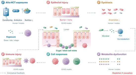
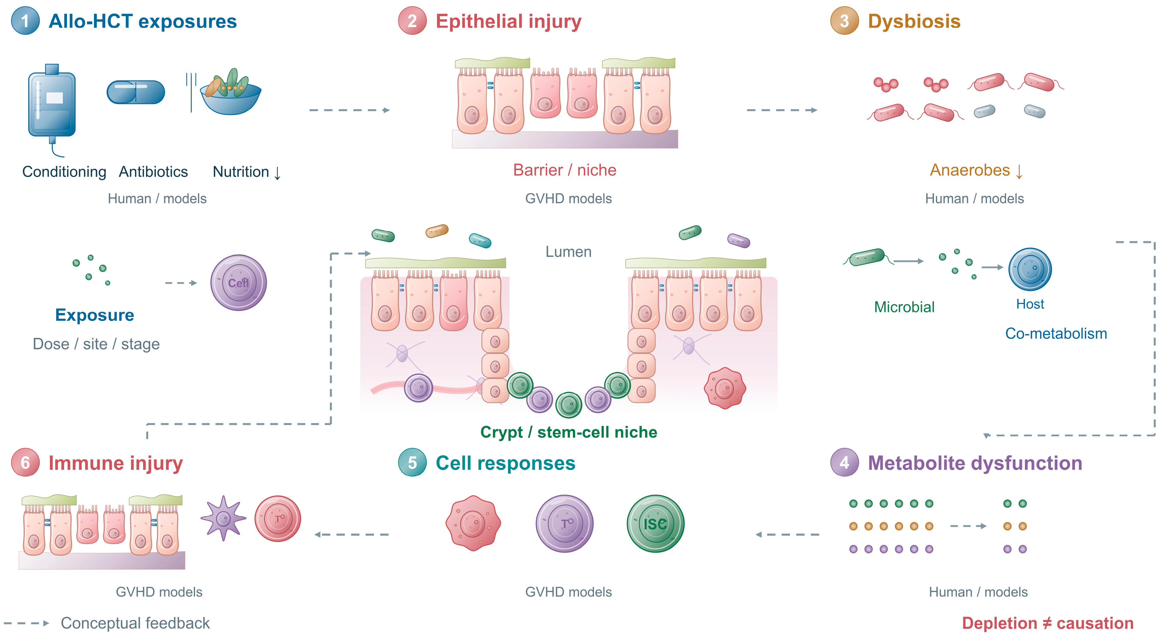
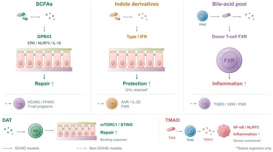
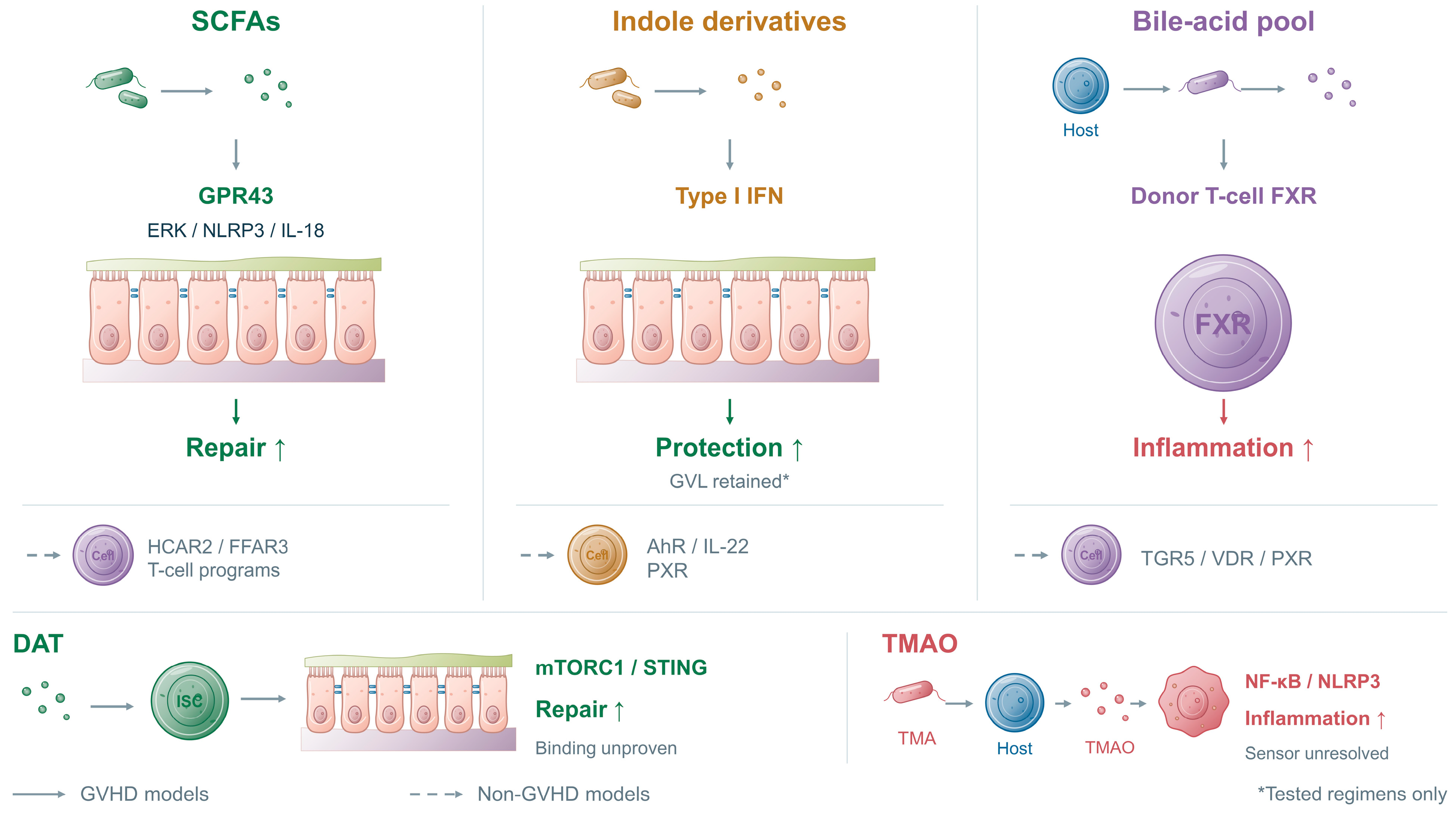
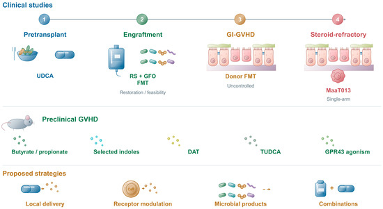
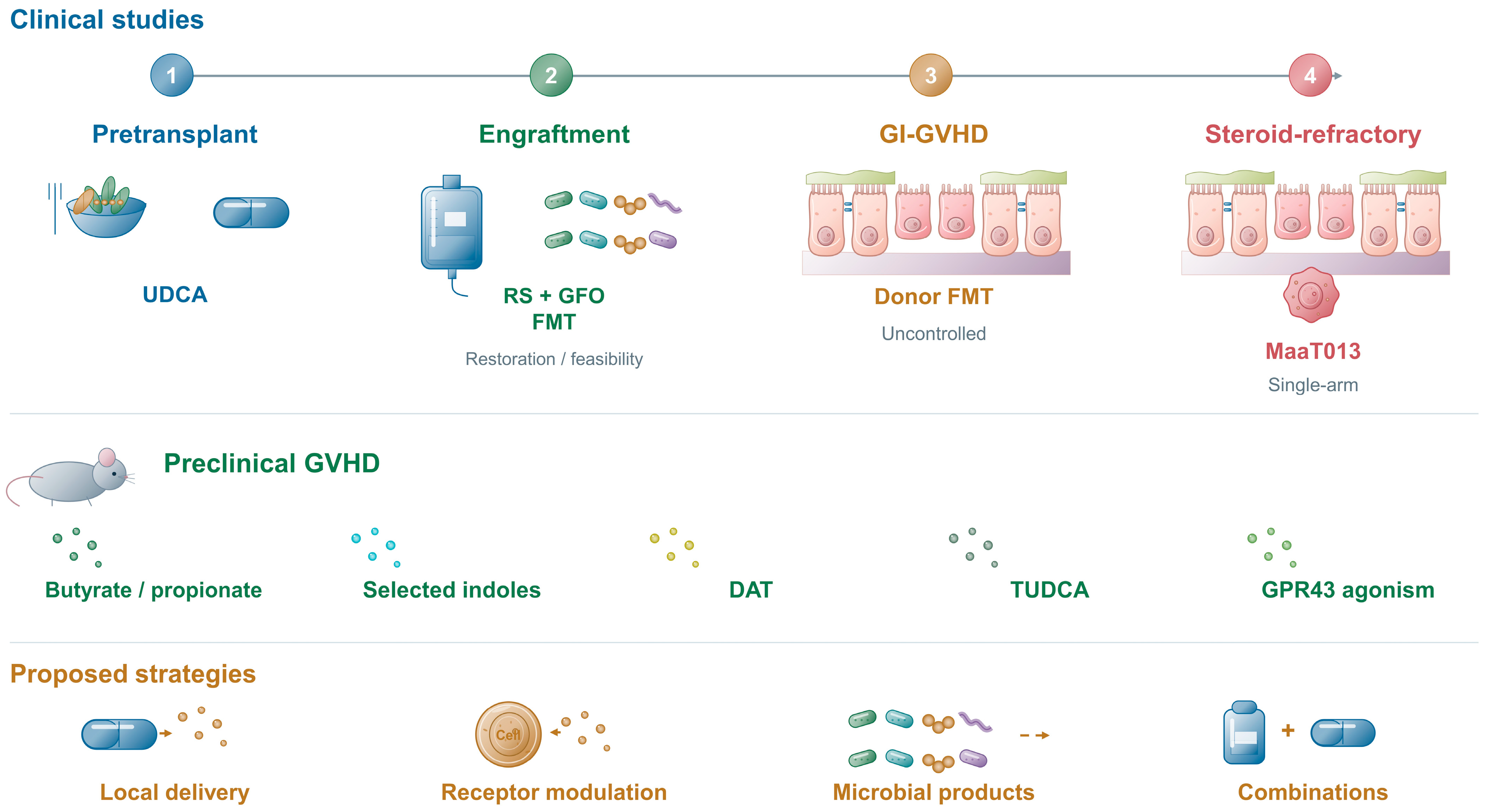

Disclaimer/Publisher’s Note: The statements, opinions and data contained in all publications are solely those of the individual author(s) and contributor(s) and not of MDPI and/or the editor(s). MDPI and/or the editor(s) disclaim responsibility for any injury to people or property resulting from any ideas, methods, instructions or products referred to in the content. |
© 2026 by the authors. Licensee MDPI, Basel, Switzerland. This article is an open access article distributed under the terms and conditions of the Creative Commons Attribution (CC BY) license.