


Abstract
Dry eye disease (DED) is a complex, multifactorial condition in which oxidative stress is increasingly recognized as a recurrent feature of ocular-surface dysfunction and as a signaling hub linking environmental stress, organelle dysfunction, innate immunity, and regulated cell death. Mitochondrial and endoplasmic-reticulum dysfunction, failure of endogenous antioxidant systems, and increased production of reactive oxygen species (ROS) can occur in response to tear hyperosmolarity, desiccating stress, aging, and other injuries. These changes activate redox-sensitive signaling pathways such as Nrf2/Keap1, SIRT3–TXNIP–NLRP3, and SLC7A11/GPX4, linking mitochondrial injury and lipid peroxidation to inflammasome activation, ferroptosis, pyroptosis, and epithelial barrier disruption. Damage to the tear film causes persistent inflammation that further damages the ocular surface. This narrative review examines oxidative stress in DED through the lens of redox checkpoints rather than a single pathogenic mechanism. We incorporate up-to-date evidence on organelle stress, antioxidant signaling, innate immune activation, and regulated cell death, and discuss their crosstalk to highlight therapeutically actionable nodes. We focus on newer redox-modulating and mitochondria-targeted strategies, while critically considering the overrepresentation of preclinical evidence, the limited validation of oxidative biomarkers, and the translational barriers that currently keep mechanism-based antioxidant therapy out of routine clinical care for patients with diverse clinical phenotypes of DED.
1. Introduction
Dry eye disease (DED) is a multifactorial, symptomatic disease characterized by loss of homeostasis of the tear film and/or ocular surface. The updated Tear Film & Ocular Surface Society Dry Eye Workshop III (TFOS DEWS III) definition refers to the instability and hyperosmolarity of the tear film, inflammation and damage of the ocular surface, and neurosensory abnormalities as etiological factors. These factors may coexist in individual patients to different degrees. This framework reframes DED no longer as mutually exclusive disease categories but rather as a spectrum. TFOS DEWS III identifies several etiological drivers of DED. It proposes clinically relevant subtypes based on tear-film component deficiencies, eyelid abnormalities, and ocular surface abnormalities. Moreover, several of these drivers may be at play at the same time [1,2,3,4]. Thus, DED should be viewed as a heterogeneous clinical spectrum in which the mechanisms responsible for loss of ocular surface homeostasis may vary between patients and may change with disease severity.
This variety is consistent with DED being linked with a variety of conditions. Epidemiological evidence identifies advanced age along with systemic, ocular, iatrogenic, and lifestyle-related factors such as Sjögren syndrome and other connective tissue disorders, diabetes, blepharitis, ocular surgery, topical medications, incomplete blinking, contact lens use, low-humidity environments, and digital screen use [5]. Human studies have shown that the lacrimal gland undergoes structural and functional changes with age, including decreased acinar density and diameter, decreased lacrimal-fluid peroxidase activity, and increasing fibrosis and atrophy. Furthermore, experimental aging models show inflammatory infiltration and accumulation of oxidative-damage markers such as 8-hydroxy-2′-deoxyguanosine (8-OHdG), nitrotyrosine, 4-hydroxy-2-nonenal (4-HNE), and lipofuscin-like material [6]. It is a crucial distinction: while the clinical association between aging and DED is strong, many mechanistic connections linking aging, oxidative damage, and ocular-surface dysfunction are largely supported by animal data. Environmental exposure follows this evidence gradient. A systematic review of 102 preclinical and clinical studies found that particulate matter and other associated pollutants induce oxidative and inflammatory signaling, apoptosis, and ocular-surface damage in cellular and animal models. However, clinical studies primarily support associations between pollution exposure and ocular irritation, conjunctivitis, and DED-related signs rather than establishing a specific redox mechanism in humans [7].
Against this heterogeneous background, oxidative stress is better supported as a feature of the DED ocular-surface environment than as a universal initiating cause. A systematic review and meta-analysis included nine studies with 333 patients suffering from DED and 165 healthy controls. It found an overall increase in oxidative-stress markers in DED (standardized mean difference [SMD] 2.39, 95% CI 1.85–2.94). However, there was substantial between-study heterogeneity (I2 = 96.1%). The signal was observed in tears and conjunctival samples and included increased levels of 4-HNE (SMD 4.75, 95% CI 1.67–7.84), malondialdehyde (MDA; SMD 3.00, 95% CI 2.55–3.45), and reactive oxygen species (ROS; SMD 1.31, 95% CI 0.94–1.68), among other markers [8]. Individual biomarkers should not be regarded as interchangeable. In a study of 44 patients with non-Sjögren DED and 33 controls, tear 4-HNE was higher in patients than in controls (0.20 ± 0.03 versus 0.02 ± 0.01 μg/mL), as was MDA (13.32 ± 4.03 versus 3.80 ± 1.05 pmol/mg), while levels of the early lipid-peroxidation marker hexanoyl-lysine were essentially similar (279.84 ± 69.98 in controls versus 283.21 ± 89.67 nmol/L in patients). Further analysis found that 4-HNE and MDA, but not hexanoyl-lysine, were associated with tear break-up time, Schirmer values, tear clearance, keratoepitheliopathy, conjunctival goblet-cell density, and symptoms. These findings, together with previous clinical and translational evidence, support the existence of a local oxidative imbalance in DED while also illustrating why oxidative biomarkers cannot yet be regarded as standardized diagnostic or causal markers [9,10].
The oxidative environment is mechanistically important, not only because of direct macromolecular damage. Physiological concentrations of ROS participate in cellular signaling, whereas excessive ROS can oxidize lipids, proteins, and nucleic acids and perturb intracellular homeostasis. Mitochondria are an important endogenous source of ROS, as demonstrated by experimental work. Mitochondrial dysfunction can further enhance the production of ROS. Other sources of ROS include the endoplasmic reticulum and some enzymatic systems, such as NADPH and xanthine oxidases. Oxidative damage may also interact with innate immune signaling. ROS-mediated damage to membranes and organelles may generate damage-associated signals that trigger responses in inflammatory pathways, including NLRP3-related signaling. However, much of the mechanistic evidence is still preclinical [11]. Antioxidant responses are actively regulated rather than passive. The Nrf2/Keap1 system coordinates transcriptional antioxidant defenses and interacts with thioredoxin/TXNIP signaling and SLC7A11-dependent redox regulation. However, the available DED literature supporting pharmacological manipulation of these pathways remains dominated by cellular and animal experiments. Direct clinical validation of Nrf2-targeted mechanisms is still missing [12]. Evidence of inflammation in humans contributes an important complementary layer of information to this area. The findings of a meta-analysis of 13 studies comprising a total of 342 patients suffering from DED and 205 controls show that tear concentrations of IL-1β, IL-6, IL-8, IL-10, IFN-γ, and TNF-α were higher in patients than in controls. However, methodological differences and variability between individuals have prevented the definition of reliable diagnostic cut-offs [13]. It follows that oxidative as well as inflammatory abnormalities coexist in human DED. But the direction and molecular intermediates of their interaction cannot be inferred from the observational data alone.
In this review, we thus use the term “redox checkpoints” as an operational framework for molecular systems that determine whether oxidative stress is buffered, repaired, or converted into inflammatory and cell-death signaling. Rather than treating ROS accumulation as a single pathogenic mechanism, we explore how environmental or metabolic stress can relate to epithelial dysfunction. This involves mitochondrial and endoplasmic-reticulum stress, Nrf2/Keap1- and SIRT3–TXNIP-related antioxidant control, mitochondrial DNA-dependent innate immune sensing, and NLRP3 inflammasome activation. The paper then evaluates regulated forms of cell death, particularly ferroptosis, pyroptosis, and necroptosis, to see if their experimental evidence can be meaningfully integrated into current models of DED. The review strongly emphasizes distinguishing mechanisms already shown in patients from those inferred from cellular or animal models, identifying tissue- and phenotype-specific differences, and determining which redox pathways and biomarkers have sufficient support to be regarded as plausible translational targets rather than mechanistically attractive but still unvalidated.
2. Materials and Methods
This narrative review was created on the basis of a structured literature search of PubMed/MEDLINE, updated through 5 September 2026. The search was conducted iteratively rather than as a single systematic-review query. Boolean combinations paired the disease terms (‘dry eye disease’ OR ‘ocular surface’) with mechanistic terms including ‘oxidative stress’, ‘reactive oxygen species’, ‘mitochondria’, ‘mitochondrial dysfunction’, ‘mitophagy’, ‘endoplasmic reticulum stress’, ‘unfolded protein response’, ‘Nrf2’, ‘Keap1’, ‘SIRT3’, ‘TXNIP’, ‘NLRP3’, ‘inflammasome’, ‘cGAS-STING’, ‘mitochondrial DNA’, ‘ferroptosis’, ‘pyroptosis’, ‘necroptosis’, ‘autophagy’, ‘lipid peroxidation’, and ‘antioxidant’. Titles and abstracts were initially evaluated for relevance to DED or ocular-surface redox biology, after which potentially relevant full texts were assessed. Studies were retained when they provided clinical evidence, mechanistically informative experimental data, or translational information directly relevant to the pathways considered in this review. Studies without direct relevance to ocular-surface biology, purely descriptive reports without mechanistic or translational relevance, and articles whose findings could not be meaningfully integrated into the review framework were not prioritized. The review methodology was informed by the Scale for the Assessment of Narrative Review Articles (SANRA), particularly concerning justification of the review, definition of aims, description of the literature search, referencing, and presentation of the level of evidence [14].
The literature search primarily focused on articles published from 2015 onwards, with particular emphasis on studies published between 2022 and 2026. Earlier publications providing pivotal clinical or mechanistic evidence were also considered when their contributions could not be adequately replaced by more recent literature.
The evidence considered included human clinical studies; systematic reviews and meta-analyses; consensus reports; translational studies; animal models; and mechanistically informative in vitro studies. Priority was given to TFOS consensus reports, systematic reviews and meta-analyses, studies published in major ophthalmic and biomedical journals, and recent studies directly addressing molecular mechanisms relevant to the conceptual framework of this review. We also inspected the reference lists of relevant reviews and primary studies for additional relevant publications.
Mechanisms were selected for dedicated subsection treatment when they were directly relevant to the redox–organelle stress framework of the review, were supported by mechanistically informative experimental evidence in DED or ocular-surface models, and had a sufficient body of literature to permit meaningful synthesis. Pathways supported only indirectly or by isolated observations were discussed more briefly where relevant. Therapeutic evidence was prioritized when an intervention directly targeted one of these mechanisms or provided controlled human evidence relevant to their translation. Apoptosis was not excluded from consideration, but it was not assigned a dedicated subsection because the regulated-cell-death section was specifically designed to examine emerging redox-coupled mechanisms—particularly ferroptosis, pyroptosis, and necroptosis—whereas apoptosis-related findings were incorporated where relevant to organelle stress and epithelial injury.
Due to the fact that the focus of this review was mechanistic integration rather than exhaustive estimation of treatment effects or disease associations, no quantitative meta-analysis, formal study-level risk-of-bias assessment, or certainty-of-evidence grading was carried out. In line with the biological model from which they were derived, the experimental findings were interpreted with particular care not to extrapolate mechanisms shown only in cultured cells or experimental DED models to established human disease. Accordingly, the Results section is a structured narrative synthesis rather than the result of a systematic-review selection process. The qualitative descriptors used in Section 3.6 were based on three features: evidence from functional perturbation experiments (pharmacological or genetic), consistency across experimental systems, and the presence or absence of concordant human ocular-surface or clinical data. These descriptors are intended to summarize translational maturity and do not represent formal risk-of-bias or certainty-of-evidence grades.
Because this was a hypothesis-guided structured narrative review rather than an agnostic systematic mapping review, the pathway-directed search strategy may preferentially retrieve evidence concerning mechanisms already represented within the proposed framework. Accordingly, omission or limited discussion of other pathways should not be interpreted as evidence of lesser biological relevance.
3. Results
3.1. Oxidative Stress as an Initiating and Amplifying Signal
The aforementioned clinical evidence demonstrates the presence of oxidative imbalance at the ocular surface in dry eye disease (DED). However, it does not clarify whether the accumulation of reactive oxygen species (ROS) is primarily a consequence of epithelial injury or plays an active role in disease propagation [8,9,10]. Mechanistic studies address this distinction by showing that ROS can function upstream of innate inflammatory signaling rather than acting just as end-products of tissue damage.
A notable translational study linked hyperosmotic stress with the ROS–NLRP3–IL-1β axis in human corneal epithelial cells and patients with environment-induced DED. Within 15 min of exposing immortalized HCE-2 human corneal epithelial cells to 500 mOsm medium, ROS generation increased. This increase in ROS formation, along with NLRP3 inflammasome formation, caspase-1 activity, and IL-1β release, was suppressed by pretreatment with 20 mM N-acetyl-L-cysteine. NLRP3 silencing independently decreased ASC, pro-caspase-1, and pro-IL-1β expression and reduced caspase-1 activity and IL-1β secretion. Together, these findings indicate a role for ROS upstream of inflammasome activation in this experimental system. Notably, this same study involved 20 patients who suffered from environment-induced DED as well as 15 controls. Conjunctival epithelial ROS fluorescence was 15.06 ± 4.48 versus 10.16 ± 2.41 RFU transcripts of NLRP3, PYCARD, CASP1, and IL1Bwere upregulated more than twofold, and tear bioactive IL-1β was 4.39 ± 0.93 versus 3.51 ± 0.58 ng/mL. Tear IL-1β also correlated positively with OSDI scores (R2 = 0.402) and negatively with tear break-up time (R2 = 0.306) [15]. Thus, the human arm supports the presence of the same signaling components identified experimentally, although it remains associative and cannot establish their temporal or causal ordering.
The temporal structure of this response has been examined in desiccating-stress models. Within just 24 h of exposure to low humidity with concomitant scopolamine-induced tear suppression, corneal epithelial NF-κB phospho-p65, nuclear NF-κB, NLRP3 protein and Nlrp3 transcripts, and caspase-1 staining were already increased. This early response was not simply a generalized upregulation of inflammatory cytokine transcription: IL-1β mRNA decreased on day 1, whereas bound and total IL-18 protein increased, with mature IL-1β subsequently increasing between days 1 and 5. These findings support early activation of epithelial innate pathways prior to full accumulation of downstream mature cytokines [16].
More recent work adds a mechanosensitive step between hyperosmolarity and inflammasome activation. Exposure of immortalized human corneal epithelial cells to 450 mOsm medium for 24 h increased PIEZO1 together with NLRP3 and IL-1β, promoted nuclear translocation of NF-κB and phosphorylation of IκBα, and increased ASC-speck formation and the mature IL-1β p17 fragment. Activation of PIEZO1 with Yoda1 reproduced NLRP3 activation, whereas PIEZO1 silencing as well as pharmacological inhibition with Dooku1 or GsMTx4 suppressed the hyperosmolarity-induced inflammasome response. In the corresponding scopolamine mouse model, topical PIEZO1 inhibition also dampened inflammasome activation and improved the damaged corneal phenotype [17].
Collectively, these findings refine the role of oxidative stress in DED. ROS can be interpreted not only as markers of lipid, protein, and DNA oxidation but also as components of a stress-sensing network. This network can convert osmotic and desiccating stimuli into innate inflammatory responses. Still, cellular and animal models provide the strongest causal evidence through ROS inhibition, NLRP3 knockdown, and PIEZO1 manipulation. Human data demonstrate concordant pathway activation. However, this sequence has not yet been established to operate uniformly across DED phenotypes.
3.2. Endoplasmic Reticulum Stress and the Unfolded Protein Response
The same signaling logic also applies upstream to endoplasmic reticulum (ER) homeostasis. When misfolded proteins accumulate, the unfolded protein response (UPR) initially helps to lower protein synthesis and increase folding capacity. But when ER stress cannot be resolved, UPR signaling can switch to inflammatory and pro-apoptotic pathways. This concept is supported by limited human evidence as well as far more detailed experimental data in DED.
Recent studies indicate a molecular link between the UPR and the oxidative and inflammasome pathways described above. The use of single-cell RNA-sequencing on 33,809 corneal epithelial cells isolated from six lacrimal-gland-excision DED mice and seven controls revealed enrichment of ER-stress and terminal-UPR signatures, notably increased GRP78 and activation of the PERK–CHOP branch. Researchers later replicated these results in a scopolamine/desiccating-stress model and in primary and immortalized human corneal epithelial cells exposed to 500 mOsm medium. The pharmacological reduction in ER stress by 4-phenylbutyric acid decreased ROS production and NLRP3/IL-1β activation induced by hyperosmolarity, whereas the induction of ER stress by tunicamycin increased expression of TXNIP. Knockdown of EIF2AK3 (encoding PERK) diminished the levels of p-eIF2α, ATF4, CHOP, and TXNIP, while silencing TXNIP reduced NLRP3, pro-IL-1β, and cleaved IL-1β; conversely, overexpressing TXNIP augmented these inflammasome-associated signals [18].
There is direct evidence of ER stress in ocular-surface samples from patients with Sjögren-associated DED. Conjunctival impression cytology from six women with Sjögren syndrome and Oxford ocular-surface staining grade ≥III, compared to six female controls, demonstrated altered localization of HSPA5/GRP78 and accumulation of the pro-apoptotic UPR mediator DDIT3/CHOP in conjunctival epithelial cells. In a complementary stratified human corneal epithelial model, exposure to hyperosmotic saline produced concentration- and time-dependent activation of the UPR: sXBP1 expression peaked at 24 h, and exposure to an additional 94 mM NaCl for 24 h significantly increased HSPA5 and DDIT3 along with epithelial cell loss and rose-bengal-positive damage. Intervention within the same model supports a functional, not merely associative, role for ER stress. The inclusion of 1 mM tauroursodeoxycholic acid (TUDCA), a chemical chaperone, significantly diminished hyperosmolarity-induced cell loss, with the viability experiments yielding p < 0.0001, and mitigated nuclear chromatin condensation, DNA fragmentation, and caspase-3 cleavage. Under hyperosmotic conditions, TUDCA also selectively reduced the transcription of the inflammatory mediators CXCL8 and IL5 [19]. These findings provide proof of principle that preservation of ER proteostasis can alter epithelial survival and inflammatory output. The intervention, however, was tested in vitro rather than as a treatment in patients with DED.
These experiments therefore position ER stress upstream of a PERK–CHOP–TXNIP–NLRP3 signaling sequence capable of coupling defective proteostasis to ROS generation and innate inflammation. Nonetheless, the hierarchy of evidence is important. Human ocular-surface evidence is currently limited to a very small and clinically specific Sjögren cohort, whereas causal manipulation of PERK, TXNIP, ROS, and NLRP3 has been conducted in experimental systems. Currently, ER-stress markers cannot be regarded as validated biomarkers that define a DED endotype or predictors of response to ER-directed therapy.
3.3. Mitochondrial Dysfunction, mtDNA Release, and Mitophagy
Mitochondria expand the range of redox responses beyond the accumulation of reactive oxygen species (ROS) because changes in mitochondrial function can simultaneously impact cellular energetics, redox balance, and the availability of mitochondrial danger signals. The experimental literature surrounding dry eye disease (DED) connects hyperosmotic and other ocular-surface stresses to changes in mitochondrial membrane potential, decreased ATP generation, disrupted mitochondrial structure and calcium homeostasis, and increased mitochondrial ROS (mtROS), providing a mechanistic basis for a self-reinforcing relationship between mitochondrial dysfunction and oxidative damage [20]. However, the available evidence is mostly based on cellular and animal studies rather than direct measurements of mitochondrial function in DED patients.
Recent experiments have begun to delineate how mitochondrial redox disturbance is coupled to inflammatory signaling. In immortalized HCE-T human corneal epithelial cells exposed to 500 mOsm medium for 24 h, hyperosmolarity increased mtROS, reduced mitochondrial membrane potential and ATP production, and produced swollen and fragmented mitochondria with disrupted cristae and sparse matrix. Overexpression of SIRT3 reversed the abnormalities, while pharmacological inhibition of SIRT3 with 50 μM 3-TYP further increased mtROS and aggravated the loss of membrane potential and ATP production. The same manipulation influenced the inflammatory output: hyperosmotic stress decreased thioredoxin and increased TXNIP, NLRP3, cleaved caspase-1, IL-1β, and IL-18, while SIRT3 overexpression produced opposite effects. Mice exposed to 0.075% benzalkonium chloride (BAC) twice a day for 14 days were also treated with topical honokiol at 16 μg/mL from days 8 to 14. This treatment similarly improved mitochondrial ultrastructure, reduced corneal ROS, and attenuated TXNIP–NLRP3 signaling [21]. These experiments therefore support an mtROS–TXNIP–NLRP3 sequence downstream of mitochondrial dysfunction. However, the evidence remains preclinical, and the study did not evaluate SIRT3 activation in patients. Notably, the authors of the study identified long-term safety, pharmacokinetics, and clinical efficacy of honokiol as unresolved issues.
The origin of oxidative injury at the subcellular level matters, as evidence from mitochondrial targeting shows. In H2O2-stressed human corneal epithelial cells, total intracellular ROS and mtROS increased approximately 4.3-fold and 2.7-fold, respectively, after exposure to 300 μM H2O2. Mitochondria-targeted SkQ1 nanoparticles showed 1.7-fold and 1.9-fold greater cytosolic- and mitochondrial-ROS scavenging activity, respectively, than free SkQ1 and better maintained mitochondrial membrane potential. Notably, exposure to oxidative stress led to 8-OHdG-positive mitochondrial DNA (mtDNA) oxidation, the cytosolic release of oxidized mtDNA (ox-mtDNA), and the colocalization of ox-mtDNA with NLRP3. SkQ1 nanoparticles markedly reduced these events. Topical application of SkQ1 nanoparticles in a BAC-induced DED model also reduced the expression of NLRP3, caspase-1, and IL-1β, along with decreased mtDNA oxidation [22]. Consequently, these findings connect mitochondrial ROS not only to generalized oxidative damage but also to the generation of an mtDNA-derived signal capable of engaging the inflammasome.
Another fate of released mtDNA is recognition by the cyclic GMP–AMP synthase–stimulator of interferon genes (cGAS–STING) pathway. In hyperosmolarity-stressed immortalized human corneal epithelial cells, cytosolic mtDNA increased along with cGAS–STING activity. CGAS or TMEM173 silencing reduced TBK1 and IRF3 phosphorylation as well as the downstream expression of CXCL10 and IFN-β. Transfection of isolated mtDNA at a concentration of 1 μg/mL reproduced pathway activation. In contrast, lowering cellular mtDNA by about 50% using ethidium bromide diminished STING, p-TBK1, p-IRF3, CXCL10, and IFN-β responses. Mechanistically, hyperosmotic stress induced mitochondrial structural damage and opening of the mitochondrial permeability transition pore (mPTP). Inhibition of mPTP opening with 5 μg/mL cyclosporin A reduced cytosolic mtDNA and cGAS–STING signaling; however, BAX knockdown did not have a comparable effect. Blocking oxidized mtDNA with exogenous 8-OHdG did not inhibit cGAS–STING activation. Taken together, these findings support a role for cytosolic mtDNA displacement in cGAS–STING activation in this experimental system, rather than establishing it as an exclusive or definitively critical signal. Importantly, the same study also included 26 patients with DED and 22 controls and demonstrated increased cGAS and STING protein levels in tear samples collected using Schirmer strips; STING levels were positively correlated with corneal fluorescein staining (rs = 0.692, p = 0.001) [23]. This differs from the ox-mtDNA–NLRP3 interaction described above [22] and suggests that mtDNA can engage different innate sensors depending on its subcellular disposition and molecular state. However, this interpretation derives from comparison across different experimental systems rather than a direct head-to-head study.
Additional human observations further support the translational relevance of cGAS–STING. In a subsequent investigation of environmental DED, hyperosmolarity-stressed primary human corneal epithelial cells and an environmental DED mouse model showed increased cGAS, STING, TBK1, IRF3, and IFN-β. In the mouse model, STING inhibition also reduced inflammatory mediators and corneal epithelial damage. More importantly, conjunctival samples from eleven patients with DED and eleven controls indicated significantly increased expression of each of the five pathway components. The DED group had an OSDI of 38.09 ± 12.05 versus 10.01 ± 1.55 in controls and a tear break-up time of 2.91 ± 0.83 versus 5.27 ± 2.24 s [24]. These findings provide human corroboration of pathway activation but remain cross-sectional and based on a small cohort. Earlier work had proposed cGAS–STING as a plausible DNA-sensing mechanism in DED before such direct experimental evidence was available [25]. Current findings move the pathway beyond that initial hypothesis, but they do not yet establish a cGAS–STING-defined clinical endotype or the efficacy of STING-directed treatment in patients.
The available PINK1/Parkin data do not support a consistently beneficial effect of increasing mitophagy, and mitochondrial quality control adds another layer of complexity to this situation. In human corneal epithelial cells exposed to BAC, concentrations up to 0.0003% produced dose-dependent oxidative and mitochondrial injury. Mechanistic experiments were subsequently performed with 0.00015% BAC, which increased intracellular and mitochondrial ROS, reduced membrane potential, ATP, glutathione redox balance, and superoxide dismutase activity, and increased malondialdehyde. Furthermore, 0.00015% BAC shifted mitochondrial dynamics toward DRP1/Fis1-mediated fission and triggered PINK1/Parkin-associated mitophagic flux. At a concentration of 25 μM, norepinephrine reduced ROS, improved mitochondrial function and viability, and decreased LC3B, PINK1, Parkin, mitochondrial–lysosomal colocalization, and other indices of the BAC-induced mitophagic response. Corresponding improvements in corneal staining, TUNEL positivity, mitochondrial fragmentation, ROS, TNF-α, and IL-1β were observed in BAC-treated mice [26]. In this model, then, protection was associated with restraining an excessive mitophagic response rather than further increasing it.
By contrast, in two additional experimental settings, PINK1-dependent mitophagy was required for protection. In hyperosmolarity-stressed human corneal epithelial cells, 20 μM cRGD-conjugated bilirubin nanoparticles (cNPs@BR) reduced ROS, NOX4, 4-HNE, and inflammatory cytokines and improved mitochondrial respiration, membrane potential, biogenesis, and dynamics. PINK1 knockdown largely abolished these mitochondrial, antioxidant, and anti-inflammatory benefits. In a 0.2% BAC mouse model of DED, treatment with topical 20 μM cNPs@BR for 7 days reduced the corneal fluorescein score from >12 in PBS-treated DED eyes to 1.5 and increased tear production to 6.62 ± 0.69 mm, compared with 5.35 ± 0.51 mm with lifitegrast treatment. Moreover, the treatment increased corneal PINK1, Parkin, and LC3B and decreased p62 [27]. Similarly, in 12-month-old mice, aging was associated with lacrimal-gland oxidative stress, mitochondrial dysfunction, and acinar-cell apoptosis. Enhancing PINK1/Parkin-associated mitophagy with rapamycin (5 mg/kg/day for one month) improved lacrimal-gland structure and tear secretion while also decreasing ROS and apoptosis. Inhibition with Mdivi-1 (10 mg/kg/day) increased fibrosis and inflammatory-cell infiltration, increased TUNEL-positive cells, and impaired mitochondrial clearance and glandular function [28].
Taken together, these seemingly conflicting results suggest that mitophagy should not be regarded as an intrinsically beneficial or harmful pathway. Instead, they support a model in which mitochondrial quality control is adaptive when it promotes effective removal of dysfunctional organelles, but may accompany or contribute to injury when strongly or persistently activated in a severely damaged mitochondrial population. The direction of therapeutic modulation is therefore likely to depend on the initiating stress, tissue compartment, mitochondrial damage burden, and state of autophagic flux. Crucially, all three studies are experimental, and it remains unknown whether altered mitophagy defines a reproducible phenotype in human DED.
3.4. Antioxidant Systems as Redox Checkpoints
The preceding findings suggest that antioxidant defense is better thought of as a series of regulatory thresholds rather than as a simple opposing force to reactive oxygen species (ROS) production. Nuclear factor erythroid 2-related factor 2 (Nrf2) gives the clearest example at the transcriptional level. Under basal conditions, Kelch-like ECH-associated protein 1 (Keap1) promotes Nrf2 degradation. Under oxidative stress, this regulation is disrupted, leading to Nrf2 accumulation, translocation to the nucleus, association with small Maf proteins, and transcription dependent on antioxidant response elements. The downstream program incorporates various cytoprotective genes, including heme oxygenase-1 (HO-1), superoxide dismutase, glutathione peroxidase, catalase, and others. In addition to these canonical enzymes, Nrf2 regulation encompasses a further set of target genes. The pathway has been reported to increase thioredoxin expression, suppress TXNIP, and induce the cystine/glutamate transporter SLC7A11/xCT, thus linking transcriptional antioxidant adaptation with several of the redox-sensitive systems outlined above [12].
However, the evidence that is specific to DED is mostly experimental. Mice deficient in Nrf2 exposed to environmental stress experience more ocular-surface oxidative damage than wild-type animals, whereas several pharmacological Nrf2 activators reduce oxidative markers and improve experimental DED phenotypes [12]. More recent work provides an example of this broader network: cNPs@BR elevated nuclear Nrf2 and HO-1 concomitant with reduced ROS, NOX4, and 4-HNE levels in hyperosmolarity-challenged corneal epithelial cells. However, the same intervention also affected NF-κB signaling and PINK1-dependent mitophagy, precluding attribution of its entire effect to Nrf2 alone [27]. Critically, the available review of Nrf2-directed therapy identifies no clinical trial directly validating the efficacy of Nrf2 agonism in DED and emphasizes that interactions between Nrf2/Keap1 and other signaling pathways remain incompletely defined [12].
Based on the framework employed in this review, Nrf2/Keap1 can be regarded as a transcriptional redox checkpoint, while SIRT3 and PINK1/Parkin-dependent mitochondrial quality control operate closer to the organelle itself. This terminology is operational for the purpose of this article and not a formally recognized molecular classification. It is also important not to go beyond what the available evidence shows: although SLC7A11/xCT is reported to be regulated by Nrf2 [12], these data alone do not establish a clinically relevant SLC7A11–GPX4 ferroptosis checkpoint in DED. The evidence linking lipid-peroxide detoxification to ferroptotic cell death is therefore considered separately in the regulated cell-death section rather than inferred from the antioxidant studies presented here.
3.5. Innate Immune Sensing as a Converter of Organelle Stress into Inflammation
In conjunction, the above studies identify innate sensing as the point where different types of cellular stress can acquire a common inflammatory output. The most studied convergence node is NLRP3. NLRP3 activation has been experimentally linked to hyperosmolarity-induced ROS [15], PIEZO1-dependent mechanosensing [17], PERK–CHOP/TXNIP signaling during ER stress [18], and mitochondrial mtROS–TXNIP signaling [21]. Mitochondrial injury provides an additional route through cytosolic ox-mtDNA interacting with NLRP3 [22]. Nonetheless, these mechanisms should not be represented as a single experimentally verified linear pathway. They were established in various models using different perturbations. However, no study has yet shown that all of these upstream signals operate simultaneously in an individual DED patient, or that they have comparable importance.
This distinction is particularly relevant when NLRP3 is compared with cGAS–STING. Both pathways have the ability to convert mitochondrial damage into inflammation. However, the experimental data available indicate different forms of mitochondrial signaling. In the study of SkQ1 nanoparticles, it was found that oxidized mtDNA released into the cytoplasm colocalized with NLRP3. Inflammasome activation was attenuated upon prevention of mtDNA oxidation and release [22]. Conversely, cGAS–STING activation under hyperosmotic stress depended on mPTP-mediated leakage of mtDNA into the cytosol, whereas experimental interference with mtDNA oxidation did not inhibit the pathway [23]. cGAS–STING then mostly signaled through TBK1–IRF3 and type I interferon-associated mediators rather than through caspase-1-dependent cytokine maturation [23,24]. As such, there does not appear to be a single innate-immune consequence of mitochondrial disruption; the location and molecular state of mitochondria-derived danger signals may influence which sensing system is engaged.
The strength of clinical support also varies across these pathways. The ROS–NLRP3 axis has concordant human ocular-surface evidence, although its causal ordering derives primarily from cell experiments [15]. For cGAS–STING, two independent human cohorts provide convergent evidence using different ocular-surface samples and analytical approaches. In one study, tear cGAS and STING protein levels were increased in 26 patients with DED compared with 22 controls, and STING levels correlated positively with corneal fluorescein staining [23]. A separate investigation demonstrated conjunctival upregulation of cGAS, STING, TBK1, IRF3, and IFN-β in 11 patients with environmental DED compared with 11 controls [24]. These observations strengthen the evidence that cGAS–STING pathway activation occurs in human DED, although the available studies remain cross-sectional and do not establish pathway dependence, prevalence across DED phenotypes, or predictive value. Thus, cGAS–STING is best regarded as an experimentally substantiated pathway with convergent human corroboration rather than a validated molecular endotype.
3.6. Regulated Cell Death: Ferroptosis, Pyroptosis, and Necroptosis
Regulated cell death is an execution layer through which redox imbalance, organelle stress, and inflammatory signaling can be converted into loss of ocular-surface epithelial cells. According to the DED literature, several partially overlapping death programs are implicated, including pyroptosis, ferroptosis, and necroptosis, but there are differences concerning the robustness of the different lines of evidence. Examinations of human tears and conjunctivae have detected pyroptosis-associated components, which include NLRP3, ASC, caspase-1, GSDMD-N, IL-1β, and IL-18 [29]. These findings provide clinical corroboration that death machinery linked with the inflammasome is present at the diseased ocular surface. Such measurements, however, cannot demonstrate the occurrence, extent, or relative contribution of lytic epithelial death in vivo. The mechanistic evidence for ferroptosis and necroptosis is even more strongly concentrated in stressed epithelial cells and experimental animal models. Consequently, these pathways should be interpreted as experimentally supported mechanisms of epithelial injury rather than as established, mutually exclusive molecular endotypes of clinical DED.
For pyroptosis, the relevant advance is not just further evidence for NLRP3 activation (discussed earlier) but showing that diverse upstream signals and gasdermin executors can convert inflammasome activity into membrane rupture. Potassium efflux provides one such entry point. In corneal epithelial cells, 10 μM nigericin reduced intracellular potassium and increased cell death together with NLRP3, ASC, cleaved caspase-1, and N-terminal GSDMD; inhibition of potassium efflux with 100 μM quinine attenuated these changes. Desiccating stress produced a similar phenotype in HCE-T cells, whereas KCNK5 silencing preserved intracellular potassium, decreased cell death and the mRNA levels of IL6, IL1A, and IL1B, and inhibited NLRP3/caspase-1/GSDMD signaling. Scopolamine/desiccating-stress mice showed similar results, with KCNK5 silencing reducing corneal epithelial defects and pyroptosis-associated proteins and partially restoring tear secretion [30]. Transcriptomic and functional experiments further associated KCNK5 with reduced TNFSF10-associated autophagic activity, with TNFSF10 supplementation alleviating the pyroptotic phenotype. Ion flux and autophagic competence thus seem able to change the threshold for inflammasome-dependent epithelial lysis rather than constituting distinct pathogenic processes.
Execution through pyroptosis is not limited to GSDMD. When human corneal epithelial cells were exposed to hyperosmotic conditions across 312–500 mOsm, membrane rupture occurred together with an increase in caspase-3 and GSDME, and GSDME expression increased when GSDMD was silenced. In GSDMD-knockout mice subjected to experimental DED, caspase-3 increased despite the absence of detectable PARP-associated apoptosis, while GSDME and pyroptosis-associated inflammatory markers remained elevated [31]. These results support an alternative caspase-3/GSDME route in the absence of canonical GSDMD-dependent execution and caution against simply equating caspase-3 activity with apoptosis in this experimental context. In contrast, the canonical NLRP3/caspase-1/GSDMD axis was robustly activated in a 0.2% BAC rat model: compared to healthy controls, NLRP3, caspase-1, GSDMD, and IL-1β increased approximately 6.2-, 5.9-, 8.8-, and 10.3-fold, respectively. The administration of free melatonin lowered these signals by 22%, 31%, 42%, and 26% relative to saline-treated BAC eyes, whereas TAT-modified melatonin liposomes resulted in even greater suppression along with reduced mtDNA oxidation and preservation of corneal epithelium and conjunctival goblet cells [32]. Since this intervention targets both mitochondrial oxidative injury and inflammasome signaling at the same time in a preservative-toxicity model, its therapeutic effect cannot specifically be attributed to inhibition of pyroptosis.
Ferroptosis is the most direct mechanistic extension of the redox-checkpoint framework because cell death occurs when iron-dependent lipid oxidation exceeds the capacity of antioxidant and lipid-repair systems. Ferroptotic phenotypes in hyperosmolar corneal epithelial cells and scopolamine/desiccating-stress models have been defined by intracellular Fe2+ accumulation, lipid peroxidation, altered GPX4 and iron-handling proteins, and characteristic mitochondrial injury. This response is not simply due to increased ROS; stressed cells simultaneously activate protective programs. AKR1C1 was induced downstream of NRF2, and selective depletion of AKR1C1 led to decreased cell viability, increased lipid peroxidation, decreased GPX4, FTL, and FTH1, and increased TFRC. Conversely, AKR1C1 overexpression reduced both lipid peroxidation and intracellular iron accumulation [33]. This offers a functional connection between the transcriptional antioxidant response mentioned in Section 3.4 and the probability that oxidative stress progresses to ferroptotic death.
GPX4 emerges as a recurrent convergence point but through more than one regulatory route. In hyperosmolar corneal epithelial cells, astaxanthin increased levels of SLC7A11 and GPX4, improved glutathione availability and ferritin-associated iron storage, reduced Fe2+ overload, ROS, and lipid peroxidation, and preserved mitochondrial structure. Ferrostatin-1 and deferoxamine also showed related anti-ferroptotic effects and were accompanied by restoration of autophagic flux [34].
Membrane lipid susceptibility to ferroptosis provides an additional regulatory axis. In BAC-treated rats, corneal lipid hydroperoxide levels were 43.7% higher than in controls. Moreover, topical inhibition of DDR1 with DDR1-IN-1 reduced lipid peroxidation and ACSL4 expression. In HCECs stressed with 450 mOsm medium, there was a roughly sixfold elevation in lipid-peroxide-positive cells. DDR1-IN-1 protected cells in a concentration-dependent manner between 0.1 and 3 μM, while overexpression of the YAP1 gene or reactivation of YAP reduced this protection. This supports a DDR1–YAP–ACSL4 mechanism rather than a generalized effect on iron uptake [35]. Autophagic dysfunction can converge on the same lipid-remodeling enzyme. Under desiccating stress, impaired autophagic flux was associated with SQSTM1 accumulation. Moreover, SQSTM1 overexpression increased ACSL4, Fe2+, ROS, malondialdehyde, and 4-HNE while reducing cell viability and GPX4. Silencing ACSL4 reversed these alterations. Inhibition of ACSL4 in vivo with rosiglitazone (5 mg/kg twice daily for 7 days) reduced corneal epithelial defects, but recovery of tear secretion and conjunctival goblet cells did not reach statistical significance [36]. This distinction is important: suppression of molecular ferroptotic markers and improvement in epithelial staining do not necessarily reflect recovery of the whole lacrimal functional unit.
SIRT1 is part of a distinct GPX4-regulatory pathway. When HCECs were exposed to 450–500 mOsm medium, there was an increase in malondialdehyde and Fe2+. There was also a reduction in glutathione. Moreover, lipid oxidation increased and mitochondrial volume decreased. Activation of SIRT1 with 1 μM SRT1720 suppressed these changes. Mechanistically, HIF1α was deacetylated and stabilized by SIRT1, and HIF1α in turn supported GPX4 expression. SRT1720 also increased corneal GPX4 and reduced lipid oxidation in scopolamine-induced DED mice [37]. Together, these experimental investigations identify multiple routes that could modify lipid-peroxide susceptibility. However, none demonstrates that pharmacological manipulation of these pathways modifies human DED.
Necroptosis presents a different relationship between cell death and inflammation, as the death machinery itself can feed back into innate sensing. In desiccating-stress DED, increased RIPK3 and MLKL were observed. RIPK3 deletion protected the corneal epithelium, while localized overexpression of RIPK3 worsened epithelial damage [38]. SQSTM1 acted downstream of RIPK3: silencing SQSTM1 decreased MLKL and phosphorylated MLKL even in RIPK3-overexpressing cells, whereas SQSTM1 overexpression augmented necroptotic signaling. This activity was dissociated from canonical autophagic flux and differs from the autophagy-dependent SQSTM1 accumulation implicated in ferroptosis above [36,38]. RIPK3 deficiency also resulted in decreased NLRP3 expression, while RIPK3 remained elevated in NLRP3-deficient animals. This places NLRP3 downstream of RIPK3 in this model. Manipulating SQSTM1 similarly altered NLRP3, ASC, caspase-1, and IL-1β [38]. Necroptosis could hence intensify inflammation through active intracellular signaling as well as through the release of intracellular contents after membrane disruption.
Another necroptotic mechanism links mitochondrial dynamics to cell death. Hyperosmotic stress induced local C5a–mitochondrial C5a receptor 1 (mtC5aR1) signaling in corneal epithelial cells. Subsequently, this process activated DRP1, causing mitochondrial fragmentation, followed by RIPK3/MLKL-dependent necroptosis. Recombinant C5a exposure decreased mitochondrial mean branch length, perimeter, aspect ratio, and branches per mitochondrion by 46%, 66%, 32%, and 64%, respectively, while increasing phosphorylated DRP1 by approximately 50%. The research found that inhibition of DRP1 with Mdivi-1 reduced phosphorylated MLKL by 43%. Additionally, it increased cell viability by 16%. This supports mitochondrial fission as a functional intermediate rather than an accompanying morphological change. The intracellular C5aR1 antagonist JPE-1375 was more effective than the membrane-impermeable antagonist PMX-53 in suppressing RIPK3/MLKL activation and led to an approximately 32% reduction in corneal staining scores in experimental DED mice [39]. These findings provide mechanistic evidence for an intracellular complement–mitochondrial–necroptotic axis, though the importance of this axis in human epithelial loss remains to be established. Related regulated-cell-death findings have also been reported by independent groups using distinct experimental models [31,34,35]. Nevertheless, several specific mechanistic axes remain supported by a relatively limited number of laboratories and require broader independent replication.
Finally, the divisions between these pathways are probably not rigid. Chrysin provides an instructive experimental example. At non-cytotoxic concentrations up to 18 μM, chrysin reduced NOX2, 4-HNE, and FACL4/ACSL4 while increasing GPX4.
Plasmid-mediated NOX2 overexpression reversed its effects on ROS, Fe2+ accumulation, and lipid peroxidation. Simultaneously, chrysin reduced STING/NLRP3 signaling and IL-1β and IL-6, effects that were lost following plasmid-mediated STING overexpression [40]. These experiments show concurrent modulation of ferroptotic and inflammatory signaling, but they do not establish that ferroptosis itself activates STING in DED; the pleiotropic nature of chrysin and the BAC model preclude that causal inference.
The emerging literature therefore supports regulated cell death as a networked consequence of redox, metabolic, organelle, and inflammatory stress rather than a collection of isolated terminal pathways [29,41]. Ferroptosis in particular is supported by increasingly detailed molecular experiments linking iron handling, GPX4-dependent antioxidant defense, lipid remodeling, autophagy, and mitochondrial function [33,34,35,36,37,41,42]. Nevertheless, recent translational assessments acknowledge that the evidence remains dominated by cultured epithelial cells and acute rodent models, which do not reproduce the chronicity, tear-film dynamics, blinking-associated mechanical stress, neuroimmune interactions, or etiological heterogeneity of human DED [42]. Clinical evidence is largely observational and does not yet demonstrate ferroptosis-specific target engagement or disease modification. Accordingly, terms such as “central driver” remain premature for clinical DED. The more relevant translational question is likely to be which death pathway becomes rate-limiting for epithelial loss in a particular tissue, disease phenotype, and stage, and whether that state can be identified prospectively in patients before pathway-directed treatment is attempted.
Experimentally, these death programs should be distinguished using combinations of molecular and functional criteria rather than individual markers. Pyroptosis is supported by gasdermin cleavage, inflammasome/caspase activation, membrane permeabilization, and release of mature inflammatory cytokines; ferroptosis by iron-dependent lipid peroxidation, altered GPX4/SLC7A11 or ACSL4-related signaling, and functional rescue by ferroptosis inhibitors or iron chelation; necroptosis by RIPK3/MLKL activation together with genetic or pharmacological interruption of this axis; and apoptosis by characteristic apoptotic morphology and caspase/PARP-associated signaling in the absence of predominant lytic gasdermin-mediated execution [29,30,31,32,33,34,35,36,37,38,39]. No single marker is fully specific: for example, caspase-3 can participate in GSDME-mediated pyroptosis rather than classical apoptosis [31]. Current DED models support substantial crosstalk between these pathways, including RIPK3–NLRP3 coupling and concurrent modulation of ferroptotic and inflammatory signaling [38,40], but they do not establish whether the different death programs are activated simultaneously, sequentially, or in distinct epithelial-cell populations in human DED.
The principal redox-sensitive pathways discussed above, together with their current level of human validation, are summarized in Table 1.
Table 1.
Principal redox-sensitive pathways implicated in DED and their current translational maturity.
3.7. Tissue and Phenotype Context
The biological meaning of oxidative stress in DED depends strongly on the tissue compartment in which it arises. The concept of the Ocular Lipid Unit emphasizes metabolic crosstalk among the lacrimal and meibomian glands, cornea, conjunctiva, and tear film rather than treating their lipid biology as independent. Within this framework, alterations in mitochondrial fatty-acid oxidation, very-long-chain fatty-acid synthesis, polyunsaturated-fatty-acid balance, and lipid peroxidation may influence both tissue metabolism and tear-film properties, while lipid molecular ordering and viscoelasticity may be more informative than absolute lipid quantity [43]. Aging provides a clear example of why this tissue context matters. Age-related DED reflects coordinated changes across the lacrimal gland, meibomian gland, cornea, conjunctiva, and their innervation rather than a simple age-dependent reduction in tear production. In the aging lacrimal gland, acinar atrophy, fibrosis, lipid accumulation, cellular senescence, increased 8-OHdG immunoreactivity, impaired mitochondrial quality control, and defective mitophagy coexist with inflammatory and secretory dysfunction. Aging of the cornea is likewise associated with reduced epithelial regenerative capacity, altered barrier-related gene expression, cumulative oxidative injury, cellular senescence, and neural degeneration, while the conjunctiva undergoes epithelial remodeling and progressive goblet-cell loss [44]. Oxidative stress in an older patient may consequently reflect a chronic failure of tissue maintenance embedded within senescence, inflammaging, mitochondrial dysfunction, and impaired regeneration rather than the same process operating during an acute environmental challenge. This distinction limits the assumption that a single antioxidant strategy should have equivalent biological effects across age-related, autoimmune, and exposure-associated DED.
The same principle applies to evaporative disease. Evidence-based MGD guidelines define low-delivery MGD clinically through symptoms together with abnormalities of the lid margin and the quantity or quality of meibomian secretion, and distinguish obstructive from atrophic disease because the underlying glandular abnormalities and therapeutic implications differ [45]. The guidelines also emphasize that no globally accepted diagnostic criteria exist and identify substantial areas in which the evidence remains limited. Thus, although altered lipid metabolism and oxidative injury may contribute biologically to MGD, they do not replace direct characterization of glandular structure and secretion. A redox signal detected in an evaporative phenotype therefore has to be interpreted alongside the glandular abnormality producing tear-film instability rather than treated as an alternative disease definition.
Conjunctival goblet cells provide a complementary example of tissue specificity. Their contribution extends beyond mucin secretion to ocular-surface barrier and immune homeostasis, but their distribution is intrinsically heterogeneous: reported density ranges from tens to thousands of cells/mm2 depending on conjunctival location. Biopsy, impression cytology, and in vivo confocal microscopy can characterize goblet-cell morphology, distribution, and density, while newer fluorescence approaches may permit non-invasive visualization, yet standardized clinical assessment of the mucin/goblet-cell compartment remains substantially less developed than imaging of the meibomian glands or aqueous tear parameters [46]. Consequently, loss or dysfunction of this compartment can coexist with redox and inflammatory abnormalities without being adequately captured by a single tear biomarker.
Sjögren-related DED further demonstrates why disease context cannot be ignored. Sjögren disease is characterized by autoimmune infiltration and dysfunction of exocrine tissues, particularly the lacrimal and salivary glands, and ocular disease initially reflects an aqueous-deficient process even though meibomian-gland abnormalities may subsequently coexist [47]. This immunological background is measurably different from that of non-Sjögren DED. A meta-analysis of 15 studies including 809 participants—302 with Sjögren-related DED, 220 with non-Sjögren DED, and 287 healthy controls—found higher tear IL-12p70, IL-17, IL-4, IL-6, IL-8, and TNF-α in Sjögren-related than in non-Sjögren disease, whereas several other cytokines did not distinguish the two phenotypes. The magnitude and consistency of these differences were also cytokine-dependent: for example, IL-6 was higher in Sjögren DED with an SMD of 1.34 (95% CI 0.04–2.64), but heterogeneity was 92%, whereas TNF-α showed a smaller but more consistent difference (SMD 0.49, 95% CI 0.19–0.79; I2 = 0%). EGF was lower in Sjögren than in non-Sjögren DED (SMD −0.72, 95% CI −1.20 to −0.24; I2 = 0%) [48]. These data argue against treating inflammatory—and by extension redox-inflammatory—signals as interchangeable across DED phenotypes.
Human proteomic evidence reinforces this point while also exposing the limitations of extrapolating acute experimental stress to chronic disease. Untargeted analysis of Schirmer-strip proteins from 12 patients with Sjögren-related DED and six healthy controls identified 111 dysregulated proteins involving redox metabolism, cellular metabolism, cytoskeletal organization, and programmed cell death; PRDX6, an antioxidant peroxiredoxin, and the mucosal chemokine CXCL17 were significantly reduced in the patient samples. In an independent group of 39 patients, PRDX6 correlated positively with TBUT (r = 0.40, p = 0.01) and Schirmer I values (r = 0.52, p = 0.0006) and negatively with OSDI (r = −0.35, p = 0.03) and Oxford staining (r = −0.38, p = 0.02); CXCL17 correlated negatively with Oxford staining (r = −0.37, p = 0.03) [49]. Yet hyperosmotic or IFN-γ stimulation of human corneal epithelial cells increased PRDX6 and CXCL17 gene expression and PRDX6 and CXCL17 protein expression, producing changes in the opposite direction from those observed in chronic Sjögren disease. Rather than invalidating the experimental models, this divergence suggests that an acute epithelial stress response and the molecular state of established autoimmune DED are not equivalent.
Environmental exposure adds another layer of variability. In a prospective comparative study of 80 healthy young adults, 50 participants used a smartphone and 30 a computer display under standardized conditions. After four hours of smartphone use, symptoms increased and both fluorescein and non-invasive tear break-up times decreased; tear HEL increased relative to baseline and one hour, while ocular-surface ROS also increased and was higher than in the computer-display group at four hours [50]. Thus, oxidative measurements can change over a period of hours even in individuals without established ocular disease, underscoring their sensitivity to recent exposure.
Experimental blue-light models provide possible mechanisms for such environmental effects but should not be conflated with routine human screen exposure. Continuous 440 nm irradiation of human corneal epithelial cells for up to 48 h increased ROS, apoptosis, and migratory dysfunction while suppressing AKT/S6K signaling; ROS scavenging with N-acetylcysteine restored viability, migration, and pathway activation, and related effects were reproduced in mice exposed to 425 nm light for 12 h/day over 14 days [51]. A separate study focused on the conjunctiva and exposed mice to 440 ± 15 nm light at 0.2 mW/cm2 for four weeks. Conjunctival iron progressively increased after two and four weeks, accompanied by lipid peroxidation and ferroptosis-like mitochondrial changes; deferiprone reduced iron accumulation and improved corneal staining and tear-film break-up time. NCOA4 manipulation further supported ferritinophagy as a mechanistic source of labile iron in this model, and 1 μM topical NCOA4–9a was tested twice daily during the four-week exposure period [52]. These findings establish biological plausibility for ROS- and iron-dependent epithelial injury under defined experimental irradiation, but the intensity, wavelength, duration, and continuity of these protocols prevent direct inference that ordinary screen exposure induces clinically relevant ferroptosis in humans.
3.8. Oxidative Biomarkers: Promise and Current Limitations
The differences described above pose the main issue for redox biomarkers. The fact that oxidative products differ between DED and healthy ocular surfaces proves biological involvement, but to be clinically useful, a biomarker must answer a more specific question. Does it identify a reproducible molecular phenotype? Does it add information above and beyond established examination? Does it predict progression or treatment response? Does it identify patients in whom a pathway-directed intervention is likely to work?
According to the pooled and individual human studies discussed earlier, oxidative imbalance can be measured in DED, but they also highlight why this has not yet been translated into a diagnostic assay [8,9,10]. A diverse array of studies measured ROS, lipid-peroxidation products, oxidized nucleic acids, and antioxidant enzymes in tears, conjunctival cells, or tissue using different analytical platforms and patient definitions. Even within a single non-Sjögren cohort, individual lipid-peroxidation products did not show uniformity; notably, 4-HNE and MDA were associated with disease parameters whilst HEL was not [10]. Therefore, oxidative stress cannot be treated as a single measurable analyte, and neither the biological stage represented by each marker nor the extent to which different assays are interchangeable has been established.
More recent investigations show that biomarker values cannot be disaggregated from phenotype and context. The PRDX6/CXCL17 signature was found specifically in Sjögren-related DED rather than in an unselected DED population, and its inverse behaviour in stressed epithelial cells relative to patient samples indicates that the direction of change can depend on whether an assay captures an acute adaptive response or established tissue dysfunction [49]. On the other hand, the smartphone study indicated that HEL and ocular-surface ROS in healthy participants could change within four hours while their tear-film stability deteriorated at the same time [50]. A measured redox abnormality may therefore reflect disease biology, phenotype, recent exposure, compensatory antioxidant activation, or some combination of these states.
Due to this context dependence, the current evidence has not yet satisfied the necessary requirements. The biological signal itself, and the measurement of that signal, can be influenced by a variety of factors, including sampling location and method, reflex tearing, assay platform, time of day and recent visual or environmental exposure, topical treatment, contact-lens use, MGD, autoimmune disease, and age.
More importantly, cross-sectional associations do not ascertain whether a marker precedes epithelial deterioration, merely accompanies established injury, or changes as a protective response. Before oxidative biomarkers can be incorporated into routine DED phenotyping, candidate signals therefore require independent replication in clearly defined clinical subtypes, standardized collection and analytical procedures, longitudinal assessment, prespecified thresholds, and evidence that they provide incremental diagnostic or predictive value over and above conventional ocular-surface assessment. Currently, their most important function is mechanistic and hypothesis-generating rather than as validated clinical decision tools.
3.9. Therapeutic Implications and the Translational Gap
The scientific complexity related to mechanistic signalling described above has not yet been translated into a redox-directed treatment framework for clinical DED. The present evidence-based management is still very much organized around the etiological drivers and severity of the disease: tear-film supplementation, conservation and stimulation; treatment of meibomian gland dysfunction and lid disease; anti-inflammatory and immunomodulatory therapy when inflammation is clinically relevant; and biologic tear substitutes or more advanced interventions for severe or refractory ocular-surface damage. According to TFOS DEWS III, the selection of treatment should be based on an etiological prescribing algorithm rather than on individual molecular pathways [3]. Oxidative or mitochondrial abnormalities may contribute to the biology of the disease but do not currently serve as independent therapeutic indications.
Human evidence is most definitive regarding whether mitochondrial oxidative stress can be targeted directly, using SkQ1. In a randomized, double-masked, multicenter study, 240 patients were randomized 1:1 to receive Visomitin, which contains 0.155 μg/mL (250 nM) SkQ1, or an otherwise identical vehicle three times daily for 6 weeks, followed by 6 weeks off treatment. Both groups showed an improvement in tear break-up time; however, at selected treatment and post-treatment time points, between-group differences favored SkQ1. The levels of total and central corneal fluorescein staining were also lower with Visomitin at week 6 and remained significantly different for up to 4 weeks following treatment cessation. Schirmer values did not differ between groups. In a post hoc subgroup with baseline total corneal staining ≥3, the apparent benefit was especially clear [53]. According to these results, mitochondria-targeted therapy has the potential to influence ocular-surface outcomes in clinically meaningful ways, but they do not establish a biomarker-selected mitochondrial DED endotype. This distinction is reinforced by the more recent TFOS DEWS III assessment: a subsequent randomized Phase II study involving 91 patients reported improvements in several secondary signs and symptoms but could not meet its prespecified primary efficacy endpoints [3]. Taken together, the controlled clinical evidence suggests biological activity and an acceptable safety profile, but does not establish consistent primary-endpoint efficacy or validate mitochondria-targeted antioxidant therapy as an effective pathway-directed treatment for unselected DED. The subsequent clinical-development program further illustrates this inconsistency. VISTA-1 (NCT03764735; n = 451) and VISTA-2 (NCT04206020; n = 610) were large randomized, double-masked, vehicle-controlled studies, and neither met its prespecified co-primary efficacy endpoints. Secondary and subgroup analyses nevertheless reported signals favoring SkQ1 for selected ocular-surface outcomes, including central corneal fluorescein staining. Because these results have been reported primarily through clinical-trial records, conference communications, and sponsor-reported analyses rather than full peer-reviewed publications, they should be interpreted accordingly.
The broader clinical literature also argues that biological plausibility is no guarantee of efficacy. In the DREAM trial undertaken in patients with moderate-to-severe DED, 535 patients were randomized to receive fish-derived n-3 fatty acids at 3000 mg/day or an olive-oil placebo for 12 months. The average OSDI improvement with n-3 supplementation was −13.9 points, compared with −12.5 points with placebo. The between-group difference was −1.9 points (95% CI −5.0 to 1.1) and was not significant. Despite a biochemical adherence rate of 85.2% to active treatment, there were also no meaningful differences between groups in corneal staining, conjunctival staining, tear break-up time, or Schirmer testing [54]. The fact that DREAM did not test a mitochondria-targeted drug should not be taken as evidence against redox-directed therapy itself. Instead, it shows the translational risk of assuming clinical benefit on the basis of anti-inflammatory or metabolically plausible effects without adequate target selection and controlled efficacy data.
On the other hand, mechanistic studies indicate that there may already be overlap between current therapies and some of the pathways discussed here. In hypertonicity-stressed HCE-T cells and BAC-treated mice, cyclosporine A increased autophagic activity while decreasing γ-H2AX, cGAS, STING, IL-6, and IL-1β, and these effects were attenuated in the presence of the autophagy inhibitor 10 mM 3-methyladenine. When used in a mouse model three times daily over 14 days, 0.05% topical cyclosporine reduced corneal staining and restored epithelial morphology [55].
These studies support an autophagy–cGAS–STING interaction downstream of cyclosporine in these models. However, they do not show that the established clinical efficacy of cyclosporine in patients is due to cGAS–STING inhibition. It is important to distinguish mechanistic reinterpretation of an effective drug from clinical validation of the newly identified pathway.
A second translational approach seeks to merge molecular targeting with enhanced ocular pharmacokinetics. Conventional topical delivery is limited by blinking, tear turnover, nasolacrimal drainage, and the epithelial barrier, with conventional topical ocular bioavailability reported to be <5% [56]. Adhesive Cu2−xSe nanoparticle-loaded AF127 hydrogels addressed retention while providing SOD- and GPX-like antioxidant activity. Cu2−xSe nanoparticles at 0.1 μg/mL decreased intracellular ROS to approximately control levels and reduced late apoptotic cells from 10.10% to 4.13% in H2O2-stressed corneal epithelial cells. AF127 was engineered to prolong ocular-surface residence through Schiff-base adhesion [56]. Nitrogen-doped graphene quantum dots similarly combined direct ROS scavenging with Nrf2 activation, while RGDS functionalization enhanced cellular uptake. RGDS@NGQDs (10 μg/mL), when used in cells stressed by H2O2, increased their viability from approximately 40% to 68%. In scopolamine-treated mice, corneal staining decreased from 12.3 ± 2.5 to 5.1 ± 2.3 after 7 days [57].
More recent platforms broaden this concept beyond general ROS removal. Tetrahedral framework nucleic acids penetrated mainly into the corneal epithelium, diminished oxidative and inflammatory signalling, including the cGAS–STING axis, and improved tear production, corneal staining, goblet-cell preservation, and epithelial thickness in a desiccating-stress/scopolamine model [58]. Hierarchical PHP-DPS@INS nanoparticles combined ocular-surface adhesion and lysosomal escape with mitochondrial targeting through SS-31 and insulin-mediated modulation of the mitochondrial inflammatory metabolite fumarate; this architecture reduced ROS and IL-1β, IL-6, and TNF-α while restoring mitochondrial structure and ocular-surface parameters in experimental DED [59]. A mitochondria-targeted CCH@PC system relying on triphenylphosphonium for mitochondrial localization and hyaluronic acid for ocular retention achieved up to 70% ROS scavenging in experimental assays and increased tear secretion from 2.20 to 5.06 mm in the murine DED model [60].
Collectively, these platforms indicate that the field is moving from systemic or nonspecific antioxidant supplementation toward local, sustained, and increasingly subcellular control of oxidative injury. Nonetheless, their sophistication presently outruns their clinical validation. Studies [56,57,58,59,60] remain based on cultured cells and experimental animals, often using acute hyperosmotic, scopolamine, BAC, or desiccating-stress models. Improvements in ROS, mitochondrial function, inflammatory proteins, corneal staining, or tear secretion do not confirm disease modification in heterogeneous human DED. Translation will require not just ocular toxicology, pharmacokinetics, retention, and reproducible manufacturing, but also proof that, in patients, the intended molecular target is engaged and that such engagement predicts a clinically meaningful benefit. In the end, the critical translational step is not just to develop a more powerful antioxidant platform, but to link a validated redox or organelle-stress phenotype to a treatment whose mechanism, exposure, and clinical response can be demonstrated in the same patient population.
The principal translational opportunities and unresolved barriers for redox-directed DED therapy are summarized in Table 2.
Table 2.
Translational status and major barriers of redox-directed approaches in DED.
4. Discussion
The principal conclusion of this review is that oxidative stress in DED is more appropriately interpreted as part of a dynamic stress-response network than as an isolated upstream lesion or a uniform excess of reactive oxygen species. Human studies establish that oxidative imbalance is detectable at the ocular surface, but they do not by themselves identify where that imbalance originates or whether it initiates disease [8]. The experimental literature provides a broader framework in which osmotic, environmental, metabolic, inflammatory, and age-associated stress can be processed through mitochondria, the endoplasmic reticulum, antioxidant systems, autophagic pathways, innate immune sensors, and regulated cell-death mechanisms [20,29]. Within this framework, ROS are neither exclusively initiating molecules nor simply terminal products of tissue injury. They participate in bidirectional interactions through which epithelial stress, organelle dysfunction, inflammation, and cell loss can reinforce one another.
The integrated redox–organelle stress network proposed in this review is summarized in Figure 1.
Figure 1.
Integrated redox–organelle stress network in dry eye disease. Environmental, osmotic, metabolic, inflammatory, and age-related stressors disturb ocular-surface redox homeostasis and promote mitochondrial and endoplasmic-reticulum dysfunction. These alterations engage innate immune pathways, particularly NLRP3 and cGAS–STING, and can converge on regulated cell-death mechanisms including pyroptosis, ferroptosis, and necroptosis. The resulting epithelial damage and barrier disruption may further amplify oxidative and inflammatory signaling through the release of damage-associated molecular patterns, cytokines, and additional ROS, creating a feed-forward network rather than a strictly linear pathogenic sequence. Redox-regulatory systems such as Nrf2/Keap1, SIRT3, mitophagy/autophagy, and SLC7A11/GPX4 modulate whether stress is buffered or propagated. The bidirectional connection between oxidative stress and mitochondrial dysfunction indicates reciprocal amplification, whereby oxidative stress can damage mitochondria and dysfunctional mitochondria can further increase ROS generation. Image(s) provided by Servier Medical Art (https://smart.servier.com), licensed under CC BY 4.0 (accessed on 5 September 2026, https://creativecommons.org/licenses/by/4.0/).
The way we view specific individual molecular pathways is also altered. The systems of Nrf2/Keap1, SIRT3, mitophagy, and SLC7A11/GPX4 allude to different layers of cellular defense instead of constituting a single established pathway. Nrf2 is involved in regulating a broad transcriptional antioxidant program and interacts with thioredoxin/TXNIP and cystine transport mechanisms [12], while SIRT3 is believed to act more proximally at the mitochondrial level: in hyperosmotic HCE-T cells, overexpression of SIRT3 preserved mitochondrial function and attenuated the mtROS–TXNIP–NLRP3 response [21].
SLC7A11 and GPX4 also control vulnerability to ferroptotic injury and lipid-peroxide accumulation in experimental DED [34].
The observations put forth here seem to support multiple redox “checkpoints”. However, we prefer to refer to these as operational constructs for the present review rather than clinically validated classifications. To date, it has not been demonstrated that any of these pathways define a stable molecular endotype in unselected DED patients.
Across these pathways, the principal mechanistic insight is that redox and organelle stress can branch into several context-dependent responses rather than a single linear cascade. Mitophagy/autophagy, mitochondrial danger-signal sensing, inflammasome activation, and regulated cell death can all be experimentally modulated, but their direction and relative importance depend on the model, tissue, stress intensity, and disease stage. None has yet been established as an obligatory pathway across human DED phenotypes [20,29].
Human evidence strengthens this distinction between biological plausibility and endotype validation. For cGAS–STING, two independent human cohorts provide convergent evidence using different ocular-surface samples and analytical approaches. Increased tear cGAS and STING protein levels were reported in 26 patients with DED compared with 22 controls, with STING levels also correlating with corneal fluorescein staining [23]. A separate investigation demonstrated conjunctival upregulation of cGAS, STING, TBK1, IRF3, and IFN-β in 11 patients with environmental DED compared with 11 controls [24]. These observations strengthen the evidence that cGAS–STING pathway activation occurs in human DED, although the available studies remain cross-sectional and do not establish pathway dependence, prevalence across DED phenotypes, or predictive value. The cGAS–STING pathway is therefore better conceptualized as a process with convergent human translational support rather than an established DED molecular endotype.
Mechanistic resolution has advanced much more rapidly than clinical validation. The overwhelming amount of causal evidence arises from hyperosmotic corneal epithelial cells, acute desiccating stress, scopolamine-treated animals, or BAC-induced ocular-surface damage. Systems created by scientists are useful precisely because they isolate experimentally manipulable processes. Yet that advantage also defines their limitation. Contemporary analyses of ferroptosis recognize that cell models lack tear-film dynamics, blink-related mechanical forces, and neuroimmune interactions. Likewise, rodent models differ from humans in ocular-surface anatomy, lacrimal-gland biology, and meibum composition [42]. An additional construct-validity limitation concerns hyperosmolarity-based in vitro models. Several mechanistic studies use prolonged exposures to 450–500 mOsm media [18,21,27], conditions substantially more severe than the tear-film osmolarities typically measured in clinical DED. Although these models provide robust and experimentally tractable pathway activation, they may reproduce acute epithelial injury more closely than the chronic, fluctuating, and lower-grade hyperosmolar stress experienced by patients. Accordingly, activation of the UPR, mitophagy, inflammasome signaling, or regulated cell-death pathways under these conditions should not be assumed to reproduce their magnitude or temporal organization in human DED.
Many commonly used animal models reproduce acute or subacute DED over days to weeks using one or a few dominant insults, whereas human DED is generally chronic and heterogeneous, with interacting contributions from aging, autoimmunity, glandular dysfunction, medications, environmental exposure, and neurosensory factors [3,42]. The ocular lipid-unit concept, as well as recent research on aging, shows that the relevant biology extends far beyond the corneal epithelium [43,44]. Sjögren-related disease provides a further distinct context, where systemic autoimmunity and lacrimal-gland inflammation fundamentally influence the upstream biology of the ocular surface [47].
These limitations are especially important when interpreting regulated cell death. Currently, clear evidence that ferroptosis can be induced and manipulated in experimental dry eye disease is accumulating. This includes altered iron handling, lipid peroxidation, GPX4-related antioxidant defense, ACSL4-dependent lipid susceptibility, and interactions with autophagy [33,34,36,41]. However, we still consider it premature to assume that ferroptosis can be a leading cause of epithelial loss in unselected human DED. According to clinical translation studies, the evidence today is largely cellular and animal-based, and the current clinical evidence is mostly observational [42]. Significantly, when the epithelial ferroptotic pathway was experimentally suppressed, not all components of the lacrimal functional unit were restored [36]. Thus, molecular target engagement and improvement of the epithelium do not necessarily mean that glandular, mucosal, or tear-film functions are recovered.
Pyroptosis and necroptosis also require caution. Research on human subjects has detected gasdermin- and inflammasome-related molecules in tears or conjunctival samples. However, this type of measurement reflects the presence or activation of components of the machinery rather than directly measuring pyroptotic epithelial death in vivo [29]. There is a more compelling causal case for necroptosis from the experimental manipulation of RIPK3 and MLKL [38], and the recently described intracellular C5a–mitochondrial C5aR1–DRP1 axis provides an additional mechanistic connection between mitochondrial fragmentation and necroptosis. Nonetheless, the human part of the latter analysis contained tear samples from only six DED patients and six controls, while the causal sequence was determined mainly in hyperosmotic epithelial cells and mice [39]. Thus, regulated cell-death pathways should be considered plausible and experimentally validated contributors. However, the relative importance of these cell-death pathways in human phenotypes remains unresolved.
The aforementioned distinction, in part, explains the translational immaturity of oxidative biomarkers relative to the molecular mechanisms that they are supposed to represent. Human studies conducted so far have established that DED is associated with altered oxidative products. However, differences between studies, as well as the lack of standardized thresholds, limit their use as diagnostic or treatment-selection tools [8]. Further studies show that redox biomarkers are highly context-dependent. Certain candidate proteins may behave differently in established human disease than in acute epithelial stress [49]. Likewise, oxidative measurements can differ after even short environmental exposure in healthy humans [50]. These observations imply that a biomarker may reflect injury, adaptation, exhaustion, or recent exposure; thus, one should not interpret any single redox abnormality as evidence of a stable molecular endotype.
As a result, future biomarker studies should be designed for clinical qualification and not just for demonstration of statistical separation from healthy controls. This necessitates predefined DED phenotypes, standardized sampling and assay procedures, longitudinal measurements, and concurrent assessment of standard clinical outcomes. The critical next step is to evaluate candidate biomarkers for their incremental value: do they predict progression, identify a mechanistically coherent subgroup, show target engagement, or alter the probability of responding to a specific therapy? Complex multi-analyte signatures of oxidative damage, antioxidant capacity, organelle stress, and inflammatory signaling may ultimately be more informative than individual ROS-related molecules, but their value will depend on prospective validation rather than increased analytical complexity alone [8,49].
Clinical and preclinical therapeutic evidence supports the same distinction. Clinical evaluation of mitochondria-targeted SkQ1 has produced secondary and subgroup signals of biological activity, but efficacy on prespecified primary endpoints has been inconsistent: three of the four controlled trials discussed here, including the two largest studies, VISTA-1 and VISTA-2, did not meet their primary or co-primary efficacy endpoints [3,53]. In contrast, DREAM reveals that biological plausibility by itself is not enough to predict clinical benefit in a heterogeneous DED population [54]. The experimental modulation of autophagy and cGAS–STING by cyclosporine provides similarly useful mechanistic information, but does not establish this pathway as the mechanism responsible for the clinical efficacy of cyclosporine [55].
The rapidly developing nanotherapeutic literature aims to solve a real translational problem by combining pathway targeting with prolonged ocular exposure. Adhesive hydrogels, ROS-scavenging nanozymes, tetrahedral nucleic-acid structures, hierarchical mitochondria-targeted systems, and sustained antioxidant nanoparticles have led to considerable molecular and ocular-surface improvements in experimental DED [56,57,58,59,60]. What is important is not the heterogeneous formulations themselves, but rather the common design principle they represent: improving topical retention while delivering the active component closer to the cellular or subcellular site of injury. Despite increased sophistication in delivery, the biological issue of patient selection remains unresolved. A formulation may show effective ROS scavenging, mitochondrial localization, or prolonged corneal retention, but may still fail clinically if the pathway targeted is not rate-limiting in the treated phenotype.
The most important next step is therefore mechanistic stratification rather than development of increasingly universal antioxidant therapies. Age-related disease may combine mitochondrial decline, cellular senescence, inflammaging, glandular remodeling, and impaired regeneration [44]; Sjögren-related DED occurs within a systemic autoimmune and lacrimal inflammatory environment [47]; and MGD-dominant disease is embedded within abnormalities of glandular and lipid homeostasis [43]. Environmental or iatrogenic phenotypes may involve yet different temporal patterns and tissue compartments. These categories can share oxidative injury without sharing the same upstream driver or therapeutic vulnerability. Future trials of redox-directed therapies should therefore link, within the same patients, a predefined clinical phenotype, a molecular signature indicating engagement of the proposed pathway, pharmacological evidence that the treatment modifies that pathway, and a clinically meaningful outcome. Such a design would allow redox biology to be tested as a treatment-selection framework rather than merely as a mechanistic description of DED.
This review also has limitations inherent to its design. As a structured narrative synthesis, it used a hypothesis-guided and pathway-directed search strategy rather than an agnostic systematic mapping approach, which may preferentially retrieve mechanisms already represented within the proposed conceptual framework. Formal study-level risk-of-bias assessment and certainty-of-evidence grading were not performed. In addition, several mechanistic pathways are supported by studies originating from a relatively limited number of research groups, reducing the extent of independent replication. Considerable heterogeneity in cell systems, animal models, osmotic conditions, exposure duration, tissue compartments, and clinical DED phenotypes further limits direct comparison across studies. Accordingly, the redox-checkpoint framework proposed here should be regarded as an integrative and hypothesis-generating model rather than an exhaustive or definitive classification of DED pathogenesis.
5. Conclusions
Oxidative stress in DED is better viewed as a component of a dynamic, tissue- and context-dependent stress-response network, rather than as a uniform excess of ROS or a single upstream cause of disease. Studies suggest that there is extensive crosstalk among redox imbalance, mitochondrial and endoplasmic-reticulum dysfunction, endogenous antioxidant defenses, innate immune sensing, and regulated cell death. Within this network, Nrf2/Keap1 and SLC7A11/GPX4 contribute to antioxidant and lipid-peroxide control. Other pathways, such as SIRT3–TXNIP–NLRP3 and mtDNA–cGAS–STING, depict how organelle dysfunction can be coupled to inflammatory signaling. Mitophagy, autophagy, ferroptosis, pyroptosis, and necroptosis add further context-dependent layers rather than being linked in a single obligatory pathogenic sequence. The translational maturity of this framework varies. Studies conducted in humans indicate local oxidative imbalance, and there is emerging evidence for selected innate immune pathways. However, detailed causal evidence for organelle quality control and regulated cell death remains predominantly preclinical. At the moment, no oxidative biomarker or redox-defined molecular endotype has been validated for diagnostic, prognostic, or treatment-selection value, and mechanism-directed antioxidant therapy has not become an established component of routine DED management. This means that the next step is no longer simply to identify additional antioxidant compounds, but also to determine which redox mechanisms are actually functionally relevant in particular tissues, disease stages, and clinical phenotypes. Prospective studies linking standardized molecular signatures with target engagement and clinically meaningful treatment responses will be needed if we are to translate redox biology from a mechanistic framework into a clinically actionable component of precision DED management.
Author Contributions
Conceptualization, M.C., M.Z., F.C., C.G., R.A.C. and A.M.R.; methodology, M.C., M.Z., F.C., C.G., R.A.C., S.M., M.A. and A.M.R.; software, M.C., M.Z., F.C., S.M., M.A. and A.M.R.; validation, M.C., M.Z., F.C., C.G., R.A.C., S.M., M.A. and A.M.R.; formal analysis, M.C., M.Z., F.C., R.A.C., S.M., M.A. and A.M.R.; investigation, M.C., M.Z., F.C., C.G., R.A.C., S.M., M.A. and A.M.R.; resources, M.C., M.Z., F.C., C.G., R.A.C., S.M., M.A. and A.M.R.; data curation, M.C., F.C., R.A.C., S.M., M.A. and A.M.R.; writing—original draft preparation, M.C., M.Z., F.C., C.G. and A.M.R.; writing—review and editing, M.C., M.Z., F.C., C.G., R.A.C., S.M., M.A. and A.M.R.; visualization, M.C., M.Z., F.C., S.M., M.A. and A.M.R.; supervision, M.Z., F.C., C.G., R.A.C. and A.M.R.; project administration, M.C., M.Z., F.C., C.G. and A.M.R. All authors have read and agreed to the published version of the manuscript.
Funding
This research received no external funding.
Institutional Review Board Statement
Not applicable.
Informed Consent Statement
Not applicable.
Data Availability Statement
No new data were created or analyzed in this study. Data sharing is not applicable to this article.
Acknowledgments
During the revision of this manuscript, ChatGPT (OpenAI, GPT-5.4) and Grammarly were used to support language editing, rephrasing, and lexical refinement. No AI tools were employed in making scientific inferences or defining the conclusions of this review. The authors retain full responsibility for the accuracy, integrity, and final content of this manuscript.
Conflicts of Interest
The authors declare no conflicts of interest.
References
- Perez, V.L.; Chen, W.; Craig, J.P.; Dogru, M.; Jones, L.; Stapleton, F.; Wolffsohn, J.S.; Sullivan, D.A. TFOS DEWS III: Executive Summary. Am. J. Ophthalmol. 2026, 282, 135–145. [Google Scholar] [CrossRef] [Scilit] [PubMed]
- Wolffsohn, J.S.; Benítez-Del-Castillo, J.M.; Loya-Garcia, D.; Inomata, T.; Iyer, G.; Liang, L.; Pult, H.; Sabater, A.L.; Starr, C.E.; Vehof, J.; et al. TFOS DEWS III: Diagnostic Methodology. Am. J. Ophthalmol. 2025, 279, 387–450. [Google Scholar] [CrossRef] [Scilit] [PubMed]
- Jones, L.; Craig, J.P.; Markoulli, M.; Karpecki, P.; Akpek, E.K.; Basu, S.; Bitton, E.; Chen, W.; Dhaliwal, D.K.; Dogru, M.; et al. TFOS DEWS III: Management and Therapy. Am. J. Ophthalmol. 2025, 279, 289–386. [Google Scholar] [CrossRef] [Scilit] [PubMed]
- Stapleton, F.; Argüeso, P.; Asbell, P.; Azar, D.; Bosworth, C.; Chen, W.; Ciolino, J.B.; Craig, J.P.; Gallar, J.; Galor, A.; et al. TFOS DEWS III: Digest. Am. J. Ophthalmol. 2025, 279, 451–553, Erratum in Am. J. Ophthalmol. 2026, 288, 350–351. https://doi.org/10.1016/j.ajo.2026.04.007. [Google Scholar] [CrossRef] [Scilit] [PubMed]
- Britten-Jones, A.C.; Wang, M.T.M.; Samuels, I.; Jennings, C.; Stapleton, F.; Craig, J.P. Epidemiology and Risk Factors of Dry Eye Disease: Considerations for Clinical Management. Medicina 2024, 60, 1458. [Google Scholar] [CrossRef] [Scilit] [PubMed] [PubMed Central]
- Kitazawa, K.; Inomata, T.; Shih, K.; Hughes, J.B.; Bozza, N.; Tomioka, Y.; Numa, K.; Yokoi, N.; Campisi, J.; Dana, R.; et al. Impact of aging on the pathophysiology of dry eye disease: A systematic review and meta-analysis. Ocul. Surf. 2022, 25, 108–118. [Google Scholar] [CrossRef] [Scilit] [PubMed]
- Iqbal, S.; Ramini, A.; Kaja, S. Impact of particulate matter and air pollution on ocular surface disease: A systematic review of preclinical and clinical evidence. Ocul. Surf. 2025, 35, 100–116. [Google Scholar] [CrossRef] [Scilit] [PubMed] [PubMed Central]
- Navel, V.; Sapin, V.; Henrioux, F.; Blanchon, L.; Labbé, A.; Chiambaretta, F.; Baudouin, C.; Dutheil, F. Oxidative and antioxidative stress markers in dry eye disease: A systematic review and meta-analysis. Acta Ophthalmol. 2022, 100, 45–57. [Google Scholar] [CrossRef] [Scilit] [PubMed]
- Seen, S.; Tong, L. Dry eye disease and oxidative stress. Acta Ophthalmol. 2018, 96, e412–e420. [Google Scholar] [CrossRef] [Scilit] [PubMed]
- Choi, W.; Lian, C.; Ying, L.; Kim, G.E.; You, I.C.; Park, S.H.; Yoon, K.C. Expression of Lipid Peroxidation Markers in the Tear Film and Ocular Surface of Patients with Non-Sjogren Syndrome: Potential Biomarkers for Dry Eye Disease. Curr. Eye Res. 2016, 41, 1143–1149. [Google Scholar] [CrossRef] [Scilit] [PubMed]
- Hu, R.; Shi, J.; Xie, C.M.; Yao, X.L. Dry Eye Disease: Oxidative Stress on Ocular Surface and Cutting-Edge Antioxidants. Glob. Chall. 2025, 9, e00068. [Google Scholar] [CrossRef] [Scilit] [PubMed] [PubMed Central]
- Zhang, Y.N.; Ouyang, W.J.; Hu, J.Y.; Liu, Z.G. Targeting Nrf2 signaling in dry eye. Int. J. Ophthalmol. 2024, 17, 1911–1920. [Google Scholar] [CrossRef] [Scilit] [PubMed] [PubMed Central]
- Roda, M.; Corazza, I.; Bacchi Reggiani, M.L.; Pellegrini, M.; Taroni, L.; Giannaccare, G.; Versura, P. Dry Eye Disease and Tear Cytokine Levels—A Meta-Analysis. Int. J. Mol. Sci. 2020, 21, 3111. [Google Scholar] [CrossRef] [Scilit] [PubMed] [PubMed Central]
- Baethge, C.; Goldbeck-Wood, S.; Mertens, S. SANRA-a scale for the quality assessment of narrative review articles. Res. Integr. Peer Rev. 2019, 4, 5. [Google Scholar] [CrossRef] [Scilit] [PubMed] [PubMed Central]
- Zheng, Q.; Ren, Y.; Reinach, P.S.; Xiao, B.; Lu, H.; Zhu, Y.; Qu, J.; Chen, W. Reactive oxygen species activated NLRP3 inflammasomes initiate inflammation in hyperosmolarity stressed human corneal epithelial cells and environment-induced dry eye patients. Exp. Eye Res. 2015, 134, 133–140. [Google Scholar] [CrossRef] [Scilit] [PubMed]
- Yu, Z.; Yazdanpanah, G.; Alam, J.; de Paiva, C.S.; Pflugfelder, S. Induction of Innate Inflammatory Pathways in the Corneal Epithelium in the Desiccating Stress Dry Eye Model. Investig. Ophthalmol. Vis. Sci. 2023, 64, 8. [Google Scholar] [CrossRef] [Scilit] [PubMed] [PubMed Central]
- Lian, L.; Ye, X.; Wang, Z.; Li, J.; Wang, J.; Chen, L.; Reinach, P.S.; Ma, X.; Chen, W.; Zheng, Q. Hyperosmotic stress-induced NLRP3 inflammasome activation via the mechanosensitive PIEZO1 channel in dry eye corneal epithelium. Ocul. Surf. 2025, 36, 106–118. [Google Scholar] [CrossRef] [Scilit] [PubMed]
- Zha, Z.; Xiao, D.; Liu, Z.; Peng, F.; Shang, X.; Sun, Z.; Liu, Y.; Chen, W. Endoplasmic Reticulum Stress Induces ROS Production and Activates NLRP3 Inflammasome Via the PERK-CHOP Signaling Pathway in Dry Eye Disease. Investig. Ophthalmol. Vis. Sci. 2024, 65, 34. [Google Scholar] [CrossRef] [Scilit] [PubMed] [PubMed Central]
- Guindolet, D.; Woodward, A.M.; Gabison, E.E.; Argüeso, P. Alleviation of Endoplasmic Reticulum Stress Enhances Human Corneal Epithelial Cell Viability under Hyperosmotic Conditions. Int. J. Mol. Sci. 2022, 23, 4528. [Google Scholar] [CrossRef] [Scilit] [PubMed] [PubMed Central]
- Ouyang, W.; Yan, D.; Hu, J.; Liu, Z. Multifaceted mitochondrial as a novel therapeutic target in dry eye: Insights and interventions. Cell Death Discov. 2024, 10, 398. [Google Scholar] [CrossRef] [Scilit] [PubMed] [PubMed Central]
- Zhang, D.; He, Y.; Yang, F.; Chu, Y.; Jiang, J.; Li, B.; Chen, Z.; Zhang, Q.; Hu, K. SIRT3 mediates mitochondrial protection and attenuates mtROS-TXNIP-NLRP3 signaling activation in dry eye disease. Exp. Eye Res. 2026, 267, 110960. [Google Scholar] [CrossRef] [Scilit] [PubMed]
- Huang, B.; Zhang, N.; Qiu, X.; Zeng, R.; Wang, S.; Hua, M.; Li, Q.; Nan, K.; Lin, S. Mitochondria-targeted SkQ1 nanoparticles for dry eye disease: Inhibiting NLRP3 inflammasome activation by preventing mitochondrial DNA oxidation. J. Control. Release 2024, 365, 1–15. [Google Scholar] [CrossRef] [Scilit] [PubMed]
- Ouyang, W.; Wang, S.; Yan, D.; Wu, J.; Zhang, Y.; Li, W.; Hu, J.; Liu, Z. The cGAS-STING pathway-dependent sensing of mitochondrial DNA mediates ocular surface inflammation. Signal Transduct. Target. Ther. 2023, 8, 371. [Google Scholar] [CrossRef] [Scilit] [PubMed] [PubMed Central]
- Tan, X.; Chen, Q.; Chen, Z.; Sun, Z.; Chen, W.; Wei, R. Mitochondrial DNA-Activated cGAS-STING Signaling in Environmental Dry Eye. Investig. Ophthalmol. Vis. Sci. 2024, 65, 33. [Google Scholar] [CrossRef] [Scilit] [PubMed] [PubMed Central]
- Ouyang, W.; Wang, S.; Hu, J.; Liu, Z. Can the cGAS-STING Pathway Play a Role in the Dry Eye? Front. Immunol. 2022, 13, 929230. [Google Scholar] [CrossRef] [Scilit] [PubMed] [PubMed Central]
- Zhao, H.; Wang, W.; Yang, Y.; Feng, C.; Lin, T.; Gong, L. Norepinephrine Attenuates Benzalkonium Chloride-Induced Dry Eye Disease by Regulating the PINK1/Parkin Mitophagy Pathway. Ocul. Immunol. Inflamm. 2025, 33, 766–780. [Google Scholar] [CrossRef] [Scilit] [PubMed]
- Huang, Y.; Wang, L.; Jin, H. CRGD-Conjugated Bilirubin Nanoparticles Alleviate Dry Eye Disease Via Activating the PINK1-Mediated Mitophagy. Investig. Ophthalmol. Vis. Sci. 2024, 65, 55. [Google Scholar] [CrossRef] [Scilit] [PubMed] [PubMed Central]
- Zhao, H.; Zhang, Y.; Ren, Y.; Wang, W. PINK1/Parkin-Mediated Mitophagy Ameliorates Mitochondrial Dysfunction in Lacrimal Gland Acinar Cells During Aging. Investig. Ophthalmol. Vis. Sci. 2024, 65, 12. [Google Scholar] [CrossRef] [Scilit] [PubMed] [PubMed Central]
- Li, J.; Bao, X.; Guo, S.; Huang, Y.; Huang, C.; Hu, J.; Liu, Z. Cell death pathways in dry eye disease: Insights into ocular surface inflammation. Ocul. Surf. 2024, 34, 535–544. [Google Scholar] [CrossRef] [Scilit] [PubMed]
- Liao, K.; Zeng, H.; Yang, X.; He, D.; Wang, B.; Yuan, J. KCNK5 Regulating Potassium Efflux and Inducing Pyroptosis in Corneal Epithelial Cells Through TNFSF10-Mediated Autophagy in Dry Eye. Investig. Ophthalmol. Vis. Sci. 2024, 65, 34. [Google Scholar] [CrossRef] [Scilit] [PubMed] [PubMed Central]
- Wang, L.; Tian, Y.; Zhang, H.; Dong, Y.; Hua, X.; Yuan, X. Caspase 3/GSDME-Mediated Corneal Epithelial Pyroptosis Promotes Dry Eye Disease. Investig. Ophthalmol. Vis. Sci. 2025, 66, 24. [Google Scholar] [CrossRef] [Scilit] [PubMed] [PubMed Central]
- Lou, Q.; Pan, L.; Xiang, S.; Li, Y.; Jin, J.; Tan, J.; Huang, B.; Nan, K.; Lin, S. Suppression of NLRP3/Caspase-1/GSDMD Mediated Corneal Epithelium Pyroptosis Using Melatonin-Loaded Liposomes to Inhibit Benzalkonium Chloride-Induced Dry Eye Disease. Int. J. Nanomed. 2023, 18, 2447–2463. [Google Scholar] [CrossRef] [Scilit] [PubMed] [PubMed Central]
- Zuo, X.; Zeng, H.; Wang, B.; Yang, X.; He, D.; Wang, L.; Ouyang, H.; Yuan, J. AKR1C1 Protects Corneal Epithelial Cells Against Oxidative Stress-Mediated Ferroptosis in Dry Eye. Investig. Ophthalmol. Vis. Sci. 2022, 63, 3. [Google Scholar] [CrossRef] [Scilit] [PubMed] [PubMed Central]
- Hou, C.; Xiao, J.; Wang, Y.; Pan, X.; Liu, K.; Lu, K.; Wang, Q. Astaxanthin activated the SLC7A11/GPX4 pathway to inhibit ferroptosis and enhance autophagy, ameliorating dry eye disease. Front. Pharmacol. 2024, 15, 1407659. [Google Scholar] [CrossRef] [Scilit] [PubMed] [PubMed Central]
- Dai, Y.; Li, Y.; Xu, J.; Zhang, J. A highly selective inhibitor of discoidin domain receptor-1 (DDR1-IN-1) protects corneal epithelial cells from YAP/ACSL4-mediated ferroptosis in dry eye. Br. J. Pharmacol. 2024, 181, 4245–4261. [Google Scholar] [CrossRef] [Scilit] [PubMed]
- Zhang, R.; Liang, L.; Liao, K.; Zeng, H.; Yang, X.; Wang, X.; Wang, B.; Yuan, J. Autophagy Impairment-Derived SQSTM1 Accumulation Promotes Ferroptosis in Corneal Epithelial Cells Through ACSL4 in Dry Eye. Investig. Ophthalmol. Vis. Sci. 2025, 66, 23. [Google Scholar] [CrossRef] [Scilit] [PubMed] [PubMed Central]
- Lian, L.; Le, Z.; Ye, X.; Wang, Z.; Li, J.; Dong, C.; Shen, Y.; Li, J.; Ren, Y.; Ma, X.; et al. SIRT1 Prevents Ferroptosis in Corneal Epithelial Cells by Enhancing HIF1α Protein Stability in Dry Eye Disease. Adv. Sci. 2026, 13, e22806. [Google Scholar] [CrossRef] [Scilit] [PubMed] [PubMed Central]
- Wang, B.; Zuo, X.; Zhang, R.; Liang, L.; Zeng, H.; Yang, X.; Wang, X.; Wang, L.; Yuan, J. RIPK3/SQSTM1-Mediated Necroptosis Activates the NLRP3 Inflammasome in Dry Eye Disease. Investig. Ophthalmol. Vis. Sci. 2026, 67, 48. [Google Scholar] [CrossRef] [Scilit] [PubMed] [PubMed Central]
- Wang, L.; Yan, H.; Shen, Y.; Xin, S.; Dong, C.; Li, J.; Ren, Y.; Chen, W.; Zheng, Q. The Intracellular C5a-mtC5aR1 Axis Promotes Necroptosis in Dry Eye Through DRP1-Mediated Mitochondrial Dysfunction. Investig. Ophthalmol. Vis. Sci. 2026, 67, 35. [Google Scholar] [CrossRef] [Scilit] [PubMed] [PubMed Central]
- Li, L.; Li, H.; Peng, H.; Liu, S.; Chen, B. Chrysin Attenuates Dry Eye Progression by Suppressing NOX2-Dependent Ferroptosis and STING/NLRP3-Mediated Inflammatory Responses. Investig. Ophthalmol. Vis. Sci. 2026, 67, 15. [Google Scholar] [CrossRef] [Scilit] [PubMed] [PubMed Central]
- Li, L.; Chen, B. Ferroptosis and organelle roles in dry eye. iScience 2025, 28, 113527. [Google Scholar] [CrossRef] [Scilit] [PubMed] [PubMed Central]
- Yang, C.; Yang, S.; Shu, X.; Jiang, L.; Shen, Y.; Huang, X.; Wei, R. Therapeutic exploitation of ferroptosis pathways in dry eye disease: Opportunities and challenges. Exp. Eye Res. 2026, 268, 111022. [Google Scholar] [CrossRef] [Scilit] [PubMed]
- Duan, H.; Sheng, P.; Zhang, Y.; Ma, B.; Yang, L.; Yoon, K.C.; Lan, Q.; Qi, H. Dysregulated lipid metabolism in dry eye disease: Pathophysiological mechanisms and emerging treatments. Ocul. Surf. 2026, 41, 185–202. [Google Scholar] [CrossRef] [Scilit] [PubMed]
- Zhang, Z.; Yuan, Y.; He, Y.; Chi, K.; Zeng, H.; Deng, Y.; Wang, B.; Yu, H.; Calder, V.L.; Yuan, J.; et al. Age-related dry eye disease: Redefining a distinct subtype driven by aging. Ocul. Surf. 2026, 41, 171–184. [Google Scholar] [CrossRef] [Scilit] [PubMed]
- Amano, S.; Shimazaki, J.; Yokoi, N.; Hori, Y.; Arita, R. Committee for Meibomian Gland Dysfunction Clinical Practice Guidelines. Meibomian Gland Dysfunction Clinical Practice Guidelines. Jpn. J. Ophthalmol. 2023, 67, 448–539. [Google Scholar] [CrossRef] [Scilit] [PubMed]
- Liu, Y.; Duan, Z.; Yuan, J.; Xiao, P. Imaging assessment of conjunctival goblet cells in dry eye disease. Clin. Exp. Ophthalmol. 2024, 52, 576–588. [Google Scholar] [CrossRef] [Scilit] [PubMed]
- Wu, K.Y.; Kulbay, M.; Tanasescu, C.; Jiao, B.; Nguyen, B.H.; Tran, S.D. An Overview of the Dry Eye Disease in Sjögren’s Syndrome Using Our Current Molecular Understanding. Int. J. Mol. Sci. 2023, 24, 1580. [Google Scholar] [CrossRef] [Scilit] [PubMed] [PubMed Central]
- Li, J.; Liu, Y.; Liu, Z.; Li, X. Tear cytokine levels in Sjogren’s syndrome-related dry eye disease compared with non-Sjogren’s syndrome-related dry eye disease patients: A meta-analysis. Medicine 2024, 103, e40669. [Google Scholar] [CrossRef] [Scilit] [PubMed] [PubMed Central]
- Akkurt Arslan, M.; Rousseau, A.; Liang, H.; Chardonnet, S.; Pionneau, C.; Rabut, G.; Réaux Le Goazigo, A.; Labetoulle, M.; Baudouin, C.; Brignole-Baudouin, F.; et al. Deficiency of antioxidant enzyme PRDX6 and mucosal chemokine CXCL17: Unraveling the factors behind oxidative stress and mucosal homeostasis disruption in dry eye disease. Biochim. Biophys. Acta Mol. Basis Dis. 2025, 1871, 167911. [Google Scholar] [CrossRef] [Scilit] [PubMed]
- Choi, J.H.; Li, Y.; Kim, S.H.; Jin, R.; Kim, Y.H.; Choi, W.; You, I.C.; Yoon, K.C. The influences of smartphone use on the status of the tear film and ocular surface. PLoS ONE 2018, 13, e0206541. [Google Scholar] [CrossRef] [Scilit] [PubMed] [PubMed Central]
- Jin, L.; Yang, Q.; Li, J.; Li, X.; Xia, Y.; Chen, Z.; Wen, Y.; Wang, L.; Wang, X.; Tong, J.; et al. The ROS/AKT/S6K axis induces corneal epithelial dysfunctions under LED blue light exposure. Ecotoxicol. Environ. Saf. 2024, 287, 117345. [Google Scholar] [CrossRef] [Scilit] [PubMed]
- Yang, Q.; Li, Q.; Xia, Y.; Ge, J.; Xie, Y.; Shen, C.; Chen, K.; Chen, J.; Shen, Y.; Tong, J.; et al. Blue LED light induces dry eye disease via NCOA4-mediated ferritinophagy and iron-overload. Ecotoxicol. Environ. Saf. 2026, 316, 120150. [Google Scholar] [CrossRef] [Scilit] [PubMed]
- Brzheskiy, V.V.; Efimova, E.L.; Vorontsova, T.N.; Alekseev, V.N.; Gusarevich, O.G.; Shaidurova, K.N.; Ryabtseva, A.A.; Andryukhina, O.M.; Kamenskikh, T.G.; Sumarokova, E.S.; et al. Results of a Multicenter, Randomized, Double-Masked, Placebo-Controlled Clinical Study of the Efficacy and Safety of Visomitin Eye Drops in Patients with Dry Eye Syndrome. Adv. Ther. 2015, 32, 1263–1279. [Google Scholar] [CrossRef] [Scilit] [PubMed] [PubMed Central]
- Dry Eye Assessment and Management Study Research Group; Asbell, P.A.; Maguire, M.G.; Pistilli, M.; Ying, G.S.; Szczotka-Flynn, L.B.; Hardten, D.R.; Lin, M.C.; Shtein, R.M. N-3 Fatty Acid Supplementation for the Treatment of Dry Eye Disease. N. Engl. J. Med. 2018, 378, 1681–1690. [Google Scholar] [CrossRef] [Scilit] [PubMed] [PubMed Central]
- Han, X.; Zhang, J.; Chen, C.; Zhang, S.; Zhao, X.; Wang, R.; Sun, X.; Jiang, P.; Li, Y. Cyclosporine A Ameliorates Inflammatory Responses in Dry Eye by Inactivating cGAS-STING Signaling Via the Initiation of Autophagy. Investig. Ophthalmol. Vis. Sci. 2025, 66, 48. [Google Scholar] [CrossRef] [Scilit] [PubMed] [PubMed Central]
- Ou, L.; Wu, Z.; Hu, X.; Huang, J.; Yi, Z.; Gong, Z.; Li, H.; Peng, K.; Shu, C.; Koole, L.H. A tissue-adhesive F127 hydrogel delivers antioxidative copper-selenide nanoparticles for the treatment of dry eye disease. Acta Biomater. 2024, 175, 353–368. [Google Scholar] [CrossRef] [Scilit] [PubMed]
- Wu, Z.; Xia, W.; Ou, L.; Zheng, L.; Hou, B.; Pan, T.; Sun, W.; Koole, L.H.; Shao, Y.; Qi, L. Utilization of Nitrogen-Doped Graphene Quantum Dots to Neutralize ROS and Modulate Intracellular Antioxidant Pathways to Improve Dry Eye Disease Therapy. Int. J. Nanomed. 2024, 19, 2691–2708. [Google Scholar] [CrossRef] [Scilit] [PubMed] [PubMed Central]
- Yan, D.; Huang, C.; Ouyang, W.; Hu, J.; Liu, Z. Unleashing Novel Therapeutic Strategies for Dry Eye: Targeting ROS and the cGAS-STING Signaling Pathway with Tetrahedral Framework Nucleic Acids. Adv. Healthc. Mater. 2024, 13, e2400198. [Google Scholar] [CrossRef] [Scilit] [PubMed]
- Xia, Y.; Zhang, Y.; Du, Y.; Wang, Z.; Cheng, L.; Du, Z. Comprehensive dry eye therapy: Overcoming ocular surface barrier and combating inflammation, oxidation, and mitochondrial damage. J. Nanobiotechnol. 2024, 22, 233. [Google Scholar] [CrossRef] [Scilit] [PubMed] [PubMed Central]
- Zhao, X.; Yang, X.; Wang, X.; Zeng, H.; Yuan, J.; Wang, B. A multifunctional mitochondria-targeted nanoparticle for the effective treatment of dry eye disease via reactive oxygen species scavenging. Mater. Today Bio 2026, 37, 102815. [Google Scholar] [CrossRef] [Scilit] [PubMed] [PubMed Central]
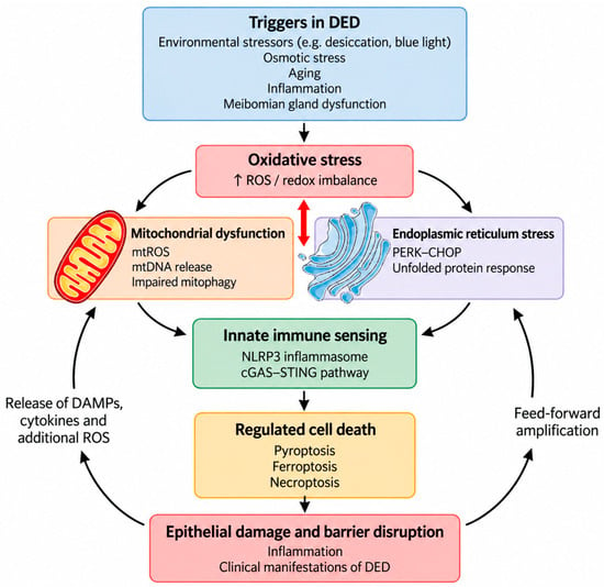
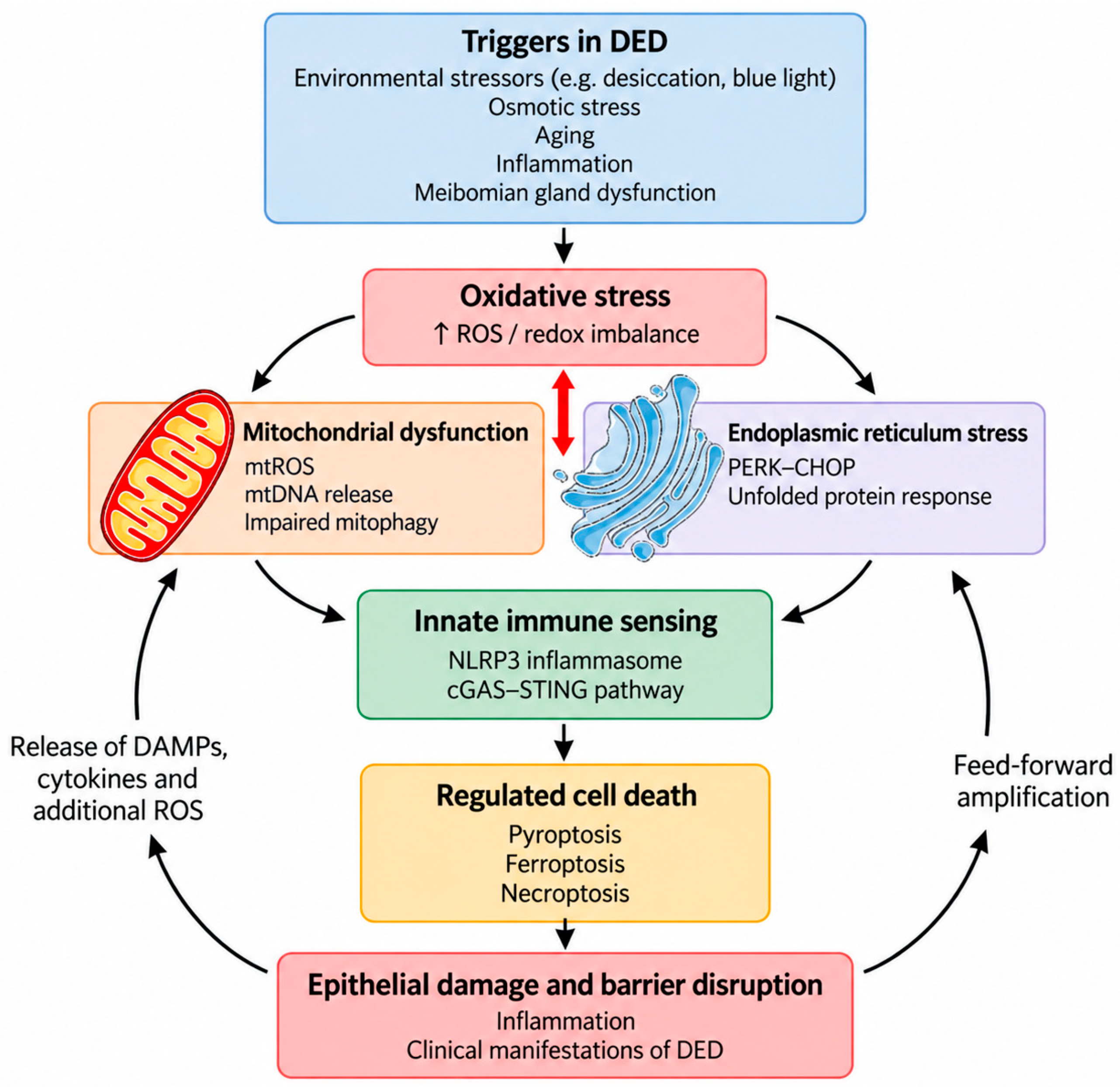

Disclaimer/Publisher’s Note: The statements, opinions and data contained in all publications are solely those of the individual author(s) and contributor(s) and not of MDPI and/or the editor(s). MDPI and/or the editor(s) disclaim responsibility for any injury to people or property resulting from any ideas, methods, instructions or products referred to in the content. |
© 2026 by the authors. Licensee MDPI, Basel, Switzerland. This article is an open access article distributed under the terms and conditions of the Creative Commons Attribution (CC BY) license.