


Abstract
This study characterized acid hydrolysates prepared from WSSV-infected (HS-WSSV) and non-infected (HS-noWSSV) Penaeus (Litopenaeus) vannamei Boone, 1931 muscle and evaluated immune-related transcript abundance and survival following WSSV challenge. Hydrolysates were maintained at pH 3.8 for 20 d, and their soluble protein profiles were assessed by SDS-PAGE. Shrimp received selected hydrolysate supernatants by intramuscular injection, and transcript abundance of Dorsal, Relish, Rab6, proPO, and PAP was evaluated at 3, 6, and 12 h post-injection. Survival was assessed after intramuscular and short-term immersion WSSV challenges. SDS-PAGE showed progressive changes in banding patterns consistent with protein degradation and distinct soluble protein profiles between HS-WSSV and HS-noWSSV preparations. Significant differences between HS-WSSV and the corresponding HS-noWSSV preparations were detected for several immune-related transcripts at specific hydrolysis and sampling times (p ≤ 0.05). Survival was evaluated using Kaplan–Meier analysis and Cox proportional hazards models with aquarium-clustered robust standard errors. In the intramuscular challenge, the 10 d HS-WSSV treatment showed the highest final survival (40.0%) and a lower mortality hazard than the WSSV-positive control (HR = 0.624, 95% CI: 0.349–1.115), although this difference was not significant (p = 0.112). In the short-term immersion challenge, the 10 d preparation produced the highest final survival (56.7%) and a significantly lower mortality hazard (HR = 0.472, 95% CI: 0.367–0.608, p < 0.001). Under the evaluated conditions, the 10 d HS-WSSV preparation was associated with early transcript-abundance changes and the most favorable survival profile, particularly following short-term immersion challenge.
1. Introduction
White spot syndrome virus (WSSV), a large enveloped double-stranded DNA virus of the family Nimaviridae, remains one of the principal infectious threats to penaeid shrimp aquaculture [1,2]. Penaeus (Litopenaeus) vannamei Boone, 1931 lacks classical adaptive immunity and relies on cellular and humoral innate responses initiated by pattern-recognition receptors and regulated by signaling pathways such as Toll and IMD [3,4]. The NF-κB-related transcription factors Dorsal and Relish participate in these pathways, whereas phagocytosis-activating protein (PAP), Rab6, and prophenoloxidase (proPO) are associated with cellular handling of pathogens and melanization-related defenses [5,6,7].
Several experimental approaches have been explored to reduce the impact of WSSV, including inactivated viral preparations, recombinant proteins, and nucleic acid-based strategies [8,9,10]. Protein hydrolysates contain soluble protein fragments and peptides whose biological effects depend on the protein source and hydrolysis conditions. Experimental studies in aquatic animals, including P. vannamei, indicate that some protein hydrolysates can modulate innate immune responses and increase resistance to infection; however, these findings have been obtained mainly with dietary or enzymatically produced hydrolysates and cannot be directly extrapolated to acid hydrolysates prepared from WSSV-infected tissue [11,12]. Acid hydrolysis cleaves tissue proteins and changes the soluble protein profile, but the composition and biological effects of hydrolysates prepared from WSSV-infected shrimp tissue remain insufficiently characterized [13,14]. Comparing infected and non-infected tissue hydrolysates may help distinguish responses associated with the infected tissue from those produced by the shrimp-muscle hydrolysate matrix. Characterizing the soluble protein profile during hydrolysis provides a first description of protein degradation and allows subsequent biological responses to be interpreted in relation to changes in the hydrolysate matrix.
We hypothesized that the soluble fraction obtained from WSSV-infected shrimp muscle would modify the early abundance of immune-related transcripts and would be associated with shrimp survival after subsequent WSSV exposure. Therefore, this study characterized the soluble protein profile of acid hydrolysates prepared from WSSV-infected and non-infected P. vannamei muscle, evaluated the relative abundance of Dorsal, Relish, PAP, Rab6, and proPO transcripts after intramuscular administration, and assessed cumulative mortality following intramuscular and short-term immersion WSSV challenges. Specifically, we investigated whether administration of acid hydrolysates prepared from WSSV-infected tissue was associated with changes in the abundance of transcripts involved in Toll, IMD, melanization, and phagocytosis-related pathways, and whether these responses were associated with altered survival following WSSV challenge.
2. Materials and Methods
2.1. Experimental Shrimp
A batch of Pacific white shrimp, P. vannamei (10 ± 2 g), was obtained from a local shrimp farm in Sinaloa, Mexico. The source farm was selected based on sanitary surveillance information provided by a representative of the Comité Estatal de Sanidad Acuícola de Sinaloa (CESASIN) in the northern Guasave zone. This information was used to identify farms and ponds with no reported detection of the pathogens included in the routine surveillance program.
Upon arrival at the laboratory, the shrimp were transferred to a 500 L tank containing seawater at a salinity of 30 g L−1, a temperature of 25 ± 2 °C, with constant aeration. The shrimp were acclimated for one week under these conditions. During acclimation, 50% of the water was renewed every three days, and the shrimp were fed twice daily, in the morning and afternoon, with a commercial diet containing 35% crude protein (Nutripec®, Purina, Mexico). A 12 h light:12 h dark photoperiod was maintained. Mortality during acclimation was <5% and occurred predominantly shortly after transport. Shrimp selected for the bioassays showed no evident external signs of disease.
To assess the WSSV status of the experimental batch, a random sample of 10 shrimp was analyzed by quantitative real-time PCR (qPCR). WSSV was not detected in the analyzed shrimp, and the batch was subsequently used in the experimental bioassays.
2.2. Experimental Design
The study was conducted in four main phases under a random allocation scheme of organisms to the different treatments. In the first phase, the WSSV stock was titrated in vivo to determine the median shrimp infectious dose (SID50 mL−1) using five serial ten-fold dilutions of the viral inoculum (10−2, 10−3, 10−4, 10−5, and 10−6), with ten shrimp evaluated per dilution, according to the Reed–Muench method [15].
In the second phase, infected muscle tissue was obtained by intramuscular inoculation of healthy shrimp with 100 μL of the viral inoculum; shrimp showing clear clinical signs of disease in a moribund state were subsequently collected.
In the third phase, two acid hydrolysates were prepared: one from WSSV-infected shrimp muscle (HS-WSSV) and another from non-infected shrimp muscle (HS-noWSSV). Both hydrolysates were maintained under acid hydrolysis for 20 d. Samples for SDS-PAGE analysis were collected before acidification (BA), immediately after acidification (AA, time 0), and at 1, 3, 5, 10, 15, and 20 d of hydrolysis. Based on the banding profiles and the objectives of the subsequent assays, BA, AA, 3 d, 10 d, and 20 d supernatants were selected for the gene-expression and viral-challenge bioassays.
In the fourth phase, the biological effects of the hydrolysates were assessed through a gene-expression bioassay and experimental WSSV challenge assays in P. vannamei injected with supernatants from HS-WSSV and HS-noWSSV, as well as through experimental WSSV challenge bioassays conducted by intramuscular injection and short-term immersion exposure.
The experimental systems were maintained at 28 ± 2 °C, 30 g L−1 salinity, pH 7.9, and dissolved oxygen above 5.0 mg L−1 by continuous aeration. No water exchange was performed during the 12 h gene-expression bioassay or the 72 h challenge assays because of their short duration. Shrimp were offered a commercial diet once daily during the gene-expression and challenge bioassays. Feed was provided to apparent satiation and remained available to the shrimp; however, feed consumption progressively decreased during the WSSV challenge as clinical signs developed. Aquaria were inspected hourly. Dead shrimp were removed immediately, whereas cumulative mortality was summarized at 6 h intervals. Shrimp lying persistently on the bottom and unable to resume normal posture or swimming were classified as moribund.
2.3. Viral Inoculum and Determination of Infectious Titer
The same WSSV strain was used for preparation of the infected hydrolysate and for the challenge bioassays. Viral titration was performed in vivo by intramuscular injection in P. vannamei shrimp maintained in 40 L aquaria containing seawater at 30 g L−1 salinity, a temperature of 28 ± 2 °C, and constant aeration. The original WSSV isolate was obtained and maintained as previously described [16,17]. To generate a fresh viral stock, healthy P. vannamei shrimp were experimentally infected with WSSV, and gill tissues from moribund animals exhibiting advanced clinical signs of white spot disease were collected. The tissues were homogenized in ten volumes (w/v) of sterile 1× PBS and centrifuged at 3000× g for 20 min at 4 °C. The resulting supernatant was subsequently centrifuged at 13,000× g for 20 min at 4 °C. The resulting supernatant was filtered through a 0.45 µm membrane, aliquoted, and stored at −70 °C until use. The same inoculum batch was used throughout the study.
Five serial ten-fold dilutions of the WSSV stock (10−2, 10−3, 10−4, 10−5, and 10−6) were prepared in sterile 1× PBS, and for each dilution, 10 shrimp (8 ± 1 g) were injected intramuscularly with 100 μL of the corresponding inoculum into the abdominal muscle between the third and fourth abdominal segments. Shrimp were monitored every 6 h for 72 h to record clinical signs and mortality. Mortality was preceded by clinical signs consistent with WSSV infection, including lethargy and atypical coloration. Dead shrimp were removed throughout the assay, and surviving organisms were sacrificed at the end of the observation period. The infectious titer of the WSSV stock was estimated using the Reed–Muench method [15]. The cumulative mortality percentages immediately above and below the 50% endpoint were 86.67% at the 10−3 dilution and 38.46% at the 10−4 dilution, respectively. The resulting proportional distance was 0.7606, corresponding to an endpoint dilution of 10−3.7606. After correction for the 0.1 mL inoculation volume, the estimated infectious titer of the WSSV stock was 5.76 × 10⁴ SID50 mL−1. Complete mortality data and the Reed–Muench calculation are provided in Supplementary Table S1.
2.4. Acid Hydrolysates from Shrimp Muscle
2.4.1. Collection of Infected Muscle Tissue
To obtain infected muscle tissue, P. vannamei shrimp were injected intramuscularly with 100 μL of the same PBS-adjusted WSSV challenge inoculum used in the intramuscular bioassays. Shrimp showing clear clinical signs of disease and a moribund state were collected and stored at −70 °C until use in the preparation of the infected hydrolysate.
2.4.2. Preparation of Acid Hydrolysates
Muscle tissue used to prepare HS-WSSV was obtained from shrimp experimentally infected with the WSSV stock described above. Moribund shrimp showing evident clinical signs of white spot disease were collected and stored frozen until processing, which was performed 3 d after collection. The HS-noWSSV control hydrolysate was prepared from muscle tissue obtained from non-infected shrimp.
For each hydrolysate, 250 g of shrimp muscle was combined with 125 mL of distilled water and homogenized using a NutriBullet NB90904 food processor (LLC, Los Angeles, CA, USA). Acid hydrolysis was initiated by adding 4.0 mL of undiluted 95% formic acid to each preparation. Additional formic acid was subsequently added as required to maintain a pH of 3.8 ± 0.2, resulting in total added volumes of 5.9 mL for HS-WSSV and 6.4 mL for HS-noWSSV. The hydrolysates were maintained in glass containers at room temperature for 20 d. The pH was monitored daily, and each preparation was manually homogenized with a spatula before pH measurement and, when necessary, acid adjustment.
Samples were collected before acidification, immediately after acidification, and after 1, 3, 5, 10, 15, and 20 d of hydrolysis. To obtain the soluble fraction used in the subsequent analyses and bioassays, 40 g of each hydrolysate sample was transferred to a 50 mL conical centrifuge tube and centrifuged at 2000× g for 20 min. The resulting supernatant was transferred to 1.5 mL microcentrifuge tubes and centrifuged again at 4 °C for 20 min at 10,000× g. The clear supernatant phase was carefully recovered, aliquoted, and stored at −70 °C until analysis and use in the bioassays.
2.4.3. Soluble Protein Quantification and SDS-PAGE Analysis
Samples were collected before acidification (BA), after acidification (AA, time 0), and during the hydrolysis process at 1, 3, 5, 10, 15, and 20 d. Once all samples had been collected, soluble protein concentration was determined using the Bradford method [18], and electrophoretic protein profiles were analyzed by sodium dodecyl sulfate-polyacrylamide gel electrophoresis (SDS-PAGE) according to Laemmli [19].
For electrophoretic analysis, 150 µL of each sample was mixed with 50 µL of loading buffer containing deionized water, 0.5 M Tris-HCl (pH 6.8), glycerol, 10% (w/v) SDS, bromophenol blue, and β-mercaptoethanol at a final concentration of 5% (v/v). Samples were heated at 95 °C for 15 min and immediately placed on ice. Proteins were separated using a 12% resolving polyacrylamide gel prepared with 1.5 M Tris-HCl (pH 8.8) and a 4% stacking gel prepared with 0.5 M Tris-HCl (pH 6.8). A volume of 10 µL of each prepared sample was loaded per well. Electrophoresis was performed using a Mini-PROTEAN® system (Bio-Rad, Hercules, CA, USA), initially at 80 V during migration through the stacking gel and subsequently at 120 V during separation through the resolving gel, for an approximate total running time of 90 min.
Following electrophoresis, the gels were stained with Coomassie Brilliant Blue and destained for 3 h using a water:methanol:acetic acid solution (70:20:10, v/v/v). The gels were subsequently maintained in distilled water for 12 h under constant agitation. Gel images were acquired using a Gel Doc XR+ imaging system (Bio-Rad) and processed using the software supplied with the equipment. The electrophoretic profiles were used to characterize changes in the soluble protein fractions during hydrolysis and to support the selection of the hydrolysis times subsequently evaluated in the biological assays.
2.5. Gene Expression Bioassay
2.5.1. Experimental Bioassay
To evaluate the effects of the hydrolysates on the relative abundance of transcripts associated with the shrimp immune response, the bioassay was conducted in 40 L aquaria maintained at 28 ± 2 °C, 30 g L−1 salinity, pH 7.9, dissolved oxygen above 5.0 mg L−1, constant aeration, and mechanical filtration. Supernatants corresponding to BA, AA, 3 d, 10 d, and 20 d from HS-WSSV and HS-noWSSV were evaluated.
For each treatment, 30 shrimp were randomly distributed among three independent aquaria, with ten shrimp per aquarium, according to a completely randomized design. Each shrimp received an intramuscular injection of 100 μL of the corresponding hydrolysate supernatant, standardized to 50 μg of total soluble protein. At 3, 6, and 12 h post-injection, two shrimp were randomly sampled from each aquarium. Hemolymph from the two shrimp collected from the same aquarium was pooled before centrifugation, generating one pooled aquarium-level biological sample. Thus, three independent pooled samples were obtained per treatment at each sampling time, with the aquarium considered the experimental unit (n = 3 aquaria).
2.5.2. Hemolymph Collection and RNA Extraction
Hemocytes were selected for transcript-abundance analysis because they are key cellular components of the shrimp innate immune response and are relevant to the immune-related genes evaluated in this study. A volume of 200 μL of hemolymph was collected from each sampled shrimp using 5% potassium oxalate prepared in sterile 1× PBS as an anticoagulant. Hemolymph from the two shrimp sampled from the same aquarium was pooled before centrifugation to obtain the hemocyte pellet. Each pooled pellet represented one aquarium-level biological sample. Total RNA was extracted from the hemocyte pellet using TRIzol® reagent (Invitrogen, Thermo Fisher Scientific, Carlsbad, CA, USA), with the samples maintained on ice throughout the procedure.
RNA concentration and purity were determined using a NanoDrop 2000 spectrophotometer (Thermo Scientific, Wilmington, DE, USA). Samples with an A260/A280 ratio between 1.8 and 2.0 were considered acceptable. Purified RNA was stored at −80 °C until use.
2.5.3. cDNA Synthesis
Prior to cDNA synthesis, RNA samples were treated with DNase to remove possible genomic DNA contamination. Reverse transcription was performed using ImProm-II™ reverse transcriptase (Promega, Madison, WI, USA) and oligo(dT)20, following the manufacturer’s instructions. Reactions were carried out in a Bio-Rad T100™ thermocycler (Bio-Rad Laboratories, Inc., Hercules, CA, USA) under the following program: 5 min at 25 °C, 60 min at 42 °C, and 15 min at 70 °C.
2.5.4. qPCR Analysis of Gene Expression
Relative expression of immune response-related genes was evaluated by quantitative real-time PCR (qPCR) using specific primers (Table 1). Amplification efficiency for each primer pair was determined from standard curves generated using serial dilutions of cDNA. The amplification efficiencies were 96.1% for Relish, 105.0% for Dorsal, 98.8% for Rab6, 106.7% for proPO, 106.7% for PAP, 98.8% for EF1α, and 100.1% for β-actin.
EF1α and β-actin were initially evaluated as candidate reference genes. Expression stability was assessed using RefFinder, and β-actin was identified as the most stable candidate reference gene under the experimental conditions and was therefore selected for normalization. Efficiency-corrected relative transcript abundance was calculated according to the Pfaffl method [20]. For preparation of the 2× master mix, 1.5 μL of 10× reaction buffer, 0.75 μL of 50 mM MgCl2, 0.3 μL of 10 mM dNTPs, 0.75 μL of 20× EvaGreen® (Biotium, Fremont, CA, USA), 0.1 μL of recombinant Taq DNA polymerase (Invitrogen), and 4.1 μL of ultrapure water were mixed. Each reaction had a final volume of 15 μL and contained 7.5 μL of this master mix, 0.35 μL of each primer (10 μM), 1.8 μL of ultrapure water, and 5 μL of cDNA. Reactions were performed in duplicate in a Bio-Rad CFX96™ qPCR system. Each pooled aquarium-level biological sample was analyzed in duplicate qPCR reactions. The duplicate reactions represented technical replicates and were not treated as independent observations. The mean Cq value of the two technical replicates was used for relative-quantification calculations after assessment of replicate agreement.
Table 1.
Primers used for immune response gene expression analysis.
2.6. WSSV Challenge Bioassays
2.6.1. Intramuscular Challenge
To evaluate the effect of prior hydrolysate administration on mortality after intramuscular WSSV challenge, shrimp were randomly distributed among 40 L aquaria containing seawater maintained at 30 g L−1 salinity, 28 ± 2 °C, pH 7.9, and dissolved oxygen above 5.0 mg L−1. Each treatment consisted of three independent aquaria with ten shrimp per aquarium.
The evaluated groups included BA, AA, 3 d, 10 d, and 20 d HS-WSSV treatments; their corresponding HS-noWSSV controls; a WSSV-positive control; and an unchallenged negative control. All groups underwent the same number of handling and injection procedures to standardize injection-related stress. Before injection, each supernatant was diluted with sterile 1× PBS to a final soluble-protein concentration of 0.5 mg mL−1, so that the 100 µL injection volume contained 50 µg of total soluble protein. During the first injection, shrimp assigned to the hydrolysate treatments received 100 μL of the corresponding HS-WSSV or HS-noWSSV supernatant. Shrimp in the WSSV-positive and negative control groups received 100 μL of sterile 1× PBS. Three hours later, all challenged groups, including the WSSV-positive control, received a second intramuscular injection of 100 μL of the same PBS-adjusted WSSV challenge inoculum. The same inoculum preparation and injection volume were used for all challenged groups. Shrimp in the unchallenged negative control received a second injection of 100 μL of sterile 1× PBS. The 3 h interval between hydrolysate administration and WSSV challenge was selected based on a preliminary transcript-abundance assay in which several post-administration sampling times were evaluated. Overall, the most pronounced early changes in transcript abundance were observed at 3 h; therefore, this interval was selected for hydrolysate administration before WSSV exposure.
Aquaria were inspected hourly. Dead shrimp were removed immediately, and cumulative mortality was recorded and summarized at 6 h intervals for 72 h. The equivalent number and timing of injections across treatments allowed injection-related handling stress to be standardized among experimental groups. At the end of each assay, water and disposable materials potentially exposed to WSSV were disinfected with sodium hypochlorite before disposal.
2.6.2. Short-Term Immersion Challenge
The short-term immersion challenge was conducted using three independent 30 L aquaria per treatment, with ten shrimp per aquarium. Seawater was maintained at 30 g L−1 salinity, 28 ± 2 °C, pH 7.9, and dissolved oxygen above 5.0 mg L−1 by continuous aeration. Treatments included the 3 d, 10 d, and 20 d HS-WSSV preparations, a WSSV-positive control, and an unchallenged negative control.
Shrimp assigned to the hydrolysate treatments received an intramuscular injection of 100 μL of the corresponding supernatant, standardized to 50 μg of total soluble protein. Shrimp in the WSSV-positive and negative control groups received 100 μL of sterile 1× PBS. Three hours later, shrimp were transferred to exposure aquaria containing 10 L of a suspension prepared from WSSV-infected shrimp muscle in saline water at a final concentration of 0.2 g L−1. Thus, each exposure aquarium received the equivalent of 2 g of WSSV-infected muscle. Shrimp were maintained in the suspension for 1 h. The unchallenged negative control underwent a corresponding 1 h mock immersion in 10 L of saline water without WSSV-infected muscle.
After exposure, shrimp were transferred to aquaria containing previously chlorinated and dechlorinated seawater. Aquaria were inspected hourly, dead shrimp were removed immediately, and cumulative mortality was recorded at 6 h intervals for 72 h.
2.7. Statistical Analysis
Transcript-abundance data were analyzed using STATISTICA version 7.0 (StatSoft Inc., Tulsa, OK, USA). For the transcript-abundance bioassay, the aquarium was considered the experimental unit. Hemolymph from two shrimp sampled from the same aquarium was pooled to generate one aquarium-level biological sample, resulting in three independent biological samples per treatment and post-injection sampling time (n = 3 aquaria). Duplicate qPCR reactions were treated as technical replicates, and the mean Cq value was used for relative quantification after assessment of agreement between technical replicates. Relative transcript abundance was calculated using the efficiency-corrected Pfaffl method, with β-actin as the reference gene. Each HS-WSSV preparation was compared with its corresponding HS-noWSSV preparation at the same hydrolysis time and post-injection sampling time using Student’s t-test. These comparisons were predefined to evaluate matched HS-WSSV versus HS-noWSSV preparations within each gene, hydrolysis time, and post-injection sampling time. Because multiple planned comparisons were performed, the transcript-abundance analysis was considered exploratory. No formal adjustment for multiple testing was applied to these comparisons; therefore, the probability of Type I error across the set of tests is increased, and individual significant p-values should be interpreted cautiously as exploratory rather than confirmatory evidence. Statistical significance for individual comparisons was established at p ≤ 0.05.
Survival analyses were performed in R version 4.6.1 [23] using the survival package version 3.8-12 and the survminer package version 0.5.2. Kaplan–Meier estimators were used to describe survival during the 72 h observation period, and global log-rank tests were used to assess overall differences among survival curves. When the global log-rank test was significant, pairwise log-rank comparisons were performed with Benjamini–Hochberg adjustment for multiple comparisons. Mortality hazard was further evaluated using Cox proportional hazards models with aquarium-clustered robust standard errors to account for the non-independence of shrimp maintained within the same aquarium. The WSSV-positive control was used as the reference category. Results are reported as hazard ratios (HRs) with 95% confidence intervals (CIs) and corresponding p-values. Statistical significance was established at p ≤ 0.05.
3. Results
3.1. Characterization of the Hydrolysates
3.1.1. pH and Soluble Protein
Following acidification, the pH of both hydrolysates decreased to 4.0 and reached 3.8 after 24 h, remaining stable through day 20. Soluble protein concentrations ranged from 1.8 to 2.5 mg mL−1 in both hydrolysates throughout the evaluated period.
3.1.2. Electrophoretic Analysis of the Hydrolysates
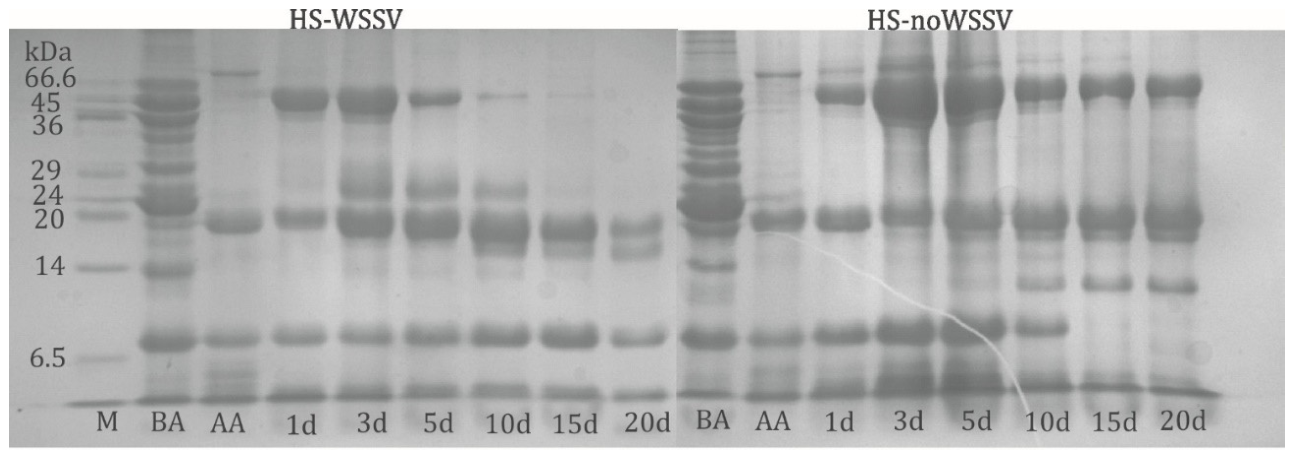
The 12% SDS-PAGE profiles (Figure 1) differed visually between HS-WSSV and HS-noWSSV during hydrolysis. Before acidification, both preparations showed similar banding patterns. As hydrolysis progressed, high-molecular-weight bands decreased in HS-WSSV but remained comparatively stable in HS-noWSSV. Consequently, bands above approximately 20 kDa became markedly less evident in HS-WSSV by the end of the monitoring period. Because the bands were not analytically identified, these results are limited to a descriptive comparison of the electrophoretic profiles.
Figure 1.
Coomassie Brilliant Blue-stained 12% SDS-PAGE profiles of the soluble protein fractions of acid hydrolysates prepared from WSSV-infected shrimp muscle (HS-WSSV) and non-infected shrimp muscle (HS-noWSSV) during 20 d of hydrolysis. Supernatants were analyzed before acidification (BA), immediately after acidification (AA, time 0), and after 1, 3, 5, 10, 15, and 20 d of hydrolysis. M, BenchMark™ Protein Ladder (Invitrogen); molecular mass markers are indicated in kDa. HS-noWSSV and HS-WSSV samples were analyzed on separate gels run under identical electrophoretic conditions, and the resulting gel images were assembled for comparative presentation. No quantitative comparison of band intensity between gels was performed.
3.2. Relative Abundance of Immune-Related Transcripts in Shrimp Injected with HS-WSSV and HS-noWSSV
3.2.1. Transcriptional Response at 3 h Post-Injection
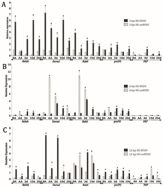
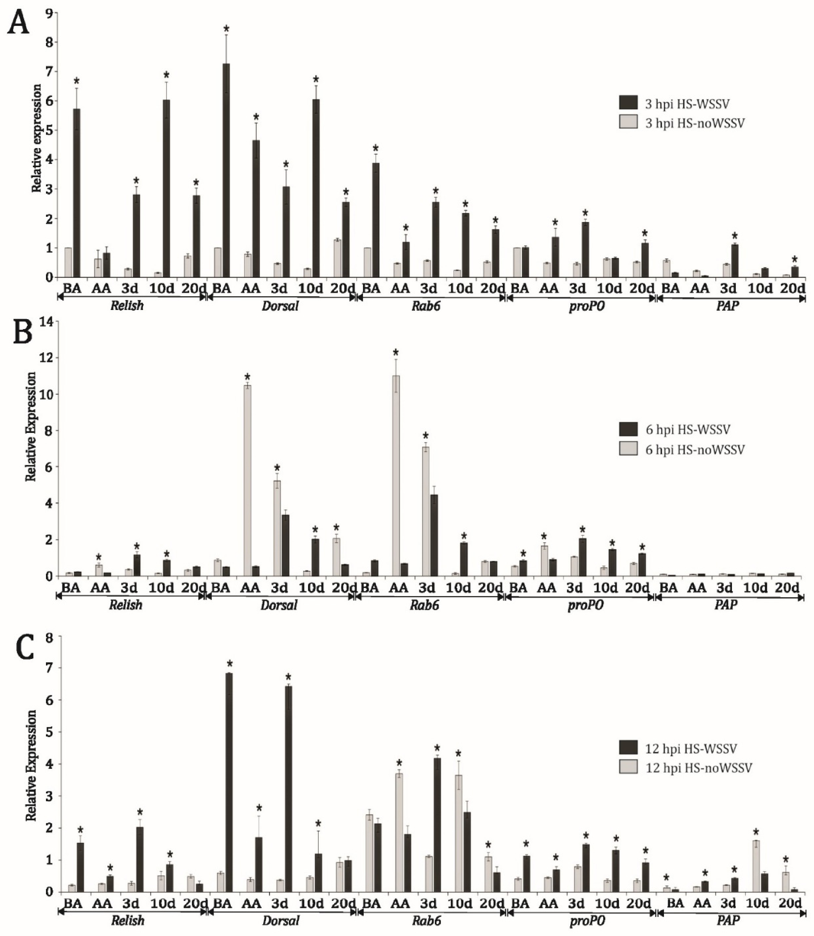
At 3 h post-injection, shrimp receiving HS-WSSV showed higher Relish transcript abundance than those receiving the corresponding HS-noWSSV preparation at BA, 3 d, 10 d, and 20 d, whereas no significant difference was detected at AA. Dorsal and Rab6 transcript abundance was higher in shrimp receiving HS-WSSV at all evaluated hydrolysis times. Higher proPO transcript abundance was detected in the HS-WSSV groups at AA, 3 d, and 20 d. For PAP, significantly higher transcript abundance in the HS-WSSV groups was observed at 3 d and 20 d (Figure 2A).
Figure 2.
Relative abundance of immune-related transcripts (Relish, Dorsal, Rab6, proPO, and PAP) in P. vannamei at 3 h (A), 6 h (B), and 12 h (C) after intramuscular injection of acid-hydrolysate supernatants. BA, before acidification; AA, immediately after acidification (time 0); hydrolysis times of 3, 10, and 20 d. Data are presented as mean ± SEM from three independent aquarium-level pooled samples per treatment (n = 3 aquaria). Asterisks indicate significant differences between HS-WSSV and the corresponding HS-noWSSV preparation within the same gene, hydrolysis time, and post-injection sampling time (p ≤ 0.05, Student’s t-test).
3.2.2. Transcriptional Response at 6 h Post-Injection
At 6 h post-injection, the direction of the transcriptional response varied among genes and hydrolysis times. Relish transcript abundance was higher in shrimp receiving HS-noWSSV at AA, whereas higher abundance was observed in the HS-WSSV groups at 3 d and 10 d. Dorsal transcript abundance was higher in the HS-noWSSV groups at AA, 3 d, and 20 d, whereas the 10 d HS-WSSV group showed the higher value. A similar bidirectional pattern was observed for Rab6, with higher transcript abundance in the HS-noWSSV groups at AA and 3 d and in the HS-WSSV group at 10 d. For proPO, higher transcript abundance was detected in the HS-WSSV groups at BA, 3 d, 10 d, and 20 d, whereas the HS-noWSSV group showed the higher value at AA. No significant differences were detected for PAP (Figure 2B).
3.2.3. Transcriptional Response at 12 h Post-Injection
At 12 h post-injection, Relish and Dorsal transcript abundance was higher in shrimp receiving HS-WSSV at BA, AA, 3 d, and 10 d, whereas no significant difference was detected at 20 d. For Rab6, higher transcript abundance was observed in the HS-noWSSV groups at AA, 10 d, and 20 d, whereas the HS-WSSV group showed the higher value at 3 d. proPO transcript abundance was higher in the HS-WSSV groups at all evaluated hydrolysis times. PAP showed a bidirectional response, with higher abundance in the HS-WSSV groups at BA, AA, and 3 d and in the HS-noWSSV groups at 10 d and 20 d (Figure 2C).
3.3. Survival Following WSSV Challenge
3.3.1. Intramuscular Challenge
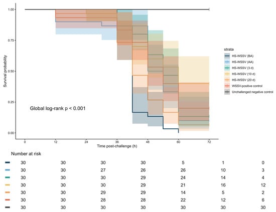
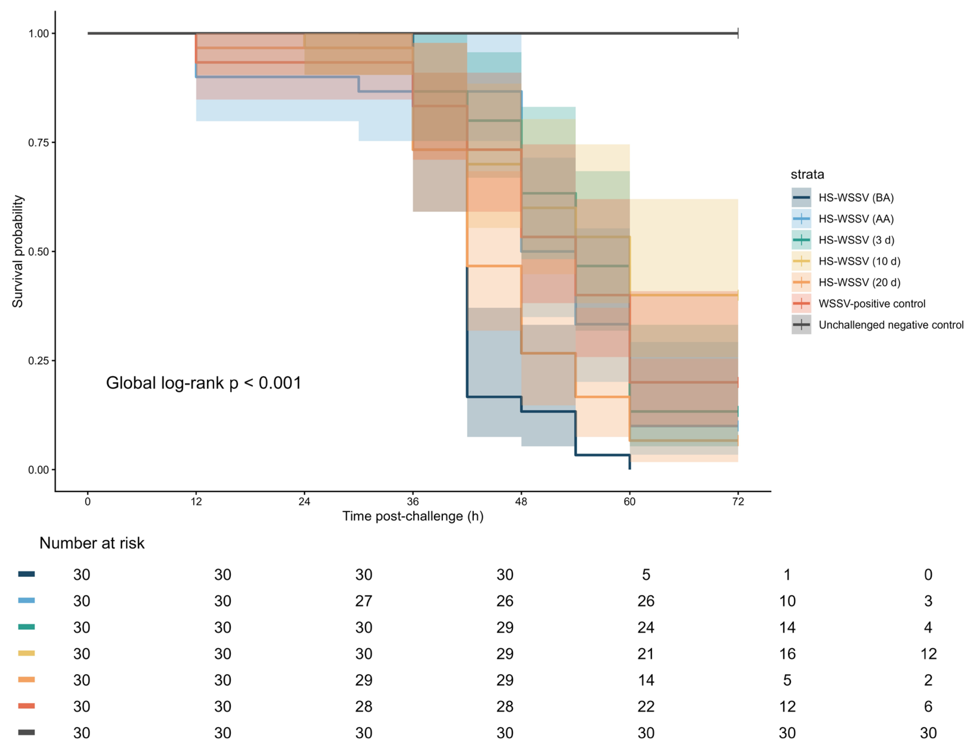
Kaplan–Meier analysis revealed significant overall differences in survival among experimental groups following WSSV challenge by intramuscular injection (global log-rank test: χ2 = 101.25, df = 6, p < 0.001). Final survival at 72 h was 0.0%, 10.0%, 13.3%, 40.0%, and 6.7% in the HS-WSSV BA, AA, 3 d, 10 d, and 20 d treatments, respectively. Final survival in the WSSV-positive control was 20.0%, whereas the unchallenged negative control maintained 100% survival throughout the observation period (Figure 3).
Figure 3.
Kaplan–Meier survival curves of P. vannamei previously administered HS-WSSV preparations and subsequently challenged with WSSV by intramuscular injection. Shaded areas represent 95% confidence intervals, and censoring marks indicate shrimp that remained alive at the end of the 72 h observation period. The number of shrimp at risk is shown below the graph. The global log-rank test indicated significant differences among the survival curves (p < 0.001). Aquaria were the experimental units, with shrimp clustered within aquaria.
Pairwise log-rank comparisons adjusted using the Benjamini–Hochberg procedure indicated that survival in the HS-WSSV 10 d treatment differed significantly from that in the BA and 20 d treatments. However, no significant differences were detected between the 10 d treatment and the AA, 3 d, or WSSV-positive control groups after adjustment for multiple comparisons. Thus, although the 10 d preparation achieved the highest final survival among the challenged groups, its superiority was not statistically supported relative to all treatments.
The Cox proportional hazards model with aquarium-clustered robust standard errors, using the WSSV-positive control as the reference group, showed that the HS-WSSV 10 d treatment was associated with a lower mortality hazard (HR = 0.624, 95% CI: 0.349–1.115, p = 0.112), corresponding to an approximate 37.6% reduction in mortality hazard relative to the positive control. Nevertheless, this reduction was not statistically significant (p = 0.112). The HS-WSSV 3 d treatment exhibited a mortality hazard similar to that of the positive control (HR = 1.01, 95% CI: 0.813–1.26, p = 0.911). In contrast, the HS-WSSV 20 d (HR = 1.94, 95% CI: 1.54–2.43, p < 0.001), AA (HR = 1.20, 95% CI: 1.15–1.25, p < 0.001), and BA preparations (HR = 3.54, 95% CI: 2.61–4.79, p < 0.001) were associated with significantly greater mortality hazards than the positive control.
3.3.2. Short-Term Immersion Challenge
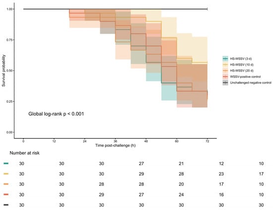
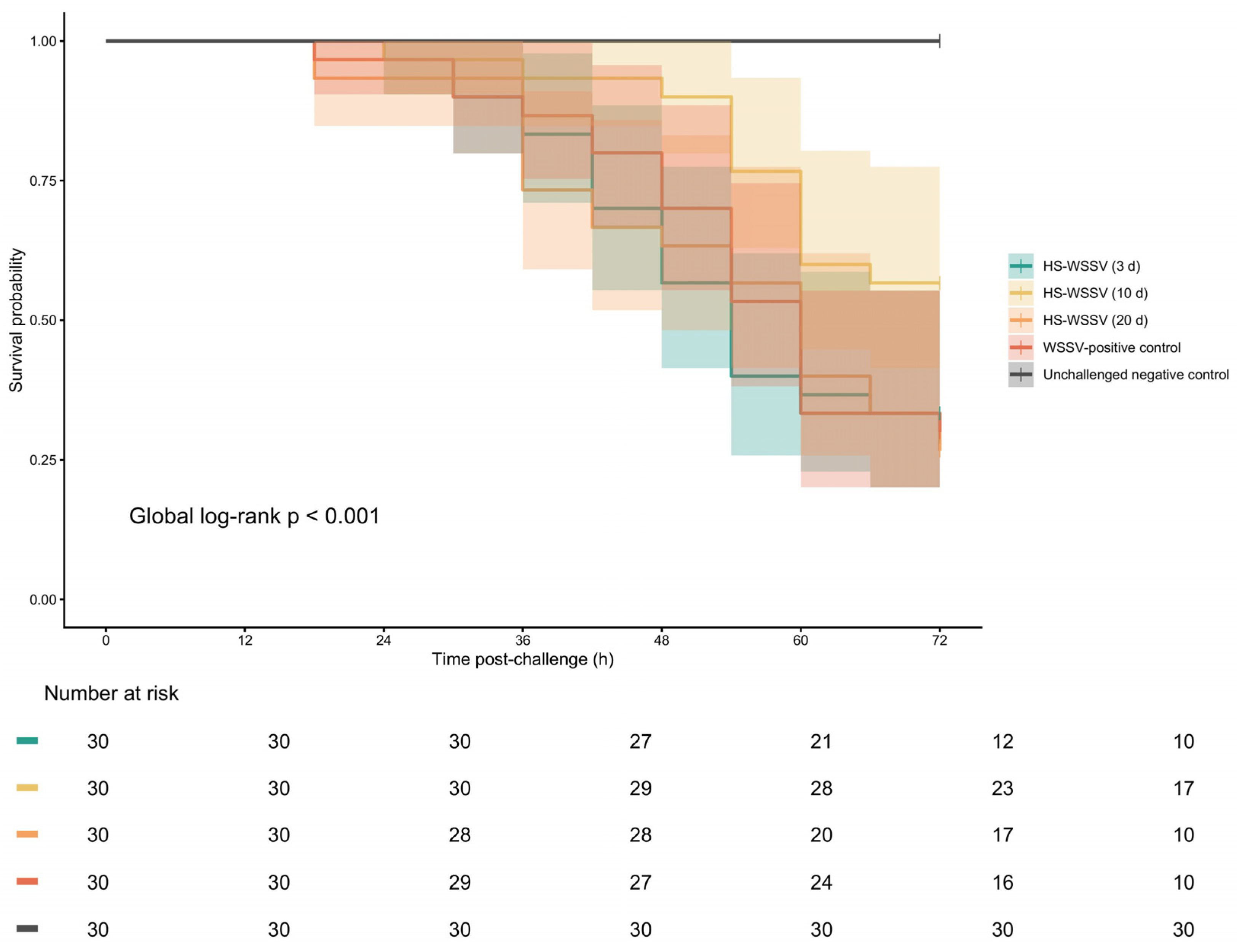
Kaplan–Meier analysis demonstrated significant overall differences among survival curves following WSSV challenge by short-term immersion (global log-rank test: χ2 = 40.25, df = 4, p < 0.001). Final survival at 72 h was 33.3%, 56.7%, and 26.7% in the HS-WSSV 3 d, 10 d, and 20 d treatments, respectively. Final survival in the WSSV-positive control was 30.0%, whereas the unchallenged negative control maintained 100% survival (Figure 4).
Figure 4.
Kaplan–Meier survival curves of P. vannamei previously administered HS-WSSV preparations and subsequently challenged with WSSV by short-term immersion. Shaded areas represent 95% confidence intervals, and censoring marks indicate shrimp that remained alive at the end of the 72 h observation period. The number of shrimp at risk is shown below the graph. The global log-rank test indicated significant differences among the survival curves (p < 0.001). Aquaria were the experimental units, with shrimp clustered within aquaria.
Pairwise log-rank comparisons showed that survival in the HS-WSSV 10 d treatment was significantly higher than that observed in the HS-WSSV 3 d and 20 d treatments and the WSSV-positive control after adjustment for multiple comparisons. No significant differences were detected among the 3 d, 20 d, and WSSV-positive control groups.
Consistent with these findings, the aquarium-clustered Cox proportional hazards model, using the WSSV-positive control as the reference group, indicated that the HS-WSSV 10 d treatment was associated with a significantly lower mortality hazard (HR = 0.472, 95% CI: 0.367–0.608, p < 0.001). This hazard ratio corresponds to an approximate 52.8% reduction in mortality hazard relative to the positive control. In contrast, neither the HS-WSSV 3 d treatment (HR = 1.05, 95% CI: 0.821–1.352, p = 0.680) nor the HS-WSSV 20 d treatment (HR = 1.07, 95% CI: 0.868–1.321, p = 0.524) differed significantly from the positive control.
4. Discussion
The cleavage of peptide bonds modifies the buffering capacity of the system, making pH monitoring and control relevant variables during hydrolysis [24]. This cleavage generates new amino (-NH2) and carboxyl (-COOH) groups that can transfer protons (H+) and alter the acid-base balance of the medium [25]. In the present study, pH was approximately 4.0 following the addition of formic acid and during the first day of hydrolysis, after which it stabilized at 3.8. Higher pH values, ranging from 7.21 to 7.38, have been reported for low-salt shrimp paste [26]; however, these values correspond to a different processing system. The low and stable pH recorded in the present study resulted from intentional acidification and created conditions favorable for the hydrolytic modification of tissue proteins.
Soluble protein concentration varied only slightly, remaining between 1.8 and 2.5 mg mL−1 in both hydrolysates throughout the 20 d period. This limited variation indicates that the amount of soluble protein recovered remained relatively stable. However, total soluble protein concentration does not provide information about changes in the molecular size or composition of the protein fragments, which were more clearly reflected in the SDS-PAGE profiles.
Some bands observed in HS-WSSV occurred within molecular-weight ranges reported for WSSV structural proteins [27]. However, correspondence in apparent molecular weight is insufficient for protein identification, and SDS-PAGE alone cannot establish whether these bands were viral or host-derived. Therefore, the electrophoretic profiles should be interpreted as comparative evidence of changes in the soluble protein matrix rather than as evidence of the presence or preservation of specific viral proteins.
The progressive disappearance of high-molecular-weight bands was more evident in HS-WSSV than in HS-noWSSV, suggesting differences in protein degradation dynamics between hydrolysates derived from infected and non-infected tissues. In HS-WSSV, the band with the highest apparent molecular weight progressively decreased until it was almost undetectable, whereas the HS-noWSSV profile remained comparatively stable. The physiological and structural alterations described in WSSV-infected shrimp tissues may have contributed to differences in the initial protein matrix and its susceptibility to hydrolysis [1,28]. Thus, the electrophoretic results indicate that the soluble protein profiles of the infected- and non-infected-tissue hydrolysates changed differently during acid hydrolysis and provide physicochemical context for the subsequent bioassays. Nevertheless, they do not identify the compounds responsible for the observed biological responses.
As penaeid shrimp lack classical adaptive immunity, their response to WSSV depends primarily on innate immune mechanisms mediated by hemocytes and signaling pathways that coordinate cellular and humoral defenses [29]. The Toll and IMD pathways are particularly relevant because they involve the NF-κB-related transcription factors Dorsal and Relish, respectively, which regulate antimicrobial peptides and other immune effectors associated with the response to WSSV [3,30]. The genes evaluated in the present study represent complementary components of this response: Dorsal and Relish are associated with immune signaling, PAP and Rab6 with cellular processes and pathogen handling, and proPO with the melanization cascade. This set of genes therefore provided markers of signaling, cellular, and humoral components involved in the shrimp response to WSSV [6].
Administration of HS-WSSV was followed by early changes in Dorsal and Relish transcript abundance. Changes in Dorsal transcript abundance were consistent with modulation of Toll-related signaling, whereas changes in Relish transcript abundance were consistent with modulation of IMD-related signaling. Toll- and IMD-related responses can occur in a coordinated manner during WSSV infection, with Dorsal and Relish acting as interconnected transcriptional regulators of immune defenses [3,31,32]. Relish has also been associated with apoptosis, hemocyte homeostasis, and phenoloxidase-related responses during WSSV infection [33]. Nevertheless, changes in transcript abundance alone do not demonstrate functional activation of either pathway or the production of downstream effectors. The results should therefore be interpreted as evidence of an early transcriptional response associated with Toll- and IMD-related immune signaling.
PAP and Rab6 extended the analysis to genes associated with cellular immune processes. PAP has been related to immune stimulation and resistance to WSSV and other pathogens, suggesting a role in hemocyte-mediated defense functions [5]. In the present study, PAP showed a comparatively lower response than Dorsal or Relish, with its clearest changes occurring at early sampling times. This pattern is compatible with a role in early cellular immune responses, although the present data do not establish the functional contribution of PAP to the observed survival outcomes.
Rab6 also showed early increases in transcript abundance after HS-WSSV administration, particularly at 3 h post-injection. This gene has been associated with phagocytosis-related responses in shrimp, and RNA interference studies support its participation in hemocyte-mediated responses against WSSV [34,35]. Rab6 has also been related to cellular events involving the WSSV protein VP466 and reorganization of the actin cytoskeleton during pathogen internalization [35]. Consequently, the early changes in Rab6 transcript abundance are compatible with modulation of cellular immune processes, although they do not demonstrate enhanced phagocytic activity.
The changes in proPO transcript abundance observed after HS-WSSV administration were consistent with modulation of the prophenoloxidase system, an important humoral defense mechanism in crustaceans that leads to phenoloxidase activity and melanization [29,36]. Although this system is commonly associated with antibacterial and antifungal defenses, its involvement in the response to WSSV has also been documented. Sutthangkul et al. [37] reported that proPO silencing increased susceptibility to WSSV, supporting the participation of this system in antiviral defense.
Collectively, HS-WSSV administration was associated with an early transcriptional response involving genes related to immune signaling, cellular processes, and humoral defenses rather than with changes restricted to a single immune component. Several significant transcriptional differences were observed across the evaluated hydrolysis and post-injection sampling times. Given the exploratory nature of these analyses and the number of pairwise comparisons performed, individual transcript-level differences should be interpreted cautiously and require confirmation in independent experiments. The 10 d preparation produced higher transcript abundance than its corresponding HS-noWSSV preparation for several genes at specific sampling times and was also associated with the most favorable survival profile in the subsequent challenge assays. However, the specific compounds responsible for these responses were not identified, and a causal relationship between transcriptional modulation and lower mortality cannot be established from the present results.
Across both challenge models, the 10 d HS-WSSV preparation showed the most favorable survival profile among the evaluated preparations. However, the magnitude and statistical support of this response differed according to the challenge route. The concordance between some of the early transcriptional responses and the subsequent survival patterns suggests that hydrolysis time was associated with differences in the biological properties of the preparations.
Following intramuscular WSSV challenge, the 10 d HS-WSSV treatment achieved the numerically highest final survival among the challenged groups, reaching 40.0% at 72 h. The Cox proportional hazards model estimated a 37.6% reduction in mortality hazard relative to the WSSV-positive control (HR = 0.624, 95% CI: 0.349–1.115, p = 0.112). However, this difference was not statistically significant, and pairwise log-rank comparisons likewise did not detect a significant difference between the 10 d treatment and the positive control after adjustment for multiple comparisons. Therefore, under the intramuscular challenge conditions, the response associated with the 10 d preparation should be interpreted as a favorable survival pattern rather than definitive evidence of superior protection.
The HS-noWSSV preparations were included as hydrolysate controls to distinguish responses potentially associated with the non-infected shrimp-muscle matrix. Because these treatments did not show a survival pattern indicating a protective effect, subsequent analyses focused on the HS-WSSV preparations.
A more clearly supported treatment-associated survival response was observed following the short-term immersion challenge. The 10 d HS-WSSV preparation produced the highest final survival, reaching 56.7% at 72 h, and was associated with an approximately 52.8% lower mortality hazard relative to the WSSV-positive control (HR = 0.472, 95% CI: 0.367–0.608, p < 0.001). This result was supported by both the pairwise log-rank comparisons and the aquarium-clustered Cox model. In contrast, the 3 d and 20 d preparations did not differ significantly from the positive control. These findings indicate that the favorable response was specifically associated with the preparation obtained after 10 d of hydrolysis under the evaluated immersion-challenge conditions rather than being a general characteristic of all HS-WSSV preparations. However, corresponding HS-noWSSV controls were not included in the short-term immersion challenge.
The survival response associated with the 10 d preparation differed between challenge routes. Under intramuscular injection, the estimated reduction in mortality hazard did not reach statistical significance, whereas under short-term immersion the 10 d preparation was associated with a significantly lower mortality hazard relative to the positive control. Direct injection introduces the challenge material into internal tissues and may produce infection dynamics that differ from those resulting from short-term immersion, which could contribute to the different survival patterns observed between challenge models. This explanation remains hypothetical, however, because the study was not designed to determine the mechanisms underlying route-dependent differences.
The short-term immersion model differs from controlled peroral challenge procedures based on feeding infected tissue or direct oral inoculation. It nevertheless provides an experimental model of waterborne WSSV exposure. Previous studies have reported reduced mortality using oral-delivery systems containing recombinant WSSV proteins or dsRNA [10,38,39,40]. However, differences in administration route, formulation, dose, challenge material, and experimental design prevent direct efficacy comparisons with the present study.
The comparatively favorable performance of the 10 d preparation in both challenge models suggests that hydrolysis time influenced the biological characteristics of the soluble fraction. The 3 d, 10 d, and 20 d treatments represented different stages of hydrolysis, whereas the interval between hydrolysate administration and WSSV challenge was fixed at 3 h. Therefore, the differences among treatments are more plausibly related to changes in the composition of the soluble hydrolysate fraction during hydrolysis than to differences in the administration-to-challenge interval. The 10 d preparation may have contained a combination or relative abundance of soluble components that differed from those present at earlier or later hydrolysis times. However, this interpretation remains hypothetical because individual proteins or peptides were not identified, residual viral material was not quantified, and the functional mechanisms underlying the survival response were not directly evaluated.
The survival response observed with the 10 d HS-WSSV preparation is compatible with previous reports describing altered responses to subsequent WSSV exposure following administration of virus-related stimuli in crustaceans. Nevertheless, the present study did not evaluate immune memory, trained immunity, epigenetic regulation, viral load, or the duration of the induced response. Accordingly, the findings should not be interpreted as evidence of trained immunity, sterilizing protection, or a specific antiviral mechanism. The present findings therefore support only an association between administration of the 10 d HS-WSSV preparation, early changes in immune-related transcript abundance, and a more favorable survival profile, particularly under short-term immersion challenge.
Taken together, HS-WSSV administration, particularly the preparation obtained after 10 d of hydrolysis, was associated with early changes in the abundance of transcripts representing several components of innate immunity and with the most favorable survival profile among the evaluated preparations. The strength of the survival evidence differed between challenge models: the intramuscular assay showed a favorable but non-significant reduction in mortality hazard relative to the WSSV-positive control, whereas the short-term immersion assay showed a statistically significant reduction in mortality hazard. However, the compounds responsible for these associations remain unidentified, and causality between transcriptional modulation and survival was not established. An additional limitation of the present study is that each hydrolysate was produced as a single preparation batch. Therefore, batch-to-batch reproducibility of the physicochemical characteristics and biological responses could not be evaluated and should be addressed in future studies using independently prepared hydrolysate batches. The 10 d HS-WSSV preparation cannot currently be recommended for commercial application because residual WSSV infectivity, biosafety, batch-to-batch reproducibility, dose standardization, and the identity of the potentially active components remain to be determined. In addition, the scalability of hydrolysate production, product stability during storage, manufacturing costs, and overall economic feasibility would need to be established before any practical application in commercial shrimp farming could be considered.
5. Conclusions
The 10 d HS-WSSV preparation was associated with early changes in immune-related transcript abundance and the most favorable survival profile among the evaluated hydrolysis times. Following intramuscular challenge, the 10 d preparation produced the highest final survival and a lower estimated mortality hazard than the WSSV-positive control, although this difference was not statistically significant. Following short-term immersion challenge, the 10 d preparation produced significantly higher survival and a lower mortality hazard than the positive control.
These findings suggest that hydrolysis time influenced the biological properties of the soluble preparations. However, the compounds responsible for the observed associations remain unidentified, and a causal relationship between transcript-abundance changes and survival was not established. The results support further investigation of the 10 d HS-WSSV preparation as a source of potentially bioactive soluble components. Future studies should evaluate residual viral infectivity, identify and characterize the potentially active fractions, measure functional immune responses and viral load, confirm batch-to-batch reproducibility using independently prepared hydrolysate batches, and validate safety and efficacy under broader experimental conditions.
Supplementary Materials
The following supporting information can be downloaded at: https://www.mdpi.com/article/10.3390/aquacj6040046/s1, Table S1: In vivo titration of the WSSV stock in P. vannamei and Reed-Muench calculation of the median shrimp infectious dose (SID50).
Author Contributions
N.M.P.-L.: Formal analysis, investigation, data curation, visualization, writing—original draft preparation. P.Á.-R.: Conceptualization, methodology, supervision, project administration, funding acquisition. M.M.-M.: Conceptualization, methodology, resources, supervision, project administration, funding acquisition, writing—original draft preparation, writing—review and editing. H.R.-G.: Resources, supervision, writing—review and editing. C.G.-J.: Investigation, data curation, visualization. G.L.-C.: Methodology, supervision, writing—review and editing. E.T.-O.: Conceptualization, supervision. C.C.-M.: Supervision, writing—review and editing. G.L.-V.: Formal analysis, investigation. D.L.B.-M.: Investigation. Author P.Á.-R. passed away prior to the publication of this manuscript. All other authors have read and agreed to the published version of this manuscript.
Funding
This research was funded by Instituto Politécnico Nacional (IPN), grant numbers SIP20250923, SIP20250582, and SIP20250469. The APC was funded by SIP-IPN.
Institutional Review Board Statement
Ethical review and approval were waived for this study because it involved an invertebrate species (Penaeus vannamei) that is not subject to ethical approval under the applicable institutional requirements. All experimental procedures were conducted in accordance with applicable national regulations and institutional guidelines for the care and use of experimental animals, and appropriate measures were taken to minimize stress and unnecessary suffering.
Informed Consent Statement
Not applicable.
Data Availability Statement
The data presented in this study are available on request from the corresponding author.
Acknowledgments
N.M.P.-L. received fellowship support from CONACYT (1088121) and BEIFI-IPN. The authors acknowledge financial support from COFAA-IPN (Comisión de Operación y Fomento de Actividades Académicas) and EDI-IPN (Estímulo al Desempeño de los Investigadores) of the Instituto Politécnico Nacional. The authors also acknowledge the valuable contribution of the late P.A.-R. to the development of this research.
Conflicts of Interest
The authors declare no conflicts of interest.
References
- Sánchez-Paz, A. White spot syndrome virus: An overview on an emergent concern. Vet. Res. 2010, 41, 43. [Google Scholar] [CrossRef] [Scilit] [PubMed]
- Verbruggen, B.; Bickley, L.; Van Aerle, R.; Bateman, K.; Stentiford, G.; Santos, E.; Tyler, C. Molecular mechanisms of White Spot Syndrome Virus infection and perspectives on treatments. Viruses 2016, 8, 23. [Google Scholar] [CrossRef] [Scilit] [PubMed]
- Li, F.; Xiang, J. Recent advances in researches on the innate immunity of shrimp in China. Dev. Comp. Immunol. 2013, 39, 11–26. [Google Scholar] [CrossRef] [Scilit] [PubMed]
- Roy, S.; Bossier, P.; Norouzitallab, P.; Vanrompay, D. Trained immunity and perspectives for shrimp aquaculture. Rev. Aquac. 2020, 12, 2351–2370. [Google Scholar] [CrossRef] [Scilit]
- Khimmakthong, U.; Deachamag, P.; Phongdara, A.; Chotigeat, W. Stimulating the immune response of Litopenaeus vannamei using the phagocytosis-activating protein (PAP) gene. Fish Shellfish Immunol. 2011, 31, 415–422. [Google Scholar] [CrossRef] [Scilit] [PubMed]
- Wang, P.-H.; Huang, T.; Zhang, X.; He, J.-G. Antiviral defense in shrimp: From innate immunity to viral infection. Antivir. Res. 2014, 108, 129–141. [Google Scholar] [CrossRef] [Scilit] [PubMed]
- Soto-Alcalá, J.; Álvarez-Ruiz, P.; Audelo-Naranjo, J.M.; Esparza-Leal, H.M.; Luis-Villaseñor, I.E.; Estrada-Godínez, J.A.; Luna-González, A.; Gámez-Jiménez, C.; Diarte-Plata, G. Transcriptional response of immune-related genes in Litopenaeus vannamei post-larvae cultured in recirculating aquaculture systems with and without biofloc. Aquac. Int. 2019, 27, 209–225. [Google Scholar] [CrossRef] [Scilit]
- Amar, E.C.; Faisan, J.P., Jr.; Gapasin, R.S.J. Field efficacy evaluation of a formalin-inactivated white spot syndrome virus (WSSV) vaccine for the preventive management of WSSV infection in shrimp grow-out ponds. Aquaculture 2021, 531, 735907. [Google Scholar] [CrossRef] [Scilit]
- Lanh, P.T.; Nguyen, H.M.; Duong, B.T.T.; Hoa, N.T.; Thom, L.T.; Tam, L.T.; Thu, H.T.; Van Nha, V.; Hong, D.D.; Mouradov, A.; et al. Generation of microalga Chlamydomonas reinhardtii expressing VP28 protein as oral vaccine candidate for shrimps against White Spot Syndrome Virus (WSSV) infection. Aquaculture 2021, 540, 736737. [Google Scholar] [CrossRef] [Scilit]
- Ramos-Carreño, S.; Giffard-Mena, I.; Zamudio-Ocadiz, J.N.; Nuñez-Rivera, A.; Valencia-Yañez, R.; Ruiz-Garcia, J.; Viana, M.T.; Cadena-Nava, R.D. Antiviral therapy in shrimp through plant virus VLP containing VP28 dsRNA against WSSV. Beilstein J. Org. Chem. 2021, 17, 1360–1373. [Google Scholar] [CrossRef] [Scilit] [PubMed]
- Khosravi, S.; Rahimnejad, S.; Herault, M.; Fournier, V.; Lee, C.-R.; Bui, H.T.D.; Jeong, J.-B.; Lee, K.-J. Effects of protein hydrolysates supplementation in low fish meal diets on growth performance, innate immunity and disease resistance of red sea bream Pagrus major. Fish Shellfish Immunol. 2015, 45, 858–868. [Google Scholar] [CrossRef] [Scilit] [PubMed]
- Tran, H.T.Q.; Ho, T.H.; Nan, F.-H.; Liu, C.-H.; Hu, Y.-F.; Chong, C.M.; de Cruz, C.R.; Karim, M.; Liu, T.-J.; Kuo, I.-P.; et al. Assessment of fish protein hydrolysate as a substitute for fish meal in white shrimp diets: Impact on growth, immune response, and resistance against Vibrio parahaemolyticus infection. Fish Shellfish Immunol. 2024, 150, 109597. [Google Scholar] [CrossRef] [Scilit] [PubMed]
- Pasupuleti, V.K.; Braun, S. State of the art manufacturing of protein hydrolysates. In Protein Hydrolysates in Biotechnology; Pasupuleti, V.K., Demain, A.L., Eds.; Springer: Dordrecht, The Netherlands, 2008; pp. 11–32. [Google Scholar] [CrossRef] [Scilit]
- He, S.; Franco, C.; Zhang, W. Functions, applications and production of protein hydrolysates from fish processing co-products (FPCP). Food Res. Int. 2013, 50, 289–297. [Google Scholar] [CrossRef] [Scilit]
- Reed, L.J.; Muench, H. A simple method of estimating fifty per cent endpoints. Am. J. Epidemiol. 1938, 27, 493–497. [Google Scholar] [CrossRef] [Scilit]
- Álvarez-Ruiz, P.; Mejía-Ruiz, C.H.; Magallón-Barajas, F.J.; Escobedo-Bonilla, C.M. Silencing Pacific white shrimp Litopenaeus vannamei LvRab7 reduces mortality in brooders challenged with white spot syndrome virus. Aquac. Res. 2013, 44, 772–782. [Google Scholar] [CrossRef] [Scilit]
- Escobedo-Bonilla, C.M.; Wille, M.; Alday-Sanz, V.; Sorgeloos, P.; Pensaert, M.B.; Nauwynck, H.J. In vivo titration of white spot syndrome virus (WSSV) in specific pathogen-free Litopenaeus vannamei by intramuscular and oral routes. Dis. Aquat. Org. 2005, 66, 163–170. [Google Scholar] [CrossRef] [Scilit] [PubMed]
- Bradford, M.M. A rapid and sensitive method for the quantitation of microgram quantities of protein utilizing the principle of protein-dye binding. Anal. Biochem. 1976, 72, 248–254. [Google Scholar] [CrossRef] [PubMed]
- Laemmli, U.K. Cleavage of structural proteins during the assembly of the head of bacteriophage T4. Nature 1970, 227, 680–685. [Google Scholar] [CrossRef] [Scilit] [PubMed]
- Pfaffl, M.W. A new mathematical model for relative quantification in real-time RT-PCR. Nucleic Acids Res. 2001, 29, e45. [Google Scholar] [CrossRef] [Scilit] [PubMed]
- Alvarez-Ruiz, P.; Luna-González, A.; Escamilla-Montes, R.; Mejía-Ruiz, C.H.; Magallón-Barajas, F.J.; Llera-Herrera, R.; Galván-Alvarez, D.A. Long-lasting effect against White Spot syndrome virus in shrimp broodstock, Litopenaeus vannamei, by LvRab7 silencing. J. World Aquac. Soc. 2015, 46, 571–582. [Google Scholar] [CrossRef] [Scilit]
- Wang, Y.-C.; Chang, P.-S.; Chen, H.-Y. Tissue expressions of nine genes important to immune defense of the Pacific white shrimp Litopenaeus vannamei. Fish Shellfish Immunol. 2007, 23, 1161–1177. [Google Scholar] [CrossRef] [Scilit] [PubMed]
- R Core Team. R: A Language and Environment for Statistical Computing; R Foundation for Statistical Computing: Vienna, Austria, 2026. [Google Scholar] [CrossRef] [Scilit]
- Qi, W.; He, Z. Enzymatic hydrolysis of protein: Mechanism and kinetic model. Front. Chem. China 2006, 1, 308–314. [Google Scholar] [CrossRef] [Scilit]
- Leone, M. Peptide bond hydrolysis: Enzymatic and non-enzymatic pathways in protein metabolism. Biochem. Pharmacol. Open Access 2024, 13, 375. [Google Scholar]
- Yang, B.; Liu, Y.; Sang, Y.; Sun, J. Bacterial diversity and physicochemical properties of low-salt shrimp paste fermented at different temperatures. LWT 2023, 187, 115277. [Google Scholar] [CrossRef] [Scilit]
- van Hulten, M.C.W.; Reijns, M.; Vermeesch, A.M.G.; Zandbergen, F.; Vlak, J.M. Identification of VP19 and VP15 of White Spot Syndrome Virus (WSSV) and glycosylation status of the WSSV major structural proteins. J. Gen. Virol. 2002, 83, 257–265. [Google Scholar] [CrossRef] [Scilit] [PubMed]
- Xiao, X.-C.; Lin, D.; Cao, K.-Y.; Sun, L.-C.; Chen, Y.-L.; Weng, L.; Zhang, L.-J.; Cao, M.-J. Properties of Pacific white shrimp (Litopenaeus vannamei) collagen and its degradation by endogenous proteinases during cold storage. Food Chem. 2023, 419, 136071. [Google Scholar] [CrossRef] [Scilit] [PubMed]
- Elbahnaswy, S.; Zahran, E.; El-Son, M.A.M.; Abd El-Gawad, E.A.; Shosha, A.M.; El Sebaei, M.G. Advances in anti-WSSV immune mechanisms of penaeid shrimp: Decoding host-pathogen interactions for WSSD resilience. Aquac. Int. 2025, 33, 506. [Google Scholar] [CrossRef] [Scilit]
- Li, C.; Weng, S.; He, J. WSSV-host interaction: Host response and immune evasion. Fish Shellfish Immunol. 2019, 84, 558–571. [Google Scholar] [CrossRef] [Scilit] [PubMed]
- Liu, Y.; Song, L.; Sun, Y.; Liu, T.; Hou, F.; Liu, X. Comparison of immune response in Pacific white shrimp, Litopenaeus vannamei, after knock down of Toll and IMD gene in vivo. Dev. Comp. Immunol. 2016, 60, 41–52. [Google Scholar] [CrossRef] [Scilit] [PubMed]
- Li, H.; Yin, B.; Wang, S.; Fu, Q.; Xiao, B.; Lü, K.; He, J.; Li, C. RNAi screening identifies a new Toll from shrimp Litopenaeus vannamei that restricts WSSV infection through activating Dorsal to induce antimicrobial peptides. PLoS Pathog. 2018, 14, e1007109. [Google Scholar] [CrossRef] [Scilit] [PubMed]
- Zhu, F.; Sun, B.; Wang, Z. The crab Relish plays an important role in white spot syndrome virus and Vibrio alginolyticus infection. Fish Shellfish Immunol. 2019, 87, 297–306. [Google Scholar] [CrossRef] [Scilit] [PubMed]
- Wu, W.; Zong, R.; Xu, J.; Zhang, X. Antiviral phagocytosis is regulated by a novel Rab-dependent complex in shrimp Penaeus japonicus. J. Proteome Res. 2008, 7, 424–431. [Google Scholar] [CrossRef] [Scilit] [PubMed]
- Ye, T.; Zong, R.; Zhang, X. The role of White Spot Syndrome Virus (WSSV) VP466 protein in shrimp antiviral phagocytosis. Fish Shellfish Immunol. 2012, 33, 350–358. [Google Scholar] [CrossRef] [Scilit] [PubMed]
- Tassanakajon, A.; Rimphanitchayakit, V.; Visetnan, S.; Amparyup, P.; Somboonwiwat, K.; Charoensapsri, W.; Tang, S. Shrimp humoral responses against pathogens: Antimicrobial peptides and melanization. Dev. Comp. Immunol. 2018, 80, 81–93. [Google Scholar] [CrossRef] [Scilit] [PubMed]
- Sutthangkul, J.; Amparyup, P.; Charoensapsri, W.; Senapin, S.; Phiwsaiya, K.; Tassanakajon, A. Suppression of shrimp melanization during White Spot Syndrome Virus infection. J. Biol. Chem. 2015, 290, 6470–6481. [Google Scholar] [CrossRef] [Scilit] [PubMed]
- Boonyakida, J.; Nakanishi, T.; Satoh, J.; Shimahara, Y.; Mekata, T.; Park, E.Y. Immunostimulation of shrimp through oral administration of silkworm pupae expressing VP15 against WSSV. Fish Shellfish Immunol. 2022, 128, 157–167. [Google Scholar] [CrossRef] [Scilit] [PubMed]
- Charoonnart, P.; Taunt, H.N.; Yang, L.; Webb, C.; Robinson, C.; Saksmerprome, V.; Purton, S. Transgenic microalgae expressing double-stranded RNA as potential feed supplements for controlling white spot syndrome in shrimp aquaculture. Microorganisms 2023, 11, 1893. [Google Scholar] [CrossRef] [Scilit] [PubMed]
- Tong, Y.; Yang, J.; Wang, L.; Chi, X.; Zhu, C.; Yin, R.; Zhang, L.; Li, Y.; Zhao, C.; Jia, R. Effects of dietary supplementation of Anabaena sp. PCC7120 expressing VP28 protein on survival and histopathology after WSSV infection in Macrobrachium nipponense. Fish Shellfish Immunol. 2023, 139, 108865. [Google Scholar] [CrossRef] [Scilit] [PubMed]
Disclaimer/Publisher’s Note: The statements, opinions and data contained in all publications are solely those of the individual author(s) and contributor(s) and not of MDPI and/or the editor(s). MDPI and/or the editor(s) disclaim responsibility for any injury to people or property resulting from any ideas, methods, instructions or products referred to in the content. |
© 2026 by the authors. Licensee MDPI, Basel, Switzerland. This article is an open access article distributed under the terms and conditions of the Creative Commons Attribution (CC BY) license.