


Abstract
Background/Objectives: Caffeine is a widely used ergogenic aid; however, evidence regarding its effects on neuromuscular and metabolic responses during resistance training remains limited. Therefore, this study aimed to analyze the effects of caffeine supplementation on blood glucose concentration and neuromuscular activation, assessed through electromyographic activity, during a muscular endurance test. Methods: Eleven resistance-trained men (25.7 ± 5.9 years) participated in this randomized, double-blind, crossover trial. Six laboratory visits were conducted, including one-repetition maximum (1RM) and maximal voluntary isometric contraction (MVIC) testing in the bench press exercise. From the second visit onward (baseline; BL), participants completed a 24 h dietary recall (24hDR), pre- and post-exercise blood glucose assessments, and a muscular endurance test performed at 80% of 1RM until concentric failure, while electromyographic activity was recorded from the clavicular and sternal portions of the pectoralis major, triceps brachii, and anterior deltoid. From the third visit onward, participants received one of the following randomized interventions 60 min before testing: low-dose caffeine (LC = 3 mg·kg−1), high-dose caffeine (HC = 6 mg·kg−1), low-dose placebo (LP = 230 mg), or high-dose placebo (HP = 460 mg). Results: There was an increase in the number of repetitions in all conditions vs. BL, with no significant differences (p > 0.05). Electromyographic activity did not differ between conditions (p > 0.05). Blood glucose decreased post-test in BL and placebo conditions (p < 0.05) but remained stable with caffeine (p > 0.05). Conclusions: Low and high doses of caffeine did not significantly affect muscular endurance performance or neuromuscular activation. However, caffeine supplementation appeared to attenuate the post-exercise decline in blood glucose concentration following exercise performed to concentric failure.
1. Introduction
The constant search for improved performance in physical exercise has frequently been associated with the use of substances capable of providing some type of ergogenic benefit. Among these substances, caffeine stands out, being considered one of the most widely consumed stimulant drugs in the world [1]. It acts as a psychoactive substance capable of inducing changes in psychological functions, in addition to exerting an ergogenic effect on physical performance [2,3]. Its main ergogenic mechanism is associated with its non-selective antagonistic action on adenosine receptors in the Central Nervous System (CNS) and in peripheral tissues, modulating physiological responses related to performance [4,5].
The neuromuscular efficiency promoted by caffeine may contribute to improved performance in Resistance Training (RT) due to its influence on skeletal muscle contraction processes through different mechanisms. Among these mechanisms are increased intracellular calcium (Ca2+) release into the sarcoplasm [6,7] and improved sensitivity of myofibrils to Ca2+ [8], as well as greater motor unit recruitment and increased conduction velocity of muscle fibers [9]. These physiological responses may favor increases in muscular strength and power, in addition to prolonging the contraction period of skeletal muscle [7].
Some studies demonstrate that work capacity may increase after caffeine ingestion, resulting in greater muscular endurance, evidenced by an increase in the number of repetitions performed [10], as well as an increase in the conduction velocity of muscle fibers [11]. These findings provide evidence related to neuromuscular parameters, involving changes in muscle contractile properties and sarcolemma excitability [11,12], suggesting a possible ergogenic response induced by caffeine.
In addition, caffeine may stimulate metabolic processes, including increased glycogenolysis and fatty acid oxidation, contributing to greater availability of energy substrates during exercise [13]. Furthermore, caffeine consumption may induce metabolic changes, such as alterations in glucose concentration [3]. Acute ingestion of this substance may promote increased metabolic stress, as glucose metabolism may be influenced, resulting in elevated plasma concentrations [14]. Even during physical exercise, these responses may be observed. According to Desbrow et al. [15], caffeine supplementation promoted increased glucose levels after the performance of a cycling time trial.
Therefore, evidence investigating the effects of caffeine in the context of resistance training (RT) is still limited. Although some studies have explored this topic, the literature still lacks investigations that examine, in an integrated manner, the metabolic and neuromuscular responses associated with acute caffeine consumption during resistance exercise. In this context, recent studies, such as Saborosa et al. (2026) [16], highlight the need for further research to deepen the understanding of the acute effects of caffeine in RT practice, contributing to strengthening scientific evidence and understanding the possible mechanisms involved.
In general, many of the ergogenic benefits attributed to caffeine during physical exercise have been observed primarily in aerobic modalities, supporting theories related to increased work capacity. However, evidence regarding its influence on neuromuscular and metabolic responses during resistance exercise remains limited. Based on the potential effects of caffeine on neuromuscular function and metabolic responses reported in previous studies, we hypothesized that caffeine supplementation would be associated with improved muscular endurance performance, as well as distinct electromyographic and glycemic responses during an acute resistance exercise session. Therefore, the present study aimed to analyze the influence of caffeine supplementation on blood glucose concentration and neuromuscular activation, assessed through electromyographic activity, during the performance of a muscular endurance test.
2. Results
2.1. Dietary Profile
The average intake of macronutrients and caffeine of the participants, obtained through the 24hDR, is presented in Table 1. The results are expressed in absolute and relative values.
Table 1.
Daily intake of macronutrients and caffeine based on the 24 h dietary recall.
2.2. Muscular Endurance Test
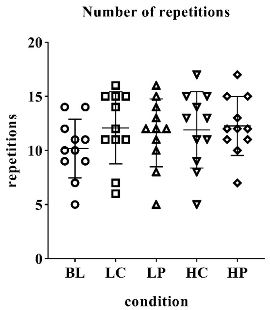
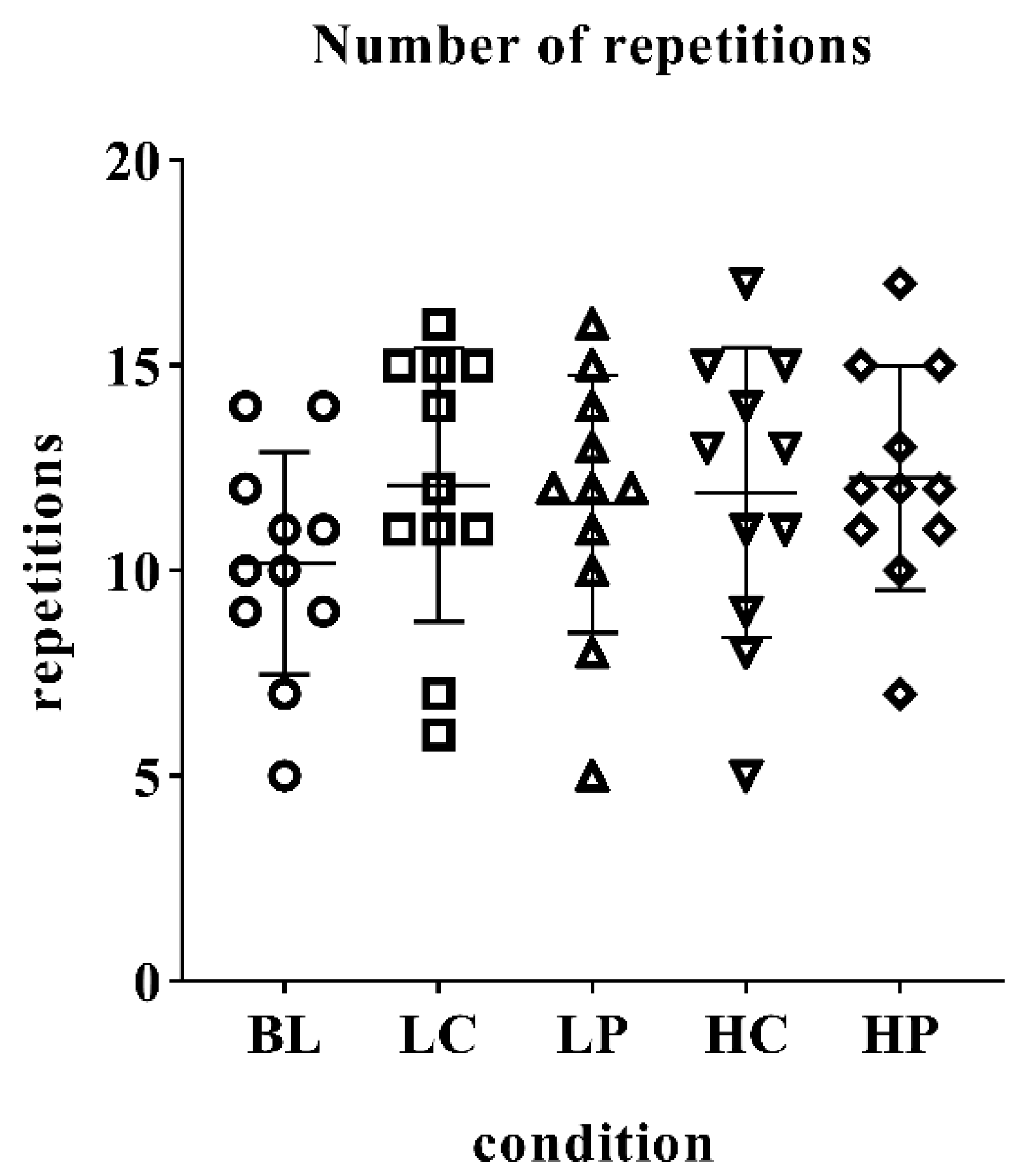
The number of repetitions achieved in each condition is shown in Figure 1. The results indicated that LC (12.09 ± 3.33 reps) and HP (12.27 ± 2.27 reps) presented the highest number of repetitions; however, when comparing the conditions, no significant differences were found (F(4,40) = 2.12; p = 0.096; ηp2 = 0.175).
Figure 1.
Number of repetitions performed under different experimental conditions. Individual values and mean ± standard deviation of the number of repetitions performed in the resistance test under the different experimental conditions. No statistically significant differences were observed between conditions. BL = baseline; LC = low-dose caffeine; LP = low-dose placebo; HC = high-dose caffeine; HP = high-dose placebo.
Table 2 shows the mean comparison between conditions for the number of repetitions, as well as the variation found based on the delta change. Our results demonstrate that the greatest mean difference occurred when comparing BL and HP (+17%), followed by LC in relation to BL (+16%). However, no significant differences were found in any of these comparisons (p > 0.05).
Table 2.
Pairwise comparisons of the number of repetitions between experimental conditions.
2.3. Electromyographic Variables
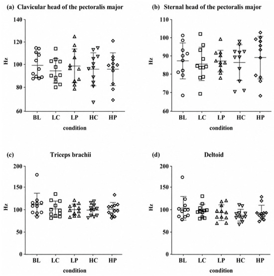
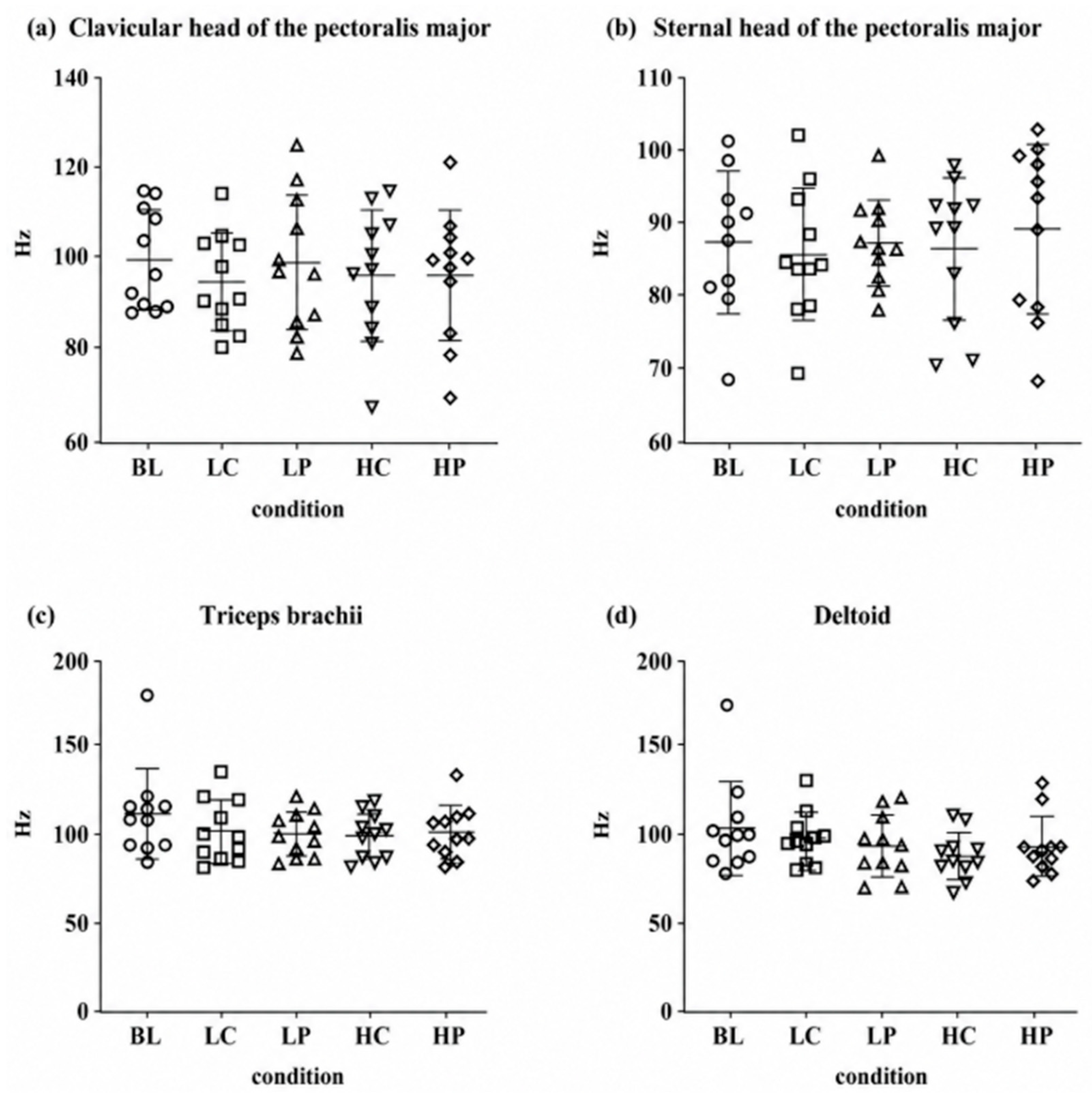
The EMGhz values of the PM, in its clavicular and sternal portions, as well as the triceps brachii and anterior deltoid, are presented in Figure 2. No statistically significant differences were found between the analyzed conditions for any of the evaluated muscles (Clavicular: F(4,40) = 0.425; p = 0.790; ηp2 = 0.041; Sternal: F(4,40) = 1.03; p = 0.403; ηp2 = 0.094; Triceps brachii: F(4,40) = 1.22; p = 0.317; ηp2 = 0.109; Anterior deltoid: F(4,40) = 1.36; p = 0.264; ηp2 = 0.120). However, when observing the mean values of each condition, it is noted that the BL condition presented the highest mean electromyographic frequency values for all analyzed muscles (Figure 2a—Clavicular: 99.3 ± 10.8 Hz; Figure 2b—Sternal: 100 ± 44.2 Hz; Figure 2c—Triceps brachii: 112 ± 25.0 Hz; Figure 2d—Anterior deltoid: 103 ± 25.6 Hz).
Figure 2.
Mean EMG frequency (Hz) of the analyzed muscles across experimental conditions. Individual values and mean ± standard deviation of the electromyographic mean frequency (EMGhz) recorded during the resistance test under different experimental conditions. Panels represent the following muscles: (a) clavicular head of the pectoralis major, (b) sternal head of the pectoralis major, (c) triceps brachii, and (d) anterior deltoid. No statistically significant differences were observed between conditions. BL = baseline; LC = low-dose caffeine; LP = low-dose placebo; HC = high-dose caffeine; HP = high-dose placebo; EMGhz = electromyographic mean frequency expressed in Hertz (Hz).
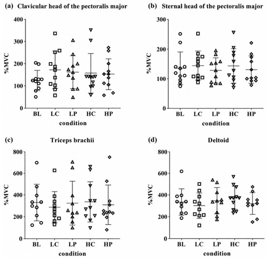
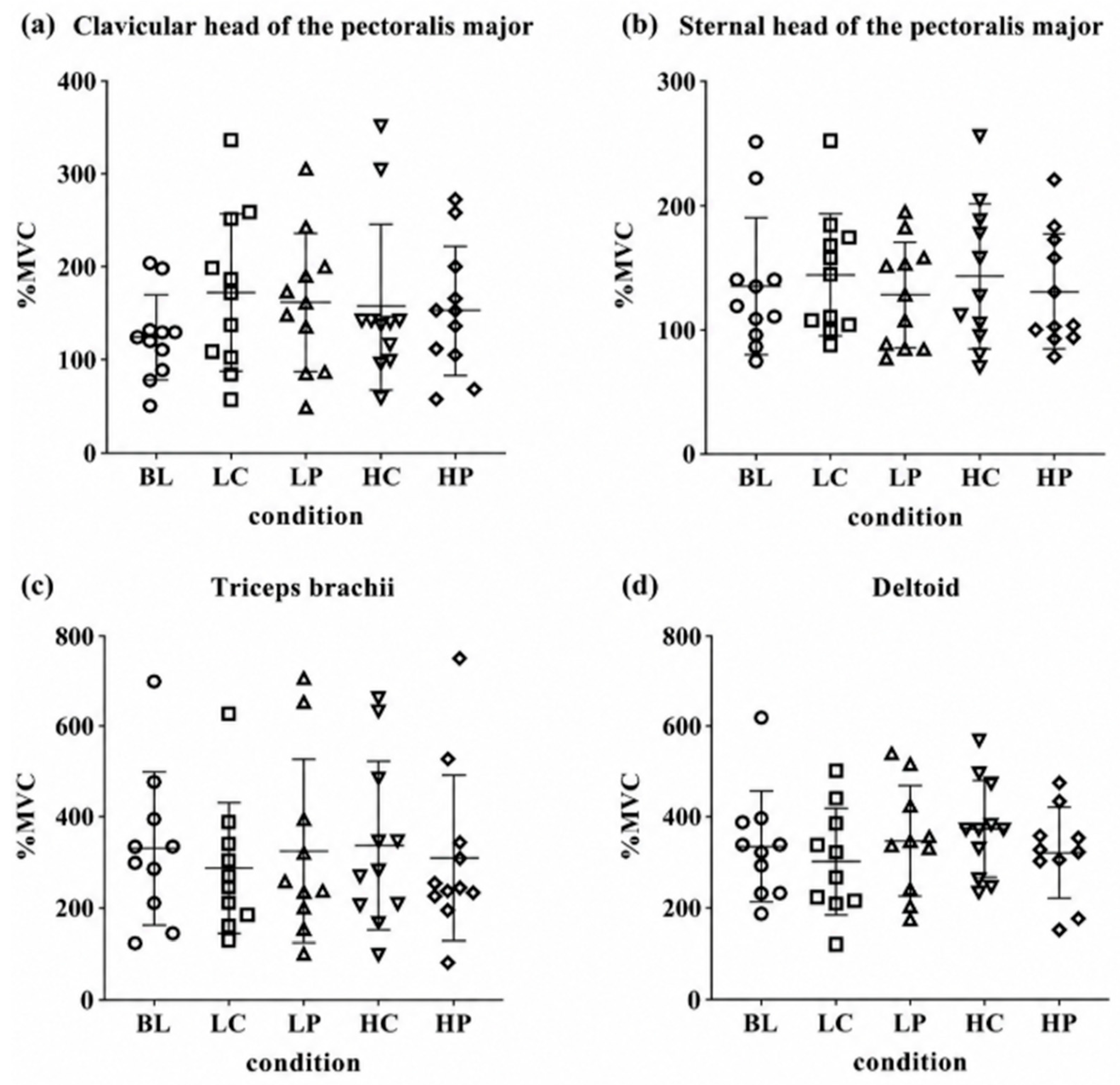
The EMGcvm activity values are presented in Figure 3. For both portions of the PM muscle, the LC condition showed the highest mean values of electromyographic activation (Figure 3a—Clavicular: 172 ± 85.5%; Figure 3b—Sternal: 144 ± 49.3%). For the triceps brachii and anterior deltoid muscles, the HC condition presented the highest mean activation values (Figure 3c—Triceps: 336 ± 185%; Figure 3d—Deltoid: 374 ± 106%). However, the repeated measures ANOVA did not indicate statistically significant differences between conditions for any of the analyzed muscles (Clavicular: F(4,40) = 1.87; p = 0.134; ηp2 = 0.158; Sternal: F(4,40) = 0.492; p = 0.742; ηp2 = 0.047; Triceps brachii: F(4,32) = 0.367; p = 0.830; ηp2 = 0.044; Anterior deltoid: F(4,32) = 0.855; p = 0.501; ηp2 = 0.097).
Figure 3.
Normalized electromyographic activity (%MVC) of the analyzed muscles across experimental conditions. Individual values and mean ± standard deviation of electromyographic activity normalized by maximal voluntary contraction (%MVC) recorded during the resistance test under different experimental conditions. Panels represent the following muscles: (a) clavicular head of the pectoralis major, (b) sternal head of the pectoralis major, (c) triceps brachii, and (d) anterior deltoid. No statistically significant differences were observed between conditions for any of the analyzed muscles (p > 0.05). BL = baseline; LC = low-dose caffeine; LP = low-dose placebo; HC = high-dose caffeine; HP = high-dose placebo; %MVC = electromyographic activity normalized by maximal voluntary contraction.
2.4. Blood Glucose
Blood glucose values (Table 3) showed a reduction in the post-test moment compared to the pre-test in all analyzed conditions. Statistically significant differences between pre- and post-test moments were observed in the BL (ΔM = 11.91; p = 0.048), LP (ΔM = 10.91; p = 0.044), and HP (ΔM = 11.27; p = 0.011) conditions. However, when comparing the conditions with each other at the post-test moment, no statistically significant differences were observed (F(4,40) = 1.28; p = 0.293; ηp2 = 0.114).
Table 3.
Blood glucose levels before and after the resistance test under different experimental conditions.
3. Discussion
The present study aimed to analyze the influence of caffeine on blood glucose concentration and neuromuscular activation, assessed through electromyographic activity, during the performance of a muscular endurance test. The results indicated that the caffeine doses used (3 and 6 mg·kg−1) contributed to the maintenance of glycemic levels after the test performed to muscular failure. However, these same dosages were not sufficient to promote significant changes in muscular endurance or neuromuscular parameters during resistance exercise.
Regarding blood glucose concentration, it was observed that the conditions without caffeine supplementation showed a significant reduction in glycemia in the post-exercise moment. On the other hand, in caffeine conditions, although a decrease in glucose also occurred, this reduction was smaller and did not reach statistical significance. These results may indicate a different glycemic response pattern in the caffeine-infused conditions. However, given the absence of significant differences between the conditions, this observation should be interpreted with caution, thus demonstrating a possible persistence between pre- and post-dose values. Previous evidence indicates that resistance exercises performed at high (90% of 1RM) or moderate intensity (70% of 1RM) are capable of acutely reducing blood glucose concentration [17]. In this sense, our results indicate that the intensity used in the muscular endurance test (80% of 1RM) was sufficient to promote a glycemic reduction when participants were not supplemented with caffeine.
Some studies indicate that caffeine may attenuate the reduction in glucose during or after physical exercise [18,19]. One of the mechanisms proposed is that caffeine may increase fatty acid oxidation through enhanced catecholamine release, potentially promoting lipolysis and reducing reliance on glucose as an energy substrate [3]. However, these physiological responses were not directly assessed in the present study. However, these effects are more frequently described in endurance exercise [20,21,22,23]. In contrast, experimental evidence suggests that caffeine may modulate intracellular pathways related to glucose uptake. Rose and Richter (2005) [24] demonstrated that caffeine may increase AMP-activated protein kinase (AMPK) phosphorylation and stimulate GLUT4 translocation [25], favoring AMPK-dependent glucose uptake in skeletal muscle. In addition, the increase in Ca2+ release during muscle contraction may also act as a signal for greater glucose uptake by the muscle [24]. Despite these possible mechanisms, the effects of caffeine on glycemic response are still inconsistent in the literature, and most studies have investigated predominantly aerobic exercise. Therefore, our findings suggest that supplementation with low and high doses of caffeine may attenuate the reduction in blood glucose induced by resistance exercise, indicating a possible modulatory effect of the substance on glycemic responses during high-intensity resistance exercise. Thus, further investigations are necessary to understand the impact of caffeine on glucose regulation during resistance exercise.
Regarding muscular endurance performance and neuromuscular activation, caffeine did not show a significant effect. The doses used in the present study (3 and 6 mg·kg−1) are consistent with those frequently described in the literature as ideal for obtaining ergogenic effects [3,26]. Although some evidence indicates that acute caffeine ingestion may increase muscular endurance [26], other studies, in agreement with our findings, demonstrate that the substance does not promote significant changes in endurance, strength, or muscular power during resistance exercise [16,27,28].
One of the physiological mechanisms proposed in the literature is the antagonism of adenosine receptors in the central nervous system, which may contribute to a reduced perception of effort and delayed central fatigue [29]. However, during resistance exercise performed with high loads and to muscular failure, as in the present study, fatigue tends to be predominantly peripheral, being influenced by factors such as metabolite accumulation, reduced phosphocreatine availability, and alterations in excitation–contraction coupling of muscle fibers [30,31]. Thus, even if caffeine may influence neural activation, performance during high-load exercise performed to failure could be primarily limited by peripheral factors related to the muscle’s contractile capacity [30,32]. In addition, the methodological characteristics of the adopted protocol, especially the performance of only one set to muscular failure in each condition, may not have been sufficient to demonstrate possible ergogenic effects of caffeine [16].
Regarding neuromuscular responses, the absence of significant differences in electromyographic parameters was similar to that reported by Nishikawa et al. (2024) [33], who did not observe changes in muscle torque or maximal voluntary contraction after caffeine ingestion. Although previous studies have suggested that caffeine may increase CNS excitability and influence mechanisms such as Na+/K+ pump activity and acetylcholine release [3,4], these physiological responses were not directly assessed in the present study. Furthermore, exercises performed to muscular failure tend to generate high levels of neuromuscular activation regardless of supplementation. Similar findings were reported by Montalvo-Alonso et al. (2025) [34], who observed improvements in muscular performance following caffeine ingestion without significant changes in electromyographic activity.
Finally, it is noteworthy that the habitual caffeine intake of the participants, assessed through the 24hDR, was lower than the values recommended by the International Society of Sports Nutrition (ISSN) for obtaining ergogenic effects [35], indicating low habitual consumption of the substance. Additionally, the results suggest a possible placebo effect, especially when participants received two capsules, since they were previously informed that higher doses would involve the ingestion of two capsules. In this context, the placebo effect can be understood as an improvement in performance or physiological responses after the administration of an inert substance, being mainly attributed to psychological mechanisms related to the individual’s expectations [36,37].
Some limitations of the present study should be acknowledged. Although the crossover design reduced interindividual variability, the relatively small sample size and the use of a single-set muscular endurance protocol may have limited the ability to detect subtle acute effects of caffeine supplementation during resistance exercise. Furthermore, the use of a single set performed to concentric failure may restrict the generalizability of the findings to resistance training sessions involving greater exercise volumes or different populations. Although the crossover design was randomized and included a washout period between experimental conditions, treatment order was not incorporated into the statistical analyses. Therefore, potential order and period effects cannot be completely excluded and should be considered when interpreting the findings. Additionally, participants’ physical activity outside the prescribed experimental sessions was not systematically monitored. As a result, variations in habitual physical activity levels may have contributed to the observed responses and represent a potential source of uncontrolled variability.
Another limitation is that only surface electromyographic activity and capillary blood glucose responses were assessed. Consequently, the physiological and neuromuscular mechanisms potentially associated with caffeine supplementation, such as alterations in neural activation, fatigue-related processes, substrate utilization, and metabolic regulation, were not directly evaluated. Moreover, the inherent variability of surface electromyographic measurements may have reduced the sensitivity to detect subtle differences between experimental conditions. Therefore, the mechanistic interpretations presented should be viewed with caution and are based primarily on previous evidence rather than direct measurements obtained in the present study.
Finally, dietary intake before each experimental session was monitored using a single 24 h dietary recall, which does not provide a comprehensive assessment of habitual dietary intake and may not fully capture day-to-day dietary variability. In addition, although participants were instructed to maintain a similar dietary pattern throughout the study and all assessments were conducted in a fed state, meal composition, energy intake, and the interval between the last meal and caffeine ingestion were not strictly standardized. Consequently, differences in feeding status may have influenced caffeine absorption and the physiological responses observed.
Despite these limitations, the present study contributes to the growing body of evidence regarding caffeine supplementation as an ergogenic aid in resistance training, particularly with respect to acute glycemic responses during resistance exercise. Nevertheless, these findings should be interpreted cautiously, given the limited evidence currently available on this outcome in resistance exercise. Future studies involving larger samples, higher training volumes, stricter dietary control, and chronic interventions are warranted to further clarify the metabolic and neuromuscular effects of caffeine supplementation during resistance training.
From a practical perspective, the present findings suggest that acute caffeine supplementation at doses of 3 or 6 mg·kg−1 may not substantially enhance muscular endurance performance or neuromuscular activation during a single set of bench press exercise performed to concentric failure in resistance-trained men. However, the maintenance of blood glucose levels observed in the caffeine conditions may indicate a distinct metabolic response that warrants further investigation. Therefore, coaches and practitioners should consider that the ergogenic effects of caffeine may not be consistently observed during low-volume resistance exercise protocols, and individual responses should be considered when recommending caffeine supplementation in resistance training settings.
4. Materials and Methods
4.1. Sample
This study employed a randomized, double-blind, crossover design. To ensure blinding and allocation concealment, a researcher who was not involved in data collection or outcome assessment was responsible for supplement preparation and generation of the randomization sequence. All participants completed all experimental conditions (LC, LP, HC, and HP), but the order of administration was randomly assigned for each participant. The randomization sequence was concealed from both participants and investigators responsible for data collection, thereby maintaining blinding throughout the study. All participants signed an Informed Consent Form (ICF). Participants were informed about the overall objectives of the study and the use of caffeine supplementation; however, they remained blinded to the experimental condition administered during each session throughout the protocol. The study was approved by the Human Research Ethics Committee (COEP; CAAE: 20221419.7.0000.5148) and conducted in accordance with the Declaration of Helsinki of the World Medical Association. This clinical trial was registered in the Brazilian Registry of Clinical Trials (ReBEC; RBR-10vqn68h).
Eleven men engaged in resistance training (25.7 ± 5.9 years; 71.10 ± 11.0 kg; 170.72 ± 6.6 cm; 12.49 ± 3.8%BF) with practical experience (7.5 ± 3.8 years) participated in the study. The inclusion criteria were age ≥ 18 years; regular resistance training practice with a minimum frequency of three sessions per week; absence of musculoskeletal injuries; no use of stimulant supplementation; and continuous resistance training experience for at least one year. Participants who did not follow the methodological procedures, had non-communicable chronic diseases, or sustained injuries during the experimental period were excluded from the study.
4.2. Experimental Design
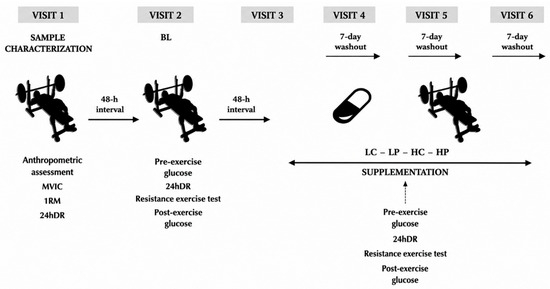
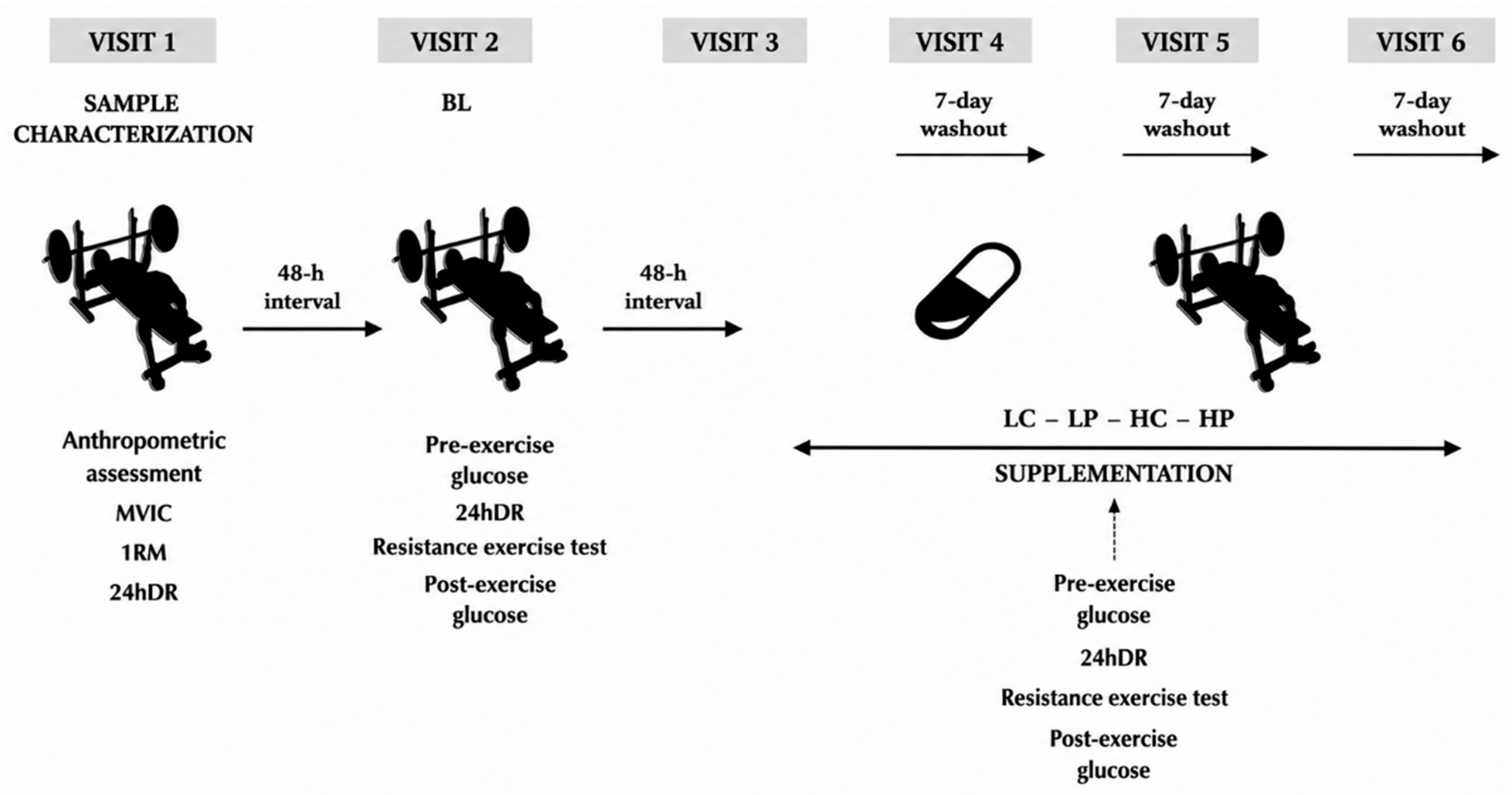
Participants attended six in-person visits to the laboratory (Figure 4). A 48 h interval was adopted between the first three visits, while a seven-day interval was established between subsequent visits, respecting the washout period. All assessments were conducted at the same time of day, as determined during the first visit. Data collection took place at the Human Movement Studies Laboratory (LEMOH), located at the Federal University of Lavras (UFLA), Minas Gerais, Brazil, in a climate-controlled and fully equipped environment (25 °C).
Figure 4.
Experimental design and timeline of the study. The study consisted of six laboratory visits. Visit 1 included sample characterization, anthropometric assessment, maximal voluntary isometric contraction (MVIC), one-repetition maximum (1RM) testing, and a 24 h dietary recall (24hDR). Visit 2 (baseline; BL) and all subsequent visits included pre- and post-exercise blood glucose measurements, a 24hDR, and a muscular endurance test. The experimental conditions were randomized and consisted of low-dose caffeine (LC), low-dose placebo (LP), high-dose caffeine (HC), and high-dose placebo (HP), with a 7-day washout period between conditions. BL = baseline; MVIC = maximal voluntary isometric contraction; 1RM = one-repetition maximum; 24hDR = 24 h dietary recall; LC = low-dose caffeine; LP = low-dose placebo; HC = high-dose caffeine; HP = high-dose placebo; Pre-exercise glucose = blood glucose measurement obtained before exercise; Post-exercise glucose = blood glucose measurement obtained after exercise.
Some recommendations were provided to the participants: to maintain a dietary pattern similar to that reported in the first 24 h dietary recall (24hDR), to avoid performing upper body exercises in the 24 h preceding each visit, to abstain from the consumption of stimulant substances during the experimental period, and to avoid alcohol consumption in the 24 h prior to the assessments. Participants were also instructed to maintain their usual physical activity habits throughout the study period. However, physical activities performed outside the experimental sessions were not systematically monitored.
The first visit aimed to characterize the sample and included anthropometric assessment, familiarization with the bench press exercise, performance of the maximal voluntary isometric contraction (MVIC) test, one-repetition maximum (1RM) test, and application of the 24hDR. The second visit was considered the baseline condition (BL). From the third visit onward, participants were subjected to the experimental conditions according to the study randomization: low caffeine (LC), low placebo (LP), high caffeine (HC), and high placebo (HP), not necessarily in this order.
From the second visit onward, all procedures were performed identically, including pre-test blood glucose assessment, application of the 24hDR, performance of the muscular endurance test, and post-test blood glucose assessment. Supplementation was provided in capsule form containing the supplements, 60 min before the test. The doses and experimental conditions were changed between visits according to the study randomization.
4.3. Procedure Protocols
4.3.1. Anthropometry
Anthropometric measurements included body mass (kg), height (cm), and body fat percentage (%BF). Body mass was assessed using a digital scale (G-tech Glass 10®, Carmy Electronic Ltd., Zhongshan, China), and height was measured using a wall-mounted stadiometer with 1 mm precision. %BF was estimated using a portable ultrasound device (BodyMetrix® BX2000, IntelaMetrix Inc., Livermore, CA, USA), applying the three-site protocol for men by Jackson and Pollock (1978) [38], with analysis performed using BodyView® software version 3.0.9.22073 (IntelaMetrix Inc., Livermore, CA, USA).
4.3.2. 24 h Dietary Recall (24hDR)
The 24hDR was applied at all visits by an experienced nutritionist. The 24 h dietary recall obtained prior to each experimental session was used to monitor pre-test dietary intake and identify the consumption of caffeine-containing foods and beverages that could potentially influence the study outcomes. This assessment was intended to control dietary factors before testing rather than to characterize participants’ habitual dietary intake. Participants were instructed to maintain their habitual dietary intake and to replicate, as closely as possible, the dietary pattern reported in the first recall before each experimental session. Therefore, all experimental sessions were conducted in a fed state. The collected data were entered into WebDiet® software, version 11.0.4 (WebDiet LTDA, Rio de Janeiro, RJ, Brazil),to analyze carbohydrate, lipid, and protein intake. The quantification of caffeine intake reported in the 24hDR was performed according to the study by Heckman, Weil, and De Mejia (2010) [39].
4.3.3. Supplementation
Supplementation was administered through standardized capsules, respecting the blinding process of the study. The caffeine and placebo capsules were prepared by a certified compounding pharmacy (Bioderme, Divinópolis, Minas Gerais, Brazil). The capsules contained either caffeine or placebo (starch), with identical appearance to prevent identification by participants. The four intervention visits (LC, LP, HC, and HP) followed the protocol: in the low-dose conditions, participants received one capsule containing caffeine (LC = 3 mg·kg−1) or placebo (LP = 230 mg). In the high-dose conditions, two capsules were administered, totaling 6 mg·kg−1 of HC or 460 mg of placebo (HP). Participants were aware that they could receive one or two capsules at each visit, according to the study randomization, without repetition of conditions.
4.3.4. One-Repetition Maximum Test (1RM)
The 1RM test was performed to determine the maximum load in the bench press exercise, executed on a guided bar (Multi Exercise with Guided Bar–Smith–Master Line, 4 inches; MAT Fitness, São José do Rio Preto, SP, Brazil). The procedures followed the recommendations of Rodríguez-Ridao et al. [40]. Initially, a warm-up of 15 movements without load was performed, followed by one set of 8 repetitions at 50% of the estimated 1RM and one set of 3 repetitions at 70% of the estimated 1RM. Subsequently, up to five attempts were performed to determine the maximum load, with 5 min intervals between sets. The 1RM value was considered the highest load lifted with full execution of the movement. The attempt was interrupted when the participant was unable to complete the concentric phase of the movement.
4.3.5. Muscular Endurance Test Protocol
The muscular endurance test was based on the protocol proposed by Karp (2001) [41], aiming to evaluate muscular endurance in a given exercise. In this test, participants were instructed to perform the maximum number of repetitions possible until concentric failure, using a previously established load. In the present study, the load corresponded to 80% of 1RM [41,42] for all participants. The exercise used was the bench press, performed on the same equipment used for the 1RM test. Initially, a standardized warm-up of 20 repetitions at 20% of 1RM was performed. Subsequently, the main phase of the test began, in which participants performed as many repetitions as possible with 80% of 1RM until concentric failure occurred. No assistance from evaluators was allowed during repetitions, except at the moment of concentric failure, and this movement was not counted.
4.3.6. Electromyography (EMG)
Surface EMG was used in all muscular endurance tests to analyze two responses: electromyographic activity normalized by maximal voluntary contraction (EMGcvm) and mean signal frequency (EMGhz). EMGcvm corresponds to the peak electromyographic activity value expressed as a percentage (%) after normalization by MVC. EMGhz represents the mean frequency of the electromyographic signal during exercise execution, expressed in Hertz (Hz). The muscles evaluated during the test were the pectoralis major (PM), in its clavicular and sternal portions, the triceps brachii, and the anterior deltoid. The reference electrode (ground) was placed on the iliac crest. Electrode placement followed the recommendations of the SENIAM project (Surface Electromyography for the Non-Invasive Assessment of Muscles), as proposed by Hermens et al. (2000) [43].
Signal acquisition was performed using a MioTool 400 electromyograph (Miotec®, Biomedical Equipment Ltd., Porto Alegre, Brazil), with four input channels, 14-bit resolution, and a sampling rate of 2000 samples·s−1 per channel, coupled to an SDS-500 sensor (Miotec®, Biomedical Equipment Ltd., Porto Alegre, Brazil)with a maximum gain of 1000 times. The electromyographic signals were filtered using a 5th-order Butterworth band-pass filter with zero phase delay, within a frequency range of 20 to 500 Hz. Signal amplitude was calculated using root mean square (RMS), with a moving window of 100 ms, through MiotecSuite 1.0 software.
To calibrate the signal acquisition channels, the MVIC test was performed. MVIC is a method used to quantify muscle strength during maximal isometric contraction, estimating 100% voluntary muscle activation and providing values in kilograms (kg) or Newtons (N) of force [44]. The test was performed in the bench press exercise, in which the bar was fixed to a Miograph load cell (Miotec®, Biomedical Equipment Ltd., Porto Alegre, Brazil), positioned on the ground. Participants were instructed to push the bar for 10 s, with the 5 most stable seconds of the contraction analyzed, without any bar movement.
4.3.7. Glycemia
Blood samples for glycemic analysis were collected before and immediately after the muscular endurance tests. Samples were obtained via capillary puncture from one of the participant’s fingers, collecting approximately 1–2 μL of fresh capillary blood, with the first drop discarded. After site asepsis with alcohol, puncture was performed using disposable lancets, and the blood was applied to reagent strips coupled to a glucometer (Accu-Chek® Active, Roche®, São Paulo, Brazil) for glycemic analysis. Values were expressed in milligrams per deciliter (mg·dL−1). The equipment used presents high accuracy and reliability for capillary blood glucose measurement [45].
4.3.8. Statistical Analysis
Descriptive statistics were used for variable analysis, with results expressed as mean ± standard deviation. The delta variation (Δ) for the number of repetitions between conditions was also calculated and expressed as a percentage (%). Data normality was verified using the Shapiro–Wilk test. Although the normality assumption for EMG variables was not fully met, repeated measures ANOVA was used due to its robustness against moderate violations of normality. Comparisons between experimental conditions (BL, LC, LP, HC, and HP) for the number of repetitions, EMGcvm, and EMGhz were performed using repeated measures ANOVA, followed by Scheffé’s post hoc test. Effect size was calculated using partial eta squared (ηp2), with values of approximately 0.01 considered small, 0.06 moderate, and ≥0.14 large. Pre- and post-test glucose levels in each condition were compared using the paired t-test. Homogeneity of variances was verified using Levene’s test (p > 0.05). Sample size was determined a priori using G*Power software (version 3.1, University of Düsseldorf, Germany). The calculation was based on a repeated-measures design, assuming an effect size (f2) of 0.55, a significance level (α) of 0.05, and a statistical power (1 − β) of 0.95. The analysis indicated a minimum required sample size of 10 participants. Therefore, the final sample of 11 participants was considered sufficient to detect the expected effects. All statistical analyses were performed using JAMOVI® software (version 2.6), adopting a significance level of α = 5% (p < 0.05). Graphs were created using GraphPad Prism® (version 8.0.2).
5. Conclusions
Caffeine supplementation, regardless of dose (3 or 6 mg·kg−1), did not significantly affect muscular endurance, electromyographic activity, or post-exercise blood glucose responses compared with placebo. Although no significant differences in blood glucose responses were observed between conditions, a descriptively different pattern of glycemic response was noted in the caffeine conditions; however, this observation warrants cautious interpretation. Therefore, the findings of the present study do not support a clear ergogenic effect of caffeine under the experimental conditions adopted, particularly within the context of an acute resistance exercise session.
Future studies are warranted to further investigate the effects of caffeine on neuromuscular and metabolic responses during resistance exercise, particularly using different exercise volumes, training protocols, and longer intervention periods. Nevertheless, the present study contributes to the existing literature by providing additional evidence regarding the acute effects of caffeine supplementation on electromyographic and glycemic responses during a muscular endurance test performed to failure in resistance-trained men.
Author Contributions
Conceptualization, G.P.S. and S.F.d.S.; methodology, G.P.S., L.B.L., B.d.C.C., R.P., A.M., P.A., A.C.P.K., H.d.S.S. and S.F.d.S.; Formal Analysis, G.P.S., C.C.D.R.d.S. and S.F.d.S.; Investigation, G.P.S., L.B.L. and H.d.S.S.; resources, P.F., N.P. and T.R.M.; data curation G.P.S., C.C.D.R.d.S. and S.F.d.S.; original draft preparation, writing—review and editing, G.P.S., L.B.L., C.E.V., A.C.P.K., H.d.S.S. and S.F.d.S.; funding acquisition, P.F., G.P.S. and S.F.d.S. All authors have read and agreed to the published version of the manuscript.
Funding
This research received no external financial support.
Institutional Review Board Statement
The study was approved by the Human Research Ethics Committee (COEP; CAAE: 20221419.7.0000.5148), entitled “Nutritional Supplements and Responses to Different Physical Activity Programs.” This clinical trial was registered in the Brazilian Registry of Clinical Trials (ReBEC; RBR-10vqn68h) (approved date 28 July 2023).
Informed Consent Statement
Informed consent was obtained from all subjects involved in the study.
Data Availability Statement
The data supporting the findings of this study are available from the corresponding author upon reasonable request.
Acknowledgments
The authors would like to thank all participants who took part in this study. Their commitment was essential to the success of this research.
Conflicts of Interest
The authors declare no conflicts of interest.
References
- Chen, B.; Zhang, C.; Xu, Z.; Li, Y.; Guo, L.; Cao, Y.; Girard, O. Effects of Caffeine Supplementation on Exercise Performance in Volleyball Players: A Systematic Review and Meta-Analysis. Nutrients 2025, 17, 1709. [Google Scholar] [CrossRef] [Scilit] [PubMed]
- Grgic, J.; Grgic, I.; Pickering, C.; Schoenfeld, B.J.; Bishop, D.J.; Pedisic, Z. Wake Up and Smell the Coffee: Caffeine Supplementation and Exercise Performance—An Umbrella Review of 21 Published Meta-Analyses. Br. J. Sports Med. 2020, 54, 681–688. [Google Scholar] [CrossRef] [Scilit] [PubMed]
- Guest, N.S.; VanDusseldorp, T.A.; Nelson, M.T.; Grgic, J.; Schoenfeld, B.J.; Jenkins, N.D.M.; Arent, S.M.; Antonio, J.; Stout, J.R.; Trexler, E.T.; et al. International Society of Sports Nutrition Position Stand: Caffeine and Exercise Performance. J. Int. Soc. Sports Nutr. 2021, 18, 1. [Google Scholar] [CrossRef] [Scilit] [PubMed]
- Grgic, J.; Mikulic, P.; Schoenfeld, B.J.; Bishop, D.J.; Pedisic, Z. The Influence of Caffeine Supplementation on Resistance Exercise: A Review. Sports Med. 2019, 49, 17–30. [Google Scholar] [CrossRef] [Scilit] [PubMed]
- Lopes, J.P.; Pliássova, A.; Cunha, R.A. The Physiological Effects of Caffeine on Synaptic Transmission and Plasticity in the Mouse Hippocampus Selectively Depend on Adenosine A1 and A2A Receptors. Biochem. Pharmacol. 2019, 166, 313–321. [Google Scholar] [CrossRef] [Scilit] [PubMed]
- Altermann, A.M.; Dias, C.S.; Luiz, M.V.; Navarro, F. A influência da cafeína como recurso ergogênico no exercício físico: Sua ação e efeitos colaterais. Rev. Bras. Nutr. Esportiva 2008, 2, 225–239. [Google Scholar]
- Olorunshola, K.V.; Achie, L.N. Caffeine Alters Skeletal Muscle Contraction by Opening of Calcium Ion Channels. Curr. Res. J. Biol. Sci. 2011, 3, 521–525. [Google Scholar]
- Magkos, F.; Kavouras, S.A. Caffeine and Ephedrine. Sports Med. 2004, 34, 871–889. [Google Scholar] [CrossRef] [Scilit] [PubMed]
- Black, C.D.; Waddell, D.E.; Gonglach, A.R. Caffeine’s Ergogenic Effects on Cycling. Med. Sci. Sports Exerc. 2015, 47, 1145–1158. [Google Scholar] [CrossRef] [Scilit] [PubMed]
- Duncan, M.J.; Oxford, S.W. The Effect of Caffeine Ingestion on Mood State and Bench Press Performance to Failure. J. Strength Cond. Res. 2011, 25, 178–185. [Google Scholar] [CrossRef] [Scilit] [PubMed]
- Bazzucchi, I.; Felici, F.; Montini, M.; Figura, F.; Sacchetti, M. Caffeine Improves Neuromuscular Function during Maximal Dynamic Exercise. Muscle Nerve 2011, 43, 839–844. [Google Scholar] [CrossRef] [Scilit] [PubMed]
- Solomonow, M.; Baten, C.; Smit, J.; Baratta, R.; Hermens, H.; D’Ambrosia, R.; Shoji, H. Electromyogram Power Spectra Frequencies Associated with Motor Unit Recruitment Strategies. J. Appl. Physiol. 1990, 68, 1177–1185. [Google Scholar] [CrossRef] [Scilit] [PubMed]
- Spineli, H.; Acioli, J.; Pinto, M.P.; Learsi, S.; Bastos-Silva, V.J.; Araujo, G.G. Caffeine Intake Improves Muscular Endurance and Induces Depletion of Anaerobic Work Capacity in the Bench Press. Motriz 2022, 28, e102200014921. [Google Scholar] [CrossRef] [Scilit]
- Shearer, J.; Graham, T.E. Performance Effects and Metabolic Consequences of Caffeine and Caffeinated Energy Drink Consumption on Glucose Disposal. Nutr. Rev. 2014, 72, 121–136. [Google Scholar] [CrossRef] [Scilit] [PubMed]
- Desbrow, B.; Biddulph, C.; Devlin, B.; Grant, G.D.; Anoopkumar-Dukie, S.; Leveritt, M.D. The Effects of Different Doses of Caffeine on Endurance Cycling Time Trial Performance. J. Sports Sci. 2012, 30, 115–120. [Google Scholar] [CrossRef] [Scilit] [PubMed]
- Saborosa, G.P.; Leite, L.B.; Forte, P.; Malheiro, A.; Afonso, P.; Arruda, D.H.; Costa, S.F.F.; de Sá Souza, H.; da Silva, S.F. Differential Effects of Low and High Caffeine Doses on Bench Press Muscular Endurance. Appl. Sci. 2026, 16, 1679. [Google Scholar] [CrossRef] [Scilit]
- Monroe, J.C.; Naugle, K.M.; Naugle, K.E. Effect of Acute Bouts of Volume-Matched High-Intensity Resistance Training Protocols on Blood Glucose Levels. J. Strength Cond. Res. 2020, 34, 445–450. [Google Scholar] [CrossRef] [Scilit] [PubMed]
- Dewar, L.; Heuberger, R. The Effect of Acute Caffeine Intake on Insulin Sensitivity and Glycemic Control in People with Diabetes. Diabetes Metab. Syndr. 2017, 11, S631–S635. [Google Scholar] [CrossRef] [Scilit] [PubMed]
- Zaharieva, D.P.; Miadovnik, L.A.; Rowan, C.P.; Gumieniak, R.J.; Jamnik, V.K.; Riddell, M.C. Effects of Acute Caffeine Supplementation on Reducing Exercise-Associated Hypoglycaemia in Individuals with Type 1 Diabetes Mellitus. Diabet. Med. 2016, 33, 488–496. [Google Scholar] [CrossRef] [Scilit] [PubMed]
- Hodgson, A.B.; Randell, R.K.; Jeukendrup, A.E. The Metabolic and Performance Effects of Caffeine Compared to Coffee during Endurance Exercise. PLoS ONE 2013, 8, e59561. [Google Scholar] [CrossRef] [Scilit] [PubMed]
- França, V.F.; Malfatti, C.R.M.; Silva, L.A.; Wietzikoski, E.C.; Osiecki, A.; Osiecki, R. Efeito da Suplementação Aguda com Cafeína na Resposta Bioquímica Durante Exercício de Endurance em Ratos. Rev. Bras. Med. Esporte 2015, 21, 372–375. [Google Scholar] [CrossRef] [Scilit]
- Glaister, M.; Gissane, C. Caffeine and Physiological Responses to Submaximal Exercise: A Meta-Analysis. Int. J. Sports Physiol. Perform. 2018, 13, 402–411. [Google Scholar] [CrossRef] [Scilit] [PubMed]
- Pettersen, S.A.; Krustrup, P.; Bendiksen, M.; Randers, M.B.; Brito, J.; Bangsbo, J.; Jin, Y.; Mohr, M. Caffeine Supplementation Does Not Affect Match Activities and Fatigue Resistance during Match Play in Young Football Players. J. Sports Sci. 2014, 32, 1958–1965. [Google Scholar] [CrossRef] [Scilit] [PubMed]
- Rose, A.J.; Richter, E.A. Skeletal Muscle Glucose Uptake during Exercise: How Is It Regulated? Physiology 2005, 20, 260–270. [Google Scholar] [CrossRef] [Scilit] [PubMed]
- Arguedas-Soley, A.; Townsend, I.; Hengist, A.; Betts, J. Acute Caffeine Supplementation and Live Match-Play Performance in Team Sports: A Systematic Review (2000–2021). J. Sports Sci. 2022, 40, 717–732. [Google Scholar] [CrossRef] [Scilit] [PubMed]
- Polito, M.D.; Souza, D.B.; Casonatto, J.; Farinatti, P. Acute Effect of Caffeine Consumption on Isotonic Muscular Strength and Endurance: A Systematic Review and Meta-Analysis. Sci. Sports 2016, 31, 119–128. [Google Scholar] [CrossRef] [Scilit]
- Williams, A.D.; Cribb, P.J.; Cooke, M.B.; Hayes, A. The Effect of Ephedra and Caffeine on Maximal Strength and Power in Resistance-Trained Athletes. J. Strength Cond. Res. 2008, 22, 464–470. [Google Scholar] [CrossRef] [Scilit] [PubMed]
- Filip-Stachnik, A.; Krzysztofik, M.; Del Coso, J.; Pałka, T.; Sadowska-Krępa, E. The Effect of Acute Caffeine Intake on Resistance Training Volume, Prooxidant-Antioxidant Balance and Muscle Damage Markers. J. Sports Sci. Med. 2023, 22, 436–446. [Google Scholar] [CrossRef] [Scilit] [PubMed]
- Spriet, L.L. Exercise and Sport Performance with Low Doses of Caffeine. Sports Med. 2014, 44, 175–184. [Google Scholar] [CrossRef] [Scilit] [PubMed]
- Grgic, J.; Trexler, E.T.; Lazinica, B.; Pedisic, Z. Effects of Caffeine Intake on Muscle Strength and Power: A Systematic Review and Meta-Analysis. J. Int. Soc. Sports Nutr. 2018, 15, 11. [Google Scholar] [CrossRef] [Scilit] [PubMed]
- Allen, D.G.; Lamb, G.D.; Westerblad, H. Skeletal Muscle Fatigue: Cellular Mechanisms. Physiol. Rev. 2008, 88, 287–332. [Google Scholar] [CrossRef] [Scilit] [PubMed]
- Grgic, J.; Schoenfeld, B.J.; Orazem, J.; Sabol, F. Effects of Resistance Training Performed to Repetition Failure or Non-Failure on Muscular Strength and Hypertrophy: A Systematic Review and Meta-Analysis. J. Sport Health Sci. 2021, 11, 202–211. [Google Scholar] [CrossRef] [Scilit] [PubMed]
- Nishikawa, T.; Hirono, T.; Holobar, A.; Kunugi, S.; Okudaira, M.; Ohya, T.; Watanabe, K. Acute Effects of Caffeine or Quercetin Ingestion on Motor Unit Firing Pattern before and after Resistance Exercise. Eur. J. Appl. Physiol. 2024, 124, 1645–1658. [Google Scholar] [CrossRef] [Scilit] [PubMed]
- Montalvo-Alonso, J.J.; del Val-Manzano, M.; Cerezo-Telléz, E.; Ferragut, C.; Valadés, D.; Rodríguez-Falces, J.; Pérez-López, A. Acute Caffeine Intake Improves Muscular Strength, Power, and Endurance Performance. Eur. J. Appl. Physiol. 2025, 125, 3259–3272. [Google Scholar] [CrossRef] [Scilit] [PubMed]
- Kreider, R.B.; Wilborn, C.D.; Taylor, L.; Campbell, B.; Almada, A.L.; Collins, R.; Cooke, M.; Earnest, C.P.; Greenwood, M.; Kalman, D.S.; et al. ISSN Exercise & Sports Nutrition Review Update: Research & Recommendations. J. Int. Soc. Sports Nutr. 2010, 7, 7. [Google Scholar] [CrossRef] [Scilit]
- Stewart-Williams, S.; Podd, J. The Placebo Effect: Dissolving the Expectancy versus Conditioning Debate. Psychol. Bull. 2004, 130, 324–340. [Google Scholar] [CrossRef] [Scilit] [PubMed]
- Beedie, C.J.; Foad, A.J. The Placebo Effect in Sports Performance. Sports Med. 2009, 39, 313–329. [Google Scholar] [CrossRef] [Scilit] [PubMed]
- Jackson, A.S.; Pollock, M.L. Generalized Equations for Predicting Body Density of Men. Br. J. Nutr. 1978, 40, 497–504. [Google Scholar] [CrossRef] [Scilit] [PubMed]
- Heckman, M.A.; Weil, J.; de Mejia, E.G. Caffeine in Foods: A Comprehensive Review on Consumption, Functionality, Safety, and Regulatory Matters. J. Food Sci. 2010, 75, R77–R87. [Google Scholar] [CrossRef] [Scilit] [PubMed]
- Rodríguez-Ridao, D.; Antequera-Vique, J.A.; Martín-Fuentes, I.; Muyor, J.M. Effect of Five Bench Inclinations on the Electromyographic Activity of the Pectoralis Major, Anterior Deltoid, and Triceps Brachii during the Bench Press Exercise. Int. J. Environ. Res. Public Health 2020, 17, 7339. [Google Scholar] [CrossRef] [Scilit] [PubMed]
- Karp, J.R. Muscle Fiber Types and Training. Strength Cond. J. 2001, 23, 21. [Google Scholar] [CrossRef]
- Hall, E.C.R.; Lysenko, E.A.; Semenova, E.A.; Borisov, O.V.; Andryushchenko, O.N.; Andryushchenko, L.B.; Vepkhvadze, T.F.; Lednev, E.M.; Zmijewski, P.; Popov, D.V.; et al. Prediction of Muscle Fiber Composition Using Multiple Repetition Testing. Biol. Sport 2021, 38, 277–283. [Google Scholar] [CrossRef] [Scilit] [PubMed]
- Hermens, H.J.; Freriks, B.; Disselhorst-Klug, C.; Rau, G. Development of Recommendations for SEMG Sensors and Sensor Placement Procedures. J. Electromyogr. Kinesiol. 2000, 10, 361–374. [Google Scholar] [CrossRef] [Scilit] [PubMed]
- Burden, A. How Should We Normalize Electromyograms Obtained from Healthy Participants? J. Electromyogr. Kinesiol. 2010, 20, 1023–1035. [Google Scholar] [CrossRef] [Scilit] [PubMed]
- Dhatt, G.S.; Agarwal, M.M.; Othman, Y.; Nair, S.C. Performance of the Roche Accu-Chek Active Glucose Meter to Screen for Gestational Diabetes Mellitus Using Fasting Capillary Blood. Diabetes Technol. Ther. 2011, 13, 1229–1233. [Google Scholar] [CrossRef] [Scilit] [PubMed]

Disclaimer/Publisher’s Note: The statements, opinions and data contained in all publications are solely those of the individual author(s) and contributor(s) and not of MDPI and/or the editor(s). MDPI and/or the editor(s) disclaim responsibility for any injury to people or property resulting from any ideas, methods, instructions or products referred to in the content. |
© 2026 by the authors. Licensee MDPI, Basel, Switzerland. This article is an open access article distributed under the terms and conditions of the Creative Commons Attribution (CC BY) license.