


Abstract
Forensic diagnosis of drowning remains challenging because postmortem findings are often supportive rather than pathognomonic. The classical diatom test has long been used as an ancillary tool, but its interpretation remains controversial because of contamination, false positives, environmental ubiquity, and heterogeneous analytical workflows. This PRISMA-ScR scoping review mapped emerging diatom-based and adjacent aquatic biological approaches proposed beyond the classical test, with emphasis on analytical innovation, forensic applicability, and medico-legal validation. Scopus, PubMed, and Web of Science Core Collection were searched for literature published between 2014 and 2026. After deduplication and screening, 165 studies were included. The literature covered targeted molecular and environmental-biological approaches, drowning-site inference, automated image analysis and scanning electron microscopy (SEM)/automated scanning electron microscopy (Auto-SEM) workflows, quantitative and tissue-to-medium comparisons, alternative substrates, contamination assessment, and medico-legal interpretation. Most evidence consisted of controlled experimental or comparative validation studies and case-based forensic applications; no study was classified as multicenter, externally validated, or operationally mature Level 5 evidence. Emerging approaches may strengthen drowning investigations by improving detection, comparison, quantification, and workflow reproducibility. However, they should currently be regarded as adjunctive tools rather than standalone proof of drowning or precise site attribution. Harmonized protocols, standardized environmental reference sampling, explicit contamination controls, external validation, and fit-for-purpose medico-legal interpretation remain central priorities.
1. Introduction
Drowning remains a difficult postmortem diagnosis because autopsy findings are usually supportive rather than pathognomonic. Drowning is also a major public-health concern, with an estimated 300,000 fatal drowning events worldwide in 2021 [1], and a recent systematic review has reaffirmed that no single autopsy finding is pathognomonic for drowning, so the diagnosis remains one of integration and exclusion [2]. Independent Global Burden of Disease estimates for the same year report 274,230 fatal drowning events and a 64% reduction in the age-standardized mortality rate since 1990, with persisting geographic and demographic heterogeneity [3]. Drowning remains the third leading cause of death among children aged 5–14 years globally, and 92% of fatal drowning events occur in low- and middle-income countries, where mortality rates are 3.2 times higher than in high-income settings. A further dimension particularly relevant to medico-legal practice is the documented but largely uncounted burden of fatal drowning among migrants, with more than 39,000 deaths reported during unsafe migration journeys since 2014 [1]. In bodies recovered from water, the forensic question often extends beyond the immediate cause of death to the timing of submersion, the compatibility between the recovery site and the site of drowning, and the influence of postmortem or environmental factors. Interpretation therefore depends on the convergence of scene information, autopsy and histology, toxicology, environmental data, and ancillary laboratory tests [2,4,5,6]. Among these ancillary approaches, diatom analysis has remained prominent because it links biological traces recovered from the body with organisms present in the aquatic environment [4,5,6,7].
The classical diatom test is based on the premise that, during active drowning, water containing diatoms or other planktonic material may be aspirated, enter the lungs, and in some circumstances disseminate to distant tissues. Diatoms are attractive forensic traces because their siliceous frustules can survive laboratory processing and may be compared with assemblages in the suspected drowning medium [4,5,6,7]. However, the test is also controversial: diatoms are environmentally widespread, may be introduced by contamination, and may be detected in low numbers or in contexts that do not establish drowning. Sampling strategies, tissue masses, digestion and filtration protocols, light microscopy or scanning electron microscopy (SEM) workflows, taxonomic expertise, and interpretive thresholds all influence evidentiary value [6,8,9,10].
Recent work has expanded the field beyond conventional microscopy-based presence/absence testing. Molecular and environmental DNA (eDNA) approaches have been explored to improve detection and environmental comparison [11,12,13,14,15,16]; image-analysis, deep-learning, artificial intelligence (AI)-assisted, You Only Look Once (YOLO)- or DiatomNet-type systems, and SEM/automated SEM (Auto-SEM) workflows aim to reduce observer dependence [17,18,19,20,21,22,23]; and quantitative strategies, including tissue-to-medium comparisons and lung-to-drowning-medium ratios (L/D ratio), seek to make interpretation more reproducible [24,25,26,27,28]. These developments differ in biological targets and forensic purposes. Some remain close to classical forensic diatomology, whereas others use diatom DNA, mixed planktonic signatures, microbial profiles, automated imaging, or standardized quantitative comparisons.
This diversity is where the phycological dimension becomes central, but also where terminology requires precision. Diatoms are not generic particles: they are algae with siliceous frustules that can be preserved through many laboratory procedures, and their morphology, taxonomy, and ecological distribution vary with the water body, season, hydrology, substrate, salinity, and local environmental conditions. When diatoms and other microalgae are translated into forensic evidence, algal morphology, ecology, molecular signatures, environmental reference sampling, and medico-legal reasoning become part of the same interpretive pathway.
Existing reviews have clarified the history, diagnostic controversy, and medico-legal interpretation of the classical diatom test [4,5,6,7,8,9,10]. The present review addresses a different question: how the broader methodological landscape emerging around forensic phycology and related aquatic biological evidence can be organized by forensic target, methodological family, and validation maturity. In this review, forensic phycology refers primarily to diatoms, algae, and other microalgae, whereas bacterioplankton, mixed microbial profiles, eDNA, metagenomic, and metabolomic approaches are treated as related aquatic or postmortem biological evidence. Its focus is not to re-evaluate the classical test alone, but to map newer diatom-based and adjacent aquatic biological approaches in relation to medico-legal readiness.
A scoping-review design was used to organize this heterogeneous literature rather than to estimate pooled diagnostic accuracy [29]. This review aimed to map emerging approaches proposed beyond the classical diatom test in forensic drowning investigations, with particular attention to forensic purpose, analytical workflow, validation maturity, and medico-legal interpretability.
2. Materials and Methods
2.1. Review Design
This scoping review mapped the extent, range, and characteristics of the literature on emerging diatom-based, algal/microalgal, molecular, metagenomic, automated, AI-assisted, quantitative, standardized, and related aquatic biological approaches in forensic drowning investigation. The review was conducted and reported in accordance with the Preferred Reporting Items for Systematic Reviews and Meta-Analyses extension for Scoping Reviews (PRISMA-ScR) [29]. No protocol was registered; however, the review questions, eligibility criteria, and charting framework were defined before the full-text eligibility assessment.
The purpose of the review was not to estimate pooled diagnostic accuracy or to perform meta-analysis. Instead, it was designed to organize a heterogeneous body of forensic, phycological, molecular, computational, and medico-legal literature into categories relevant to drowning investigations and to identify the main methodological and validation gaps.
2.2. Review Questions
The review was guided by the following questions:
- Which emerging diatom-based, molecular, metagenomic, automated, AI-assisted, quantitative, and standardized approaches have been proposed beyond the classical diatom test?
- Which forensic functions do these approaches address, including drowning diagnosis, drowning-medium comparison, site inference, detection/classification, standardization, contamination control, and medico-legal interpretation?
- Which methodological, interpretive, and validation gaps limit operational or court-oriented use?
2.3. Information Sources and Search Strategy
The final search used three bibliographic sources: Scopus, PubMed, and Web of Science Core Collection. Records published from 1 January 2014 to 15 May 2026 were considered. English-language filtering was applied where available. Article and review filters were applied where available; PubMed article-type filtering was not applied at the export stage and publication types were assessed during screening. The search combined drowning/submersion terms with diatom, planktonic, microalgal, molecular, eDNA/metagenomic, automation, SEM/Auto-SEM, quantitative, validation, contamination, and medico-legal interpretation concepts.
All complete database-specific search strings, restrictions, search dates, and exported record numbers are reported in Supplementary Table S1; these operational details are not repeated in the main text.
All final database exports were imported into a master screening dataset before deduplication.
2.4. Eligibility Criteria
Records were eligible when they addressed forensic drowning investigations or postmortem diatom/plankton analysis and examined at least one method, marker, interpretation problem, or validation issue relevant to the review objective. Eligible evidence sources included original research articles, experimental studies, methodological studies, validation studies, case reports or case series, computational or AI studies, database or mapping studies, reviews, systematic reviews, and other high-relevance interpretive papers.
Records were included when they addressed one or more of the following: diatom test or postmortem diatom analysis; diatom-based, plankton-based, microalgal, molecular, DNA-based, polymerase chain reaction (PCR)/quantitative polymerase chain reaction (qPCR), 18S ribosomal ribonucleic acid/deoxyribonucleic acid (18S rRNA/rDNA) or ribulose-1,5-bisphosphate carboxylase/oxygenase large-subunit gene (rbcL), microarray, sequencing, metabarcoding, or metagenomic methods relevant to drowning; drowning-site inference or drowning-medium comparison; automated, AI-assisted, deep learning, machine learning, image-analysis, Auto-SEM, or classification methods applied to diatoms or drowning evidence; quantitative or standardized indicators such as the L/D ratio, tissue-to-medium comparisons, standardized sampling, digestion/filtration workflows, or method validation; and medico-legal interpretation, contamination, false positives, reliability, reproducibility, or evidentiary validation of diatom, plankton, or microalgal evidence.
Records were excluded when they were clearly unrelated to forensic drowning or aquatic forensic evidence, including non-forensic algal ecology, paleontology, paleoecology, geology, carbonate-platform drowning, stratigraphy, harmful algal bloom ecology or toxicology without forensic drowning relevance, generic drowning pathology without a relevant diatom/plankton/microalgal/molecular/automated component, generic forensic AI unrelated to drowning evidence, and publication types without sufficient methodological content. Full texts were also excluded when they were the wrong records, duplicate publications, outside the time frame, or not usable for extraction.
2.5. Selection Process
Database exports were merged and deduplicated using identifiers and bibliographic metadata, including DOI, PMID, database identifiers, normalized titles, authors, year, and journal. Title/abstract screening followed conservative scoping-review logic: records were included if clearly relevant, excluded only if clearly irrelevant, and marked as uncertain when relevance could not be determined from the title and abstract alone.
Records classified as Include or Uncertain at title/abstract screening were taken forward for full-text retrieval. Full texts were retrieved through reference-management software, institutional or legal access routes, manual searching, publisher pages, repository pages, author-uploaded sources, and document-delivery attempts when necessary. Reports that could not be retrieved after these attempts were classified only as reports sought for retrieval but not retrieved and were not assessed for content eligibility on the basis of the title or abstract alone.
Retrieved full texts were assessed against the eligibility criteria, and reasons for full-text exclusion were recorded using standardized categories. Title/abstract screening, full-text eligibility assessment, initial data charting, and initial validation-level assignment were performed by one reviewer. Uncertain records, borderline eligibility decisions, thematic-domain assignments, and validation-level classifications were subsequently checked by the other authors with relevant forensic, phycological, molecular, and medico-legal expertise. Disagreements or interpretive uncertainties were resolved by discussion and consensus.
2.6. Data Charting
Data were charted from each included study using a structured extraction form. The charted fields included record identifier, title, authors, year, journal, country or setting, dominant evidence descriptor, sample type, organism or marker, analytical method, forensic target, main findings, reported advantages, reported limitations, validation level, thematic domain, relevance to the review, source file, and notes for synthesis. High-relevance reviews and systematic reviews were retained when they informed methodological context, interpretive frameworks, validation concerns, or reference tracking; their conclusions were not treated as independent original diagnostic-performance evidence.
Validation levels were categorized as follows: 1 = conceptual or narrative; 2 = proof-of-concept; 3 = experimental controlled validation; 4 = case-based forensic application; and 5 = multicenter, externally validated, or operationally mature. This validation-level classification was used as a pragmatic descriptive maturity-mapping framework developed for this scoping review to summarize apparent methodological maturity. It was not intended as a formal risk-of-bias tool, a validated certainty-of-evidence scale, or a pooled diagnostic-accuracy assessment.
2.7. Synthesis of Results
The results were synthesized descriptively and thematically. Because the included literature was methodologically heterogeneous, no pooled quantitative synthesis was attempted. Five descriptive layers were kept distinct: thematic domains described substantive contributions; methodological families described technical platforms or workflows; dominant evidence descriptors summarized the primary contribution of each record for descriptive counting; validation levels described apparent methodological maturity; and relevance ratings described direct relevance to the review questions.
Thematic-domain and methodological-family assignments were non-mutually exclusive because individual studies could contribute simultaneously to method development, quantitative standardization, and medico-legal interpretation. Dominant evidence descriptors were used only to summarize the primary contribution of each record; they should not be read as mutually exclusive analytical categories. Summary tables were prepared to describe thematic distribution, methodological families, forensic applications, validation levels, and recurrent limitations.
3. Results
3.1. Study Selection
The database searches identified 487 records after the application of database filters. After the removal of 268 duplicate records, 219 unique records were screened at the title/abstract level. Thirty-four records were excluded at the title/abstract screening, leaving 185 reports sought for full-text retrieval. Of these, 18 reports could not be retrieved after EndNote-assisted retrieval, manual searching, institutional or library attempts, and legal web searching. These 18 reports were not assessed for content eligibility.
A total of 167 reports were retrieved and assessed for full-text eligibility. Two full texts were excluded: one generic forensic AI review unrelated to diatoms or drowning evidence, and one general drowning-pathology discussion without a specific diatom, plankton, microalgal, molecular, or automated analytical focus. Therefore, 165 studies were included in this scoping review. The study-selection process is summarized in Figure 1.
Figure 1.
PRISMA-ScR flow diagram. Flow diagram summarizing record identification, deduplication, title/abstract screening, full-text retrieval, full-text eligibility assessment, and final inclusion in the scoping review. Reports not retrieved were counted as retrieval failures and were not assessed for full-text eligibility.
3.2. General Characteristics of Included Studies
The 165 included studies were published between 2014 and 2026. The annual distribution was uneven, with the largest number of included studies charted in 2022 (n = 28), followed by 2025 (n = 17), 2021 (n = 16), 2023 (n = 14), 2024 (n = 13), 2018 (n = 12), and 2019 (n = 12). The remaining years contributed between 7 and 11 studies each.
The charted country or setting most frequently recorded was China (n = 79), followed by Japan (n = 22), India (n = 20), Italy (n = 14), and the United Kingdom (n = 9). Smaller numbers of studies were charted from Romania, Australia, Portugal, Hungary, the United States, Sweden, Canada, South Korea, and Poland; five studies had a country or setting that was not clearly reported.
Using a dominant evidence descriptor assigned to each record for descriptive counting, the included corpus was led by experimental animal studies (n = 76) and molecular assay or metagenomic studies (n = 44). AI or computational studies accounted for 22 records, case reports or case series for 11, reviews for 6, systematic reviews for 3, methodological or validation studies for 2, and database or mapping studies for 1. These descriptors summarize the primary contribution of each record; they should not be interpreted as mutually exclusive technical categories, because animal experiments, molecular assays, AI workflows, validation designs, and review articles may overlap analytically or thematically. The complete charting of included studies is reported in Supplementary Table S2.
3.3. Thematic Distribution of Included Studies
The evidence did not form a single methodological stream. Instead, the included studies clustered around four partly overlapping thematic functions: molecular or environmental comparison, automation and classification, quantitative standardization, and medico-legal interpretation. Domain assignment was therefore non-mutually exclusive and should be distinguished from the methodological-family synthesis, which describes technical platforms or workflows rather than thematic contributions. Quantitative or standardized indicators were the most represented area (Domain C; n = 148, 89.7%), followed by medico-legal interpretation, validation, contamination, and reliability (Domain D; n = 133, 80.6%). Molecular, metagenomic, DNA-based, or site-inference approaches (Domain A) were represented by 83 studies (50.3%), and automation, image analysis, and AI-assisted detection or classification (Domain B) by 51 studies (30.9%). No study remained in the unclear/other category after re-audit.
The overlap between domains was itself informative. The most frequent combinations were C; D (n = 40, 24.2%), A; C; D (n = 37, 22.4%), B; C; D (n = 23, 13.9%), and A; B; C; D (n = 19, 11.5%). These combinations show that many studies did not simply introduce a technique; they also addressed reproducibility, comparison, contamination, or interpretation. The thematic distribution is summarized in Table 1.
Table 1.
Thematic distribution of included studies.
Domain assignment was non-mutually exclusive and thematic; therefore, individual studies could contribute to more than one domain. Domains should not be read as study-design categories or as one-to-one methodological platforms. Only the most frequent domain combinations are shown. The complete domain-combination table is provided in Supplementary Table S3; complete study charting is provided in Supplementary Table S2.
The methodological-family synthesis is set out in Table 2.
Table 2.
Methodological families and forensic applications. Summary of the main methodological families identified among included studies, including representative techniques, principal forensic applications, and recurrent caveats or validation gaps. Methodological families were treated as non-mutually exclusive technical/workflow categories, as the same study could contribute to more than one analytical or interpretive category. SEM/Auto-SEM was charted as a methodological family, whereas Domain B captures automation, image analysis, and AI-assisted detection/classification as thematic contributions; the two counts should therefore not be read as one-to-one equivalents.
3.4. Molecular, Microbial, Environmental-Mapping, and Site-Inference Approaches
These studies addressed two related forensic functions: detecting aquatic biological signals in postmortem samples and comparing them with environmental sources. The molecular and environmental-biological component of the corpus was methodologically diverse, ranging from targeted PCR/qPCR and marker-based assays to sequencing, metabarcoding, metagenomic diatom analysis, microbial profiling, diatomological mapping, water-body databases, and environmental comparisons.
Targeted molecular and molecular-adjacent work focused on whether aquatic biological signals could be recovered or interpreted more reproducibly than by morphology alone. This subgroup included DNA-binding, PCR-based, silicon/tissue-distribution, quantitative, veterinary, and morphology-adjacent studies relevant to biological signal recovery and drowning interpretations [30,31,32,33,34]. Related studies addressed organ–medium correlations, the comparison of microwave digestion-vacuum filtration-automated scanning electron microscopy (MD-VF-Auto SEM) with multiplex PCR, digestion variables, diatom DNA extraction, and closed-organ or case-based interpretations [35,36,37,38,39]. Case-based and contamination-oriented studies, including mud aspiration, resuscitation or postmortem changes, and maggot-based evidence, broadened this evidence base [40,41,42,43].
A second cluster used broader environmental-biological signals for comparison with drowning media or potential drowning sites. Bacterioplankton/PCR and microbiota-based studies examined non-diatom microbial indicators relevant to drowning diagnosis or interpretations [44,45,46]. Algal, planktonic, and related animal or case-context studies contributed complementary biological detection evidence [47,48,49,50,51]. Chloroplast, cyanobacterial, multiplex-PCR, and diatom DNA barcode workflows further contributed detection and environmental-comparison evidence [52,53,54,55]. Further studies addressed contamination blind spots, sternal aspirates, diatom DNA extraction from water and tissues, and the comparison of plankton testing methods [56,57,58,59,60].
Site inference and environmental comparisons were also pursued through diatomological mapping, water-body databases, and regional reference resources. These studies covered rivers, lakes, dams, drowning media, and other local aquatic settings [61,62,63,64,65,66,67,68]. Water-body database studies and comparative sampling protocols characterized spatial structure in diatom assemblages [69,70,71,72,73,74,75,76], while regional mapping and site-specific profiles expanded the evidence across additional aquatic contexts [77,78,79,80,81,82,83,84]. Additional regional freshwater applications were also represented [85]. AI-supported database work and water-body characterization studies further developed reference resources for site-oriented interpretations [86,87,88,89,90,91,92,93].
Studies based primarily on bacterial or mixed microbial profiles were treated as adjacent aquatic biological approaches relevant to forensic site inference, rather than as strictly diatom- or microalgal-based evidence. They were retained because they inform environmental comparisons while relying on different biological markers and interpretive assumptions. This adjacent evidence included bacterial composition and random-forest site inferences [94], broader photoautotrophic microbial perspectives [95], object-detection and optical-classification applications connected with detection or site-oriented tasks [96,97], and substrate-colonization evidence [98]. Within this subgroup, recent quantitative work has shown that random-forest classifiers trained on 16S ribosomal ribonucleic acid (16S rRNA) microbial-community data may discriminate antemortem drowning from postmortem immersion with an area under the curve (AUC) of 0.96 and a Day-1 accuracy of 98.22%, declining to 86.11% by Day 7 [60], and that microbial site-attribution models can achieve Day-1 accuracies above 95% within a single species while losing substantial performance when transferred across species (down to approximately 61% in a mouse-to-rabbit application) [99]. Further regional and integrative comparison studies confirmed the importance of local environmental context in site-oriented interpretations. [100,101,102].
3.5. Automation, Image Analysis, and AI-Assisted Detection/Classification
Automation-oriented studies addressed a different bottleneck: the time, expertise, and subjectivity required to locate and classify candidate diatoms. Fifty-one studies mapped to this area, including semi-automated detection, image-analysis workflows, AI-assisted classification, deep learning, YOLO-based systems, DiatomNet, automated search systems, and selected SEM/Auto-SEM or microwave digestion-vacuum filtration-automated SEM (MD-VF-Auto SEM) workflows.
The studies mainly pursued faster screening, reduced observer dependence, more reproducible image-based classifications, and the scalable processing of diatom evidence. In the methodological-family synthesis, automation, image analysis, and AI-assisted classification accounted for 31 studies (18.8%), while SEM or Auto-SEM workflows accounted for 78 studies (47.3%). The difference reflects the purpose of the two classifications. Domain B captures studies in which automation, image analysis, or AI-assisted detection/classification was a substantive thematic contribution, whereas SEM/Auto-SEM was charted as a broader methodological family. Some SEM studies overlapped with Domain B when SEM was part of automated search or standardized detection, whereas other SEM studies used SEM primarily as an imaging or detection platform without being AI- or automation-oriented. Recurrent limitations included dataset annotation, model transparency, external validation, equipment dependence, and deployment across laboratories (Table 2).
The computational subgroup included image-analysis and deep-learning classification studies [103,104,105,106,107,108], together with object-detection, DiatomNet/YOLO-type, and AI-assisted search systems [109,110,111,112,113,114,115,116].
3.6. Quantitative and Standardized Indicators
The largest body of evidence concerned the move from simple presence/absence findings toward reproducible quantification and standardized comparisons. Domain C included 148 studies (89.7%) and brought together work on diatom counts, lung-to-drowning-medium logic, tissue-to-medium comparisons, organ distribution, digestion and filtration workflows, SEM/Auto-SEM-based counting, and standardized analytical procedures.
Morphological microscopy and digestion-filtration workflows were the largest methodological family (n = 88, 53.3%). SEM and Auto-SEM workflows accounted for 78 studies (47.3%), and quantitative indicators or standardized comparisons for 40 studies (24.2%). Together, these studies addressed method optimization, tissue/drowning-medium comparison, organ distribution, higher-resolution detection, and more structured interpretations. However, these counts should not be interpreted as evidence that SEM/Auto-SEM has replaced light microscopy; the latter remains historically broader and more accessible, whereas SEM/Auto-SEM requires equipment-intensive workflows and external validation. Recurring caveats were heterogeneity in sampling, digestion and filtration, taxonomic expertise, equipment requirements, organ-specific differences, environmental background variation, and the limited comparability of thresholds across contexts (Table 2).
Quantitative, tissue-to-medium, organ-distribution, the L/D ratio, and abundance-comparison studies formed the central group of structured indicators. Key examples included the quantitative investigation of diatom dispersion in lung tissue [24], quantitative analysis for drowning diagnosis [25], lung tissue-to-drowning-medium comparison [26], concordance analysis of diatom types and patterns in lung tissue and the drowning medium [27], relationships between diatom abundances in rat organs and environmental waters [28], and L/D ratio validation [112]. Automation, SEM-based, and AI-assisted detection studies intersected with quantitative or standardized workflows when they supported countable, reproducible detection or search efficiency [103,104,105,106,107]. Object-detection, AI-search, and related SEM-associated workflows further developed this automation-oriented strand [108,109,110,111,112]. Methodological studies on digestion, filtration, enzymatic, hypochlorite, hydrogen-peroxide/protease, and ethanol-related processing evaluated recovery and comparability [117,118,119,120,121,122,123], while experimental lung-tissue concentration, diagnostic-ratio, and retrospective quantitative studies added further structured evidence [124,125]. Further case-based or interpretive work addressed individualization, methodological limits, trace linkage, diagnostic algorithms, imaging-assisted assessment, and difficult case interpretations [126,127,128,129,130,131,132,133].
A related set of studies broadened the evidentiary setting beyond routine lung or organ sampling. Context-specific evidence included archeological or historical water-death remains, tissue samples, lung-weight findings, and human case series [134,135,136,137,138,139]. Digestion, filtration, and extraction methods were addressed across conventional and newer workflows [140,141,142,143,144,145]. Water-body mapping, databasing, phytoplankton ecology, site comparison, and forensic diatom-flora studies supported environmental interpretations [146,147,148,149,150,151]. Veterinary or non-human applications and trace transfer to clothing or fabrics were represented in separate studies [152,153]. Alternative or specialized substrates and interpretive settings included experimental lung-injection testing [154], closed organs [155], vitreous humor [156], bone marrow or bone [157,158,159], bone and tooth [160], synovial fluid [161], poisoning-related interpretive contexts [162], and maggots [43]. These records emphasize that transfer, persistence, contamination, decomposition, and processing effects require matrix-specific validation rather than direct extrapolation from lung tissue or drowning-medium analysis.
3.7. Medico-Legal Interpretation, Contamination, and Validation
The interpretive evidence base showed that the main medico-legal issue is not simply whether an aquatic biological signal can be detected, but how that signal should be weighed in context. Domain D included 133 studies (80.6%) addressing contamination, false-positive findings, reliability, reproducibility, forensic interpretations, evidentiary caution, case-based applications, and the diagnostic or medico-legal validation of diatom, plankton, microalgal, molecular, or automated methods.
Contamination, false-positive analysis, and medico-legal interpretations formed a distinct methodological family of 50 studies (30.3%). These studies were mainly relevant to court-oriented interpretations, diagnostic reliability, contamination control, and the limits of evidentiary weight. Across this group, emerging methods were generally framed as supportive or adjunctive rather than as standalone proof of drowning. Recurrent limitations included environmental ubiquity, sampling and laboratory variability, incomplete external validation, limited inter-laboratory comparability, and uncertain operational thresholds.
Across this subset, the recurrent pattern was that analytical positivity required contextual qualification: the studies did not treat detection as self-explanatory, but linked interpretation to contamination control, case circumstances, tissue or matrix selection, and comparison with the drowning environment.
Reviews and studies addressing false-positive results, contamination sources, and medico-legal interpretations provide the main basis for weighing positive diatom findings in context [5,6,8,9,10]. Additional work on contamination blind spots, non-drowning entry pathways, closed-organ interpretations, and laboratory consumables reinforces the need for strict contamination control [56,115,155,163]. Case-based and validation-oriented studies added practical examples of how analytical findings may be interpreted alongside scene, autopsy, environmental, and laboratory information [164,165,166].
3.8. Validation Level and Forensic Applicability
The validation-level synthesis confirmed that the field remains active but not yet operationally mature. Controlled experimental or comparative validation studies dominated the corpus (Level 3; n = 110, 66.7%), followed by case-based forensic applications (Level 4; n = 36, 21.8%), conceptual or narrative studies (Level 1; n = 14, 8.5%), and proof-of-concept studies (Level 2; n = 5, 3.0%). No study was categorized as Level 5, defined as multicenter, externally validated, or operationally mature.
Relevance to the review was categorized as High for 72 studies (43.6%), Moderate for 69 (41.8%), and Low for 24 (14.5%). The absence of Level 5 evidence does not negate the value of experimental or case-based work, but it indicates that the mapped evidence has not yet demonstrated broad multicenter or externally validated operational readiness. Validation levels and medico-legal limitations are summarized in Table 3.
Table 3.
Validation level and medico-legal limitations. Classification of included studies according to a pragmatic descriptive maturity-mapping framework and relevance to the review, with associated medico-legal limitations relevant to forensic applicability. The Level 1–5 system was not a formal risk-of-bias or certainty-of-evidence grading tool.
4. Discussion
4.1. Principal Findings
This scoping review shows that recent research on diatom-based evidence in drowning investigations is no longer centered only on the question of whether diatoms can be detected after death. The included literature instead reflects a broader methodological shift: aquatic biological traces are being explored as diagnostic supports, environmental-comparison tools, quantitative indicators, automated image-analysis targets, and medico-legal evidence requiring cautious interpretations.
The classical diatom test remains the conceptual reference point, but most contemporary work attempts to address its weaknesses through molecular or environmental-biological detection, SEM/Auto-SEM workflows, AI-assisted classifications, quantitative tissue-to-medium comparisons, contamination assessment, and validation-oriented designs. The validation profile remains the central limiting factor. Although the corpus was large and methodologically active, no included study was categorized as multicenter, externally validated, or operationally mature Level 5 evidence. This supports a cautious conclusion: emerging approaches may strengthen forensic interpretations, but they are not yet standalone proof of drowning or precise site attribution.
Figure 2 summarizes this integrated fit-for-purpose framework and the main pre-analytical, analytical, and interpretive barriers identified by the scoping map.
Figure 2.
Conceptual overview of the transition from classical diatom testing to integrated forensic phycology and related aquatic biological evidence. The scheme illustrates the expansion from conventional microscopy-based diatom testing to a broader framework incorporating molecular/metagenomic approaches, automated or AI-assisted detection, quantitative standardization, medico-legal interpretation, and fit-for-purpose forensic validation. The pre-analytical, analytical, and interpretive layers indicate key conditions influencing evidentiary robustness and forensic applicability. MD-VF-Auto SEM is included as a promising but validation-dependent workflow, not as an established replacement for conventional methods and not as a method that removes contamination or interpretive uncertainty.
4.2. Methodological Advances Beyond the Classical Diatom Test
4.2.1. Molecular, eDNA, Metagenomic, and Microbial Approaches
Molecular, eDNA, metabarcoding, metagenomic, and microbial-profiling approaches are being explored because they address some limitations inherent in morphology-based diatom analyses while introducing different validation requirements. Classical diatom testing depends on the recovery of identifiable frustules, operator expertise, digestion or filtration conditions, the organ sampling strategy, and comparison with environmental material. DNA-based methods offer a complementary route: rather than relying exclusively on morphologically intact diatom structures, they may detect diatom, planktonic, algal, or microbial signals using genetic targets. These methods differ substantially in target, resolution, and validation needs, ranging from targeted PCR/qPCR or marker-based assays to broader sequencing, metabarcoding, metagenomic, and microbial-profiling approaches (Table 2). Examples include PCR-capillary electrophoresis for forensic diatom testing [11], 18S rRNA gene arrays for the forensic detection of diatoms [12], qPCR-based or real-time qPCR approaches [15,16], and rbcL-based pyrosequencing for drowning-site inferences [13].
The potential contribution of these methods lies in their ability to support analytical sensitivity, target specificity, and comparisons between biological samples and drowning media. Molecular assays may be useful when diatoms are scarce, degraded, or difficult to classify morphologically. They may also allow the detection of planktonic or microbial targets that are not limited to classical diatom morphology. This is reflected in studies that examined bacterioplankton or microbial indicators as diagnostic supports [44,46], as well as studies that used microbial community profiling and random forest approaches for site inferences [94,101]. These approaches broaden the evidentiary landscape beyond strict forensic phycology and should be described as related aquatic biological evidence when they rely primarily on bacterial, mixed microbial, or other non-algal signals.
However, the charted evidence also indicates that the forensic interpretation of molecular findings is not straightforward. Detection of a molecular target is not equivalent to proof of drowning unless the sampling context, contamination controls, the postmortem interval, reference-water sampling, and the background distribution of the target are carefully addressed. A positive molecular signal does not necessarily demonstrate intact diatom frustules, and it cannot be directly equated with antemortem drowning. Marker selection is a central issue. Targets such as 18S rRNA/rDNA and rbcL can be informative for diatoms, but they differ in taxonomic resolution, amplification behavior, reference-database availability, and susceptibility to degradation or inhibition. In addition, molecular workflows may be affected by DNA degradation in forensic samples, extraction difficulty, amplification inhibition, contamination, background eDNA signals, incomplete or misannotated barcode databases, cost, and technical platform requirements. This is why molecular methods should be understood as extending and complementing, not replacing, morphological examinations.
Site inference represents one of the most attractive but also most demanding applications. Studies on metagenomic diatom analyses and microbial profiling suggest that environmental biological signatures may contribute to identifying or narrowing possible drowning sites [13,14,87,90]. Diatomological mapping and environmental-comparison studies similarly indicate the potential value of structured reference-water or water-body characterization for site-oriented reconstructions [64,70,77,78,89]. Nevertheless, bacterial or mixed microbial-profile studies should be understood as adjacent aquatic biological approaches, not as strictly diatom- or microalgal-based evidence. The distinction between broad environmental discrimination and precise geographic attribution is critical. A method may differentiate between water types or demonstrate similarity between a body sample and a reference medium, yet still fall short of reliably identifying a specific site, particularly when seasonal variation, hydrology, local microhabitats, and sampling intervals are not standardized. Environmental reference sampling must therefore be treated as a core component of the method, not as an ancillary step.
The future value of these molecular and environmental-biological approaches depends on clearer validation designs, including blinded comparisons, defined thresholds, negative controls, cross-laboratory testing, and the standardized reporting of environmental reference samples. Without these elements, molecular sensitivity may increase the amount of information available to the forensic pathologist but may also increase interpretive uncertainties.
4.2.2. Light Microscopy, SEM/Auto-SEM, and AI-Assisted Image Analysis
A separate line of development concerns image-based detection and automation rather than molecular detection. Conventional light microscopy remains the historically broadest and most accessible platform for forensic diatom examinations and has been applied across a wider range of laboratories, countries, and case contexts. Its limitations include dependence on preparation quality, examiner expertise, background debris, damaged or partial frustules, and taxonomic interpretations. SEM and Auto-SEM may offer high-resolution imaging and support standardized search or counting workflows, but they should not be presented as replacements for conventional light microscopy in routine forensic practice without broader validation.
The charted evidence contained a distinct automation and AI cluster. The included studies covered digital whole-slide image analysis using convolutional neural networks [17], optical or light-microscopy-based database and classification approaches [78,97], object detection systems and YOLO-based models [18,19,96,109,116], DiatomNet [20], comparisons among deep learning image-classification algorithms [23], and AI-based automatic diatom identification systems in practical cases [106]. In parallel, SEM, Auto-SEM, and MD-VF-Auto SEM workflows were widely represented as a methodological family (n = 78; 47.3%), reflecting high-resolution imaging and semi-automated or automated searching (Table 2). This representation should be interpreted cautiously: a substantial part of the SEM/Auto-SEM evidence remains concentrated in a limited number of research environments, and external inter-laboratory reproducibility has not yet reached operational maturity.
Within this methodological family, the microwave digestion-vacuum filtration-automated SEM workflow should be interpreted as an emerging and methodologically valuable technical route, rather than as an established routine standard. Microwave digestion may improve tissue removal and downstream filtration, but the high-temperature and high-pressure digestion steps may also damage or fragment diatom frustules. Such fragmentation can reduce morphologically interpretable material, affect taxonomic identification, and introduce bias into quantitative counting if intact valves and fragments are not distinguished by predefined criteria. In addition, SEM scanning and the automated search do not eliminate contamination risks. Residual diatoms from digestion vessels, filtration devices, membranes, slides, reagents, water, or the laboratory environment remain relevant unless controlled through validated cleaning or single-use procedures, procedural blanks, recovery controls, and contamination controls. Reported high yields or striking counts obtained with MD-VF-Auto SEM are informative, but they require independent replication by different laboratories, operators, instruments, sample matrices, and aquatic environments before supporting routine forensic implementation. Cost, access to microwave digestion systems and SEM platforms, maintenance, calibration, and specialized training also limit generalizability to ordinary forensic laboratories. Accordingly, MD-VF-Auto SEM should be regarded as a promising assistive workflow for detection, enrichment, screening, and counting, but not as a method that has resolved contamination, comparability, or medico-legal interpretation problems [5,6,21,22,37,103,105,107,155,163]. Routine use should therefore remain subject to further validation and practical applicability assessments [5,6].
The main value of automation lies in the potential to reduce manual screening time, support standardized detection workflows, and limit dependence on individual examiner experience. Automated image analyses may be useful when large numbers of particles or fields must be screened, when diatom counts are needed for quantitative comparisons, or when rare target structures are embedded in complex backgrounds. In this sense, automation is not merely a technical convenience; it is connected to reproducibility and auditability. However, it should currently be framed as assistive detection, counting, triage, and classification support, not as autonomous forensic proof.
Yet the review also shows why computational performance should not be equated with medico-legal validity. AI systems depend on annotated datasets, image acquisition conditions, taxonomic diversity, preprocessing, and the representativeness of training and testing data. This dependence is particularly important because diatom taxonomy remains complex and evolving: species boundaries, expert criteria, new genera, and annotation standards remain active issues. A detector trained on one set of expert-labeled images, one preparation method, one microscope, one environmental region, or one taxonomic convention may not perform equivalently in another laboratory or case context. External validation and laboratory transferability are therefore essential. In addition, the forensic meaning of an AI output depends on how it is integrated into an evidentiary workflow. An algorithm may classify particles accurately under test conditions, but forensic interpretation still requires knowledge of sample origin, contamination controls, organ selection, comparison with the drowning medium, and case circumstances.
Auto-SEM and image-based automation also raise practical questions. These systems may support standardized counting and higher-throughput screening, but they require equipment availability, calibration, quality control, specialized operation and maintenance, and agreed interpretive thresholds. Their use may reduce observer dependence at one stage of the process while shifting the validation burden to image acquisition, training-data curation, algorithmic decision thresholds, and inter-laboratory reproducibility. Therefore, the future role of AI and Auto-SEM in forensic diatom analyses is likely to be assistive rather than autonomous. AI may help locate, count, classify, or prioritize candidate diatoms, but the final interpretation should remain embedded within a validated forensic pathway.
4.2.3. Quantitative Comparison and Standardization
A third development concerns quantification and standardization. Quantitative and standardization-oriented studies show that the central methodological concern in the recent literature is reproducibility. The simple presence or absence of diatoms is insufficient because diatoms and other planktonic material may be environmentally ubiquitous, may contaminate samples, and may be detected in small numbers under conditions that do not necessarily establish drowning. In addition, different organs, tissues, digestion methods, filtration systems, microscopes, and counting strategies may produce non-comparable results. The movement beyond presence/absence is therefore a necessary step toward making diatom evidence more reproducible.
Quantitative approaches in the charted literature included diatom counts, tissue-to-medium comparisons, the L/D ratio or lung-to-drowning-medium logic, organ-specific distributions, diatom dispersion in lung tissue, and method comparisons involving digestion, filtration, microscopy, SEM, and Auto-SEM (Table 2). Examples include quantitative investigation of diatom dispersion in lung tissue [24], quantitative analysis of diatoms in drowning diagnosis [25], quantitative comparison of diatoms in lung tissue and drowning medium [26], concordance analysis of diatom types and patterns in lung tissue and drowning medium [27], relationships between diatom abundances in rat organs and environmental waters [28], and L/D ratio validation [112]. These studies illustrate the attempt to transform diatom evidence into a more structured comparative signal.
The value of such indicators is that they can potentially distinguish between incidental or background findings and findings that are more consistent with the aspiration and circulation of the drowning medium. However, quantitative indicators require a high degree of pre-analytical and analytical harmonization. Counts are affected by tissue weights, the organ sampled, sampling locations, digestion times, filtration media, microscope types, counting fields, operator criteria, and the density and composition of diatoms in the water sample. If these variables are not standardized, a quantitative value may appear more objective than it really is. This is particularly relevant for workflows involving microwave digestion and vacuum filtration, because digestion-induced frustule fragmentation may change the number of countable structures and may therefore affect comparisons between tissues, organs, and drowning media [6,21,37,103,105].
4.2.4. Alternative Matrices and Trace-Transfer Contexts
Alternative matrices also appear in the charted evidence, including clothing and fabrics [9,153], maggots [43], vitreous humor [156], bone marrow and bone [157,158,159], bone and tooth [160], synovial fluid [161], closed organs and postmortem or resuscitation-related contexts [39,42,56,155,162], and other specialized or archeological/veterinary matrices [134,139,151,152,167]. These studies are relevant because they expand the potential evidentiary context of diatom analyses beyond routine lung or organ sampling. However, they also require matrix-specific validations. The behavior of diatoms in clothing, bone, maggots, synovial fluid, vitreous humor, or closed organs cannot be assumed to match their behavior in lung tissue or the drowning medium. Transfer, persistence, contamination, decomposition, and postmortem redistribution may differ across substrates.
4.2.5. Cross-Cutting Standardization Requirements
The main implication is that standardization should not be limited to laboratory protocols. It must also include case documentation, water-reference collection, sampling locations, sample mass, digestion and filtration parameters, imaging and counting criteria, reporting thresholds, contamination controls, and interpretive language. Only when these elements are harmonized can quantitative indicators support comparability across studies and laboratories.
4.3. Standardization, Validation, and Medico-Legal Interpretation
The medico-legal significance of diatom and planktonic evidence lies not only in detection but in interpretations. Domain D was represented by 133 studies (80.6%), and the methodological-family synthesis identified contamination, false-positive analyses, and medico-legal interpretations as a distinct cluster of 50 studies (30.3%) (Table 1 and Table 2). This prominence confirms that the main challenge is not whether diatoms can be detected, but what their detection means in a specific forensic context.
Contamination and false-positive findings remain central concerns. Diatoms may be present in the environment, on surfaces, in laboratory reagents, in instruments, or in water used during sampling or processing. They may also enter samples through mechanisms unrelated to antemortem drowning, depending on decomposition, handling, the aspiration of mud or debris, or case-specific circumstances. The included evidence contained studies and reviews directly addressing false-positive results, contamination, and the medico-legal interpretation of diatom findings [5,6,8,9,10]. These records support a cautious interpretation: analytical detection alone does not determine the manner, cause, or circumstances of death.
A specific and under-discussed interpretive variable is the toxicological status of the deceased. A recent forensic appraisal on this point indicates that alcohol or drug intoxication may alter the macroscopic and histological pattern of drowning findings and may also affect the yield or distribution of diatom evidence through changes in aspiration dynamics, agonal time, respiratory effort, and circulatory functions [168]. Case-based evidence also illustrates that intoxication and unusual case dynamics can complicate the reconstruction of water-related deaths [169]. Toxicological status should therefore be discussed as a contextual confounder that may influence detection patterns, rather than as a direct source of contamination or a standalone explanation for false-positive or false-negative findings. Forensic reports that integrate diatom or planktonic findings should explicitly comment on the toxicological status as part of the overall interpretive framework.
A further, recently described and methodologically instructive source of contamination concerns the laboratory consumables themselves. Commercial microscope slides cleaned with diatomaceous earth have been documented as a previously unreported source of fossil-diatom contamination, capable of producing reproducible false-positive findings even under stringent analytical conditions [163]. This observation reinforces a general principle that emerges across the included literature: contamination control must include not only reagents, water, and sampling instruments, but also slides, vials, filters, and other consumables, with mandatory blank controls processed in parallel with case materials.
The distinction between analytical validity and forensic validity is essential. A method may be analytically sensitive, may detect diatom DNA, may classify images accurately, or may recover diatoms from a particular tissue. Nevertheless, forensic validity requires that the finding be interpretable in relation to the autopsy findings, investigative circumstances, toxicology, environmental evidence, sample handling, and alternative explanations. A technically positive result may have limited probative value if the environmental reference is inadequate, if contamination controls are not reported, or if the detected organism is common across many possible water sources.
This distinction is particularly important for emerging technologies. Molecular assays, metagenomic profiles, and AI-assisted classifications can increase the amount and granularity of information obtained from samples. However, more data do not automatically resolve the medico-legal questions. In some circumstances, additional sensitivity may detect low-level background or contamination; in others, machine learning may provide a visually compelling classification that remains under-validated for courtroom use. Therefore, emerging methods should be reported with explicit uncertainties, including limitations of sampling, detection thresholds, reference data, and external validations.
Accordingly, the key question is no longer only whether diatoms, planktonic material, or related molecular signals can be detected. The central question is whether the complete sampling–analysis–interpretation chain is demonstrably fit for the forensic purpose at hand: supporting a drowning diagnosis, contributing to drowning-medium comparisons or site inferences, assessing contamination or false positives, enabling quantitative comparisons, or informing court-oriented medico-legal interpretations. A workflow that is useful for high-throughput screening may not be sufficiently validated for precise site attribution; likewise, a sensitive molecular assay may require different controls and interpretive thresholds when used for contamination assessments rather than diagnostic support. This fit-for-purpose framing helps separate analytical promise from evidentiary weight.
The evidence base also emphasizes that diatom or planktonic evidence should be integrated with conventional forensic pathology rather than isolated from it. Findings may support drowning diagnoses, strengthen site comparisons, or add environmental context, but they should be interpreted alongside autopsy signs, scene information, toxicological results, the circumstances of recovery, and the exclusion of alternative causes of death. The most defensible medico-legal position is not that a single test proves drowning, but that a coherent pattern of pathological, circumstantial, environmental, and analytical findings may support or weaken a drowning hypothesis.
4.4. Implications for Forensic Phycology, Related Aquatic Biological Evidence, and Future Research
The review is directly relevant to forensic phycology because the evidentiary value of diatoms and other microalgae depends on ecological, morphological, taxonomic, molecular, and environmental knowledge. Here, “forensic phycology” is used for forensic applications involving algae and microalgae, particularly diatoms. When the evidence concerns bacterioplankton, mixed microbial profiles, eDNA, metagenomics, metabolomics, or other non-algal markers, the broader expression “forensic phycology and related aquatic biological evidence” is more appropriate. Diatoms are especially relevant because their siliceous frustules can be well preserved and can support morphological and taxonomic comparisons with environmental assemblages, while their distribution varies with the water body, season, hydrology, salinity, substrate, pollution, and sampling conditions.
The reviewed evidence suggests that the field is moving toward integration rather than replacement. Molecular methods may improve sensitivity or environmental comparisons, but they require reference databases and taxonomic understanding. AI-assisted detection may accelerate classifications, but it requires curated image sets, expert annotation, taxonomic stability, and external validations. Quantitative indicators may improve reproducibility, but they depend on knowledge of environmental abundance and species distributions. Diatomological mapping and site-inference studies are explicitly ecological as well as forensic [64,70,77,87,89,90].
For forensic pathology, this creates both opportunities and responsibilities. The opportunity is that phycological, molecular, microbial, and computational evidence can provide information that conventional autopsy findings cannot provide alone, particularly in equivocal drowning cases or site-inference questions. The responsibility is that such evidence must be interpreted within a medico-legal framework that recognizes uncertainties, alternative explanations, and the limits of validation. A multidisciplinary model involving forensic pathologists, phycologists, molecular biologists, microscopists, bioinformaticians, and legal medicine specialists is therefore more appropriate than a single-test model.
This integration is especially important because the end user of the evidence may be a court, not a laboratory. Court-oriented interpretations require transparent methods, defined limitations, reproducible workflows, and language that does not overstate the strength of the evidence. Forensic phycology and related aquatic biological evidence should therefore aim not only to improve detection but also to improve reporting standards and interpretive frameworks.
The most important research gap identified by this scoping review is the absence of studies categorized as multicenter, externally validated, or operationally mature (Level 5) (Table 3). This does not mean that the field lacks useful evidence. Rather, it means that much of the evidence remains controlled, local, single-laboratory, case-based, or proof-of-concept. Future research should therefore prioritize designs that test whether promising methods remain reliable across laboratories, water bodies, sample types, instruments, operators, and case conditions.
Several priorities follow. Harmonized protocols are needed for tissue and organ sampling, drowning-medium collection, sample masses, anatomical sites, timing, storage, contamination control, and documentation. Curated molecular and image-reference datasets are also needed, together with standardized environmental sampling for site-inference studies. DNA-based methods require reference data that support interpretations across regions and water types, while AI models require annotated image sets that include taxonomic diversity, debris, damaged frustules, variable preparations, and external test data. Future validations should also include explicit contamination controls, blinded designs, real forensic casework where possible, and more structured reporting of methods, controls, uncertainties, and interpretive limits.
Several of these priorities have been articulated with operational detail in a recent systematic review of the diatom test for fatal drowning, which screened 372 records and included only 17 studies meeting predefined scientific-quality criteria [6]. Among the methodological gaps explicitly identified are the near-absent use of positive (spike) controls to quantify diatom loss during digestion and centrifugation, the inconsistent comparison between tissue and water-medium profiles (performed in only 53% of included studies), and the limited involvement of certified diatomologists in taxonomic classifications (reported in only one of seventeen studies). The same review proposed concrete recommendations for forensic laboratories: the systematic use of single-use sterile instruments with bi- or tri-distilled water along the entire processing chain; adoption of recovery-rate testing through diatom spiking; the standardization of tissue volumes and replicate sampling; alignment with European standards (EN) 13946 [170] for diatom digestion and EN 14407 [171] for taxonomic enumeration; inter-laboratory intercalibration exercises analogous to existing freshwater-algae networks; and routine collaboration with independent expert diatomologists [6]. These recommendations are directly relevant to forensic phycology and to broader aquatic biological evidence workflows, and provide a concrete starting point for future harmonization efforts across the methodological families mapped in this review.
Looking beyond strictly phycological evidence, a parallel methodological frontier is emerging from postmortem metabolomics combined with machine-learning classifiers, which have recently been proposed to discriminate drowning from other causes of death on the basis of high-dimensional metabolic signatures rather than morphological or genetic markers. In a large forensic cohort of 503 drowning cases and four matched control groups (hangings, chronic heart disease, intoxications, and trauma; n = 497–516 each), binary orthogonal partial least-squares discriminant analysis (OPLS-DA) models distinguished drowning from each comparator with sensitivities of 83–87% and specificities of 78–89%, and validation-set AUC values for drowning of 0.87. Notably, postmortem submersion time and post-submersion intervals had minimal impact on metabolomic classifications, while the main source of misclassification was prolonged in-hospital care before death. Significantly enriched pathways included glycerophospholipid metabolism, steroid hormone biosynthesis, fatty-acid activation, and cytochrome P450 drug metabolism, suggesting that the metabolic signature of drowning reflects pathway-level biological changes rather than only circumstantial grouping [172]. Such approaches do not replace forensic phycology and are not phycological evidence in the strict sense. Instead, they exemplify a broader trajectory: the future of forensic drowning diagnosis is likely to rely on the convergence of complementary signatures (diatoms, eDNA, microbial profiles, metabolomic fingerprints, automated imaging) interpreted within a multidisciplinary, validated framework rather than on any single ancillary test.
4.5. Strengths and Limitations
This review used a PRISMA-ScR framework to map a heterogeneous and interdisciplinary body of evidence across three major bibliographic sources: Scopus, PubMed, and Web of Science Core Collection. The workflow included deduplication, title/abstract screening, full-text retrieval, eligibility assessment, standardized exclusion reasons, and structured data charting, while integrating technical, phycological, computational, and medico-legal domains rather than focusing on one analytical platform alone.
A further strength is the distinction between methodological families and validation maturity. Grouping conventional microscopy, SEM/Auto-SEM, PCR/qPCR, sequencing, AI-assisted image analyses, environmental mapping, and medico-legal interpretations under a single “diatom test” label would obscure important differences in forensic targets and validation needs.
The review also has limitations. Eighteen reports sought for retrieval were not obtained and were not assessed for content eligibility, which may have led to the underrepresentation of some regional or specialized evidence. Because the search was limited to Scopus, PubMed, and the Web of Science Core Collection, studies available only through national or language-specific databases may have been missed. Some records required additional bibliographic matching because of title variants, scanned files, translated materials, or incomplete metadata. The included studies were also heterogeneous in design, sample types, analytical platforms, forensic targets, and reporting quality, which precluded a meta-analysis and limits direct comparison across studies.
As a scoping review, this study mapped evidence rather than grading certainty or estimating diagnostic accuracy. It did not perform pooled sensitivity or specificity analyses, nor a formal risk-of-bias assessment; the Level 1–5 categories were used only to describe the mapped methodological maturity. Title/abstract screening, full-text eligibility assessment, initial charting, and validation-level assignment were performed by one reviewer and subsequently checked by the other authors for uncertain or interpretively important records. This approach provided consistency across a heterogeneous evidence base, but it is less robust than fully independent duplicate screening and should be considered when interpreting the map. The findings should therefore be read as a structured map of available evidence and gaps, not as a quantitative validation of any single method.
5. Conclusions
This scoping review indicates that forensic diatom evidence has moved beyond the classical presence/absence test. Contemporary research increasingly addresses how diatom-based and related aquatic biological evidence should be generated, compared, quantified, standardized, and interpreted for forensic purposes.
The main implication is integrative rather than replacement-based. Classical morphology, phycological expertise, molecular methods, computational tools, and medico-legal reasoning may provide complementary information when embedded in transparent and validated workflows. Within such workflows, the quality and reliability of the final interpretations remain closely linked to the expertise of the human operator, particularly in specimen handling, microscopic recognition, comparative assessments, and medico-legal contextualization. However, analytical promise should not be confused with forensic proof. Because no included study reached multicenter, externally validated, or operationally mature Level 5 evidence, emerging approaches should remain adjunctive tools rather than standalone proof of drowning or precise site attribution. No single methodological family, including MD-VF-Auto SEM, SEM/Auto-SEM, AI-assisted detection, or DNA-based analyses, should currently be treated as a replacement for validated, context-integrated forensic interpretations.
Future work should prioritize harmonized sampling and processing protocols, standardized environmental reference collection, explicit contamination controls, curated molecular and image-reference datasets, the external validation of AI models, inter-laboratory reproducibility testing, and the transparent reporting of uncertainties. The most robust path forward is a validated framework for forensic phycology and related aquatic biological evidence, in which microscopy, phycology, molecular biology, bioinformatics, automation, and forensic pathology provide complementary evidence while preserving medico-legal caution.
Supplementary Materials
The following supporting information can be downloaded at: https://www.mdpi.com/article/10.3390/phycology6030079/s1, Supplementary Table S1. Full search strategies; Supplementary Table S2. Complete charting of included studies; Supplementary Table S2a. Methodological and forensic characteristics of included studies; Supplementary Table S2b. Main findings, reported advantages, and reported limitations of included studies; Supplementary Table S3. Complete thematic distribution of included studies and domain combinations. The PRISMA-ScR checklist is provided as a separate standalone checklist file.
Author Contributions
Conceptualization, P.B., A.P., R.Z. and G.G.; methodology, P.B., P.P., A.P., R.Z. and G.G.; validation, P.P., A.P., R.Z. and G.G.; formal analysis, P.B., C.C., M.G., S.T. and M.M.; investigation, P.B., C.C., M.G., S.T., M.M., P.P. and A.P.; data curation, P.B., C.C. and M.G.; writing—original draft preparation, P.B.; writing—review and editing, P.B., C.C., M.G., S.T., M.M., P.P., A.P., R.Z. and G.G.; visualization, P.B.; supervision, A.P., R.Z. and G.G.; project administration, P.B. and G.G. All authors have read and agreed to the published version of the manuscript.
Funding
This research received no external funding.
Institutional Review Board Statement
Not applicable.
Informed Consent Statement
Not applicable.
Data Availability Statement
No new empirical data were generated in this study. All data extracted and synthesized from the included literature are provided in the article and Supplementary Materials. Further inquiries can be directed to the corresponding author.
Conflicts of Interest
The authors declare no conflicts of interest.
Abbreviations
The following abbreviations are used in this manuscript:
| AI | artificial intelligence |
| AUC | area under the curve |
| Auto-SEM | automated scanning electron microscopy |
| DiatomNet | automated diatom detection/classification system |
| eDNA | environmental DNA |
| EN | European Standard |
| L/D | lung-to-drowning-medium ratio |
| MD-VF-Auto SEM | microwave digestion-vacuum filtration-automated scanning electron microscopy |
| OPLS-DA | orthogonal partial least squares discriminant analysis |
| PCR | polymerase chain reaction |
| qPCR | quantitative polymerase chain reaction |
| rbcL | ribulose-1,5-bisphosphate carboxylase/oxygenase large-subunit gene |
| rDNA | ribosomal deoxyribonucleic acid |
| rRNA | ribosomal ribonucleic acid |
| SEM | scanning electron microscopy |
| YOLO | You Only Look Once |
| 16S rRNA | 16S ribosomal ribonucleic acid |
| 18S rRNA/rDNA | 18S ribosomal ribonucleic acid/deoxyribonucleic acid |
References
- World Health Organization. Global Status Report on Drowning Prevention 2024; World Health Organization: Geneva, Switzerland, 2024. [Google Scholar]
- Tyr, A.; Heldring, N.; Winskog, C.; Zilg, B. Diagnosing fatal drownings: A review of the postmortem findings. Forensic Sci. Int. 2024, 364, 112251. [Google Scholar] [CrossRef] [Scilit] [PubMed]
- Zhu, W.; He, X.; San, R.; Chen, N.; Han, T.; Zhang, S.; Xu, Y.; Jin, S.; Xu, Y.; Jiang, Y. Global, regional, and national drowning trends from 1990 to 2021: Results from the 2021 Global Burden of Disease Study. Acad. Emerg. Med. 2024, 31, 1212–1222. [Google Scholar] [CrossRef] [Scilit] [PubMed]
- Tambuzzi, S.; Gentile, G.; Zoia, R. Forensic Diatom Analysis: Where Do We Stand and What Are the Latest Diagnostic Advances? Diagnostics 2024, 14, 2302. [Google Scholar] [CrossRef] [Scilit] [PubMed]
- Ludes, B.P.; Chambre, A.; Delabarde, T. The diatom test in the field of forensic medicine: A review of a long-standing question. Int. J. Leg. Med. 2025, 139, 597–605. [Google Scholar] [CrossRef] [Scilit] [PubMed]
- Tyr, A.; Lunetta, P.; Zilg, B.; Winskog, C.; Heldring, N. The medico-legal interpretation of diatom findings for the diagnosis of fatal drowning: A systematic review. Int. J. Leg. Med. 2025, 139, 729–746. [Google Scholar] [CrossRef] [Scilit] [PubMed]
- Zhou, Y.Y.; Cao, Y.J.; Huang, J.; Deng, K.F.; Ma, K.J.; Zhang, T.Y.; Chen, L.Q.; Zhang, J.; Huang, P. Research advances in forensic diatom testing. Forensic Sci. Res. 2020, 5, 98–105. [Google Scholar] [CrossRef] [Scilit] [PubMed]
- Shen, X.F.; Liu, Y.; Xiao, C.; Zheng, C.; Huang, J.F.; Shi, H.; Xu, Q.Y.; Cheng, J.D.; Liu, C.; Zhao, J. Analysis of false-positive results of diatom test in the diagnosis of drowning—Would not be an impediment. Int. J. Leg. Med. 2019, 133, 1819–1824. [Google Scholar] [CrossRef] [Scilit] [PubMed]
- Dahiya, P.; Makwana, M.D.; Chaniyara, P.; Bhatia, A. A comprehensive review of forensic diatomology: Contemporary developments and future trajectories. Egypt. J. Forensic Sci. 2024, 14, 2. [Google Scholar] [CrossRef] [Scilit]
- Al-Najjar, A.S.; Shata, F.N.; Ba Mhel, O.; Gutob, Y.; Alhazmi, M.; Alharbi, Z.; Choghari, R.A.; Qashar, A.A.; Basudan, B.; Aljohani, N.; et al. Diatom Analysis in Drowning: A Critical Review of Reliability, Contamination, and Medico-Legal Interpretation. Cureus 2026, 18, e100870. [Google Scholar] [CrossRef] [Scilit] [PubMed]
- Yu, Z.; Liu, C.; Hu, S.; Zhao, J.; Wang, H.; Zhou, Y.; Chen, L.; Xie, W.; Wang, H. PCR—Capillary electrophoresis is a new method for forensic diatom testing. J. Pure Appl. Microbiol. 2014, 8, 1467–1474. [Google Scholar]
- Jiang, L.; Xiao, C.; Zhao, J.; Jiang, T.; Lin, J.; Xu, Q.Y.; Liu, C.; Cai, W.W. Development of 18S rRNA gene arrays for forensic detection of diatoms. Forensic Sci. Int. 2020, 317, 110482. [Google Scholar] [CrossRef] [Scilit] [PubMed]
- Fang, T.; Liao, S.P.; Chen, X.G.; Zhao, Y.C.; Zhu, Q.; Cao, Y.Y.; Wang, Q.Y.; Zhang, S.; Gao, Z.H.; Yang, Y.W.; et al. Forensic drowning site inference employing mixed pyrosequencing profile of DNA barcode gene (rbcL). Int. J. Leg. Med. 2019, 133, 1351–1360. [Google Scholar] [CrossRef] [Scilit] [PubMed]
- Nakanishi, H.; Takada, A.; Yoneyama, K.; Kodama, S.; Sakai, K.; Saito, K. The utility of drowning site inference through metagenomic diatom analysis. Leg. Med. 2024, 71, 102548. [Google Scholar] [CrossRef] [Scilit] [PubMed]
- Zhang, X.F.; Chen, L.; Chen, X.H.; Zhao, J.; Lin, T.C.; Gu, D.E.; Wang, F.; Liu, Z.Y.; Meng, S.Y.; Yang, X.Y.; et al. Diagnosis of Drowning by qPCR Detection of Plankton DNA in Cardiac Blood of Cadavers. J. Forensic Med. 2025, 41, 477–481. [Google Scholar] [CrossRef] [Scilit] [PubMed]
- Yu, Z.H.; Xu, Q.Y.; Xiao, C.; Li, H.; Wu, W.B.; Du, W.A.; Zhao, J.; Liu, H.; Wang, H.J.; Liu, C. SYBR Green real-time qPCR method: Diagnose drowning more rapidly and accurately. Forensic Sci. Int. 2021, 321, 110720. [Google Scholar] [CrossRef] [Scilit] [PubMed]
- Zhou, Y.Y.; Zhang, J.; Huang, J.; Deng, K.F.; Zhang, J.H.; Qin, Z.Q.; Wang, Z.Y.; Zhang, X.F.; Tuo, Y.; Chen, L.Q.; et al. Digital whole-slide image analysis for automated diatom test in forensic cases of drowning using a convolutional neural network algorithm. Forensic Sci. Int. 2019, 302, 109922. [Google Scholar] [CrossRef] [Scilit] [PubMed]
- Gu, G.S.; Gan, S.W.; Deng, J.H.; Du, Y.K.; Qiu, Z.W.; Liu, J.J.; Liu, C.; Zhao, J. Automated diatom detection in forensic drowning diagnosis using a single shot multibox detector with plump receptive field. Appl. Soft Comput. 2022, 122, 108885. [Google Scholar] [CrossRef] [Scilit]
- Yu, W.M.; Xiang, Q.Q.; Hu, Y.C.; Du, Y.K.; Kang, X.D.; Zheng, D.Y.; Shi, H.; Xu, Q.Y.; Li, Z.G.; Niu, Y.; et al. An improved automated diatom detection method based on YOLOv5 framework and its preliminary study for taxonomy recognition in the forensic diatom test. Front. Microbiol. 2022, 13, 963059. [Google Scholar] [CrossRef] [Scilit] [PubMed]
- Zhang, J.; Vieira, D.N.; Cheng, Q.; Zhu, Y.Z.; Deng, K.F.; Zhang, J.H.; Qin, Z.Q.; Sun, Q.R.; Zhang, T.Y.; Ma, K.J.; et al. DiatomNet v1.0: A novel approach for automatic diatom testing for drowning diagnosis in forensically biomedical application. Comput. Methods Programs Biomed. 2023, 232, 107434. [Google Scholar] [CrossRef] [Scilit] [PubMed]
- Zhao, J.; Wang, Y.Z.; Wang, G.P.; Ma, Y.B.; Shi, H.; Wen, J.F.; Li, X.Y.; Hu, S.L.; Chen, F.; Liu, C. Application of the microwave digestion-vacuum filtration-automated scanning electron microscopy method for diatom detection in the diagnosis of drowning. J. Forensic Leg. Med. 2015, 33, 125–128. [Google Scholar] [CrossRef] [Scilit] [PubMed]
- Hu, S.L.; Chen, L.; Ma, J.; Li, L.G.; Ming, Y.; Wu, J.J.; Liu, Y.; Guo, S.Y.; Zhou, Y.C. Technical note MD-VF-Auto SEM: A method of the forensic diatom test that may help end the longstanding “war-on-diatoms”. Forensic Sci. Int. 2026, 380, 112803. [Google Scholar] [CrossRef] [Scilit] [PubMed]
- Zhu, Y.Z.; Zhang, J.; Cheng, Q.; Yu, H.X.; Deng, K.F.; Zhang, J.H.; Qin, Z.Q.; Zhao, J.; Sun, J.H.; Huang, P. Comparison among Four Deep Learning Image Classification Algorithms in AI-based Diatom Test. J. Forensic Med. 2022, 38, 31–39. [Google Scholar] [CrossRef] [Scilit] [PubMed]
- Hagen, D.; Obermayer, A.; Stoiber, W.; Steinbacher, P.; Zhao, J.; Monticelli, F.C.; Pittner, S. Quantitative investigation of diatom dispersion in lung tissue of confirmed drowning incidents. Int. J. Leg. Med. 2025, 139, 1597–1607. [Google Scholar] [CrossRef] [Scilit] [PubMed]
- Du, Y.K.; Zhang, T.Y.; Liu, J.J.; Liu, C.; Kang, X.D.; Zheng, D.Y.; Shi, H.; Xu, Q.Y.; Ma, K.J.; Zhao, J. Application of Diatoms Quantitative Analysis in the Diagnosis of Drowning. J. Forensic Med. 2022, 38, 110–113. [Google Scholar] [CrossRef] [Scilit] [PubMed]
- Zhao, J.; Ma, Y.B.; Liu, C.; Wen, J.F.; Hu, S.L.; Shi, H.; Zhu, L.Y. A quantitative comparison analysis of diatoms in the lung tissues and the drowning medium as an indicator of drowning. J. Forensic Leg. Med. 2016, 42, 75–78. [Google Scholar] [CrossRef] [Scilit] [PubMed]
- Du, Y.K.; Liu, J.J.; Li, Q.; Li, H.; Kang, X.D.; Zheng, D.Y.; Shi, H.; Xu, Q.Y.; Liu, C.; Wang, H.J.; et al. Concordance analysis of diatom types and patterns in lung tissue and drowning medium in laboratory animal model. Int. J. Leg. Med. 2022, 136, 911–917. [Google Scholar] [CrossRef] [Scilit] [PubMed]
- Ren, L.; Chen, Y.Q.; Zhou, Q.L.; Liu, Z.Z.; Li, Y.; Liu, Q. Relationships between Diatom Abundances in Rat Organs and in Environmental Waters. Curr. Med. Sci. 2021, 41, 981–986. [Google Scholar] [CrossRef] [Scilit] [PubMed]
- Tricco, A.C.; Lillie, E.; Zarin, W.; O’Brien, K.K.; Colquhoun, H.; Levac, D.; Moher, D.; Peters, M.D.J.; Horsley, T.; Weeks, L.; et al. PRISMA Extension for Scoping Reviews (PRISMA-ScR): Checklist and Explanation. Ann. Intern. Med. 2018, 169, 467–473. [Google Scholar] [CrossRef] [Scilit] [PubMed]
- Seo, Y.; Ichida, D.; Sato, S.; Kuroki, K.; Kishida, T. An improved method for the diatom test utilizing DNA binding ability of silica. J. Forensic Sci. 2014, 59, 779–784. [Google Scholar] [CrossRef] [Scilit] [PubMed]
- Pierucci, G.; Merlano, F.; Chen, Y.; Sturini, M.; Maraschi, F.; Profumo, A. Haematic silicon in drowning. J. Forensic Leg. Med. 2016, 39, 22–26. [Google Scholar] [CrossRef] [Scilit] [PubMed]
- Piegari, G.; De Biase, D.; d’Aquino, I.; Prisco, F.; Fico, R.; Ilsami, R.; Pozzato, N.; Genovese, A.; Paciello, O. Diagnosis of Drowning and the Value of the Diatom Test in Veterinary Forensic Pathology. Front. Vet. Sci. 2019, 6, 404. [Google Scholar] [CrossRef] [Scilit] [PubMed]
- Liu, Q.; Chen, Y.Q.; Ren, L. Forensic analysis of diatom in the spleen and heart in Sprague-Dawley rats. Aust. J. Forensic Sci. 2019, 51, 466–475. [Google Scholar] [CrossRef] [Scilit]
- Zhang, P.P.; Kang, X.D.; Zhang, S.R.; Xiao, C.; Ma, Y.B.; Shi, H.; Xu, Q.Y.; Zhao, J.; Chen, L.F.; Liu, C. The length and width of diatoms in drowning cases as the evidence of diatoms penetrating the alveoli-capillary barrier. Int. J. Leg. Med. 2020, 134, 1037–1042. [Google Scholar] [CrossRef] [Scilit] [PubMed]
- Zhao, J.; Zhang, P.P.; Xue, Y.; Kang, X.D.; Zhang, S.R.; Ma, Y.B.; Shi, H.; Cheng, J.D.; Liu, Y.Z.; Liu, C.; et al. Correlation analysis of diatom content in the organs and drowning mediums for the drowning death cases. Aust. J. Forensic Sci. 2021, 53, 191–198. [Google Scholar] [CrossRef] [Scilit]
- Zhang, J.-M.; Lin, T.-C.; Liao, Z.-Y.; Du, Y.-K.; Yu, Z.-H.; Liu, J.-J.; Wu, S.-Q.; Kang, X.-D.; Xu, Q.-Y.; Shi, H.; et al. Comparison of Application of MD-VF-Auto SEM Method and Plankton Gene Multiplex PCR System in the Diagnosis of Drowning. J. Forensic Med. 2022, 38, 114–118. [Google Scholar] [CrossRef] [Scilit] [PubMed]
- Liu, J.-J.; Du, Y.-K.; Zhao, J.; Kang, X.-D.; Yu, Z.-H.; Zheng, D.-Y.; Shi, H.; Xu, Q.-Y.; Chen, L.-F.; Liu, C. Effects of Digestive Temperature and Time on Diatom Test. J. Forensic Med. 2022, 38, 77–81. [Google Scholar] [CrossRef] [Scilit] [PubMed]
- Cai, J.; Wang, B.; Hu, S.-L.; Qu, Y.-H.; Song, T.; Chen, J.-H.; Deng, J.-Q. Improved Glass Bead-Vortex Oscillation Method for DNA Extraction from Diatom. J. Forensic Med. 2022, 38, 119–126. [Google Scholar] [CrossRef] [Scilit] [PubMed]
- Tsuneya, S.; Yoshida, M.; Hoshioka, Y.; Chiba, F.; Inokuchi, G.; Torimitsu, S.; Iwase, H. Relevance of diatom testing on closed organs of a drowned cadaver who died after receiving treatment for 10 days: A case report. Leg. Med. 2023, 60, 102168. [Google Scholar] [CrossRef] [Scilit] [PubMed]
- Tsuneya, S.; Nakajima, M.; Yoshida, M.; Hoshioka, Y.; Chiba, F.; Inokuchi, G.; Torimitsu, S.; Iwase, H. Detection of diatoms in a case of mud aspiration at a coastal area. Leg. Med. 2024, 66, 102354. [Google Scholar] [CrossRef] [Scilit] [PubMed]
- Szűcs, D.; Fejes, V.; Sipos, K.; Simon, G.; Poór, V.S. Combined use of microscopic and molecular techniques to detect aquatic microorganisms in drowning-related cases and exploring the risk factors over a 6-year period (2018–2023) in Hungary. Leg. Med. 2025, 76, 102663. [Google Scholar] [CrossRef] [Scilit] [PubMed]
- Tsuneya, S.; Nakajima, M.; Uemura, Y.; Torimitsu, S.; Takahagi, Y.; Inokuchi, G.; Iwase, H.; Makino, Y. Diatom testing for closed organs should be carefully performed and interpreted: The effect of post-mortem change and resuscitation. Leg. Med. 2025, 79, 102735. [Google Scholar] [CrossRef] [Scilit] [PubMed]
- Badu, I.K.; Asante, D.; Agyemang, E.D.; Kwaku Duah, K.; Adokoh, C.K.; Girela-Lopez, E. Diatoms in maggots; a potential tool for drowning diagnosis—A preliminary study. Med. Sci. Law. 2025, 65, 36–43. [Google Scholar] [CrossRef] [Scilit] [PubMed]
- Rutty, G.N.; Bradley, C.J.; Biggs, M.J.P.; Hollingbury, F.E.; Hamilton, S.J.; Malcomson, R.D.G.; Holmes, C.W. Detection of bacterioplankton using PCR probes as a diagnostic indicator for drowning; the Leicester experience. Leg. Med. 2015, 17, 401–408. [Google Scholar] [CrossRef] [Scilit] [PubMed]
- Lee, S.Y.; Woo, S.K.; Lee, S.M.; Ha, E.J.; Lim, K.H.; Choi, K.H.; Roh, Y.H.; Eom, Y.B. Microbiota Composition and Pulmonary Surfactant Protein Expression as Markers of Death by Drowning. J. Forensic Sci. 2017, 62, 1080–1088. [Google Scholar] [CrossRef] [Scilit] [PubMed]
- Kakizaki, E.; Sonoda, A.; Sakai, M.; Yukawa, N. Simple detection of bacterioplankton using a loop-mediated isothermal amplification (LAMP) assay: First practical approach to 72 cases of suspected drowning. Forensic Sci. Int. 2018, 289, 289–303. [Google Scholar] [CrossRef] [Scilit] [PubMed]
- Li, P.; Xu, Q.-Y.; Chen, L.; Liu, C.; Zhao, J.; Wang, Y.-Z.; Yu, Z.-L.; Hu, S.-L.; Wang, H.-J. Value of specific 16S rDNA fragment of algae in diagnosis of drowning: An experiment with rabbits. Nan Fang Yi Ke Da Xue Xue Bao = J. South. Med. Univ. 2015, 35, 1215–1218. [Google Scholar]
- Uchigasaki, S.; Tie, J.; Haseba, T.; Cui, F.L.; Ohno, Y.; Isobe, E.; Isahai, I.; Tsutsumi, H. Real-time PCR assay for the detection of picoplankton DNA distribution in the tissues of drowned rabbits. Leg. Med. 2016, 20, 33–36. [Google Scholar] [CrossRef] [Scilit] [PubMed]
- Fucci, N.; Campobasso, C.P.; Mastrogiuseppe, L.; Puccinelli, C.; Marcheggiani, S.; Mancini, L.; Marino, L.; Pascali, V.L. Diatoms in drowning cases in forensic veterinary context: A preliminary study. Int. J. Leg. Med. 2017, 131, 1573–1580. [Google Scholar] [CrossRef] [Scilit] [PubMed]
- Nakanishi, M.; Nakayama, A.; Kasuda, S.; Kudo, R.; Yuui, K.; Ishitani, A.; Hatake, K. A sensitive and time-saving method for the diagnosis of drowning by multiplex PCR. J. Nara Med. Assoc. 2018, 69, 77–85. [Google Scholar]
- Yuan, W.-Y.; Tang, X.-H.; Zhou, S.-P.; Yu, W.-D. Application of Specific Fragment Length Polymorphism of Algae rDNA in Identification of Drowning Cases. J. Forensic Med. 2018, 34, 516–519. [Google Scholar] [CrossRef] [PubMed]
- Vinayak, V. Chloroplast gene markers detect diatom DNA in a drowned mice establishing drowning as a cause of death. Electrophoresis 2020, 41, 2144–2148. [Google Scholar] [CrossRef] [Scilit] [PubMed]
- Li, H.; Xu, Q.-Y.; Liu, C.; Xiao, C.; Zhao, J.; Yu, Z.-H.; Yang, X.-Y.; Li, Y.; Wan, L.-H. Application of Chlorophyte ChlB Gene and Cyanophyte NIES Gene in the Detection of Drowning-Related Plankton. J. Forensic Med. 2021, 37, 58–64. [Google Scholar] [CrossRef] [PubMed]
- Xiao, C.; Xu, Q.Y.; Li, H.; Zhu, X.L.; Yu, Z.H.; Zhao, J.; Li, Y.; Liu, H.; Shi, H.; Liu, C. Development and application of a multiplex PCR system for drowning diagnosis. Electrophoresis 2021, 42, 1270–1278. [Google Scholar] [CrossRef] [Scilit] [PubMed]
- Liu, M.Y.; Zhao, Y.; Sun, Y.Z.; Wu, P.; Zhou, S.L.; Ren, L. Diatom DNA barcodes for forensic discrimination of drowning incidents. FEMS Microbiol. Lett. 2021, 367, fnaa145. [Google Scholar] [CrossRef] [Scilit] [PubMed]
- Kakizaki, E.; Shinkawa, N.; Sonoda, A.; Yukawa, N. Conventional diatom testing using strong acid: Notable false-positive results caused by an underestimated contamination source (blind spot). Forensic Sci. Int. 2022, 330, 111131. [Google Scholar] [CrossRef] [Scilit] [PubMed]
- Szűcs, D.; Fejes, V.; Kozma, Z.; Poór, V.S.; Sipos, K. Sternal aspirate sampling of Bacillariophyceae (diatoms) and Cyanobacteria in suspected drowning cases. J. Forensic Leg. Med. 2022, 85, 102298. [Google Scholar] [CrossRef] [Scilit] [PubMed]
- Zhou, Y.-C.; Wang, B.; Cai, J.; Xu, Y.-Z.; Qin, X.-S.; Ha, S.; Cong, B.; Chen, J.-H.; Deng, J.-Q. Extraction of Diatom DNA from Water Samples and Tissues. J. Vis. Exp. 2023, 201, e65792. [Google Scholar] [CrossRef] [Scilit] [PubMed]
- Zhang, X.-F.; Su, Q.; Chen, X.-H.; Wu, W.-B.; Zheng, D.-Y.; Zhao, J.; Chen, L.; Xu, Q.-Y.; Liu, C. Comparison of Three Drowning-related Plankton Testing Methods in Drowning Diagnosis. J. Forensic Med. 2025, 41, 244–251. [Google Scholar] [CrossRef] [Scilit] [PubMed]
- Su, Q.; Zhang, X.F.; Chen, X.H.; Yu, Z.H.; Wu, W.B.; Xiang, Q.Q.; Yang, C.L.; Zhao, J.; Chen, L.; Xu, Q.Y.; et al. Microbial community profiling for forensic drowning diagnosis across locations and submersion times. BMC Microbiol. 2025, 25, 244. [Google Scholar] [CrossRef] [Scilit] [PubMed]
- Lin, C.Y.; Yen, W.C.; Hsieh, H.M.; Tsai, L.C.; Huang, T.Y.; Huang, C.C.; Yu, Y.J.; Shun, C.T.; Wu, J.T.; Chou, C.L.; et al. Diatomological investigation in sphenoid sinus fluid and lung tissue from cases of suspected drowning. Forensic Sci. Int. 2014, 244, 111–115. [Google Scholar] [CrossRef] [Scilit] [PubMed]
- Sasidharan, A.; Umadethan, B.; Resmi, S.; Sundaram, K.R.; Pillay, V.V.; Ajay, B. Diatoms—A study on its temporo-spatial variations with special reference to temperature and salinity. J. South India Medicolegal Assoc. 2014, 6, 14–21. [Google Scholar] [CrossRef] [Scilit]
- Badu, I.K.; Girela, E.; Beltrán, C.M.; Ruz-Caracuel, I.; Jimena, I. Diatoms in forensic analysis: A practical approach in rats. Med. Sci. Law. 2015, 55, 228–235. [Google Scholar] [CrossRef] [Scilit] [PubMed]
- Zhuo, L.; Lin, S.; Song, P. A Future Perspective in Forensic Medicine in China: Diatomological Mapping of Water Bodies. J. Forensic Sci. Med. 2016, 2, 107–110. [Google Scholar] [CrossRef] [Scilit]
- Singh, R.; Zolawmsangi; Jasuja, O.P. A preliminary study on the diatom flora of various drowning sites in Mizoram rivers. Anil Aggrawal’s Internet J. Forensic Med. Toxicol. 2016, 17. [Google Scholar]
- Coelho, S.; Ramos, P.; Ribeiro, C.; Marques, J.; Santos, A. Contribution to the determination of the place of death by drowning—A study of diatoms’ biodiversity in Douro river estuary. J. Forensic Leg. Med. 2016, 41, 58–64. [Google Scholar] [CrossRef] [Scilit] [PubMed]
- Rácz, E.; Könczöl, F.; Tóth, D.; Patonai, Z.; Porpáczy, Z.; Kozma, Z.; Poór, V.S.; Sipos, K. PCR-based identification of drowning: Four case reports. Int. J. Leg. Med. 2016, 130, 1303–1307. [Google Scholar] [CrossRef] [Scilit] [PubMed]
- Kaushik, N.; Pal, K.S.; Sharma, A.; Thakur, G. Role of Diatoms in Diagnosis of Death Due to Drowning: Case Studies. Int. J. Med. Toxicol. Forensic Med. 2017, 7, 59–65. [Google Scholar] [CrossRef] [Scilit]
- Idota, N.; Tsuboi, H.; Takaso, M.; Tojo, M.; Kinebuchi, T.; Nakamura, M.; Ichioka, H.; Shintani-Ishida, K.; Ikegaya, H. Comparison between Temperature Gradient Gel Electrophoresis of Bacterial 16S rDNA and Diatom Test for Diagnosis of Drowning. J. Forensic Sci. 2018, 63, 752–757. [Google Scholar] [CrossRef] [Scilit] [PubMed]
- Kaur, A.; Jasuja, O.P.; Singh, R. Diatomological mapping of Renuka lake in Himachal Pradesh: Significant in forensic investigation of drowning deaths. Anil Aggrawals Internet J. Forensic Med. Toxicol. 2018, 19, 1. [Google Scholar]
- Bak, M.; Witkowski, A.; Dobosz, S.; Kociolek, J.P.; Pietras, L. Diatoms and drowning: Can diatoms enter the lungs after the initial drowning event? Rom. J. Leg. Med. 2018, 26, 29–36. [Google Scholar] [CrossRef] [Scilit]
- Zhao, Y.C.; Chen, X.G.; Yang, Y.W.; Zhao, X.H.; Zhang, S.; Gao, Z.H.; Fang, T.; Wang, Y.F.; Zhang, J. Potential forensic biogeographic application of diatom colony consistency analysis employing pyrosequencing profiles of the 18S rDNA V7 region. Int. J. Leg. Med. 2018, 132, 1611–1620. [Google Scholar] [CrossRef] [Scilit] [PubMed]
- Hu, D.; Pi, Z.-Y.; Zhang, Z.-R.; Chen, Y.-X.; Xing, Y.-M.; Cheng, B.-W. Application of 18S rDNA Clone Library to Detect Diatom Population Diversity in Dianchi. J. Forensic Med. 2019, 35, 444–447. [Google Scholar] [CrossRef] [PubMed]
- Li, Z.; Liu, X.R.; Yu, Y.F.; Huang, H.J.; Li, X.L.; Ji, Q.; Li, K.; Yu, Y.J.; Li, D.; Mao, Z.S.; et al. Barcoding for diatoms in the Yangtze River from the morphological observation and 18S rDNA polymorphic analysis. Forensic Sci. Int. 2019, 297, 81–89. [Google Scholar] [CrossRef] [Scilit] [PubMed]
- Voloshynovych, V.M.; Kasala, R.O.; Stambulska, U.Y.; Voloshynovych, M.S. Determination the presence of amplification products of 16s rRNA microcystis aeruginosa as a biomarker of drowning. Rom. J. Leg. Med. 2019, 27, 16–21. [Google Scholar] [CrossRef] [Scilit]
- Liu, M.Y.; Zhao, Y.; Sun, Y.Z.; Li, Y.A.; Wu, P.; Zhou, S.L.; Ren, L. Comparative study on diatom morphology and molecular identification in drowning cases. Forensic Sci. Int. 2020, 317, 110552. [Google Scholar] [CrossRef] [Scilit] [PubMed]
- Zhang, L.; Nie, Q.Y.; Dai, Y.L.; Zhu, S.S.; Wang, J.B.; Wang, W.; Tan, X.B.; Zhang, P.; Li, J.B. Diatomological mapping of water bodies in Chongqing section of the Yangtze River and Jialing River. Int. J. Leg. Med. 2020, 134, 1375–1385. [Google Scholar] [CrossRef] [Scilit] [PubMed]
- Zhang, J.; Zhou, Y.Y.; Vieira, D.N.; Cao, Y.J.; Deng, K.F.; Cheng, Q.; Zhu, Y.Z.; Zhang, J.H.; Qin, Z.Q.; Ma, K.J.; et al. An efficient method for building a database of diatom populations for drowning site inference using a deep learning algorithm. Int. J. Leg. Med. 2021, 135, 817–827. [Google Scholar] [CrossRef] [Scilit] [PubMed]
- Bhati, K.; Walia, M.; Gullaiya, J. Diatoms databank of Delhi, India: Forensic identification of diatoms based on morphology. J. Forensic Med. Toxicol. 2021, 38, 43–47. [Google Scholar] [CrossRef] [Scilit]
- Bhagyalakshmi, R.; Kumar, P.; Rai, S.; Nanda, A.; Cimba, S.N.; Singh, J. Forensic Database Management of Diatoms: A Tool for Comparison. Indian J. Forensic Med. Pathol. 2021, 14, 575–582. [Google Scholar]
- Cai, J.; Wang, B.; Chen, J.H.; Deng, J.Q. Application Progress of High-Throughput Sequencing Technology in Forensic Diatom Detection. Fa Yi Xue Za Zhi 2022, 38, 20–30. [Google Scholar] [CrossRef] [Scilit] [PubMed]
- Li, H.; Kang, X.D.; Zheng, D.Y.; Zhang, P.P.; Xiao, C.; Yu, Z.H.; Shi, H.; Xu, Q.Y.; Zhao, J.; Liu, C.; et al. Are diatom types or patterns in the organs and water samples of drowning cases always consistent? Aust. J. Forensic Sci. 2022, 54, 376–385. [Google Scholar] [CrossRef] [Scilit]
- Yu, H.-X.; Zhang, J.; Zhu, Y.-Z.; Cheng, Q.; Yu, X.-T.; Huang, P.; Dang, Y.-H.; Shi, G.-F. Bibliometrics Analysis in English and Chinese Literature on Drowning in Forensic Medicine from 1991 to 2020. J. Forensic Med. 2022, 38, 98–109. [Google Scholar] [CrossRef] [Scilit] [PubMed]
- Voloshynovych, V.M.; Kozan, N.M.; Voloshynovych, V.M.; Kotsyubynska, J.Z.; Voloshynovych, M.S.; Zelenchuk, G.M.; Chadiuk, V.O. Current State and Prospects of Forensic-Medical Diagnosis of Drowning: A Review of the Literature. [Obecny stan i perspektywy sądowo-lekarskiej diagnostyki utonięć. Przegląd literatury]. Probl. Forensic Sci. 2022, 129, 49–73. [Google Scholar] [CrossRef] [Scilit]
- Abhishek, A.; Kumar, R.; Rani, S.; Chauhan, S. Forensic Study of Diatoms in Freshwater Sample Around Patna Region, Bihar, India. Indian J. Forensic Med. Pathol. 2021, 14, 240–245. [Google Scholar]
- Li, P.; Wang, P.; Zhang, Y.G.; Hu, K.; Yuan, M.J.; Zhang, L.L. Change Rule of Diatom in Zhangweixin River in Dezhou. Fa Yi Xue Za Zhi 2022, 38, 92–97. [Google Scholar] [CrossRef] [Scilit] [PubMed]
- Kaur, A.; Singh, R. Diatomological mapping of eight waterbodies of Himachal Pradesh by using Scanning Electron Microscopy- A forensic perspective. Anil Aggrawals Internet J. Forensic Med. Toxicol. 2022, 23, 1. [Google Scholar]
- Wu, J.L.; Xu, G.J.; Wei, X.X.; Yu, Y.J.; Wang, S.J.; Ren, W.L.; Cui, W.; Su, X.N. Distribution of Diatoms in the Navigable Sections of Beijing-Hangzhou Grand Canal. Fa Yi Xue Za Zhi 2022, 38, 86–91. [Google Scholar] [CrossRef] [Scilit] [PubMed]
- Chakraborty, S.; Rajan, S. Forensic Characterization of Waterbodies in Malnad Region by Diatomological Mapping. J. Forensic Med. Sci. Law. 2022, 31, 65–70. [Google Scholar]
- Sharma, T.; Rai, J. Forensic Study of Diatoms Distribution in Gomti River, Lucknow and Effect of Seasonal Variation on Diatomological Mapping. Ann. Agri Bio Res. 2022, 27, 249–251. [Google Scholar]
- Zhu, Y.-Z.; Zhang, J.; Cheng, Q.; Deng, K.-F.; Ma, K.-J.; Zhang, J.-H.; Zhao, J.; Sun, J.-H.; Huang, P.; Qin, Z.-Q. Research Progress of Automatic Diatom Test by Artificial Intelligence. J. Forensic Med. 2022, 38, 14–19. [Google Scholar] [CrossRef] [Scilit] [PubMed]
- Liu, C.; Cong, B. Review and Prospect of Diagnosis of Drowning Deaths in Water. J. Forensic Med. 2022, 38, 3–13. [Google Scholar] [CrossRef] [Scilit] [PubMed]
- Zehra, A.; Kaloo, Z.A.; Sachdeva, M.P. Diversity and distribution of diatoms in aquatic ecosystems of Kashmir Himalaya: Future implications for forensic science. J. Punjab Acad. Forensic Med. Toxicol. 2023, 23, 93–98. [Google Scholar] [CrossRef] [Scilit]
- Su, Q.; Yang, C.L.; Chen, L.; She, Y.Q.; Xu, Q.Y.; Zhao, J.; Liu, C.; Sun, H.Y. Inference of drowning sites using bacterial composition and random forest algorithm. Front. Microbiol. 2023, 14, 1213271. [Google Scholar] [CrossRef] [Scilit] [PubMed]
- Bhardwaj, N.; Ahluwalia, A.S.; Pal, S.K.; Mandotra, S.K. Potential of photoautotrophic microbial organisms in deciphering forensic issues. Leg. Med. 2023, 62, 102223. [Google Scholar] [CrossRef] [Scilit] [PubMed]
- Tournois, L.; Hatsch, D.; Ludes, B.; Delabarde, T. Automatic detection and identification of diatoms in complex background for suspected drowning cases through object detection models. Int. J. Leg. Med. 2024, 138, 659–670. [Google Scholar] [CrossRef] [Scilit] [PubMed]
- Ma, A.; Chen, M.; Yu, Q.H.; Chen, L.Q.; Huang, P.; Zhang, J. A simple optical microscopy-based diatom classification approach for forensic drowning site inference: A pilot study. Int. J. Leg. Med. 2025, 139, 2207–2218. [Google Scholar] [CrossRef] [Scilit] [PubMed]
- Nurdin, J.; Dufan, M.; Nofrita. Diatom Colonization on Cowhide Substrate in Batang Kuranji River, Padang City, for Forensic Drowning Diagnosis. Pak. J. Biol. Sci. 2025, 28, 649–659. [Google Scholar] [CrossRef] [Scilit] [PubMed]
- Su, Q.; Zhang, X.F.; Chen, X.H.; Yu, Z.H.; Wu, W.B.; Xiang, Q.Q.; Yang, C.L.; Zhao, J.; Chen, L.; Xu, Q.Y.; et al. Integrating microbial profiling and machine learning for inference of drowning sites: A forensic investigation in the Northwest River. Microbiol. Spectr. 2025, 13, e01321-24. [Google Scholar] [CrossRef] [Scilit] [PubMed]
- Rana, A.S.; Verma, P.; Mahajan, M. Forensic diatomology of Pong dam reservoir: Implications for drowning diagnosis and site identification. J. Forensic Leg. Med. 2026, 118, 103094. [Google Scholar] [CrossRef] [Scilit] [PubMed]
- Zhang, X.F.; Su, Q.; Chen, X.H.; Wang, S.J.; Du, J.Y.; Chen, L.; Xu, Q.Y.; Liu, C.; Zhao, J. Inference of drowning sites of cases in the Pearl river based on microbial community profiling and random forest algorithm. Int. J. Leg. Med. 2026, 140, 2463–2477. [Google Scholar] [CrossRef] [Scilit] [PubMed]
- Ju, Y.F.; Xu, N.; Liang, X.W.; Ding, J.; Yu, T.; Jiang, M. Using aquatic microbial communities for forensic site identification: Insights from phytoplankton and diatom studies. Appl. Ecol. Environ. Res. 2026, 24, 2939–2968. [Google Scholar] [CrossRef] [Scilit]
- Wang, Y.; Zhao, J.; Li, P.; Hu, S.; Wang, H.; Wang, H.; Liu, C. Evaluation of three methods for forensic diatom test. Nan Fang Yi Ke Da Xue Xue Bao = J. South. Med. Univ. 2015, 35, 427–431. [Google Scholar]
- Zhao, J.; Wang, Y.Z.; Zhang, Y.J.; Hu, S.L.; Liu, C. Types of diatoms in China’s three major rivers and the possible application for an automatic forensic diatom test. Aust. J. Forensic Sci. 2015, 47, 268–274. [Google Scholar] [CrossRef] [Scilit]
- Zhao, J.; Liu, C.; Bardeesi, A.S.A.; Wu, Y.D.; Ma, Y.B.; Hu, S.L.; Shi, H.; Cheng, J.D. The Diagnostic Value of Quantitative Assessment of Diatom Test for Drowning: An Analysis of 128 Water-related Death Cases using Microwave Digestion-Vacuum Filtration-Automated Scanning Electron Microscopy. J. Forensic Sci. 2017, 62, 1638–1642. [Google Scholar] [CrossRef] [Scilit] [PubMed]
- Zhou, Y.Y.; Cao, Y.J.; Yang, Y.; Wang, Y.L.; Deng, K.F.; Ma, K.J.; Chen, Y.J.; Qin, Z.Q.; Zhang, J.H.; Huang, P.; et al. Application of Artificial Intelligence Automatic Diatom Identification System in Practical Cases. Fa Yi Xue Za Zhi 2020, 36, 239–242. [Google Scholar] [CrossRef] [PubMed]
- Yu, W.M.; Xue, Y.; Knoops, R.; Yu, D.Y.; Balmashnova, E.; Kang, X.D.; Falgari, P.; Zheng, D.Y.; Liu, P.F.; Chen, H.; et al. Automated diatom searching in the digital scanning electron microscopy images of drowning cases using the deep neural networks. Int. J. Leg. Med. 2021, 135, 497–508. [Google Scholar] [CrossRef] [Scilit] [PubMed]
- Deng, J.H.; Guo, W.Q.; Zhao, Y.W.; Liu, J.J.; Lai, R.H.; Gu, G.S.; Zhang, Y.L.; Li, Q.; Liu, C.; Zhao, J. Identification of diatom taxonomy by a combination of region-based full convolutional network, online hard example mining, and shape priors of diatoms. Int. J. Leg. Med. 2021, 135, 2519–2530. [Google Scholar] [CrossRef] [Scilit] [PubMed]
- Chen, J.; Liu, X.R.; Yang, J.W.; Chen, Y.Q.; Wang, C.; Ou, M.Y.; Wu, J.Y.; Yu, Y.J.; Li, K.; Chen, P.; et al. Construction and Application of YOLOv3-Based Diatom Identification Model of Scanning Electron Microscope Images. J. Forensic Med. 2022, 38, 46–52. [Google Scholar] [CrossRef] [Scilit] [PubMed]
- Yu, D.-Y.; Liu, J.-J.; Liu, C.; Du, Y.-K.; Huang, P.; Zhang, J.; Yu, W.-M.; Hu, Y.-C.; Zhao, J.; Cheng, J.-D. Evaluation of Inspection Efficiency of Diatom Artificial Intelligence Search System Based on Scanning Electron Microscope. J. Forensic Med. 2022, 38, 40–45. [Google Scholar] [CrossRef] [Scilit] [PubMed]
- Yu, H.-X.; Zhu, Y.-Z.; Zhao, T.-Q.; Cheng, Q.; Li, Z.-R.; Yin, W.-J.; Chen, M.; Cai, H.-X. Enzymatic Digestion Method Coupled with Artificial Intelligence Techniques in Forensic Drowning Diatom Detection. J. Sun Yat-Sen. Univ. (Med. Sci.) 2023, 44, 430–438. [Google Scholar] [CrossRef]
- Hagen, D.; Pittner, S.; Zhao, J.; Obermayer, A.; Stoiber, W.; Steinbacher, P.; Monticelli, F.C.; Gotsmy, W. Validation and optimization of the diatom L/D ratio as a diagnostic marker for drowning. Int. J. Leg. Med. 2023, 137, 939–948. [Google Scholar] [CrossRef] [Scilit] [PubMed]
- Wu, Y.H.; Luo, L.S.T.; Li, Y.Y.; Sun, Y.T.; Huang, X.Y.; Zhou, Y.C.; Wang, Y.; Wang, Y.Q.; Zeng, D.B.; Yun, L.B. A comparative assessment of time-consuming and laborious diatom analysis: Brief experimentation with suggestion of automatic identification. Forensic Sci. Int. 2024, 355, 111939. [Google Scholar] [CrossRef] [Scilit] [PubMed]
- Deng, J.H.; Yang, J.F.; Wei, H.M.; Gu, G.S.; Xiang, Q.Q.; Du, Y.K.; Liu, C.; Chen, Z.Q.; Fei, L.K.; Zhao, J. A high-efficiency local and global detector for diatom-based drowning diagnosis. Eng. Appl. Artif. Intell. 2024, 138, 109296. [Google Scholar] [CrossRef] [Scilit]
- Du, Y.K.; Xiang, Q.Q.; Niu, Y.; Liu, L.; Liu, J.J.; Su, Q.; Li, Z.G.; Shi, H.; Xu, Q.Y.; Wang, H.J.; et al. Diatoms pass through the gastrointestinal barrier and lead to false-positive: An animal experiment. Forensic Sci. Med. Pathol. 2024, 20, 351–359. [Google Scholar] [CrossRef] [Scilit] [PubMed]
- Deng, J.H.; Yang, J.F.; Gu, G.S.; Liu, C.; Kang, X.D.; Zheng, D.Y.; Shi, H.; Zhao, J. DiaDet-R: A lightweight and accurate rotated detector for diatom detection in drowning diagnostics. Expert Syst. Appl. 2025, 265, 125958. [Google Scholar] [CrossRef] [Scilit]
- Xu, Q.Y.; Liu, C.; Zhao, J.; Li, Y.; Wang, Y.Z.; Li, P.; Han, Y.L. Comparison of digestion methods complied with two nanofiltration membranes on the efficiency of diatoms recovery. J. Nanosci. Nanotechnol. 2016, 16, 7129–7133. [Google Scholar] [CrossRef] [Scilit]
- Kakizaki, E.; Sonoda, A.; Shinkawa, N.; Yukawa, N. A new enzymatic method for extracting diatoms from organs of suspected drowning cases using papain: Optimal digestion and first practical application. Forensic Sci. Int. 2019, 297, 204–216. [Google Scholar] [CrossRef] [Scilit] [PubMed]
- Tsuneya, S.; Nakajima, M.; Makino, Y.; Torimitsu, S.; Yamaguchi, R.; Iwase, H. A quantitative comparison between using sodium hypochlorite as a digestion method for the diatom test and the conventional method using fuming nitric acid. Forensic Sci. Int. 2021, 329, 111086. [Google Scholar] [CrossRef] [Scilit] [PubMed]
- Marezza, F.; Marchetti, C.; Lanzi, G.; Lusetti, A.; Bertani, A.; Cecchi, R. Diatom extraction: A new technique with heated H2O2. A technical note. Leg. Med. 2021, 50, 101861. [Google Scholar] [CrossRef] [Scilit] [PubMed]
- Tsuneya, S.; Nakajima, M.; Makino, Y.; Torimitsu, S.; Yamaguchi, R.; Iwase, H. The effect of ethanol or long-time reaction on the diatom test in water samples using sodium hypochlorite. Leg. Med. 2022, 57, 102054. [Google Scholar] [CrossRef] [Scilit] [PubMed]
- Tsuneya, S.; Nakajima, M.; Makino, Y.; Torimitsu, S.; Yamaguchi, R.; Iwase, H. Effect of ethanol on the diatom test using nitric acid or sodium hypochlorite. Leg. Med. 2023, 61, 102190. [Google Scholar] [CrossRef] [Scilit] [PubMed]
- Guo, W.Q.; Chen, M.; Ma, A.; Huang, P.; Zhang, J. Application of Protease-Hydrogen Peroxide Digestion Method in Forensic Diatom Examination. Fa Yi Xue Za Zhi 2024, 40, 317–323. [Google Scholar] [CrossRef] [Scilit] [PubMed]
- Zhao, J.; Luo, Y.Z.; Wang, Y.Z.; Xiao, C.; Shi, H.; Hu, S.L.; Kang, X.D.; Xu, Q.Y.; Cheng, J.D.; Liu, C. Application of Quantitative Analysis of Diatoms in Lung Tissue for the Diagnosis of Drowning of Experimental Animals. Fa Yi Xue Za Zhi 2019, 35, 706–709. [Google Scholar] [CrossRef] [PubMed]
- Kalashnikov, D.P.; Polukhin, N.V.; Zakharov, S.N.; Kislov, M.A.; Syrova, D.A.; Zolotenkova, G.V. Quantitative Assessment of the Diagnostic Value of Drowning Signs Based on a Retrospective Analysis of Drowning Cases. Russ. J. Forensic Med. 2025, 11, 210–222. [Google Scholar] [CrossRef] [Scilit]
- Pal, S.K.; Sharma, A.; Sehgal, A.; Kumar, V. Individualisation of the biological remains from anthropological perspective-A case study. Med.-Leg. Update 2014, 14, 41–45. [Google Scholar] [CrossRef] [Scilit]
- Myskowiak, J.-B.; Saccardy, C. Diatoms. Utility of the research and limits of the method. Investigations from medicolegal institutes and laboratories. IRCGN statement of accounts 2014–2016. Rev. DE Med. Leg. 2017, 8, 143–152. [Google Scholar] [CrossRef] [Scilit]
- More, S.; Bera, S. Role of fresh water diatoms in linking victim, suspect and object with aquatic crime scene using physical evidences: First experimental study from India. Anil Aggrawal’s Internet J. Forensic Med. Toxicol. 2018, 19, e2. [Google Scholar]
- Marella, G.L.; Feola, A.; Marsella, L.T.; Mauriello, S.; Giugliano, P.; Arcudi, G. Diagnosis of Drowning, an Everlasting Challenge in Forensic Medicine: Review of the Literature and Proposal of a Diagnostic Algorithm. Acta Medica Mediterr. 2019, 35, 919–927. [Google Scholar]
- Wang, Z.Q.; Ma, K.J.; Zou, D.H.; Liu, N.G.; Li, Z.D.; Shao, Y.; Chen, Y.J. Diagnosis of drowning using postmortem computed tomography combined with endoscopic autopsy: A case report. Medicine 2020, 99, 19182. [Google Scholar] [CrossRef] [Scilit] [PubMed]
- Khurshid, A.; Shah, M.U.; Khurshid, M.; Sohail, A.; Ali, G. Diatom-Positive Cadaver: Drowning or Homicide? Cureus 2021, 13, e18312. [Google Scholar] [CrossRef] [Scilit] [PubMed]
- Dervisevic, E.; Camdzic, N.; Masic, E.; Katica, M.; Suta, N.; Spahic, E.; Lazovic, E.; Besic, A. Biological markers as an additional method of determining drowning. Open Vet. J. 2024, 14, 3047–3054. [Google Scholar] [CrossRef] [Scilit] [PubMed]
- Lee, D.Y.; Kim, H.; Kim, G.E.; Jung, E.-B.; Lee, S.D.; Kim, M.-Y. Immersion Deaths in Seoul: Implications of Decomposition for Postmortem Diagnosis of Drowning. J. Korean Med. Sci. 2026, 41, e112. [Google Scholar] [CrossRef] [Scilit] [PubMed]
- Carlie, A.; Arcini, C.; Druid, H.; Risberg, J. Archaeology, forensics and the death of a child in Late Neolithic Sweden. Antiquity 2014, 88, 1148–1163. [Google Scholar] [CrossRef] [Scilit]
- Sitthiwong, N.; Ruangyuttikarn, W.; Vongvivach, S.; Peerapornpisal, Y. Detection and identification of diatoms in tissue samples of drowning victims. Chiang Mai J. Sci. 2014, 41, 1020–1031. [Google Scholar]
- Rubio, L.; Martín-Cazorla, F.; Gaitán, M.J.; Ramos, V.; Santos, I. Influence of postmortem findings on lung weight of drowning victims. Rom. J. Leg. Med. 2014, 22, 89–96. [Google Scholar] [CrossRef] [Scilit]
- Jakhar, P.; Saini, E.; Kadian, A.; Verma, K. Diatoms as cardinal key to drowning case studies. J. Forensic Med. Toxicol. 2015, 32, 17–19. [Google Scholar]
- Pal, S.K.; Sharma, A.; Sehgal, A.; Rana, A. Diagnosing Death with Diatoms: A Retrospective Study of Forensic Cases in Himachal Pradesh, India. Int. J. Med. Toxicol. Forensic Med. 2017, 7, 124–137. [Google Scholar] [CrossRef] [Scilit]
- Andrade, P.; Goff, J.; Pearce, R.; Cundy, A.; Sear, D.; Castro, V. Evidence for a mid-Holocene drowning from the Atacama Desert coast of Chile. J. Archaeol. Sci. 2022, 140, 105565. [Google Scholar] [CrossRef] [Scilit]
- Wang, H.P.; Liu, Y.; Zhao, J.; Hu, S.L.; Wang, Y.Z.; Liu, C.; Zhang, Y.J. A simple digestion method with a Lefort aqua regia solution for diatom extraction. J. Forensic Sci. 2015, 60, S227–S230. [Google Scholar] [CrossRef] [Scilit] [PubMed]
- Fucci, N.; Pascali, V.L.; Puccinelli, C.; Marcheggiani, S.; Mancini, L.; Marchetti, D. Evaluation of two methods for the use of diatoms in drowning cases. Forensic Sci. Med. Pathol. 2015, 11, 601–605. [Google Scholar] [CrossRef] [Scilit] [PubMed]
- Kakizaki, E.; Yukawa, N. Simple protocol for extracting diatoms from lung tissues of suspected drowning cases within 3 h: First practical application. Forensic Sci. Int. 2015, 251, 179–185. [Google Scholar] [CrossRef] [Scilit] [PubMed]
- Zhao, J.; Wang, Y.Z.; Liu, C.; Hu, S.L.; Shi, H.; Wen, J.F. Detection of Diatoms by a Combination of Membrane Filtration and Transparentness. J. Forensic Sci. 2016, 61, 1643–1646. [Google Scholar] [CrossRef] [Scilit] [PubMed]
- Niu, Y.; Zhao, J.; Li, X.; Shi, H.; Liu, C.; Hu, S.L.; Wen, J.F.; Dai, W.L.; Zheng, D.Y.; Cheng, J.D.; et al. Separation of diatoms from digestive solution: A combination of membrane filtering and vacuum pumping. Aust. J. Forensic Sci. 2018, 50, 361–370. [Google Scholar] [CrossRef] [Scilit]
- Besta, T.; Hamrová, H.; Tomásek, P.; Dohnalová, P.; Markvartová, J.; Hauer, T.; Svejdová, T.; Zubícková, K.; Zagatová, I.; Lefnar, R.; et al. HNO3-PCF: A new lossless light microscopy method for forensic diatom drowning analysis. Forensic Sci. Int. 2025, 370, 112449. [Google Scholar] [CrossRef] [Scilit] [PubMed]
- Saini, P.; Kushwaha, K.P.S. Morphological Study of Diatoms in 24 Different Water Bodies of Haryana, India. Anil Aggrawal’s Internet J. Forensic Med. Toxicol. 2016, 17. [Google Scholar] [CrossRef] [Scilit]
- Saini, E.; Khanagwal, V.P.; Singh, R. A systematic databasing of diatoms from different geographical localities and sites of Haryana for advancing validation of forensic diatomology. Data Brief. 2017, 10, 63–68. [Google Scholar] [CrossRef] [Scilit] [PubMed]
- Carballeira, R.; Vieira, D.N.; Febrero-Bande, M.; Muñoz Barús, J.I. A valid method to determine the site of drowning. Int. J. Leg. Med. 2018, 132, 487–497. [Google Scholar] [CrossRef] [Scilit] [PubMed]
- Thakar, M.K.; Luthra, D.; Khattar, J.S. Forensic studies of phytoplankton ecology of two water bodies of Kurukshetra area of Haryana, State in India. Egypt. J. Forensic Sci. 2018, 8, 38. [Google Scholar] [CrossRef] [Scilit]
- Zalat, A.A.; El-Sheekh, M.M.; El-Shenody, R.A.; El-Hashash, M.S. Role of Diatom Flora in the Forensic Diagnosis of Drowning Cases from some Water Bodies in the Delta Region. Egypt. J. Aquat. Biol. Fish. 2023, 27, 421–442. [Google Scholar] [CrossRef] [Scilit]
- Bogusz, M.; Bogusz, I.; Zelazna-Wieczorek, J. The possibilities and limitations of comparative diatomaceous analysis for confirming or excluding the site of an incident—Case studies. Forensic Sci. Int. 2023, 346, 111644. [Google Scholar] [CrossRef] [Scilit] [PubMed]
- Rubini, S.; Frisoni, P.; Russotto, C.; Pedriali, N.; Mignone, W.; Grattarola, C.; Giorda, F.; Pautasso, A.; Barbieri, S.; Cozzi, B.; et al. The diatoms test in veterinary medicine: A pilot study on cetaceans and sea turtles. Forensic Sci. Int. 2018, 290, e19–e23. [Google Scholar] [CrossRef] [Scilit] [PubMed]
- Magni, P.A.; Mohan, M.; Vadiveloo, A.; Moheimani, N.R. Transferability of Australian diatoms to clothing: Assessment of several extraction methods on different fabric types under laboratory conditions. Forensic Sci. Int. 2020, 312, 110297. [Google Scholar] [CrossRef] [Scilit] [PubMed]
- Kihara, Y.; Makino, Y.; Nakajima, M.; Tsuneya, S.; Tanaka, A.; Yamaguchi, R.; Torimitsu, S.; Hayama, S.; Iwase, H. Experimental water injection into lungs using an animal model: Verification of the diatom concentration test to diagnose drowning. Forensic Sci. Int. 2021, 327, 110983. [Google Scholar] [CrossRef] [Scilit] [PubMed]
- Sonoda, A.; Kakizaki, E.; Shinkawa, N.; Matsuda, H.; Yukawa, N. Conventional diatom testing using strong acid: (II) Number and types of diatoms detected in closed organs and lungs of 80 autopsy cases using only new Kjeldahl flasks. Forensic Sci. Int. 2022, 341, 111510. [Google Scholar] [CrossRef] [Scilit] [PubMed]
- Tambuzzi, S.; Gentile, G.; Bailo, P.; Andreola, S.; Zoja, R. Use of cadaveric vitreous humor as an innovative substrate for diatoms research and forensic diagnosis of drowning. Int. J. Leg. Med. 2022, 136, 1745–1754. [Google Scholar] [CrossRef] [Scilit] [PubMed]
- Marshall, M.; Chraibi, V.; Morgan, R. A method for bone marrow extraction of diatoms for forensic science applications. J. Forensic Sci. 2023, 68, 1343–1351. [Google Scholar] [CrossRef] [Scilit] [PubMed]
- Marshall, M.; Chraibi, V.; Morgan, R. Colonization of diatoms in and on porcine bone substrate and considerations of diatom ecology for forensic science. J. Forensic Sci. 2023, 68, 1237–1244. [Google Scholar] [CrossRef] [Scilit] [PubMed]
- Bogusz, I.; Bogusz, M.; Borowska, B.; Szleszkowski, L.; Zelazna-Wieczorek, J. A critical assessment of the diatom test of rib bone marrow as a supporting procedure in the case of drowning. J. Forensic Leg. Med. 2024, 107, 102753. [Google Scholar] [CrossRef] [Scilit] [PubMed]
- Dervišević, E.; Katica, M.; Mašić, E.; Čamdžić, N.; Ajanović, Z.; Dervišević, L.; Salihbegović, A.; Sarajlić, N. Bone and Tooth: Substrates for determining Drowning: A new diagnostic procedure in forensic medicine Practice? Leg. Med. 2024, 66, 102366. [Google Scholar] [CrossRef] [Scilit] [PubMed]
- Tambuzzi, S.; Gentile, G.; Bailo, P.; Primavera, R.; Andreola, S.; Zoja, R. Could synovial fluid be a useful substrate for the forensic analysis of diatoms? J. Forensic Sci. 2024, 69, 1926–1931. [Google Scholar] [CrossRef] [Scilit] [PubMed]
- Kihara, Y.; Tsuneya, S.; Nakajima, M.; Torimitsu, S.; Chiba, F.; Tanaka, A.; Tanaka, H.; Osada, K.; Saka, K.; Noritake, K.; et al. Technical note: Quantitative and qualitative analysis of diatom testing in methomyl poisoning case. Int. J. Leg. Med. 2026, 140, 837–843. [Google Scholar] [CrossRef] [Scilit] [PubMed]
- Lunetta, P.; Virri, J.; Weckström, J. The diatom test for drowning: An unreported source of diatom contamination. Int. J. Leg. Med. 2025, 139, 2815–2818. [Google Scholar] [CrossRef] [Scilit] [PubMed]
- Muccino, E.; Crudele, G.D.L.; Gentile, G.; Marchesi, M.; Rancati, A.; Zoja, R. Suicide drowning in the non-coastal territory of Milan. Int. J. Leg. Med. 2015, 129, 777–784. [Google Scholar] [CrossRef] [Scilit] [PubMed]
- Du, Y.K.; Liu, J.J.; Kang, X.D.; Yu, Z.H.; Zheng, D.Y.; Shi, H.; Xu, Q.Y.; Ren, J.J.; Liu, C.; Zhao, J. Pathway of Diatoms Enter Experimental Rabbits through the Lymphatic System of the Digestive Tract. Fa Yi Xue Za Zhi 2022, 38, 67–70. [Google Scholar] [CrossRef] [Scilit] [PubMed]
- Torres-Portugal, M.; Madureira-Carvalho, A.; Leal, S.; Sousa, A.; Ribeiro, C.; Couto, C. Freshwater and estuarine diatom composition and seasonal variation: The influence of environmental factors in supporting forensic applications. Aust. J. Forensic Sci. 2026. [Google Scholar] [CrossRef] [Scilit]
- Ishikawa, N.; Miake, Y.; Kitamura, K.; Yamamoto, H. A new method for estimating time since death by analysis of substances deposited on the surface of dental enamel in a body immersed in seawater. Int. J. Leg. Med. 2019, 133, 1421–1427. [Google Scholar] [CrossRef] [Scilit] [PubMed]
- Poulain, C.; Mathieu, O.; Gouetta, V.; Baccino, É.; Peyron, P.A. Influence of the toxicological status on the diagnosis of fatal drowning. Int. J. Leg. Med. 2023, 137, 1471–1479. [Google Scholar] [CrossRef] [Scilit] [PubMed]
- Handlos, P.; Handlosová, K.; Dokoupil, M.; Gebauerová, V.; Uvíra, M. Drowning during methamphetamine intoxication precipitated by improper fluid administration: Medico-legal case analysis. Forensic Sci. Med. Pathol. 2025, 22, 779–783. [Google Scholar] [CrossRef] [Scilit] [PubMed]
- EN 13946:2014; Water Quality—Guidance for the Routine Sampling and Preparation of Benthic Diatoms from Rivers and Lakes. European Committee for Standardization: Brussels, Belgium, 2014.
- EN 14407:2014; Water Quality—Guidance for the Identification and Enumeration of Benthic Diatom Samples from Rivers and Lakes. European Committee for Standardization: Brussels, Belgium, 2014.
- Elmsjö, A.; Helander, A.; Druid, H.; Kugelberg, F.C. Machine learning and metabolic signatures of drowning: A pathway to uncovering cause of death in aquatic environments. Forensic Sci. Int. 2026, 383, 112894. [Google Scholar] [CrossRef] [Scilit] [PubMed]
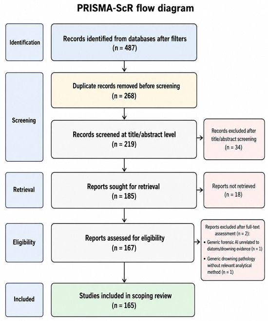
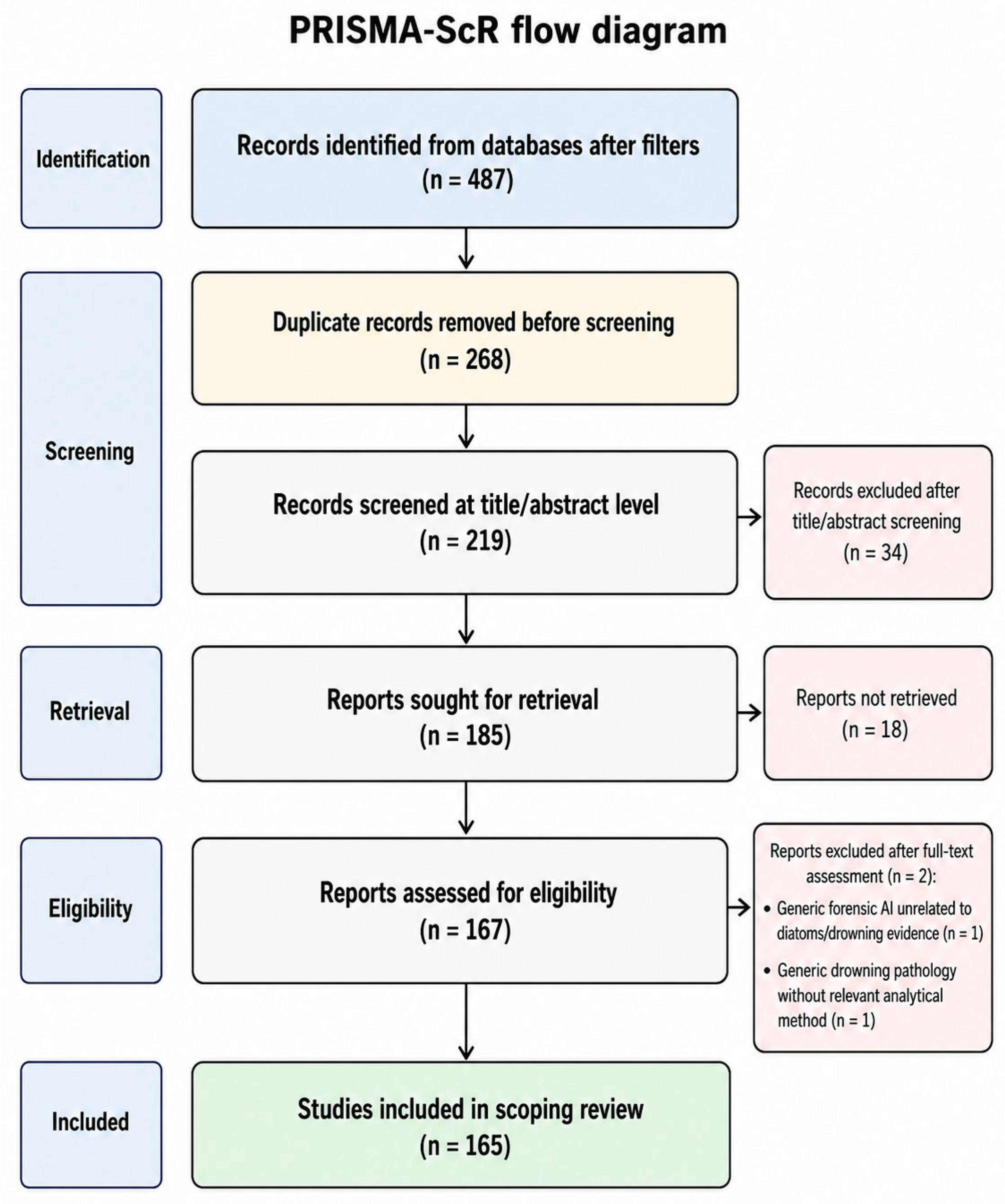
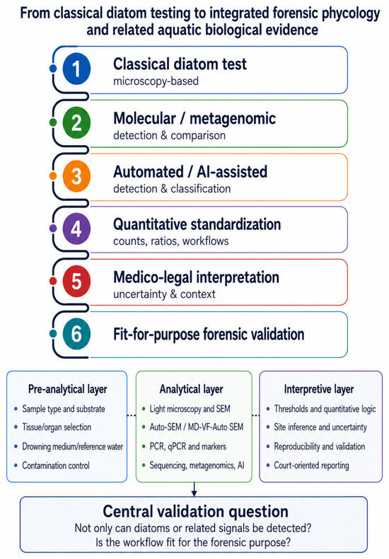
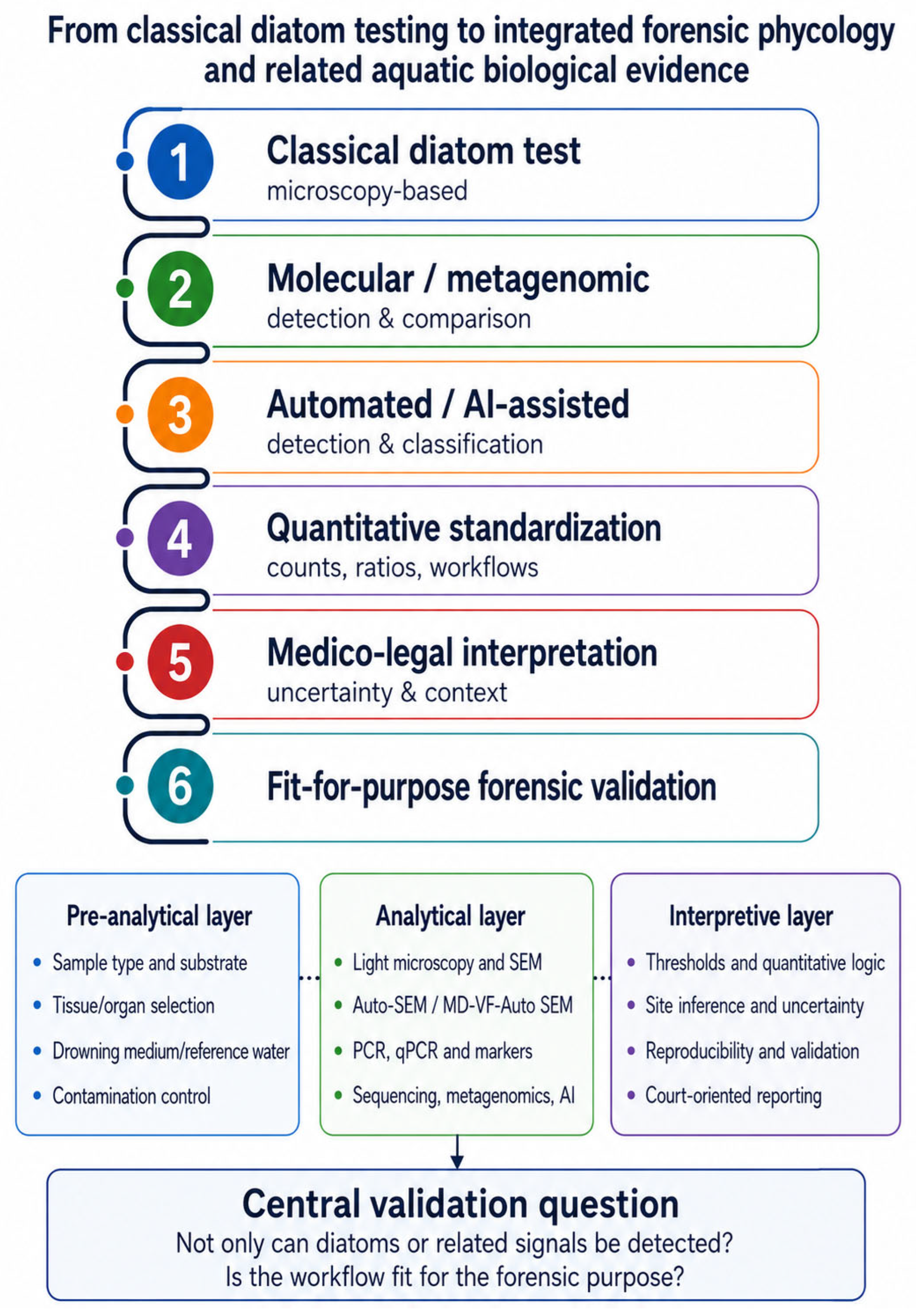

Disclaimer/Publisher’s Note: The statements, opinions and data contained in all publications are solely those of the individual author(s) and contributor(s) and not of MDPI and/or the editor(s). MDPI and/or the editor(s) disclaim responsibility for any injury to people or property resulting from any ideas, methods, instructions or products referred to in the content. |
© 2026 by the authors. Licensee MDPI, Basel, Switzerland. This article is an open access article distributed under the terms and conditions of the Creative Commons Attribution (CC BY) license.