


Abstract
Taxonomic clarity within the genus Antithamnion is critical for understanding its molecular phylogeny and biodiversity. Here we report Antithamnion hubbsii for the first time from the Korean coast. This finding highlights the need to re-evaluate its relationship with the previously reported, morphologically very similar A. nipponicum in this region, raising the question of whether the newly identified A. hubbsii represents a local variant of A. nipponicum or a recently introduced invasive species via nearby ports. Specimens collected from Gangneung were analyzed using plastid-encoded rbcL and psaA genes, confirming their identity as A. hubbsii. Morphological features such as indeterminate lateral axes, oppositely arranged pinnae and pinnules, and distinctive adaxial gland cells supported this identification. Molecular analyses revealed minimal divergence between A. hubbsii and A. nipponicum (1–3 bp in rbcL, none in psbA), and contrasting results from different species delimitation methods. Phylogenetic analyses nevertheless placed the Korean specimens in a strongly supported A. hubbsii/A. nipponicum clade. Taken together, our results suggest that the North American invasive A. nipponicum and the Korean A. hubbsii may represent a single species with broad intraspecific variation. Definitive resolution will require molecular analyses of the type specimens of both taxa.
1. Introduction
Marine ecosystems serve as habitats for various algal species, which play crucial roles as primary producers. Red algae (Rhodophyta) have adapted to diverse environments and are widely distributed throughout the world’s oceans [1]. Among them, the genus Antithamnion has received continuous attention from marine biologists due to its morphological diversity. For instance, Antithamnion nipponicum has been introduced to new regions, such as the Norwegian coast, highlighting the impact of globalization on marine ecosystems [1]. Later studies, including DNA barcoding data by Rueness [2], reported sequences of A. hubbsii from Norway, suggesting that earlier identifications of A. nipponicum were incorrect. Additionally, Antithamnion sparsum has been recently recorded in the Northwest Atlantic, supported by molecular data, underscoring that our knowledge of Antithamnion distribution and taxonomy continues to expand [3].
The taxonomic history of Antithamnion has been marked by considerable confusion. Ref. [4] suggested that A. hubbsii Dawson and A. nipponicum Yamada et Inagaki were conspecific but distinct from A. pectinatum (Montagne) Brauner. Later, Athanasiadis and Tittley [5] classified similar material from the Azores as A. pectinatum, while tentatively reducing A. nipponicum to synonymy. Subsequently, Athanasiadis [6] revisited the type material and formally proposed that A. nipponicum should be treated as a synonym of A. pectinatum, with A. hubbsii recognized as the correct name for the invasive species. These studies highlight the historical difficulty in distinguishing morphologically similar species within the genus and illustrate how changing interpretations of type specimens have shaped the nomenclatural treatment of A. hubbsii and A. nipponicum. However, misidentifications within the genus Antithamnion remain common due to the high degree of morphological similarity among species [4]. In particular, A. nipponicum and A. hubbsii possess very similar morphological features, making them difficult to distinguish using morphology alone. Previous studies emphasized that the key identifying features of A. hubbsii include indeterminate lateral axes of pinnae, oppositely arranged pinnules, and gland cells located on the adaxial surface of pinnules [4]. Recent phylogenetic analyses of the tribe Ceramieae have further demonstrated that morphological homoplasies are frequent across the group, complicating species delimitation and highlighting the necessity of molecular evidence for accurate identification [7].
Until now, only A. nipponicum has been reported from Korean waters, with no confirmed presence of A. hubbsii. However, through detailed morphological observations and molecular phylogenetic studies of specimens collected from the eastern coast of Gangneung, it was revealed that some specimens previously identified as A. nipponicum are A. hubbsiDi. This represents the first report of A. hubbsii in Korean waters, deepening our understanding of red algal biodiversity in Korea.
The introduction pathway of marine invasive species is often linked to international maritime activities. In the case of A. hubbsii in Korean waters, two potential introduction vectors are worthy of investigation. First, given that A. hubbsii has been well-documented along the Pacific coast of North America [4], increasing cruise ship traffic between North American ports and Korean destinations could serve as a potential vector. Cruise ships carry large volumes of ballast water and provide hull surface area for biofouling organisms, both of which are recognized as major pathways for marine species introductions [8]. Second, considering that Japan is geographically proximate to Korea and that A. hubbsii has been historically reported under the name A. nipponicum in Japanese waters, natural dispersal or smaller-scale maritime activities between Japan and Korea could have facilitated the introduction. This highlights how historical taxonomic uncertainty has complicated assessments of its true distribution, even though the introduction itself is most likely related to natural or anthropogenic dispersal pathways rather than misidentification.
Our research employs both traditional morphological and modern molecular approaches to accurately identify and characterize A. hubbsii from Korean waters. This integrated approach is essential for distinguishing between morphologically similar species and establishing phylogenetic relationships between Korean specimens and those from other regions. By documenting this new record and investigating its introduction pathway, this study contributes to our understanding of marine biodiversity in Korea and provides valuable information for monitoring and managing non-native species in Korean coastal ecosystems.
2. Materials and Methods
2.1. Sampling, Culture and Microscopy
The samples were collected in the rocky intertidal of Sungeut beach (37°49′2.6″ N, 128°53′40.5″ E), Gangneung-si, Gangwon-do, Republic of Korea, on 6 January 2024. Culture conditions consisted of sterilized seawater supplemented with IMR (Institute of Marine Resources) medium as described by [9]. Samples were maintained at 20 °C under cool-white fluorescent light and a photon fluence rate at approximately 25 µmol photons m−2 s−1, with a 16 h light ⁄ 8 h dark cycle. Culture maintenance involved transfer to new IMR medium every 7–14 days. Samples including the type specimen were deposited at the National Marine Biodiversity Institute of Korea (MARBIK), Seocheon, Republic of Korea (MABIK: AL001003219-AL00103221).
Live imaging was performed using an Olympus BX54 research microscope featuring differential interference contrast (DIC) capabilities and integrated with a Samsung iPolis imaging system (Samsung, Suwon, Republic of Korea). Nuclear visualization was performed following cell fixation in culture media using microwave irradiation for a few seconds. Subsequently, cells were stained with Hoeschst 33342 (1 μL/mL in the IMR medium) for 15 min under dark conditions prior to microscopic examination. Light and fluorescence microscopy images were merged using Adobe Photoshop CS6 software (Adobe Systems, San Jose, CA, USA). The images were then analyzed using ImageJ software (NIH, Bethesda, MD, USA; version 1.54g, http://imagej.nih.gov/ij/) to quantify the length of the cells and the diameter of the nuclei.
2.2. Molecular Analysis
Total DNA was extracted from fresh or dried samples using the Chelex method [10]. The partial sequence of the plastid-encoded ribulose-1, 5-bisphosphate carboxylase/oxygenase large subunit gene (rbcL) was amplified using the primer pairs F321 and R1150 [11]. PCR conditions followed a touchdown PCR as follows: initial denaturation at 94 °C for 4 min, followed by 10 cycles of 94 °C for 1 min, 55 °C for 30 s, and a decrease in annealing temperature by 1 °C per cycle, and 72 °C for 1 min; followed by 25 cycles of 94 °C for 1 min, 45 °C for 30 s, and 72 °C for 1 min and a final step at 72 °C for 5 min. Additionally, the partial sequence of the plastid-encoded photosystem I P700 chlorophyll a apoprotein A1 gene (psaA) was amplified using the primer pairs psaA130F (5′-AACWACWACTTGGATTCGAA-3′) and psaA1760R (5′-CCTCTWCCWGGWCATCWCAWGG-3′) [12]. The PCR conditions for psaA followed a touchdown protocol: initial denaturation at 94 °C for 2 min, followed by 5 cycles of 94 °C for 30 s, 45 °C for 30 s, and 72 °C for 1 min; then 35 cycles of 94 °C for 30 s, 46.5 °C for 30 s, 72 °C for 1 min, and a final extension at 72 °C for 7 min. PCR products were purified using ZymocleanTM Gel DNA recovery Kit (Zymo Research, Irvine, CA, USA). PCR products were sent for commercial Sanger sequencing using both sets of PCR primers (Cosmogene Tech, Daejeon, Republic of Korea). Sequences were edited and assembled in Geneious® Prime 2025.1.2 (https://www.geneious.com).
For phylogenetic analyses, the newly determined rbcL and psaA sequences were analyzed alongside sequences from various Antithamnion and Anthithamnionella species, plus related genera downloaded from GenBank. Details of all specimens used in this study, including collection information, voucher numbers, and GenBank accession numbers, are presented in Table 1 The dataset was aligned using MAFFT in Geneious® Prime 2025.1.2 (https://www.geneious.com) together with our new sequences of Antithamnion hubbsii collected from Korea (PV453999). Phylogenetic analyses were performed using the maximum likelihood (ML) method with IQ-TREE 2.3.4 [13]. Codon positions were partitioned and the best-fit model was determined using ModelFinder [14] and partition model [15]. The rbcL DNA substitution models were selected for each codon (i.e., first codon: TN+F+I; second codon: F81+F+I; third codon: HKY+F+G4) were selected. The psaA DNA substitution models were selected for each codon (i.e., first codon: TIM2+F+G4; second codon: TIM+F+I; third codon: TPM3u+F+I) were selected. Support for each internal branch was determined by non-parametric bootstrap (500 replicates) [16]. Bayesian inference analysis was conducted with MrBayes v. 3.2 [17] with partitioned codons using variable rates and six rate categories. Two parallel runs of Markov chain Monte Carlo were performed for 5,000,000 generations, sampling every 1000 generations. Estimated sample sizes, split frequencies, and stationarity were checked after each run. After analysis, 10% of generations were removed as burn-in and the posterior probabilities were visualized in Figtree v1.1.4 [18].
Table 1.
Collections of Antithamnion and outgroup taxa from which rbcL and psaA were obtained.
Three DNA-based species delimitation methods were used to determine species status. For the Assemble Species by Automatic Partitioning (ASAP) method, the Kimura (K80) Ts/Tv 2.0 model was selected and analyzed from the ASAP online platform (Muséum National d’Histoire Naturelle, Paris, France; originally available at https://bioinfo.mnhn.fr/abi/public/asap/, accessed on 6 May 2025; see also https://bio.tools/asap-assemble). A tree-based method (Poisson-Tree processes, PTP; [19]) was used online using the ML tree topology. Additionally, the Automatic Barcode Gap Discovery (ABGD) method was applied using the online ABGD platform (https://bioinfo.mnhn.fr/abi/public/abgd/abgdweb.html, accessed on 6 May 2025) with default parameters, including a relative gap width (X) of 1.5 and prior intraspecific divergence (P) values ranging from 0.001 to 0.1 [20]. The final tree was then edited using Adobe Illustrator 2024 (Adobe, San Jose, CA, USA).
3. Results
3.1. Morphological Observations
Genus: Antithamnion
Species: Antithamnion hubbsii
Type locality: Intertidal, Pacific Grove, CA, USA; [21]
Collection site in Korea: Antithamnion hubbsii collected from Sungeut beach, Gangneung-si, Gangwon-do (37°49′2.6″ N, 128°53′40.5″ E); collected 6 January 2024.
Habitat: Intertidal, epiphytic on other macroalgae.
Holotype: Dawson E.Y. 1925. Specimens collected from intertidal zone, Pacific Grove, CA, USA; deposited in the University of California Herbarium, Berkeley (UC), accession number UC 261983.
Specimens examined: KNU000620- KNU000622 (Sungeut beach (37°49′2.6″ N, 128°53′40.5″ E), Gangneung-si, Republic of Korea, 30 March 2013, Eunyoung Shim and Soo Yeon Kim). deposited in the National Marine Biodiversity Institute of Korea (MABIK AL00103219- AL00103221).
GenBank accession number: PV453999 (rbcL), PV454032 (psaA)
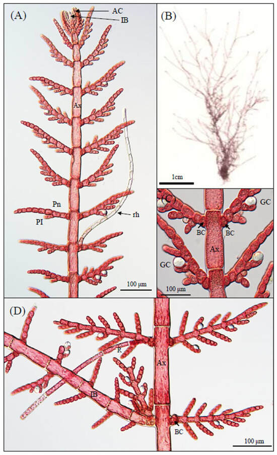
Description: Antithamnion hubbsii is a delicate red alga with a filamentous thallus organization displaying distinct morphological features (Figure 1A). Thalli are generally small, reaching approximately 1–5 cm in height. A representative herbarium specimen shows the overall thallus morphology with a slender, upright habit and densely branched axes (Figure 1B). Both diploid tetrasporophytes and haploid gametophytes exhibit a consistent pinnate branching pattern (Figure 1A). In diploid individuals, however, some branches may appear pectinate due to rhizoid development. The thallus consists of both prostrate and erect axes. The erect axis comprises a main and indeterminate lateral axis of unlimited growth bearing opposite pinnae of determinate growth (Figure 1D). Indeterminate lateral axes are irregularly produced from the basal cell of pinnae in the erect axes (Figure 1D).
Figure 1.
Morphological features of Antithamnion hubbsii. tetrasporophyte. Tetrasporophyte of A. hubbsii growing attached to intertidal rocks with distinctive bilateral branching pattern. (A) Light microscopy image showing pinnate branching pattern. AC: apical cell; IB: indeterminate branch; Pn: pinnule; PI: pinna; rh: rhizoid. Scale bar = 100 μm. (B) Higher magnification of central axis showing cellular organization. Ax: axial cell; BC: basal cell; GC: gland cell. Scale bar = 100 μm. (C) Herbarium specimen of A. hubbsii tetrasporophyte showing overall thallus morphology and branching pattern. Scale bar = 1 cm. (D) Close-up view of lateral branches showing the origin of indeterminate branches (IB) from the basal cells of whorl branches (paired or irregularly produced). Note the presence of gland cells (GC) at the adaxial surface of pinnules. Ax: axial cell; BC: basal cell. Scale bar = 100 μm.
Vegetative features: The main axis consists of prominent axial cells (Ax) that are cylindrical and elongated (Figure 1A). Branching is generally distichous (arranged in two rows), forming a regular pinnate pattern (Figure 1A), although some individuals exhibit irregular or one-sided branching. Apical cells (ACs) are prominent at the tips of growing branches and are responsible for indeterminate growth (Figure 1A). Rhizoids (rh) develop from the lower cell of lateral branches and serve as attachment structures (Figure 1A). Each branch originates from a diminutive basal cell (BC) at the nodes of the main axis (Figure 1C). A distinctive feature is the presence of spherical gland cells (GCs) situated adjacent to basal cells of lateral branches (Figure 1C). These gland cells are colorless to pale and conspicuous under light microscopy, borne on pinnae, adaxially, and covering 2 cells (Table 2). Indeterminate lateral branches arise irregularly from the basal cells of pinnae in the erect axes (Figure 1D).
Table 2.
Distinguishing characteristics of local and morphologically similar Antithamnion spp. as well as Antithamnionella.
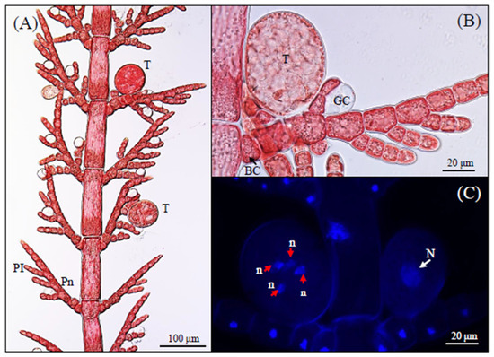
Reproductive structures: The tetrasporophyte bears spherical tetrasporangia (T) that develop on specialized determinate short branches (Figure 2A). Tetrasporangia are typically borne singly or in pairs (occasionally visible in Figure 2A), most often on the adaxial side of pinnae, adjacent to gland cells (Figure 2B). However, some sporangia can also be positioned abaxially, as observed in a few specimens (Figure 1A). Each tetrasporangium is characterized by a distinctive 1-cell pedicel structure that differentiates it from other species in the genus (Table 2), although in some herbarium specimens (Figure 1B) the sporangia appear sessile or with a very short pedicel. Nuclear staining reveals the presence of a single large nucleus (N) in ordinary vegetative cells (Figure 2C; white arrow). Within each tetrasporangium, nuclear staining confirmed the presence of four small nuclei (n) corresponding to the four daughter cells formed during meiotic division (Figure 2C; red arrows), confirming the typical pattern of tetrasporogenesis seen in other red algae.
Figure 2.
Tetrasporangial development in A. hubbsii. (A) Light microscopy image of tetrasporophyte showing mature tetrasporangia (T) distributed along the thallus. Pn: pinnule; PI: pinna. Scale bar = 100 μm. (B) Higher magnification of a mature tetrasporangium (T) showing characteristic 1-cell pedicel structure. BC: basal cell; GC: gland cell. Scale bar = 20 μm. (C) Fluorescence microscopy image of DAPI-stained tetrasporangia showing four nuclei (n) within a single tetrasporangium, confirming meiotic division. N: nucleus of vegetative cell. Scale bar = 20 μm.
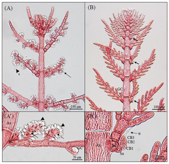
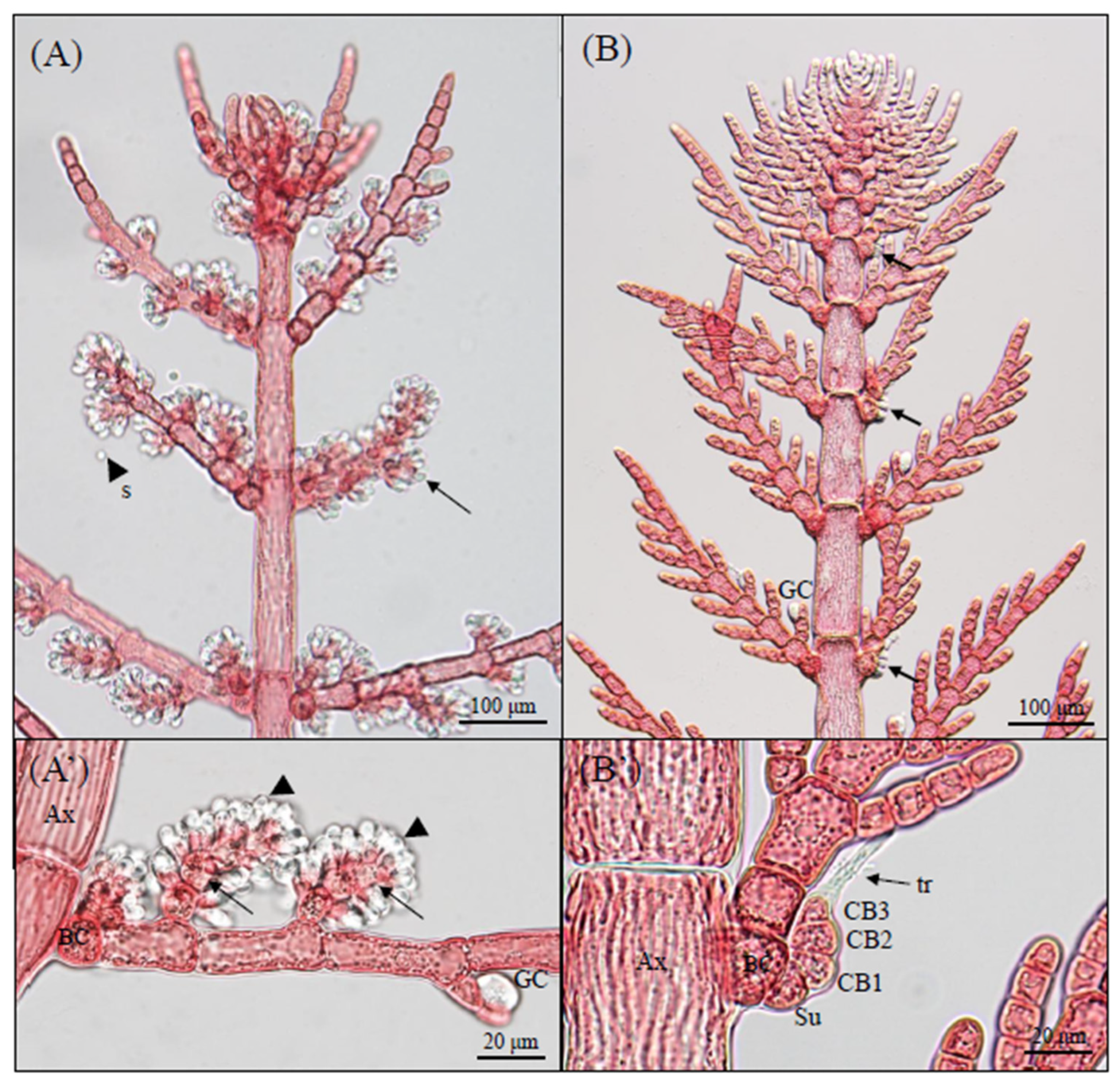
Sexual dimorphism: Male and female gametophytes show distinct morphological differences (Figure 3). Reproductive structures were observed after culturing gametophytes derived from tetraspores for 3–6 months under laboratory conditions. Male gametophytes display dense clusters of spermatangia (arrowheads) borne on spermatangial parent cells on special branches on both the adaxial and abaxial sides of pinnules, giving a tufted appearance (Figure 3A,A’). Female gametophytes maintain a more regular branching pattern with procarps (arrows) developing on the basal cells of pinnae (Figure 3B). Detailed examination of the female reproductive structures reveals the development of two carpogonial branch cells (CB1-3) arranged in a row, with the terminal carpogonium extending a trichogyne (tr) for spermatial reception (Figure 3B’). The carpogonial branch is supported by a distinct supporting cell (Su) connected to the basal cell (BC). The branching in female gametophytes appears more orderly and pinnate compared to the clustered organization in male plants.
Figure 3.
Morphological comparison of male and female reproductive structures in red algae A. hubbsii. (A) Male gametophyte with spermatangia (arrowheads) on spermatangial parent cells of pinnule branches; (A’) detail of spermatangial clusters showing axial cell (Ax), basal cell (BC), and gland cell (GC). (B) Female gametophyte with procarps (arrows) borne on basal cells of pinna; (B’) carpogonial branch with CB1-CB3, axial cell (Ax), basal cell (BC), supporting cell (Su), and trichogyne (tr). Abbreviations: Ax, axial cell; BC, basal cell; Su, supporting cell; tr, trichogyne; Au, auxiliary cell; Cp, carpogonium; Cy, cystocarp; Ft, foot cell; Fu, fusion cell; G, gonimoblast; Gi, gonimoblast initial; GI, first gonimolobes.
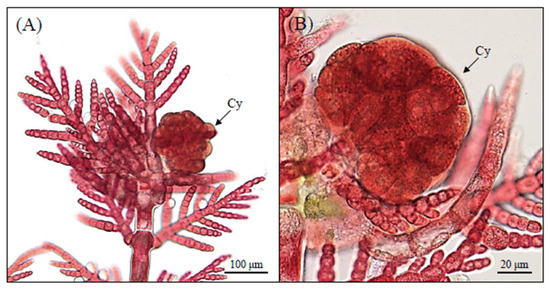
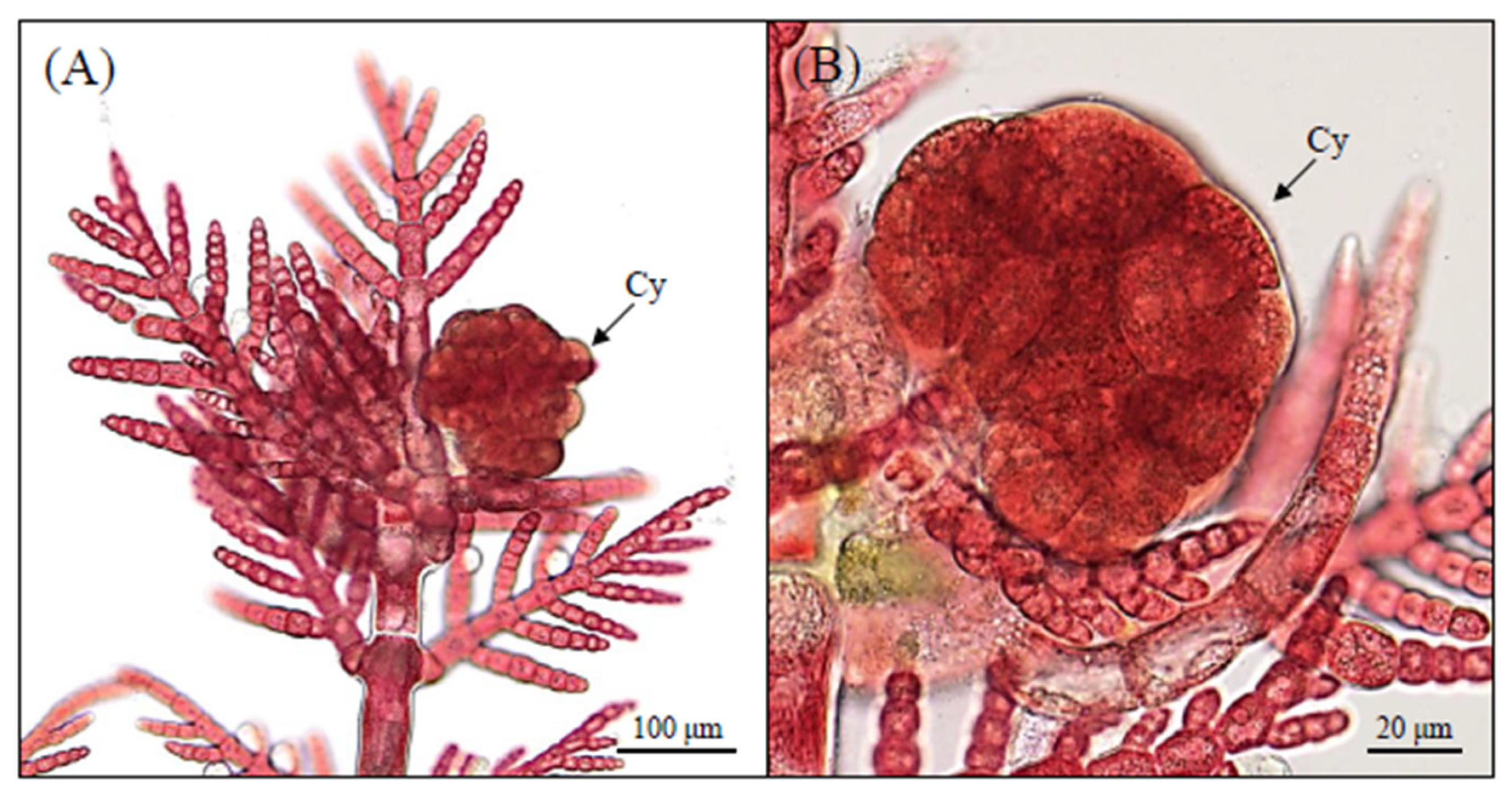
Carposporophyte development: Following fertilization, the carpogonium develops into a carposporophyte (Figure 4A). Carposporophytes were observed after co-culturing male and female specimens for at least 5 months. The development is initiated when the carpogonium establishes a connection with the auxiliary cell, from which gonimoblast initials proliferate. The mature carposporophyte appears as a distinctive, spherical to slightly lobed structure attached to the female thallus, with carposporangia arranged in clusters. At higher magnification (Figure 4B), the cystocarp is clearly visible as a compact, multi-lobed body containing numerous developing carposporangia.
Figure 4.
Development from carpogonium to cystocarp in A. hubbsii. (A) Fertilized carpogonium develops into a mature carposporophyte embedded within the female thallus. The cystocarp appears as a spherical to slightly lobed structure containing clustered carposporangia. Scale bar = 100 μm. (B) Higher magnification showing a compact, multi-lobed cystocarp (Cy) with numerous developing carposporangia, indicating active differentiation within the carposporophyte. Cy: cystocarp. Scale bar = 20 μm.
Distinguishing characteristics: As shown in Table 2, A. hubbsii can be distinguished from morphologically similar species by a combination of features. It has paired indeterminate laterals, pinnate arrangement in diploid phase and adaxially pectinate in haploid phase, and distichous branching plane. These characteristics, together with the distinctive gland cell arrangement and 1-cell pedicel of tetrasporangia, clearly differentiate A. hubbsii from other related species such as A. nipponicum, A. cruciatum, and A. defectum. While A. hubbsii shares some features with A. nipponicum, such as paired indeterminate laterals and distichous branching, our molecular analyses demonstrate that they represent distinct species.
This combination of morphological features, particularly the arrangement of pinnae, location of gland cells, and pattern of tetrasporangia development, supports the identification of this specimen as Antithamnion hubbsii, consistent with its phylogenetic placement shown in the molecular analyses.
3.2. Phylogenetic Analyses
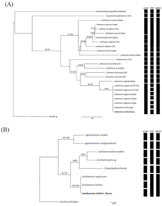
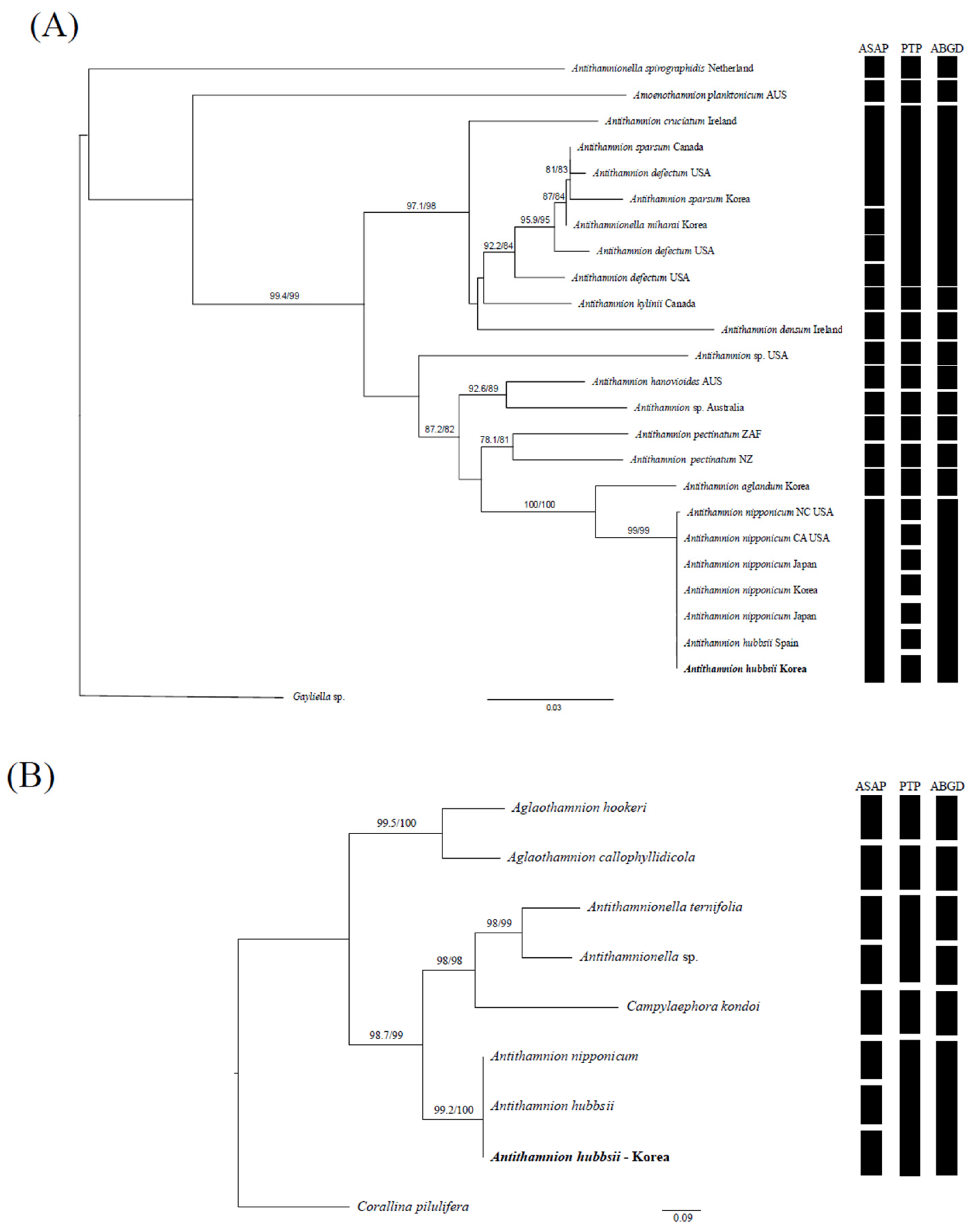
The 1467 base pairs (bp) of rbcL and 1523 bp of psaA were sequenced from samples collected in Korea (Table 1, Figure 5). Molecular phylogenetic analyses based on both rbcL (Figure 5A) and psaA (Figure 5B) sequences revealed that specimens collected from Sungeut Beach in Gangneung, Republic of Korea, clustered with previously reported A. hubbsii from California and Spain, as well as sequences deposited in GenBank under the name A. nipponicum from Japan. In the rbcL phylogeny, our Korean A. hubbsii (PV453999) formed a strongly supported clade with A. hubbsii from California (AY591930) with strong support (98/99 ML/Bayesian). This cluster also included sequences identified as A. nipponicum from Japan and Spain, showing nearly identical sequences with only 3 bp differences across 1467 bp (~0.2%). Such minimal divergence is generally regarded as intraspecific variation rather than interspecific differentiation. The psaA gene analysis similarly grouped our Korean A. hubbsii specimen (PV454032) with previously reported A. hubbsii from Spain (MK814610) and sequences deposited as A. nipponicum (AY295136, Japan) with high support (99.4/100). These sequences were either identical or showed very low divergence (0–0.34%), again consistent with intraspecific variation.
Figure 5.
Maximum Likelihood (ML) phylogenetic trees of Antithamnion species based on (A) rbcL and (B) psaA gene sequences. Numbers at nodes indicate bootstrap support values (%) for ML analysis. The trees were rooted using Cystoclonium purpureum and Ceramium piluliferoides as outgroups for rbcL and psaA, respectively. Newly sequenced A. hubbsii specimens from Korea are indicated in bold. The matrices to the right of each tree summarize species delimitation results based on three methods: ASAP (Assemble Species by Automatic Partitioning), PTP (Poisson Tree Processes), and ABGD (Automatic Barcode Gap Discovery). Black squares indicate groups recognized as separate species by each method. Conflicting results between ASAP and PTP analyses highlight the taxonomic uncertainty between A. hubbsii and A. nipponicum.
Both markers consistently placed the Korean specimens within the A. hubbsii lineage together with sequences historically labeled as A. nipponicum. This indicates that the Korean material is genetically indistinguishable from A. hubbsii populations in North America, Spain, and Japan. Species delimitation methods provided conflicting outcomes: while ASAP and ABGD supported the recognition of A. hubbsii from Gangneung as a distinct entity, PTP merged it with A. nipponicum. These discrepancies likely reflect methodological sensitivity rather than biological separation.
Taken together, these results provide the first confirmed record of A. hubbsii in Korean waters and highlight that previously reported A. nipponicum in this region almost certainly represents the same taxon.
4. Discussion
The present study documents the first confirmed occurrence of Antithamnion hubbsii in Korean waters, based on both detailed morphological characteristics and molecular phylogenetic evidence. The species has likely been misidentified as A. nipponicum in previous surveys due to their striking morphological similarities. This finding highlights the taxonomic complexity within the genus Antithamnion and emphasizes the importance of integrating molecular tools with traditional morphological approaches for accurate species identification in red algae [22].
The taxonomic history of Antithamnion species has been marked by numerous revisions and reclassifications over the past century. The genus was first established by [23] and has since undergone significant taxonomic refinement. Our findings contribute to this ongoing taxonomic discourse by providing molecular evidence for the presence of A. hubbsii in Korean waters. The extremely close genetic relationship between A. hubbsii and A. nipponicum revealed in our analyses (0–0.3% divergence in rbcL and psaA) reinforces the taxonomic challenges within this genus. While [3] previously suggested these taxa might represent the same species based on minimal genetic differences, our analyses also support this interpretation. Although species delimitation methods gave conflicting outcomes—PTP tended to oversplit rbcL sequences into multiple units, whereas ASAP indicated distinction in psaA—the overall evidence, including identical or nearly identical sequences across multiple regions (Japan, Spain, North America, Korea), is more consistent with treating these records as a single species. This highlights the ongoing methodological challenges in delimiting species boundaries within closely related red algal taxa [24,25].
Although the origin of A. hubbsii in Korean waters remains uncertain, its occurrence necessitates a re-evaluation of regional red algal biodiversity, particularly in taxa with cryptic or morphologically similar species. Previous studies have shown that minimal genetic divergence can mask true taxonomic identity, highlighting the necessity for refined field sampling and the application of multi-locus molecular approaches [22,26]. Recent DNA-based floristic surveys in nearby regions, such as the study of red algal diversity around Tanegashima Island, Japan [27], also highlight the value of molecular approaches in uncovering cryptic diversity. Thus, integrating such regional datasets will be critical for resolving taxonomic uncertainties within Antithamnion and for assessing broader biogeographic patterns [27].
Despite these insights, distinguishing A. hubbsii from A. nipponicum remains challenging [4,7]. No DNA sequences are available from type specimens of either species and current molecular datasets rely on recently collected material matching the protologues morphologically [22,25]. We therefore acknowledge, and share, the concern that the independence of these two species as distinct taxa remains difficult to resolve with certainty [24,25]. In particular, the origin of indeterminate laterals is a critical diagnostic feature: in A. nipponicum they often replace whorl branches, whereas in A. hubbsii they originate from the basal cells of whorl-branches [5]. Our observations of Korean specimens support this latter condition, although some irregularities in paired development were noted. This highlights the difficulty of relying on a single morphological character, especially in the absence of DNA sequences from type specimens, and reinforces the need for further integrative studies combining morphology and molecular data.
Taken together, our study provides the first confirmed record of Antithamnion hubbsii in Korean waters and underscores the longstanding taxonomic ambiguity between this species and A. nipponicum. The extremely small genetic divergence observed between the two taxa, combined with the conflicting outcomes of species delimitation methods, suggests that they may represent a single species with broad intraspecific variation rather than two distinct evolutionary lineages [24,25]. This interpretation is consistent with previous reports of historical misidentifications and overlapping morphological features in different regions [7,22]. Considering that A. nipponicum has been regarded as an invasive species in North America [4,28], clarifying whether the Korean A. hubbsii represents a native population, a regional variant of A. nipponicum, or a recent introduction via shipping routes is of particular ecological and biogeographic importance. To reach a definitive conclusion, molecular phylogenetic analyses of the type specimens of both taxa will be essential, thereby resolving their taxonomic status and providing a stronger framework for biodiversity monitoring and the management of potential introductions in coastal ecosystems [25,29].
Author Contributions
Data curation, Investigation, Methodology, Validation, Visualization, Writing—original draft preparation, E.S.; Investigation, Methodology, Resources, S.Y.K.; Investigation, Methodology, C.S.K.; Project administration, Supervision, Writing—review and editing, G.H.K. All authors have read and agreed to the published version of the manuscript.
Funding
This work was supported by the management of Marine and Fishery Bio-resources Center (2025) funded by the National Marine Biodiversity Institute of Korea (MABIK) and Korea Institute of Marine Science & Technology Promotion (KIMST) funded by the Ministry of Oceans and Fisheries, Republic of Korea (RS-2025-02303933) and the National Institute of Fisheries Science, Ministry of Oceans and Fisheries, Republic of Korea (R2025025).
Data Availability Statement
The sequence data supporting the findings of this study are openly available in the GenBank database at accession numbers AY594700 and PV454032.
Acknowledgments
The authors thank Giuseppe C. Zuccarello for valuable comments on the manuscript.
Conflicts of Interest
The authors declare no conflicts of interest.
References
- Rueness, J.; Heggøy, E.; Husa, V.; Sjøtun, K. First report of the Japanese red alga Antithamnion nipponicum on the west coast of Norway. Aquat. Invasions 2007, 2, 431–434. [Google Scholar] [CrossRef] [Scilit]
- Rueness, J. DNA barcoding of select freshwater and marine red algae (Rhodophyta). Cryptogam. Algol. 2010, 31, 377–386. [Google Scholar]
- Brooks, C.M.; Krumhansl, K. First record of the Asian Antithamnion sparsum Tokida, (Ceramiales, Rhodophyta) in Nova Scotia, Canada. BioInvasions Rec. 1932, 12, 745–752. [Google Scholar] [CrossRef] [Scilit]
- Cho, T.O.; Won, B.Y.; Fredericq, S. Antithamnion nipponicum (Ceramiaceae, Rhodophyta), incorrectly known as A. pectinatum in western Europe, is a recent introduction along the North Carolina and Pacific coasts of North America. Eur. J. Phycol. 2007, 40, 323–335. [Google Scholar] [CrossRef] [Scilit]
- Athanasiadis, A.; Tittley, I. Antithamnioid algae (Rhodophyta, Ceramiaceae) newly recorded from the Azores. Phycologia 1994, 33, 77–80. [Google Scholar] [CrossRef] [Scilit]
- Athanasiadis, A. Typification of Antithamnion nipponicum Yamada et Inagaki (Antithamnieae, Ceramioideae, Ceramiaceae, Ceramiales, Rhodophyta). Bot. Mar. 2009, 52, 256–261. [Google Scholar] [CrossRef] [Scilit]
- Díaz-Tapia, P.; Maggs, C.A.; Le Gall, L.; Verbruggen, H. Phylogenetic analysis of the red algal tribe Ceramieae reveals multiple morphological homoplasies but defines new genera. Cryptogam. Algol. 2023, 44, 101–134. [Google Scholar]
- Carlton, J.T.; Geller, J.B. Ecological roulette: The global transport of nonindigenous marine organisms. Science 1993, 261, 78–82. [Google Scholar] [CrossRef] [Scilit]
- Klochkova, T.A.; Kang, S.-H.; Cho, G.Y.; Pueschel, C.M.; West, J.A.; Kim, G.H. Biology of a terrestrial green alga, Chlorococcum sp. (Chlorococcales, Chlorophyta), collected from the Miruksazi stupa in Korea. Phycologia 2006, 45, 349–358. [Google Scholar] [CrossRef] [Scilit]
- Zuccarello, G.C.; Burger, G.; West, J.A.; King, R.J. A mitochondrial marker for red algal intraspecific relationships. Mol. Ecol. 1999, 8, 1443–1447. [Google Scholar] [CrossRef] [Scilit]
- Kang, J.H.; Jang, J.E.; Kim, J.H.; Kim, M.S. DNA barcoding of the genus Grateloupia (Rhodophyta) from Korea: An assessment of DNA barcoding markers for species identification. Diversity 2020, 12, 3. [Google Scholar]
- Yoon, H.S.; Hackett, J.D.; Bhattacharya, D. A single origin of the peridinin- and fucoxanthin-containing plastids in dinoflagellates through tertiary endosymbiosis. Proc. Natl. Acad. Sci. USA 2002, 99, 11724–11729. [Google Scholar] [CrossRef] [Scilit]
- Minh, B.Q.; Schmidt, H.A.; Chernomor, O.; Schrempf, D.; Woodhams, M.D.; von Haeseler, A.; Lanfear, R. IQ-TREE 2: New models and efficient methods for phylogenetic inference in the genomic era. Mol. Biol. Evol. 2020, 37, 1530–1534. [Google Scholar] [CrossRef] [Scilit] [PubMed]
- Kalyaanamoorthy, S.; Minh, B.Q.; Wong, T.K.F.; Von Haeseler, A.; Jermiin, L.S. ModelFinder: Fast model selection for accurate phylogenetic estimates. Nat. Methods 2017, 14, 587–589. [Google Scholar] [CrossRef] [Scilit]
- Chernomor, O.; von Haeseler, A.; Minh, B.Q. Terrace aware data structure for phylogenomic inference from supermatrices. Syst. Biol. 2016, 65, 997–1008. [Google Scholar] [CrossRef] [Scilit] [PubMed]
- Felsenstein, J. Confidence limits on phylogenies: An approach using the bootstrap. Evolution 1985, 39, 783–791. [Google Scholar] [CrossRef] [Scilit] [PubMed]
- Ronquist, F.; Teslenko, M.; van der Mark, P.; Ayres, D.L.; Darling, A.; Höhna, S.; Larget, B.; Liu, L.; Suchard, M.A.; Huelsenbeck, J.P. MrBayes 3.2: Efficient Bayesian phylogenetic inference and model choice across a large model space. Syst. Biol. 2012, 61, 539–542. [Google Scholar] [CrossRef] [Scilit]
- Rambaut, A. FigTree v1.4: Tree Figure Drawing Tool. Institute of Evolutionary Biology, University of Edinburgh. 2009. Available online: http://tree.bio.ed.ac.uk/software/figtree/ (accessed on 6 May 2025).
- Zhang, J.; Kapli, P.; Pavlidis, P.; Stamatakis, A. A general species delimitation method with applications to phylogenetic placements. Bioinformatics 2013, 29, 2869–2876. [Google Scholar] [CrossRef] [Scilit]
- Puillandre, N.; Lambert, A.; Brouillet, S.; Achaz, G. ABGD, Automatic Barcode Gap Discovery for primary species delimitation. Mol. Ecol. 2012, 21, 1864–1877. [Google Scholar] [CrossRef] [Scilit]
- Dawson, E.Y. Studies of intertidal algae at Pacific Grove, California. Bull. Calif. Acad. Sci. 1925, 24, 31–59. [Google Scholar]
- Freshwater, D.W.; Tudor, K.; O’Shaughnessy, K.; Wysor, B. DNA barcoding in the red algal order Gelidiales: Comparison of COI with rbcL and verification of the “barcoding gap”. Cryptogam. Algol. 2010, 31, 435–449. [Google Scholar]
- Nägeli, C. Die neueren Algensysteme und Versuch zur Begründung eines eigenen Systems der Algen und Florideen. Neue Denkschr. Der Allg. Schweiz. Ges. Für Die Gesammten Nat. 1847, 9, 1–275. [Google Scholar]
- Payo, D.A.; Leliaert, F.; Verbruggen, H.; D’Hondt, S.; Calumpong, H.P.; De Clerck, O. Extensive cryptic species diversity and fine-scale endemism in the marine red alga Portieria in the Philippines. Proc. R. Soc. B Biol. Sci. 2013, 280, 20122660. [Google Scholar] [CrossRef] [Scilit] [PubMed]
- Leliaert, F.; Verbruggen, H.; Vanormelingen, P.; Steen, F.; López-Bautista, J.M.; Zuccarello, G.C.; De Clerck, O. DNA-based species delimitation in algae. Eur. J. Phycol. 2014, 49, 179–196. [Google Scholar] [CrossRef] [Scilit]
- Steen, H.; Scrosati, R. Intraspecific competition in Fucus serratus and Fucus evanescens (Phaeophyceae: Fucales) germlings: Effects of settlement density, nutrient supply, and temperature. Mar. Biol. 2004, 144, 61–70. [Google Scholar]
- Suzuki, M.; Terada, R. DNA-based floristic survey of red algae (Rhodophyta) growing in the mesophotic coral ecosystems (MCEs) offshore of Tanegashima Island, northern Ryukyu Archipelago, Japan. PLoS ONE 2025, 20, e0316067. [Google Scholar] [CrossRef] [Scilit]
- Olenin, S.; Alemany, F.; Cardoso, A.C.; Gollasch, S.; Goulletquer, P.; Lehtiniemi, M.; McCollin, T.; Minchin, D.; Miossec, L.; Occhipinti-Ambrogi, A.; et al. Marine Strategy Framework Directive–Task Group 2 Report: Non-Indigenous Species; Office for Official Publications of the European Communities: Luxembourg, 2011. [Google Scholar]
- Williams, S.L.; Smith, J.E. A global review of the distribution, taxonomy, and impacts of introduced seaweeds. Annu. Rev. Ecol. Evol. Syst. 2007, 38, 327–359. [Google Scholar] [CrossRef] [Scilit]

Disclaimer/Publisher’s Note: The statements, opinions and data contained in all publications are solely those of the individual author(s) and contributor(s) and not of MDPI and/or the editor(s). MDPI and/or the editor(s) disclaim responsibility for any injury to people or property resulting from any ideas, methods, instructions or products referred to in the content. |
© 2025 by the authors. Licensee MDPI, Basel, Switzerland. This article is an open access article distributed under the terms and conditions of the Creative Commons Attribution (CC BY) license (https://creativecommons.org/licenses/by/4.0/).